Supporting Information - pnas.org · Supporting Information Pastorelli et al....
7
Supporting Information Pastorelli et al. 10.1073/pnas.0912678107 SI Materials and Methods Patients. Colonic endoscopic biopsies from males and females, aged 18–65 yr, and affected by CD or UC and noninflamed CON were harvested from patients undergoing flexible sigmoidoscopy or colonoscopy for diagnostic or surveillance purposes at the University of Virginia Digestive Health Center (UVA DHC) and were used for total RNA and protein extraction. Similar gender- and age-matched colonic surgical specimens were obtained from either UC or CD patients, as well as noninflammatory CON, who were admitted to the UVA DHC or the Cleveland Clinic Foun- dation (CCF) and underwent therapeutic bowel resection for in- flammatory disease or for malignant and other nonmalignant noninflammatory conditions. Full-thickness colon tissues were used for IHC (UVA) and as a source for mucosal IEC and LPMC (CCF). Anonymous unstained slides from infectious colitis and diverticulitis patients were obtained as formalin-fixed, paraffin- embedded endoscopic biopsies or surgical specimens from the UVA Biorepository and Tissue Research Facility. Whole blood samples were collected from UC and CD patients who underwent medical visits at the UVA DHC (disease activity study), the Gastroenterology and Endoscopy Unit of IRCCS Fondazione Mangiagalli, Regina Elena and Ospedale Maggiore of Milan and the Gastroenterology and Endoscopy Unit of IRCCS Policlinico San Donato for diagnosis, follow-up, and reactivation of IBD (disease activity and anti-TNF studies); age-, gender- and ethni- cally- matched healthy individuals were used as CON. Whole blood samples were collected from UC and CD patients treated with Infliximab (5-10 mg/kg, i.v.) 1 h prior and 2 h after each in- fusion at T 0 , and 2, 6, 14, 22, 30, and 38 wks. All diagnoses were confirmed by clinical, macroscopic, and histologic criteria. All studies were approved by the Internal Review Board of the UVA Health System, CCF, IRCCS Fondazione Mangiagalli, Regina Elena and Ospedale Maggiore of Milan and IRCCS Policlinico San Donato. Animals. SAMP1/YitFc (SAMP) and AKR/J (AKR) mice were maintained under SPF conditions in the Animal Resource Core Facility of Case Western Reserve University. All protocols were approved by the Institutional Animal Care and Use Committee (IACUC) of Case Western Reserve University. Tissue Harvest and Histologic Assessment of SAMP Ileitis. Terminal ilea were removed from mice, rinsed with cold PBS, opened longitudinally, and submerged in Bouin’s solution (LabChem). Fixed tissues were processed, paraffin-embedded, sectioned at 4 μm, stained with H&E, and resulting ileal sections were histo- logically evaluated by a single pathologist in a blinded fashion, using a validated semiquantitative scoring system as described (1). Isolation and Culture of MLN and Spleen Cells. MLNs and spleens were collected from experimental mice and processed for cell isolation as described (1); briefly, organs were aseptically re- moved and passed through a 70-μm cell strainer to obtain single cell suspensions. Lysis of erythrocytes was performed with ACK lysis buffer (Lonza). Resulting cells were cultured in RPMI medium 1640 with 10% FBS and 1% penicillin/streptomycin, in the presence of immobilized anti-CD3 (5 μg/mL) and soluble anti-CD28 (1 μg/mL) with or without rmIL-33 (10 ng/mL) for 48 h. Supernatants were subsequently collected for further analysis. Human and Mouse Serum Collection. Whole blood was drawn (5 mL) from patients by using a 22-gauge syringe into purple-topped vaccutainers and from anesthetized mice by cardiac puncture, and centrifuged at 2,500 × g for 20 min. Serum was collected from each sample, immediately aliquoted, and stored at −80 °C until further assayed. Isolation of IEC and LPMC. For human IEC, colonic tissues were collected and processed immediately after surgical resection as described (2). Briefly, specimens were opened longitudinally, rinsed, examined for gross morphological changes, and repre- sentative full-thickness samples were obtained. Intestinal mucosa was stripped from the muscularis mucosa, cut into strips, and freed of mucus by sequential washes in HBSS containing 10 mM DTT (Sigma-Aldrich). IEC were isolated by repeated incu- bations in HBSS (Invitrogen/GIBCO) containing 1 mM EDTA (Sigma-Aldrich). Mononuclear, red blood, and dead cells were removed by using a 40% Percoll Plus gradient (GE Healthcare), where IEC equilibrated at the interface; resulting preparations enriched for IEC were collected, washed twice in PBS, and subsequently counted. Using an immunoperoxidase method, all cells with epithelial morphology were stained with anti-keratin mAb, with <1% contaminating LPMC, as assessed by staining with a monoclonal antibody directed against a leukocyte com- mon Ag (CD45). For human LPMC, stripped mucosa, freed of mucus and IEC, was digested overnight with collagenase and DNase (both from ThermoScientific). The resulting crude cell suspensions were purified by using a Ficoll-Hypaque Plus gra- dient (GE Healthcare), and the preparations were preferentially enriched for LPMC, washed, and collected. IEC from experimental mice were isolated as described (3); briefly, mouse ileal segments were minced into 1-cm pieces and repeatedly incubated in a solution containing 1 mM EDTA (Sigma-Aldrich) with vigorous shaking. Contaminating cells were removed by using a 40% Percoll Plus gradient (GE Healthcare), and purity was checked. Flow Cytometry. LPMC from UC mucosa were used for flow cytometric analysis. Cell suspensions were prepared in PBS containing 2% FBS. Cells (10 6 ) were stained with appropriate combinations of FITC-, PE-, and Pacific Blue-labeled mouse anti-human CD4 (RPA-T4), CD8 (HIT8a), CD11b (ICRF44) (BD Pharmingen), and CD68 eBioY1/82A (Y1/82A) (eBio- sciences). Polyclonal goat anti-human IL-33 IgG (AF3625) and polyclonal goat anti-human ST2 IgG (AF523) (R&D Systems) were used in combination with donkey anti-goat IgG FITC- and APC-labeled secondary antibodies (F0108 and F0109) (R&D Systems). Intracellular staining for IL-33 was performed ac- cording to eBioscience permeabilization buffer kit instructions. Flow cytometric analysis was performed by using a BD LSRII based on BD FACS diva software (Becton Dickinson). Cell Culture. The HT-29 intestinal epithelial cancer cell line (ATCC) was grown to 80% confluency in RPMI medium 1640 media, supplemented with 10% FBS and 2% penicillin/strepto- myocin (all from Invitrogen/GIBCO), and cultured with or without rhTNF (10 ng/mL; Peprotech) for 0, 2, 6, 12, 24, and 48 h. Supernatants were immediately collected, aliquoted, and frozen at −20 °C for later assays; cells were harvested for total protein extraction. Total RNA Extraction and Reverse Transcription. Endoscopic bi- opsies, mouse ilea, and freshly isolated IEC were placed in RNA Later (Applied Biosystems), left at 4 °C overnight, then frozen and stored at −80 °C. Total RNA was isolated by using the Pastorelli et al. www.pnas.org/cgi/content/short/0912678107 1 of 7
Transcript of Supporting Information - pnas.org · Supporting Information Pastorelli et al....

Supporting InformationPastorelli et al. 10.1073/pnas.0912678107SI Materials and MethodsPatients. Colonic endoscopic biopsies from males and females,aged 18–65 yr, and affected by CD or UC and noninflamed CONwere harvested from patients undergoing flexible sigmoidoscopyor colonoscopy for diagnostic or surveillance purposes at theUniversity of Virginia Digestive Health Center (UVA DHC) andwere used for total RNA and protein extraction. Similar gender-and age-matched colonic surgical specimens were obtained fromeither UC or CD patients, as well as noninflammatory CON, whowere admitted to the UVA DHC or the Cleveland Clinic Foun-dation (CCF) and underwent therapeutic bowel resection for in-flammatory disease or for malignant and other nonmalignantnoninflammatory conditions. Full-thickness colon tissues wereused for IHC (UVA) and as a source for mucosal IEC and LPMC(CCF). Anonymous unstained slides from infectious colitis anddiverticulitis patients were obtained as formalin-fixed, paraffin-embedded endoscopic biopsies or surgical specimens from theUVA Biorepository and Tissue Research Facility. Whole bloodsamples were collected from UC and CD patients who underwentmedical visits at the UVA DHC (disease activity study), theGastroenterology and Endoscopy Unit of IRCCS FondazioneMangiagalli, Regina Elena and Ospedale Maggiore of Milan andthe Gastroenterology and Endoscopy Unit of IRCCS PoliclinicoSan Donato for diagnosis, follow-up, and reactivation of IBD(disease activity and anti-TNF studies); age-, gender- and ethni-cally- matched healthy individuals were used as CON. Wholeblood samples were collected from UC and CD patients treatedwith Infliximab (5-10 mg/kg, i.v.) 1 h prior and 2 h after each in-fusion at T0, and 2, 6, 14, 22, 30, and 38 wks. All diagnoses wereconfirmed by clinical, macroscopic, and histologic criteria. Allstudies were approved by the Internal Review Board of the UVAHealth System, CCF, IRCCS Fondazione Mangiagalli, ReginaElena and Ospedale Maggiore of Milan and IRCCS PoliclinicoSan Donato.
Animals. SAMP1/YitFc (SAMP) and AKR/J (AKR) mice weremaintained under SPF conditions in the Animal Resource CoreFacility of Case Western Reserve University. All protocols wereapproved by the Institutional Animal Care and Use Committee(IACUC) of Case Western Reserve University.
Tissue Harvest and Histologic Assessment of SAMP Ileitis. Terminalilea were removed from mice, rinsed with cold PBS, openedlongitudinally, and submerged in Bouin’s solution (LabChem).Fixed tissues were processed, paraffin-embedded, sectioned at4 μm, stained with H&E, and resulting ileal sections were histo-logically evaluated by a single pathologist in a blinded fashion,using a validated semiquantitative scoring system as described (1).
Isolation and Culture of MLN and Spleen Cells. MLNs and spleenswere collected from experimental mice and processed for cellisolation as described (1); briefly, organs were aseptically re-moved and passed through a 70-μm cell strainer to obtain singlecell suspensions. Lysis of erythrocytes was performed with ACKlysis buffer (Lonza). Resulting cells were cultured in RPMImedium 1640 with 10% FBS and 1% penicillin/streptomycin, inthe presence of immobilized anti-CD3 (5 μg/mL) and solubleanti-CD28 (1 μg/mL) with or without rmIL-33 (10 ng/mL) for 48h. Supernatants were subsequently collected for further analysis.
HumanandMouseSerumCollection.Whole blood was drawn (5 mL)from patients by using a 22-gauge syringe into purple-topped
vaccutainers and from anesthetized mice by cardiac puncture,and centrifuged at 2,500 × g for 20 min. Serum was collectedfrom each sample, immediately aliquoted, and stored at −80 °Cuntil further assayed.
Isolation of IEC and LPMC. For human IEC, colonic tissues werecollected and processed immediately after surgical resection asdescribed (2). Briefly, specimens were opened longitudinally,rinsed, examined for gross morphological changes, and repre-sentative full-thickness samples were obtained. Intestinal mucosawas stripped from the muscularis mucosa, cut into strips, andfreed of mucus by sequential washes in HBSS containing 10 mMDTT (Sigma-Aldrich). IEC were isolated by repeated incu-bations in HBSS (Invitrogen/GIBCO) containing 1 mM EDTA(Sigma-Aldrich). Mononuclear, red blood, and dead cells wereremoved by using a 40% Percoll Plus gradient (GE Healthcare),where IEC equilibrated at the interface; resulting preparationsenriched for IEC were collected, washed twice in PBS, andsubsequently counted. Using an immunoperoxidase method, allcells with epithelial morphology were stained with anti-keratinmAb, with <1% contaminating LPMC, as assessed by stainingwith a monoclonal antibody directed against a leukocyte com-mon Ag (CD45). For human LPMC, stripped mucosa, freed ofmucus and IEC, was digested overnight with collagenase andDNase (both from ThermoScientific). The resulting crude cellsuspensions were purified by using a Ficoll-Hypaque Plus gra-dient (GE Healthcare), and the preparations were preferentiallyenriched for LPMC, washed, and collected.IEC from experimental mice were isolated as described (3);
briefly, mouse ileal segments were minced into 1-cm pieces andrepeatedly incubated in a solution containing 1 mM EDTA(Sigma-Aldrich) with vigorous shaking. Contaminating cells wereremoved by using a 40% Percoll Plus gradient (GE Healthcare),and purity was checked.
Flow Cytometry. LPMC from UC mucosa were used for flowcytometric analysis. Cell suspensions were prepared in PBScontaining 2% FBS. Cells (106) were stained with appropriatecombinations of FITC-, PE-, and Pacific Blue-labeled mouseanti-human CD4 (RPA-T4), CD8 (HIT8a), CD11b (ICRF44)(BD Pharmingen), and CD68 eBioY1/82A (Y1/82A) (eBio-sciences). Polyclonal goat anti-human IL-33 IgG (AF3625) andpolyclonal goat anti-human ST2 IgG (AF523) (R&D Systems)were used in combination with donkey anti-goat IgG FITC- andAPC-labeled secondary antibodies (F0108 and F0109) (R&DSystems). Intracellular staining for IL-33 was performed ac-cording to eBioscience permeabilization buffer kit instructions.Flow cytometric analysis was performed by using a BD LSRIIbased on BD FACS diva software (Becton Dickinson).
Cell Culture. The HT-29 intestinal epithelial cancer cell line(ATCC) was grown to 80% confluency in RPMI medium 1640media, supplemented with 10% FBS and 2% penicillin/strepto-myocin (all from Invitrogen/GIBCO), and cultured with orwithout rhTNF (10 ng/mL; Peprotech) for 0, 2, 6, 12, 24, and 48 h.Supernatants were immediately collected, aliquoted, and frozenat −20 °C for later assays; cells were harvested for total proteinextraction.
Total RNA Extraction and Reverse Transcription. Endoscopic bi-opsies, mouse ilea, and freshly isolated IEC were placed in RNALater (Applied Biosystems), left at 4 °C overnight, then frozenand stored at −80 °C. Total RNA was isolated by using the
Pastorelli et al. www.pnas.org/cgi/content/short/0912678107 1 of 7

RNeasy kit (Qiagen) and reverse-transcribed by using the Super-Script III kit (Invitrogen), both by manufacturers’ instructions.
Preparation and Quantification of Total Protein Extracts. Total pro-tein extracts were prepared from endoscopic biopsies, mouse ilea,freshly isolated IEC, and cultured HT-29 cells as described (2).Briefly, samples were placed in RIPA lysis and extraction buffercontaining Halt protease and phosphatase inhibitor mixtures (allfrom Pierce Biotechnology), with biopsies homogenized for 30 sby using a Brinkmann hand-held polytron. Protein was also ex-tracted from cytoplasmic and nuclear cell fractions from HT-29cells, using NE-PER nuclear and cytoplasmic extraction reagents(Thermo Scientific), according to manufacturers’ instructions.Tissue and cell protein extracts were then centrifuged and im-mediately frozen at −20 °C for later assays. Total protein ex-tracts were quantified by using a modification of the Bradfordcolorimetric procedure (Bio-Rad Protein Assay; Bio-Rad Lab-oratories).
Quantification of ST2 and Cytokines Protein Levels. Human IL-33protein concentrations in patient serum and cell culture super-natantsweremeasured by sandwichELISA (Apotech), with a SLDof 5pg/mL, or by thehuman IL-33DuoSet (R&DSystems). Serumand cell culture supernatant concentrations of human sST2 werealso measured by sandwich ELISA (Medical & Biological Labo-ratories; or ST2 Duo Set, R&D Systems), both with an SLD of 32pg/mL. Mouse serum IL-33 concentrations were measured bysandwich ELISA with the mouse IL-33 Duo Set (R&D Systems).All ELISAs were performed according to manufacturers’ in-structions. Cytokines inMLN and spleen cell culture supernatantsweremeasured with theQuansysmultiplex ELISA array (QuansysBioscience), according to manufacturer’s instructions.
IL-33 and ST2 RT-PCR. Expression of human IL-33 and ST2, sST2and ST2L and mouse IL-33 and ST2 mRNA transcripts relativeto human GAPDH and mouse β-actin (housekeeping genes)was measured in endoscopic biopsies, mouse ileal tissue, andfreshly isolated IEC by using the following primers: human IL-33sense (5′-CACCCCTCAAATGAATCAGG-3′), human IL-33antisense (5′-GGAGCTCCACAGAGTGTTCC-3′) (4), humanST2 sense (5′-GACGGCGACCAGGTCCTT-3′), human ST2antisense (5′-GGGCTCCGATTACTGGAAACA-3′), humansST2 sense (5′- CGTGTAGAGCACTTTGTTCA-3′), humansST2 antisense (5′- GGCAAACTCCTTATTGTGAG-3′) humanST2L sense (5′-GAAAAAACGCAAACCTAACT-3′), humanST2L antisense (5′- TCAGAAACACTCCTTACTTG-3′) (5),human GAPDH sense (5′-CCATCACCATCTTCCAGGAG-3′),human GAPDH antisense (5′-CCTGCTTCACCACCTTCTTG-3′)(6), mouse IL-33 sense (5′- TCCTTGCTTGGCAGTATCCA-3′),mouse IL-33 antisense (5′-TGCTCAATGTGTCAACAGACG-3′)(7), mouse ST2 sense (5′-GCAATTCTGACACTTCCCATG-3′),mouse ST2 antisense (5′- ACGATTTACTGCCCTCCGTA-3′) (7),mouse β-actin sense (5′-AGCTGCGTTTTACACCCTTT-3′), andmouse β-actin antisense (5′-AAGCCATGCCAATGTTGTCT-3′)(7). RT-PCRs were performed as described (3), using a Bio-RadiCycler iQ Real Time Detection System and software (Bio-RadLaboratories). Reaction mixture consisted of 10% volume first-strand synthesis in a total volume of 25 μL that included SYBR
Green core reagents (Applied Biosystems) and 250 nM final con-centration of primers. Thermal cycling conditions were 95 °C/2 minfollowed by 42 cycles of 95 °C/15 s, 60 °C/15 s, and 72 °C/15 s.Expression of IL-33 and ST2 were normalized to GAPDH.
Western Blot Analysis. Western blot analysis of total proteinextracts from endoscopic biopsies, mouse ilea, freshly isolatedIEC and HT-29 cells, or 1:100 diluted sera was performed asdescribed (2). Briefly, samples were mixed 1:1 with Laemmlibuffer and equally loaded (25 μg of total protein per lane), afterdenaturation, on a 10.5–14% Tris·HCl Criterion precast gel(Bio-Rad Laboratories); human or mouse recombinant IL-33(Alexis) was used as positive control for IL-33; rhST2 (Medical& Biological Laboratories or R&D Systems) was used as positivecontrols for ST2. Samples were electrophoretically separatedand transferred on a nitrocellulose membrane (Bio-Rad Labo-ratories). Blots were blocked in a 5% nonfat milk/TBS solutionand incubated with specific primary antibodies: polyclonal goatanti-human IL-33 IgG (AF3625), recognizing Ser112-Thr270 C-terof IL-33 (R&D Systems), anti-mouse IL-33 IgG (AF3626) ata 1:250 dilution, human ST2 IgG (AF523) or mouse ST2 IgG(AF1004) at a 1:250 dilution (R&D Systems), monoclonal (cloneAC-74) mouse anti-β-actin (A2228) at a 1:5000 dilution (Sigma)overnight, anti-tubulin (ab7750; Abcam), or anti-TATA boxbinding protein (ab818; Abcam). After washing with TBS con-taining 0.1% Tween 20 (Sigma), a polyclonal HRP-conjugatedrabbit anti-goat IgG (HAF017) at a 1:1000 dilution (R&D Sys-tems) or a polyclonal HRP-conjugated goat anti-mouse IgG(catalog No. 12–349) at a 1:2000 dilution (Upstate Cell SignalingSolutions) were used as secondary antibodies. Blots were thendeveloped by enhanced chemiluminescence Western blottingsubstrate (Pierce) according to manufacturer’s instructions.
Immunohistochemistry. IHC staining was performed as described(2), using a polyclonal goat anti-human IL-33 IgG (AF3625;R&D Systems) or goat anti-mouse IL-33 IgG (AF3626; R&DSystems) at a 1:200 dilution and a monoclonal (clone HB12)mouse anti-human ST2 IgG (D065-3; Medical & BiologicalLaboratories) or polyclonal goat anti-mouse ST2 IgG (AF1004;R&D Systems) at a 1:100 dilution. Tissue samples were fixed byimmersion in 10% neutral buffered formalin (Thermo Scien-tific), processed, embedded in paraffin, and 3-μm-thick sectionswere placed on glass slides. Sections were then deparaffinizedand incubated in normal serum (Vector Labs) for nonspecificblocking. Samples were blocked for endogenous peroxidase ac-tivity by using 1% H2O2. After overnight incubation with primaryantibodies at 4 °C, slides were rinsed in PBS and incubated withan appropriate biotinylated secondary antibody (Vector Labs).Slides were then rinsed in PBS and incubated with VectastainABC Kit (Vector Labs) for 30 min. Immunoreactive cells werevisualized by addition of a diaminobenzidine substrate (VectorLabs) and were counterstained with hematoxylin. All incubationswere conducted at room temperature unless otherwise noted.Negative controls were prepared under identical conditions asdescribed above, replacing primary antibodies with a goat IgGisotype Ab.
1. Bamias G, et al. (2005) Proinflammatory effects of TH2 cytokines in a murine model ofchronic small intestinal inflammation. Gastroenterology 128:654–666.
2. PizarroTT,etal. (1999) IL-18,anovel immunoregulatorycytokine, isup-regulated inCrohn’sdisease: Expression and localization in intestinal mucosal cells. J Immunol 162:6829–6835.
3. Olson TS, et al. (2006) The primary defect in experimental ileitis originates froma nonhematopoietic source. J Exp Med 203:541–552.
4. Carriere V, et al. (2007) IL-33, the IL-1-like cytokine ligand for ST2 receptor, isa chromatin-associated nuclear factor in vivo. Proc Natl Acad Sci USA 104:282–287.
5. Lécart S, et al. (2002) Activated, but not resting human Th2 cells, in contrast to Th1 andT regulatory cells, produce soluble ST2 and express low levels of ST2L at the cellsurface. Eur J Immunol 32:2979–2987.
6. Schwab A, et al. (2005) Functional role of Na+-HCO3- cotransport in migration oftransformed renal epithelial cells. J Physiol 568:445–458.
7. Verri WA, Jr, et al. (2008) IL-33 mediates antigen-induced cutaneous andarticular hypernociception in mice. Proc Natl Acad Sci USA 105:2723–2728.
Pastorelli et al. www.pnas.org/cgi/content/short/0912678107 2 of 7
Fig. S1. sST2, but not ST2L, is up-regulated in UC inflamed mucosa. Colonic biopsies were harvested from inflamed areas of UC (n = 10), CD (n = 9), andnoninflamed CON (n = 9) patients, and total RNA was extracted and processed for RT-PCR. sST2 (Left) and ST2L (Right) were normalized to GAPDH and relativemRNA levels calculated as fold increase over control.
Fig. S2. IL-33 localizes to the cytoplasm and nucleus of IEC and is expressed by macrophages and B-cells/plasma cells within the intestinal LP compartment. (A)Cytoplasmic and perinuclear localization of IL-33 by IHC in surgically resected colon tissues from UC (n = 4). (B) Representative Western blot for IL-33 on cy-toplasmic (C) and nuclear (N) extracts from HT-29 IEC. Tubulin and TATA box binding protein (TBP) were used as internal housekeeping controls for cytoplasmicand nuclear fractions, respectively. (C) IHC on surgically resected colon tissues from UC (n = 4) immunolocalizes IL-33 to cells morphologically consistent withmacrophages (arrows) and plasma cells (block arrows). (D) Representative FACS analysis of LPMC isolated from UC colon using fluorochrome-conjugatedantibodies against IL-33, CD11b, and CD19. (Original magnification: ×63.)
Fig. S3. IL-33 and ST2 are localized to adipose tissue and endothelial cells within the colon. IHC staining for IL-33 and ST2 in surgically resected colon tissuesfrom UC, CD, and noninflammatory CON (n = 4 per experimental group). (A and B) IL-33 staining of endothelial cells (arrows) and inflammatory cells withinadipose tissue (arrowheads) in representative UC INV (A) and CD INV (B) colon specimens. (C) IL-33 staining pattern for colonic endothelial cells and adiposetissue in CON. (D) Cluster of inflammatory cells stained for ST2 (arrowheads) in adipose tissue from INV UC. (E) ST2 localized to adipocytes (arrows) andscattered inflammatory cells of representative CD INV colonic resection. (F) ST2 staining of normal adipose tissue from CON. (Original magnification: A, B, andD–F: ×20; C, ×10.)
Pastorelli et al. www.pnas.org/cgi/content/short/0912678107 3 of 7
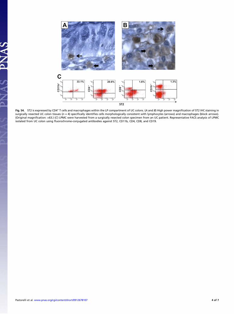
Fig. S4. ST2 is expressed by CD4+ T cells and macrophages within the LP compartment of UC colons. (A and B) High power magnification of ST2 IHC staining insurgically resected UC colon tissues (n = 4) specifically identifies cells morphologically consistent with lymphocytes (arrows) and macrophages (block arrows).(Original magnification: ×63.) (C) LPMC were harvested from a surgically resected colon specimen from an UC patient. Representative FACS analysis of LPMCisolated from UC colon using fluorochrome-conjugated antibodies against ST2, CD11b, CD4, CD8, and CD19.
Pastorelli et al. www.pnas.org/cgi/content/short/0912678107 4 of 7
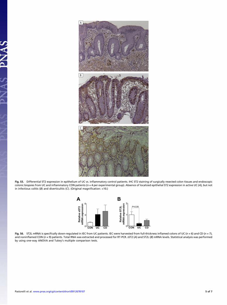
Fig. S5. Differential ST2 expression in epithelium of UC vs. inflammatory control patients. IHC ST2 staining of surgically resected colon tissues and endoscopiccolonic biopsies from UC and inflammatory CON patients (n = 4 per experimental group). Absence of localized epithelial ST2 expression in active UC (A), but notin infectious colitis (B) and diverticulitis (C). (Original magnification: ×10.)
Fig. S6. ST2L mRNA is specifically down-regulated in IEC from UC patients. IEC were harvested from full-thickness inflamed colons of UC (n = 6) and CD (n = 7),and noninflamed CON (n = 9) patients. Total RNA was extracted and processed for RT-PCR. sST2 (A) and ST2L (B) mRNA levels. Statistical analysis was performedby using one-way ANOVA and Tukey’s multiple comparison tests.
Pastorelli et al. www.pnas.org/cgi/content/short/0912678107 5 of 7

Fig. S7. Anti-TNF modulates the circulating IL-33/sST2 ratio in IBD patients, although not as potently in CD vs. UC patients. Serum samples were assayed fromIBD patients (UC, n = 9; CD, n = 11) undergoing Infliximab therapy (5-10 mg/kg, i.v.). Whole blood was drawn 1 h prior and 2 h after each infusion (total numberof infusions = 100). IL-33 and sST2 protein levels were measured by ELISA. (A) IL-33/sST2 ratio measured 1 h before each Infliximab infusion throughouttreatment protocol (T0, n = 15; W2, n = 15; W6, n = 11; W14, n = 12; W22, n = 10; W30, n = 9; W38, n = 5); W, week. (B) Individual data of CD patients (n = 7) at T0and at end of induction regimen (W14). Statistical analysis was performed by using paired t test for paired data.
Fig. S8. TNF stimulation increases intracellular f-IL-33 and sST2 expression in IEC. HT-29 IEC were cultured with or without TNF (10 ng/mL) for 0, 2, 6, 12, 24,and 48 h (n = 4 per condition). Total RNA and protein were extracted and processed for RT-PCR, Western blots, and ELISA, and supernatants were harvested forELISA. (A) IL-33 (Left), sST2 (Center), and ST2L (Right) were normalized to GAPDH and relative mRNA levels calculated as fold increase over unstimulatedsamples. (B) Western blots (Upper) and ELISAs (Lower) were performed on cell lysates for IL-33 (Left) and ST2 (Right). (C) IL-33 (Left) and sST2 (Right) proteinlevels were measured by ELISA. Statistical analysis was performed by using Student t test for paired data. Dotted line, SLD.
Pastorelli et al. www.pnas.org/cgi/content/short/0912678107 6 of 7
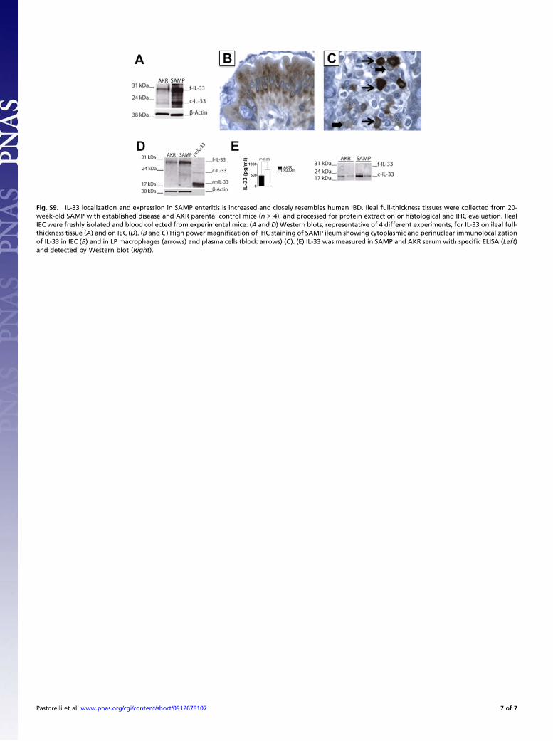
Fig. S9. IL-33 localization and expression in SAMP enteritis is increased and closely resembles human IBD. Ileal full-thickness tissues were collected from 20-week-old SAMP with established disease and AKR parental control mice (n ≥ 4), and processed for protein extraction or histological and IHC evaluation. IlealIEC were freshly isolated and blood collected from experimental mice. (A and D) Western blots, representative of 4 different experiments, for IL-33 on ileal full-thickness tissue (A) and on IEC (D). (B and C) High power magnification of IHC staining of SAMP ileum showing cytoplasmic and perinuclear immunolocalizationof IL-33 in IEC (B) and in LP macrophages (arrows) and plasma cells (block arrows) (C). (E) IL-33 was measured in SAMP and AKR serum with specific ELISA (Left)and detected by Western blot (Right).
Pastorelli et al. www.pnas.org/cgi/content/short/0912678107 7 of 7