Studying the Microbial World (microscopes) Supplemental instruction Designed by Pyeongsug Kim ©2010...
18
Studying Studying the Microbial World the Microbial World (microscopes) (microscopes) Supplemental instruction Designed by Pyeongsug Kim ©2010 [email protected] Picture from http://microbemagic.ucc.ie/about_microbes/good_bad_ugly.html Fall 2010 For Dr. Wright’s Bio 7/27 Class pdated: August 07, 2010
-
date post
22-Dec-2015 -
Category
Documents
-
view
213 -
download
0
Transcript of Studying the Microbial World (microscopes) Supplemental instruction Designed by Pyeongsug Kim ©2010...

Studying Studying the Microbial Worldthe Microbial World
(microscopes)(microscopes)Supplemental instruction
Designed by Pyeongsug Kim ©2010 [email protected]
Picture from http://microbemagic.ucc.ie/about_microbes/good_bad_ugly.html
Fall 2010
For Dr. Wright’s Bio 7/27 Class
Updated: August 07, 2010

Microbes are too small to see…MicroscopeMicroscope
Picture from http://www.toonpool.com/cartoons/MICROSCOPE%20LOOKING_27378 http://www2.sunysuffolk.edu/gambier/by14practical.htm http://www.jeol.com/PRODUCTS/ElectronOptics/TransmissionElectronMicroscopesTEM/300kV/JEM3100F/tabid/128/Default.aspx
•Light microscopes
The development light microscopes contributed to the study of microbiology
Designed by Pyeongsug Kim, ©2010 www.science-i.com
http://www.photosfan.com/2008-02/
•Electron microscopes

MicroscopeMicroscope*Useful of a microscope depends on it’s resolution.
___________the number of times an image is
enlarged.10x, 40x, 60x, 100x
Magnification
__________a measurement of clarity, the
smallest distance between two points on a specimen that can still be distinguished.
Resolution
Designed by Pyeongsug Kim, ©2009

• Light microscope - glass lens; 1,000x
• Electron microscope - electron lens; 100,000x - shorter than the waves of visible light.
• Atomic force microscope -an atomic scale; no preparation need.
MicroscopeMicroscope
/
Designed by Pyeongsug Kim, ©2010 www.science-i.com
Picture from http://nano.mtu.edu/afm.htm http://www.photosfan.com/2008-02

Light microscopeLight microscope-Light pass through a specimen. ; 1000x-Some need to stain bacteria.; some don’t increase contrast b/w microorganisms and surroundings.
-Bright-field (most common) Phase-constrast, Interference, Dark-field, Confocal, Fluorensence
Picture from http://asia.cnet.com/crave/2009/07/22/turn-your-mobile-phone-into-a-portable-microscope/ http://www.rp-photonics.com/fluorescence_microscopy.html http://www.microscopy-uk.org.uk/mag/indexmag.html?http://www.microscopy-uk.org.uk/mag/artoct06/dr-diatoms2.html
Designed by Pyeongsug Kim, ©2010 www.science-i.com

Which type of microscope are you using in Bio27 Lab. class?
Bright-field microscopeor compound microscope•A compound light microscope is also called a bright field microscope. We usually call compound microscope.
Designed by Pyeongsug Kim, ©2010 www.science-i.com
1) ______________1) ______________ - Most common - Fixed specimen(killed) or moving organism (live) eg) E.Coli stained by Scarlet Euglena in pond water…
Picture from http://www.inmagine.com/00124/00124080-photo http://www.sciencetoybox.com/cells.htm
Bright-field microscope
Tardigrades (water bears) under a compound microscope -Youtube.com

Compound Microscope
Eyepiece (oculars)
Arm
Stage
Opening of Stage
Fine-adjustmentCoarse-adjustment
Base
Illuminator
Iris diaphragm
Iris diaphragm lever
Stage clips
High-power objective
Low-power objective
Nosepiece
Body tube
In the Dr. Hughey’s Bio3 class slide

2) _____________ - amplifies differences in refractive index to
create contrast. - a darker appearance of the denser material. increasing contrast. -does not require staining to view the slidedoes not require staining to view the slide. -Makes unstained cells more readily visible. -Best for observing intracellular structures
Phase-contrast
Designed by Pyeongsug Kim, ©2010 www.science-i.com
Paramecium bursaria on phase contrast microscopeYoutube.com
Can we see microbes alive? ____YES!
Light microscope Light microscope (Cont’d)(Cont’d)

Can we see microbes alive? ____
Designed by Pyeongsug Kim, ©2010 www.science-i.com
3) ___________ -Light is directed toward the specimen
at an angle. -can see unstained cells Makes unstained cells more readily
visible. -stand out as bright objects against a
dark background.
Dark field
YES!
Paramecium Darkfield.aviYoutube.com
Light microscope Light microscope (Cont’d)(Cont’d)

Designed by Pyeongsug Kim, ©2010 www.science-i.com
•Bright-field – most widely used, specimen is darker than surrounding field
•Dark-field – brightly illuminated specimens surrounded by dark field
•Phase-contrast – transforms subtle changes in light waves passing through the specimen into differences in light intensity, best for observing intracellular structures
Light microscope Light microscope (Cont’d)(Cont’d)

4) ___________ - Nomarski DIC(differential interfence contrast) - Two light beams - 3D image!!3D image!!
Interference
Rotifer; Synchaeta sp via DIC microscopy
Youtube.com
Can we see microbes alive? ____ YES!
Designed by Pyeongsug Kim, ©2010 www.science-i.com
Light microscope Light microscope (Cont’d)(Cont’d)

Light microscope Light microscope (Cont’d)(Cont’d)
5) ___________ -Light onto the specimen (not pass through) cells attached to soil particles or other opaque materials
-UV light -Fluorescent dyes and Tags Uses dyes that emit visible light when bombarded
with shorter uv rays. Can stain intracellular structures Can stain live cells Can distinguish living and dead cells Can be used in quantitative analysis
Fluorescence
Designed by Pyeongsug Kim, ©2010 www.science-i.com
Can we see microbes alive? ____ YES!

6) ___________ -Light onto the specimen (not pass through) -3D image!!3D image!! -Can see inside intact cell in DETAIL. a miniature CAT scan for cells.
- Cell division -may use fluorescent dyes to see cellular location or to determine molecule compound.
- For thick structure eg) community of organism.
Confocal scanning laser
CAT scan for brain
Light microscope Light microscope (Cont’d)(Cont’d)
Designed by Pyeongsug Kim, ©2010 www.science-i.com
Can we see microbes alive? ____ YES!
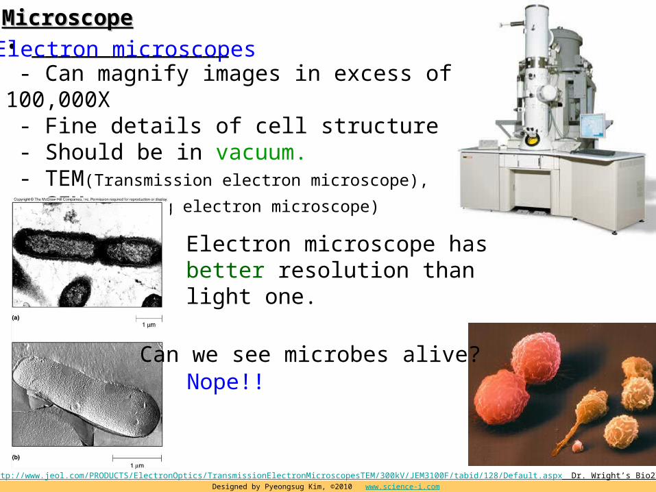
• _______________ - Can magnify images in excess of 100,000X - Fine details of cell structure - Should be in vacuum. - TEM(Transmission electron microscope),
SEM(scanning electron microscope)
Electron microscopesMicroscopeMicroscope
Picture from http://www.jeol.com/PRODUCTS/ElectronOptics/TransmissionElectronMicroscopesTEM/300kV/JEM3100F/tabid/128/Default.aspx Dr. Wright’s Bio27 PPTDesigned by Pyeongsug Kim, ©2010 www.science-i.com
Electron microscope has better resolution than light one.
Can we see microbes alive? Nope!!

Electron microscope (Cont’d)Electron microscope (Cont’d)
1) _____________________ -To see inside cell. (organelles) - transmits electrons through the specimen -Thin sectioning Freeze fracturing, freeze etching -Vacuum required. -Artifacts are concern.
2) ______________________ -To see surface of the cell. -Vacuum NOT required. - a whole, metal-coated specimen with
electrons - 3D image!!3D image!! Very expensive~~~~
TEM(Transmission electron microscope)
SEM(scanning electron microscope)
Designed by Pyeongsug Kim, ©2010 www.science-i.com
Should kill the specimen!!
Should kill the specimen!!

Avian flu virus by scanning microscopes
Designed by Pyeongsug Kim, ©2010 www.science-i.com
Picture from http://blog.silive.com/health/2008/10/climate_change_could_promote_t.html http://ibexinc.wordpress.com/2008/12/31/
electron microscope image of a virus (bacteriophage )
Virus can be seen through a electron microscope.Virus can be seen through a electron microscope.
Flu virus by electron microscopes
Designed by Pyeongsug Kim, ©2010 www.science-i.com
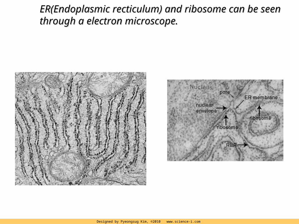
ER(Endoplasmic recticulum) and ribosome can be seen ER(Endoplasmic recticulum) and ribosome can be seen through a electron microscope.through a electron microscope.

Atomic force microscopeAtomic force microscope
Designed by Pyeongsug Kim, ©2010 www.science-i.com
First ever real time film of DNA-enzyme interactionYoutube.com
-Atomic scale eg. Interaction between DNA and enzyme.-Atomic force microscope has much better resolution than electron microscope.
Picture from http://nano.mtu.edu/afm.htm