
Structure of the Cytoplasmic Loop between Putative Helices ... · position 124 labeled with a Cys...
7
pubs.acs.org/Biochemistry Published on Web 04/29/2009 r 2009 American Chemical Society 5284 Biochemistry 2009, 48, 5284–5290 DOI: 10.1021/bi8020668 Structure of the Cytoplasmic Loop between Putative Helices II and III of the Mannitol Permease of Escherichia coli: A Tryptophan and 5-Fluorotryptophan Spectroscopy Study † Erwin P. P. Vos, ‡ Marcel Bokhove, ‡ Ben H. Hesp, § and Jaap Broos* ,‡ ‡ Department of Biophysical Chemistry, Groningen Biomolecular Science and Biotechnology Institute, and § Zernike Institute for Advanced Materials, University of Groningen, Nijenborgh 4, 9747 AG Groningen, The Netherlands Received November 7, 2008; Revised Manuscript Received April 28, 2009 ABSTRACT: In this work, four single tryptophan (Trp) mutants of the dimeric mannitol transporter of Escherichia coli, EII mtl , are characterized using Trp and 5-fluoroTrp (5-FTrp) fluorescence spectroscopy. The four positions, 97, 114, 126, and 133, are located in a region shown by recent studies to be involved in the mannitol translocation process. To spectroscopically distinguish between the Trp positions in each subunit of dimeric EII mtl , 5-FTrp was biosynthetically incorporated because of its much simpler photophysics compared to those of Trp. The steady-state and time-resolved fluorescence methodologies used point out that all four positions are in structured environments, both in the absence and in the presence of a saturating concentration of mannitol. The fluorescence decay of all 5-FTrp-containing mutants was highly homogeneous, suggesting similar microenvironments for both probes per dimer. However, Stern-Volmer quenching experiments using potassium iodide indicate different solvent accessibilities for the two probes at positions 97 and 133. A5A ˚ two-dimensional (2D) projection map of the membrane-embedded IIC mtl dimer showing 2-fold symmetry is available. The results of this work are in better agreement with a 7 A ˚ projection map from a single 2D crystal on which no symmetry was imposed. The mannitol permease of Escherichia coli, EII mtl , 1 is respon- sible for the transport of mannitol across the cytoplasmic membrane and for its concomitant phosphorylation to mannitol 1-phosphate. The protein belongs to the phosphoenolpyruvate (PEP)-dependent phosphotransferase system (PTS), a system consisting of different sugar-specific EII transporters which phosphorylate their sugar during translocation (1, 2). The phosphate group is donated by PEP and transferred via the cytoplasmic proteins EI and HPr to EII. In EII mtl , the phosphate group is donated to His554 in the A domain and subsequently transferred to Cys384 in the B domain, which phosphorylates mannitol bound at the C domain (IIC mtl ). The A and B domains (each 14 kDa) and the C domain (36 kDa) are covalently linked. The three-dimensional (3D) structures of the A and B domains have been determined (3, 4). For the membrane-embedded C domain, harboring the sugar translocation pathway, a 5 A ˚ two- dimensional (2D) projection map is available (5). The functional oligomeric state of EII mtl is a dimer, and it contains one mannitol binding site (6). Currently, little is known about the structure of IIC domains of the PTS sugar translocators. Topology studies predict six to eight TMHs (7-9). Recent studies (8, 10) suggest that residues 70-134, presented as a cytoplasmic loop in the IIC mtl topology model of Sugiyama et al. (7) (Figure 1), are involved in the mannitol translocation process. Some of these residues are accessible only from the periplasmic side, while others are accessible only from the cytoplasmic side (8). Conformational changes upon mannitol binding or upon EII mtl phosphorylation in this region have been reported (8, 10). Moreover, residue position 124 labeled with a Cys can form a disulfide bridge with Cys384 in the B domain (11). In two recent topology models, residues 70-134 are presented as forming two short R-helices, protruding into the membrane (8), or presented as a transmem- brane helix and a periplasmic loop (9). IIC mtl is known to be remarkably resistant to trypsin degradation, and therefore, the cytoplasmic loop formed by residues 70-134 is likely structured. Tryptophan phosphorescence spectroscopy was used to in- vestigate structural details of this “active” region of the IIC mtl domain (10). Four single Trp mutants of EII mtl containing a Trp at positions 97, 114, 126, and 133 were analyzed (mutants W97, W114, W126, and W133, respectively). All mutants were created by F to W substitution in the Trp-less protein. One remarkable outcome of this study was that the microenvironment of W97 † This work was supported by The Netherlands Foundation for Chemical Research (CW) with financial aid from The Netherlands Organization for the Advancement of Scientific Research (NWO). *To whom correspondence should be addressed. Phone: +31 50 3634277. Fax: +31 50 3634800. E-mail: [email protected]. 1 Abbreviations: EII mtl , mannitol-specific transporting and phosphor- ylating enzyme from E. coli; 5-FTrp, 5-fluorotryptophan; 5-FNATA, N-acetyl-DL-5-fluorotryptophanamide; PTS, phosphoenolpyruvate-de- pendent group translocation system; TMH, transmembrane helix; mannitol, D-mannitol; PEP, phosphoenolpyruvate; S-V, Stern-Vol- mer; fwhh, full width at half-height; TCSPC, time-correlated single- photon counting; EII mtl , wild-type EII mtl with tryptophans at positions 30, 42, 109, and 117; Trp-less EII mtl , EII mtl in which the four native tryptophans are replaced with Phe. W97, W114, W126, and W133 are the single-Trp-containing EII mtl mutants based on Trp-less EII mtl . Downloaded by UNIV OF GRONINGEN on September 17, 2009 | http://pubs.acs.org Publication Date (Web): April 29, 2009 | doi: 10.1021/bi8020668
Transcript of Structure of the Cytoplasmic Loop between Putative Helices ... · position 124 labeled with a Cys...

pubs.acs.org/Biochemistry Published on Web 04/29/2009 r 2009 American Chemical Society
5284 Biochemistry 2009, 48, 5284–5290
DOI: 10.1021/bi8020668
Structure of the Cytoplasmic Loop between Putative Helices II and III of the MannitolPermease of Escherichia coli: A Tryptophan and 5-Fluorotryptophan Spectroscopy Study†
Erwin P. P. Vos,‡ Marcel Bokhove,‡ Ben H. Hesp,§ and Jaap Broos*,‡
‡Department of Biophysical Chemistry, Groningen Biomolecular Science and Biotechnology Institute, and §Zernike Institute forAdvanced Materials, University of Groningen, Nijenborgh 4, 9747 AG Groningen,
The Netherlands
Received November 7, 2008; Revised Manuscript Received April 28, 2009
ABSTRACT: In this work, four single tryptophan (Trp) mutants of the dimeric mannitol transporter ofEscherichia coli, EIImtl, are characterized using Trp and 5-fluoroTrp (5-FTrp) fluorescence spectroscopy. Thefour positions, 97, 114, 126, and 133, are located in a region shown by recent studies to be involved in themannitol translocation process. To spectroscopically distinguish between the Trp positions in each subunit ofdimeric EIImtl, 5-FTrp was biosynthetically incorporated because of its much simpler photophysics comparedto those of Trp. The steady-state and time-resolved fluorescence methodologies used point out that all fourpositions are in structured environments, both in the absence and in the presence of a saturating concentrationof mannitol. The fluorescence decay of all 5-FTrp-containing mutants was highly homogeneous, suggestingsimilar microenvironments for both probes per dimer. However, Stern-Volmer quenching experiments usingpotassium iodide indicate different solvent accessibilities for the two probes at positions 97 and 133.A 5 A two-dimensional (2D) projection map of the membrane-embedded IICmtl dimer showing 2-foldsymmetry is available. The results of this work are in better agreement with a 7 A projection map from a single2D crystal on which no symmetry was imposed.
The mannitol permease of Escherichia coli, EIImtl,1 is respon-sible for the transport of mannitol across the cytoplasmicmembrane and for its concomitant phosphorylation to mannitol1-phosphate. The protein belongs to the phosphoenolpyruvate(PEP)-dependent phosphotransferase system (PTS), a systemconsisting of different sugar-specific EII transporters whichphosphorylate their sugar during translocation (1, 2). Thephosphate group is donated by PEP and transferred via thecytoplasmic proteins EI and HPr to EII. In EIImtl, the phosphategroup is donated to His554 in the A domain and subsequentlytransferred to Cys384 in the B domain, which phosphorylatesmannitol bound at the C domain (IICmtl). The A and B domains(each 14 kDa) and the C domain (36 kDa) are covalently linked.The three-dimensional (3D) structures of the A and B domainshave been determined (3, 4). For the membrane-embedded C
domain, harboring the sugar translocation pathway, a 5 A two-dimensional (2D) projection map is available (5). The functionaloligomeric state of EIImtl is a dimer, and it contains onemannitolbinding site (6).
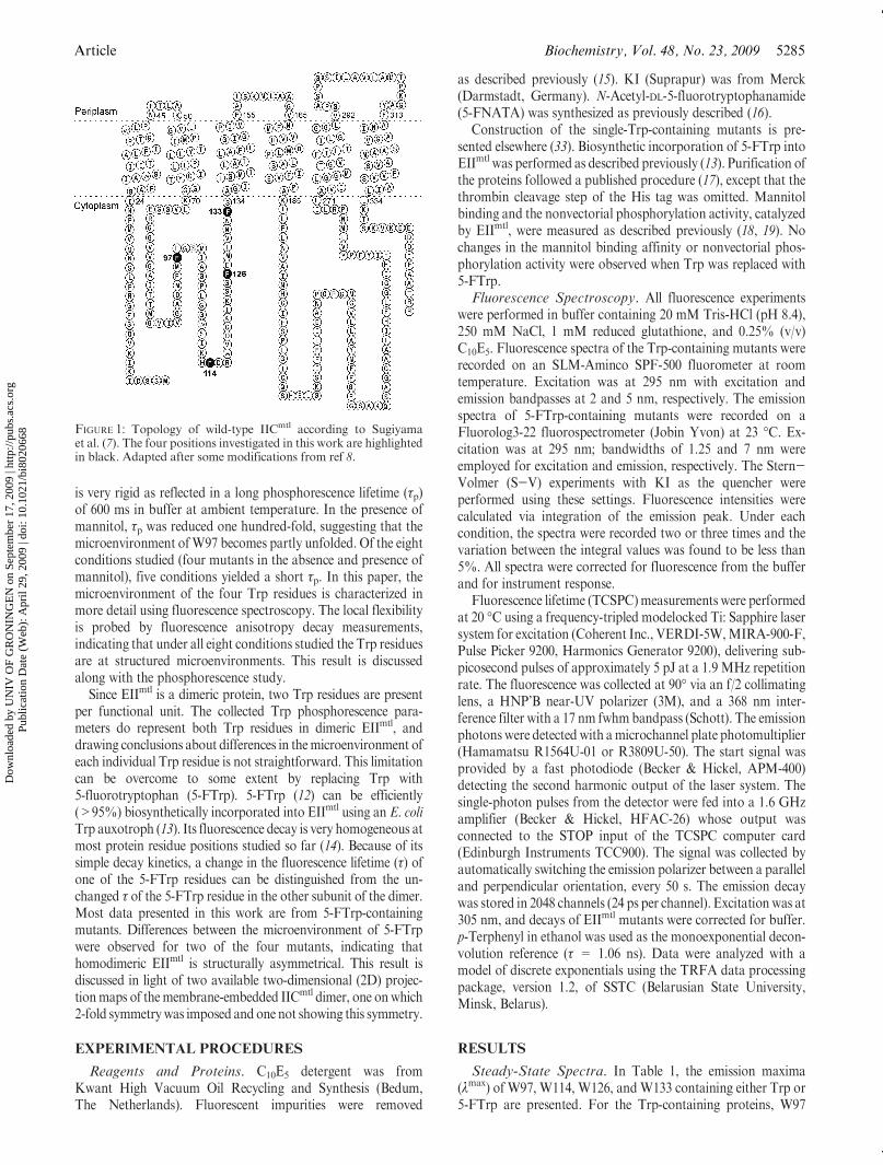
Currently, little is knownabout the structure of IIC domains ofthe PTS sugar translocators. Topology studies predict six to eightTMHs (7-9). Recent studies (8, 10) suggest that residues70-134, presented as a cytoplasmic loop in the IICmtl topologymodel of Sugiyama et al. (7) (Figure 1), are involved in themannitol translocation process. Some of these residues areaccessible only from the periplasmic side, while others areaccessible only from the cytoplasmic side (8). Conformationalchanges upon mannitol binding or upon EIImtl phosphorylationin this region have been reported (8, 10). Moreover, residueposition 124 labeled with a Cys can form a disulfide bridge withCys384 in the B domain (11). In two recent topology models,residues 70-134 are presented as forming two short R-helices,protruding into the membrane (8), or presented as a transmem-brane helix and a periplasmic loop (9). IICmtl is known to beremarkably resistant to trypsin degradation, and therefore, thecytoplasmic loop formed by residues 70-134 is likely structured.
Tryptophan phosphorescence spectroscopy was used to in-vestigate structural details of this “active” region of the IICmtl
domain (10). Four single Trp mutants of EIImtl containing a Trpat positions 97, 114, 126, and 133 were analyzed (mutants W97,W114, W126, andW133, respectively). All mutants were createdby F to W substitution in the Trp-less protein. One remarkableoutcome of this study was that the microenvironment of W97
†This work was supported by The Netherlands Foundation forChemical Research (CW) with financial aid from The NetherlandsOrganization for the Advancement of Scientific Research (NWO).*To whom correspondence should be addressed. Phone: +31 50
3634277. Fax: +31 50 3634800. E-mail: [email protected]: EIImtl, mannitol-specific transporting and phosphor-
ylating enzyme from E. coli; 5-FTrp, 5-fluorotryptophan; 5-FNATA,N-acetyl-DL-5-fluorotryptophanamide; PTS, phosphoenolpyruvate-de-pendent group translocation system; TMH, transmembrane helix;mannitol, D-mannitol; PEP, phosphoenolpyruvate; S-V, Stern-Vol-mer; fwhh, full width at half-height; TCSPC, time-correlated single-photon counting; EIImtl, wild-type EIImtl with tryptophans at positions30, 42, 109, and 117; Trp-less EIImtl, EIImtl in which the four nativetryptophans are replaced with Phe. W97, W114, W126, and W133 arethe single-Trp-containing EIImtl mutants based on Trp-less EIImtl.
Dow
nloa
ded
by U
NIV
OF
GR
ON
ING
EN
on
Sept
embe
r 17
, 200
9 | h
ttp://
pubs
.acs
.org
P
ublic
atio
n D
ate
(Web
): A
pril
29, 2
009
| doi
: 10.
1021
/bi8
0206
68
Article Biochemistry, Vol. 48, No. 23, 2009 5285
is very rigid as reflected in a long phosphorescence lifetime (τp)of 600 ms in buffer at ambient temperature. In the presence ofmannitol, τp was reduced one hundred-fold, suggesting that themicroenvironment ofW97 becomes partly unfolded. Of the eightconditions studied (four mutants in the absence and presence ofmannitol), five conditions yielded a short τp. In this paper, themicroenvironment of the four Trp residues is characterized inmore detail using fluorescence spectroscopy. The local flexibilityis probed by fluorescence anisotropy decay measurements,indicating that under all eight conditions studied the Trp residuesare at structured microenvironments. This result is discussedalong with the phosphorescence study.
Since EIImtl is a dimeric protein, two Trp residues are presentper functional unit. The collected Trp phosphorescence para-meters do represent both Trp residues in dimeric EIImtl, anddrawing conclusions about differences in themicroenvironment ofeach individual Trp residue is not straightforward. This limitationcan be overcome to some extent by replacing Trp with5-fluorotryptophan (5-FTrp). 5-FTrp (12) can be efficiently(>95%) biosynthetically incorporated into EIImtl using an E. coliTrp auxotroph (13). Its fluorescence decay is very homogeneous atmost protein residue positions studied so far (14). Because of itssimple decay kinetics, a change in the fluorescence lifetime (τ) ofone of the 5-FTrp residues can be distinguished from the un-changed τ of the 5-FTrp residue in the other subunit of the dimer.Most data presented in this work are from 5-FTrp-containingmutants. Differences between the microenvironment of 5-FTrpwere observed for two of the four mutants, indicating thathomodimeric EIImtl is structurally asymmetrical. This result isdiscussed in light of two available two-dimensional (2D) projec-tionmaps of themembrane-embedded IICmtl dimer, one onwhich2-fold symmetrywas imposed andone not showing this symmetry.
EXPERIMENTAL PROCEDURES
Reagents and Proteins. C10E5 detergent was fromKwant High Vacuum Oil Recycling and Synthesis (Bedum,The Netherlands). Fluorescent impurities were removed
as described previously (15). KI (Suprapur) was from Merck(Darmstadt, Germany). N-Acetyl-DL-5-fluorotryptophanamide(5-FNATA) was synthesized as previously described (16).
Construction of the single-Trp-containing mutants is pre-sented elsewhere (33). Biosynthetic incorporation of 5-FTrp intoEIImtl was performed as described previously (13). Purification ofthe proteins followed a published procedure (17), except that thethrombin cleavage step of the His tag was omitted. Mannitolbinding and the nonvectorial phosphorylation activity, catalyzedby EIImtl, were measured as described previously (18, 19). Nochanges in the mannitol binding affinity or nonvectorial phos-phorylation activity were observed when Trp was replaced with5-FTrp.Fluorescence Spectroscopy. All fluorescence experiments
were performed in buffer containing 20 mM Tris-HCl (pH 8.4),250 mM NaCl, 1 mM reduced glutathione, and 0.25% (v/v)C10E5. Fluorescence spectra of the Trp-containing mutants wererecorded on an SLM-Aminco SPF-500 fluorometer at roomtemperature. Excitation was at 295 nm with excitation andemission bandpasses at 2 and 5 nm, respectively. The emissionspectra of 5-FTrp-containing mutants were recorded on aFluorolog3-22 fluorospectrometer (Jobin Yvon) at 23 �C. Ex-citation was at 295 nm; bandwidths of 1.25 and 7 nm wereemployed for excitation and emission, respectively. The Stern-Volmer (S-V) experiments with KI as the quencher wereperformed using these settings. Fluorescence intensities werecalculated via integration of the emission peak. Under eachcondition, the spectra were recorded two or three times and thevariation between the integral values was found to be less than5%. All spectra were corrected for fluorescence from the bufferand for instrument response.
Fluorescence lifetime (TCSPC)measurements were performedat 20 �C using a frequency-tripled modelocked Ti: Sapphire lasersystem for excitation (Coherent Inc., VERDI-5W,MIRA-900-F,Pulse Picker 9200, Harmonics Generator 9200), delivering sub-picosecond pulses of approximately 5 pJ at a 1.9 MHz repetitionrate. The fluorescence was collected at 90� via an f/2 collimatinglens, a HNP’B near-UV polarizer (3M), and a 368 nm inter-ference filter with a 17 nm fwhmbandpass (Schott). The emissionphotonswere detected with amicrochannel plate photomultiplier(Hamamatsu R1564U-01 or R3809U-50). The start signal wasprovided by a fast photodiode (Becker & Hickel, APM-400)detecting the second harmonic output of the laser system. Thesingle-photon pulses from the detector were fed into a 1.6 GHzamplifier (Becker & Hickel, HFAC-26) whose output wasconnected to the STOP input of the TCSPC computer card(Edinburgh Instruments TCC900). The signal was collected byautomatically switching the emission polarizer between a paralleland perpendicular orientation, every 50 s. The emission decaywas stored in 2048 channels (24 ps per channel). Excitationwas at305 nm, and decays of EIImtl mutants were corrected for buffer.p-Terphenyl in ethanol was used as the monoexponential decon-volution reference (τ = 1.06 ns). Data were analyzed with amodel of discrete exponentials using the TRFA data processingpackage, version 1.2, of SSTC (Belarusian State University,Minsk, Belarus).
RESULTS
Steady-State Spectra. In Table 1, the emission maxima(λmax) of W97, W114, W126, andW133 containing either Trp or5-FTrp are presented. For the Trp-containing proteins, W97
FIGURE 1: Topology of wild-type IICmtl according to Sugiyamaet al. (7). The four positions investigated in this work are highlightedin black. Adapted after some modifications from ref 8.
Dow
nloa
ded
by U
NIV
OF
GR
ON
ING
EN
on
Sept
embe
r 17
, 200
9 | h
ttp://
pubs
.acs
.org
P
ublic
atio
n D
ate
(Web
): A
pril
29, 2
009
| doi
: 10.
1021
/bi8
0206
68
5286 Biochemistry, Vol. 48, No. 23, 2009 Vos et al.
shows the most blue-shifted λmax (at 318 nm) andW126 the mostred-shifted λmax (at 330 nm). For the 5-FTrp-containing proteins,the same trend is visible except that the spectra are approximately5 nm red-shifted. 5-FTrp emission spectra are known to beslightly red-shifted compared to Trp emission spectra (16).Effect of Mannitol on the Emission Spectra. Mutants
W114, W126, and W133 show, like wild-type EIImtl, a highaffinity for mannitol (Kd ∼ 100 nM) (10). For W97, a loweraffinity was measured (Kd ∼ 2 μM) (10). The effects of asaturating concentration of mannitol (50 μM) on the emissionspectra of the Trp-containing mutants are presented in Table 1.Mannitol binding induces only minor changes (<5%) in thespectra of W114 and W126. The emission intensity of W133increases 5%. For W97, the effect of mannitol is very strong asthe emission intensity drops 46%. A similar trend is evident forthe 5-FTrp-containing mutants, although the change in emissionis significantly smaller for the W97 mutant (-13%). The broad-ness (fwhh) of the emission spectra of the W97 and W126mutants, containing 5-FTrp, increases upon mannitol binding,while a narrowing was observed for W133 containing 5-FTrp(Table 1). Ameasurable change in λmax uponmannitol binding (2nm) was observed only for W126 containing 5-FTrp.Time-Resolved FluorescenceData of 5-FTrp-Containing
Mutants. InFigures 2 and 3, the 5-FTrp decays are presented forW97, W114, W126, and W133, together with reduced residuals(χr
2) and the autocorrelation function. The fit parameters arelisted in Table 2. For W97 andW114, a single exponential couldadequately fit the decays. For W126 and W133, three exponen-tials were needed for a proper fit; the decays are dominated byτ = 5.3 ns (R = 0.93) and τ = 4.5 ns (R = 0.95) values,respectively. In the presence of mannitol, the fluorescence ofW97 decays essentially with the same τ as in the absence ofmannitol. A 19% drop in amplitude (R) is detected, showing thatthe decrease in steady-state emission upon mannitol binding isdue to the formation of dark ground-state complexes. Similarly, adrop in R was also observed for W133 (-10% for τ = 4.5 ns),together with no change in τ.
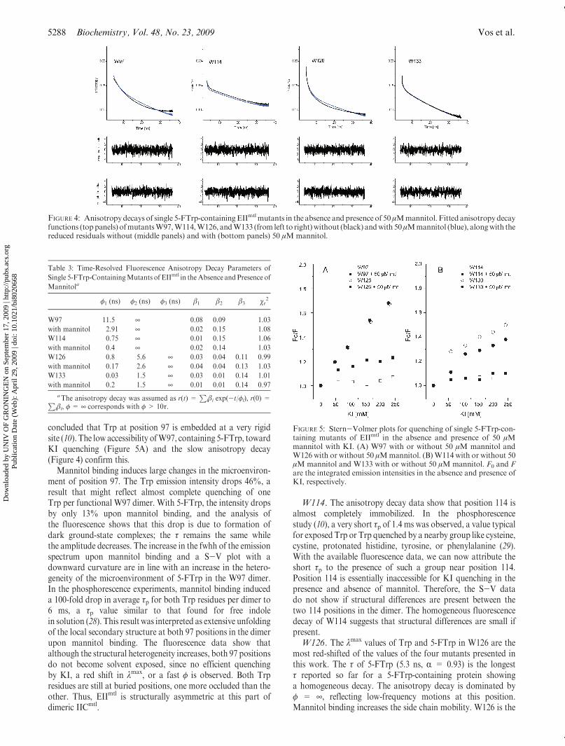
To investigate the microenvironment of the four 5-Trp posi-tions in more detail, the anisotropy decays were recorded in theabsence and presence of mannitol (Figure 4 and Table 3). Thedecays could be properly fitted using two or three rotationalcorrelation times (φ). For the sake of clarity, only the modeled fitfunctions are presented in Figure 4. All anisotropy decays aredominated by φ=¥, typical for immobilized 5-FTrp side chains.Because dimeric EIImtl, solubilized by detergent, is a largecomplex (>200 kDa), depolarization due to protein tumblingcannot be assessed with 5-FTrp.Solvent Accessibility of the 5-FTrp Positions toward KI.
Stern-Volmer (S-V) experiments were performed with the fourmutants using KI as the collisional quencher. KI and notacrylamide was used as the quencher because the latter likelypartitions into the C10E5 micelle surrounding EIImtl, in this waybiasing the quenching data. Moreover, acrylamide can quenchthe singlet Trp state quite efficiently via a through spacemechanism (20).
EIImtl is a dimeric protein under the conditions used in thiswork (21). When the two 5-FTrp residues in the dimer exhibitsimilar solvent accessibilities, a linear S-Vplot is expected.Whenthe solvent accessibility is different for the twoprobes, a S-Vplotshowing a downward curvature is expected (22). 5-FTrp atposition 126 shows a high and essentially similar solvent acces-sibility in the presence and absence of mannitol (Figure 5).
For mutant W97 without mannitol, a very low solvent accessi-bility for KI was measured. In the presence of mannitol, afraction of the protein becomes more solvent accessible atposition 97. The opposite situation was observed for W133;addition of mannitol decreases the accessibility of 5-FTrp atposition 133 for KI. The S-V plots for W133 under bothconditions show a downward curvature, indicating differentmicroenvironments of each 133 position in the dimer.
Since the quenching of 5-FTrp for KI has not been reportedbefore, the KI sensitivity of this probe was investigated using theneutral analogue 5-FNATA (τ = 4.4 ns) (14), dissolved in thesame buffer as the EIImtl mutants. A S-V constant of 20 M-1
was observed, corresponding to a collisional quenching constant(kq) of 4.5 � 109 M-1 s-1. KI quenching of NATA under theseconditions yielded a kq of 4.3 � 109 M-1 s-1. This shows thatfluoro substitution of Trp at the 5 position does not affect itssensitivity for quenching by iodide.
DISCUSSION
In this work, a fluorescence study of four single Trpmutants ofEIImtl, all containing a Trp in the first putative cytoplasmic loopof the IICmtl topology model of Sugiyama et al. (7), is presented(Figure 1). Comparison of the fluorescence lifetimes, rotationalcorrelation times, and solvent accessibilities, in the absence andpresence of mannitol, shows that this part of the protein under-goes large structural changes upon ligand binding. Most of theexperiments have been conducted with mutants containing 5-FTrp, biosynthetically incorporated using an E. coli Trp auxo-troph. In an earlier report, the same positions have been studiedusing Trp phosphorescence spectroscopy (10). Both techniquesyield complementary information; in fluorescence, dynamicfeatures on the nanosecond time scale are probed while Trpphosphorescence is remarkably sensitive to the local fluidity andcan report on conformational changes in the microsecond tosecond time domain. A new feature introduced here is the abilityto distinguish between the microenvironments of the two Trppositions in each subunit in dimeric EIImtl. For this, we tookadvantage of the simple fluorescence decay kinetics of 5-FTrpand combined time-resolved data with iodide quenching results.Comparison of Trp and 5-FTrp as Intrinsic Protein
Probes. A difference between Trp and 5-FTrp, relevant forprotein fluorescence studies, is the 0.3 eV increase in ionizationpotential upon 5-fluoro substitution (16). The higher ionizationpotential efficiently suppresses electron transfer from the excitedstate to nearby amide groups acting as electron acceptors.This photoinduced electron transfer process is the dominant
Table 1: Steady-State Fluorescence of Single Trp and 5-FTrp-Containing
Mutants of EIImtl
λmax (nm)
emission
intensity (% change)
with mannitol
fwhh
(nm)
fwhh with
mannitol
(nm)
W97 Trp 318 -46
W97 5-FTrp 323 -13 44 46.9
W114 Trp 324 3
W114 5-FTrp 329 3 48.3 48.7
W126 Trp 330 -4
W126 5-FTrp 334 (336
withmannitol)
0 54.6 55.5
W133 Trp 321 5
W133 5-FTrp 325 -8 45.7 44.8
Dow
nloa
ded
by U
NIV
OF
GR
ON
ING
EN
on
Sept
embe
r 17
, 200
9 | h
ttp://
pubs
.acs
.org
P
ublic
atio
n D
ate
(Web
): A
pril
29, 2
009
| doi
: 10.
1021
/bi8
0206
68

Article Biochemistry, Vol. 48, No. 23, 2009 5287
nonradiative depopulation process of the Trp singlet state (23).The large variation in quantum yield (Q) observed for Trp indifferent proteins could be related to the tuning of this process bythe proteinmatrix (24). 5-Fluoro substitution in Trp increases theQ and τ by ∼50% (14). It also reduces the fluorescence decayheterogeneity found for Trp embedded in proteins since eachrotamer is either not or only minimally quenched by amidegroups, an observation that explains the monoexponential decay
often observed for 5-FTrp in proteins (16, 25). Compared to thatof Trp, less variation in Q is expected for 5-FTrp-containingproteins undergoing a conformational change since a newestablished rotamer population likely yields a similar Q. Thedata presented inTable 1 show that the variation inQ, induced bymannitol binding, is equal or smaller for the 5-FTrp-containingproteins than for the Trp-containing proteins.
Isolation of a new batch of protein can result in a Trp rotamerdistributionwhich differs froma previous batch since the rotamerdistribution, “frozen” by the proteinmatrix, can be dependent onexperimental conditions used during isolation. We noticed thatthe reproducibility of the lifetime of 5-FTrp-containing proteinswas very high; typically, a variation of <0.2 ns was foundbetween different protein batches. The low dependence on thelifetime for the rotamer distribution might be the reason for this.
Below the spectral properties of each mutant are discussedfollowed by a discussion of what information this work providesabout the presence of (a)symmetry in the EIImtl dimer.W97. The λmax of Trp in W97 is 318 nm, typical for a Trp in
either a hydrophobic (26) or a rigid environment, where thestabilization of the large excited-state dipole moment of Trp islimited (27). The position of the 0-0 vibrational band in thephosphorescence spectrum of W97 was typical for a polar site.On the basis of all the phosphorescence data recorded, it could be
FIGURE 3: Fluorescence decays of single 5-FTrp-containing EIImtl mutants in the presence of 50 μMmannitol. Experimental fluorescence decays(toppanels) ofmutantsW97,W114,W126, andW133 (from left to right), alongwith the reduced residuals (middle panels) and the autocorrelationfunctions of the residuals (bottom panels).
FIGURE 2: Fluorescence decays of single 5-FTrp-containing EIImtl mutants. Experimental fluorescence decays (top panels) of mutants W97,W114, W126, and W133 (from left to right), along with the reduced residuals (middle panels) and the autocorrelation functions of the residuals(bottom panels).
Table 2: Time-Resolved Fluorescence Intensity Decay Parameters of
Single 5-FTrp-Containing Mutants of EIImtl in the Absence and Presence
of Mannitola
τ1 (ns) τ2 (ns) τ3 (ns) R1 R2 R3 χr2
W97 4.6 1.0 1.01
with mannitol 4.6 8.6 0.98 0.02 1.11
W114 4.7 1.0 1.07
with mannitol 1.6 4.7 0.02 0.98 1.03
W126 0.01 2.2 5.3 0.01 0.06 0.93 1.01
with mannitol 3.0 5.4 0.01 0.99 1.07
W133 0.9 4.5 8.3 0.03 0.95 0.02 1.01
with mannitol 1.2 4.6 13.0 0.04 0.95 0.01 0.95
aThe total intensity was assumed as I(t) =P
iRi exp(-t/τi), wherePiRi = 1.
Dow
nloa
ded
by U
NIV
OF
GR
ON
ING
EN
on
Sept
embe
r 17
, 200
9 | h
ttp://
pubs
.acs
.org
P
ublic
atio
n D
ate
(Web
): A
pril
29, 2
009
| doi
: 10.
1021
/bi8
0206
68
5288 Biochemistry, Vol. 48, No. 23, 2009 Vos et al.
concluded that Trp at position 97 is embedded at a very rigidsite (10). The low accessibility ofW97, containing 5-FTrp, towardKI quenching (Figure 5A) and the slow anisotropy decay(Figure 4) confirm this.
Mannitol binding induces large changes in the microenviron-ment of position 97. The Trp emission intensity drops 46%, aresult that might reflect almost complete quenching of oneTrp per functional W97 dimer. With 5-FTrp, the intensity dropsby only 13% upon mannitol binding, and the analysis ofthe fluorescence shows that this drop is due to formation ofdark ground-state complexes; the τ remains the same whilethe amplitude decreases. The increase in the fwhh of the emissionspectrum upon mannitol binding and a S-V plot with adownward curvature are in line with an increase in the hetero-geneity of the microenvironment of 5-FTrp in the W97 dimer.In the phosphorescence experiments, mannitol binding induceda 100-fold drop in average τp for both Trp residues per dimer to6 ms, a τp value similar to that found for free indolein solution (28). This result was interpreted as extensive unfoldingof the local secondary structure at both 97 positions in the dimerupon mannitol binding. The fluorescence data show thatalthough the structural heterogeneity increases, both 97 positionsdo not become solvent exposed, since no efficient quenchingby KI, a red shift in λmax, or a fast φ is observed. Both Trpresidues are still at buried positions, one more occluded than theother. Thus, EIImtl is structurally asymmetric at this part ofdimeric IICmtl.
W114. The anisotropy decay data show that position 114 isalmost completely immobilized. In the phosphorescencestudy (10), a very short τp of 1.4 ms was observed, a value typicalfor exposedTrp or Trp quenched by a nearby group like cysteine,cystine, protonated histidine, tyrosine, or phenylalanine (29).With the available fluorescence data, we can now attribute theshort τp to the presence of such a group near position 114.Position 114 is essentially inaccessible for KI quenching in thepresence and absence of mannitol. Therefore, the S-V datado not show if structural differences are present between thetwo 114 positions in the dimer. The homogeneous fluorescencedecay of W114 suggests that structural differences are small ifpresent.W126. The λmax values of Trp and 5-FTrp in W126 are the
most red-shifted of the values of the four mutants presented inthis work. The τ of 5-FTrp (5.3 ns, R = 0.93) is the longestτ reported so far for a 5-FTrp-containing protein showinga homogeneous decay. The anisotropy decay is dominated byφ = ¥, reflecting low-frequency motions at this position.Mannitol binding increases the side chain mobility. W126 is the
FIGURE 4: Anisotropydecays of single 5-FTrp-containingEIImtlmutants in the absence andpresenceof 50μMmannitol. Fitted anisotropydecayfunctions (toppanels) ofmutantsW97,W114,W126, andW133 (from left to right)without (black) andwith 50μMmannitol (blue), alongwith thereduced residuals without (middle panels) and with (bottom panels) 50 μMmannitol.
Table 3: Time-Resolved Fluorescence Anisotropy Decay Parameters of
Single 5-FTrp-ContainingMutants of EIImtl in theAbsence and Presence of
Mannitola
φ1 (ns) φ2 (ns) φ3 (ns) β1 β2 β3 χr2
W97 11.5 ¥ 0.08 0.09 1.03
with mannitol 2.91 ¥ 0.02 0.15 1.08
W114 0.75 ¥ 0.01 0.15 1.06
with mannitol 0.4 ¥ 0.02 0.14 1.03
W126 0.8 5.6 ¥ 0.03 0.04 0.11 0.99
with mannitol 0.17 2.6 ¥ 0.04 0.04 0.13 1.03
W133 0.03 1.5 ¥ 0.03 0.01 0.14 1.01
with mannitol 0.2 1.5 ¥ 0.01 0.01 0.14 0.97
aThe anisotropy decay was assumed as r(t) =P
iβi exp(-t/φi), r(0) =Piβi, φ = ¥ corresponds with φ > 10τ.
FIGURE 5: Stern-Volmer plots for quenching of single 5-FTrp-con-taining mutants of EIImtl in the absence and presence of 50 μMmannitol with KI. (A) W97 with or without 50 μM mannitol andW126 with or without 50 μMmannitol. (B)W114with or without 50μM mannitol and W133 with or without 50 μM mannitol. F0 and Fare the integrated emission intensities in the absence and presence ofKI, respectively.
Dow
nloa
ded
by U
NIV
OF
GR
ON
ING
EN
on
Sept
embe
r 17
, 200
9 | h
ttp://
pubs
.acs
.org
P
ublic
atio
n D
ate
(Web
): A
pril
29, 2
009
| doi
: 10.
1021
/bi8
0206
68
Article Biochemistry, Vol. 48, No. 23, 2009 5289
only mutant presented in this study for which an essentially linearS-V relation was observed, and mannitol binding had no measur-able impact on solvent accessibility. Apparently, the solventaccessibility of both 5-FTrp positions in the dimer is similar. Akq of 6.2� 108 M-1 s-1 could be calculated, a value 7 times lowerthat the kq of 5-FNATA under these conditions (see above).W133. The λmax of Trp inW133 is at a blue position; binding
of mannitol induces a 5% increase in intensity for the Trp-containing protein, while a decrease of 8% is observed with 5-FTrp at position 133. Analysis of the 5-FTrp fluorescence decayshowed that this decrease can be attributed to the formation ofdark ground-state complexes (Table 2).
The characterization of W133 using phosphorescence spectro-scopy revealed a remarkable structuring of the triplet emissionspectrum upon mannitol binding, as well as a 4-fold increase inthe average τp (10). This mannitol binding-induced structuring isalso reflected in the S-V plot. It makes position W133 lesssensitive to KI quenching (Figure 5) and induces a sharpening ofthe emission spectrum (Table 1). In the anisotropy decay, a fastcomponent (φ=0.03 ns), typical of a freely rotating 5-FTrp sidechain, increases to 0.2 ns upon mannitol binding, an observationin line with the phosphorescence and S-V data. The S-V dataalso reveal different solvent accessibilities for both W133 posi-tions in dimeric EIImtl. Like at position 97, EIImtl is structurallydifferent at both 133 positions in the dimer.Asymmetry in Dimeric EIImtl. S-V plots showing down-
ward curvature as obtained for W97 and W133 suggest that thetwo 5-FTrp residues in the dimer have different microenviron-ments. However, the fluorescence decay kinetics of the fourmutants in the absence and presence of mannitol are veryhomogeneous, indicating that both 5-FTrp residues in each dimerexhibit the same lifetime. Earlier, we reported a linear correlationbetween the τ of 5-FTrp-containing EIImtl mutants and λmax, anda slope of 0.06 ns/nmwas found (16). A similar relationshipwith apositive slope between τ and the λmax of indole derivativessolubilized inaqueousandorganic solvents hasbeen reported (30).Thus, only a change inmicroenvironment resulting in a significantshift in λmax of 5-FTrp is expected to have a measurable effect onτ. The solvent dependence of the 5-FTrp λmax is similar to that forTrp (16), and the λmax of Trp in proteins is known to vary from304 nm in a mutant of transhydrogenase from Rhodospirillumrubrum (31) to 350 nm when at a water-exposed position.
The presence of two different lifetimes in each mutant wasanalyzed by fitting the fluorescence decay with an extra expo-nential while splitting the amplitudes of the main lifetimecontribution in equal halves and fixing these values during thefitting. In this way, the data can be properly fitted (Table 4),although the fit statistics are not quite as good as the fitspresented in Table 2. The obtained fit results show that the twoτ values differ by e0.2 ns from the τ (Table 2) found for thatprotein. Therefore, the current time-resolved data sets show noevidence for the presence of large differences in the decay kineticsof the two 5-FTrp residues in each dimer.New Insights into the Structure and Mechanism of
EIImtl. In the phosphorescence study of the four mutants, shortτp values, typical for free indole, were found under five of theeight conditions studied (four mutants in the absence andpresence of mannitol). The τp value of a single Trp-containingprotein in buffer at ambient temperature is the most informativephosphorescence parameter for the local structure as a goodempirical relation between τp and local viscosity has beenpresented (32). However, this relationship breaks down if the
Trp is close to a nearby quenching group like cysteine, cystine,protonated histidine, tyrosine, or phenylalanine (29). This workclearly demonstrates that the four studied Trp positions are atstructured positions as for each mutant a large contribution ofimmobilized 5-FTrp is present in the anisotropy decay (φ = ¥),both in the absence and in the presence of mannitol. The S-Vdata are also in line with the Trp residues at structured positionsas the solvent accessibility for iodide ranged from a limitedaccessibility (W126) to inaccessible in the case of W114. Thesolvent accessibility of these four Trp positions has not beenreported previously. Interestingly, the S-V data together withthe fluorescence decay kinetics inform about the presence andextent of (a)symmetry in dimeric EIImtl. As for each mutant ahomogeneous decay was recorded, one can conclude that struc-tural differences between the two Trp microenvironments in thedimer are of limited order. S-V data forW97 andW133 disclosedifferences in structure between the subunits.
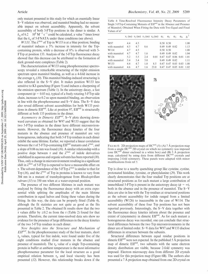
Structural differences between equal residue positions indimeric EIImtl were not expected. In the published 5 A projectionmap of dimeric EIImtl, two subunits with the same electrondensity distribution are visible, because 2-fold symmetry wasimposed. Electron microscopy data from different 2D crystalswas used for this projection map (Figure 6B). The authors alsopresented a 7 A projection map obtained from one 2D crystal on
Table 4: Time-Resolved Fluorescence Intensity Decay Parameters of
Single 5-FTrp-Containing Mutants of EIImtl in the Absence and Presence
of Mannitol, Obtained When Fitting the Data with Two Equal and Fixed
Values of Ra
τ1 (ns) τ2 (ns) τ3 (ns) τ4 (ns) R1 R2 R3 R4 χr2
W97 4.4 4.5 0.50 0.50 1.00
with mannitol 4.5 4.7 8.6 0.49 0.49 0.02 1.13
W114 4.7 4.5 0.50 0.50 1.08
with mannitol 4.7 4.7 1.6 0.49 0.49 0.02 1.04
W126 5.2 5.5 2.4 0.01 0.47 0.47 0.06 0.01 1.01
with mannitol 5.4 5.4 3.0 0.49 0.49 0.02 1.11
W133 4.4 4.7 1.0 8.5 0.47 0.47 0.03 0.03 1.08
with mannitol 4.6 4.7 1.6 23.0 0.47 0.47 0.05 0.01 1.02
FIGURE 6: 2D projection maps of IICmtl (5): (A) 7 A projection mapfrom a single IICmtl 2D crystal on which no symmetry was imposed(one IICmtl dimer enclosed in a white box) and (B) 5 A projectionmap, calculated by using data from different IICmtl crystals andimposing 2-fold symmetry. These panels were adapted with minormodifications from ref 5.
Dow
nloa
ded
by U
NIV
OF
GR
ON
ING
EN
on
Sept
embe
r 17
, 200
9 | h
ttp://
pubs
.acs
.org
P
ublic
atio
n D
ate
(Web
): A
pril
29, 2
009
| doi
: 10.
1021
/bi8
0206
68
5290 Biochemistry, Vol. 48, No. 23, 2009 Vos et al.
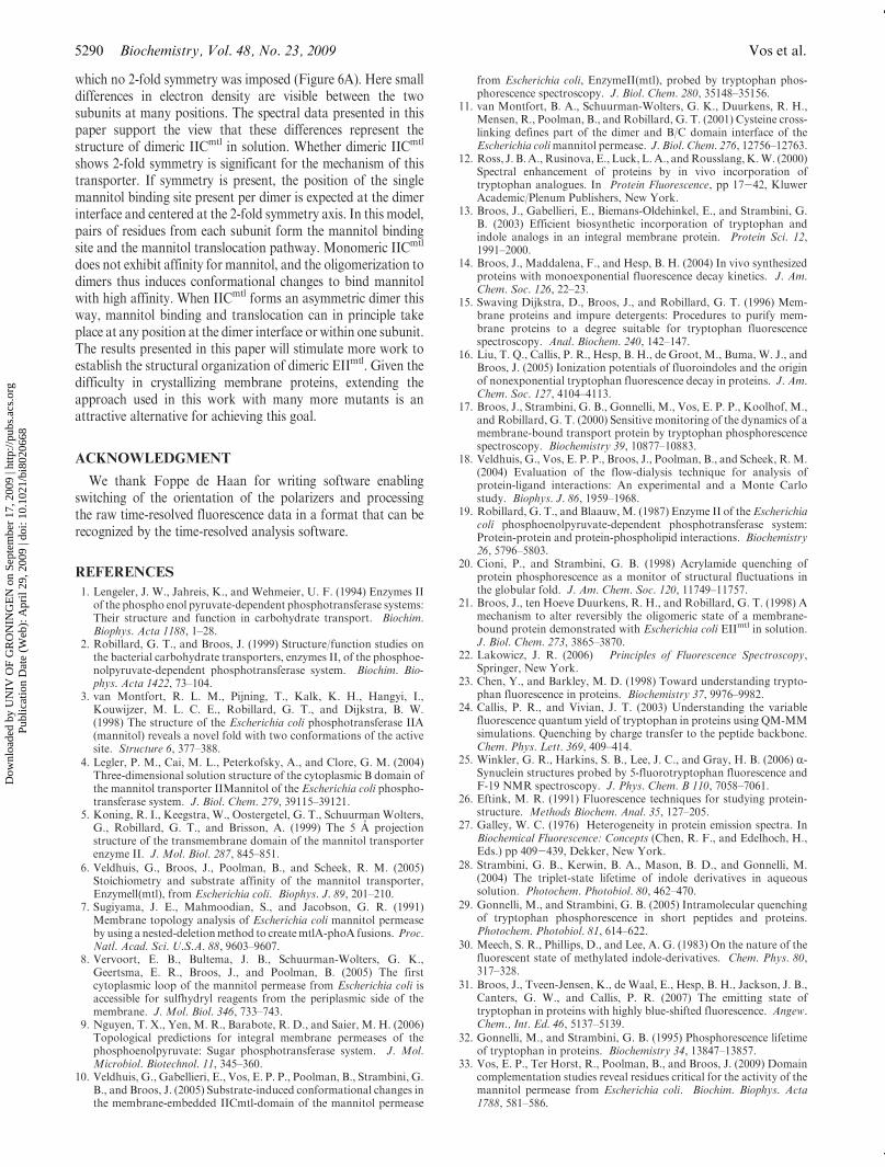
which no 2-fold symmetry was imposed (Figure 6A). Here smalldifferences in electron density are visible between the twosubunits at many positions. The spectral data presented in thispaper support the view that these differences represent thestructure of dimeric IICmtl in solution. Whether dimeric IICmtl
shows 2-fold symmetry is significant for the mechanism of thistransporter. If symmetry is present, the position of the singlemannitol binding site present per dimer is expected at the dimerinterface and centered at the 2-fold symmetry axis. In this model,pairs of residues from each subunit form the mannitol bindingsite and the mannitol translocation pathway. Monomeric IICmtl
does not exhibit affinity for mannitol, and the oligomerization todimers thus induces conformational changes to bind mannitolwith high affinity. When IICmtl forms an asymmetric dimer thisway, mannitol binding and translocation can in principle takeplace at any position at the dimer interface or within one subunit.The results presented in this paper will stimulate more work toestablish the structural organization of dimeric EIImtl. Given thedifficulty in crystallizing membrane proteins, extending theapproach used in this work with many more mutants is anattractive alternative for achieving this goal.
ACKNOWLEDGMENT
We thank Foppe de Haan for writing software enablingswitching of the orientation of the polarizers and processingthe raw time-resolved fluorescence data in a format that can berecognized by the time-resolved analysis software.
REFERENCES
1. Lengeler, J. W., Jahreis, K., and Wehmeier, U. F. (1994) Enzymes IIof the phospho enol pyruvate-dependent phosphotransferase systems:Their structure and function in carbohydrate transport. Biochim.Biophys. Acta 1188, 1–28.
2. Robillard, G. T., and Broos, J. (1999) Structure/function studies onthe bacterial carbohydrate transporters, enzymes II, of the phosphoe-nolpyruvate-dependent phosphotransferase system. Biochim. Bio-phys. Acta 1422, 73–104.
3. van Montfort, R. L. M., Pijning, T., Kalk, K. H., Hangyi, I.,Kouwijzer, M. L. C. E., Robillard, G. T., and Dijkstra, B. W.(1998) The structure of the Escherichia coli phosphotransferase IIA(mannitol) reveals a novel fold with two conformations of the activesite. Structure 6, 377–388.
4. Legler, P. M., Cai, M. L., Peterkofsky, A., and Clore, G. M. (2004)Three-dimensional solution structure of the cytoplasmic B domain ofthe mannitol transporter IIMannitol of the Escherichia coli phospho-transferase system. J. Biol. Chem. 279, 39115–39121.
5. Koning, R. I., Keegstra, W., Oostergetel, G. T., Schuurman Wolters,G., Robillard, G. T., and Brisson, A. (1999) The 5 A projectionstructure of the transmembrane domain of the mannitol transporterenzyme II. J. Mol. Biol. 287, 845–851.
6. Veldhuis, G., Broos, J., Poolman, B., and Scheek, R. M. (2005)Stoichiometry and substrate affinity of the mannitol transporter,Enzymell(mtl), from Escherichia coli. Biophys. J. 89, 201–210.
7. Sugiyama, J. E., Mahmoodian, S., and Jacobson, G. R. (1991)Membrane topology analysis of Escherichia coli mannitol permeaseby using a nested-deletionmethod to createmtlA-phoA fusions. Proc.Natl. Acad. Sci. U.S.A. 88, 9603–9607.
8. Vervoort, E. B., Bultema, J. B., Schuurman-Wolters, G. K.,Geertsma, E. R., Broos, J., and Poolman, B. (2005) The firstcytoplasmic loop of the mannitol permease from Escherichia coli isaccessible for sulfhydryl reagents from the periplasmic side of themembrane. J. Mol. Biol. 346, 733–743.
9. Nguyen, T. X., Yen, M. R., Barabote, R. D., and Saier, M. H. (2006)Topological predictions for integral membrane permeases of thephosphoenolpyruvate: Sugar phosphotransferase system. J. Mol.Microbiol. Biotechnol. 11, 345–360.
10. Veldhuis, G., Gabellieri, E., Vos, E. P. P., Poolman, B., Strambini, G.B., and Broos, J. (2005) Substrate-induced conformational changes inthe membrane-embedded IICmtl-domain of the mannitol permease
from Escherichia coli, EnzymeII(mtl), probed by tryptophan phos-phorescence spectroscopy. J. Biol. Chem. 280, 35148–35156.
11. van Montfort, B. A., Schuurman-Wolters, G. K., Duurkens, R. H.,Mensen, R., Poolman, B., and Robillard, G. T. (2001) Cysteine cross-linking defines part of the dimer and B/C domain interface of theEscherichia colimannitol permease. J. Biol. Chem. 276, 12756–12763.
12. Ross, J. B.A., Rusinova, E., Luck, L.A., andRousslang,K.W. (2000)Spectral enhancement of proteins by in vivo incorporation oftryptophan analogues. In Protein Fluorescence, pp 17-42, KluwerAcademic/Plenum Publishers, New York.
13. Broos, J., Gabellieri, E., Biemans-Oldehinkel, E., and Strambini, G.B. (2003) Efficient biosynthetic incorporation of tryptophan andindole analogs in an integral membrane protein. Protein Sci. 12,1991–2000.
14. Broos, J., Maddalena, F., and Hesp, B. H. (2004) In vivo synthesizedproteins with monoexponential fluorescence decay kinetics. J. Am.Chem. Soc. 126, 22–23.
15. Swaving Dijkstra, D., Broos, J., and Robillard, G. T. (1996) Mem-brane proteins and impure detergents: Procedures to purify mem-brane proteins to a degree suitable for tryptophan fluorescencespectroscopy. Anal. Biochem. 240, 142–147.
16. Liu, T. Q., Callis, P. R., Hesp, B. H., de Groot, M., Buma, W. J., andBroos, J. (2005) Ionization potentials of fluoroindoles and the originof nonexponential tryptophan fluorescence decay in proteins. J. Am.Chem. Soc. 127, 4104–4113.
17. Broos, J., Strambini, G. B., Gonnelli, M., Vos, E. P. P., Koolhof, M.,and Robillard, G. T. (2000) Sensitive monitoring of the dynamics of amembrane-bound transport protein by tryptophan phosphorescencespectroscopy. Biochemistry 39, 10877–10883.
18. Veldhuis, G., Vos, E. P. P., Broos, J., Poolman, B., and Scheek, R.M.(2004) Evaluation of the flow-dialysis technique for analysis ofprotein-ligand interactions: An experimental and a Monte Carlostudy. Biophys. J. 86, 1959–1968.
19. Robillard, G. T., and Blaauw,M. (1987) Enzyme II of the Escherichiacoli phosphoenolpyruvate-dependent phosphotransferase system:Protein-protein and protein-phospholipid interactions. Biochemistry26, 5796–5803.
20. Cioni, P., and Strambini, G. B. (1998) Acrylamide quenching ofprotein phosphorescence as a monitor of structural fluctuations inthe globular fold. J. Am. Chem. Soc. 120, 11749–11757.
21. Broos, J., ten Hoeve Duurkens, R. H., and Robillard, G. T. (1998) Amechanism to alter reversibly the oligomeric state of a membrane-bound protein demonstrated with Escherichia coli EIImtl in solution.J. Biol. Chem. 273, 3865–3870.
22. Lakowicz, J. R. (2006) Principles of Fluorescence Spectroscopy,Springer, New York.
23. Chen, Y., and Barkley, M. D. (1998) Toward understanding trypto-phan fluorescence in proteins. Biochemistry 37, 9976–9982.
24. Callis, P. R., and Vivian, J. T. (2003) Understanding the variablefluorescence quantum yield of tryptophan in proteins using QM-MMsimulations. Quenching by charge transfer to the peptide backbone.Chem. Phys. Lett. 369, 409–414.
25. Winkler, G. R., Harkins, S. B., Lee, J. C., and Gray, H. B. (2006) R-Synuclein structures probed by 5-fluorotryptophan fluorescence andF-19 NMR spectroscopy. J. Phys. Chem. B 110, 7058–7061.
26. Eftink, M. R. (1991) Fluorescence techniques for studying protein-structure. Methods Biochem. Anal. 35, 127–205.
27. Galley, W. C. (1976) Heterogeneity in protein emission spectra. InBiochemical Fluorescence: Concepts (Chen, R. F., and Edelhoch, H.,Eds.) pp 409-439, Dekker, New York.
28. Strambini, G. B., Kerwin, B. A., Mason, B. D., and Gonnelli, M.(2004) The triplet-state lifetime of indole derivatives in aqueoussolution. Photochem. Photobiol. 80, 462–470.
29. Gonnelli, M., and Strambini, G. B. (2005) Intramolecular quenchingof tryptophan phosphorescence in short peptides and proteins.Photochem. Photobiol. 81, 614–622.
30. Meech, S. R., Phillips, D., and Lee, A. G. (1983) On the nature of thefluorescent state of methylated indole-derivatives. Chem. Phys. 80,317–328.
31. Broos, J., Tveen-Jensen, K., de Waal, E., Hesp, B. H., Jackson, J. B.,Canters, G. W., and Callis, P. R. (2007) The emitting state oftryptophan in proteins with highly blue-shifted fluorescence. Angew.Chem., Int. Ed. 46, 5137–5139.
32. Gonnelli, M., and Strambini, G. B. (1995) Phosphorescence lifetimeof tryptophan in proteins. Biochemistry 34, 13847–13857.
33. Vos, E. P., Ter Horst, R., Poolman, B., and Broos, J. (2009) Domaincomplementation studies reveal residues critical for the activity of themannitol permease from Escherichia coli. Biochim. Biophys. Acta1788, 581–586.
Dow
nloa
ded
by U
NIV
OF
GR
ON
ING
EN
on
Sept
embe
r 17
, 200
9 | h
ttp://
pubs
.acs
.org
P
ublic
atio
n D
ate
(Web
): A
pril
29, 2
009
| doi
: 10.
1021
/bi8
0206
68