Stressed Memories: How Acute Stress Affects - Erno Hermans
9
Behavioral/Systems/Cognitive Stressed Memories: How Acute Stress Affects Memory Formation in Humans Marloes J. A. G. Henckens, 1,3 * Erno J. Hermans, 1,2 * Zhenwei Pu, 1,3 Marian Joe ¨ls, 3 and Guille ´n Ferna ´ndez 1,2,3 1 Donders Institute for Brain, Cognition and Behaviour, Radboud University Nijmegen, and 2 Department of Neurology, Radboud University Nijmegen Medical Centre, 6500 HB Nijmegen, The Netherlands, and 3 Swammerdam Institute for Life Sciences–Center for NeuroScience, University of Amsterdam, 1098 SM Amsterdam, The Netherlands Stressful, aversive events are extremely well remembered. Such a declarative memory enhancement is evidently beneficial for survival, but the same mechanism may become maladaptive and culminate in mental diseases such as posttraumatic stress disorder (PTSD). Stress hormones are known to enhance postlearning consolidation of aversive memories but are also thought to have immediate effects on attentional, sensory, and mnemonic processes at memory formation. Despite their significance for our understanding of the etiology of stress-related mental disorders, effects of acute stress at memory formation, and their brain correlates at the system scale, remain elusive. Using an integrated experimental approach, we probed the neural correlates of memory formation while participants underwent a controlled stress induction procedure in a crossover design. Physiological (cortisol level, heart rate, and pupil dilation) and subjective measures confirmed acute stress. Remarkably, reduced hippocampal activation during encoding predicted stress-enhanced memory performance, both within and between participants. Stress, moreover, amplified early visual and inferior temporal responses, suggesting that hypervigilant processing goes along with enhanced inferior temporal information reduction to relay a higher proportion of task- relevant information to the hippocampus. Thus, acute stress affects neural correlates of memory formation in an unexpected manner, the understanding of which may elucidate mechanisms underlying psychological trauma etiology. Introduction Information encoded into memory during stressful experiences is generally well remembered (Kim and Diamond, 2002), espe- cially if this information is relevant to the stressor (Joe ¨ls et al., 2006; Sandi and Pinelo-Nava, 2007; Smeets et al., 2009). Al- though this phenomenon represents adaptive behavior, dysregu- lation of the underlying mechanism might result in psychological trauma and thus potentially mental disease (McEwen, 2004; de Kloet et al., 2005). Past research has put strong emphasis on the mechanisms by which acute stress enhances memory consolida- tion (Roozendaal et al., 2006). It is widely assumed that rapidly unfolding neurochemical events during the initial stress phase exert immediate effects on attentional, sensory, and mnemonic processes (de Kloet et al., 2005). However, such putative ef- fects of acute stress have received little attention and remain poorly understood. The effects of stress on memory are thought to be mediated through hormones and neurotransmitters released by two inter- acting effector systems: the (nor)epinephrine (NE) sympathetic system and the hypothalamic–pituitary–adrenal (HPA) axis. Un- der stress, the sympathetic system, with the locus ceruleus (LC) at its core, shifts toward a tonically active state (Aston-Jones and Cohen, 2005; Valentino and Van Bockstaele, 2008). This shift causes an increase in NE tone almost in the entire brain, including the medial temporal lobe (MTL) (Valentino and Van Bockstaele, 2008; Sara, 2009), the key structure of the declarative memory system (Squire and Zola-Morgan, 1991). This increased NE tone, which is associated with peripheral effects such as pupil dilation, supports neural plasticity that underlies memory for- mation (Roozendaal et al., 2006) and causes a surge of arousal that is thought to lead to hypervigilance and prioritized pro- cessing of information relevant to the stressor (Aston-Jones and Bloom, 1981; Ramos and Arnsten, 2007). On a slightly longer timescale, the HPA axis increases the release of glu- cocorticoids, which also modulate MTL plasticity (Lupien and Lepage, 2001; de Kloet et al., 2005; Roozendaal et al., 2006; McEwen, 2007). Together, neuromodulators active during acute stress can therefore be hypothesized to induce a system level reorganization of mnemonic processes, geared toward more effective memory encoding. To tackle this issue, we used functional magnetic resonance imaging (fMRI) to probe effects of controlled stress induction on the neural substrates of memory formation. To satisfy the puta- tive requirements for stress-enhanced memory to occur (Joe ¨ls et al., 2006; Sandi and Pinelo-Nava, 2007), we maximized overlap between stressor and learning material by fully embedding a learning task in a stressful context created by strongly aversive movie clips (Qin et al., 2009; van Marle et al., 2009). To isolate Received March 11, 2009; revised June 17, 2009; accepted July 13, 2009. This work was supported by Grant 918.66.613 from the Dutch Organization for Scientific Research (Nederlandse Organisatie voor Wetenschappelijk Onderzoek). We thank D. J. de Quervain and L. Nadel for instructive discussions on the data presented here. *M.J.A.G.H. and E.J.H. contributed equally to this work. Correspondence should be addressed to Marloes J. A. G. Henckens, Donders Institute for Brain, Cognition and Behaviour, Centre for Cognitive Neuroimaging, Radboud University Nijmegen, P.O. Box 9101, 6500 HB Nijmegen, The Netherlands. E-mail: [email protected]. DOI:10.1523/JNEUROSCI.1184-09.2009 Copyright © 2009 Society for Neuroscience 0270-6474/09/2910111-09$15.00/0 The Journal of Neuroscience, August 12, 2009 • 29(32):10111–10119 • 10111
Transcript of Stressed Memories: How Acute Stress Affects - Erno Hermans

Behavioral/Systems/Cognitive
Stressed Memories: How Acute Stress Affects MemoryFormation in Humans
Marloes J. A. G. Henckens,1,3* Erno J. Hermans,1,2* Zhenwei Pu,1,3 Marian Joels,3 and Guillen Fernandez1,2,3
1Donders Institute for Brain, Cognition and Behaviour, Radboud University Nijmegen, and 2Department of Neurology, Radboud University NijmegenMedical Centre, 6500 HB Nijmegen, The Netherlands, and 3Swammerdam Institute for Life Sciences–Center for NeuroScience, University of Amsterdam,1098 SM Amsterdam, The Netherlands
Stressful, aversive events are extremely well remembered. Such a declarative memory enhancement is evidently beneficial for survival,but the same mechanism may become maladaptive and culminate in mental diseases such as posttraumatic stress disorder (PTSD). Stresshormones are known to enhance postlearning consolidation of aversive memories but are also thought to have immediate effects onattentional, sensory, and mnemonic processes at memory formation. Despite their significance for our understanding of the etiology ofstress-related mental disorders, effects of acute stress at memory formation, and their brain correlates at the system scale, remain elusive.Using an integrated experimental approach, we probed the neural correlates of memory formation while participants underwent acontrolled stress induction procedure in a crossover design. Physiological (cortisol level, heart rate, and pupil dilation) and subjectivemeasures confirmed acute stress. Remarkably, reduced hippocampal activation during encoding predicted stress-enhanced memoryperformance, both within and between participants. Stress, moreover, amplified early visual and inferior temporal responses, suggestingthat hypervigilant processing goes along with enhanced inferior temporal information reduction to relay a higher proportion of task-relevant information to the hippocampus. Thus, acute stress affects neural correlates of memory formation in an unexpected manner, theunderstanding of which may elucidate mechanisms underlying psychological trauma etiology.
IntroductionInformation encoded into memory during stressful experiencesis generally well remembered (Kim and Diamond, 2002), espe-cially if this information is relevant to the stressor (Joels et al.,2006; Sandi and Pinelo-Nava, 2007; Smeets et al., 2009). Al-though this phenomenon represents adaptive behavior, dysregu-lation of the underlying mechanism might result in psychologicaltrauma and thus potentially mental disease (McEwen, 2004; deKloet et al., 2005). Past research has put strong emphasis on themechanisms by which acute stress enhances memory consolida-tion (Roozendaal et al., 2006). It is widely assumed that rapidlyunfolding neurochemical events during the initial stress phaseexert immediate effects on attentional, sensory, and mnemonicprocesses (de Kloet et al., 2005). However, such putative ef-fects of acute stress have received little attention and remainpoorly understood.
The effects of stress on memory are thought to be mediatedthrough hormones and neurotransmitters released by two inter-acting effector systems: the (nor)epinephrine (NE) sympathetic
system and the hypothalamic–pituitary–adrenal (HPA) axis. Un-der stress, the sympathetic system, with the locus ceruleus (LC) atits core, shifts toward a tonically active state (Aston-Jones andCohen, 2005; Valentino and Van Bockstaele, 2008). This shiftcauses an increase in NE tone almost in the entire brain, includingthe medial temporal lobe (MTL) (Valentino and Van Bockstaele,2008; Sara, 2009), the key structure of the declarative memorysystem (Squire and Zola-Morgan, 1991). This increased NEtone, which is associated with peripheral effects such as pupildilation, supports neural plasticity that underlies memory for-mation (Roozendaal et al., 2006) and causes a surge of arousalthat is thought to lead to hypervigilance and prioritized pro-cessing of information relevant to the stressor (Aston-Jonesand Bloom, 1981; Ramos and Arnsten, 2007). On a slightlylonger timescale, the HPA axis increases the release of glu-cocorticoids, which also modulate MTL plasticity (Lupien andLepage, 2001; de Kloet et al., 2005; Roozendaal et al., 2006;McEwen, 2007). Together, neuromodulators active duringacute stress can therefore be hypothesized to induce a systemlevel reorganization of mnemonic processes, geared towardmore effective memory encoding.
To tackle this issue, we used functional magnetic resonanceimaging (fMRI) to probe effects of controlled stress induction onthe neural substrates of memory formation. To satisfy the puta-tive requirements for stress-enhanced memory to occur (Joels etal., 2006; Sandi and Pinelo-Nava, 2007), we maximized overlapbetween stressor and learning material by fully embedding alearning task in a stressful context created by strongly aversivemovie clips (Qin et al., 2009; van Marle et al., 2009). To isolate
Received March 11, 2009; revised June 17, 2009; accepted July 13, 2009.This work was supported by Grant 918.66.613 from the Dutch Organization for Scientific Research (Nederlandse
Organisatie voor Wetenschappelijk Onderzoek). We thank D. J. de Quervain and L. Nadel for instructive discussionson the data presented here.
*M.J.A.G.H. and E.J.H. contributed equally to this work.Correspondence should be addressed to Marloes J. A. G. Henckens, Donders Institute for Brain, Cognition and
Behaviour, Centre for Cognitive Neuroimaging, Radboud University Nijmegen, P.O. Box 9101, 6500 HB Nijmegen,The Netherlands. E-mail: [email protected].
DOI:10.1523/JNEUROSCI.1184-09.2009Copyright © 2009 Society for Neuroscience 0270-6474/09/2910111-09$15.00/0
The Journal of Neuroscience, August 12, 2009 • 29(32):10111–10119 • 10111

neural activity related to successful mem-ory encoding, we used a well establishedsubsequent memory paradigm (cf. Dolcoset al., 2004). Crucially and in contrast toprevious studies that looked into the ef-fects of arousing items on memory for-mation (Cahill, 2003; Richardson et al.,2003; Dolcos et al., 2004), we implementeda crossover design with separated stressand nonstress control sessions. Thus,the present study allowed us to assess pro-longed modulations of mnemonic opera-tions caused by a protracted state of acutestress.
Materials and MethodsParticipantsEighteen young (ages, 19 –31 years; median, 22years), right-handed, healthy male volunteersgave informed consent to participate in thestudy. Individuals who met any of the follow-ing criteria were excluded from participation:history of head injury, treatment with psycho-tropic medications, narcotics, �-blockers, ste-roids, or any other medication that affects CNS or endocrine systems,medical illness within the 3 weeks before testing, self-reported mental orsubstance use disorder, daily tobacco use, regular nightshift work, cur-rent stressful episode or major life event, previous exposure to slides usedin the study [i.e., International Affective Picture System (IAPS) (Lang etal., 1999)], and regularly viewing extremely violent movies or playingviolent computer games. Moreover, volunteers with high scores on de-pression [score above 8 on the Beck Depression Inventory (Beck et al.,2002)] were excluded from participation. The study was in accor-dance with institutional guidelines of the local ethics committee(Commissie Mensgebonden Onderzoek region Arnhem-Nijmegen,The Netherlands) and the declaration of Helsinki.
Study designIn a counterbalanced crossover design, all men underwent two sessions,separated by 1 month, of intentional episodic memory encoding duringfMRI. Memory was tested 24 h after each fMRI session by cued recall(CR). Both neutral and negative pictures were encoded, which were ei-ther embedded in a stressful or neutral control context created by shortmovie clips (Fig. 1). This allowed us to investigate brain activation duringmemory formation in a coherent stressful experience as a function oflater remembrance, both within [contrasting brain activation during theprocessing of subsequently remembered and forgotten items (Wagner etal., 1999)] and between (relating brain activation to memory perfor-mance across subjects) subjects. Physiological [cortisol level, heart rate(HR), and pupil dilation], and psychological [negative affect (NA)] in-dices were measured to confirm successful stress induction. Data wereanalyzed with the factors stress (stress induction vs control context),subsequent memory (later remembered vs later forgotten items), anditem valence (negative vs neutral pictures).
ProcedureBefore arrival. To minimize differences in baseline cortisol levels, weinstructed participants not to use any recreational drugs for 3 d and torefrain from drinking alcohol, exercising, and smoking for 24 h beforeeach session. Furthermore, participants were requested not to brush theirteeth, floss, or eat and drink anything but water for 2 h before all sessions,enabling adequate saliva sampling for cortisol assessment. To reduce theimpact of diurnal variation in cortisol levels, all testing was performed inthe afternoon, between 2:00 P.M. and 6:00 P.M., when hormone levelsare relatively stable.
Arrival. On the first day, participants rested 30 min before taking thefirst saliva sample. To increase familiarity with the procedure and mini-mize task repetition effects, participants were explicitly informed about
all details of the memory experiment. A financial reward was promisedproportional to the participant’s performance in the recall test to encour-age encoding. Furthermore, participants were asked to complete Spiel-berger’s Trait Anxiety Inventory (van der Ploeg et al., 1980) and theNEO-Five Factor Inventory (Costa and McCrae, 1992).
Scanning. Participants lay supine in the scanner and viewed the screenthrough a mirror positioned on the head coil. They were asked to lie asstill as possible, keep their eyes open, and look directly and continuouslyat the center of the screen in front of them. Four movie fragments wereused to create the appropriate context, shown before, between, and at theend of picture encoding (dividing the encoding session in three blocks)(Fig. 1). Participants were instructed to view each movie clip and picturefor the entire time that it was displayed. Pictures belonged to two cate-gories, with either a neutral or negative picture valence. Participants wereasked to memorize and rate the valence of each picture. Ratingswere given with right-hand button presses, with the index finger fornegative and the middle finger for neutral pictures. Pictures were shownin a pseudorandom order (no more than two pictures of the same valenceconsecutively), and all first slides were neutral to avoid ceiling effects inrecall that might result from the combined effect of arousal and primacyon memory. Slides were presented for 5 s with a 4 – 8 s intertrial interval(fixation cross). After completion of the encoding task, a structural scanwas performed.
Subsequent memory test. Participants came back the subsequent day toperform a cued recall test, lasting 75 min. One- or two-word written cuesfor each picture (with similar valence as the picture) were provided,describing the readily identifiable gist of the picture, i.e., which is themost salient feature of the scene depicted on the picture. Participantswere asked to write down as many characteristics of all pictures as theypossibly could remember, providing enough relevant characteristicsso that an outsider could identify each picture and discriminate itfrom similar studied pictures (Dolcos et al., 2004). A short introduc-tion was written to help the participants in listing characteristics. Onerater evaluated initially the written descriptions provided by the par-ticipants, and only pictures with a description that allowed both iden-tification and discrimination were classified as remembered. Pictureswith no recollection of characteristics were considered forgotten. Pic-ture descriptions that could not clearly be linked to a particular pic-ture were scored as a nonresponse and not included in the analyses.Subsequently, a second rater, blind to the study condition, indepen-dently re-rated all responses in the memory test to probe reliability.Inter-rater correspondence was very high (95.6%) and comparable
Figure 1. Experimental design. IAPS pictures (Lang et al., 1999) were encoded during fMRI scanning in either a stressful or aneutral control condition generated by short movie clips. Psychological and physiological measures were obtained to monitor theeffectiveness of stress induction. Memory was tested 24 h later in a cued recall test. S, Saliva sample; P, PANAS questionnaire(Watson et al., 1988).
10112 • J. Neurosci., August 12, 2009 • 29(32):10111–10119 Henckens et al. • Stressed Memories
with other studies using similar designs (Buchanan and Lovallo, 2001;Payne et al., 2006, 2007).
Stimulus materialsStressor. Four short movie fragments were used to create the propercontext (1 � 140 s, 3 � 90 s). They were either selected from a distressingmovie [Irreversible (2002), Gaspar Noe] or a neutral control movie[Comment j’ai tue mon pere (2001), Anne Fontaine]. Selected fragmentswere comparable in amount of speech, human presence, luminance, andlanguage. The stressful movie clips contained scenes with aggressive be-havior and violence against men and women. Occasionally, people in thevideo could be heard shouting and crying out in anger, pain, or distress.Previous studies have confirmed the effectiveness of these movie clips ininducing stress (Qin et al., 2009; van Marle et al., 2009). Although con-siderably distressing, the film content was approved by the NICAM(Dutch Institute for Audiovisual Media) for viewers above 16 years. Par-ticipants were informed before the experiment that watching the filmcould be stressful and that they could terminate the experiment at anypoint. This stress induction method was chosen because it meets thecriteria described by Joels et al. (2006) for stress-enhanced memory tooccur, i.e., close spatiotemporal proximity and content overlap of stres-sor and task (the memory encoding was part of a continuous and coher-ent stressful episode experienced within an fMRI environment). Thisoverlap in content was achieved by parallelizing studied pictures andmovies based on content features, both depicting real-life, emotionallysalient stimuli. To be more precise, the movies used in the stress condi-tion contained, for example, male to male and male to female violence,mutilations, and injuries, which were also present in many negative IAPSphotographs. There was also considerable overlap between the neutralmovie and neutral pictures. Examples of scenes shown in both are, forexample, people eating, talking, and walking.
Pictures. Three stimulus sets were created for picture encoding, two ofwhich were used per participant. Each set consisted of 80 negative and 80neutral pictures, supplemented with 41 null events (fixation). Pictureswere selected from both a standard set of affective pictures [IAPS (Lang etal., 1999)] and an additional set of newly rated pictures. New pictureswere downloaded from the internet and selected on the authors’ assess-ment of emotionality and similarity to IAPS pictures. New pictures wererated on a scale from 1 to 9 on both arousal and valence using the Self-Assessment Manikin (SAM) scales (Bradley and Lang, 1994) by an addi-tional group of 20 male volunteers. To ensure reliable rating that did notsignificantly differ from IAPS ratings and to serve as a reference frame,positive and negative IAPS pictures were added to this test set. All selectednegative slides were chosen for their moderate-to-high arousal quality(average � SE arousal score, 5.5 � 0.7) and negative valence (average �SE valence score, 3.1 � 0.7), rated on a 1–9 point rating scale as deter-mined by the SAM (Bradley and Lang, 1994). Neutral slides were selectedfor their relatively low arousal (average � SE arousal score, 2.5 � 0.7) andneutral valence (average � SE valence score, 5.3 � 0.3). Used picture setscontained �50% newly rated neutral and 15% newly rated negative pic-tures and were matched on chromatic features and complexity, whereasoverlap in content within one set was minimized. Used stimulus sets didnot differ in mean arousal and valence ratings.
Stress measuresSaliva collection and analysis. Cortisol levels were measured from saliva atfive time points: baseline measurements at the beginning of the experi-ment (twice) (t � 30 and 45 min), immediately after the first movie clip(t � 90 min), immediately after the last movie clip (t � 135 min), and atthe end of the experiment (t � 165 min).
Saliva was collected using a commercially available collection device(Salivette; Sarstedt). For each sample, the participant first placed thecotton swab provided in each Salivette tube in his mouth and chewedgently on it for 1 min to produce saliva. Third and fourth samples weretaken in the scanner. Swabs were handed over to the participants, andthey were instructed not to move their head while chewing. The swab wasthen placed back in the Salivette tube, and the samples were stored in afreezer at �25°C until assayed. Laboratory analyses were performed atthe Department of Biopsychology, Technical University of Dresden
(Dresden, Germany). After thawing, Salivettes were centrifuged at 3000rpm for 5 min, which resulted in a clear supernatant of low viscosity.Salivary free cortisol concentrations were subsequently measured using acommercially available chemiluminescence immunoassay with high sen-sitivity of 0.16 ng/ml (IBL Inc.). For analyses, area under the curve withrespect to increase was calculated and analyzed for cortisol levels ex-pressed as baseline percentage of each session (average level of measure-ments 1 and 2).
Heart rate. Cardiac rhythm of the participants was measured duringscanning, using a pulse oximeter placed on their left index finger. Partic-ipants were instructed to keep their hands as still as possible during themeasurement. Heart rate frequency was calculated using in-house soft-ware. Data of one subject was discarded from analyses because of exces-sive artifacts in the recorded signal.
Pupil diameter. A commercial MR-compatible eye-tracking devicefrom SensoMotoric Instruments (MEyeTrack-LR) mounted on the scan-ner bed was used to measure eye movements and pupil diameter at asampling rate of 50 Hz. Moreover, eye-tracking confirmed attentiveviewing of all slides and movie fragments.
Eye pupil data were analyzed using in-house software implemented inMatlab 7.5 (MathWorks), which was based on methods described previ-ously by others (Siegle et al., 2003). Eyeblink artifacts were identified bydifferentiating the signal to detect eye pupil changes occurring too rap-idly to represent actual dilation. Blinks were removed from the signalusing linear interpolation. Scanner pulses recorded simultaneously en-abled synchronization with stimulus presentation. Pupil diameter foreach trial was normalized to the average 1 s prestimulus onset baseline.The averaged normalized pupil diameter during picture presentation wasused as response measure. These were collapsed over trials within stressinduction and picture valence conditions. Because of data loss or exces-sive artifacts in the recorded signal in either of the sessions, data of fivesubjects were not included into analyses. It is important to note that thismethod does not measure absolute pupil diameter.
Psychological measures. Mood state was assessed using the Positive andNegative Affect Schedule (PANAS) questionnaire (Watson et al., 1988) atthree time points: at the beginning of the experiment (t � 30 min),immediately after the first movie clip (t � 90 min), and immediately afterthe last movie clip (t � 135 min). Picture valence ratings (neutral ornegative), which were obtained during picture encoding blocks, werescored as either corresponding (“correct”) or not corresponding (“incor-rect”) with a priori categorizations. Furthermore, average reaction times(RTs) were calculated for those items with correct rating.
Behavioral and physiological statistical analysis. Behavioral and physio-logical data were analyzed in SPSS 15.0 (SPSS) using repeated measuresANOVAs and paired samples t test statistics. When no main effects or inter-actions involving the order factor were significant, this factor was omitted.Furthermore, in cortisol data analyses, the difference in time of day betweenboth sessions was entered as a covariate. � was set at 0.05 throughout.
MRI acquisition. Participants were scanned in a Siemens TIM Trio 3.0Tesla MRI scanner equipped with an eight-channel phased array headcoil. Blood oxygenation level-dependent T2*-weighted gradient echoplanar images (EPIs) were acquired with the following parameters: rep-etition time (TR), 2.18 s; echo time (TE), 25 ms; flip angle (FA), 90°; 37axial slices approximately aligned with anterior commissure–posteriorcommissure plane; slice-matrix size, 64 � 64; slice thickness, 3.0 mm;slice gap, 0.3 mm; field of view (FOV), 212 � 212 mm 2. Because of itsrelatively short TE, this sequence yields optimal contrast-to-noise ratio inthe medial temporal lobe.
A high-resolution anatomical image was acquired for each participantusing a T1-weighted three-dimensional magnetization-prepared rapid gra-dient echo sequence combined with generalized autocalibrating partiallyparallel acquisitions (GRAPPA) (Griswold et al., 2002). The followingparameters were used: TE, 2.96 ms; TR, 2300 ms; FA, 8°; FOV, 256 �256 � 192 mm; voxel size, 1 mm isotropic; GRAPPA acceleration factor2. The total duration of each MRI session was �1 h.
fMRI data analysis. Data were analyzed using statistical parametricmapping software (SPM5; University College London, London, UK) andin-house software. The first five EPI volumes were discarded to allow forT1 equilibration. Before analysis, the images of the three encoding blocks
Henckens et al. • Stressed Memories J. Neurosci., August 12, 2009 • 29(32):10111–10119 • 10113

were separately motion corrected using rigid body transformations andleast sum of squares minimization. Subsequently, they were temporallyadjusted to account for differences in sampling times across differentslices. All functional images were then coregistered with the high-resolution T1-weighted structural image using normalized mutual infor-mation maximization. The anatomical image was subsequently used tonormalize all scans into MNI152 (Montreal Neurological Institute)space. All functional images were resampled with a voxel size of 2 mmisotropic. Finally, all images were smoothed with an isotropic 8 mmfull-width at half-maximum Gaussian kernel to accommodate residualfunctional/anatomical variance between subjects.
Data were analyzed using a general linear model, in which individualevents were modeled based on stress, subsequent memory, and itemvalence. Regressors were temporally convolved with the canonical hemo-dynamic response function of SPM5. The six covariates corresponding tothe movement parameters obtained from the realignment procedurewere also included in the model. To reduce unspecific differences be-tween scan sessions, global normalization using proportional scaling wasapplied. The single-subject parameter estimates from each session andcondition obtained from the first-level analysis were included in subse-quent random effects analyses. For the second-level analysis, a factorialANOVA was used, with stress induction (stress vs control context), pic-ture valence (negative vs neutral), and subsequent memory (remem-bered vs forgotten) as within-subject factors. � for statistical tests was setat 0.05, family-wise error rate corrected using Gaussian random fieldtheory. Based on our a priori hypothesis about their involvement inmemory and attention, data for the regions of interest—MTL and ventralvisual stream—were corrected for a reduced search region (based ontheir size) and small volume corrected using a sphere with 15 mm radius.Statistical tests for all other regions corrected for a whole-brain searchregion.
To test the regional overlap between the main effects of memory andstress, conjunction analyses were performed using the minimum statisticcompared with the conjunction null method as implemented withinSPM5 (Nichols et al., 2005). We used a reduced search volume with aradius of 10 mm (approximating the underlying spatial resolution of thefMRI signal) centered on the maxima of the main contrasts as proposedby Friston et al. (2005). For purpose of visualization of the overlap ofboth contrasts, the less conservative minimum statistic compared withthe global null method with a threshold of p � 0.001, uncorrected, wasused in Figure 3C.
To assess the relationship between neural activity and memory perfor-mance across subjects, mean activity of the anatomically defined hip-pocampus was extracted [using the automated anatomical labeling ofactivations (Tzourio-Mazoyer et al., 2002)], and the differences in re-sponses between the stress and control conditions were entered in regres-sion analyses as a predictor for the difference in memory performance.Visualizations of activations were created using MRIcron (http://www.sph.sc.edu/comd/rorden/mricron/) by superimposing statistical para-metric maps thresholded at p � 0.001, uncorrected, onto a canonicalT1-weighted image in standard MNI152 space.
ResultsEffectiveness of stress induction: physiological measuresPhysiological measures confirmed successful stress induction.Area under the curve measures of salivary cortisol levels indicatedthat HPA axis activity was elevated throughout the picture en-coding procedure in the stress condition (F(1,15) � 6.49; p � 0.02)(Fig. 2A). Moreover, heart rate frequency (mean � SD; HRcontrol,59.26 � 9.36 beats per minute; HRstress, 65.95 � 9.69 beats perminute), which is associated with elevated sympathetic tonus,was increased (F(1,16) � 12.34; p � 0.003). Finally, pupil dilationresponses to pictures were decreased (F(1,11) � 4.90; p � 0.05)(Fig. 2B). Given the direct association of LC activity and pupildilation, this finding is consistent with the notion that phasic LCresponses diminish against a background of enhanced tonic ac-tivity (Aston-Jones and Cohen, 2005). Moreover, in agreement
with previous literature (Bradley et al., 2008), a significant effectof item valence was observed in pupil dilation responses, withnegative pictures causing more dilation, indicating stronger pha-sic sympathetic responses, than neutral ones (F(1,11) � 52.08; p �0.001) (supplemental Fig. S1A, available at www.jneurosci.org assupplemental material). However, this measure did not yield anysignificant interaction effects between item valence and stress(F(1,12) � 1).
Effectiveness of stress induction: psychological measuresStress induction led to an increase in subjective stress, as mea-sured by elevated self-reported negative affect (PANAS question-naire) measured just before the encoding blocks (mean � SD;NAcontrol, 13.97 � 4.62; NAstress, 16.08 � 4.79; F(1,17) � 7.21; p �0.02). Picture ratings obtained during encoding blocks werehighly consistent with predetermined picture categories, with94.7 � 0.3% corresponding (correct) responses. Whereas reac-tion times for (correct only) picture rating were independent ofpicture valence (F(1,17) � 1), stress induced a trend toward slowerreaction times (mean � SD; RTcontrol, 1.39 � 0.33 s; RTstress,1.51 � 0.34 s; F(1,17) � 3.57; p � 0.08).
Effectiveness of stress induction: memory enhancementMemory was tested in a CR test (Dolcos et al., 2004) the sub-sequent day. Stress enhanced memory performance: picturesencoded during the stressful experience were more often remem-bered 1 d later than pictures encoded in the control condition(mean � SD; CRcontrol, 69.33 � 20.67 pictures; CRstress, 75.83 �18.96 pictures; F(1,17) � 4.42; p � 0.05). This stress effect onpicture encoding did not change over time during the encodingsession (as evidenced by a nonsignificant stress by encoding blockinteraction, F(1,17) � 1), indicating that this stress modulationwas a rather stable state during the entire scanning session. Asexpected, memory performance was better for negative than forneutral pictures (mean � SD; CRneutral, 31.19 � 10.88 pictures;CRnegative, 41.39 � 10.17 pictures; F(1,17) � 51.41; p � 0.001)(supplemental Fig. S1B, available at www.jneurosci.org as sup-plemental material). However, this picture valence effect did notinteract with stress induction (F(1,17) � 1).
Brain activation maps: main effects of stress, memory, andpicture valenceImaging data were analyzed using a random effects ANOVA withstress (stress induction vs control context), subsequent memory(later remembered vs later forgotten items), and item valence(negative vs neutral pictures) as within-subject factors. Given
A B
Figure 2. Physiological effects of stress. A, B, The stress induction procedure increased (areaunder the curve) cortisol levels (expressed as percentage of baseline) (45–135 min) (A) andreduced mean phasic pupil dilation (expressed as ratio of baseline diameter) after the initiallight reflex (B). Significance refers to the observed within-subject effects, and the error barsrepresent SEM of the between-subject variance. *p � 0.05.
10114 • J. Neurosci., August 12, 2009 • 29(32):10111–10119 Henckens et al. • Stressed Memories
strong neurophysiological evidence for its involvement in mem-ory formation and stress–memory interactions, the MTL, andmore specifically, the hippocampus (Joels et al., 2004), was ourmain region of interest. Furthermore, we focused on stress-induced changes in both lower- and higher-order visual process-ing regions, known to be modulated by vigilance (Munk et al.,1996). Therefore, data for the MTL structures and the ventralvisual stream were thresholded at p � 0.05, small volume cor-rected (r � 15 mm). A threshold of p � 0.05 whole-brain cor-rected was applied to all other regions.
We first identified brain responses to pictures in general thatwere affected by stress induction. Larger responses to picturepresentation for the stress induction than the control conditionwere found in visual areas: activation in regions of the primaryvisual cortex, right inferior temporal region, and fusiform gyrus,associated with higher-order visual processing and attention(Moran and Desimone, 1985; Heinze et al., 1994), was elevated bystress induction (Table 1, Fig. 3A). Second, regions supportingsuccessful memory formation were identified. In line with previ-ous literature of picture encoding (Brewer et al., 1998; Dolcos etal., 2004), regions displaying larger neural activity during encod-
ing of subsequently remembered than subsequently forgottenpictures were the bilateral fusiform gyrus extending into the para-hippocampal region, inferior temporal gyrus, inferior frontalcortex, inferior parietal gyrus, precentral gyrus, and the middle/superior occipital lobe. Negative effects of subsequent memorywere found in the cuneus, precuneus, lingual gyrus, posteriorcingulate cortex, and middle frontal cortex (Fig. 3B).
As expected, brain imaging results also revealed strong maineffects of item valence (supplemental Table S1, available at www.jneurosci.org as supplemental material), with encoding activitybeing greater for negative than for neutral items in regions asso-ciated with visual processing (including the middle occipital andmiddle temporal gyri) (Lang et al., 1998; Wagner et al., 1998).Additional differences in activation were observed in the amyg-dala, fusiform gyrus, cerebellum, brainstem, thalamus, and infe-rior frontal cortex; regions typically activated in tasks involvingemotional processing and arousal (Phan et al., 2002). Item va-lence and memory effects interacted in an extended medial tem-poral region, which showed larger subsequent memory effects fornegative than for neutral pictures, reflecting better memory per-formance for these items (supplemental Table S1, available atwww.jneurosci.org as supplemental material). These findings areconsistent with other studies concerning emotional subsequentmemory effects (Dolcos et al., 2004; Dougal et al., 2007). In linewith behavioral and physiological measures, however, picturevalence effects did not interact with stress induction.
Brain activation maps: conjunction and interaction effects ofstress and memoryTo examine the main question at issue, how stress affects memoryformation, we first identified those brain regions in which activitywas modulated by both stress and memory formation indepen-dently (i.e., overlapping effects), leaving the actual underlyingmemory processes unaffected. Both factors were associated withdifferential activity in the primary visual cortex and inferior tem-poral gyrus. To ensure actual spatial overlap, conjunction analy-ses (using the minimum statistic compared with the conjunctionnull method) over the two orthogonal contrasts (Nichols et al.,2005) were performed. Activity in the primary visual cortex wassignificantly increased after stress induction and was negativelyassociated with subsequent remembrance (Fig. 3C), indicatingthat stress-induced activation of this region was related to lesseffective memory formation. In contrast, in the inferior temporalgyrus, a combined positive stress induction and subsequentmemory effect was found (Fig. 3C). Enhanced activation afterstress induction in this region was apparently associated withbetter memory formation.
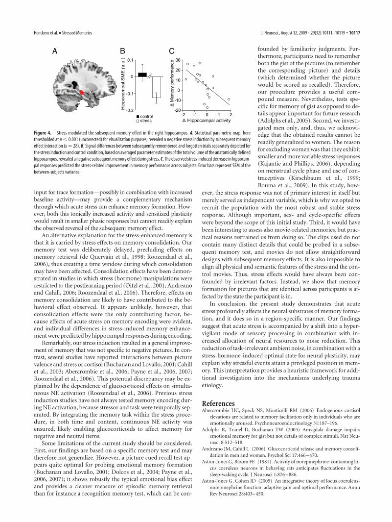
Second, we investigated whether stress interacted with mem-ory processes and thus influenced the subsequent memory effectitself. Stress induction modulated the subsequent memory effectfocally in the right hippocampus (Table 1, Fig. 4A,B). Most in-terestingly, the observed interaction was carried by a negativesubsequent memory effect in the stress induction condition: hip-pocampal responses to pictures were lower during encoding ofsubsequently remembered compared with forgotten items. Todetermine whether this effect was related to the observed in-creases in memory performance and thus could explain observedvariance in stress effects on memory performance across partici-pants, mean activity of the anatomically defined hippocampus(bilateral) was extracted (Tzourio-Mazoyer et al., 2002) and thedifferences in activity between the stress and control conditionswere entered into regression analyses as a predictor for the differ-ence in memory performance. The decrease in hippocampal re-
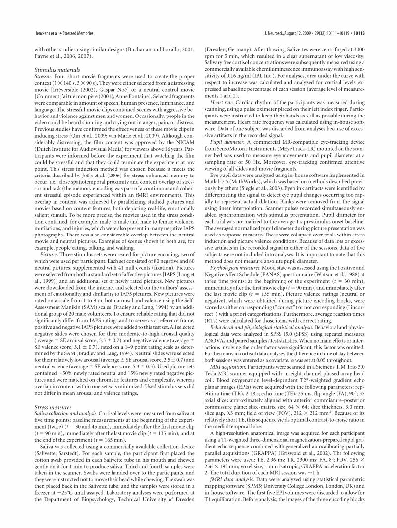
Table 1. Brain regions revealing significant main, interaction, or conjunctioneffects
Coordinates
Peak T scoreRegion x y z
Main effect of subsequent memoryRemembered � forgotten
Middle occipital gyrus, L �26 �68 36 6.64���Middle occipital gyrus, R 30 �68 38 7.39���Inferior temporal gyrus, L �46 �62 �6 7.94���Inferior temporal gyrus, R 54 �56 �10 8.69���Fusiform gyrus, L �34 �32 �20 4.46 ��
Fusiform gyrus, R 34 �32 �22 4.15 ��
Inferior parietal lobule, L �44 �44 56 6.63���Inferior parietal lobule, R 36 �52 56 5.31��Inferior frontal gyrus, L �50 34 6 8.47���Inferior frontal gyrus, R 52 6 22 6.21���
54 38 6 5.93���Forgotten � remembered
Cuneus, L �4 �90 24 4.86�Cuneus, R 16 �64 34 8.46���Lingual gyrus, L �16 �62 �4 5.74��Middle frontal gyrus, R 38 34 34 5.04�
28 52 22 4.87�Main effect of stress
Stress � controlSuperior occipital gyrus, L �8 �94 8 5.09�Superior occipital gyrus, R 16 �92 20 4.99�Lingual gyrus, R 8 �72 �2 5.86���Fusiform gyrus, L �36 �66 �16 3.88 �
Fusiform gyrus, R 28 �70 �6 5.28�28 �50 �2 4.88�
Inferior temporal gyrus, R 46 �48 �18 4.06 ��
Stress by SME interaction (negative)Hippocampus, R 28 �26 �8 4.29 ��
Forgotten � remembered during stressHippocampus, R 28 �26 �8 5.01�
Stress by SME conjunctionRemembered � forgotten and stress � control
Inferior temporal gyrus, R 48 �52 �6 3.20 †
Stress � control and forgotten � rememberedLingual gyrus, L �8 �76 �6 4.21 ��
�20 �62 �4 3.68 �
The peak x, y, z coordinates are given in MNI152 standard space coordinates. L and R denote left and right. SME,Subsequent memory effect. �p � 0.05 whole-brain corrected; ��p � 0.01 whole-brain corrected; ���p � 0.001whole-brain corrected; �p � 0.05 small volume corrected; ��p � 0.01 small volume corrected, †p � 0.05 smallvolume corrected (r � 10 mm) centered on the maximum of the main contrast.
Henckens et al. • Stressed Memories J. Neurosci., August 12, 2009 • 29(32):10111–10119 • 10115

sponse to pictures predicted the stress-induced improvement in memoryperformance (r � �0.615; p � 0.007), pro-viding complementary evidence that re-duced hippocampal activity is related to anincrease in memory performance understress (Fig. 4C).
DiscussionHere we show that acute stress profoundlyaffected the neural correlates of memoryformation, and it did so in a region-specific manner. Reduced hippocampalresponses were associated with bettermemory formation under stress, bothwithin and across subjects. Furthermore,in early visual areas, stress led to an in-crease of activity, which was accompa-nied by a negative subsequent memoryeffect, whereas stress-enhanced activa-tion in inferior temporal regions was ac-companied by a positive subsequentmemory effect.
The stress induction increased bothpsychological stress, as indicated by ele-vated self-reported negative affect, andphysiological stress: both activity of theHPA axis and sympathetic tonus was in-creased. Moreover, decreased pupil dilation responses werefound, which is widely regarded as a relatively direct index of LCactivity (Koss, 1986), with stimulus-locked pupil dilation reflect-ing a phasic LC response. During states of stress, the LC shiftstoward a tonically hyperactive state, which is thought to result ina hypervigilant processing state and a concomitant decrease instimulus-coupled phasic LC activity (Aston-Jones and Cohen,2005). Our finding of a decreased pupil dilation response duringstress, together with the slightly elevated reaction times for pic-ture rating, support this interpretation of a stress-induced hyper-vigilant state of unfocussed processing.
The stress-enhanced activity in the primary visual cortexmight also support the notion of such a state change. Previousstudies have shown that both attentional and emotional statesmodulate visual processing (Wang et al., 2006; Vuilleumier andDriver, 2007) and that hypervigilance is accompanied by poten-tiation of sensory input (Munk et al., 1996). The widespreadneocortical projections of the LC might recruit additional neuralresources to process an excess of sensory information. The nega-tive conjunction of stress and subsequent memory effects in thisregion might indicate that the stress-induced activation, how-ever, is supraoptimal for memory formation and likely containslarge amounts of task-irrelevant information. Because this effectin itself is not related to better memory, additional factors arenecessary to explain stress-induced memory enhancement.
One possible explanation for this memory improvement may layin stress-enhanced filtering of excess sensory information in the ven-tral visual stream (Kastner et al., 1998; Kastner and Pinsk, 2004).Visual-selective attention modulates the inferior temporal cortex(Moran and Desimone, 1985), and lesions in these regions lead toattentional deficits (De Weerd et al., 1999). Under conditions of lowattentional selection, cortical representations of simultaneous visualstimuli interact in a mutually suppressive manner. Attentional selec-tion of a single stimulus results in diminishment of the suppressiveinfluence of nearby stimuli, thus providing a neural basis for filtering
out irrelevant information (Kastner et al., 1998). Moreover, it hasbeen proposed that tonic LC states are mirrored by increased activa-tion of a ventral frontoparietal attention network, enhancing selec-tive processing of salient stimuli (Corbetta et al., 2008). In line withthis, we observed bilateral subsequent memory effects in these infe-rior temporal regions but also stress-induced activity increases. Thelatter can be taken to reflect reduction of ambient noise by focusingon task-relevant information. Conjunction effects of stress and sub-sequent memory, without interaction, indicate that activity in thisregion is modulated relatively independently by stress and memoryformation. Thus, elevated stress may increase the likelihood of suc-cessful memory formation.
Consequently, adequate noise reduction may have led to lessinformation relayed to the hippocampus. In line with this idea,the hippocampus showed less activity for later remembered thanfor later forgotten items under stress. Moreover, the overall de-crease in hippocampal responses predicted the stress-related im-provement in memory performance across subjects. Possibly,during stress, hippocampal input during subsequently forgottenitems might be characterized by a large proportion of irrelevantinformation, thwarting clean separation between task-related and-unrelated information as required for the subsequent memory test.Thus, our findings suggest that stress-related memory improve-ments are related to a combination of increased noise reductionaccompanied by, or leading to, a decreased hippocampal response.
In addition to these alterations in sensory and mnemonic op-erations, stress may promote a neural state optimized for mem-ory formation. LC activation elevates hippocampal NE levels,leading to tonically increased activity (Berridge and Foote, 1991).Therefore, the level of hippocampal activity might have been gen-erally higher during the stress compared with the control condi-tion, but fMRI cannot detect such slowly modulated changes inbaseline activity. Furthermore, corticosteroids and NE lower thethreshold for synaptic modification (Groc et al., 2008). There-fore, sensitization of hippocampal plasticity, requiring less neural
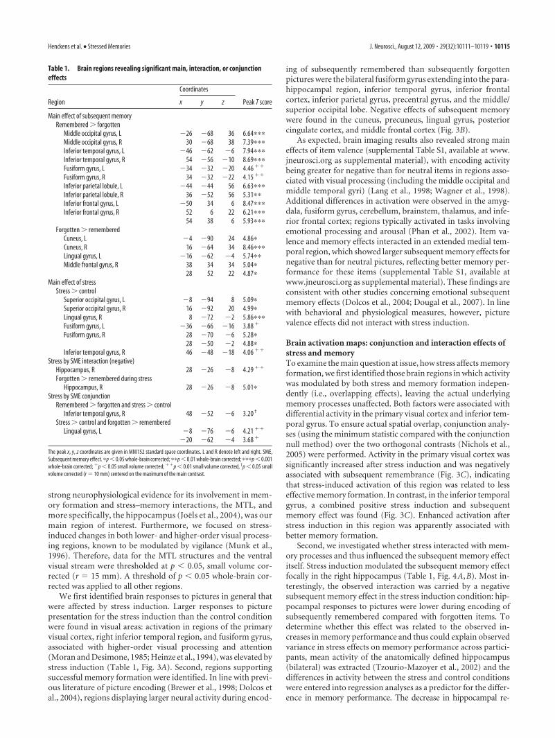
A B C
Figure 3. Brain regions affected by stress induction and memory (y � �72, �59). A, Stress induction increased responsive-ness within the primary visual cortex and right inferior temporal region, centered on the fusiform gyrus. B, Positive (in red)subsequent memory effects in large inferior temporal and superior parietal regions and negative (in blue) subsequent memoryeffects in posterior midline structures comprising the cuneus and the lingual gyrus. C, Conjunctions of positive effects of stressinduction with positive (in red) or negative (in blue) subsequent memory effects. These figures show that enhanced recruitment ofthe primary visual cortex after stress induction was detrimental to memory formation. In contrast, stress-enhanced inferiortemporal activation proved beneficial. All statistical parametric maps are thresholded at p � 0.001, uncorrected, using minimumstatistic/global null methods for conjunction effects, for visualization purposes. For formal statistical tests, see Table 1.
10116 • J. Neurosci., August 12, 2009 • 29(32):10111–10119 Henckens et al. • Stressed Memories
input for trace formation—possibly in combination with increasedbaseline activity—may provide a complementary mechanismthrough which acute stress can enhance memory formation. How-ever, both this tonically increased activity and sensitized plasticitywould result in smaller phasic responses but cannot readily explainthe observed reversal of the subsequent memory effect.
An alternative explanation for the stress-enhanced memory isthat it is carried by stress effects on memory consolidation. Ourmemory test was deliberately delayed, precluding effects onmemory retrieval (de Quervain et al., 1998; Roozendaal et al.,2006), thus creating a time window during which consolidationmay have been affected. Consolidation effects have been demon-strated in studies in which stress (hormone) manipulations wererestricted to the postlearning period (Oitzl et al., 2001; Andreanoand Cahill, 2006; Roozendaal et al., 2006). Therefore, effects onmemory consolidation are likely to have contributed to the be-havioral effect observed. It appears unlikely, however, thatconsolidation effects were the only contributing factor, be-cause effects of acute stress on memory encoding were evident,and individual differences in stress-induced memory enhance-ment were predicted by hippocampal responses during encoding.
Remarkably, our stress induction resulted in a general improve-ment of memory that was not specific to negative pictures. In con-trast, several studies have reported interactions between picturevalence and stress or cortisol (Buchanan and Lovallo, 2001; Cahillet al., 2003; Abercrombie et al., 2006; Payne et al., 2006, 2007;Roozendaal et al., 2006). This potential discrepancy may be ex-plained by the dependence of glucocorticoid effects on simulta-neous NE activation (Roozendaal et al., 2006). Previous stressinduction studies have not always tested memory encoding dur-ing NE activation, because stressor and task were temporally sep-arated. By integrating the memory task within the stress proce-dure, in both time and content, continuous NE activity wasensured, likely enabling glucocorticoids to affect memory fornegative and neutral items.
Some limitations of the current study should be considered.First, our findings are based on a specific memory test and maytherefore not generalize. However, a picture cued recall test ap-pears quite optimal for probing emotional memory formation(Buchanan and Lovallo, 2001; Dolcos et al., 2004; Payne et al.,2006, 2007); it shows robustly the typical emotional bias effectand provides a cleaner measure of episodic memory retrievalthan for instance a recognition memory test, which can be con-
founded by familiarity judgments. Fur-thermore, participants need to rememberboth the gist of the pictures (to rememberthe corresponding picture) and details(which determined whether the picturewould be scored as recalled). Therefore,our procedure provides a useful com-pound measure. Nevertheless, tests spe-cific for memory of gist as opposed to de-tails appear important for future research(Adolphs et al., 2005). Second, we investi-gated men only, and, thus, we acknowl-edge that the obtained results cannot bereadily generalized to women. The reasonfor excluding women was that they exhibitsmaller and more variable stress responses(Kajantie and Phillips, 2006), dependingon menstrual cycle phase and use of con-traceptives (Kirschbaum et al., 1999;Bouma et al., 2009). In this study, how-
ever, the stress response was not of primary interest in itself butmerely served as independent variable, which is why we opted torecruit the population with the most robust and stable stressresponse. Although important, sex- and cycle-specific effectswere beyond the scope of this initial study. Third, it would havebeen interesting to assess also movie-related memories, but prac-tical reasons restrained us from doing so. The clips used do notcontain many distinct details that could be probed in a subse-quent memory test, and movies do not allow straightforwarddesigns with subsequent memory effects. It is also impossible toalign all physical and semantic features of the stress and the con-trol movies. Thus, stress effects would have always been con-founded by irrelevant factors. Instead, we show that memoryformation for pictures that are identical across participants is af-fected by the state the participant is in.
In conclusion, the present study demonstrates that acutestress profoundly affects the neural substrates of memory forma-tion, and it does so in a region-specific manner. Our findingssuggest that acute stress is accompanied by a shift into a hyper-vigilant mode of sensory processing in combination with in-creased allocation of neural resources to noise reduction. Thisreduction of task-irrelevant ambient noise, in combination with astress-hormone-induced optimal state for neural plasticity, mayexplain why stressful events attain a privileged position in mem-ory. This interpretation provides a heuristic framework for addi-tional investigation into the mechanisms underlying traumaetiology.
ReferencesAbercrombie HC, Speck NS, Monticelli RM (2006) Endogenous cortisol
elevations are related to memory facilitation only in individuals who areemotionally aroused. Psychoneuroendocrinology 31:187–196.
Adolphs R, Tranel D, Buchanan TW (2005) Amygdala damage impairsemotional memory for gist but not details of complex stimuli. Nat Neu-rosci 8:512–518.
Andreano JM, Cahill L (2006) Glucocorticoid release and memory consoli-dation in men and women. Psychol Sci 17:466 – 470.
Aston-Jones G, Bloom FE (1981) Activity of norepinephrine-containing lo-cus coeruleus neurons in behaving rats anticipates fluctuations in thesleep-waking cycle. J Neurosci 1:876 – 886.
Aston-Jones G, Cohen JD (2005) An integrative theory of locus coeruleus-norepinephrine function: adaptive gain and optimal performance. AnnuRev Neurosci 28:403– 450.
A B C
Figure 4. Stress modulated the subsequent memory effect in the right hippocampus. A, Statistical parametric map, herethresholded at p � 0.001 (uncorrected) for visualization purposes, revealed a negative stress induction by subsequent memoryeffect interaction (x � 28). B, Signal differences between subsequently remembered and forgotten trials separately depicted forthe stress induction and control condition, based on averaged parameter estimates of the total volume of the anatomically definedhippocampus, revealed a negative subsequent memory effect during stress. C, The observed stress-induced decrease in hippocam-pal responses predicted the stress-related improvement in memory performance across subjects. Error bars represent SEM of thebetween-subjects variance.
Henckens et al. • Stressed Memories J. Neurosci., August 12, 2009 • 29(32):10111–10119 • 10117

Beck AT, Steer R, Brown GK (2002) Beck Depression Inventory-II-NL.Handleiding. De Nederlandse versie van de Beck Depression Inventory,Ed 2. Lisse, The Netherlands: Swets Test Publishers.
Berridge CW, Foote SL (1991) Effects of locus coeruleus activation on elec-troencephalographic activity in neocortex and hippocampus. J Neurosci11:3135–3145.
Bouma EM, Riese H, Ormel J, Verhulst FC, Oldehinkel AJ (2009) Adoles-cents’ cortisol responses to awakening and social stress; effects of gender,menstrual phase and oral contraceptives. The TRAILS study. Psychoneu-roendocrinology 34:884 – 893.
Bradley MM, Lang PJ (1994) Measuring emotion: the Self-AssessmentManikin and the Semantic Differential. J Behav Ther Exp Psychiatry25:49 –59.
Bradley MM, Miccoli L, Escrig MA, Lang PJ (2008) The pupil as a measureof emotional arousal and autonomic activation. Psychophysiology45:602– 607.
Brewer JB, Zhao Z, Desmond JE, Glover GH, Gabrieli JD (1998) Makingmemories: brain activity that predicts how well visual experience will beremembered. Science 281:1185–1187.
Buchanan TW, Lovallo WR (2001) Enhanced memory for emotional mate-rial following stress-level cortisol treatment in humans. Psychoneuroen-docrinology 26:307–317.
Cahill L (2003) Similar neural mechanisms for emotion-induced memory im-pairment and enhancement. Proc Natl Acad Sci U S A 100:13123–13124.
Cahill L, Gorski L, Le K (2003) Enhanced human memory consolidationwith post-learning stress: interaction with the degree of arousal at encod-ing. Learn Mem 10:270 –274.
Corbetta M, Patel G, Shulman GL (2008) The reorienting system of thehuman brain: from environment to theory of mind. Neuron 58:306 –324.
Costa PT Jr, McCrae RR (1992) Revised NEO Personality Inventory (NEO-PI-R) and the Five Factor Inventory (NEO-FFI): professional manual.Odessa, FL: Psychological Assessment Resources.
de Kloet ER, Joels M, Holsboer F (2005) Stress and the brain: from adapta-tion to disease. Nat Rev Neurosci 6:463– 475.
de Quervain DJ, Roozendaal B, McGaugh JL (1998) Stress and glucocor-ticoids impair retrieval of long-term spatial memory. Nature394:787–790.
De Weerd P, Peralta MR 3rd, Desimone R, Ungerleider LG (1999) Loss ofattentional stimulus selection after extrastriate cortical lesions in ma-caques. Nat Neurosci 2:753–758.
Dolcos F, LaBar KS, Cabeza R (2004) Interaction between the amygdala andthe medial temporal lobe memory system predicts better memory foremotional events. Neuron 42:855– 863.
Dougal S, Phelps EA, Davachi L (2007) The role of medial temporal lobe in itemrecognition and source recollection of emotional stimuli. Cogn Affect BehavNeurosci 7:233–242.
Friston KJ, Penny WD, Glaser DE (2005) Conjunction revisited. Neuroim-age 25:661– 667.
Griswold MA, Jakob PM, Heidemann RM, Nittka M, Jellus V, Wang J, KieferB, Haase A (2002) Generalized autocalibrating partially parallel acquisi-tions (GRAPPA). Magn Reson Med 47:1202–1210.
Groc L, Choquet D, Chaouloff F (2008) The stress hormone corticosteroneconditions AMPAR surface trafficking and synaptic potentiation. NatNeurosci 11:868 – 870.
Heinze HJ, Mangun GR, Burchert W, Hinrichs H, Scholz M, Munte TF, GosA, Scherg M, Johannes S, Hundeshagen H (1994) Combined spatial andtemporal imaging of brain activity during visual selective attention inhumans. Nature 372:543–546.
Joels M, Karst H, Alfarez D, Heine VM, Qin Y, van Riel E, Verkuyl M, Lucassen PJ,Krugers HJ (2004) Effects of chronic stress on structure and cell function inrat hippocampus and hypothalamus. Stress 7:221–231.
Joels M, Pu Z, Wiegert O, Oitzl MS, Krugers HJ (2006) Learning understress: how does it work? Trends Cogn Sci 10:152–158.
Kajantie E, Phillips DI (2006) The effects of sex and hormonal status on thephysiological response to acute psychosocial stress. Psychoneuroendocri-nology 31:151–178.
Kastner S, Pinsk MA (2004) Visual attention as a multilevel selection pro-cess. Cogn Affect Behav Neurosci 4:483–500.
Kastner S, De Weerd P, Desimone R, Ungerleider LG (1998) Mechanisms ofdirected attention in the human extrastriate cortex as revealed by func-tional MRI. Science 282:108 –111.
Kim JJ, Diamond DM (2002) The stressed hippocampus, synaptic plasticityand lost memories. Nat Rev Neurosci 3:453– 462.
Kirschbaum C, Kudielka BM, Gaab J, Schommer NC, Hellhammer DH(1999) Impact of gender, menstrual cycle phase, and oral contraceptiveson the activity of the hypothalamus-pituitary-adrenal axis. PsychosomMed 61:154 –162.
Koss MC (1986) Pupillary dilation as an index of central nervous systemalpha 2-adrenoceptor activation. J Pharmacol Methods 15:1–19.
Lang PJ, Bradley MM, Fitzsimmons JR, Cuthbert BN, Scott JD, Moulder B,Nangia V (1998) Emotional arousal and activation of the visual cortex:an fMRI analysis. Psychophysiology 35:199 –210.
Lang PJ, Bradley MM, Cuthbert BN (1999) International Affective PictureSystem (IAPS): technical manual and affective ratings. Gainesville, FL:University of Florida.
Lupien SJ, Lepage M (2001) Stress, memory, and the hippocampus: can’tlive with it, can’t live without it. Behav Brain Res 127:137–158.
McEwen BS (2004) Protection and damage from acute and chronic stress:allostasis and allostatic overload and relevance to the pathophysiology ofpsychiatric disorders. Ann N Y Acad Sci 1032:1–7.
McEwen BS (2007) Physiology and neurobiology of stress and adaptation:central role of the brain. Physiol Rev 87:873–904.
Moran J, Desimone R (1985) Selective attention gates visual processing inthe extrastriate cortex. Science 229:782–784.
Munk MH, Roelfsema PR, Konig P, Engel AK, Singer W (1996) Role ofreticular activation in the modulation of intracortical synchronization.Science 272:271–274.
Nichols T, Brett M, Andersson J, Wager T, Poline JB (2005) Valid conjunc-tion inference with the minimum statistic. Neuroimage 25:653– 660.
Oitzl MS, Reichardt HM, Joels M, de Kloet ER (2001) Point mutation in themouse glucocorticoid receptor preventing DNA binding impairs spatialmemory. Proc Natl Acad Sci U S A 98:12790 –12795.
Payne JD, Jackson ED, Ryan L, Hoscheidt S, Jacobs JW, Nadel L (2006) Theimpact of stress on neutral and emotional aspects of episodic memory.Memory 14:1–16.
Payne JD, Jackson ED, Hoscheidt S, Ryan L, Jacobs WJ, Nadel L (2007)Stress administered prior to encoding impairs neutral but enhances emo-tional long-term episodic memories. Learn Mem 14:861– 868.
Phan KL, Wager T, Taylor SF, Liberzon I (2002) Functional neuroanatomyof emotion: a meta-analysis of emotion activation studies in PET andfMRI. Neuroimage 16:331–348.
Qin S, Hermans EJ, van Marle HJ, Luo J, Fernandez G (2009) Acute psycho-logical stress reduces working memory-related activity in the dorsolateralprefrontal cortex. Biol Psychiatry 66:25–32.
Ramos BP, Arnsten AF (2007) Adrenergic pharmacology and cognition: fo-cus on the prefrontal cortex. Pharmacol Ther 113:523–536.
Richardson MP, Strange BA, Duncan JS, Dolan RJ (2003) Preserved ver-bal memory function in left medial temporal pathology involves reor-ganisation of function to right medial temporal lobe. Neuroimage 20[Suppl 1]:S112–S119.
Roozendaal B, Okuda S, de Quervain DJ, McGaugh JL (2006) Glucocorti-coids interact with emotion-induced noradrenergic activation in influ-encing different memory functions. Neuroscience 138:901–910.
Sandi C, Pinelo-Nava MT (2007) Stress and memory: behavioral effects andneurobiological mechanisms. Neural Plast 2007:78970.
Sara SJ (2009) The locus coeruleus and noradrenergic modulation of cogni-tion. Nat Rev Neurosci 10:211–223.
Siegle GJ, Steinhauer SR, Stenger VA, Konecky R, Carter CS (2003) Use ofconcurrent pupil dilation assessment to inform interpretation and anal-ysis of fMRI data. Neuroimage 20:114 –124.
Smeets T, Wolf OT, Giesbrecht T, Sijstermans K, Telgen S, Joels M (2009)Stress selectively and lastingly promotes learning of context-related higharousing information. Psychoneuroendocrinology. Advance online pub-lication. Retrieved Mar. 30, 2009. doi:10.1016/j.psyneuen.2009.03.001.
Squire LR, Zola-Morgan S (1991) The medial temporal lobe memory sys-tem. Science 253:1380 –1386.
Tzourio-Mazoyer N, Landeau B, Papathanassiou D, Crivello F, Etard O,Delcroix N, Mazoyer B, Joliot M (2002) Automated anatomical labelingof activations in SPM using a macroscopic anatomical parcellation of theMNI MRI single-subject brain. Neuroimage 15:273–289.
Valentino RJ, Van Bockstaele E (2008) Convergent regulation of locus co-eruleus activity as an adaptive response to stress. Eur J Pharmacol583:194 –203.
10118 • J. Neurosci., August 12, 2009 • 29(32):10111–10119 Henckens et al. • Stressed Memories

van der Ploeg HM, Defares PB, Spielberger CD (1980) Handleiding bij deZelf-Beoordelings Vragenlijst, ZBV: Een Nederlandse vertaling van deSpielberger State—Trait Anxiety Inventory. Lisse, The Netherlands: Swetsand Zeitlinger.
van Marle HJ, Hermans EJ, Qin S, Fernandez G (2009) From specificityto sensitivity: how acute stress affects human amygdala function.Biol Psychiatry. Advance online publication. Retrieved July 11, 2009.doi:10.1016/j.biopsych.2009.05.014.
Vuilleumier P, Driver J (2007) Modulation of visual processing by attentionand emotion: windows on causal interactions between human brain re-gions. Philos Trans R Soc Lond B Biol Sci 362:837– 855.
Wagner AD, Schacter DL, Rotte M, Koutstaal W, Maril A, Dale AM, Rosen
BR, Buckner RL (1998) Building memories: remembering and forget-ting of verbal experiences as predicted by brain activity. Science281:1188 –1191.
Wagner AD, Koutstaal W, Schacter DL (1999) When encoding yields re-membering: insights from event-related neuroimaging. Philos Trans RSoc Lond B Biol Sci 354:1307–1324.
Wang L, LaBar KS, McCarthy G (2006) Mood alters amygdala activationto sad distractors during an attentional task. Biol Psychiatry 60:1139 –1146.
Watson D, Clark LA, Tellegen A (1988) Development and validation of briefmeasures of positive and negative affect: the PANAS scales. J Pers SocPsychol 54:1063–1070.
Henckens et al. • Stressed Memories J. Neurosci., August 12, 2009 • 29(32):10111–10119 • 10119