Streptococci. Streptococcus Gram positive spherical or ovoid cells, arranged in chains or pairs Non...
20
Streptococci
-
Upload
marilynn-watkins -
Category
Documents
-
view
224 -
download
0
Transcript of Streptococci. Streptococcus Gram positive spherical or ovoid cells, arranged in chains or pairs Non...

Streptococci
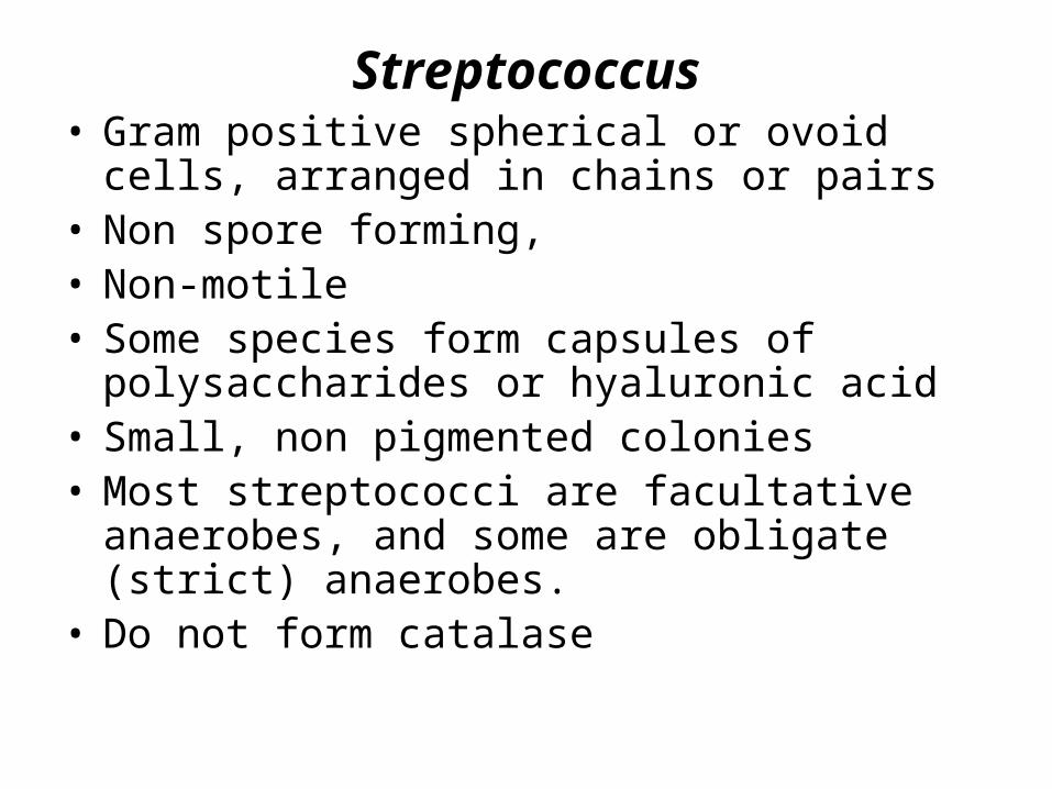
Streptococcus• Gram positive spherical or ovoid cells, arranged
in chains or pairs• Non spore forming,• Non-motile• Some species form capsules of polysaccharides
or hyaluronic acid• Small, non pigmented colonies• Most streptococci are facultative anaerobes, and
some are obligate (strict) anaerobes.• Do not form catalase
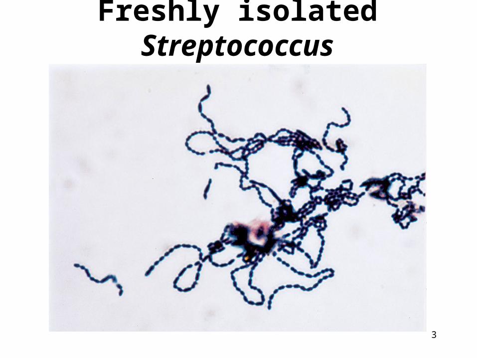
Freshly isolated Streptococcus
3

Classification
1. Based on their hemolytic properties on blood agar
2. Lancefield Grouping– species-specific CHO cell wall antigens– groups designated A-H, K-V
• some not groupable (e.g., Streptococcus pneumoniae and viridians streptococci).
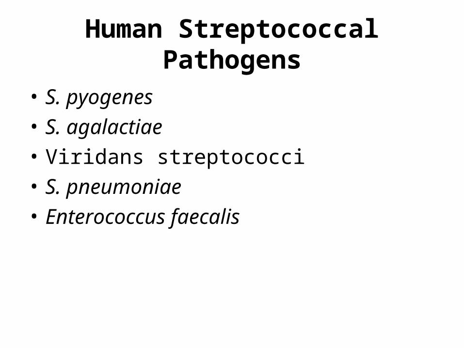
• S. pyogenes• S. agalactiae• Viridans streptococci• S. pneumoniae• Enterococcus faecalis
Human Streptococcal Pathogens

Streptococcus pyogenesPyogenes means pus producing
• Inhabits throat, nasopharynx, occasionally skin• It is the main human pathogen associated with
local or systemic invasion and poststreptococcal immunologic disorder.
• Gram positive cocci in chains
• Lancefield Serological Group A
• Beta Hemolytic on blood agar


Clinical disease: • pharyngitis (streptococcal sore throat),
osteomyelitis, endocarditis, septic arthritis, and meningitis.
• It may also infect the skin, causing erysipelas, impetigo, or cellulitis.
• two autoimmune diseases, rheumatic fever and acute glomerulonephritis.
• Other toxigenic S. pyogenes infections may lead to streptococcal
toxic shock syndrome, scarlet fever.

Laboratory diagnosis 1-Blood agar: very small, white to
grey colonies approximately 1mm in diameter surrounded by a zone of beta hemolysis.
2- Sensitive to bacitracin. 3-Serological test: streptolysin O
stimulate Anti- streptolysin O (ASO) antibody: ASO test) is indicative of previous pharyngeal infection.

2-Group B streptococci (GBS or Streptococcus agalactiae):
• Often found in the upper respiratory tract and genitourinary tract of healthy adults
• Gram-positive cocci • Typically form short chains in clinical
specimens and longer chains in laboratory media.
• A small zone of beta hemolysis on Blood agar, although some strains are non-hemolytic.
• Resist to bacitracin.
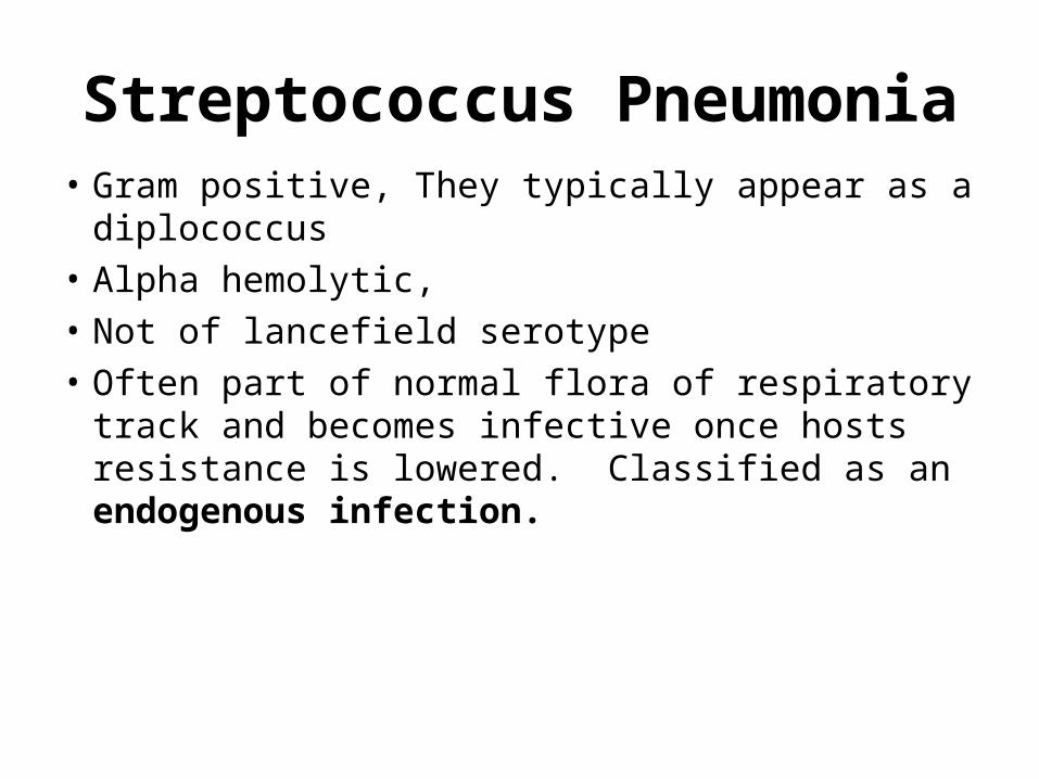
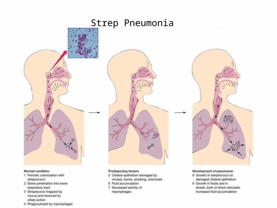
Streptococcus Pneumonia• Gram positive, They typically appear as a
diplococcus• Alpha hemolytic,• Not of lancefield serotype• Often part of normal flora of respiratory
track and becomes infective once hosts resistance is lowered. Classified as an endogenous infection.

Strep Pneumonia

Cultivation and Diagnosis
1. Gram stain of specimen – presumptive identification
2. -hemolytic
3. optochin sensitivity4. Quellung test or capsular swelling
reaction The test consists of mixing a loopful of colony with equal quantity of specific antiserum and then examining microscopically at 1000X for capsular swelling.
5. bile solubility

Hemolysis patterns on blood agar

Not optochin sensitive
Optochin sensitive
IdentificationIdentification
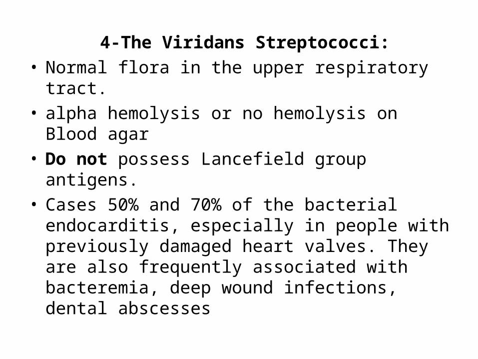
4-The Viridans Streptococci: • Normal flora in the upper respiratory tract.• alpha hemolysis or no hemolysis on Blood
agar• Do not possess Lancefield group antigens.• Cases 50% and 70% of the bacterial
endocarditis, especially in people with previously damaged heart valves. They are also frequently associated with bacteremia, deep wound infections, dental abscesses

5-The Genus Enterococcus (Enterococcus faecalis):
• Enterococci are streptococci typically occurring in pairs and short chains • Lancefield group D streptococci, these
are non-hemolytic (gamma)• Normal colonists of human large
intestine• Cause opportunistic urinary, wound,
septicemia and endocarditis• hydrolyze esculin and grow in the
presence of 40% bile salts

Laboratory diagnosis: • To distinguish group D streptococci,
the Bile Esculin agar method is performed.
• Enterococci grow in the presence of the bile salts in the medium. They hydrolyze the esculin, producing esculetin which reacts with the iron salts in the medium turning the agar black