Storage of Toxic Substances
63
Chapter 5 Absorption, Distribution, Metabolism, and Elimination of Toxics
Transcript of Storage of Toxic Substances
Chapter 5
Absorption, Distribution, Metabolism, and Elimination of
Toxics
Introduction
• The amount toxicant that actually reaches the target tissue is
dependent upon the amount absorbed, the distribution of
the substance throughout the body, the metabolism (change
in the chemical and physical characteristics) of the substance,
and the rate of excretion.
• The amount of a substance that actually reaches the target
tissue is the net result of the interaction between absorption,
distribution, metabolism, and excretion.

Absorption of Toxic Substances
• The cell membrane is selective permeable;
therefore, only certain substances are
able to pass through it.
• The selective permeability is determined
by molecular size, lipid 9fat0 solubility,
and electrical charge associated with the
toxic molecule, as well as by cell
membrane active and passive transport
mechanisms.
• The amount and rate of toxic substance
transported across these barriers is
determined by the characteristics
associated with cell membrane transport
and the physico-chemical attributes of
the toxic substance.
Mechanisms of Absorption (1)
• Passive diffusion – Most toxic substances move across the cell membrane by diffusion.
– Diffusion is dependent on three basic factors:
• the concentration gradient across the cell membrane,
• lipid solubility, and
• The electrical charge associated with the molecule.
– The rate of diffusion is primarily based on the differences in the
concentration of the substance across the cell membrane, the
surface area and thickness of the cell membrane.
– Lipid solubility is also an important factor in determining the
diffusion rate of a toxic substance since 75% of the cell membrane
is composed of lipids.

Mechanisms of Absorption (2)
– The electrical charge associated with a toxic substance molecule
or atom will also affect the rate of diffusion.
» In general, nonionized toxic substances diffuse more readily
across the cell membrane than ionized substances.
» Organic substances such as nitrous oxide, ethylene, and
divinyl ether diffuse across the cell membrane of the alveoli
easily because they do not have an electrical charge and are
lipid-soluble.

Mechanisms of Absorption (3)
• Carrier-mediated transport – Facilitated diffusion (transport) and active transport are important
when:
• moving molecules across the cell membrane against a concentration
gradient;
• the size of the toxic molecule is too large to diffuse through the pores of
the cell membrane; and
• there is low lipid solubility and the substance has an electrical charge
associated with it.
– When toxic substances are present in the small intestine, in
some cases they will compete with the nutrients for the
binding sites associated with the active transport mechanism.
• For instance, calcium is actively transported from the small intestine
across the cell membrane by a calcium-binding protein (CaBP).
• Toxic substances such as cadmium, lead or strontium will also bind to
the CaBP because their physio-chemical characteristics are similar to
calcium.
Mechanisms of Absorption (4)
– High protein diets appear to reduce the toxicity of lead and
cadmium, because amino acids derived from proteins compete for
the same protein-carrier molecules.
• Facilitated diffusion – It is characterized by a cell membrane protein attaching to a
substance outside the cell and transporting it across the cell
membrane. This is similar to the active transport mechanism.
– No energy is expended and transport of the substance is from an
area of high concentration to an area of low concentration.
– This mechanism is used to transport substances that are too large
to diffuse through the cell membrane pores or that have an
electrical charge associated with them.
Mechanisms of Absorption (5)
• Phagocytosis and pinocytosis – Phagocytosis plays an important role in the disposition of
particulates that enter the respiratory tract.
– Particulates such as asbestos, silica dust, or uranium dioxide are
engulfed by white blood cells found throughout the respiratory tract,
particularly the alveoli.
– These particulate-containing cells may then remain in the
respiratory system or be transported out of the systems by the
ciliary-clearance mechanism.
– Pinocytosis occurs frequently in the lining of the lumen in the small
intestine. Water soluble substances and particulates appear to be
transported via this mechanism.

Dose-Response Curves (2)
• The threshold is the dose below which no effect is detected
or above which an effect is first observed.
• The threshold information is useful information in
extrapolating animal data to humans and calculating what
may be considered a safe human dose for a given toxic
substance.
• The threshold dose (ThD0.0) is measured as mg/kg/day. It
is assumed that humans are as sensitive as the test animal
used. To determine the equivalent dose in man the ThD0.0
is multiplied by the average weight of a man, which is
considered to be 70 kg.

Skin Absorption (1)
• All toxicants that pass through the skin do so as a result of
passive diffusion.
• The rate at which a toxic substance is absorbed is
determined by
– its ability to penetrate the keratinized outer layer of the epidermis;
– the physico-chemical properties of the toxic substance.
• Absorption is enhanced as a result of damage to the skin’s
outer layer.
• Application of certain chemicals such as methyl and ethyl
alcohol, hexane, and acetone are lipid-soluble and can be
used to alter skin permeability by degrading the lipid
barrier of the cell membrane.

Skin Absorption (2)
• Absorption of toxic substances is enhanced in areas of the skin with a well developed blood supply.
• Once the substance diffuses through the epidermis it is absorbed into the bloodstream, which will carry it to other parts of the body.

Lung Absorption (1)
• Gases and vapors of volatile compounds diffuse across the alveolar cell membrane and are absorbed into the bloodstream.
• Although lipid solubility is important in determining the rate of absorption, an even more important factor is the solubility of the toxic substance in the blood and its interaction with components of the blood.
Lung Absorption (2)
• Particulates 5 μm or larger in diameter are usually trapped in the nose and upper portions of the respiratory tract. These particles are removed from the respiratory tract by nose wiping, blowing, or by sneezing.
• Particulates 2~5 μm in diameter are trapped in the trachea and bronchial region of the respiratory tract. These particles are primarily removed by the mucociliary-escalator.
• Particles 1 μm in diameter and smaller reach the alveoli. These particulates may be dissolved and absorbed into the bloodstream.
• Some particulates (asbestos, fiberglass) that are phagocytized remain in the lungs where they may have adverse effects and result in the development of respiratory disease.

Gastrointestinal Absorption
• Most of the absorption in the digestive tract occurs in the
small intestine.
• The primary mechanism of absorption is by diffusion but, as
we already know, faciliated and active transport also occurs.
• Increase absorption will occur if food conatining the toxic
substance remains in the digestive tract for a longer period
of time.

Distribution of Toxic Substances (1)
• Absorption into and out of the bloodstream occurs in
only one part of the circulatory system: the
capillaries.
• Anatomy of the circulatory system

Distribution of Toxic Substances (2)
• Anatomy of the heart – The right atrium receives low-oxygenated blood from the systemic
(body) circulation and the left atrium receives oxygen-rich blood
from the lungs.
– The ventricles are thick, muscular chambers. The right ventricle
pumps blood through the pulmonary artery to the extensive
vascular network (pulmonary circulation) located in the lungs.
– The pulmonary capillaries are in close association with the alveoli
of the lungs; this facilitates the diffusion of substances --- such as
oxygen, carbon dioxide, or airborne toxic substances --- between
the circulatory system and inhaled air.
– Oxygenated blood returns to the left ventricle via the pulmonary
veins and left atria.

Distribution of Toxic Substances (3)
• Anatomy of the vascular system
– Arterioles are small-diameter arteries; the blood flows
from arteriles to capillaries.
– Venules are small veins that collect blood from
capillaries; several venules unite to form larger veins.
– There are 10 billion capillaries in the body. They are
composed of a single layer of flat endotheilial cells,
having a diameter of 4-8 μm, and are the principal site
of exchange of substances between the blood and the
surrounding tissues.
– Diffusion is the primary mechsnism responsible for the
movement of gases, lipid-soluble substances, and water-
soluble molecules.

Distribution of Toxic Substances (4)
• The limiting factor --- in terms of transporting toxic
substances from the capillaries --- appears to be the size of
the molecule.
• Pinocytosis seems to be the major mechanism for
transporting these types of substances. Pinocytosis is a
relatively slow process; therefore, large toxic molecules will
remain in the blood for a longer period of time.
• The competition for protein-carrier molecules may affect
the rate of absorption and subsequent toxicity of a
substance.

Distribution of Toxic Substances (5)
• There are several mechanisms that oppose distribution to
the taregt tissue:
– binding to plasma protein;
– distribution to storage sites; and
– specialized barriers.
• Binding to plasma proteins
– When toxic substances enter the bloodstream they may bind with
plasma proteins such as albumin, transferrin, globulin, and
lipoproteins.
– Most toxic substances will bind with the plasma protein albumin.
– There is always some portion of the toxic substance that is not
bound and is in equilibrium with the bound portion.

Distribution of Toxic Substances (6)
• As the undound toxicant passes through the endotheilial
cells of the capillary into the extravascular space (a space
between the wall of the capillary and the tissue cell
membrance, filled with fluid), the bound toxicant
disassociates (separates) from the protein to maintain an
equilibrium between the bound and unbound toxicant in
the blood.
• The anount of free toxicant in the extravascular space is in
equilibrium with the amount of free toxiciant in the blood.
• A dynamic equilibrium exists between the bound and
unbound forms of a toxicant. The equilibrium will be
determined by factors that affect absorption, subsequently
affecting toxicity.

Storage of Toxic Substances (1)
• The toxicants may be stored in the target tissue, possibly
resulting in an adverse response, or it may be stored in
other tissue types, which may bot be readily affected.
• The toxicants are fat-soluble and are easily absorbed into
fatty deposits throughout the body where they may be
sotred for long periods of time.
– Lead is readily absorbed and stored in bone. Lead also affects
nerve tissue.
– In general, storage of these substances in these tissue types will
have no effect on their primary target tissue, the nerve tissue.
Storage of Toxic Substances (2)
• Liver and kidneys have a high affinity for toxic
substances and store more toxicants than nay other tissue
in the body. The reason for the high affinity is not clearly
understood but may be attributable to anatomical and
physiological characteristics associated with both organs.
• The ability to concentrate toxic substances in these
organs is advantageous for the detoxification and the
excretion of toxic substances; however, it may also
increase the occurrence of adverse effects in the liver and
kidneys.

Storage of Toxic Substances (3)
• Structure of the liver
– The liver is the single largest organ of the body and can
weigh as much as 1.4 kg. It is comprised of two lobes,
each of which is divided into numerous functional units
called lobules. A lobule consist of cords of liver cells
arranged around a central vein.
– Most venous blood passes through the liver before it
reaches the heart. The liver receives blood from the
lower extremities, kidneys, spleen, and gastrointestinal
tract. The hepatic portal vein carries nutrient rich, but
oxygen poor blood from the digestive tract to the liver.
Storage of Toxic Substances (4)
• The blood from the branches of the hepatic portal vein
passes into spaces called hepatic sinusoids located between
the lobular cords. In the sinusoids, venous blood is mixed
with oxygen-rich and nutrient-poor blood from the hepatic
artery, the second major source of blood to the liver.
• The blood from the sinusoids drains toward the center of
the lobule and into the central vein and then exists the liver
through the hepatic veins.

Storage of Toxic Substances (5)
• Absorption of toxic substances in the liver is affected by
the same factors that regulate absorption in other tissue
types.
• Lipophilic substances, such as organochlorine pesticides
and organic solvents (trichloroethane, methyl chloroform),
are readily absorbed. If they are not biotransformed into
water-soluble substances they will be retained in the liver
for long periods of time.
• Active transport mechanisms facilitate concentration of
toxicants in the liver, subsequently causing liver damage.
The hepatocytes absorb copper (Cu2+) and iron (Fe2+) from
the blood by protein-carrier molecules in the cell
membrane.

Storage of Toxic Substances (6)
• The liver is a major storage site for water-insoluble toxic
substances such as heavy metals.
• There are two other features associated with the liver,
which facilitate the absorption and concentration of
toxicants. – As it passes through the liver, the blood enters the sinusoids located
between strands of hepatocytes. The layer of endothelial cells
lining the sinusoids is discontinuous with small and large fenestrae
(pores). The fenestrae allow larger, blood borne molecules to pass
through the endothelial lining and to be absorbed by the
hepatocytes.
– The liver cells also have a high concentration of the intracellular
protein metallothionein. Methallothionein has a high affinity
(binding up to 99%) for many different kinds of toxic metals such
as Cd, Hg and Pb.

Storage of Toxic Substances (7)
– When bound to metallothionein the protein metal complex does not
readily pass through the cell membrane. Therefore, the affinity of
metals for the cell membrane transport mechanisms, the presence
of fenestrae, and the high affinity of metals for intracellular
metallothionein facilitate the storage of some toxicants in the liver
at higher concentrations than in the surrounding tissue.

Storage of Toxic Substances (8)
• Structure and function of the kidneys
– The kidneys are two bean-shapes organs located on the
posterior wall of the abdominal cavity. Each kidney is
divided into two parts, the outer portion referred to as
the cortex and the inner portion referred to as the
medulla.
– Within the medulla are cone-shaped structures called
renal pyramids. Urine passes from the tips of the
pyramids to a large funnel-like structure called the
renal pelvis. The renal pelvis narrows to form the
ureter, which connects the kidneys with the urinary
bladder.

Storage of Toxic Substances (9)
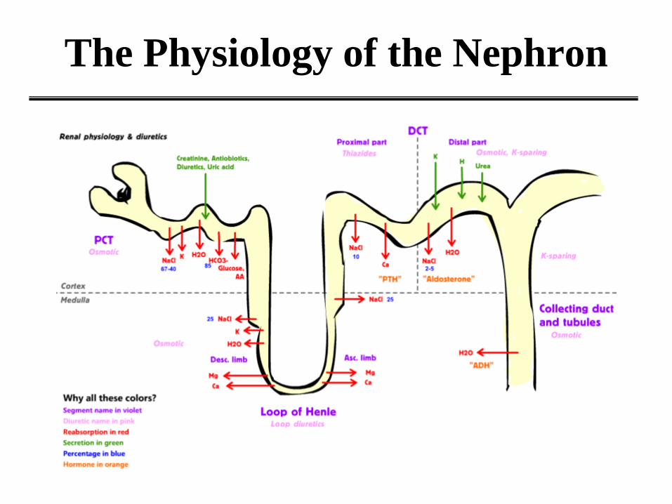
• The functional unit of the kidney is the nephron, parts of
which are found in the renal cortex and the medulla.
Tehre are approximately 1.3 million nephrons in each
kidney.
• Each nephron is composed of the following structures:
– renal corpuscle
– a proximal convoluted tubule,
– a Loop of Henle
– a distal convoluted tubule
• The distal convoluted tubule empties into a colecting duct,
which transports urine toward the renal plevis area where
it enters the ureter.
Storage of Toxic Substances (10)
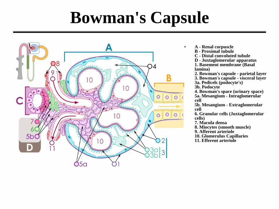
• The renal corpuscle is composed of Bowman’s capsule and
a ball-like arrangement of capillaries referred to as the
glomerulus.
• Substances carried in the blood are transported to the
renal corpuscle of the nephron.
• The kidneys receive approximately 25% of the total
cardiac output, which is about 1.2~1.3 liters of blood per
minute. The perfusion rate for the kidneys is greater than
that for the heart, brain or liver.
– The perfusion rate is the amount of blood delivered to tissues or
organs over a specific period of time.
• The primary functions of the kidneys are to remove
metabolic waste from the body and help maintain
homeostasis of the kidney.

Storage of Toxic Substances (11)
• Large molecules such as proteins do not easily pass
through the walls of Bowman’s capsule.
• The negative charge associated with both albumin and the
cell membrane of capillaries repel each other, which
further hinders the movement of albumin from the blood
into the nephron.
• Once a substance enters the kidneys it may be reabsorbed
by either active or passive transport mechanisms along the
various parts of the nephron.
Storage of Toxic Substances (12)
• The proximal convoluted tubule is the primary site of
reabsorption of water and solutes. Protein, amino acids,
glucose molecules, as well as sodium, potassium, and
chloride ions are actively transported from the proximal
convolute tubule to the peritubular capillaries. Water from
the tubule also enters the capillaries as a result of osmosis.
• Active transport mechanisms predominate in the
ascending limb of the Loop of Henle. Passive processes
exist primarily in the distal convoluted tubule.
Storage of Toxic Substances (13)
• Secretion of various substances from the peritubular
capillaries to the nephron can also occur. Ammonia
diffuses from the capillaries to the nephron tubules.
• Substances such as hydrogen and potassium ions, as well
as penicillin, are actively transported to the lumen of the
nephron.
Storage of Toxic Substances (14)
• Unbound metals like Cd and Hg can be reabsorbed by
active transport mechanisms in the cells of the proximal
convoluted tubule. Once in the cell they bind to
metallothionein resulting in concentration of these
toxicants in the kidneys, possibly causing adverse effects.
• The kidneys store 10 times the amount of Cd found in the
liver, and it can be stored for 10 years or more.
• Bioaccumulation in the kidneys may ultimately cause
kidney damage and, if severe enough, result in complete
renal failure.

Storage of Toxic Substances (15)
• Storage in fat
– Storage of toxicants in the fat initially results in a lower
concentration of the toxicant reaching the target tissue.
Like storage in the plasma protein, liver and kidneys
the toxic substance in the fat is in equilibrium with the
toxicant in the blood.
– Larger amounts of the toxicant are released when fat
reserves are mobilized in response to increased
metabolism of fats, such as during periods of fasting or
starvation. Therefore, signs of intoxication may occur
under starvation condition even though they may not
have appeared during the initial exposure.

Storage of Toxic Substances (16)
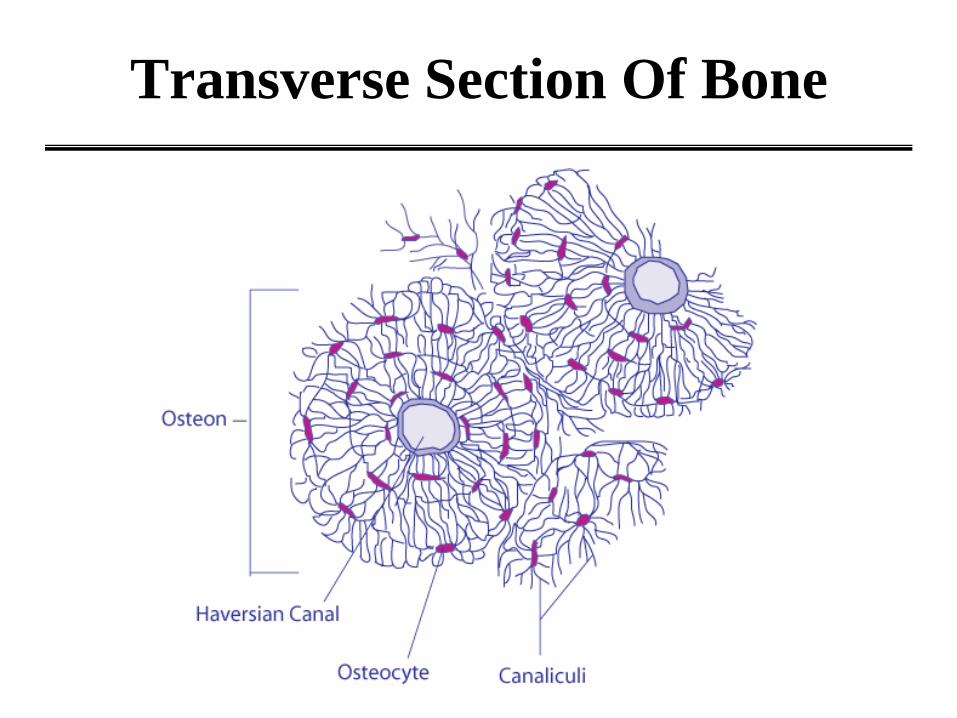
• Storage in bone – Bone is the major storage site for calcium in the body. It is
composed of bone cells called osteocytes and an extracellular
matrix arranged in sheets called lamellae.
– The extracellular matrix is composed of a mineral complex called
hydroxyapatite, which is composed primarily of calcium and
phosphate ions.
– When blood levels of calcium are low, the hydroxyapatite is broken
down, and calcium is released from the bone into bloodstream.
When the calcium levels increase in the blood, calcium is absorbed
into the hydroxyapatite of the bone.
– It is during the exchange between blood and bone that calcium can
be replaced by toxic substances such as lead and strontium.

Storage of Toxic Substances (17)
• These substances have a similar size and electrical charge
as calcium. 90% of the total body burden of lead is in the
bone. Significant amounts can remain in the bone for
10~20 years.
• Storage of toxic substances in the bone may or may not
have a detrimental effect. Lead is not toxic to bone.
However, it may be released from the bone resulting in
nerve damage. Mobilization of the lead can occur in older
individuals suffering from osteoporosis.

Storage of Toxic Substances (18)
• Mobilized lead in the bone of expectant mothers may be
passed on the fetus. The nervous system of the fetus is
very susceptible to the toxic effects of substances such as
lead and may result in birth defects associated with the
nervous system.
• The hydroxide ion (OH-) is also a component of the
metabolic process associated with bone metabolism.
Chronic exposure to elevated levels of fluoride may result
in the fluoride ion (F-) replacing the hydroxide ion during
normal bone metabolism. Increased deposition of fluoride
in the bone can result in fluorosis, which is characterized
by weakening of the bone.

Specialized Barrier: Blood-Brain Barrier (1)
• The blood-brain barrier is created by the close associated
of brain capillaries with specialized cells formed in the
nervous system. This barrier effectively decreases the type
and amount of toxic substances that are transferred from
the blood to the brain tissue.
• Reduced absorption of toxicants is attributed to several
unique characteristics associated with the blood brain
barrier:
– the closely packed endothelial cells of the capillaries;
– the astrocytes; and
– The low protein content of the interstitial fluid.

Specialized Barrier: Blood-Brain Barrier (2)
• Normally, capillary endothelial cells are loosely joined to
each other with pores, about 4 nm in diameter, located
between the cells. The capillary endothelial cells of the
blood-brain barrier are more closely arranged to each
other, therefore, few or no pores exist.
• Lipid-soluble substances like ethanol easily diffuse through
the phospholipid cell membrane and into the fluid
surrounding the brain. Water soluble substances such as
glucose --- which is necessary for nerve cell functioning ---
must be transported across the blood-brain barrier by
carrier-mediated mechanisms.
Specialized Barrier: Blood-Brain Barrier (3)
• The capillaries are surrounded by cellular processes from
astrocytes. Astrocytes are specialized cells found in the
nervous system.
• The cell membrane of the astrocyte has a high lipid content
that helps to form an effective barrier, slowing the rate of
movement of water-soluble molecules.
• The interstitial fluid is the liquid between the external wall
of the capillary and the cell membrane of the surrounding
tissue; it has a low protein content which reduced amounts
of the protein-toxic substance complex reaches the brain
tissue.
• The hydrophobic cell membrane of the astrocyte processes
slows or inhibits the passage of toxicants.

Redistribution of Toxic Substances
• Toxic substances have a tendency to concentrate initially in
well perfused tissues such as the liver and kidneys.
However, other tissue types may have a greater affinity for
the toxicant and over time --- because of the dynamic
equilibrium discussed previously --- the toxicant will
eventually be transported and stored in these tissues.

Metabolism (biotransformation)
• The difference in toxicity between genders seems to be
primarily influence by the sex hormones (estrogen and
testosterone), which can affect metabolism.
• Some organophosphate pesticides are more toxic to
females than males.
– Parathion is metabolized more rapidly in females resulting in a
higher concentration of the more toxic intermediate, paraoxon.
– Male rats are 10 times more susceptible to liver damage than
female rats as a result of chronic oral exposure to DDT.
State of Health
• The liver and the kidney are important organs for
detoxifying and removing toxic substances. Therefore,
conditions that lead to liver or kidney disease enhance the
toxic effects of substances normally detoxified by these
organs.
Transverse Section Of Bone

Individual Bone Structure

The Nephron of the Kidney
Bowman's Capsule
• A - Renal corpuscle B - Proximal tubule C - Distal convoluted tubule D - Juxtaglomerular apparatus 1. Basement membrane (Basal lamina) 2. Bowman's capsule - parietal layer 3. Bowman's capsule - visceral layer 3a. Pedicels (podocyte's) 3b. Podocyte 4. Bowman's space (urinary space) 5a. Mesangium - Intraglomerular cell 5b. Mesangium - Extraglomerular cell 6. Granular cells (Juxtaglomerular cells) 7. Macula densa 8. Miocytes (smooth muscle) 9. Afferent arteriole 10. Glomerulus Capillaries 11. Efferent arteriole
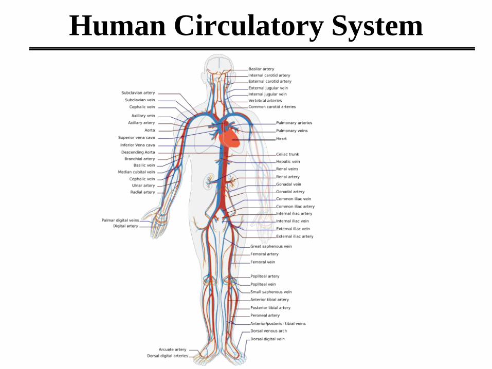
Human Circulatory System

Diagram of the Human Heart
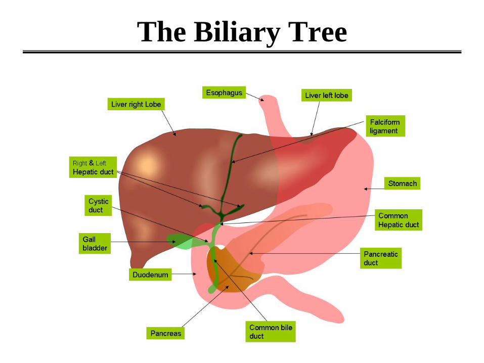
The Biliary Tree
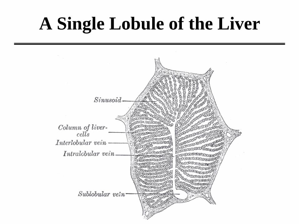
A Single Lobule of the Liver

Human Liver Sinusoid

The Hepatic Artery
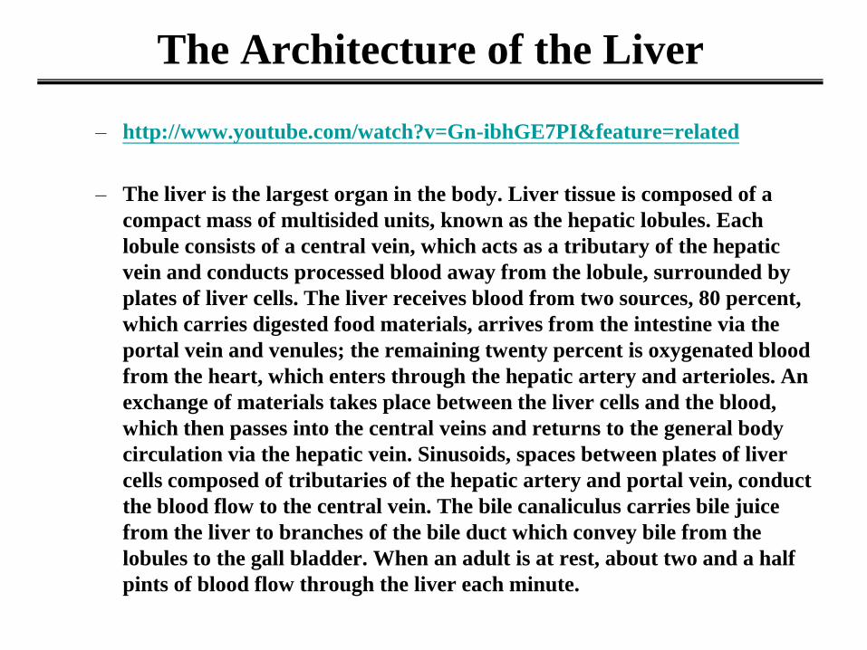
The Architecture of the Liver
– http://www.youtube.com/watch?v=Gn-ibhGE7PI&feature=related
– The liver is the largest organ in the body. Liver tissue is composed of a
compact mass of multisided units, known as the hepatic lobules. Each
lobule consists of a central vein, which acts as a tributary of the hepatic
vein and conducts processed blood away from the lobule, surrounded by
plates of liver cells. The liver receives blood from two sources, 80 percent,
which carries digested food materials, arrives from the intestine via the
portal vein and venules; the remaining twenty percent is oxygenated blood
from the heart, which enters through the hepatic artery and arterioles. An
exchange of materials takes place between the liver cells and the blood,
which then passes into the central veins and returns to the general body
circulation via the hepatic vein. Sinusoids, spaces between plates of liver
cells composed of tributaries of the hepatic artery and portal vein, conduct
the blood flow to the central vein. The bile canaliculus carries bile juice
from the liver to branches of the bile duct which convey bile from the
lobules to the gall bladder. When an adult is at rest, about two and a half
pints of blood flow through the liver each minute.

Cardiovascular System
• http://www.youtube.com/watch?v=rBQOLiFto6Q&feature
=related

General Anatomy of the Liver

Renal Pyramids
• 1. Renal pyramid
2. Efferent artery
3. Renal artery
4. Renal vein
5. Renal hilum
6. Renal pelvis
7. Ureter
8. Minor calyx
9. Renal capsule
• 10. Inferior renal capsule
11. Superior renal capsule
12. Afferent vein
13. Nephron
14. Minor calyx
15. Major calyx
16. Renal papilla
17. Renal column

Nephron
The Physiology of the Nephron