stage III colorectal cancer. Oncoimmunology, 6(12...
http://www.diva-portal.org This is the published version of a paper published in Oncoimmunology. Citation for the original published paper (version of record): Malesci, A., Bianchi, P., Celesti, G., Basso, G., Marchesi, F. et al. (2017) Tumor-associated macrophages and response to 5-fluorouracil adjuvant therapy in stage III colorectal cancer. Oncoimmunology, 6(12): e1342918 https://doi.org/10.1080/2162402X.2017.1342918 Access to the published version may require subscription. N.B. When citing this work, cite the original published paper. Permanent link to this version: http://urn.kb.se/resolve?urn=urn:nbn:se:umu:diva-144130
Transcript of stage III colorectal cancer. Oncoimmunology, 6(12...

http://www.diva-portal.org
This is the published version of a paper published in Oncoimmunology.
Citation for the original published paper (version of record):
Malesci, A., Bianchi, P., Celesti, G., Basso, G., Marchesi, F. et al. (2017)Tumor-associated macrophages and response to 5-fluorouracil adjuvant therapy instage III colorectal cancer.Oncoimmunology, 6(12): e1342918https://doi.org/10.1080/2162402X.2017.1342918
Access to the published version may require subscription.
N.B. When citing this work, cite the original published paper.
Permanent link to this version:http://urn.kb.se/resolve?urn=urn:nbn:se:umu:diva-144130

Full Terms & Conditions of access and use can be found athttp://www.tandfonline.com/action/journalInformation?journalCode=koni20
OncoImmunology
ISSN: (Print) 2162-402X (Online) Journal homepage: http://www.tandfonline.com/loi/koni20
Tumor-associated macrophages and responseto 5-fluorouracil adjuvant therapy in stage IIIcolorectal cancer
Alberto Malesci, Paolo Bianchi, Giuseppe Celesti, Gianluca Basso, FedericaMarchesi, Fabio Grizzi, Giuseppe Di Caro, Tommaso Cavalleri, LorenzaRimassa, Richard Palmqvist, Alessandro Lugli, Viktor. H. Koelzer, MassimoRoncalli, Alberto Mantovani, Shuji Ogino & Luigi Laghi
To cite this article: Alberto Malesci, Paolo Bianchi, Giuseppe Celesti, Gianluca Basso, FedericaMarchesi, Fabio Grizzi, Giuseppe Di Caro, Tommaso Cavalleri, Lorenza Rimassa, RichardPalmqvist, Alessandro Lugli, Viktor. H. Koelzer, Massimo Roncalli, Alberto Mantovani, ShujiOgino & Luigi Laghi (2017) Tumor-associated macrophages and response to 5-fluorouraciladjuvant therapy in stage III colorectal cancer, OncoImmunology, 6:12, e1342918, DOI:10.1080/2162402X.2017.1342918
To link to this article: https://doi.org/10.1080/2162402X.2017.1342918
© 2017 The Author(s). Published by Taylor &Francis Group, LLC© Alberto Malesci, PaoloBianchi, Giuseppe Celesti, Gianluca Basso,Federica Marchesi, Fabio Grizzi, Giuseppe DiCaro, Tommaso Cavalleri, Lorenza Rimassa,Richard Palmqvist, Alessandro Lugli, Viktor.H. Koelzer, Massimo Roncalli, AlbertoMantovani, Shuji Ogino and Luigi Laghi
View supplementary material
Accepted author version posted online: 12Jul 2017.Published online: 19 Jul 2017.
Submit your article to this journal
Article views: 447 View related articles
View Crossmark data Citing articles: 1 View citing articles

ORIGINAL RESEARCH
Tumor-associated macrophages and response to 5-fluorouracil adjuvant therapyin stage III colorectal cancer
Alberto Malescia,b, Paolo Bianchic,d, Giuseppe Celestic, Gianluca Bassoc,d, Federica Marchesie, Fabio Grizzie,Giuseppe Di Caroe, Tommaso Cavalleric, Lorenza Rimassaf, Richard Palmqvistg, Alessandro Luglih,i, Viktor. H. Koelzer i,Massimo Roncallij,k, Alberto Mantovanie,k, Shuji Oginol,m,n, and Luigi Laghib,c
aDepartment of Biotechnologies and Translational Medicine, University of Milan, via Manzoni 56, 20089 Rozzano (Milan), Italy; bDepartment ofGastroenterology, Humanitas Clinical and Research Center, via Manzoni 56, 20089 Rozzano (Milan), Italy; cLaboratory of Molecular Gastroenterology,Department of Gastroenterology, Humanitas Clinical and Research Center, via Manzoni 56, 20089 Rozzano (Milan), Italy; dClinical InvestigationLaboratory, Humanitas Clinical and Research Center, via Manzoni 56, 20089 Rozzano (Milan), Italy; eDepartment of Immunology and Inflammation,Humanitas Clinical and Research Center, via Manzoni 113, Rozzano (Milan), Italy; fHumanitas Cancer Center, Humanitas Clinical and Research Center,Via Manzoni 56, Rozzano 20089 (Milan), Italy; gDepartment of Medical Biosciences, Pathology, Umea
�University, Umea
�, Sweden; hClinical Pathology
Division, Institute of Pathology, University of Bern, Bern, Switzerland; iTranslational Research Unit, Institute of Pathology, University of Bern, Bern,Switzerland; jDepartment of Pathology, Humanitas Clinical and Research Center, via Manzoni 56, 20089 Rozzano (Milan), Italy; kDepartment ofBiomedical Sciences, Humanitas University, Via Manzoni 113, 20089 Rozzano (Milan), Italy; lDivision of MPE Molecular Pathological Epidemiology,Department of Pathology, Brigham and Women’s Hospital, and Harvard Medical School, Boston, MA 02215 USA; mDepartment of Oncologic Pathology,Dana-Farber Cancer Institute, Boston, MA 02215 USA; nDepartment of Epidemiology, Harvard T.H. Chan School of Public Health, Boston, MA 02215 USA
ARTICLE HISTORYReceived 9 June 2017Revised 9 June 2017Accepted 9 June 2017
ABSTRACTTumor-associated macrophages (TAMs) play a role in tumor development and progression. Wehypothesized that abundance of TAMs might modify efficacy of 5-fluorouracil chemotherapy in colorectalcancer. We measured the density of CD68C TAMs at the invasive front of primary tumor of colorectalcarcinoma (PT-TAMs; n D 208), at available matched metastatic lymph node (LN-TAMs; n D 149), and in anindependent set of primary colorectal cancers (PT-TAMs, n D 111). The hazard ratios for disease-freesurvival were computed by Cox proportional-hazards model. In exploratory analysis, the interactionbetween TAMs and 5-fluorouracil adjuvant therapy was significant (PT-TAMs, p D 0.02; LN-TAMs, p D0.005). High TAMs were independently associated with better disease-free survival only in 5-fluorouracil-treated patients (PT-TAMs, HR 0.23; 95%CI, 0.08–0.65; p D 0.005; LN-TAMs, HR 0.13; 95%CI, 0.04–0.43; p D0.001). The independent predictive value of PT-TAMs was replicated in the external set (HR, 0.14; 95%CI0.02–1.00; p D 0.05). In an in vitro experiment, 5-fluorouracil and macrophages showed a synergistic effectand increased colorectal cancer cell death. High densities of TAMs, particularly in metastatic lymph-nodes,identify stage III colorectal cancer patients benefitting from 5-fluorouracil adjuvant therapy.
Abbreviations: TAMs, Tumor-associated macrophages; TANs, tumor-associated neutrophils; PT-TAMs, TAMs at theinvasive front of primary tumors; LN-TAMs, () TAMs in the nodal metastasis; IRA, immune-reactive area
KEYWORDSAdjuvant therapy; Colorectalcancer; Innate immunity;Outcome; Tumor associatemacrophages
Introduction
Adjuvant chemotherapy is the standard post-surgical treatmentof patients with stage III colorectal cancer. In this setting, fluo-ropyrimidines improve 5-year survival rates by 10–15%, anadditional 4–6% being contributed by their combination withoxaliplatin.1-3 Even among patients receiving adjuvant therapy,one third still progresses to metastatic disease,4-6 a rate notimproved by biologic agents.5,7-9 These facts indicate thatmechanisms of disease eradication achieved by chemotherapyare unlikely to cooperate with those of disease control exertedby biologic agents,10 putting forward the lack of markers pre-dicting the efficacy of chemotherapy in stage III colorectal
cancer. Molecular features which define colorectal cancer sub-types with heterogeneous prognosis (such as KRAS/BRAF andmicrosatellite status), do not predict responsiveness to adjuvantchemotherapy10,11 as they do in metastatic setting, or alikePI3K-mutations toward aspirin responsiveness in adjuvant set-ting.12 Also tumor-infiltrating lymphocytes (TILs), which effec-tively forecast the outcome of patients with early colorectalcancer, are unlikely to act as predictive factor in node-positivepatients.13-16 Under this respect, other key players of the tumormicroenvironment deserving consideration are the tumor-asso-ciated macrophages (TAMs). Acting as promoters of cancer
CONTACT Alberto Malesci [email protected]; Luigi Laghi [email protected] Humanitas Clinical and Research, Dept. of Gastroenterology,Via Manzoni, 56, 20089 Rozzano (Milan) Italy.
Supplemental data for this article can be accessed on the publisher’s website.© 2017 Alberto Malesci, Paolo Bianchi, Giuseppe Celesti, Gianluca Basso, Federica Marchesi, Fabio Grizzi, Giuseppe Di Caro, Tommaso Cavalleri, Lorenza Rimassa, Richard Palmqvist, AlessandroLugli, Viktor. H. Koelzer, Massimo Roncalli, Alberto Mantovani, Shuji Ogino and Luigi Laghi. Published by Taylor & Francis Group, LLCThis is an Open Access article distributed under the terms of the Creative Commons Attribution-NonCommercial-NoDerivatives License (http://creativecommons.org/licenses/by-nc-nd/4.0/),which permits non-commercial re-use, distribution, and reproduction in any medium, provided the original work is properly cited, and is not altered, transformed, or built upon in any way.
ONCOIMMUNOLOGY2017, VOL. 6, NO. 12, e1342918 (11 pages)https://doi.org/10.1080/2162402X.2017.1342918
progression,17,18 their high density usually correlates with worseprognosis, with the notable exceptions of colorectal cancer.19-24
However, in some neoplastic disease, such as follicular lympho-mas, TAM prognostic impact heavily depends upon treat-ment.25-27 Preclinical studies have also depicted a complex dualrole for TAMs in modulating the response to anticancer thera-pies.28-30 In some cancers, selected chemotherapeutic agentsenhance the anti-tumor activity of TAMs by eliciting adaptiveresponses able to determine immunogenic cell death,31,32 bydepleting immunosuppressive TAMs,33,34 or by reprogram-ming macrophages to an antitumor phenotype.35,36 Althoughscanty clinical data are available on interactions of TAMs withchemotherapy efficacy in solid tumors, high loads of TAMscorrelated with prolonged survival in 5-fluorouracil-treatedpatients with gastric cancer,37 and partially corrected the poorprognosis of patients resected for pancreatic cancer and treatedwith adjuvant gemcitabine.38 In keeping with these data, werecently reported the favorable prognostic interaction of 5-fluo-rouracil adjuvant therapy in colorectal cancer patients withhigh densities of tumor-associated neutrophils (TANs), a nativecomponent of the tumor infiltrate otherwise associated withpoor clinical outcome.39-41
This study was aimed to see whether the favorable prognos-tic association between TAMs densities and colorectal canceroutcome reflects a beneficial interaction of macrophages with5-fluorouracil-based adjuvant chemotherapy. To this aim, thepredictive value of TAMs at the invasive front of primarytumors (PT-TAMs) and of nodal metastasis (LN-TAMs) wastested in a consecutive series of stage III colorectal cancer, andin external set of primary tumors. In vitro we conducted experi-ments investigating the interplay of 5-fluorouracil with macro-phages in determining colorectal cancer cell death.
Results
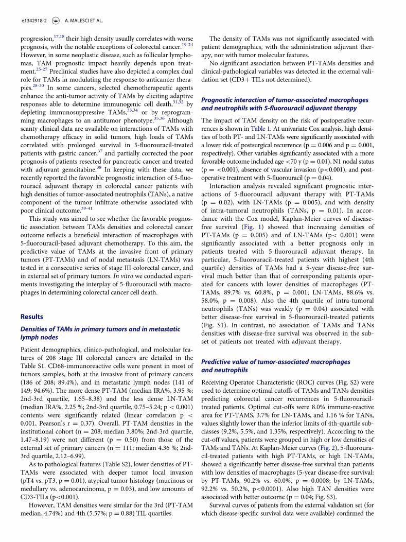
Densities of TAMs in primary tumors and in metastaticlymph nodes
Patient demographics, clinico-pathological, and molecular fea-tures of 208 stage III colorectal cancers are detailed in theTable S1. CD68-immunoreactive cells were present in most oftumors samples, both at the invasive front of primary cancers(186 of 208; 89.4%), and in metastatic lymph nodes (141 of149; 94.6%). The more dense PT-TAM (median IRA%, 3.95 %;2nd-3rd quartile, 1.65–8.38) and the less dense LN-TAM(median IRA%, 2.25 %; 2nd-3rd quartile, 0.75–5.24; p < 0.001)contents were significantly related (linear correlation p <
0.001, Pearson’s r D 0.37). Overall, PT-TAM densities in theinstitutional cohort (n D 208; median 3.80%; 2nd-3rd quartile,1.47–8.19) were not different (p D 0.50) from those of theexternal set of primary cancers (n D 111; median 4.36 %; 2nd-3rd quartile, 2.12–6.99).
As to pathological features (Table S2), lower densities of PT-TAMs were associated with deeper tumor local invasion(pT4 vs. pT3, p D 0.01), atypical tumor histology (mucinous ormedullary vs. adenocarcinoma, p D 0.03), and low amounts ofCD3-TILs (p<0.001).
However, TAM densities were similar for the 3rd (PT-TAMmedian, 4.74%) and 4th (5.57%; p D 0.88) TIL quartiles.
The density of TAMs was not significantly associated withpatient demographics, with the administration adjuvant ther-apy, nor with tumor molecular features.
No significant association between PT-TAMs densities andclinical-pathological variables was detected in the external vali-dation set (CD3C TILs not determined).
Prognostic interaction of tumor-associated macrophagesand neutrophils with 5-fluorouracil adjuvant therapy
The impact of TAM density on the risk of postoperative recur-rences is shown in Table 1. At univariate Cox analysis, high densi-ties of both PT- and LN-TAMs were significantly associated witha lower risk of postsurgical recurrence (p D 0.006 and p D 0.001,respectively). Other variables significantly associated with a morefavorable outcome included age<70 y (pD 0.01), N1 nodal status(p D <0.001), absence of vascular invasion (p<0.001), and post-operative treatment with 5-fluorouracil (pD 0.04).
Interaction analysis revealed significant prognostic inter-actions of 5-fluorouracil adjuvant therapy with PT-TAMs(p D 0.02), with LN-TAMs (p D 0.005), and with densityof intra-tumoral neutrophils (TANs, p D 0.01). In accor-dance with the Cox model, Kaplan-Meier curves of disease-free survival (Fig. 1) showed that increasing densities ofPT-TAMs (p D 0.005) and of LN-TAMs (p< 0.001) weresignificantly associated with a better prognosis only inpatients treated with 5-fluorouracil adjuvant therapy. Inparticular, 5-fluorouracil-treated patients with highest (4thquartile) densities of TAMs had a 5-year disease-free sur-vival much better than that of corresponding patients oper-ated for cancers with lower densities of macrophages (PT-TAMs, 89.7% vs. 60.8%, p D 0.001; LN-TAMs, 88.6% vs.58.0%, p D 0.008). Also the 4th quartile of intra-tumoralneutrophils (TANs) was weakly (p D 0.04) associated withbetter disease-free survival in 5-fluorouracil-treated patients(Fig. S1). In contrast, no association of TAMs and TANsdensities with disease-free survival was observed in the sub-set of patients not treated with adjuvant therapy.
Predictive value of tumor-associated macrophagesand neutrophils
Receiving Operator Characteristic (ROC) curves (Fig. S2) wereused to determine optimal cutoffs of TAMs and TANs densitiespredicting colorectal cancer recurrences in 5-fluorouracil-treated patients. Optimal cut-offs were 8.0% immune-reactivearea for PT-TAMS, 3.7% for LN-TAMs, and 1.16 % for TANs,values slightly lower than the inferior limits of 4th-quartile sub-classes (9.2%, 5.5%, and 1.35%, respectively). According to thecut-off values, patients were grouped in high or low densities ofTAMs and TANs. At Kaplan-Meier curves (Fig. 2), 5-fluoroura-cil-treated patients with high PT-TAMs, or high LN-TAMs,showed a significantly better disease-free survival than patientswith low densities of macrophages (5-year disease-free survival:by PT-TAMs, 90.2% vs. 60.0%, p D 0.0008; by LN-TAMs,92.2% vs. 50.2%, p<0.0001). Also high TAN densities wereassociated with better outcome (p D 0.04; Fig. S3).
Survival curves of patients from the external validation set (forwhich disease-specific survival data were available) confirmed the
e1342918-2 A. MALESCI ET AL.
better outcome of 5-fluorouracil-treated patients with high densi-ties of PT-TAMs (p D 0.03), together with the lack of any impacton the outcome of untreated subjects (pD 0.62) (Fig. S4).
At Cox univariate analysis (Table 2), high PT-TAMs (p D0.002), high LN-TAMs (p D 0.001), high TANs (p D 0.04), N1nodal status (p<0.001), and absence of vascular invasion (p D0.008), were associated with better disease-free survival of 5-fluorouracil-treated patients from the institutional cohort. Afavorable predictive value of high PT-TAMs (p D 0.05), N1nodal status (p D 0.03), and absence of vascular invasion (p D0.05) was also observed in the external set. In multivariate mod-els adjusted by nodal status and vascular invasion (Table 3),only PT-TAMs (p D 0.005) and LN-TAMs (p D 0.001) main-tained an independent predictive value. PT-TAMs resulted to
be an independent outcome predictor also in the external vali-dation set (p D 0.05).
Synergic colorectal cancer cell killing by macrophagesand 5-fluorouracil
At fluorescence-activated cell sorting (FACS), the death rate ofSW480 cells was not increased by short-term (24-hour) expo-sure to 10 mM 5-fluorouracil (Fig. 3A, top panels), even if can-cer cells were co-cultured with un-polarized macrophages(Fig. 3A, middle panels). In contrast, co-culture of tumor cellswith M1-polarized macrophages (Fig. 3A, bottom panels)almost doubled the cell death rate, which was further increasedby exposure to 5-fluorouracil.
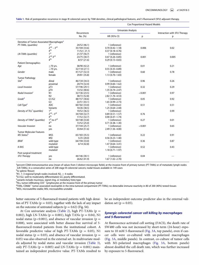
Table 1. Risk of postoperative recurrence in stage III colorectal cancer by TAM densities, clinical-pathological features, and 5-fluorouracil (5FU) adjuvant therapy.
Cox Proportional Hazard Models
RecurrencesUnivariate Analysis
Interaction with 5FU TherapyNo. (%) HR (95% CI) p p
Densities of Tumor-Associated Macrophagesa
PT-TAMs, (quartiles) 1st 24/52 (46.1) 1 (reference)2nd ¡3rd 35/104 (33.6) 0.70 (0.42–1.18) 0.006 0.024th 11/52 ( 21.1) 0.37 (0.18–0.76)
LN-TAMs (quartiles) 1st 21/37 (56.7) 1 (reference)2nd-3rd 23/75 (30.7) 0.47 (0.26–0.85) 0.001 0.0054th 8/37 (21.6) 0.29 (0.13–0.65)
Patient DemographicsAge �70 yrs. 38/90 (42.2) 1 (reference) 0.01 0.21
<70 yrs. 32/118 (27.1) 0.55 (0.35–0.89)Gender male 41/127 (32.3) 1 (reference) 0.60 0.78
female 29/81 (35.8) 1.13 (0.70–1.83)Tumor PathologySiteb distal 46/134 (34.3) 1 (reference) 0.96 0.26
proximal 24/74 (32.4) 0.99 (0.60–1.62)Local Invasion pT3 57/196 (29.1) 1 (reference) 0.32 0.29
pT4 13/32 (40.6) 1.35 (0.74–2.47)Nodal Invasionc N1 32/136 (23.5) 1 (reference) <0.001 0.97
N2 38/72 (52.8) 2.82 (1.76–4.53)Graded G1/G2 48/157 (30.6) 1 (reference) 0.05 0.92
G3 22/51 (43.1) 1.65 (0.99–2.73)Cell-Typee ADC 60/182 (33.0) 1 (reference) 0.51 0.51
Variants 10/26 (38.5) 1.25 (0.64–2.44)Density of TILsf (quartiles) 1st 19/52 (36.5) 1 (reference) 0.99
2nd¡3rd 34/104 (32.7) 0.89 (0.51–1.57) 0.764th 17/52 (32.7) 0.90 (0.47–1.74)
Density of TANsg (quartiles) 1st-to-3rd 50/148 (33.8) 1 (reference) 0.27 0.014th 13/52 (25.0) 0.71 (0.38–1.30)
Vascular Invasion no 37/144 (25.7) 1 (reference) <0.001 0.65yes 33/64 (51.6) 2.49 (1.56–4.00)
Tumor Molecular FeaturesMS Statush MSS 65/183 (35.5) 1 (reference) 0.22 0.91
MSI 5/25 (20.0) 0.56 (0.23–1.40)BRAF wild-type 64/194 (33.0) 1 (reference) 0.36 0.87
mutated 6/14 (42.8) 1.47 (0.64–3.41)K-RAS wild-type 1 (reference) 0.52 0.43
mutated 1.18 (0.71–1.97)Post-surgical treatment5FU Therapy yes 44/146 (30.1) 1 (reference) 0.04 —
no 26/62 (41.9) 1.67 (1.02–2.70)
apercent CD68-immunoreactive area (mean of values from 3 distinct microscopic fields) at the invasive front of primary tumors (PT-TAMs) or of metastatic lymph nodes(LN-TAMs), in a consecutive series of 208 stage III colorectal cancers; nodal tissues available in 149 casesbto splenic flexurecN1, 1–3 regional lymph nodes involved; N2, � 4 nodesdG1/G2, well-to moderately differentiated; G3, poorly differentiated.evariants include mucinous, signet-ring, or medullary histo-typefTILs, tumor-infiltrating CD3C lymphocytes at the invasive front of the primary tumorgTANs, CD66bC tumor-associated neutrophils in the intra-tumoral compartment (PT-TANs); no detectable immune-reactivity in 80 of 200 (40%) tested tissueshMSS, microsatellite-stable; MSI, microsatellite-unstable
ONCOIMMUNOLOGY e1342918-3

In independent experiments with SW480 and HT29 cells,the tumoricidal activity induced by co-culture with M1-macro-phages and 5-fluorouracil exposure exceeded the sum of thecytotoxic effects of macrophages and 5-fluorouracil alone(Fig. 3B). Cancer cells co-cultured wit M1-macrophages andexposed to 5-fluorouracil had a death rate significantly superiorto that of corresponding 5-fluorouracil -untreated cells(SW480, p<0.01; HT29, p<0.001) which, in turn, had a deathrate significantly higher than that of cells exposed to 5-fluoro-uracil in the absence of macrophages or after co-culturing withun-polarized macrophages (p<0.05 for both SW480 andHT29).
Macrophages preliminarily cultured with the supernatant of5-fluorouracil-treated SW480 colorectal cancer cells and thenco-cultured with tumor cells displayed a cytotoxicity signifi-cantly greater (p<0.01) than that of control macrophages or ofmacrophages pre-cultured with conditioned medium from 5-fluorouracil-untreated colorectal cancer cells (Fig. 3C and 3D).
The exposure of un-polarized (M0) macrophages to 5-fluo-rouracil increased the expression of the M1-marker HLA-DRbut not that of the M2-marker CD163 (Panel E), while drug-exposure of M1-macrophages was accompanied by a significantrelease of TRAIL (p<0.01) and TNFa (p<0.05) pro-apoptoticcytokines (Panel F).
Discussion
We first report that densities of TAMs in the primary tumorand in metastatic lymph nodes act as an independent predictivefactor in patients resected for stage III colorectal cancer and
treated with 5-fluorouracil adjuvant therapy. Together with thein-vitro evidence that macrophage greatly enhance colorectalcancer cell killing by exposure to 5-fluorouracil, clinical datastrongly support a synergism of TAMs with fluoro-pyrimidines.
In contrast to their association with poor prognosis in sev-eral tumors, high densities of TAMs favorably influence thepostsurgical clinical outcome of colorectal cancer.19-24 Thepresent study clarifies that the association between high TAMsdensities and better survival in patients with stage III colorectalcancer results from their interaction with 5-fluorouracil, thedrug at the basis of adjuvant therapy for decades. Given thatsuch an association has been shown to be stage-dependent andvirtually limited to stage III disease,22-24 the synergism with 5-fluorouracil likely accounts for the entire prognostic value ofTAMs in colorectal cancer. Consistently, we found no correla-tion between TAMs and outcome in our institutional cohort ofmore than 2 hundred stage II patients (HR 0.98; 95% CI 0.88–1.08; p D 0.66; data not shown).
Our results draw considerable strength from the fact thatTAMs exhibited their predictive role when measured at distinctanatomic sites. Actually, high densities of macrophages in met-astatic lymph nodes (LN-TAMs) were associated with disease-free survival of 5-fluorouracil-treated patients more closelythan high loads of TAMs measured in primary tumors (PT-TAMs), which might reflect a more accurate identification ofhigh-TAM infiltrates at the nodal invasive front. As to the den-sity of LN-TAMs, Onishi and coll., reported that a high densityof CD169C, but not of CD68C, macrophages was associatedwith better survival of patients with stage I-IV colorectal
Figure 1. Disease-free survival (DFS) in stage III colorectal cancer, by TAM-density quartiles. Kaplan-Meier Curves. Patients were stratified by quartile distribution of TAMsdensities at the invasive front of their primary tumor (PT-TAMs, upper panels) or of metastatic lymph-nodes (LN-TAMs, lower panels). Higher densities of both PT-TAMsand LN-TAMs were significantly associated with better disease-free survival in patients treated with 5-fluorouracil (left panels), but not in the subset of untreated patients(right panels). Also higher densities of tumor-associated neutrophils (PT-TANs) were weakly associated with the survival of 5-fluorouracil -treated patients (see Fig. S1).P values are for Log-Rank test.
e1342918-4 A. MALESCI ET AL.

cancer. The lack of association of CD68C LN-TAMs with prog-nosis is likely due to small study population, and to the absenceof stage stratification with respect to chemotherapy treatment.42
The prognostic interaction of LN-TAMs anyhow supports theconcept that macrophages can synergize with 5-fluorouracil inmetastatic microenvironment, where cytotoxic drugs areexpected to act. Supporting this interpretation, Cavnar et al.recently reported the association between high-TAM infiltra-tion of colorectal cancer liver metastases and better survival, ina cohort of patients treated with peri-surgical chemotherapy.43
Noticeably, the overall predictive value of TAMs essentiallyresulted from the excellent outcome of cancers with very highdensities of CD68C cells. Such cancers represented approxi-mately 30% of the entire series (28.8 % by high PT-TAMs,36.4% by high LN-TAMS), a fraction roughly equaling thereduction in the relative risk of death that is expected from 5-fluorouracil adjuvant therapy in stage III colorectal cancer.
The densities of tumor-infiltrating CD3C lymphocytes(TILs), although weakly correlated with PT-TAMs loads, hadno prognostic value in our series of stage III colorectal cancer.This finding challenges the concept of a stage-independent cor-relation between TILs and colorectal cancer prognosis,44 ratherindicating that the established protective role of TILs in stage II
disease13, 15, 44, 45 is lost once nodal metastases develop. In addi-tion, the lack of prognostic impact for TILs densities in stage IIIcolorectal cancer implies that their correlation with PT-TAMsis not extended to the very dense infiltrates of TAMs thataccount for the prediction of 5-fluorouracil responsiveness.Taken together, results from combined analysis of CD3C andCD68C cells support a disease model in which native immuneevasion accompanies the development of micro-metastases,that can be best eradicated by adjuvant therapy in patients withhigh TAM loads.
CD68 pan-macrophage marker does not allow the sub-clas-sification of TAMs. However, in-vitro experiments indicatethat 5-fluorouracil exposure favor macrophage polarizationtoward a M1 anti-tumor role. These findings are in keepingwith in-vivo studies showing that the skewing of TAMs toM1-like phenotype contributes to the anti-tumor and anti-angiogenic effects of pharmacological agents in mice.35,36 Fur-thermore, the unique exposure of colorectal cancer microenvi-ronment to commensal enteric microbiota, which has beenrecently shown to shape the immune cells response to cyto-toxic agents,46,47 might contribute to TAMs polarization andeventually account for the predictive value of macrophages incolorectal cancer. On the other hand, TAMs subpopulationsare variably represented in distinct microenvironments of thesame tumor,46 and expression of polarization markers is con-siderably heterogeneous in colorectal cancer.23 Accordingly,the polarization of TAMs in the resected primary tumor mighteven be irrelevant to their successive synergism with post-sur-gical chemotherapy, so that TAMs densities might simply pre-dict the likelihood of a robust response toward micro-metastases. This situation somehow resembles the one occur-ring in follicular lymphoma, in which administered therapeuticregimes skew the predictive role of CD68C TAMs (refs 24-26). Ina wider perspective, our data add to those pointing to the involve-ment of tumor targeting immune responses in determining theclinical efficacy of conventional chemotherapy regimens, besidestheir cytostatic and cytotoxic effects.48,49 The pathways mediatingthe interaction of macrophages with 5-fluorouracil remain to beidentified, although the release of TNFa/TRAIL in vitro by M1macrophages exposed to 5-fluorouracil suggests the involvementof these molecules in immunogenic cell death of colorectal cancercells.50
The study is limited by the size of the external validation set,and by the fact that most patients were treated with 5-fluoro-uracil alone (93.2% in the institutional cohort, 63.1% in the val-idation set). At any event, the prevailing impact of 5-fluorouracil in reducing the progression of stage III colorectalcancer makes it likely TAMs would maintain a strong predic-tive value also in cohorts of patients treated with oxaliplatincombination. Due to the relevant clinical implication of thelack of substantial survival benefit of adjuvant therapy inpatients with low-TAM, our findings deserve validation inpathological archives from randomized controlled trials.
This study confirms that intra-tumoral densities of neutro-phils (TANs), another native component of the immune infil-trate of colorectal cancer, are weakly associated with betteroutcome in 5-fluorouracil-treated patients. However, the loss ofTANs predictive value at multivariate analysis endorses TAMsas the most promising marker of response to adjuvant therapy.
Figure 2. Disease-free survival (DFS) in 5-fluorouracil-treated patients with stage IIIcolorectal cancer, by high/low TAMs. Kaplan-Meier Curves. Patients were classifiedby high/low density of TAMs, measured at the invasive front of their primary colo-rectal cancer (PT-TAMs) or of metastatic lymph-nodes (LN-TAMs), and defined byoptimal cut-offs at receiver operator characteristic (ROC) curves (Fig. S2). P valuesare for Log-Rank test. Also high densities of intra-tumoral neutrophils (PT-TANs)were weakly (p D 0.04) associated with better disease-free survival (Fig. S3). Theassociation of high PT-TAMs with better disease-specific survival was confirmed inthe external validation set (Fig. S4).
ONCOIMMUNOLOGY e1342918-5

Figure 3. Chemotherapy and macrophage synergism in cytotoxic function in vitro. A) Facs plots of SW480 colorectal cancer cells co-cultured with un-polarized (M0) ormacrophages polarized toward a cytotoxic phenotype by IFNg/LPS stimulation (M1). Tumor cell death was evaluated by Annexin V/7-AAD staining. 24-hour 5-fluorouraciltreatment on colorectal cancer cells alone did not significantly increase tumor cell death compared with untreated cells (top). Macrophages induced detectable tumorcell death, which was further enhanced by treatment with 5-fluorouracil (bottom). One representative of 5 experiments is shown. B) Histograms representative of 3 inde-pendent experiments performed with 2 MSS cell lines (SW480 and HT29): cytotoxicity of M1 macrophages toward colorectal cancer cells (�; P � 0.05) was significantlyincreased by the addition of 5-fluorouracil in the 2 microsatellite stable cell lines, SW480 (��; P � 0.005) and HT29 (���P � 0.001). Histograms show means § standarderror. C) Culture of macrophages with supernatant of 5-fluorouracil-treated colorectal cancer cells (MfCM) significantly increased macrophage cytotoxicity (right) comparedwith control macrophages (left) or macrophages cultured with supernatant of control colorectal cancer cells (MfCT) (middle). One representative of 2 experiments isshown. D) Histograms representative of 2 independent experiments (���; P � 0.001; ��; p � 0.01). E) Effect of chemotherapy on macrophage polarization markers. 5-Fluo-rouracil treatment of M0 macrophages increased the expression of the M1-marker HLA-DR, while the M2-marker CD163 did not shown any change. F) Effect of chemo-therapy on macrophages-M1 cytotoxicity. TRAIL and TNFa expression was measured by ELISA in cellular extracts from macrophages stimulated with LPS/IFNg andsubsequently exposed to 5-fluorouracil for 24 hours. One representative of 2 experiments is shown. Bars represent means§ standard error. (�: p <0.05; ��: p � 0.01).
e1342918-6 A. MALESCI ET AL.
By revealing that high densities of TAMs strongly predictthe efficacy of 5-fluorouracil in stage III colorectal cancer, weprovide an explanation for the unique prognostic association ofTAMs with colorectal cancer outcome, contributing a steptoward the personalized management of the disease. In particu-lar, the measurement of TAMs in metastatic lymph nodes isproposed as an independent and powerful tool to forecast che-motherapy efficacy. Our results also encourage evaluatingimmune-therapeutic interventions aimed at TAMs recruitmentand polarization as a way to improve the response rate of node-positive colorectal cancer to 5-fluorouracil-based adjuvanttherapy.
Materials and Methods
Tissue samples and patients
The study was conducted on specimens of primary tumor andof metastatic lymph nodes obtained from a consecutive seriesof 208 patients resected for stage III colorectal cancer at theHumanitas Research Hospital between 1997 and 2006. Tissuesfrom patients having undergone neo-adjuvant chemo-radio-therapy for rectal cancer were excluded from the series to avoid
possible interferences with TAMs. After study approval by theHospital Ethics Committee, the informed consent to tissueanalysis and data treatment was obtained from each patient bythe referring physician or by a clinician involved in the study.Patient demographics and tumor pathological features wereavailable at the hospital intranet system. The same source pro-vided most of clinical data relative to post-surgical treatmentand follow-up.
The administration of 5-fluorouracil-based adjuvant therapywas always prescribed on clinical grounds and not in the con-text of prospective trials. Adjuvant therapy was less frequently(p<0.001) administered to �70-year old patients (39/90, 43%)than to younger patients (107/118, 90.7%). Only in 10 of 146(6.8%) treated patients oxaliplatin was part of the therapeuticregimen. The 5-year follow-up schedule, as ordered at dis-charge, included yearly abdominal CT scan (with abdominalUS at 6 months), yearly thorax imaging (X-rays or CT scan),quarterly measurement of CEA levels, and biannual clinicalevaluation. The mean ( § SD) follow-up of patient included inthe study was 4.82 § 2.56 y. External institutions, referringphysicians or patient families were contacted to obtain clinicaldata not available in hospital records. The database was finallycompleted with data on tumor microsatellite status, K-RAS(codon12 and 13), and BRAF mutation status (codon 1799 hotspot) determined by the Laboratory of Molecular Gastroenter-ology, as described previously.51
Primary-tumor tissues were also obtained from 2 externalcohorts of stage III colorectal cancer. Eighty-two tissue sectionswere collected from the Department of Medical Biosciences ofthe University of Umea
�(Sweden), while 29 colorectal cancer
tissue micro-arrays came from the Institute of Pathology of theUniversity of Bern (Switzerland). Tissues from these subsetswere merged to create a validation set external to the originalcohort. Demographics, pathological data and disease-specificsurvival were made available for corresponding patients. Alsoamong these patients, 5-fluorouracil-based adjuvant therapywas prescribed according to recommendations in force at thetime of surgery, and oxaliplatin was part of the adjuvant ther-apy in 22 of 60 (36.7%) patients. The mean ( § SD) follow-upin the overall external set was 4.82 § 2.56 y.
Immunohistochemical staining
Serial sections of formalin-fixed and paraffin-embedded tissueswere preliminary stained with hematoxylin-eosin to select areasadequately representing the tumor invasive front. Adequatesections were obtained from all the 208 primary tumors andfrom 149 matched metastatic lymph nodes, nodal micrometa-stasis being insufficiently represented in the archive material of59 patients.
Immediately adjacent tissue sections were used for immuno-histochemical analysis. For each specimen, 2-mm thick forma-lin-fixed sections were de-paraffined, rehydrated and exposedto an antigen-retrieval system (Diva Decloaker solution, Bio-care Medical). After blocking endogenous peroxidase (Peroxi-dazed-1 and Background Sniper, Biocare Medical), slides wereincubated at room temperature with 3% hydrogen peroxide for10 minutes and then with 1:50 PBS-diluted primary monoclo-nal antibody (anti-CD68, KP-1 clone, Dako) for 60 minutes.
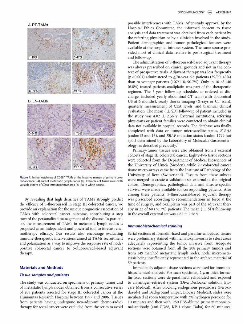
Figure 4. Immunostaining of CD68C TAMs at the invasive margin of primary colo-rectal cancer (A) and of metastatic lymph-nodes (B). Examples of tissue areas withvariable extent of CD68-immunoreative area (% IRA in white boxes).
ONCOIMMUNOLOGY e1342918-7

After exposure to MACH 4 Universal HRP Polymer (BiocareMedical) for 30 minutes at room temperature Space Import-Export, Italy) to reveal bound antibodies, 30-diaminobenzidinetetrahydrochloride was used as a chromogen (DABchromogenX50, ChemMate, Dako) and nuclei were lightly counterstained
with a freshly made hematoxylin solution (Harris Hematoxylin,DiaPath).
Staining for TANs and CD3C TILs was respectively per-formed with an anti-CD66b monoclonal antibody (cloneG10F5; BD Pharmingen, CA) in 200 specimens of primary
Table 2. Outcome predictors in 5-fluorouracil-treated patients with stage III colorectal cancer (Cox univariate analysis).
Disease-Free Survival Institutional Cohort (n D 146) Disease-Specific Survival External Validation Set (n D 60)
No. of Recurrences (%) HR (95% CI) p No. of Deaths (%) HR (95% CI) p
Densities of Tumor-Associated Macrophages (TAMs) and Neutrophils (TANs)PT-TAMsa low 40/104 (38.5) 1 (reference) 0.002 22/49 (44.9) 1 (reference) 0.05
high 4/42 (9.5) 0.20 (0.07–0.57) 1/11 (9.1) 0.14 (0.02–1.00)LN-TAMsa low 32/68 (47.1) 1 (reference) 0.001 NA
high 3/39 (7.7) 0.12 (0.04–0.41)PT-TANsb low 34/101 (33.7) 1 (reference) 0.04 NA
high 6/40 (15.0) 0.41 (0.17–0.97)Patient DemographicsAge �70 yrs. 14 /35 (40.0) 1 (reference) 0.15 5/14 (35.7) 1 (reference) 0.47
<70 yrs. 30/111 (27.0) 0.63 (0.33–1.18) 18/46 (39.1) 0.69 (0.25–1.88)Gender male 27/91 (29.7) 1 (reference) 0.84 9/27 (33.3) 1 (reference) 0.38
female 17/55 (30.9) 1.06 (0.58–1.95) 14/33 (42.4) 1.45 (0.63–3.36)Tumor PathologySite distal 28/96 (29.2) 1 (reference) 0.59 17/41 (41.5) 1 (reference) 0.71
proximal 16/50 (32.0) 1.18 (0.64–2.19) 6/19 (31.6) 0.83 (0.33–2.13)Local Invasion pT3 34/122 (27.9) 1 (reference) 0.13 17/47 (36.2) 1 (reference) 0.14
pT4 10/24 (41.7) 1.72 (0.85–3.49) 6/13 (46.1) 2.05 (0.80–5.31)Nodal Invasion N1 21/99 (15.1) 1 (reference) < 0.001 9/32 (28.1) 1 (reference) 0.03
N2 23/47 (48.9) 2.74 (1.52–4.96) 14/28 (50.0) 2.50 (1.08–5.80)Grade G1/G2 30/111 (27.0) 1 (reference) 0.13 13/38 (34.2) 1 (reference) 0.35
G3 14/35 (40.0) 1.62 (0.86–3.07) 10/22 (45.4) 1.48 (0.65–3.38)Cell-Type ADC 38/127 (29.9) 1 (reference) 0.83 11/34 (32.3) 1 (reference) 0.77
variants 6/19 (31.6) 1.10 (0.46–2.60) 12/26 (46.1) 0.88 (0.38–2.04)Density of TILsc(quartiles) 1st-to-3rd 31/107 (26.4) 1 (reference) 0.51 NA
4th 13/39 (27.8) 1.24 (0.65–2.37)Vascular Invasion no 25/104 (24.0) 1 (reference) 0.008 16/50 (32.0) 1 (reference) 0.05
yes 19/42 (45.2) 2.24 (1.23–4.08) 7/10 (70.0) 2.47 (1.01–6.01)Tumor Molecular FeaturesMS Status MSS 41/129 (31.8) 1 (reference) 0.34 22/54 (40.7) 1 (reference) 0.19
MSI 41/129 (31.8) 0.57 (0.18–1.84) 1/6 (16.7) 0.26 (0.04–1.97)BRAF wild-type 41/139 (29.5) 1 (reference) 0.27 9/24 (37.5) 1 (reference) 0.77
mutated 3/7 (42.9) 1.93 (0.60–6.24) 2/3 (66.6) 1.26 (0.27–5.96)K-RAS wild-type 27/94 (28.7) 1 (reference) 0.89 NA
mutated 14/47 (29.8) 1.05 (0.55–2.00)
NA, not availableapercent CD68-immunoreactive area at the invasive front of primary tumors (PT-TAMs) or of metastatic lymph nodes (LN-TAMs, n D 107); low and high defined by anoptimal cut-off of 8.0% for PT-TAMs and of 3.7% for LN-TAMs (see Receiver Operator Characteristic curve, Fig. 4)bpercent CD66b-immune-reactive area in the intratumoral compartment (PT-TANs, nD 141); low and high defined by an optimal cut-off of 1.16 % (see Receiver OperatorCharacteristic curve, Fig. 4)
c1st and 2nd quartiles not definable due to the absence of detectable immunoreactivity in 80 of 200 tested tissues
Table 3. Cox multivariate analysis of TAM and TAN densities, and other pathological features predicting the outcome of stage III colorectal cancer treated with 5-fluoro-uracil adjuvant therapya
Biomarker (High vs. Low) Nodal Status (N2 vs. N1) Vascular Invasion (Y vs. N)
HR (95% CI) p HR (95%CI) p HR (95%CI) p
A. Disease-Free Survival in the Institutional Cohortby PT-TAMsb 0.23 (0.08–0.65) 0.005 2.43 (1.34–4.42) 0.001 2.02 (1.11–3.68) 0.02by LN-TAMsb 0.13 (0.04–0.43) 0.001 2.35 (1.19–4.63) 0.01 2.36 (1.21–4.62) 0.01by TANsc 0.53 (0.22–1.30) 0.17 2.47 (1.31–4.67) 0.005 2.29 (1.22–4.30) 0.01B. Disease-Specific Survival in the External Validation Setby PT-TAMsb 0.14 (0.02–1.00) 0.05 2.54 (1.09–5.90) 0.03 1.57 (0.58–4.21) 0.37
aDistinct models were constructed to weight the predictive value of each candidate biomarker. Only variables with a p value less than 0.10 at univariate analysis (seeTable 3) were entered.bpercent CD68-immunoreactive area (IRA) at the invasive front of the primary tumor (PT-TAMs) or of metastatic lymph nodes (LN-TAMs). Low vs. high densities are definedby optimal cut-offs from Receiver Operator Characteristic curves (8.0% IRA for PT-TAMS and 3.7% IRA for LN-TAMs)
cpercent CD66b-immunoreactive (IRA) within the primary tumor (TANs). Low vs. high densities are defined by optimal cut-offs from Receiver Operator Characteristiccurves (1.16% IRA )
e1342918-8 A. MALESCI ET AL.
tumors and with a monoclonal antibody (clone F7.2.38, M7254;Dako) in 208 tissue sample of primary tumors, as describedpreviously13,39 (Fig. S5).
Computer-aided image analysis
An expert pathologist (M. R.) who was completely blind to clin-ical data selected 3 non-contiguous microscopic areas of CD68-stained slides. At 10x magnification, cancer tissue was to repre-sent approximately 50% of each microscopic field both for theinvasive front of primary tumors and for metastatic nodal nests(Fig. 4). A digital image (1280 £ 960 pixels, 2.21 pixel/micronresolution) covering 0.25 mm2 of each selected area, wasobtained by a 5-megapixel color camera (Olympus XC50). Anad hoc software developed at the Humanitas Research Hospital(patent WO 2005 006255), using a segmentation algorithmbased on RGB (red, green and blue) scale, was used to automat-ically measure the immune-reactive area in digitized images.13
In calculating the percent immune-reactive area (% IRA), thesystem corrected microscopic images for unfilled natural holes,vascular spaces, or histological artifacts (Fig. 5). The value of %CD68-IRA attributed to each tumor or nodal specimen was themean of values found at analysis of 3 distinct microscopicfields. The value of % CD66b-IRA for TANs was similarly cal-culated as the mean of 3 distinct intra-tumoral regions.39
In-vitro experiments
Human mononuclear cells were obtained from the peripheralblood of healthy volunteers by gradient centrifugation withHistopaque-1077 (Sigma-Aldrich, Milan, Italy). Monocyteswere isolated using the Monocyte Isolation Kit II (MACS Milte-nyi Biotec, Italy), and residual T and B cells were removed byplastic adherence. Monocytes were differentiated into macro-phages by 6-day incubation with macrophage colony-stimulat-ing factor (M-CSF, Tebu-Bio, Italy) at the concentration of100 ng/ml. Polarization of macrophages toward the M1-likephenotype was obtained on day 6 by stimulating macrophageswith interferon-gamma (IFNg, 25 ng/ml, Tebu-Bio, Italy) for20-hour and with lipopolysaccharides (LPS, 50 ng/ml) for4 hours.
HT29 and SW480 colorectal cancer cells, obtained byATCC�, were cultured in RPMI-1640 medium (Euroclone,Italy) supplemented with 10% fetal bovine serum (FBS). Afterlabeling with the fluorescent cell tracer carboxyfluorescein
diacetate succinimidyl ester (CFSE, Becton Dickinson, Italy),cells were co-cultured with M1 macrophages, at a 1:1 ratio, in10% FBS medium for 24 hours. To test the early effect of 5-fluorouracil on cell viability, 2,4-dihydroxy-5-fluoropyrimidine(5-fluorouracil, Sigma-Aldrich, Italy) at a 10 mM concentrationwas added to tumor cells alone or in co-culture with M1 macro-phages. To see whether the cytotoxic effect of macrophages wasenhanced by cell treatment with 5-fluorouracil, the superna-tants of SW480 cells treated with 20 mM 5-fluorouracil for72 hours, or of untreated cells as a control, were added in a 1:3ratio to the medium of differentiated macrophages, before 24-hour co-culturing on day 6. The rate of tumor cell death wasassessed by flow cytometry. To restrict the analysis of dead cellsto CFSE-labeled tumor cells, co-cultured macrophages werelabeled with an anti-HLA-DR-PE human antibody (cloneAC122, Miltenyi Biotec, Italy). After cell staining for annexin-V and 7-aminoactinomycin, dead tumor cells were quantifiedby a FACS Canto System (Becton Dickinson, Italy).
To assess the effect of 5-fluorouracil on macrophage polari-zation and on the expression of apoptosis-related cytokines,macrophages were exposed to 20 mM 5-fluorouracil for24 hours. For subsequent analysis of HLA-DR and CD163polarization markers, M0 macrophages were preliminary incu-bated with 1% human serum on ice for 20 minutes to block Fc-receptor sites. Cells were then washed with FACS buffer(PBS-/- C 0,5% BSA C 0,05% Sodium Azide), centrifuged at1300rpm for 5 minutes, incubated with staining antibodies(mouse anti-human CD16-PercPcy5, 3G8 clone, as isotypecontrol; HLA-DR-PE, G46–6 clone; CD163-BV421, GHI/61clone - Becton Dickinson, Italy) on ice for 20 minutes, andwashed with FACS buffer. Stained cells were finally measuredusing FACS Canto System and FlowJo single-cell analysis soft-ware. For analysis of 5-fluorouracil-induced changes in cyto-kines, M1-polarized macrophages were washed from LPS- andIFNg and then cellular extracts were tested for protein-normal-ized levels of TRAIL and TNFa by specific ELISA assays (Pro-dotti Gianni, Italy).
Statistical analysis
The Mann-Witney test was used to analyze the associations ofCD68C TAMs densities with patient demographics and withpathological or molecular features of colorectal cancer. Areceiver operating characteristic (ROC) curve was constructedto check the predictive performance of CD68-immunoreactivearea in patients with stage III cancer. Cox proportional hazardmodels were used to evaluate TAMs densities, other molecular,clinical-pathological variables and their interactions, as out-come predictors. Cox multivariate analysis was done by enter-ing only variables with a p value less than 0.10 at univariateanalysis. By a backward step-wise elimination approach, non-significant variables were removed from the model. Disease-free-survival and disease-specific survival were calculated fromdiagnosis until March 1, 2014, which was the date of data cen-soring. Kaplan-Meier curves were plotted and log-rank testwere used to analyze the survival of colorectal cancer patientsgrouped according to TAM densities. The Students’ t test wasused to compare rates of in vitro cell death in different experi-mental settings. All the statistical analyses were performed
Figure 5. Capture of immune reactive areas at image analysis. Example of com-puter assisted selection of immune reactive areas spots by red green and blue(RGB) color segmentation of the original digital microphotograph. Immune-reac-tive area, brown. Not stained tissue, yellow. The percent ratio between immunereactive areas and total area was automatically calculated.
ONCOIMMUNOLOGY e1342918-9
using STATA (version 13.1). For each statistical test, 2-sided Pvalues < 0.05 were considered statistically significant.
Author contributions
AM and LL had full access to all data in the study and take responsibility ofthe integrity of the data and the accuracy of the data analysis. Study conceptand design: LL, PB, AM. Acquisition, analysis, or interpretation of data: PB,FM, FG, GC, GB, GDC, TC, LL, LR. Drafting of the manuscript: AM, PB,LL, and SO. Critical revision of the manuscript for important intellectualcontent: AM, PB, GC, AM, SO, LL. Statistical analysis: AM, PB, LL, GC.Administrative, technical, or material support: RP, AL, VHK, MR.
Disclosure of potential conflicts of interest
No potential conflicts of interest were disclosed.
Acknowledgments
Thanks to Mrs Valentina Giatti for the help in the managing of thepatients database.
Funding
This work was supported by Associazione Italiana per la Ricerca sul Can-cro (AIRC), Investigator Grant - IG 2014,00 Number 16092 (to L.L.), andby USA National Institute of Health grant R35CA197735 (to S.O.); NodalAward (to S.O.) from Dana-Farber Harvard Cancer Center. The fundingsource did not have access to the raw data and had no role in study design;data collection, analysis, or interpretation; or writing of the report. Thecorresponding author had full access to all the data and final responsibilityfor the decision to submit the Article for publication.
ORCID
Viktor. H. Koelzer http://orcid.org/0000-0001-9206-4885
References
1. Twelves C, Wong A, Nowacki MP, Abt M, Burris H, 3rd, Carrato A,Cassidy J, Cervantes A, Fagerberg J, Georgoulias V, et al. Capecitabineas adjuvant treatment for stage III colon cancer. N Eng J Med 2005;352:2696-704; PMID:15987918; https://doi.org/10.1056/NEJMoa043116
2. Andre T, Boni C, Mounedji-Boudiaf L, Navarro M, Tabernero J, Hick-ish T, et al. Oxaliplatin, fluorouracil, and leucovorin as adjuvant treat-ment for colon cancer. N Eng J Med 2004; 350:2343-51;PMID:15175436; https://doi.org/10.1056/NEJMoa032709
3. Andre T, Boni C, Navarro M, Tabernero J, Hickish T, Topham C,Bonetti A, Clingan P, Bridgewater J, Rivera F, et al. Improved overallsurvival with oxaliplatin, fluorouracil, and leucovorin as adjuvanttreatment in stage II or III colon cancer in the MOSAIC trial. J ClinOncol 2009; 27:3109-16; PMID:19451431; https://doi.org/10.1200/JCO.2008.20.6771
4. Yothers G, O’Connell MJ, Allegra CJ, Kuebler JP, Colangelo LH, Pet-relli NJ, Wolmark N. Oxaliplatin as adjuvant therapy for colon cancer:updated results of NSABP C-07 trial, including survival and subsetanalyses. J Clin Oncol 2011; 29:3768-74; PMID:21859995; https://doi.org/10.1200/JCO.2011.36.4539
5. Alberts SR, Sargent DJ, Nair S, Mahoney MR, Mooney M, ThibodeauSN, Smyrk TC, Sinicrope FA, Chan E, Gill S, et al. Effect of oxaliplatin,fluorouracil, and leucovorin with or without cetuximab on survivalamong patients with resected stage III colon cancer: a randomizedtrial. JAMA 2012; 307:1383-93; PMID:22474202; https://doi.org/10.1001/jama.2012.385
6. Shi Q, Andre T, Grothey A, Yothers G, Hamilton SR, Bot BM, HallerDG, Van Cutsem E, Twelves C, Benedetti JK, et al. Comparison of out-comes after fluorouracil-based adjuvant therapy for stages II and IIIcolon cancer between 1978 to 1995 and 1996 to 2007: evidence of stagemigration from the ACCENT database. J Clin Oncol 2013; 31:3656-63;PMID:23980089; https://doi.org/10.1200/JCO.2013.49.4344
7. de Gramont A, Van Cutsem E, Schmoll HJ, Tabernero J, Clarke S,Moore MJ, Cunningham D, Cartwright TH, Hecht JR, Rivera F, et al.Bevacizumab plus oxaliplatin-based chemotherapy as adjuvant treat-ment for colon cancer (AVANT): a phase 3 randomised controlledtrial. Lancet Oncol 2012; 13:1225-33; PMID:23168362; https://doi.org/10.1016/S1470-2045(12)70509-0
8. Allegra CJ, Yothers G, O’Connell MJ, Sharif S, Petrelli NJ, Lopa SH,Wolmark N. Bevacizumab in stage II-III colon cancer: 5-year updateof the National Surgical Adjuvant Breast and Bowel Project C-08 trial.J Clin Oncol 2013; 31:359-64; PMID:23233715; https://doi.org/10.1200/JCO.2012.44.4711
9. Taieb J, Tabernero J, Mini E, Subtil F, Folprecht G, Van Laethem JL,Thaler J, Bridgewater J, Petersen LN, Blons H, et al. Oxaliplatin, fluo-rouracil, and leucovorin with or without cetuximab in patients withresected stage III colon cancer (PETACC-8): an open-label, rando-mised phase 3 trial. Lancet Oncol 2014; 15:862-73; PMID:24928083;https://doi.org/10.1016/S1470-2045(14)70227-X
10. Dienstmann R, Salazar R, Tabernero J. Personalizing colon canceradjuvant therapy: selecting optimal treatments for individual patients.J Clin Oncol 2015; 33:1787-96; PMID:25918287; https://doi.org/10.1200/JCO.2014.60.0213
11. Sinicrope FA, Shi Q, Smyrk TC, Thibodeau SN, Dienstmann R, Guin-ney J, Bot BM, Tejpar S, Delorenzi M, Goldberg RM, et al. Molecularmarkers identify subtypes of stage III colon cancer associated withpatient outcomes. Gastroenterology 2015; 148:88-99; PMID:25305506;https://doi.org/10.1053/j.gastro.2014.09.041
12. Liao X, Lochhead P, Nishihara R, Morikawa T, Kuchiba A, YamauchiM, Imamura Y, Qian ZR, Baba Y, Shima K, et al. Aspirin use, tumorPIK3CA mutation, and colorectal-cancer survival. N Eng J Med 2012;367:1596-606; PMID:23094721; https://doi.org/10.1056/NEJMoa1207756
13. Laghi L, Bianchi P, Miranda E, Balladore E, Pacetti V, Grizzi F, Alla-vena P, Torri V, Repici A, Santoro A, et al. CD3C cells at the invasivemargin of deeply invading (pT3-T4) colorectal cancer and risk of post-surgical metastasis: a longitudinal study. Lancet Oncol 2009; 10:877-84;PMID:19656725; https://doi.org/10.1016/S1470-2045(09)70186-X
14. Mlecnik B, Tosolini M, Kirilovsky A, Berger A, Bindea G, Meatchi T,Bruneval P, Trajanoski Z, Fridman WH, Pag�es F, et al. Histopatho-logic-based prognostic factors of colorectal cancers are associated withthe state of the local immune reaction. J Clin Oncol 2011; 29:610-8;PMID:21245428; https://doi.org/10.1200/JCO.2010.30.5425
15. Galon J, Mlecnik B, Bindea G, Angell HK, Berger A, Lagorce C, LugliA, Zlobec I, Hartmann A, Bifulco C, et al. Towards the introductionof the ‘Immunoscore’ in the classification of malignant tumours. JPathol 2014; 232:199-209; PMID:24122236; https://doi.org/10.1002/path.4287
16. Nosho K, Baba Y, Tanaka N, Shima K, Hayashi M, Meyerhardt JA,Giovannucci E, Dranoff G, Fuchs CS, Ogino S. Tumour-infiltrating T-cell subsets, molecular changes in colorectal cancer, and prognosis:cohort study and literature review. J Pathol 2010; 222:350-66;PMID:20927778; https://doi.org/10.1002/path.2774
17. Mantovani A, Allavena P, Sica A, Balkwill F. Cancer-related inflam-mation. Nature 2008; 454:436-44; PMID:18650914; https://doi.org/10.1038/nature07205
18. Coussens LM, Zitvogel L, Palucka AK. Neutralizing tumor-promotingchronic inflammation: a magic bullet? Science 2013; 339:286-91;PMID:23329041; https://doi.org/10.1126/science.1232227
19. Zhang QW, Liu L, Gong CY, Shi HS, Zeng YH, Wang XZ, Zhao YW,Wei YQ. Prognostic significance of tumor-associated macrophages insolid tumor: a meta-analysis of the literature. Plos One 2012; 7:e50946;PMID:23284651; https://doi.org/10.1371/journal.pone.0050946
20. Biswas SK, Allavena P, Mantovani A. Tumor-associated macrophages:functional diversity, clinical significance, and open questions. SeminImmunopathol 2013; 35:585-600; PMID:23657835; https://doi.org/10.1007/s00281-013-0367-7
e1342918-10 A. MALESCI ET AL.

21. Forssell J, Oberg A, Henriksson ML, Stenling R, Jung A, Palmqvist R.High macrophage infiltration along the tumor front correlates withimproved survival in colon cancer. Clin Cancer Res 2007; 13:1472-9;PMID:17332291; https://doi.org/10.1158/1078-0432.CCR-06-2073
22. Zhou Q, Peng RQ, Wu XJ, Xia Q, Hou JH, Ding Y, Zhou QM, Zhang X,Pang ZZ,WanDS, et al. The density ofmacrophages in the invasive frontis inversely correlated to liver metastasis in colon cancer. J Transl Med2010; 8:13; PMID:20141634; https://doi.org/10.1186/1479-5876-8-13
23. Algars A, Irjala H, Vaittinen S, Huhtinen H, Sundstrom J, Salmi M,Ristam€aki R, Jalkanen S. Type and location of tumor-infiltrating mac-rophages and lymphatic vessels predict survival of colorectal cancerpatients. Int J Cancer 2012; 131:864-73; PMID:21952788; https://doi.org/10.1002/ijc.26457
24. Edin S, Wikberg ML, Dahlin AM, Rutegard J, Oberg A, OldenborgPA, Palmqvist R. The distribution of macrophages with a M1 or M2phenotype in relation to prognosis and the molecular characteristicsof colorectal cancer. Plos One 2012; 7:e47045; PMID:23077543;https://doi.org/10.1371/journal.pone.0047045
25. de Jong D, Koster A, Hagenbeek A, Raemaekers J, Veldhuizen D,Heisterkamp S, de Boer JP, van Glabbeke M. Impact of the tumormicroenvironment on prognosis in follicular lymphoma is dependenton specific treatment protocols. Haematologica 2009; 94:70-7;PMID:19059937; https://doi.org/10.3324/haematol.13574
26. Taskinen M, Karjalainen-Lindsberg ML, Nyman H, Eerola LM, LeppaS. A high tumor-associated macrophage content predicts favorableoutcome in follicular lymphoma patients treated with rituximab andcyclophosphamide-doxorubicin-vincristine-prednisone. Clin CancerRes 2007; 13:5784-9; PMID:17908969; https://doi.org/10.1158/1078-0432.CCR-07-0778
27. Kridel R, Xerri L, Gelas-Dore B, Tan K, Feugier P, Vawda A, CanioniD, Farinha P, Boussetta S, Moccia AA, et al. The prognostic impact ofCD163-positive macrophages in follicular lymphoma: A study fromthe BC cancer agency and the lymphoma study association. Clin Can-cer Res 2015; 21:3428-35; PMID:25869385; https://doi.org/10.1158/1078-0432.CCR-14-3253
28. De Palma M, Lewis CE. Macrophage regulation of tumor responses toanticancer therapies. Cancer cell 2013; 23:277-86; PMID:23518347;https://doi.org/10.1016/j.ccr.2013.02.013
29. Mantovani A, Allavena P. The interaction of anticancer therapies withtumor-associated macrophages. J Exp Med 2015; 212:435-45;PMID:25753580; https://doi.org/10.1084/jem.20150295
30. Mantovani A, Marchesi F, Malesci A, Laghi L, Allavena P. Tumour-associated macrophages as treatment targets in oncology. Nat Rev ClinOncol 2017; 14(7):399-416; PMID:28117416; https://doi.org/10.1038/nrclinonc.2016.217
31. Mantovani A, Polentarutti N, Luini W, Peri G, Spreafico F. Role ofhost defense merchanisms in the antitumor activity of adriamycin anddaunomycin in mice. J Natl Cancer Inst 1979; 63:61-6; PMID:286835;https://doi.org/10.1093/jnci/63.1.61
32. Kroemer G, Galluzzi L, Kepp O, Zitvogel L. Immunogenic cell death incancer therapy. Annu Rev Immunol 2013; 31:51-72; PMID:23157435;https://doi.org/10.1146/annurev-immunol-032712-100008
33. Alizadeh D, Larmonier N. Chemotherapeutic targeting of cancer-induced immunosuppressive cells. Cancer Res 2014; 74:2663-8;PMID:24778417; https://doi.org/10.1158/0008-5472.CAN-14-0301
34. Germano G, Frapolli R, Belgiovine C, Anselmo A, Pesce S, Liguori M,Erba E, Uboldi S, Zucchetti M, Pasqualini F, et al. Role of macrophagetargeting in the antitumor activity of trabectedin. Cancer Cell 2013;23:249-62; PMID:23410977; https://doi.org/10.1016/j.ccr.2013.01.008
35. Kodumudi KN,Woan K, Gilvary DL, Sahakian E,Wei S, Djeu JY. A novelchemoimmunomodulating property of docetaxel: suppression ofmyeloid-derived suppressor cells in tumor bearers. Clin Cancer Res 2010; 16:4583-94; PMID:20702612; https://doi.org/10.1158/1078-0432.CCR-10-0733
36. Rolny C, Mazzone M, Tugues S, Laoui D, Johansson I, Coulon C,Squadrito ML, Segura I, Li X, Knevels E, et al. HRG inhibits tumorgrowth and metastasis by inducing macrophage polarization and ves-sel normalization through downregulation of PlGF. Cancer Cell 2011;19:31-44; PMID:21215706; https://doi.org/10.1016/j.ccr.2010.11.009
37. Wang B, Xu D, Yu X, Ding T, Rao H, Zhan Y, Zheng L, Li L. Associa-tion of intra-tumoral infiltrating macrophages and regulatory T cells
is an independent prognostic factor in gastric cancer after radicalresection. Ann Surg Oncol 2011; 18:2585-93; PMID:21347781; https://doi.org/10.1245/s10434-011-1609-3
38. Di Caro G, Cortese N, Castino GF, Grizzi F, Gavazzi F, Ridolfi C, et al.Dual prognostic significance of tumour-associated macrophages inhuman pancreatic adenocarcinoma treated or untreated with chemo-therapy. Gut 2016; 65(10):1710-20; PMID:26156960; https://doi.org/10.1136/gutjnl-2015-309193
39. Galdiero MR, Bianchi P, Grizzi F, Di Caro G, Basso G, Ponzetta A,Bonavita E, Barbagallo M, Tartari S, Polentarutti N, et al. Occurrenceand significance of tumor-associated neutrophils in patients with colo-rectal cancer. Int J Cancer 2016; 139:446-56; PMID:26939802; https://doi.org/10.1002/ijc.30076
40. Galdiero MR, Bonavita E, Barajon I, Garlanda C, Mantovani A, JaillonS. Tumor associated macrophages and neutrophils in cancer. Immu-nobiology 2013; 218:1402-10; PMID:23891329; https://doi.org/10.1016/j.imbio.2013.06.003
41. Shen MX, Hu PP, Donskov F, Wang GH, Liu Q, Du JJ. Tumor-associ-ated neutrophils as a new prognostic factor in cancer: A systematicreview and meta-analysis. Plos One 2014; 6; 9(6); PMID:24906014;https://doi.org/10.1371/journal.pone.0098259
42. Ohnishi K, Komohara Y, Saito Y, Miyamoto Y, Watanabe M, Baba H,Takeya M. CD169-positive macrophages in regional lymph nodes areassociated with a favorable prognosis in patients with colorectal carci-noma. Cancer science 2013; 104:1237-44; PMID:23734742; https://doi.org/10.1111/cas.12212
43. Cavnar MJ, Turcotte S, Katz SC, Kuk D, Gonen M, Shia J, Allen PJ,Balachandran VP, D’Angelica MI, Kingham TP, et al. Tumor-Associ-ated Macrophage Infiltration in Colorectal Cancer Liver Metastases isAssociated With Better Outcome. Ann Surg Oncol 2017; 24(7):1835-1842; PMID:28213791; https://doi.org/10.1245/s10434-017-5812-8
44. Galon J, Costes A, Sanchez-Cabo F, Kirilovsky A, Mlecnik B, Lagorce-Pages C, Tosolini M, Camus M, Berger A, Wind P, et al. Type, density,and location of immune cells within human colorectal tumors predictclinical outcome. Science 2006; 313:1960-4; PMID:17008531; https://doi.org/10.1126/science.1129139
45. Pages F, Berger A, Camus M, Sanchez-Cabo F, Costes A, Molidor R,Mlecnik B, Kirilovsky A, Nilsson M, Damotte D, et al. Effector mem-ory T cells, early metastasis, and survival in colorectal cancer. N Eng JMed 2005; 353:2654-66; PMID:16371631; https://doi.org/10.1056/NEJMoa051424
46. Laoui D, Van Overmeire E, Di Conza G, Aldeni C, Keirsse J, Morias Y,Movahedi K, Houbracken I, Schouppe E, Elkrim Y, et al. Tumor hypoxiadoes not drive differentiation of tumor-associated macrophages but ratherfine-tunes the M2-like macrophage population. Cancer Res 2014; 74:24-30; PMID:24220244; https://doi.org/10.1158/0008-5472.CAN-13-1196
47. Viaud S, Saccheri F, Mignot G, Yamazaki T, Daillere R, Hannani D,Enot DP, Pfirschke C, Engblom C, Pittet MJ, et al. The intestinalmicrobiota modulates the anticancer immune effects of cyclophospha-mide. Science 2013; 342:971-6; PMID:24264990; https://doi.org/10.1126/science.1240537
48. Galluzzi L, Buque A, Kepp O, Zitvogel L, Kroemer G. ImmunologicalEffects of Conventional Chemotherapy and Targeted AnticancerAgents. Cancer cell 2015; 28:690-714; PMID:26678337; https://doi.org/10.1016/j.ccell.2015.10.012
49. Tesniere A, Schlemmer F, Boige V, Kepp O, Martins I, Ghiringhelli F,Aymeric L, Michaud M, Apetoh L, Barault L, et al. Immunogenicdeath of colon cancer cells treated with oxaliplatin. Oncogene 2010;29:482-91; PMID:19881547; https://doi.org/10.1038/onc.2009.356
50. Nagasaki E, Takahara A, Koido S, Sagawa Y, Aiba K, Tajiri H, YagitaH, Homma S. Combined Treatment With Dendritic Cells and 5-fluo-rouracil Elicits Augmented NK Cell-mediated Antitumor ActivityThrough the Tumor Necrosis Factor-alpha Pathway. J Immunother2010; 33:467-74; PMID:20463601; https://doi.org/10.1097/CJI.0b013e3181d36726
51. Malesci A, Laghi L, Bianchi P, Delconte G, Randolph A, Torri V, Car-naghi C, Doci R, Rosati R, Montorsi M, et al. Reduced likelihood ofmetastases in patients with microsatellite-unstable colorectal cancer.Clin Cancer Res 2007; 13:3831-9; PMID:17606714; https://doi.org/10.1158/1078-0432.CCR-07-0366
ONCOIMMUNOLOGY e1342918-11