
Spilman Notch quin g-secretase prion
6
A -secretase inhibitor and quinacrine reduce prions and prevent dendritic degeneration in murine brains Patricia Spilman*, Pierre Lessard † , Mamta Sattavat*, Clarissa Bush*, Thomas Tousseyn* ‡ , Eric J. Huang*, Kurt Giles †§ , Todd Golde ¶ , Pritam Das ¶ , Abdul Fauq ¶ , Stanley B. Prusiner †§ , and Stephen J. DeArmond* † Departments of *Pathology and § Neurology and † Institute for Neurodegenerative Disease, University of California, San Francisco, CA 94143; ‡ Laboratory for Neuronal Cell Biology and Gene Transfer, Department for Human Genetics, Flanders Institute for Biotechnology 4, Katholieke Universiteit Leuven, 3000 Leuven, Belgium; and ¶ Department of Neuroscience, Mayo Clinic College of Medicine, Jacksonville, FL 32224 Contributed by Stanley B. Prusiner, University of California, San Francisco, CA, April 15, 2008 (sent for review December 7, 2007) In prion-infected mice, both the Notch-1 intracellular domain tran- scription factor (NICD) and the disease-causing prion protein (PrP Sc ) increase in the brain preceding dendritic atrophy and loss. Because the drug LY411575 inhibits the -secretase-catalyzed cleavage of Notch-1 that produces NICD, we asked whether this -secretase inhibitor (GSI) might prevent dendritic degeneration in mice with scrapie. At 50 d postinoculation with Rocky Mountain Laboratory (RML) prions, mice were given GSI orally for 43– 60 d. Because we did not expect GSI to produce a reduction of PrP Sc levels in brain, we added quinacrine (Qa) to the treatment regimen. Qa inhibits PrP Sc formation in cultured cells. The combination of GSI and Qa reduced PrP Sc by 95% in the neocortex and hippocampus but only 50% in the thalamus at the site of prion inoculation. The GSI plus Qa combination prevented dendritic atrophy and loss, but GSI alone did not. Even though GSI reduced NICD levels to a greater extent than GSI plus Qa, it was unable to prevent dendritic degeneration. Whether a balance between NICD and dendrite growth-stimulating factors was achieved with GSI plus Qa but not GSI alone remains to be determined. Although the combination of GSI and Qa diminished PrP Sc in the brains of RML-infected mice, GSI toxicity prevented us from being able to assess the effect the GSI plus Qa combination on incubation times. Whether less toxic GSIs can be used in place of LY411575 to prolong survival remains to be determined. Creutzfeldt–Jakob disease neuropathology prion disease therapy scrapie P rion diseases are rapidly progressive, invariably fatal neuro- degenerative disorders caused by the accumulation of PrP Sc , the pathogenic isoform of the prion protein (1–6). A wealth of evidence argues that PrP Sc is the sole component of the infec- tious prion particle. To date, no pharmacologic intervention has effectively cleared PrP Sc from the brain or prevented neurode- generation in humans with Creutzfeldt–Jakob disease (CJD) or experimental animals with scrapie (7–9). By the time most patients are diagnosed with prion disease, pronounced neuro- logical deficits are present. CJD is a rapidly progressive illness that generally leads to death in 3– 6 months. At autopsy, synaptic degeneration and nerve cell death and widespread astrocytic gliosis are found. In prion-infected rodents, neurodegeneration begins with the accumulation of PrP Sc in neuronal membranes, followed by dendritic atrophy and loss and finally nerve cell death (1, 4, 10). During studies on the pathogenesis of experimental scrapie in mice, we found that the release of the Notch-1 intracellular domain transcription factor (NICD) preceded early dendritic atrophy in scrapie-infected mice (4). Notch-1 activation re- quires multiple enzymatic cleavage steps culminating in the -secretase-mediated release of NICD (11–13). Translocation of NICD to neuronal nuclei stimulates expres- sion of genes whose gene products, such as the Hes family of proteins, inhibit expression of genes that maintain dendrites and axons, particularly during CNS development (14, 15). To test the hypothesis that PrP Sc -related activation of Notch-1 repressor signaling pathways is a major cause of early dendritic atrophy, we treated prion-infected mice with the -secretase inhibitor (GSI) LY411575 (16, 17). Toward enhancing the efficacy of the GSI, we added the drug quinacrine (Qa), which inhibits PrP Sc formation in cultured cells (18, 19). Although Qa alone modestly lowered PrP Sc levels in vivo, it failed to prolong incubation times in prion-infected, wild-type mice (20, 21). Subsequent studies revealed that the levels of Qa in the brains of these animals were too low to be therapeutic (K.G. and S.B.P., unpublished data). During studies of Qa metabolism in mice, we found that Qa efflux from the brain is controlled by multidrug-resistant (MDR) proteins, members of the P-glycoprotein ATP binding cassette transport- ers (22). Disappointingly, MDR-deficient mice accumulate high levels of Qa in their brains but fail to show prolonged survival after prion inoculation (K.G. and S.B.P., unpublished data). When GSI and Qa were administered together, they reduced PrP Sc levels 50% in the thalamus around the site of inoculation and 95% in the cortex and hippocampus. Long axonal projec- tions connect the thalamus to the cerebral cortex. GSI appeared to decrease axonal transport of PrP Sc , halting the spread of prion disease from one brain region to another. Qa alone seemed to diminish dendritic atrophy and loss. Although GSI and Qa suppressed the levels of both PrP Sc and NICD and prevented dendritic atrophy, this combination therapy did not prolong the lives of prion-inoculated mice compared with uninoculated controls due to GSI toxicity. Whether other less toxic GSIs can be used in place of LY411575 to prolong survival remains to be determined. Results Experimental Design. Wild-type, male CD1 mice were inoculated at 7 weeks of age in the right thalamus with 30 l of a 10% brain homogenate containing 10 6 ID 50 units of Rocky Mountain Laboratory (RML) prions (6). Age-matched controls were in- oculated with the same volume of a 10% brain homogenate from normal, uninfected CD1 mice. RML-inoculated CD1 mice showed clinical signs of disease beginning at 120 d postinocu- lation (dpi) [supporting information (SI) Fig. S1]. Treatment with GSI alone, Qa alone, or a combination of GSI plus Qa was begun at 50 dpi when PrP Sc accumulation was well established in the thalamus and just beginning in the neocortex and hippocam- pus (6). Both drugs were given ad libitum by the oral route mixed Author contributions: P.S., E.J.H., S.B.P., and S.J.D. designed research; P.S., P.L., C.B., and K.G. performed research; T.G., P.D., and A.F. contributed new reagents/analytic tools; M.S. and T.T. analyzed data; and P.S., S.B.P., and S.J.D. wrote the paper. The authors declare no conflict of interest. To whom correspondence may be addressed. E-mail: [email protected] or [email protected] . This article contains supporting information online at www.pnas.org/cgi/content/full/ 0803671105/DCSupplemental. © 2008 by The National Academy of Sciences of the USA www.pnas.orgcgidoi10.1073pnas.0803671105 PNAS July 29, 2008 vol. 105 no. 30 10595–10600 NEUROSCIENCE
-
Upload
patricia-spilman -
Category
Documents
-
view
15 -
download
2
Transcript of Spilman Notch quin g-secretase prion

A �-secretase inhibitor and quinacrine reduce prionsand prevent dendritic degeneration in murine brainsPatricia Spilman*, Pierre Lessard†, Mamta Sattavat*, Clarissa Bush*, Thomas Tousseyn*‡, Eric J. Huang*, Kurt Giles†§,Todd Golde¶, Pritam Das¶, Abdul Fauq¶, Stanley B. Prusiner†§�, and Stephen J. DeArmond*†�
Departments of *Pathology and §Neurology and †Institute for Neurodegenerative Disease, University of California, San Francisco, CA 94143; ‡Laboratory forNeuronal Cell Biology and Gene Transfer, Department for Human Genetics, Flanders Institute for Biotechnology 4, Katholieke Universiteit Leuven, 3000Leuven, Belgium; and ¶Department of Neuroscience, Mayo Clinic College of Medicine, Jacksonville, FL 32224
Contributed by Stanley B. Prusiner, University of California, San Francisco, CA, April 15, 2008 (sent for review December 7, 2007)
In prion-infected mice, both the Notch-1 intracellular domain tran-scription factor (NICD) and the disease-causing prion protein (PrPSc)increase in the brain preceding dendritic atrophy and loss. Becausethe drug LY411575 inhibits the �-secretase-catalyzed cleavage ofNotch-1 that produces NICD, we asked whether this �-secretaseinhibitor (GSI) might prevent dendritic degeneration in mice withscrapie. At 50 d postinoculation with Rocky Mountain Laboratory(RML) prions, mice were given GSI orally for 43–60 d. Because wedid not expect GSI to produce a reduction of PrPSc levels in brain,we added quinacrine (Qa) to the treatment regimen. Qa inhibitsPrPSc formation in cultured cells. The combination of GSI and Qareduced PrPSc by �95% in the neocortex and hippocampus but only�50% in the thalamus at the site of prion inoculation. The GSI plusQa combination prevented dendritic atrophy and loss, but GSIalone did not. Even though GSI reduced NICD levels to a greaterextent than GSI plus Qa, it was unable to prevent dendriticdegeneration. Whether a balance between NICD and dendritegrowth-stimulating factors was achieved with GSI plus Qa but notGSI alone remains to be determined. Although the combination ofGSI and Qa diminished PrPSc in the brains of RML-infected mice, GSItoxicity prevented us from being able to assess the effect the GSIplus Qa combination on incubation times. Whether less toxic GSIscan be used in place of LY411575 to prolong survival remains to bedetermined.
Creutzfeldt–Jakob disease � neuropathology � prion disease � therapy �scrapie
Prion diseases are rapidly progressive, invariably fatal neuro-degenerative disorders caused by the accumulation of PrPSc,
the pathogenic isoform of the prion protein (1–6). A wealth ofevidence argues that PrPSc is the sole component of the infec-tious prion particle. To date, no pharmacologic intervention haseffectively cleared PrPSc from the brain or prevented neurode-generation in humans with Creutzfeldt–Jakob disease (CJD) orexperimental animals with scrapie (7–9). By the time mostpatients are diagnosed with prion disease, pronounced neuro-logical deficits are present. CJD is a rapidly progressive illnessthat generally leads to death in 3–6 months. At autopsy, synapticdegeneration and nerve cell death and widespread astrocyticgliosis are found.
In prion-infected rodents, neurodegeneration begins with theaccumulation of PrPSc in neuronal membranes, followed bydendritic atrophy and loss and finally nerve cell death (1, 4, 10).During studies on the pathogenesis of experimental scrapie inmice, we found that the release of the Notch-1 intracellulardomain transcription factor (NICD) preceded early dendriticatrophy in scrapie-infected mice (4). Notch-1 activation re-quires multiple enzymatic cleavage steps culminating in the�-secretase-mediated release of NICD (11–13).
Translocation of NICD to neuronal nuclei stimulates expres-sion of genes whose gene products, such as the Hes family ofproteins, inhibit expression of genes that maintain dendrites andaxons, particularly during CNS development (14, 15). To test the
hypothesis that PrPSc-related activation of Notch-1 repressorsignaling pathways is a major cause of early dendritic atrophy, wetreated prion-infected mice with the �-secretase inhibitor (GSI)LY411575 (16, 17).
Toward enhancing the efficacy of the GSI, we added the drugquinacrine (Qa), which inhibits PrPSc formation in cultured cells(18, 19). Although Qa alone modestly lowered PrPSc levels invivo, it failed to prolong incubation times in prion-infected,wild-type mice (20, 21). Subsequent studies revealed that thelevels of Qa in the brains of these animals were too low to betherapeutic (K.G. and S.B.P., unpublished data). During studiesof Qa metabolism in mice, we found that Qa efflux from thebrain is controlled by multidrug-resistant (MDR) proteins,members of the P-glycoprotein ATP binding cassette transport-ers (22). Disappointingly, MDR-deficient mice accumulate highlevels of Qa in their brains but fail to show prolonged survivalafter prion inoculation (K.G. and S.B.P., unpublished data).
When GSI and Qa were administered together, they reducedPrPSc levels 50% in the thalamus around the site of inoculationand 95% in the cortex and hippocampus. Long axonal projec-tions connect the thalamus to the cerebral cortex. GSI appearedto decrease axonal transport of PrPSc, halting the spread of priondisease from one brain region to another. Qa alone seemed todiminish dendritic atrophy and loss. Although GSI and Qasuppressed the levels of both PrPSc and NICD and preventeddendritic atrophy, this combination therapy did not prolong thelives of prion-inoculated mice compared with uninoculatedcontrols due to GSI toxicity. Whether other less toxic GSIs canbe used in place of LY411575 to prolong survival remains to bedetermined.
ResultsExperimental Design. Wild-type, male CD1 mice were inoculatedat 7 weeks of age in the right thalamus with 30 �l of a 10% brainhomogenate containing 106 ID50 units of Rocky MountainLaboratory (RML) prions (6). Age-matched controls were in-oculated with the same volume of a 10% brain homogenate fromnormal, uninfected CD1 mice. RML-inoculated CD1 miceshowed clinical signs of disease beginning at �120 d postinocu-lation (dpi) [supporting information (SI) Fig. S1]. Treatmentwith GSI alone, Qa alone, or a combination of GSI plus Qa wasbegun at 50 dpi when PrPSc accumulation was well established inthe thalamus and just beginning in the neocortex and hippocam-pus (6). Both drugs were given ad libitum by the oral route mixed
Author contributions: P.S., E.J.H., S.B.P., and S.J.D. designed research; P.S., P.L., C.B., andK.G. performed research; T.G., P.D., and A.F. contributed new reagents/analytic tools; M.S.and T.T. analyzed data; and P.S., S.B.P., and S.J.D. wrote the paper.
The authors declare no conflict of interest.
�To whom correspondence may be addressed. E-mail: [email protected] [email protected] .
This article contains supporting information online at www.pnas.org/cgi/content/full/0803671105/DCSupplemental.
© 2008 by The National Academy of Sciences of the USA
www.pnas.org�cgi�doi�10.1073�pnas.0803671105 PNAS � July 29, 2008 � vol. 105 � no. 30 � 10595–10600
NEU
ROSC
IEN
CE

in a chocolate drink containing pulverized lab chow to mask thebitter taste of Qa. The (S)-enantiomer of LY411575 was dis-solved in DMSO to ensure uniform mixing in the chocolatedrink; the final concentration of DMSO was �0.007% in alldrink preparations unless specified. Although 100-fold higherconcentrations of DMSO were reported to reduce the rate ofPrPSc accumulation in the brain (23), we found no effect on PrPSc
levels in prion-infected mice fed the chocolate drink with 0.007%DMSO compared with those fed the chocolate drink withoutDMSO (data not shown). The doses of GSI chosen for this studywere 5 or 10 mg�kg�1�d�1; the Qa dose was 40 mg�kg�1�d�1 (24).Prion-infected, control mice fed a diet with DMSO but lackingeither GSI or Qa were designated ‘‘DMSO.’’
All currently available GSIs are accompanied by adverse sideeffects in mice, including hunched posture, hair loss, and marked(�10 g) weight loss (25). GSI at a concentration of 10mg�kg�1�d�1 combined with 40 mg�kg�1�d�1 Qa produced severeside effects by 13 d of treatment in both uninfected control andRML-infected mice, which required euthanasia (Fig. S1). InRML-infected mice and uninfected control mice given 5mg�kg�1�d�1 GSI combined with 40 mg�kg�1�d�1 Qa, six of ninemice survived 43 d of treatment. At this time point, all six
surviving mice were killed; notably, five mice were free of signsof GSI toxicity. Uninfected control mice given 5 or 10mg�kg�1�d�1 GSI alone developed the same side effects asRML-infected mice, indicating that GSI, not prion disease, wasresponsible for the observed toxicity. Given the extreme adverseeffects of 10 mg�kg�1�d�1 GSI, we used only mice given 5mg�kg�1�d�1 GSI alone or in combination with 40 mg�kg�1�d�1
Qa for the analysis reported here.
GSI and Qa Reduce PrPSc. GSI plus Qa for 43 d (93 dpi) decreasedthe levels of PrPSc in the cortex and hippocampus of RML-infected mice by �95% compared with untreated, controlDMSO mice that were inoculated intracerebrally with RMLprions, fed chocolate drink with DMSO, and killed 110 dpi(Fig. 1 A, B, D, and G). In addition to these infected controlDMSO mice, we killed some at 93 dpi as controls for thetreated group. When these control DMSO mice were com-pared with the treated group, a similar 95% reduction of PrPSc
was found in the treated mice (Fig. S2).In the thalamus, where prion replication was initiated, GSI
plus Qa treatment reduced PrPSc levels by only �50% (Fig. 1 C,D, and G). In comparison, Qa alone reduced PrPSc by only �20%
Fig. 1. Combined GSI and Qa therapy reduced PrPSc by 95% in the neocortex and hippocampus and by 50% in the thalamus from prion-infected mice. (A–C)Western immunoblots of protease-resistant PrPSc in the neocortex (A), hippocampus (B), and thalamus (C) are shown from mice given chocolate drink with 0.007%DMSO containing no drug for 60 d (DMSO), Qa alone for 60 d (Qa), GSI alone for 56 d (GSI), and GSI plus Qa for 43 d. Film exposure time was adjusted to revealPrPSc in the neocortex and hippocampus from mice treated with GSI plus Qa. Adjacent bar graphs show densitometry estimates of the relative PrPSc levels in therespective Western blots (integrated optical density units per milligram of total protein). Means and standard deviations are shown. Student’s t test probabilities(n � 3 mice each): *, P � 0.05; **, P � 0.01. (D–G) Protease-resistant PrPSc immunostained in histoblots of coronal sections of the cerebral hemisphere throughthe dorsal hippocampus where it overlies the thalamus are shown. Samples were taken from RML-infected mice at 100 dpi given chocolate drink with DMSOonly (D), Qa only (E), GSI only (F), and combined GSI plus Qa (G). CC, corpus callosum; f, fimbria of fornix; Hp, hippocampus; IC, internal capsule; Nc, neocortex;Th, thalamus.
10596 � www.pnas.org�cgi�doi�10.1073�pnas.0803671105 Spilman et al.

in the thalamus (Fig. 1 C–E) and cortex (Fig. 1 A, D, and E). GSIalone reduced PrPSc by �50% in the cortex (Fig. 1 A, D, and F)and unexpectedly increased PrPSc by �20% in the thalamus (Fig.1 C, D, and F). In the hippocampus, GSI alone and Qa alone hadno effect on PrPSc levels (Fig. 1 B and D–F). The reductions inPrPSc levels in the cortex and hippocampus with GSI plus Qawere greater than the sums of the reductions from treatmentswith either GSI or Qa alone, suggesting that the two drugs worksynergistically to decrease PrPSc. Reductions in PrPSc levels werenot due to diminished expression of PrPC because PrPC levels inthe cortex were unchanged in uninfected controls given choco-late drink with or without DMSO and those treated with GSI,Qa, or GSI plus Qa (Fig. S3). The uninfected control mice wereinoculated with brain homogenate from uninfected mice.
GSI and Qa Prevent Dendritic Degeneration. Among the earliestneurodegenerative changes in experimental scrapie are short-ening of dendrites and decreased numbers of dendritic branchesand spines (4, 10). To analyze quantitatively neurodegenerativechanges in our treated mice, we estimated the ‘‘dendrite load,’’a measure of the area of a gray matter field occupied by Golgisilver-impregnated dendrites and nerve cell bodies. With GSIplus Qa treatment, dendrite loads were similar to those ofuninfected controls or even increased (Fig. 2). As shown,degenerative changes in RML-infected, control DMSO micewere severe at 93 dpi (compare with Fig. 2 A–C) (4). Thus, thecombination of GSI and Qa prevented dendritic atrophy and lossin both the cortex and hippocampus, consistent with �95%reductions of PrPSc in the same brain regions. In the thalamus,GSI plus Qa therapy reduced PrPSc levels by only �50% (Fig.1C) yet preserved the dendrite load (Fig. 2 C and F).
NICD Levels and Dendritic Degeneration. On the basis of the findingthat synaptosomal PrPSc accumulation is temporally associatedwith both increased nuclear NICD and degeneration of dendrites
(4), we hypothesized that the accumulation of NICD causesdendritic atrophy and loss. Consistent with that hypothesis,RML-infected DMSO mice have low cortical dendrite loads andhigh NICD levels (Figs. 2D and 3A). Contrary to this hypothesis,low cortical dendrite loads and low NICD levels were found inRML-infected mice treated with GSI alone (Fig. 3). Plotting thecortical dendrite loads (Fig. 2D) as a function of cortical nuclearNICD levels (Fig. 3B) shows that the most severe loss ofdendrites occurred at extreme NICD concentrations (Fig. 3C).The lowest NICD concentration was in prion-infected micetreated with GSI alone, and the highest in prion-infected, controlDMSO mice. In contrast, intermediate levels of NICD in micetreated with either Qa alone or GSI plus Qa, which were nearuninfected control levels, were associated with the preservationof dendrites (Fig. 3C).
GSI Toxicity Dependence on Dose and Duration of Exposure. Thebeneficial effects of combining GSI with Qa to treat priondisease were marred by severe, adverse drug reactions, leadingto premature deaths: Treated, prion-infected mice died substan-tially sooner than untreated, prion-infected mice (Fig. S1 A).Administration of 10 mg�kg�1�d�1 GSI plus Qa resulted in deathafter �13 d of treatment in both infected and uninfected mice.Lower doses of GSI alone or with Qa were tolerated for longertreatment durations, but all animals eventually had to be eutha-nized within �56 d of continuous treatment (Fig. S1). Weattribute this toxicity mainly to the GSI, but Qa also seems tocontribute to the adverse effects. Infected mice treated with 5mg�kg�1�d�1 GSI alone survived longer than those given GSIplus Qa. Notably, Qa is known to affect hepatic, cardiac, andCNS function; jaundice, cardiomyopathy, slowing of electricalconduction in the heart, and seizures are among the known sideeffects (26, 27). The adverse side effects related to the GSI arelikely in part due to inhibition of Notch-1 signaling, whichencourages goblet cell differentiation in the gut and a shift
Fig. 2. Dual GSI plus Qa therapy prevented dendritic degeneration more effectively than GSI or Qa alone. (A–C) Golgi silver staining of dendrites in layer 6 ofthe cortex (A), the hippocampus CA1 region (B), and the medial nuclei of the thalamus (C) shows severe dendrite loss in infected DMSO mice and preventionof loss with GSI plus Qa treatment. (D–F) Measurements of dendrite loads in each of the three brain regions as a function of treatment are shown. Note themicroscopic field used to measure dendrite loads in the cortex spanned cortical layers 5 and 6, whereas the micrographs in A show cortex layer 6 only. Student’st test probabilities (n � 3 mice each): *, P � 0.05; **, P � 0.01. Scale bars represent 30 �m and apply to each image in the same row.
Spilman et al. PNAS � July 29, 2008 � vol. 105 � no. 30 � 10597
NEU
ROSC
IEN
CE
toward secretory function at the expense of absorptive function(16, 28, 29). Whether Qa-induced cardiac abnormalities orGSI-induced malnutrition were the immediate causes of death isbeing investigated.
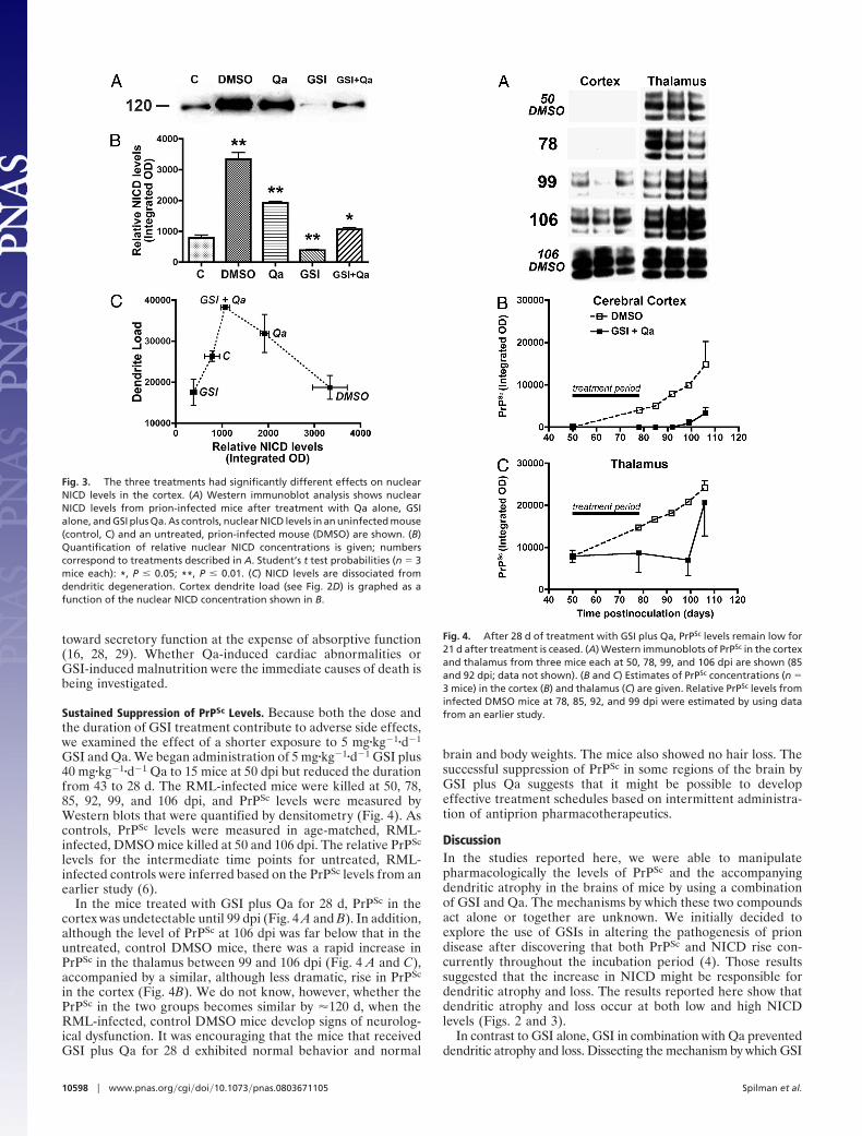
Sustained Suppression of PrPSc Levels. Because both the dose andthe duration of GSI treatment contribute to adverse side effects,we examined the effect of a shorter exposure to 5 mg�kg�1�d�1
GSI and Qa. We began administration of 5 mg�kg�1�d�1 GSI plus40 mg�kg�1�d�1 Qa to 15 mice at 50 dpi but reduced the durationfrom 43 to 28 d. The RML-infected mice were killed at 50, 78,85, 92, 99, and 106 dpi, and PrPSc levels were measured byWestern blots that were quantified by densitometry (Fig. 4). Ascontrols, PrPSc levels were measured in age-matched, RML-infected, DMSO mice killed at 50 and 106 dpi. The relative PrPSc
levels for the intermediate time points for untreated, RML-infected controls were inferred based on the PrPSc levels from anearlier study (6).
In the mice treated with GSI plus Qa for 28 d, PrPSc in thecortex was undetectable until 99 dpi (Fig. 4 A and B). In addition,although the level of PrPSc at 106 dpi was far below that in theuntreated, control DMSO mice, there was a rapid increase inPrPSc in the thalamus between 99 and 106 dpi (Fig. 4 A and C),accompanied by a similar, although less dramatic, rise in PrPSc
in the cortex (Fig. 4B). We do not know, however, whether thePrPSc in the two groups becomes similar by �120 d, when theRML-infected, control DMSO mice develop signs of neurolog-ical dysfunction. It was encouraging that the mice that receivedGSI plus Qa for 28 d exhibited normal behavior and normal
brain and body weights. The mice also showed no hair loss. Thesuccessful suppression of PrPSc in some regions of the brain byGSI plus Qa suggests that it might be possible to developeffective treatment schedules based on intermittent administra-tion of antiprion pharmacotherapeutics.
DiscussionIn the studies reported here, we were able to manipulatepharmacologically the levels of PrPSc and the accompanyingdendritic atrophy in the brains of mice by using a combinationof GSI and Qa. The mechanisms by which these two compoundsact alone or together are unknown. We initially decided toexplore the use of GSIs in altering the pathogenesis of priondisease after discovering that both PrPSc and NICD rise con-currently throughout the incubation period (4). Those resultssuggested that the increase in NICD might be responsible fordendritic atrophy and loss. The results reported here show thatdendritic atrophy and loss occur at both low and high NICDlevels (Figs. 2 and 3).
In contrast to GSI alone, GSI in combination with Qa preventeddendritic atrophy and loss. Dissecting the mechanism by which GSI
Fig. 3. The three treatments had significantly different effects on nuclearNICD levels in the cortex. (A) Western immunoblot analysis shows nuclearNICD levels from prion-infected mice after treatment with Qa alone, GSIalone, and GSI plus Qa. As controls, nuclear NICD levels in an uninfected mouse(control, C) and an untreated, prion-infected mouse (DMSO) are shown. (B)Quantification of relative nuclear NICD concentrations is given; numberscorrespond to treatments described in A. Student’s t test probabilities (n � 3mice each): *, P � 0.05; **, P � 0.01. (C) NICD levels are dissociated fromdendritic degeneration. Cortex dendrite load (see Fig. 2D) is graphed as afunction of the nuclear NICD concentration shown in B.
Fig. 4. After 28 d of treatment with GSI plus Qa, PrPSc levels remain low for21 d after treatment is ceased. (A) Western immunoblots of PrPSc in the cortexand thalamus from three mice each at 50, 78, 99, and 106 dpi are shown (85and 92 dpi; data not shown). (B and C) Estimates of PrPSc concentrations (n �3 mice) in the cortex (B) and thalamus (C) are given. Relative PrPSc levels frominfected DMSO mice at 78, 85, 92, and 99 dpi were estimated by using datafrom an earlier study.
10598 � www.pnas.org�cgi�doi�10.1073�pnas.0803671105 Spilman et al.

plus Qa prevents dendritic atrophy and loss is likely to be compli-cated (4, 30, 31). In addition to Notch-1, �26 different proteins are�-secretase substrates, some of which yield signaling peptides whencleaved (32), such as ErbB-4, which stimulates dendrite growth (33,34). Exploring the effects of Qa on �-secretase activity will be ofinterest to probe the possibility that Qa might raise the levels of oneor more growth factors that preserve dendrites or alter the ratio ofinhibitory to stimulatory factors for dendrites.
We began by administering GSIs to prion-infected mice midwaythrough the incubation period. In this study, 10 mg�kg�1�d�1
LY411575 was tolerated poorly, but the 50% lower dose allowed usto give the drug for up to 43 d. Treatment with 5 mg�kg�1�d�1 GSIalone increased the level of PrPSc in the thalamus, had no effect onthe level in hippocampus (Fig. 1C), but decreased the level of PrPSc
in the cortex by nearly 50% (Fig. 1A). Although this findingsuggested that GSI either selectively inhibited PrPSc formation orstimulated PrPSc clearance in the cortex, GSI more likely interferedwith PrPSc transport from the thalamus to the cortex. In earlierstudies, we and others demonstrated the initial replication of prionsat the site of injection with transport along axons (35, 36). In thestudies described here, PrPSc formation was initiated by intratha-lamic inoculation of prions, and presumably anterograde axonaltransport of PrPSc occurred along the monosynaptic thalamocorti-cal system of axons that connects the thalamus with the cortex (1,6, 37).
After 28 d of oral administration of 5 mg�kg�1�d�1 GSIcombined with Qa, PrPSc levels remained higher in the thalamusthan those in the cortex. Earlier studies showed that some PrPSc
is released from degenerating neurons and their processes intothe extracellular space of the CNS, where it accumulates inperivascular, subpial, and subependymal spaces (38, 39), and istaken up by activated microglia and reactive astrocytes (5, 37).Additionally, histoblots indicate that a substantial portion of theresidual thalamic PrPSc is located in large bundles of axons withinor immediately adjacent to the thalamus (Fig. 1G). Despite onlyreducing thalamic PrPSc levels by �50%, GSI plus Qa main-tained dendrite loads to control levels, raising the possibility thatPrPSc was either cleared from neurons but remained in theextracellular space or phagocytosed by glial cells, where it did notcause dendritic atrophy and loss.
From studies of Qa in cultured ScN2a cells, it appears that Qainhibits the conversion of PrPC into PrPSc. Studies with Qaattached to beads suggest that Qa binds preferentially to PrPSc
(40). As shown in Fig. 1, 40 mg�kg�1�d�1 Qa reduced PrPSc levelsminimally in the cortex and thalamus of CD1 mice but not thehippocampus. The addition of 5 mg�kg�1�d�1 GSI produced a95% decrease in PrPSc levels of in cortex and hippocampus butonly a 50% decline in the thalamus. The effect of Qa plus GSIwas much greater than the sum of the two, demonstrating thesynergistic reduction in PrPSc. Interestingly, the use of pentosanpolysulfate (PPS) combined with iron(III) meso-tetra(4-sulfonatophenyl)porphine (FeTAP) produced a synergistic pro-longation of incubation times in prion-infected mice (8). Al-though the combination of PPS and FeTAP had to beadministered intracerebrally to effect a prolongation of theincubation period, the oral administration of 4-pyridinecarbox-aldehyde-2-[4-(5-oxazolyl)phenyl]hydrazone (cpd-B) produceda doubling of the incubation time in RML-infected Tga20 micewhen started midway through the incubation period (41).
Cpd-B was the most effective in prolonging the incubationtimes of RML-infected mice but had little effect on the incuba-
tion times of either Syrian hamsters or Tg(SHaPrP)7 miceinoculated intracerebrally with 263K prions (41). Combiningcpd-B with a GSI will be of interest in the continuing search foran effective therapeutic regimen for CJD. Although the mech-anism of action of cpd-B is unclear, learning how this compounddiminishes the levels of PrPSc in cultured cells and brains ofRML-infected mice will be important. Whether such a prionstrain-specific pharmacotherapeutic can be adapted for thetreatment of CJD is unknown. Low intermittent doses of a GSImight prove useful especially if one was found to act synergis-tically with a therapeutic agent such as cpd-B, which appears tobe substantially more efficacious than Qa.
Despite the lack of success in identifying an effective therapyfor CJD, the strategy of developing compounds that reduce PrPSc
levels in cultured cells and the brains of rodents continues toseem reasonable. Disruption of the PrP gene prevents PrPSc
formation and illness in mice (42, 43). Moreover, �95% sup-pression of PrPC expression in mice harboring an inducible PrPtransgene delays the onset of neurological dysfunction, resultingin incubation times that exceed 400 d (44). The discovery ofprotease-sensitive forms of PrPSc (sPrPSc) that can cause patho-logical changes in the CNS has brought a new dimension to thestudy of therapeutics (45–49). Future investigations will need tomeasure both sPrPSc and protease-resistant PrPSc in response totherapeutics designed to treat the prion diseases (50).
Materials and MethodsAnimals and Diet. Male CD1 mice were inoculated at 7 weeks of age with 30�l of either RML prions (51) or a 10% brain homogenate from uninfected CD1mice. Inocula were injected directly into the right thalamus.
The chocolate-flavored mouse liquid diet was prepared by adding waterand cocoa powder (Hershey) to a commercially available liquid rat diet, LD’82(Bio-Serv Inc.). LY411575 was prepared in the Chemical Synthesis Core at theMayo Clinic according to the method of Fauq et al. (52) and solubilized inDMSO (Sigma–Aldrich) in a stock solution of 100 mg/ml. The final concentra-tion of DMSO in GSI-containing drink was 0.007%. Qa was obtained fromSigma. Drugs were mixed with the liquid diet. To deliver 40 mg�kg�1�d�1 Qaper mouse, 900 g of LD’82 was mixed with 4 liters of water, 50 g of cocoa, and0.315 g of Qa. DMSO also was added to the chocolate drink with Qa alone toa final concentration of 0.007%. The mice were fed ad libitum. Mice notreceiving treatment were given chocolate drink with 0.007% DMSO.
Treatment and Diagnosis. Treated animals were fed chocolate drink, preparedwith the drugs as described above. Untreated, control mice were givenchocolate drink with DMSO alone unless otherwise specified. Groups of ninemice received either DMSO alone, Qa alone at 40 mg�kg�1�d�1, GSI alone at 5mg�kg�1�d�1, or a combination of GSI and Qa at the doses specified. One groupof uninfected control mice and RML-infected mice also were given GSI at 10mg�kg�1�d�1. Treatment was initiated at 50 dpi and continued for 43 d (Figs.1–3) or 28 d (Fig. 4).
Mice were monitored twice a week for neurological signs by observingtheir gait, mobility, posture, clasping reflex, righting reflex, behavior, andbody condition. After onset of signs, they were monitored daily. Euthanasiawas performed if two or more of the following neurological signs werepresent: mild ataxia, slight head tremors, head tilt, tail rigidity, dysmetria,clasping, or circling.
Tissue Dissection, Fixation, and Analysis. These methods are described in the SIText.
ACKNOWLEDGMENTS. We thank the staff at the Hunter’s Point animalfacility, and we thank Ms. Hang Nguyen and Bernadette DeArmond, MD,MPH, for editing the manuscript. This work was funded by National Institutesof Health Grants AG10770, AG02132, AG25531, and AG021601 and by theStephen and Patricia Schott Family Fund.
1. Bouzamondo-Bernstein E, et al. (2004) The neurodegeneration sequence in priondiseases: Evidence from functional, morphological and ultrastructural studies of theGABAergic system. J Neuropathol Exp Neurol 63:882–899.
2. Cox DL, Sing RR, Yang S (2006) Prion disease: Exponential growth requires membranebinding. Biophys J 90:L77–L79.
3. Hecker R, et al. (1992) Replication of distinct scrapie prion isolates is region specific inbrains of transgenic mice and hamsters. Genes Dev 6:1213–1228.
4. Ishikura N, et al. (2005) Notch-1 activation and dendritic atrophy in prion disease. ProcNatl Acad Sci USA 102:886–891.
5. Mallucci G, et al. (2003) Depleting neuronal PrP in prion infection prevents disease andreverses spongiosis. Science 302:871–874.
Spilman et al. PNAS � July 29, 2008 � vol. 105 � no. 30 � 10599
NEU
ROSC
IEN
CE

6. Tatzelt J, Groth DF, Torchia M, Prusiner SB, DeArmond SJ (1999) Kinetics of prionprotein accumulation in the CNS of mice with experimental scrapie. J Neuropathol ExpNeurol 58:1244–1249.
7. Trevitt CR, Collinge J (2006) A systematic review of prion therapeutics in experimentalmodels. Brain 129:2241–2265.
8. Kocisko DA, Caughey B, Morrey JD, Race RE (2006) Enhanced antiscrapie effect usingcombination drug treatment. Antimicrob Agents Chemother 50:3447–3449.
9. Wisniewski T, Sigurdsson EM (2007) Therapeutic approaches for prion and Alzheimer’sdiseases. FEBS J 274:3784–3798.
10. Jeffrey M, et al. (2000) Synapse loss associated with abnormal PrP precedes neuronaldegeneration in the scrapie-infected murine hippocampus. Neuropathol Appl Neuro-biol 26:41–54.
11. De Strooper B, et al. (1999) A presenilin-1-dependent �-secretase-like protease medi-ates release of Notch intracellular domain. Nature 398:518–522.
12. Mumm JS, Kopan R (2000) Notch signaling: From the outside in. Dev Biol 228:151–165.13. Okochi M, et al. (2002) Presenilins mediate a dual intramembranous �-secretase
cleavage of Notch-1. EMBO J 21:5408–5416.14. Allen T, Lobe CG (1999) A comparison of Notch, Hes and Grg expression during murine
embryonic and post-natal development. Cell Mol Biol (Noisy-le-grand) 45:687–708.15. Kageyama R, Ohtsuka T (1999) The Notch-Hes pathway in mammalian neural devel-
opment. Cell Res 9:179–188.16. Wong GT, et al. (2004) Chronic treatment with the �-secretase inhibitor LY-411,575
inhibits �-amyloid peptide production and alters lymphopoiesis and intestinal celldifferentiation. J Biol Chem 279:12876–12882.
17. Minter LM, et al. (2005) Inhibitors of �-secretase block in vivo and in vitro T helper type1 polarization by preventing Notch upregulation of Tbx21. Nat Immunol 6:680–688.
18. Doh-ura K, Iwaki T, Caughey B (2000) Lysosomotropic agents and cysteine proteaseinhibitors inhibit scrapie-associated prion protein accumulation. J Virol 74:4894–4897.
19. Korth C, May BCH, Cohen FE, Prusiner SB (2001) Acridine and phenothiazine derivativesas pharmacotherapeutics for prion disease. Proc Natl Acad Sci USA 98:9836–9841.
20. Collins SJ, et al. (2002) Quinacrine does not prolong survival in a murine Creutzfeldt–Jakob disease model. Ann Neurol 52:503–506.
21. Barret A, et al. (2003) Evaluation of quinacrine treatment for prion diseases. J Virol77:8462–8469.
22. Huang Y, et al. (2006) Quinacrine is mainly metabolized to mono-desethyl quinacrineby CYP3A4/5 and its brain accumulation is limited by P-glycoprotein. Drug MetabDispos 34:1136–1144.
23. Shaked GM, Engelstein R, Avraham I, Kahana E, Gabizon R (2003) Dimethyl sulfoxidedelays PrPSc accumulation and disease symptoms in prion-infected hamsters. Brain Res983:137–143.
24. Yung L, et al. (2004) Pharmacokinetics of quinacrine in the treatment of prion disease.BMC Infect Dis 4:53–59.
25. Barten DM, Meredith JE, Jr, Zaczek R, Houston JG, Albright CF (2006) �-Secretaseinhibitors for Alzheimer’s disease: Balancing efficacy and toxicity. Drugs R&D 7:87–97.
26. Goodman LS, Gilman A (1955) The Pharmacological Basis of Therapeutics (Macmillan,New York).
27. Wallace DJ (2002) in Dubois’ Lupus Erythematosis, eds Wallace DJ, Hahn BH (Lippincott,Philadelphia), pp 1149–1172.
28. Milano J, et al. (2004) Modulation of notch processing by �-secretase inhibitors causesintestinal goblet cell metaplasia and induction of genes known to specify gut secretorylineage differentiation. Toxicol Sci 82:341–358.
29. van Es JH, et al. (2005) Notch/�-secretase inhibition turns proliferative cells in intestinalcrypts and adenomas into goblet cells. Nature 435:959–963.
30. Redmond L, Ghosh A (2001) The role of Notch and Rho GTPase signaling in the controlof dendritic development. Curr Opin Neurobiol 11:111–117.
31. Sestan N, Artavanis-Tsakonas S, Rakic P (1999) Contact-dependent inhibition of corticalneurite growth mediated by notch signaling. Science 286:741–746.
32. Parks AL, Curtis D (2007) Presenilin diversifies its portfolio. Trends Genet 23:140–150.33. Goldsmit Y, Erlich S, Pinkas-Kramarski R (2001) Neuregulin induces sustained reactive
oxygen species generation to mediate neuronal differentiation. Cell Mol Neurobiol21:753–769.
34. Vaskovsky A, Lupowitz Z, Erlich S, Pinkas-Kramarski R (2000) ErbB-4 activation pro-motes neurite outgrowth in PC12 cells. J Neurochem 74:979–987.
35. Taraboulos A, et al. (1992) Regional mapping of prion proteins in brains. Proc Natl AcadSci USA 89:7620–7624.
36. Brandner S, et al. (1996) Normal host prion protein necessary for scrapie-inducedneurotoxicity. Nature 379:339–343.
37. Kovacs GG, Preusser M, Strohschneider M, Budka H (2005) Subcellular localization ofdisease-associated prion protein in the human brain. Am J Pathol 166:287–294.
38. DeArmond SJ, et al. (1985) Identification of prion amyloid filaments in scrapie-infectedbrain. Cell 41:221–235.
39. DeArmond SJ, et al. (1987) Changes in the localization of brain prion proteins duringscrapie infection. Neurology 37:1271–1280.
40. Phuan P-W, et al. (2007) Discriminating between cellular and misfolded prion proteinby using affinity to 9-aminoacridine compounds. J Gen Virol 88:1392–1401.
41. Kawasaki Y, et al. (2007) Orally administered amyloidophilic compound is effective inprolonging the incubation periods of animals cerebrally infected with prion diseasesin a prion strain-dependent manner. J Virol 81:12889–12898.
42. Bueler H, et al. (1993) Mice devoid of PrP are resistant to scrapie. Cell 73:1339–1347.43. Prusiner SB, et al. (1993) Ablation of the prion protein (PrP) gene in mice prevents
scrapie and facilitates production of anti-PrP antibodies. Proc Natl Acad Sci USA90:10608–10612.
44. Safar JG, et al. (2005) Prion clearance in bigenic mice. J Gen Virol 86:2913–2923.45. Safar J, et al. (1998) Eight prion strains have PrPSc molecules with different conforma-
tions. Nat Med 4:1157–1165.46. Tzaban S, et al. (2002) Protease-sensitive scrapie prion protein in aggregates of
heterogeneous sizes. Biochemistry 41:12868–12875.47. Tremblay P, et al. (2004) Mutant PrPSc conformers induced by a synthetic peptide and
several prion strains. J Virol 78:2088–2099.48. Safar JG, et al. (2005) Diagnosis of human prion disease. Proc Natl Acad Sci USA
102:3501–3506.49. Pastrana MA, et al. (2006) Isolation and characterization of a proteinase K-sensitive
PrPSc fraction. Biochemistry 45:15710–15717.50. Colby DW, et al. (2007) Prion detection by an amyloid seeding assay. Proc Natl Acad Sci
USA 104:20914–20919.51. Chandler RL (1962) Encephalopathy in mice. Lancet 279:107–108.52. Fauq AH, Simpson K, Maharvi GM, Golde T, Das P (2007) A multigram chemical synthesis
of the �-secretase inhibitor LY411575 and its diastereoisomers. Bioorg Med Chem Lett17:6392–6395.
10600 � www.pnas.org�cgi�doi�10.1073�pnas.0803671105 Spilman et al.


