Skull base anatomy by Dr. Aditya Tiwari
59
ANATOMY OF SKULL BASE By- Dr. Aditya Tiwari, Resident, Dept. of E.N.T.
-
Upload
aditya-tiwari -
Category
Health & Medicine
-
view
2.167 -
download
0
Transcript of Skull base anatomy by Dr. Aditya Tiwari

ANATOMY OF SKULL BASE
By-Dr. Aditya Tiwari,Resident, Dept. of E.N.T.

2
The skull base represents a central and complex bone structure of the skull that forms the floor of the cranial cavity on which the brain lies.
It separates brain from facial structures and suprahyoid neck.
Anatomical knowledge of this particular region is important for under-standing several pathologic conditions as well as for planning surgical procedures.
Introduction

3
The human skull consists of three components: (1) Membranous neurocranium Forms at bones of skull (2) Cartilaginous neurocranium / chondrocranium Forms majority of skull base (3) Viscerocranium or facial skeletonThe basicranium Develops from cartilage precursors, with a
small component from membranous bone. Development of the cartilaginous skull base begins around
the 40th day of gestation, with conversion of mesenchyme into cartilage.
Occipital sclerotomal mesenchyme concentrates around the notochord & extends cephalically forming the floor of brain.
Embryology of the skull base

4
The parachordal cartilage – Around the notochord. Sclerotomal cartilage – Occipital bone. 2 hypophyseal cartilage – Fuse to form basisphenoid cartilage. 2 presphenoid cartilage – body of sphenoid. ‘Orbitosphenoid and Alisphenoid – wings of sphenoid.
The chondrification centres of skull
5
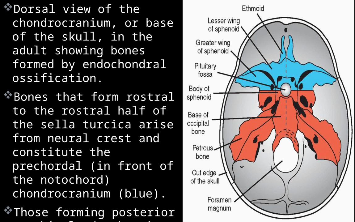
Dorsal view of the chondrocranium, or base of the skull, in the adult showing bones formed by endochondral ossification.
Bones that form rostral to the rostral half of the sella turcica arise from neural crest and constitute the prechordal (in front of the notochord) chondrocranium (blue).
Those forming posterior to this landmark arise from paraxial mesoderm (chordal chondrocranium) (red).
6
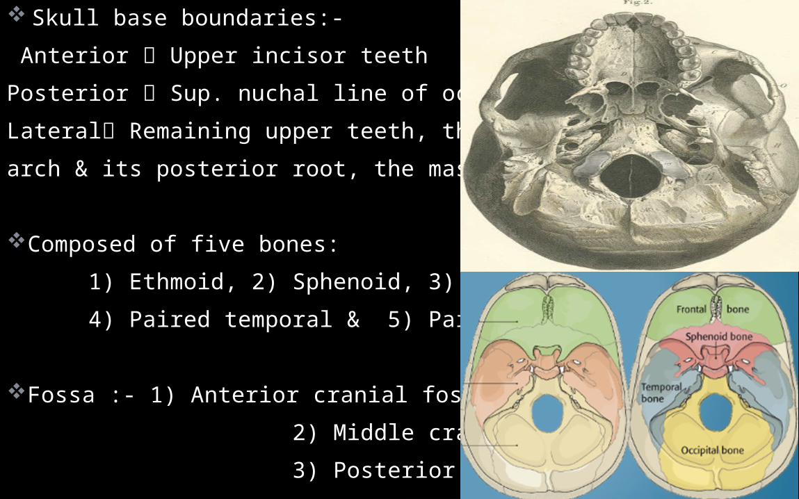
Skull base boundaries:- Anterior Upper incisor teethPosterior Sup. nuchal line of occipital bone,Lateral Remaining upper teeth, the zygomaticarch & its posterior root, the mastoid process
Composed of five bones: 1) Ethmoid, 2) Sphenoid, 3) Occipital , 4) Paired temporal & 5) Paired frontal bones
Fossa :- 1) Anterior cranial fossa, 2) Middle cranial fossa, & 3) Posterior cranial fossa.
7Anatomy skull base
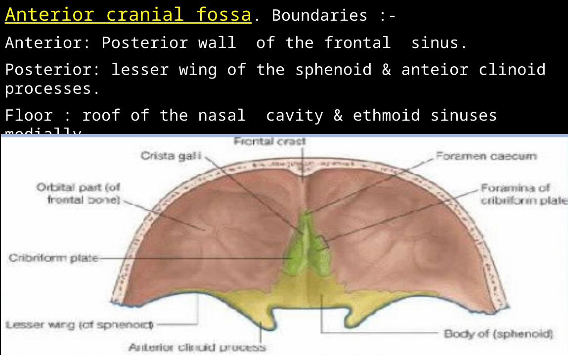
Anterior cranial fossa. Boundaries :- Anterior: Posterior wall of the frontal sinus. Posterior: lesser wing of the sphenoid & anteior clinoid processes. Floor : roof of the nasal cavity & ethmoid sinuses medially. Lateral wall : thick and strong orbital plates of the frontal bone
8Anatomy skull base
Middle cranial fossa
Post. Cranial fossa
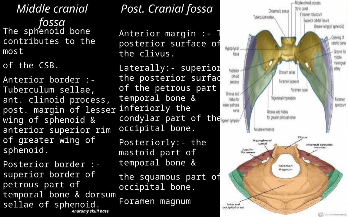
Anterior margin :- The posterior surface of the clivus. Laterally:- superiorly the posterior surface of the petrous part of temporal bone & inferiorly the condylar part of the occipital bone. Posteriorly:- the mastoid part of temporal bone & the squamous part of occipital bone. Foramen magnum
The sphenoid bone contributes to the most of the CSB.Anterior border :- Tuberculum sellae, ant. clinoid process, post. margin of lesser wing of sphenoid & anterior superior rim of greater wing of sphenoid. Posterior border :- superior border of petrous part of temporal bone & dorsum sellae of sphenoid.

9Anatomy skull base
Behind the faciomaxillary bones, its made up of the occipital bone, temporal bones and part of the sphenoid bones.
Occipital bone Has foramen magnum squamous part, petrous part & basi
occiput. Ant margin of F. magnum gives attachment to membrana
tectoria, vertical limb of the cruciform ligament, and the apical & pair of alar ligaments of the odontoid peg
Occipital condyle attaches to atlas. Ant condylar canal12th CN Post.condylar canal vein from the sigmoid sinus to the suboccipitalvenous plexus.
Osteology of the skull base

10
It shows sup & inf nuchal line and ext occipital proturbance & crest.
Each half of the occipital region is subdivided into four areas. two alongside the foramen magnum receive the recti.
Rectus capitis post minor Supplied by C1 Extend the head . Rectus capitis post major supplied by C1 extend & rotate the head.
Bwt sup & inf nuchal lines, medial area receives semispinalis capitis, supplied segmentally by post primary rami of the spinal nerves Chief extensor of the head.Lateral area, superior oblique muscle Supplied by C1 lateral flexor of the head

11Anatomy skull base
Temporal bone Its made up of squamosal, mastoid, petrous & tympanic parts
which ossify separately and later fuse creating squamotympanic & petrosquamos fissure
Has styloid process Behind its base, stylomastoid foramen & post the mastoid bone has the digastric notch, medial to which there is a groove for the occipital artery. Stylomastoid foramen transmits facial nerve.
12
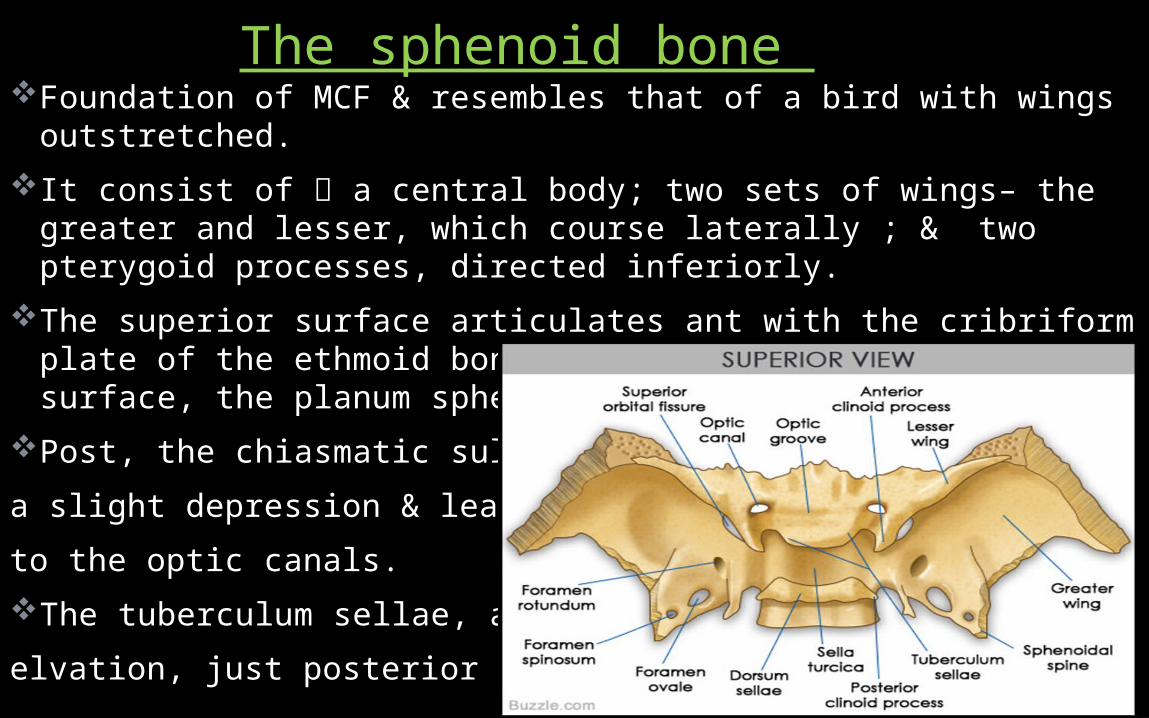
Foundation of MCF & resembles that of a bird with wings outstretched.
It consist of a central body; two sets of wings– the greater and lesser, which course laterally ; & two pterygoid processes, directed inferiorly.
The superior surface articulates ant with the cribriform plate of the ethmoid bone & contains a smooth central surface, the planum sphenoidal.
Post, the chiasmatic sulcus forms a slight depression & leads laterally to the optic canals.The tuberculum sellae, a bony elvation, just posterior to this sulcus.
The sphenoid bone

13
Followed, posteriorly by sella turcica & dorsum sellae. The dorsum sellae terminates laterally into the posterior clinoid
processes The anterior surface of the body of sphenoid forms the roof &
posterior wall of nasopharynx The body houses the sphenoid sinus .� Lesser wings� Forms medial portion of orbital apex. Greater wings – Course upward & laterally� from both sides of the sphenoid body-forms floor of MCF, posterolateral orbit & lateral calvaria.
14
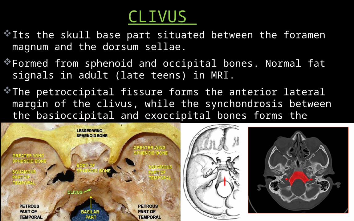
Its the skull base part situated between the foramen magnum and the dorsum sellae.
Formed from sphenoid and occipital bones. Normal fat signals in adult (late teens) in MRI.
The petroccipital fissure forms the anterior lateral margin of the clivus, while the synchondrosis between the basioccipital and exoccipital bones forms the posterior lateral margins.
CLIVUS

15
Situated centrally in the skull base forming roof of the nasopharynx, Boundaries Formed by the line of attachment of the pharyngeal wall.
The pharyngobasilar fascia is attached to the skull base and medial pterygoid plates thickened post into a pharyngeal ligament that continues inferiorly as the pharyngeal raphe.
Separated from the prevertebral muscles post by prevertebral fascia. Tubal area lies just lateral to pharyngeal area, comprises the region occupied by eustachian tube
Pharyngeal & Tubal area

16
The pharyngobasilar fascia is attached to undersurface of the tube, & two'paratubal' muscles arise one on each side of it.
The levator palati arises medially (within the pharynx) & the tensor palati arises laterally (outside the pharynx).
Both muscles are partly attached to the tube, and open it during swallowing
17
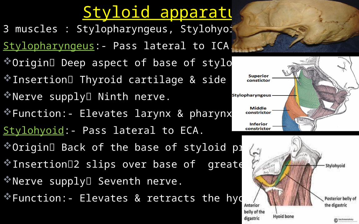
3 muscles : Stylopharyngeus, Stylohyoid, Styloglossus.
Stylopharyngeus:- Pass lateral to ICA.Origin Deep aspect of base of styloid process.Insertion Thyroid cartilage & side wall of pharynx. Nerve supply Ninth nerve.Function:- Elevates larynx & pharynx.Stylohyoid:- Pass lateral to ECA.Origin Back of the base of styloid processInsertion2 slips over base of greater cornu of hyoidNerve supply Seventh nerve.Function:- Elevates & retracts the hyoid.
Styloid apparatus

18Anatomy skull base
Styloglossus:- Pass lat. To ICA & then swings forwards med. to lingual nerve
Origin front of the styloid process &upper part of stylohyoid ligament
Insertion Side of the tongue Nerve supply Hypoglossal nerve,Function:- Retract the tongue .
19Anatomy skull base
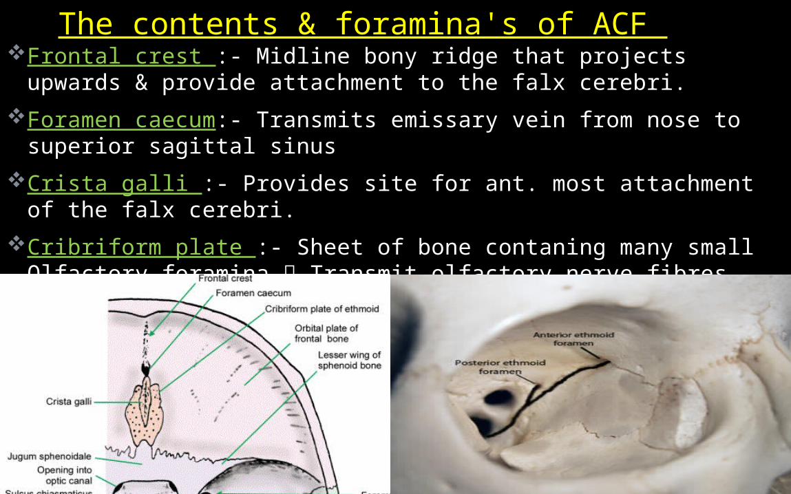
Frontal crest :- Midline bony ridge that projects upwards & provide attachment to the falx cerebri.
Foramen caecum:- Transmits emissary vein from nose to superior sagittal sinus
Crista galli :- Provides site for ant. most attachment of the falx cerebri.
Cribriform plate :- Sheet of bone contaning many small Olfactory foramina Transmit olfactory nerve fibres into the nasal cavity.
Ant & post ethmoidal foramen:-Transmits Ant & post.ethmoidal artery,nerve,vein
The contents & foramina's of ACF

20Anatomy skull base
Cavernous SinusSituated on each side of the body of sphenoid bone & extend from sup. orbital fissure ant to petrous apex post.Receives :- Sup.& inf. ophthalmic vein , Sphenoparietal sinus. Drains into:- Petrosal sinus, Pterygoid plexus, Basilar plexus.Contents:- 1) CN III, IV, V1, V2 & VI 2) ICA
The contents & foramina's of MCF Only
anatomic location in the body in which an
artery travels completely through a venous
structure

21Anatomy skull base
Triangular shaped fissure bounded med. body of sphenoid, sup. lesser wing, & inf. greater wing and is completed lat frontal bone as greater & lesser wings converge.
Optic strut separates optic canal from superior orbital fissure.Optic canal & superior orbital fissure together form the orbital
apex.Contents are as in dia-
Superior orbital fissure
Abducent nerve is most likely to damage 1st in sup. orbital fissure
syndrome
22Anatomy skull base
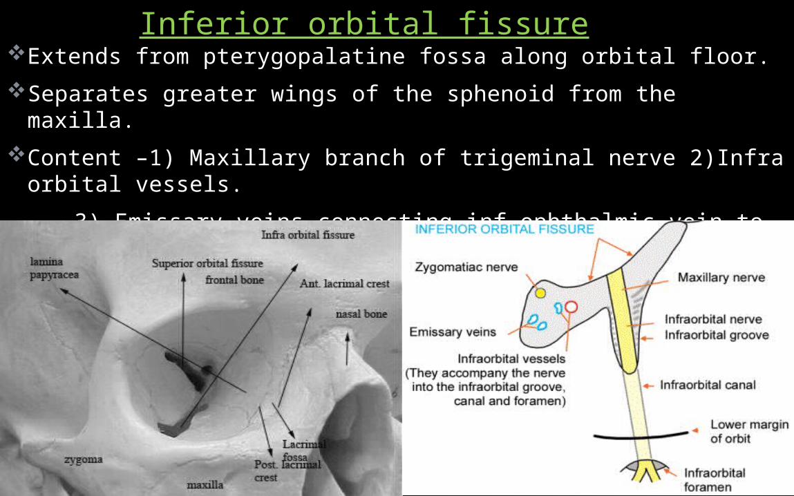
Extends from pterygopalatine fossa along orbital floor.Separates greater wings of the sphenoid from the maxilla.�Content –1) Maxillary branch of trigeminal nerve 2)Infra orbital
vessels. 3) Emissary veins connecting inf ophthalmic vein to pterygoid venous plexus. 4) Zygomatic nerve.
Inferior orbital fissure
23Anatomy skull base
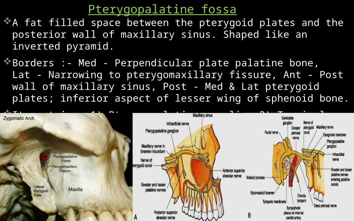
A fat filled space between the pterygoid plates and the posterior wall of maxillary sinus. Shaped like an inverted pyramid.
Borders :- Med - Perpendicular plate palatine bone, Lat - Narrowing to pterygomaxillary fissure, Ant - Post wall of maxillary sinus, Post - Med & Lat pterygoid plates; inferior aspect of lesser wing of sphenoid bone.
It contains:-1) Pterygopalatine ganglion 2) Terminal third of the maxillary artery, 3) CN V2 4) Greater & deep petrosal nerve.
Pterygopalatine fossa
24
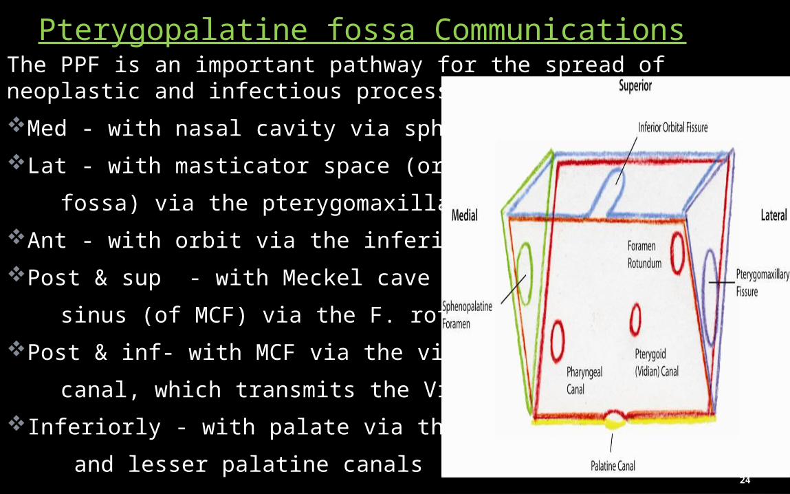
The PPF is an important pathway for the spread of neoplastic and infectious processes:Med - with nasal cavity via sphenopalatine F.Lat - with masticator space (or infratemporal fossa) via the pterygomaxillary fissure. Ant - with orbit via the inferior orbital fissure. Post & sup - with Meckel cave & cavernous sinus (of MCF) via the F. rotundum. Post & inf- with MCF via the vidian canal, which transmits the Vidian nerve.Inferiorly - with palate via the greater and lesser palatine canals
Pterygopalatine fossa Communications

25

26Anatomy skull base
Dural invagination at posterior aspect of cavernous sinus. Contains gasserian ganglion (trigeminal).
Dural layers shows thin peripheral enhancement.In MRI, 3 sensory divisions of trigeminal nerve can be visualized leaving the gasserian ganglion
Optic canal Formed by the lesser wing of sphenoid. The contents are :- Optic nerve .
Ophthalmic Artery.Sympathetic fibers from carotid plexus
MECKEL’S CAVE
27Anatomy skull base
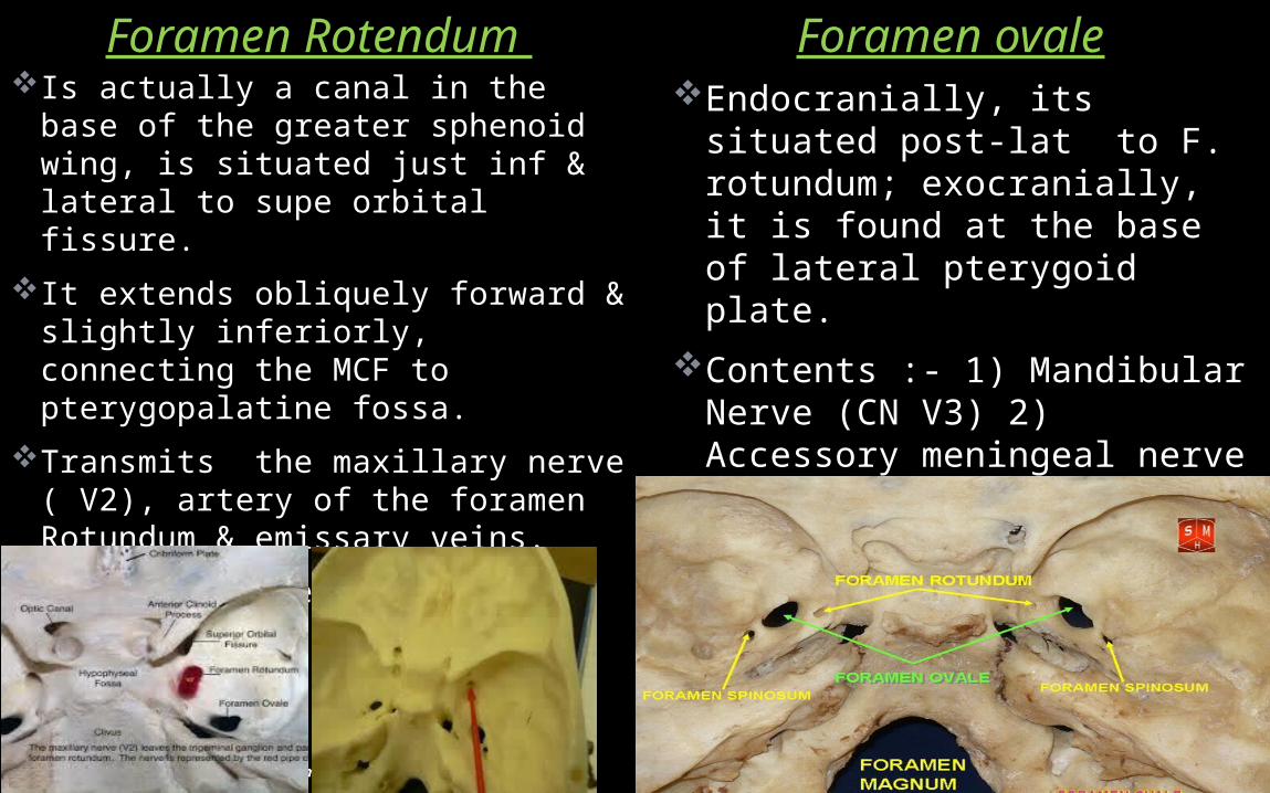
Foramen Rotendum Foramen ovaleEndocranially, its situated
post-lat to F. rotundum; exocranially, it is found at the base of lateral pterygoid plate.
Contents :- 1) Mandibular Nerve (CN V3) 2) Accessory meningeal nerve3) Lesser petrosal nerve 4)Emissary vein 5)Occasionally ant. trunk of middle meningeal artery
Is actually a canal in the base of the greater sphenoid wing, is situated just inf & lateral to supe orbital fissure.
It extends obliquely forward & slightly inferiorly, connecting the MCF to pterygopalatine fossa.
Transmits the maxillary nerve ( V2), artery of the foramen Rotundum & emissary veins.
Best visualized by means of coronal CT

28
Foramen spinosum Foramen Lacerum Its located at the base of medial
pterygoid plate, ant to the petrous apex.
Structures passing whole length:1) Meningeal branch of Ascending pharyngeal artery 2) Emissary vein
Other structures partially traversing:3) Internal carotid artery4) Greater petrosal nerve.
Its an aperture in the greater wing of the sphenoid posterolateral to foramen ovale.
Contents :- 1) Middle meningeal artery & vein. 2) Emissary vein.
3) Nervous spinosus (Meningeal branch of mandibular nerve)

29
Vidian CanalAlso c/a pterygoid canal.Located in the floor of sphenoid sinus at the junction of the
pterygoid process & the sphenoid body connecting the pterygopalatine fossa ant & the foramen lacerum posteriorly.
Contents:- 1) Vidian Artery ( Br. Of Maxillary Artery). 2) Vidian Nerve (greater superficial petrosal nerve & deep petrosal nerve )

30
Its a passage within petrous temporal bone & transmits the ICA & sympathetic plexus enters the MCF from the neck.
Its initially directed superiorly, then turns anteromedially to reach up to the petrous apex.
CAROTID SHEATH Fibrous connective tissue that surrounds vascular compartment of the neck. Layers:- Investing fascia, pretracheal fascia & prevertebral fascia. Cervical part of the sympathetic trunk is embedded in
prevertebral fascia immediately post. to sheath.
Carotid canal
31Anatomy skull base
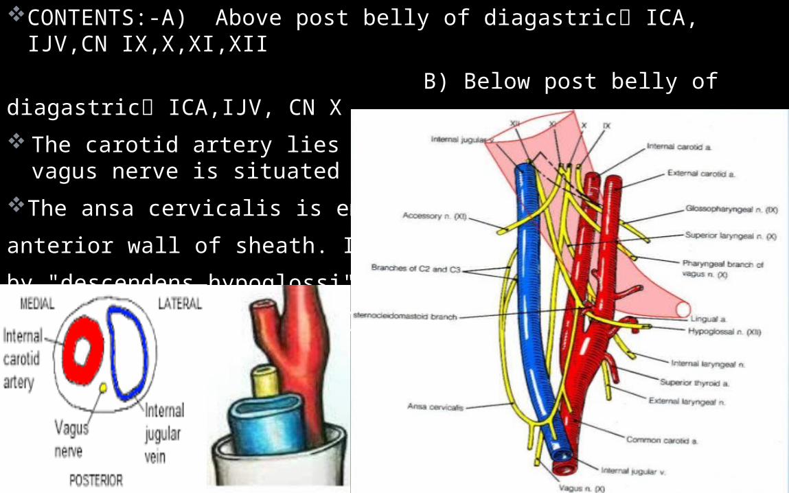
CONTENTS:-A) Above post belly of diagastric ICA, IJV,CN IX,X,XI,XII
B) Below post belly of diagastric ICA,IJV, CN X The carotid artery lies medial to the IJV, and the vagus nerve is
situated post between the two vessels.The ansa cervicalis is embedded inanterior wall of sheath. It is formed by "descendens hypoglossi" (C1) & "descendens cervicalis" (C2-C3).

32
For diagnostic imaging purposes, MCF further divided into 1) Midline sagittal, 2) Off-midline parasagittal, & 3) Lateral compartmentsBy drawing vertical lines passing medially to the petroclival fissure
(medial 2 red lines) and just lateral to the foramen ovale (lateral 2 red lines) respectively
Lateral compartment of MCF Formed by sphenoid triangle, squamous part of temporal bone, & temporomandibular joint
33Anatomy skull base
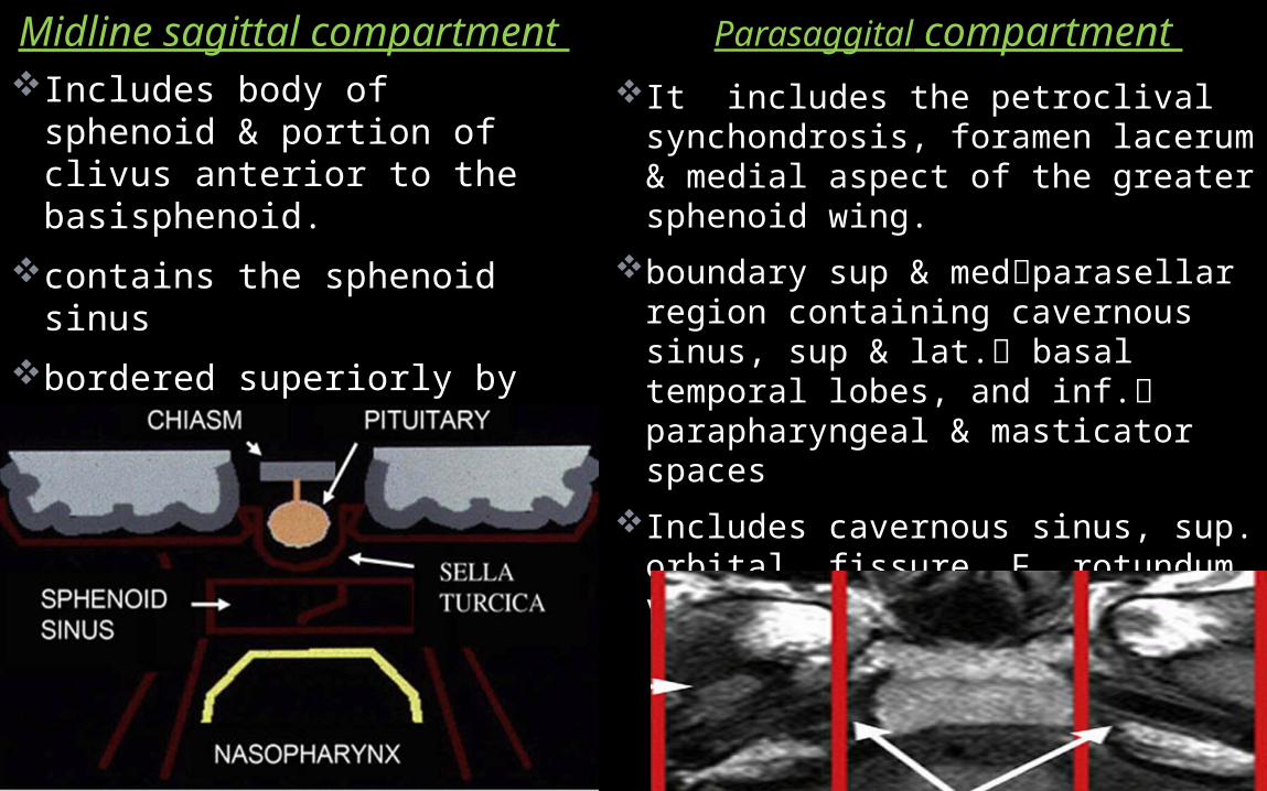
Midline sagittal compartment Parasaggital compartment It includes the petroclival
synchondrosis, foramen lacerum & medial aspect of the greater sphenoid wing.
boundary sup & medparasellar region containing cavernous sinus, sup & lat. basal temporal lobes, and inf. parapharyngeal & masticator spaces
Includes cavernous sinus, sup. orbital fissure, F. rotundum, vidian canal , & F. lacerum.
Includes body of sphenoid & portion of clivus anterior to the basisphenoid.
contains the sphenoid sinusbordered superiorly by sella
turcica and inf by roof & posterior wall of the nasopharynx
34
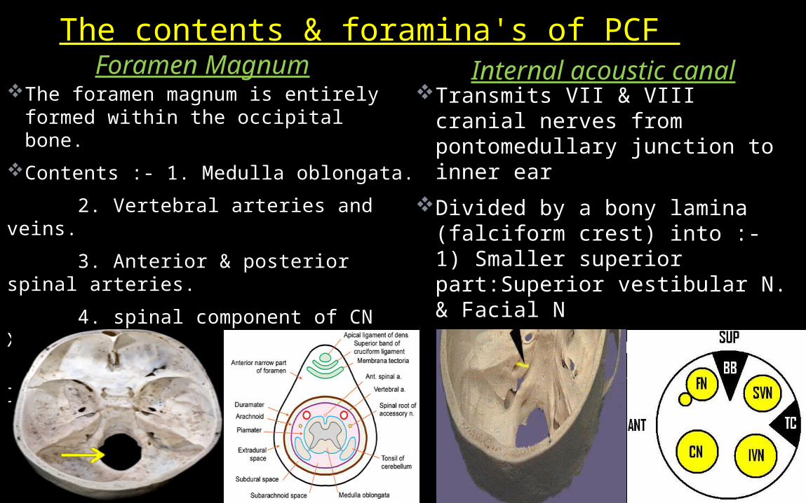
Foramen Magnum Internal acoustic canalTransmits VII & VIII cranial
nerves from pontomedullary junction to inner ear
Divided by a bony lamina (falciform crest) into :- 1) Smaller superior part:Superior vestibular N. & Facial N
2) Larger Inferior part:- Inferior vestibular N. & Cochlear nerve
The foramen magnum is entirely formed within the occipital bone.
Contents :- 1. Medulla oblongata. 2. Vertebral arteries and veins. 3. Anterior & posterior spinal arteries. 4. spinal component of CN XI. 5. Tectorial membrane & alar ligaments.
The contents & foramina's of PCF

35
Located behind the carotid canal & formed in front by petrous portion of the temporal, & behind by occipital.
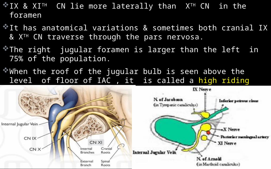
Imaging based subclassification by bony ridge, jugular spine, separate jugular foramen into 2 partsA) Pars nervosa: smaller & ant-med compart.- i) Inf petrosal sinus. Ii) Jacobson’s Nerve. B) Pars vascularis: larger & post-lat compart.- i) IJV. ii) Arnold’s nerve. iii) Spinal accessory nerve
Jugular foramen
36Anatomy skull base
IX & XITH CN lie more laterally than XTH CN in the foramenIt has anatomical variations & sometimes both cranial IX & XTH CN
traverse through the pars nervosa. The right jugular foramen is larger than the left in 75% of the
population. When the roof of the jugular bulb is seen above the level of floor
of IAC , it is called a high riding jugular bulb, which is more common on the right side.
This is a dangerous variant & exposing during translabyrinthine surgery.
37Anatomy skull base
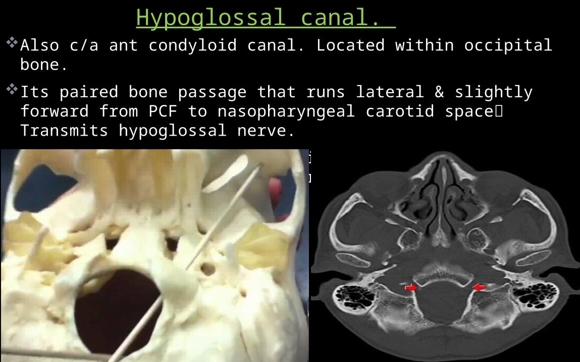
Also c/a ant condyloid canal. Located within occipital bone.Its paired bone passage that runs lateral & slightly forward from
PCF to nasopharyngeal carotid space Transmits hypoglossal nerve.
Intracanalicular enhancement is always present (emissary veins), with linear filling defects ( nerve rootlets).
Hypoglossal canal.
38
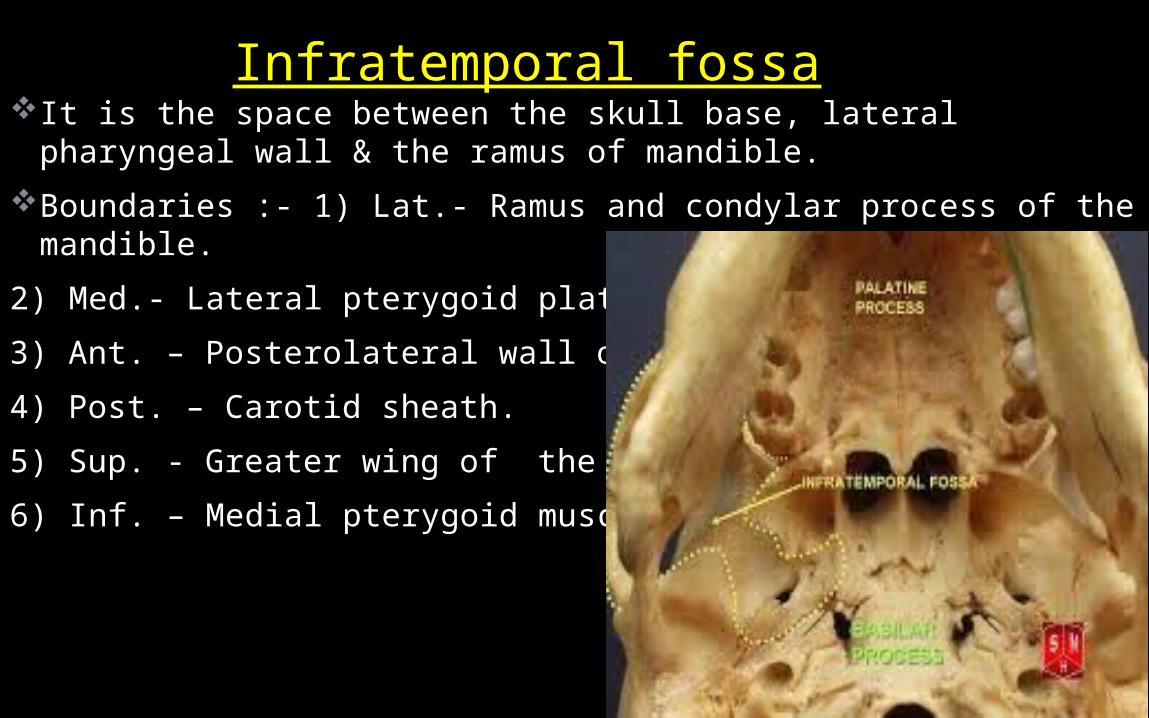
It is the space between the skull base, lateral pharyngeal wall & the ramus of mandible.
Boundaries :- 1) Lat.- Ramus and condylar process of the mandible.
2) Med.- Lateral pterygoid plate. �3) Ant. – Posterolateral wall of maxilla. 4) Post. – Carotid sheath. 5) Sup. - Greater wing of the sphenoid bone. 6) Inf. – Medial pterygoid muscles� .
Infratemporal fossa
39Anatomy skull base
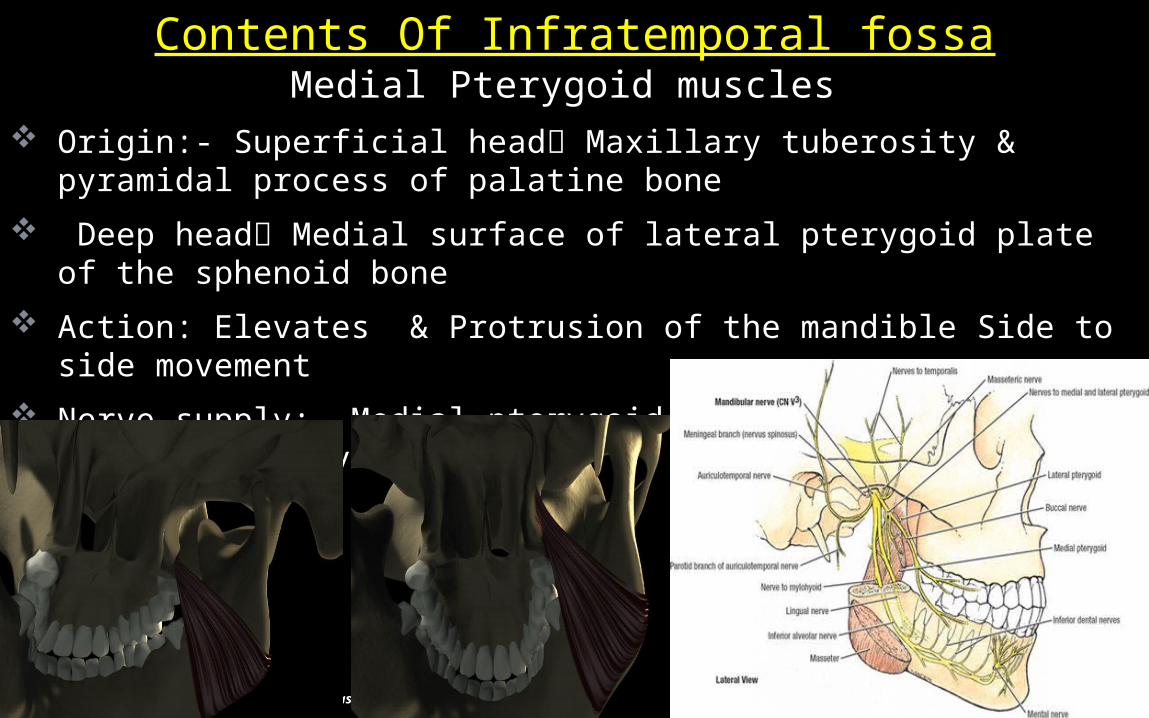
Medial Pterygoid muscles Origin:- Superficial head Maxillary tuberosity & pyramidal
process of palatine bone Deep head Medial surface of lateral pterygoid plate of the
sphenoid bone Action: Elevates & Protrusion of the mandible Side to side
movement Nerve supply:- Medial pterygoid nerve branch of mandibular
nerve Blood Supply:- Branch of mandibular nerve
Contents Of Infratemporal fossa

40Anatomy skull base
Origin:- Upper head infra-temporal surface & crest of greater wing of sphenoid
Lower head Lateral surface of the lateral pterygoid plateAction: Depression & Protrusion of the mandible, Side to side
movementNerve supply:- branches from the masseteric or buccal nerve,
branch of the ant. trunk of the mandibular nerveBlood Supply:- Pterygoid vessels from Maxillary artery
Lateral Pterygoid muscles

41Anatomy skull base
Its the larger of the 2 terminal branches of ECA Arises behind neck of the mandible Imbedded in the substance of parotid gland Pass forward bwt ramus of mandible & sphenomandibular ligament Then runs sup or deep to the lateral pterygoid muscle Pterygomaxillary fissure Pterygopalatine fossa.
MAXILLARY ARTERY

42Anatomy skull base
Described in 3 parts: before, on & beyond the lat. pterygoid muscle, with 5 branches coming from each part.
From 1ST & 3RD parts all branches enter bony foramina; from 2nd part none of the branches go through bony foramina
BRANCHES OF MAXILLARY ARTERY

43
PLEXUS:- Lies within & on lat surface of lat pterygoid muscle, & receives branches of maxillary artery.
Drains into two short, large maxillary vein to join superficial temporal vein & form retromandibular vein.
Communicating veins: 1) Inferior orbital fissure Inferior opthalmic vein
2) a connecting vein passes vertically down from the cavernous sinus 3) deep facial vein to join ant facial vein.
The pterygoid plexus & maxillary veins

44Anatomy skull base
The mandibular nerveCourse:- Lat. wall of cavernous sinus Passing through foramen
rotundum Enter pterygopalatine fossa The pterygomaxillary fissure Infratemporal fossa
Divided into 4 groups: 1) In cranium,2) In pterygopalatine fossa, 3) In infraorbital canal, 4) On the face.
In the cranium:- Middle meningeal nerve in the meninges

45Anatomy skull base
From the pterygopalatine fossa:- 1) Infraorbital nerve via Infraorbital canal 2)Zygomatic nerve (zygomaticotemporal nerve, zygomaticofacial nerve) via Inferior orbital fissure 3) Nasal Branches (nasopalatine) via Sphenopalatine foramen 4) Superior alveolar nerves (Ant, mid. & post. superior alveolar nerves)
5) Palatine Nerves (Greater & lesser palatine nerve)including the Nasopalatine nerve 6)Pharyngeal nerve
In the infraorbital canal Anterior superior alveolar nerve, Infraorbital nerve

46Anatomy skull base
Parapharyngeal , masticator, carotid & retropharyngeal spaces seen in close contact with the skull base along their cephalad aspect .
Parapharyngeal space extends caudally to the submandibular space & cranially abuts the base skull Contains fat, which acts as a medium for infection.
Relation of skull base to the deep facial spaces

47
Masticator space connects the mandible to the skull base.Odontogenic infections & oropharyngeal sq. cell ca. can tract
along masticator space to the base skull. Intracranial extension of the tumor can occur via mandibular
nerve (perineural spread) through the foramen ovale.Jugular vein thrombosis & neural tumors such as schwannoma,
neurofibromas & paraganglioma are seen in the carotid space.
Rt masseter space abscess
Perineural V3 spread via F.ovale
Rt acoustic neuroma
48
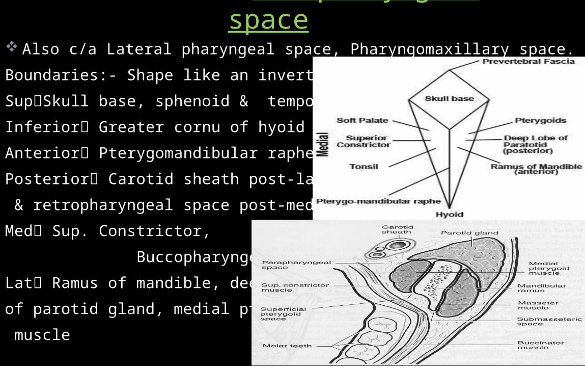
Also c/a Lateral pharyngeal space, Pharyngomaxillary space.Boundaries:- Shape like an inverted pyramidSupSkull base, sphenoid & temporal bones.Inferior Greater cornu of hyoid bone Anterior Pterygomandibular raphe.Posterior Carotid sheath post-lat. & retropharyngeal space post-med.Med Sup. Constrictor, Buccopharyngeal fasciaLat Ramus of mandible, deep lobe of parotid gland, medial pterygoid muscle
Parapharyngeal space

49
Has two compartments Prestyloid compartmentContains 2 muscles Tensor palati & Levator palati muscles2 artery Ascending palatine & ascending pharyngeal artery & int.
jugular vein. Retrostyloid compartment
It is neurovascular space, & contains the carotid sheath.
50Anatomy skull base
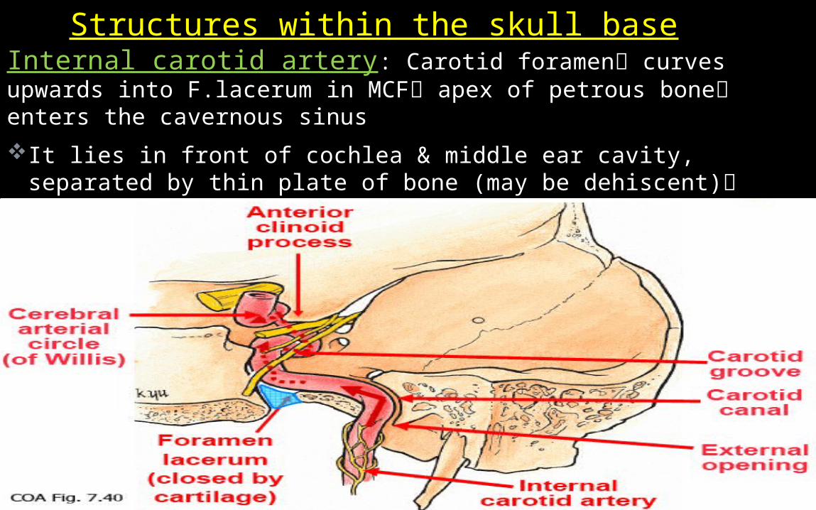
Internal carotid artery: Carotid foramen curves upwards into F.lacerum in MCF apex of petrous bone enters the cavernous sinusIt lies in front of cochlea & middle ear cavity, separated by thin
plate of bone (may be dehiscent) gives off small intrapetrous branches, including carotico-tympanic artery feeding vessels for a glomus tumour.
Structures within the skull base
51
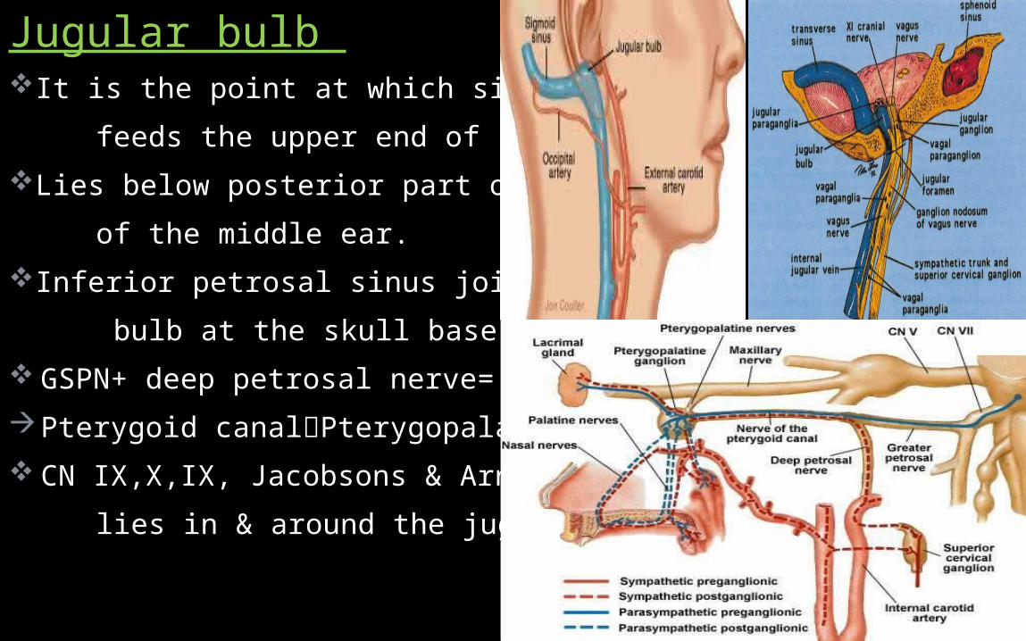
Jugular bulb It is the point at which sigmoid sinus feeds the upper end of IJV.Lies below posterior part of the floor of the middle ear.Inferior petrosal sinus joints jugular bulb at the skull base GSPN+ deep petrosal nerve= vidian N.Pterygoid canalPterygopalatine G. CN IX,X,IX, Jacobsons & Arnold nerve lies in & around the jugular foramen.
52Anatomy skull base
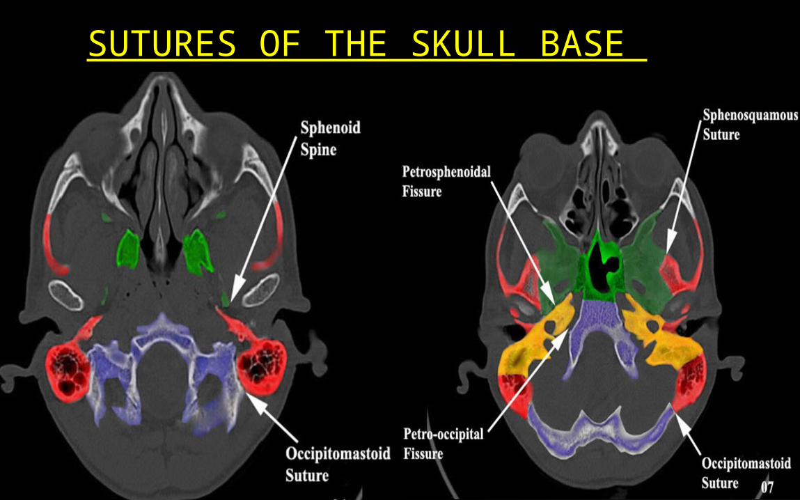
SUTURES OF THE SKULL BASE
53Anatomy skull base
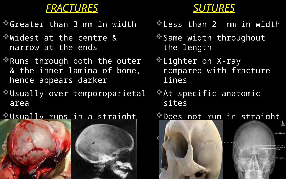
FRACTURES SUTURESLess than 2 mm in widthSame width throughout the
lengthLighter on X-ray compared
with fracture linesAt specific anatomic sitesDoes not run in straight lineCurvaceous
Greater than 3 mm in widthWidest at the centre & narrow at
the endsRuns through both the outer &
the inner lamina of bone, hence appears darker
Usually over temporoparietal area
Usually runs in a straight lineAngular turns
54Anatomy skull base
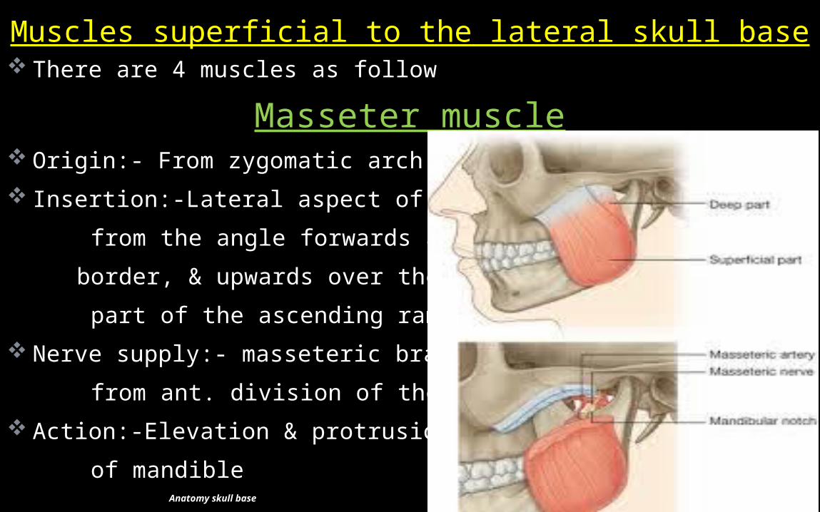
There are 4 muscles as follow
Masseter muscle Origin:- From zygomatic arch Insertion:-Lateral aspect of mandible from the angle forwards along the lower border, & upwards over the lower part of the ascending ramus. Nerve supply:- masseteric branch from ant. division of the mandibular N. Action:-Elevation & protrusion of mandible
Muscles superficial to the lateral skull base
55Anatomy skull base
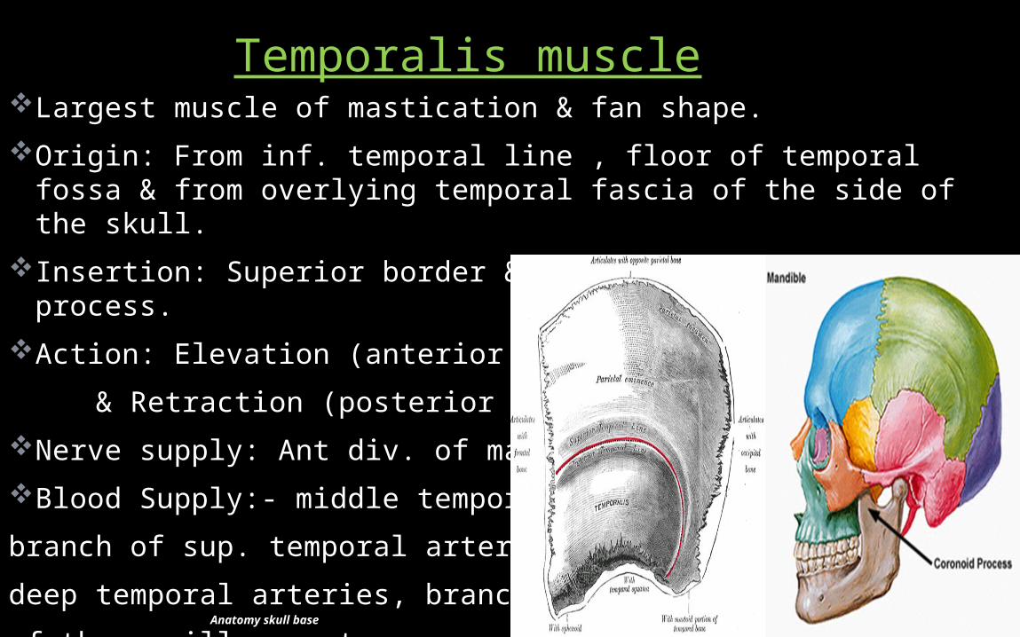
Largest muscle of mastication & fan shape.Origin: From inf. temporal line , floor of temporal fossa & from
overlying temporal fascia of the side of the skull.Insertion: Superior border & medial tip of the coronoid process.Action: Elevation (anterior fibers) & Retraction (posterior fibers)Nerve supply: Ant div. of mandibular N.Blood Supply:- middle temporal artery, branch of sup. temporal artery deep temporal arteries, branches of the maxillary artery
Temporalis muscle
56Anatomy skull base
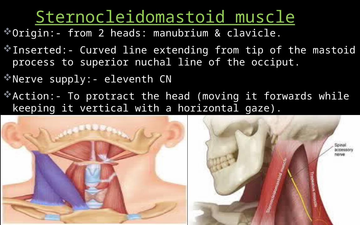
Origin:- from 2 heads: manubrium & clavicle.Inserted:- Curved line extending from tip of the mastoid process to
superior nuchal line of the occiput.Nerve supply:- eleventh CN Action:- To protract the head (moving it forwards while keeping it
vertical with a horizontal gaze).
Sternocleidomastoid muscle
57
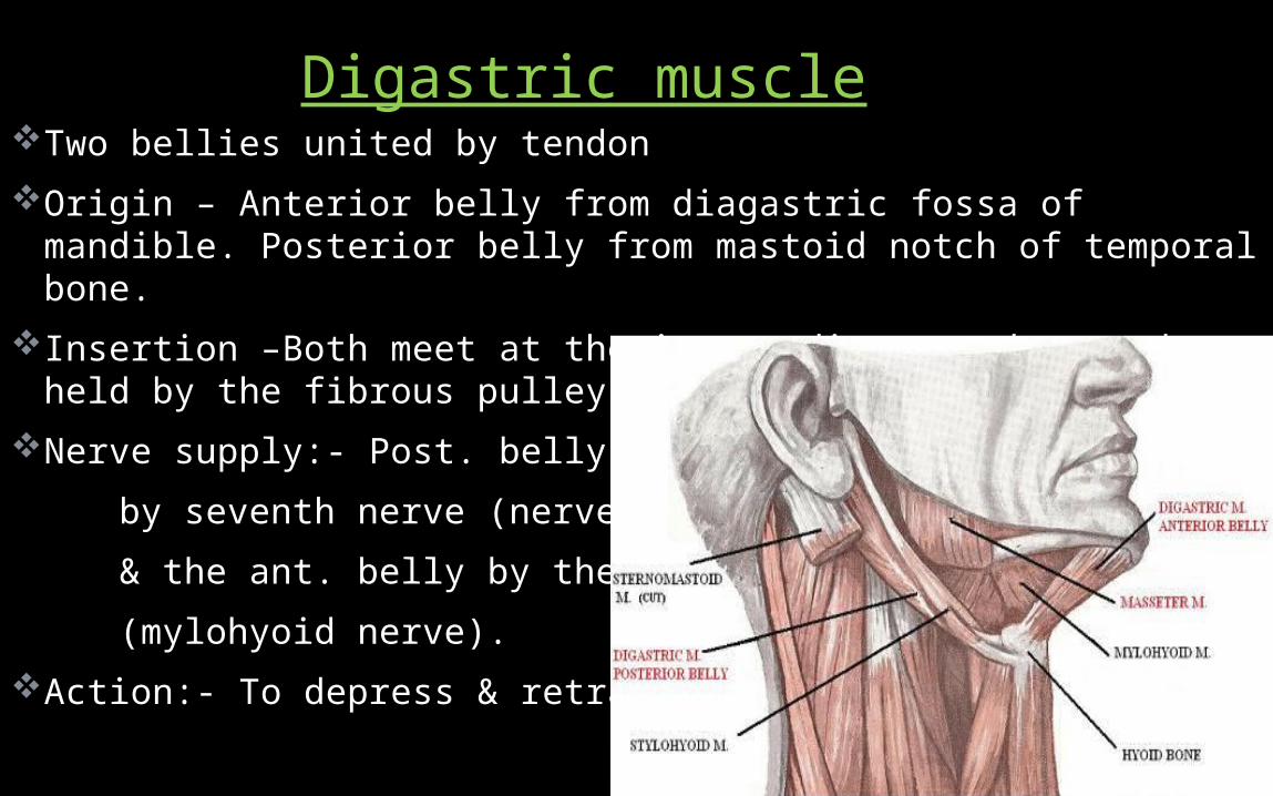
Two bellies united by tendonOrigin – Anterior belly from diagastric fossa of mandible. Posterior
belly from mastoid notch of temporal bone.Insertion –Both meet at the intermediate tendon and held by the
fibrous pulley.Nerve supply:- Post. belly is supplied by seventh nerve (nerve to digastric) & the ant. belly by the fifth nerve (mylohyoid nerve).Action:- To depress & retract the chin
Digastric muscle

58
Mastoid proces is absent It forms during 1st year as SCM muscles complete their devp.& pull on petromastoid parts of temporal bones.
Styloid process is absentStylomastoid foramen is exposed on the lateral surface of
the skull – (facial nerve is vulnerable to injury)Glabella and superciliary arches are not developed.Paranasal sinuses are rudimentary or absent, (only maxillary
sinus are usually identifiable. Frontal sinus is absent)Ext. acoustic meatus is short, straight and wholly
cartilagenousOssification is incomplete – many bones are still in several
pieces united by fibrous tissue or cartilage
Characteristics Of Fetal Skull

59Anatomy skull base