SingleMoleculeAnalysisofaRedFluorescentRecAProtein...
10
Single Molecule Analysis of a Red Fluorescent RecA Protein Reveals a Defect in Nucleoprotein Filament Nucleation That Relates to Its Reduced Biological Functions * Received for publication, April 6, 2009 Published, JBC Papers in Press, May 5, 2009, DOI 10.1074/jbc.M109.004895 Naofumi Handa ‡§¶1 , Ichiro Amitani ‡§1 , Nathan Gumlaw , Steven J. Sandler , and Stephen C. Kowalczykowski ‡§2 From the Departments of ‡ Microbiology and § Molecular and Cellular Biology, University of California, Davis, California 95616, the ¶ Department of Medical Genome Sciences, Graduate School of Frontier Sciences, University of Tokyo, Shirokanedai, Tokyo 108-8639, Japan, and the Department of Microbiology, University of Massachusetts, Amherst, Massachusetts 01003 Fluorescent fusion proteins are exceedingly useful for moni- toring protein localization in situ or visualizing protein behavior at the single molecule level. Unfortunately, some proteins are rendered inactive by the fusion. To circumvent this problem, we fused a hyperactive RecA protein (RecA803 protein) to mono- meric red fluorescent protein (mRFP1) to produce a functional protein (RecA-RFP) that is suitable for in vivo and in vitro anal- ysis. In vivo, the RecA-RFP partially restores UV resistance, con- jugational recombination, and SOS induction to recA cells. In vitro, the purified RecA-RFP protein forms a nucleoprotein fil- ament whose k cat for single-stranded DNA-dependent ATPase activity is reduced 3-fold relative to wild-type protein, and which is largely inhibited by single-stranded DNA-binding pro- tein. However, RecA protein is also a dATPase; dATP supports RecA-RFP nucleoprotein filament formation in the presence of single-stranded DNA-binding protein. Furthermore, as for the wild-type protein, the activities of RecA-RFP are further enhanced by shifting the pH to 6.2. As a consequence, RecA-RFP is proficient for DNA strand exchange with dATP or at lower pH. Finally, using single molecule visualization, RecA-RFP was seen to assemble into a continuous filament on duplex DNA, and to extend the DNA 1.7-fold. Consistent with its attenu- ated activities, RecA-RFP nucleates onto double-stranded DNA 3-fold more slowly than the wild-type protein, but still requires 3 monomers to form the rate-limited nucleus needed for filament assembly. Thus, RecA-RFP reveals that its attenu- ated biological functions correlate with a reduced frequency of nucleoprotein filament nucleation at the single molecule level. The fusion of native proteins to various fluorescent proteins has found widespread use in biology. If the fusion protein retains proper function, then the behavior and localization of the protein can be followed in living cells (1). Complementing the single-cell analysis, it is now possible to image the behavior of a fluorescent protein at the single molecule level (2– 8). How- ever, despite the growing popularity of fusion protein studies, a detailed biochemical analysis of the fusion protein is much less common, even though such examination is crucial for molecu- lar interpretations. Thus, an in vivo and in vitro analysis of the function of a fusion protein relative to the wild-type protein is an essential prerequisite. Homologous recombination is an important process not only for generating genetic variation, but also for maintaining genomic integrity through the repair of DNA breaks. In Esche- richia coli, recombinational repair of double-stranded DNA (dsDNA) 3 breaks is mediated by the RecBCD pathway, whereas the repair of ssDNA gaps is mediated by the RecF pathway (9). Both of these recombination pathways require the functions of RecA protein. RecA protein is essential to recombinational DNA repair (9 –11). RecA-like proteins are ubiquitous and highly conserved (12, 13). The ATP-bound form of the protein binds to ssDNA and polymerizes along the DNA to form an extended nucleo- protein filament (14 –16). This is the functional form of the protein that interacts with dsDNA to search for a homologous sequence. Upon finding homology, RecA protein promotes the exchange of identical DNA strands to produce the heterodu- plex joint molecules. The joint molecules can be converted into Holliday junctions and resolved by the RuvABC proteins to produce recombinant DNA products (17). The binding of RecA protein to ssDNA is competitive with the ssDNA binding (SSB) protein (18, 19). The assembly of RecA protein onto ssDNA that is complexed with SSB protein is a kinetically slow process, which is catalyzed by so-called medi- ator or loading proteins (20). RecBCD enzyme is one such RecA-loading protein (21, 22), but an additional set of loading proteins are the RecF, RecO, and RecR proteins that can form various subassemblies to facilitate the RecA-mediated displace- ment of SSB from ssDNA (23–26). In addition, a class of muta- tions that map to recA itself were isolated as suppressors of RecF function (srf) that produced mutant RecA proteins with * This work was supported, in whole or in part, by National Institutes of Health Grant GM-62653 (to S. C. K.). This work was also supported by “Grants-in- aid for Scientific Research” from the Japan Society for the Promotion of Science (JSPS) (1770001 and 19790316) and Ministry of Education, Culture, Sports, Science and Technology (MEXT) Grants 17049008 and 19037006 (to N. H.). 1 Both authors contributed equally to this work. 2 To whom correspondence should be addressed: Dept. of Microbiology, University of California, One Shields Ave., Briggs Hall, Rm. 310, Davis, CA 95616-8665. Tel.: 530-752-5938; Fax: 530-752-5939; E-mail: [email protected]. 3 The abbreviations used are: dsDNA, double-stranded DNA; RecA-RFP, RecA fused to monomeric red fluorescent protein; RecA-GFP, RecA fused to green fluorescent protein; ssDNA, single-stranded DNA; SSB, single- stranded DNA-binding protein; mRFP, monomeric red fluorescent protein; MES, 2-(N-morpholino)ethanesulfonic acid; DTT, dithiothreitol; ATPS, adenosine 5-O-(thiotriphosphate). THE JOURNAL OF BIOLOGICAL CHEMISTRY VOL. 284, NO. 28, pp. 18664 –18673, July 10, 2009 © 2009 by The American Society for Biochemistry and Molecular Biology, Inc. Printed in the U.S.A. 18664 JOURNAL OF BIOLOGICAL CHEMISTRY VOLUME 284 • NUMBER 28 • JULY 10, 2009 at University of California, Davis on July 31, 2009 www.jbc.org Downloaded from
Transcript of SingleMoleculeAnalysisofaRedFluorescentRecAProtein...
Single Molecule Analysis of a Red Fluorescent RecA ProteinReveals a Defect in Nucleoprotein Filament Nucleation ThatRelates to Its Reduced Biological Functions*
Received for publication, April 6, 2009 Published, JBC Papers in Press, May 5, 2009, DOI 10.1074/jbc.M109.004895
Naofumi Handa‡§¶1, Ichiro Amitani‡§1, Nathan Gumlaw�, Steven J. Sandler�, and Stephen C. Kowalczykowski‡§2
From the Departments of ‡Microbiology and §Molecular and Cellular Biology, University of California, Davis, California 95616, the¶Department of Medical Genome Sciences, Graduate School of Frontier Sciences, University of Tokyo, Shirokanedai, Tokyo 108-8639,Japan, and the �Department of Microbiology, University of Massachusetts, Amherst, Massachusetts 01003
Fluorescent fusion proteins are exceedingly useful for moni-toringprotein localization in situor visualizingproteinbehaviorat the single molecule level. Unfortunately, some proteins arerendered inactive by the fusion. To circumvent this problem, wefused a hyperactive RecA protein (RecA803 protein) to mono-meric red fluorescent protein (mRFP1) to produce a functionalprotein (RecA-RFP) that is suitable for in vivo and in vitro anal-ysis. In vivo, the RecA-RFPpartially restoresUV resistance, con-jugational recombination, and SOS induction to recA� cells. Invitro, the purified RecA-RFP protein forms a nucleoprotein fil-ament whose kcat for single-stranded DNA-dependent ATPaseactivity is reduced �3-fold relative to wild-type protein, andwhich is largely inhibited by single-stranded DNA-binding pro-tein. However, RecA protein is also a dATPase; dATP supportsRecA-RFP nucleoprotein filament formation in the presence ofsingle-stranded DNA-binding protein. Furthermore, as for thewild-type protein, the activities of RecA-RFP are furtherenhancedby shifting thepH to6.2.As a consequence, RecA-RFPis proficient for DNA strand exchange with dATP or at lowerpH. Finally, using single molecule visualization, RecA-RFP wasseen to assemble into a continuous filament on duplex DNA,and to extend the DNA �1.7-fold. Consistent with its attenu-ated activities, RecA-RFP nucleates onto double-stranded DNA�3-fold more slowly than the wild-type protein, but stillrequires�3monomers to form the rate-limited nucleus neededfor filament assembly. Thus, RecA-RFP reveals that its attenu-ated biological functions correlate with a reduced frequency ofnucleoprotein filament nucleation at the single molecule level.
The fusion of native proteins to various fluorescent proteinshas found widespread use in biology. If the fusion proteinretains proper function, then the behavior and localization ofthe protein can be followed in living cells (1). Complementing
the single-cell analysis, it is now possible to image the behaviorof a fluorescent protein at the singlemolecule level (2–8). How-ever, despite the growing popularity of fusion protein studies, adetailed biochemical analysis of the fusion protein is much lesscommon, even though such examination is crucial for molecu-lar interpretations. Thus, an in vivo and in vitro analysis of thefunction of a fusion protein relative to the wild-type protein isan essential prerequisite.Homologous recombination is an important process not only
for generating genetic variation, but also for maintaininggenomic integrity through the repair of DNA breaks. In Esche-richia coli, recombinational repair of double-stranded DNA(dsDNA)3 breaks ismediated by the RecBCDpathway, whereasthe repair of ssDNA gaps is mediated by the RecF pathway (9).Both of these recombination pathways require the functions ofRecA protein.RecA protein is essential to recombinational DNA repair
(9–11). RecA-like proteins are ubiquitous andhighly conserved(12, 13). The ATP-bound form of the protein binds to ssDNAand polymerizes along the DNA to form an extended nucleo-protein filament (14–16). This is the functional form of theprotein that interacts with dsDNA to search for a homologoussequence. Upon finding homology, RecA protein promotes theexchange of identical DNA strands to produce the heterodu-plex joint molecules. The joint molecules can be converted intoHolliday junctions and resolved by the RuvABC proteins toproduce recombinant DNA products (17).The binding of RecA protein to ssDNA is competitive with
the ssDNA binding (SSB) protein (18, 19). The assembly ofRecAprotein onto ssDNA that is complexedwith SSBprotein isa kinetically slow process, which is catalyzed by so-calledmedi-ator or loading proteins (20). RecBCD enzyme is one suchRecA-loading protein (21, 22), but an additional set of loadingproteins are the RecF, RecO, and RecR proteins that can formvarious subassemblies to facilitate theRecA-mediated displace-ment of SSB from ssDNA (23–26). In addition, a class of muta-tions that map to recA itself were isolated as suppressors ofRecF function (srf) that produced mutant RecA proteins with
* This work was supported, in whole or in part, by National Institutes of HealthGrant GM-62653 (to S. C. K.). This work was also supported by “Grants-in-aid for Scientific Research” from the Japan Society for the Promotion ofScience (JSPS) (1770001 and 19790316) and Ministry of Education, Culture,Sports, Science and Technology (MEXT) Grants 17049008 and 19037006(to N. H.).
1 Both authors contributed equally to this work.2 To whom correspondence should be addressed: Dept. of Microbiology,
University of California, One Shields Ave., Briggs Hall, Rm. 310, Davis,CA 95616-8665. Tel.: 530-752-5938; Fax: 530-752-5939; E-mail:[email protected].
3 The abbreviations used are: dsDNA, double-stranded DNA; RecA-RFP, RecAfused to monomeric red fluorescent protein; RecA-GFP, RecA fused togreen fluorescent protein; ssDNA, single-stranded DNA; SSB, single-stranded DNA-binding protein; mRFP, monomeric red fluorescent protein;MES, 2-(N-morpholino)ethanesulfonic acid; DTT, dithiothreitol; ATP�S,adenosine 5�-O-(thiotriphosphate).
THE JOURNAL OF BIOLOGICAL CHEMISTRY VOL. 284, NO. 28, pp. 18664 –18673, July 10, 2009© 2009 by The American Society for Biochemistry and Molecular Biology, Inc. Printed in the U.S.A.
18664 JOURNAL OF BIOLOGICAL CHEMISTRY VOLUME 284 • NUMBER 28 • JULY 10, 2009
at University of C
alifornia, Davis on July 31, 2009
ww
w.jbc.org
Dow
nloaded from
an enhanced intrinsic ability to displace SSB from ssDNA (27).One such mutant is the RecA803 protein, in which valine 37 ismutated to methionine (28, 29). This mutant RecA protein dis-plays a higher intrinsic rate of nucleoprotein filament assemblyon ssDNA, which is responsible for its enhanced capacity todisplace DNA-bound SSB protein.RecAproteinwas successfully fused to green fluorescent pro-
tein (GFP) and was visualized in living bacteria (30). The RecA-GFP protein foci were seen to appear after UV irradiation andto be dependent on the recB and recF gene products. Althoughthis protein is clearly functional in vivo, it was unfortunately,largely insoluble in vitro, thereby limiting large scale purifica-tion.4 Therefore, to facilitate biochemical use, an alternativefusion proteinwas constructed. In the present study, themono-meric red fluorescent protein (mRFP1 (31)) was fused to thecarboxyl terminus of the RecA803 protein (referred to as RecA-RFP). The hyperactive RecA803 was used because it assembleson ssDNAmore rapidly and competes better with SSB than thewild-type proteins and, as will be shown below, fusion tomRFP1 resulted in attenuated activity; thus, fusion to a hyper-active RecA protein permitted retention of at least partial func-tion. The purified fluorescent protein binds to DNA but showsattenuated ATP and dATP hydrolysis activities. Althoughnucleoprotein filament assembly is inhibited by SSB proteinunder typical reaction conditions, we found that nucleoproteinfilament formation and enzymatic activities are restored whendATP is substituted for ATP, or when the pH is lowered to 6.2.These characteristics are similar to those of the partially defec-tive RecA142mutant protein (32, 33), thereby showing that theRFP fusion converted a hypermorphic protein to a hypomor-phic RecA fusion protein. Fortunately, because the behavior ofthis RecA-RFP protein closely fits the biochemical profile of apreviously characterized mutant RecA protein, we couldunderstand its behavior. By observing assembly on single mol-ecules of dsDNA, we could see that nucleation of a RecA-RFPfilament was �3-fold slower than for the wild-type protein.Importantly, these findings lend direct single molecule supportto conclusions from ensemble studies where it was shown thatbiological function of the RecA protein correlates with its abil-ity to displace SSB protein that, in turn, is related to the rate ofRecA protein nucleation onto DNA (34).
EXPERIMENTAL PROCEDURES
Plasmids and Bacterial Strains—Plasmids derived frompBR322 encoding the fluorescent RecA proteins (pSJS1379 andpNG1) were constructed as described previously (30) and wereconfirmed by DNA sequencing. The construct on pNG1 isrecAo1403, recA803, 4151::mrfp-1. The recA-mrfp-1 constructwas made in the same manner as recA-gfp (30), with the linkerbeing amino acid residues GSI. Both plasmids carry therecAo1403 mutation, which is a promoter mutation thatincreases recA expression; plasmids pSJS1379 and pNG1 carryrecA-gfp (A206T) and recA803-mrfp-1, respectively. E. coliK12strains, AB1157 (F��� supE44 thr-1 ara-14 leuB6 �(gpt-proA)62 lacY1 tsx-33 galK2 hisG4 rfbD1mgl-51 rpsL31 kdgK51xyl-5 mtl-1 argE3 thi-1) and BIK733, a �(srl-recA)306::Tn10
derivative of AB1157 (35), were used for measurement of UVsensitivity. Strain SS2081, which is JC19328 (sulB103 lacMS286�80dII lacBK1 argE3 his-4 thi-1 xyl-5 mtl-1 SmR T6R�(srl-recA)306::Tn10 (36)) carrying pNG1, was used for RecA-RFP purification.Media—E. coli cells were grown in L broth (1.0% Bacto-tryp-
tone, 0.5% yeast extract, and 0.5% sodium chloride) (40). Anti-biotics were used as required at the following concentrations:ampicillin (amp) at 100 �g/ml; kanamycin (kan) at 50 �g/ml;and tetracycline (tet) at 10 �g/ml.RecA-RFP Protein Purification—RecA-RFP protein was puri-
fied as follows. SS2081 strain was grown overnight at 37 °C in Lbroth containing amp, kan, and tet. The cells were harvestedand stored in buffer (250 mM Tris-HCl (pH 7.5) and 25%sucrose) at 1 g/ml. Until use, the harvested cells were kept fro-zen at �80 °C. Cells were lysed using 1.3 mg/ml lysozyme and0.4% Brij-35 in the presence of 0.5 mM phenylmethylsulfonylfluoride, and then centrifuged in a BeckmanTi45 at 41,000 rpmfor 45min. To the cleared lysate, ammonium sulfate (Am2SO4)was added to 35% saturation. After centrifugation, the superna-tant was recovered and the fluorescent proteinwas precipitatedby addition of Am2SO4 to 45% saturation. After centrifugation,the pellet was dissolved in R buffer (20 mM Tris-HCl (pH 7.5),10% glycerol, 0.1 mMDTT, and 0.1 mM EDTA), and the samplewas dialyzed against R buffer overnight. The solution wasloaded onto a Q-Sepharose column (GE) equilibrated with Rbuffer. The RecA-RFP fractions were eluted by running a linearsalt gradient to 500mMNaCl in R buffer. The RecA-RFP elutedat �300mMNaCl. The pooled fractions were precipitated with55% saturated Am2SO4, centrifuged, and then dialyzed againstP buffer (20 mM potassium phosphate (pH 6.5), 10% glycerol,0.1 mM DTT, and 0.1 mM EDTA) overnight. The dialyzed sam-ple was loaded onto an ssDNA-cellulose column that was equil-ibrated with P buffer, and the fluorescent protein was eluted bywashing with P buffer containing 200mMNaCl and 1mMATP.The pooled protein fractions were dialyzed overnight againstTEDS buffer (20 mM Tris-HCl (pH 7.5), 0.1 mM EDTA, 0.1 mM
DTT, and 100mMNaCl). The dialyzed sample was loaded ontoMono Q HR10/10 column (GE) equilibrated with TEDS bufferand was eluted with a salt gradient (100–1000 mM NaCl). Thepool after the Mono Q column was concentrated by dialysisagainst storage buffer (20mMTris-HCl (pH7.5), 1mMDTT, 0.1mM EDTA, and 10% glycerol). The concentration of the fluo-rescent RecA protein was determined using the Bradford assay(Bio-Rad), using wild-type RecA protein as the standard. Theyield of RecA-RFP was �4.4 mg from 4 liters of culture, and itsstock concentration was 21.5 �M. A Hewlett-Packard HP8453spectrophotometer was used.Other Proteins andReagents—SSBprotein (37) andwild-type
RecA protein (38) were purified as described previously. Allchemicals were reagent grade and solutions were preparedusing NanoPure water.DNA Substrates Used for Biochemical Analysis—Poly(dT)
(�220 nucleotides in length) was purchased from GE Health-care. M13 mp7 ssDNA and dsDNAwere isolated as follows. Atan A600 of �0.2, a fresh culture (400 ml) of XL-1 blue wasinfectedwith 2.4� 1011M13mp7 phage. After 3 h of shaking at37 °C, cells were pelleted by centrifugation. The supernatant4 N. Handa and S. C. Kowalczykowski, unpublished observations.
RecA-RFP Protein
JULY 10, 2009 • VOLUME 284 • NUMBER 28 JOURNAL OF BIOLOGICAL CHEMISTRY 18665
at University of C
alifornia, Davis on July 31, 2009
ww
w.jbc.org
Dow
nloaded from
was used for preparation of the ssDNA (below). The pellet waswashed with 20 ml of 50 mM Tris-HCl (pH 7.5) containing 10%sucrose, and M13 dsDNA was purified using QIAtip100 col-umns (Qiagen) according to the manufacturer’s instructions.The M13 dsDNA was precipitated in ethanol and suspend in100�l of TE buffer (10mMTris-HCl (pH 7.5) and 1mMEDTA).M13 mp7 ssDNA was prepared from the supernatant
described above. The filtered (0.2 �m) supernatant (50 ml) wasultracentrifuged in a Beckman 70Ti at 25,000 rpm for 60min at4 °C. The resulting clear pellet was suspended in 500 �l of 10mM Tris-HCl (pH 7.5), 10 mM MgSO4, and 50 mM NaCl; then,20 �l of 0.5 M EDTA (pH 8.0), 25 �l of 10% SDS, and 2.5 �l ofProteinase K (1 mg/ml) were added. After mixing, the solutionwas incubated at 65 °C for 30min. The cooled solutionwas thenextracted with an equal volume of phenol, followed by phenol/chloroform, and then ether. The DNA was precipitated in eth-anol and suspended in 200 �l of TE buffer. Oligonucleotidesand EcoRI-linearizedM13dsDNAwere labeled at the 5�-end byT4 polynucleotide kinase using [�-32P]ATP; unincorporated[�-32P]ATP was removed using a MicroSpin S-200 HR column(GE).UV Sensitivity Measurement—Cultures of exponentially
growing cells (in L broth with amp, kan, and tet for selection ofthe plasmid and the host drug marker linked to the recA dele-tion) were diluted in L broth and spread on L agar plates. Theplates were irradiated with UV light (254 nm) for various times.Colonies were scored after growth at 37 °C for 20 h in the dark.SOS Induction—Strains were grown in L broth at 37 °C,
washed in phosphate buffer, and UV irradiated for 10 s at 0.5J/m2. The cells were then diluted into L broth and assayed for�-galactosidase activity (in duplicate) at the indicated times.The isogenic strains are derivatives of DM4000 and the differ-ent alleles were introduced by P1 transduction (39).Conjugational Recombination—Conjugal matings were per-
formed as described previously (39). The isogenic strains usedwere: wild-type RecA (JC13509; recAo� recA�), RecA-RFP(SS2009; recAo1403 recA803,4151::rfpI), and RecA-GFP(SS1741; recAo1403 recA4136::gfp); their full genotype wasdescribed previously (30).ATP and dATP Hydrolysis Assays—Both ATPase and dAT-
Pase activity were measured by following the proceduredescribed previously (19, 25, 40) in a buffer containing 20 mM
TrisOAc (pH 7.5), 10 mM Mg(OAc)2, 0.1 mM DTT, 0.1 mg/mlbovine serumalbumin, 5% glycerol, 1mMATPor dATP, 1.5mM
phosphoenolpyruvate, 0.75 mM nicotine adenine dinucleotide(NADH), 10 units/ml pyruvate kinase, 10 units/ml lactate dehy-drogenase, and 5% glycerol at 37 °C. Reactions were initiated byaddition of 5 �M (nucleotides) of poly(dT) or M13 ssDNA. Forexperiments conducted at pH 6.2, 25 mM MES buffer (pH 6.2)was used instead of TrisOAc (pH 7.5). Where indicated, SSBprotein (1 or 0.45�M)was added subsequent to RecAprotein toongoing reactions. The rates of any DNA-independent ATPhydrolysis were subtracted from the reported DNA-dependentrates.DNA Strand Exchange Assay—The procedure previously
described was used (32), except for the buffer. Standard buffer(25mMTrisOAc (pH 7.5), 10mMMg(OAc)2, and 0.1mMDTT)containing 3 mM phosphoenolpyruvate, 80 units/ml pyruvate
kinase, 1 mM ATP, 5 �M (nucleotides) M13 ssDNA, 0.45 �M
SSB, 10 �M (nucleotides) linear M13 dsDNA (linearized usingEcoRI restriction endonuclease, and 5�-end labeled with[�-32P]ATP), and 3 �M of either wild-type or fluorescent RecAprotein were used. The reaction was initiated by addition of thelinear dsDNA. Samples were withdrawn at the indicated times,and analyzed by agarose gel (0.8%) electrophoresis in TAEbuffer at 25 volts for 16–17 h.Single Molecule Visualization of RecA-RFP Nucleoprotein
Filaments—The experimental procedure was similar to thatreported, with slight modifications (8). A three-channel flowcell was used to generate separate laminar flow channels of�1.5mmwidth each. Bacteriophage�DNA, ligated to a 3�-bio-tinylated oligonucleotide complementary to cosR, was attachedto streptavidin-coated 1-�m polystyrene beads (Bangs Labora-tories, Inc.). The DNA-bead complex was trapped in the firstchannel in 40 mM TrisOAc (pH 8.2), 15% sucrose, and 30 mM
DTT. The trapped DNA-bead complex was then moved to thethird channel containing the indicated concentration of RecA-RFP protein in 40 mMMES (pH 6.2), 15% sucrose, 30 mMDTT,2 mM Mg(OAc)2, and 1 mM ATP�S. The DNA-bead complexwas incubated in the third channel for 5 min to form the nucle-oprotein filament; the resultant fluorescent RecA-DNA-beadcomplex was then moved back to the second (observation)channel that contained the same solution, but lacked the fluo-rescent RecA protein. The flow rate was 120 �m/s; the temper-ature was 37 � 1 °C. For nucleation studies, the buffer compo-sition in each channel was changed to permit comparison topreviously published nucleation frequencies (8): the capturechannel contained 20 nM YOYO-1 (Invitrogen), 20 mM Tri-sOAc (pH 8.2), 20% sucrose, and 30 mM DTT; the buffer inchannel 2 contained 20 mM TrisOAc (pH 8.2), 20% sucrose, 30mM DTT, 5 mM Mg(OAc)2, and 0.5 mM ATP�S; and the bufferin channel 3 contained 20 mM MES (pH 6.2), 20% sucrose, 30mMDTT, 1mMMg(OAc)2, and 0.5mMATP�S. The nucleationreactions were performed at 30 � 1 °C. Molecule lengths andfluorescence intensity profiles of RecA-RFP protein clustersformed on the DNA were determined using ImageJ; a clusterforming on the ssDNA end of any � DNA molecule wasexcluded from the nucleation rate determinations.
RESULTS
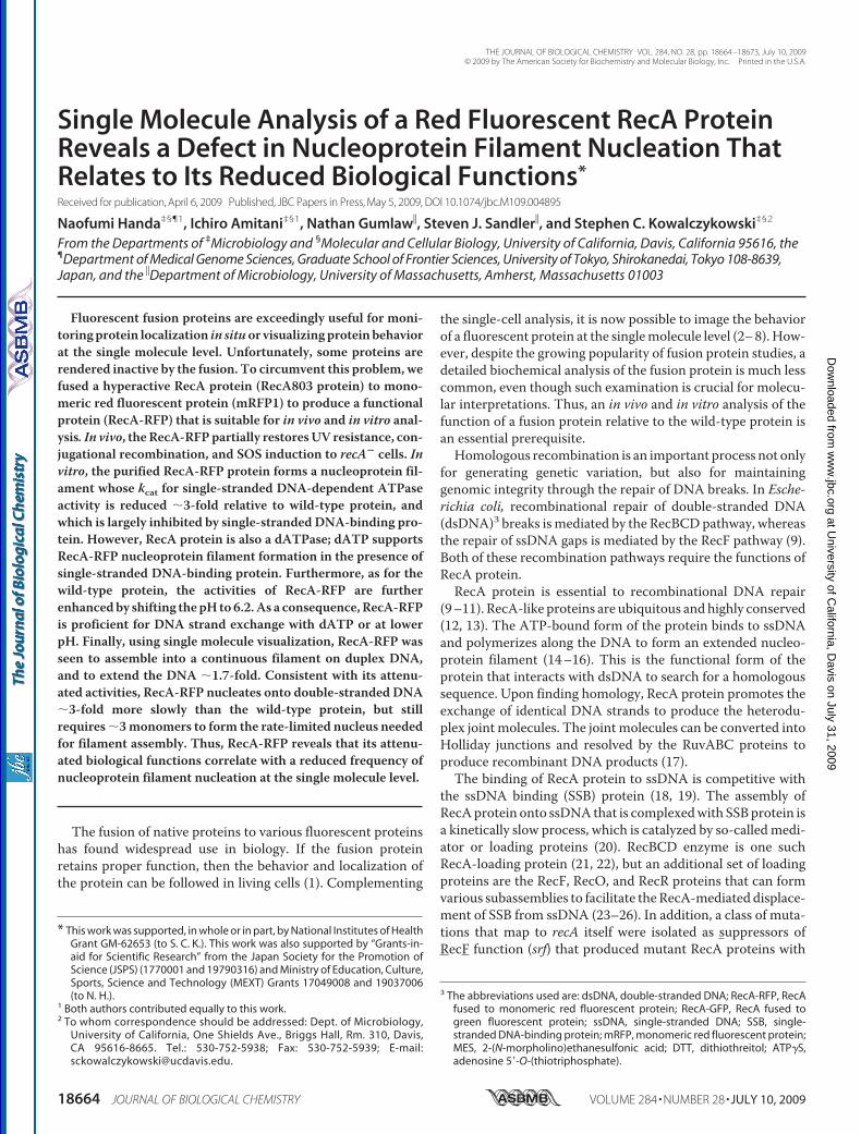
RecA-RFPPartially Complements the PhenotypicDeficienciesof a recA Null Strain—A fusion of RecA protein to green fluo-rescent protein (GFP) resulted in a protein that partiallyrestored recombination and survival to cells after UV irradia-tion, and was used to visualize RecA focus formation at sites ofDNA damage in individual living E. coli cells (30). To define thephenotype of the RecA-RFP protein, and to permit comparisonto the RecA-GFP protein previously described, these functionswere also examined. The recA803-mrfp1 (hereafter referred toas RecA-RFP) construct partially complements the UV-repairdeficiency of a recA null strain to a level comparable with that ofthe wild-type recA-gfp construct (Fig. 1A).The partial complementation of UV survival suggests that
RecA-RFP is at least partially active for recombinational DNArepair. To test recombination function directly, conjugationalrecombination was examined. Table 1 shows that recombina-
RecA-RFP Protein
18666 JOURNAL OF BIOLOGICAL CHEMISTRY VOLUME 284 • NUMBER 28 • JULY 10, 2009
at University of C
alifornia, Davis on July 31, 2009
ww
w.jbc.org
Dow
nloaded from
tion by RecA-RFP is reduced only about 2-fold relative to wild-type, and is the same, within error, as the previously character-ized RecA-GFP fusion.A third biological function of the RecA protein is induction
of the SOS-response via proteolytic cleavage of the LexArepressor. This activity was measured by using a reporter genecomprising a fusion of �-galactosidase to the LexA-regulatedsulA gene (39). The results show that RecA-RFP induces theexpression of this reporter gene in response toUV irradiation toalmost the same extent as wild-type (Fig. 1B), indicating thatRecA-RFP is proficient in LexA repressor cleavage in vivo.These collective results indicated that RecA-RFP was at leastpartially functional in vivo; as a consequence, the protein waspurified and characterized in vitro.Purification of RecA-RFP Protein—First, we attempted to
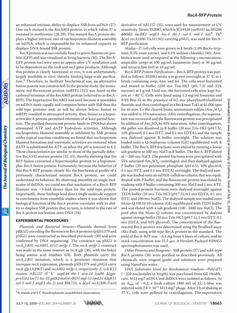
purify the RecA-RFP fusion protein using a procedure that isstandard for the wild-type RecA (38); however, that procedurewas unsuccessful. Consequently, we modified the protocol (see“Experimental Procedures”) and succeeded in purifying the flu-orescent protein. As presented in Fig. 2, the excitation andemission peaks of RecA-RFP are 582 and 608 nm, respectively.We also attempted purification of the fusion of wild-type RecAprotein with green fluorescent protein, using both the sameprocedure and a standard RecA purification protocol (38);however, the RecA-GFP protein was less soluble, and it showed
little activity in vitro.5 Therefore, we decided to focus on onlythe RecA-RFP protein.RecA-RFP Protein Has ssDNA-dependent ATPase and dAT-
Pase Activities—RecA protein has an ATPase activity that isstimulated by DNA. We first examined the ssDNA-dependentATPase activity using an ssDNA, poly(dT), which is devoid ofDNA secondary structure. Fig. 3A shows that the rate of ATPhydrolysis by RecA-RFP increases with increasing protein con-centration, until it saturates at 1.6 �M RecA-RFP. This concen-tration corresponds to saturation occurring at amolar ratio of 1RecA-RFP per �3.1 nucleotides, which is the canonical valuefor nucleoprotein filament formation by wild-type RecA pro-tein. However, in the plateau region, the rate of ATP hydrolysisis only 13�MATP/min, which corresponds to a kcat value of 8.1min�1. In comparison, the ATPase activity of the native RecAprotein saturates at approximately the same protein concentra-tion (1.5 �M), but exhibits a 3-fold greater rate of ATP hydrol-ysis (39 �M ATP/min), which corresponds to the expected kcatof 26 min�1.RecA protein is also a dATPase (40), and similar results were
obtained when dATP hydrolysis by RecA-RFP was examined
5 N. Handa, I. Amitani, and S. C. Kowalczykowski, unpublished observations.
FIGURE 1. Fluorescent proteins fused to RecA partially suppress the UVsensitivity and SOS induction defects of a recA� strain. A, fresh log-phasecultures were diluted, plated onto L plates, and irradiated at various doses ofUV light. Open squares, AB1157 (wild-type); open circles, BIK733 (�recA) withthe pBR322 empty vector; closed circles, BIK733 with pNG1 (recA803-mrfp1);closed triangles, BIK733 with pSJS1379 (recA-gfp). B, cells were assayed (induplicate) for �-galactosidase activity originating from a fusion to the sulAgene at the indicated times. The isogenic strains are derivatives of DM4000(39); open squares, DM4000 (recA�); open circles, SS2060 (�(recA)::kan); andclosed circles, SS2065 (recA803-mrfp1). The data are the mean � S.E. of at leastthree independent experiments.
FIGURE 2. Absorption, excitation, and fluorescence emission spectra ofRecA-RFP protein. A, absorption spectrum of RecA-RFP protein (4 �M) instorage buffer. B, fluorescence spectra of RecA-RFP (215 nM) in storage buffer.The excitation spectrum was obtained by measuring the fluorescence at 608nm, and emission spectrum was as obtained by exciting at 582 nm. Eachspectrum was normalized to the peak value in each scan.
TABLE 1Relative frequency of recombinationAll strains are derivatives of JC13509 and are provided under “Experimental Proce-dures.” Conjugal matings are the averages of three independent experiments.
Conjugational recombination(relative frequency)
RecA (wild-type) 1RecA-RFP 0.47 � 0.19RecA-GFP 0.64 � 0.21
RecA-RFP Protein
JULY 10, 2009 • VOLUME 284 • NUMBER 28 JOURNAL OF BIOLOGICAL CHEMISTRY 18667
at University of C
alifornia, Davis on July 31, 2009
ww
w.jbc.org
Dow
nloaded from
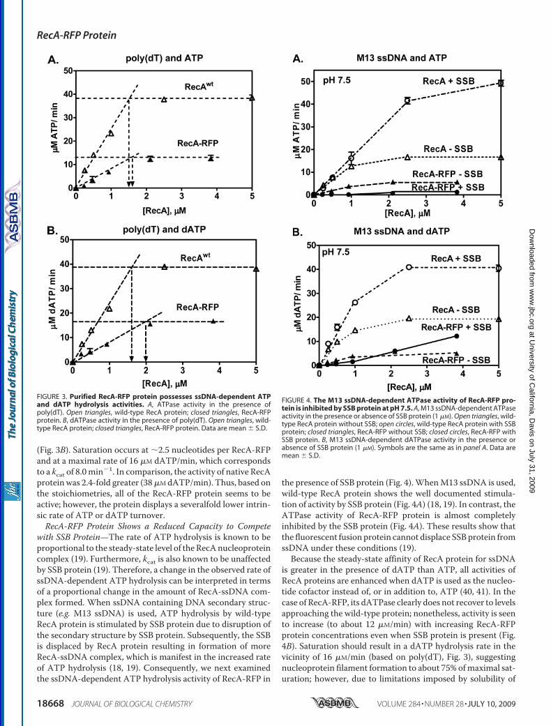
(Fig. 3B). Saturation occurs at �2.5 nucleotides per RecA-RFPand at a maximal rate of 16 �M dATP/min, which correspondsto a kcat of 8.0min�1. In comparison, the activity of native RecAproteinwas 2.4-fold greater (38�MdATP/min). Thus, based onthe stoichiometries, all of the RecA-RFP protein seems to beactive; however, the protein displays a severalfold lower intrin-sic rate of ATP or dATP turnover.RecA-RFP Protein Shows a Reduced Capacity to Compete
with SSB Protein—The rate of ATP hydrolysis is known to beproportional to the steady-state level of theRecAnucleoproteincomplex (19). Furthermore, kcat is also known to be unaffectedby SSB protein (19). Therefore, a change in the observed rate ofssDNA-dependent ATP hydrolysis can be interpreted in termsof a proportional change in the amount of RecA-ssDNA com-plex formed. When ssDNA containing DNA secondary struc-ture (e.g. M13 ssDNA) is used, ATP hydrolysis by wild-typeRecA protein is stimulated by SSB protein due to disruption ofthe secondary structure by SSB protein. Subsequently, the SSBis displaced by RecA protein resulting in formation of moreRecA-ssDNA complex, which is manifest in the increased rateof ATP hydrolysis (18, 19). Consequently, we next examinedthe ssDNA-dependent ATP hydrolysis activity of RecA-RFP in
the presence of SSB protein (Fig. 4).WhenM13 ssDNA is used,wild-type RecA protein shows the well documented stimula-tion of activity by SSB protein (Fig. 4A) (18, 19). In contrast, theATPase activity of RecA-RFP protein is almost completelyinhibited by the SSB protein (Fig. 4A). These results show thatthe fluorescent fusion protein cannot displace SSBprotein fromssDNA under these conditions (19).Because the steady-state affinity of RecA protein for ssDNA
is greater in the presence of dATP than ATP, all activities ofRecA proteins are enhanced when dATP is used as the nucleo-tide cofactor instead of, or in addition to, ATP (40, 41). In thecase of RecA-RFP, its dATPase clearly does not recover to levelsapproaching the wild-type protein; nonetheless, activity is seento increase (to about 12 �M/min) with increasing RecA-RFPprotein concentrations even when SSB protein is present (Fig.4B). Saturation should result in a dATP hydrolysis rate in thevicinity of 16 �M/min (based on poly(dT), Fig. 3), suggestingnucleoprotein filament formation to about 75% ofmaximal sat-uration; however, due to limitations imposed by solubility of
FIGURE 3. Purified RecA-RFP protein possesses ssDNA-dependent ATPand dATP hydrolysis activities. A, ATPase activity in the presence ofpoly(dT). Open triangles, wild-type RecA protein; closed triangles, RecA-RFPprotein. B, dATPase activity in the presence of poly(dT). Open triangles, wild-type RecA protein; closed triangles, RecA-RFP protein. Data are mean � S.D.
FIGURE 4. The M13 ssDNA-dependent ATPase activity of RecA-RFP pro-tein is inhibited by SSB protein at pH 7.5. A, M13 ssDNA-dependent ATPaseactivity in the presence or absence of SSB protein (1 �M). Open triangles, wild-type RecA protein without SSB; open circles, wild-type RecA protein with SSBprotein; closed triangles, RecA-RFP without SSB; closed circles, RecA-RFP withSSB protein. B, M13 ssDNA-dependent dATPase activity in the presence orabsence of SSB protein (1 �M). Symbols are the same as in panel A. Data aremean � S.D.
RecA-RFP Protein
18668 JOURNAL OF BIOLOGICAL CHEMISTRY VOLUME 284 • NUMBER 28 • JULY 10, 2009
at University of C
alifornia, Davis on July 31, 2009
ww
w.jbc.org
Dow
nloaded from
the RecA-RFP stock solution, higher concentrations of RecA-RFP could not be tested. These findings show that RecA-RFPnucleoprotein filament formation is occurring in the presenceof dATP and SSB protein.Previously, we showed that mutant RecA proteins (RecA142
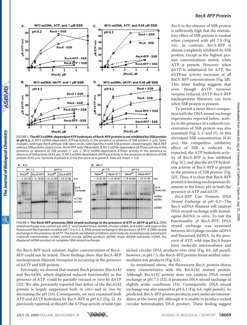
and RecA430), which displayed reduced functionality in thepresence of ATP, could be partially rescued in vitro by dATP(32). We also previously reported that defect of the RecA142protein is largely suppressed both in vitro and in vivo bydecreasing the pH (33). Consequently, we next examined bothATP and dATP hydrolysis by RecA-RFP at pH 6.2 (Fig. 5). Aspreviously reported, at this pH, theATPase activity of wild-type
RecA in the absence of SSB proteinis sufficiently high that the stimula-tory effect of SSB protein is modestwhen compared with pH 7.5 (Fig.5A). In contrast, RecA-RFP isalmost completely inhibited by SSBprotein, except at the highest pro-tein concentrations tested, whenATP is present. However, whendATP is substituted for ATP, thedATPase activity increases at allRecA-RFP concentrations (Fig. 5B).This latter finding suggests thateven though dATP turnoverremains reduced, dATP-RecA-RFPnucleoprotein filaments can formwhen SSB protein is present.To permit a more direct compar-
ison with the DNA strand exchangeexperiments reported below, activ-ity in the presence of a reduced con-centration of SSB protein was alsoexamined (Fig. 5, C and D). At thisdecreased SSB concentration (0.45�M), the competitive inhibitoryeffect of SSB is reduced. Asexpected, the ATP hydrolysis activ-ity of RecA-RFP is less inhibited(Fig. 5C), and also the dATP hydrol-ysis activity of RecA-RFP is greaterin the presence of SSB protein (Fig.5D). Thus, it is clear that RecA-RFPprotein is forming nucleoprotein fil-aments at the lower pH in both thepresence of ATP and dATP.RecA-RFP Can Promote DNA
Strand Exchange at pH 6.2—TheRecA-ssDNA filament will catalyzeDNA strand exchange with homol-ogous dsDNA in vitro. To test thefunctionality of RecA-RFP, DNAstrand exchange was examinedbetweenM13 phage circular ssDNAand linearized dsDNA. In the pres-ence of ATP, wild-type RecA formsjoint molecule intermediates and
nicked circular DNA products over time (Fig. 6A, left panel);however, at pH 7.5, the RecA-RFP protein forms neither inter-mediates nor products (Fig. 6A).As mentioned above, the fluorescent RecA protein shares
many characteristics with the RecA142 mutant protein.Although RecA142 protein does not catalyze DNA strandexchange at pH 7.5 (32), it promotes DNA strand exchange atslightly acidic conditions (33). Consequently, DNA strandexchange was also assayed at pH 6.2 (Fig. 6A, right panels). Aspredicted, RecA-RFP protein produces jointmolecule interme-diates at the lower pH, although it is unable to produce nickedcircular heteroduplex DNA product. These finding suggest
FIGURE 5. The M13 ssDNA-dependent ATP hydrolysis of RecA-RFP protein is not inhibited by SSB proteinat pH 6.2. A, M13 ssDNA-dependent ATPase activity in the presence or absence of SSB protein (1 �M). Opentriangles, wild-type RecA without SSB; open circles, wild-type RecA with SSB protein; closed triangles, RecA-RFPwithout SSB protein; closed circles, RecA-RFP with SSB protein. B, M13 ssDNA-dependent dATPase activity in thepresence or absence of SSB protein (1 �M). C, M13 ssDNA-dependent ATPase activity in the presence orabsence of SSB protein (0.45 �M). D, M13 ssDNA-dependent dATPase activity in the presence or absence of SSBprotein (0.45 �M). Symbols in panels B–D are the same as in panel A. Data are mean � S.D.
FIGURE 6. The RecA-RFP promotes DNA strand exchange in the presence of ATP or dATP at pH 6.2. DNAstrand exchange was carried out with 5�-end labeled linear dsDNA, circular ssDNA, and either wild-type or thefluorescent RecA protein at either pH 7.5 or 6.2. A, DNA strand exchange in the presence of ATP. B, DNA strandexchange in the presence of dATP. The bands are labeled as follows: joint molecule, homologously paired jointmolecule intermediate; ncDNA, nicked circular dsDNA product; dsDNA, linear dsDNA substrate; ssDNA, thedisplaced ssDNA product of complete DNA strand exchange.
RecA-RFP Protein
JULY 10, 2009 • VOLUME 284 • NUMBER 28 JOURNAL OF BIOLOGICAL CHEMISTRY 18669
at University of C
alifornia, Davis on July 31, 2009
ww
w.jbc.org
Dow
nloaded from
that, although functional, the RecA-RFP nucleoprotein fila-ments are incomplete, in agreement with the ATP hydrolysisdata.DNA strand exchange was also conducted using dATP
instead of ATP (Fig. 6B). Now, the fluorescent RecA producesboth joint molecule intermediates and some nicked circularDNA products, even at pH 7.5 (left panels). At the lower pH,RecA-RFP forms evenmore jointmolecule intermediates (rightpanel). However, the yield of DNA strand exchange product isdecreased for wild-type RecA with dATP at pH 6.2 (Fig. 6B).This seemingly unexpected result is likely due to enhancedbinding of wild-type RecA protein to the dsDNA substrateunder these conditions (33), causing an inhibition of DNAstrand exchange (42); this inhibitory phenomenon is character-istic of, and well documented for, the eukaryotic Rad51 proteinthat readily binds duplex DNA to reduceDNA strand exchange(43).RecA-RFP Can Be Used to Visualize Nucleoprotein Filament
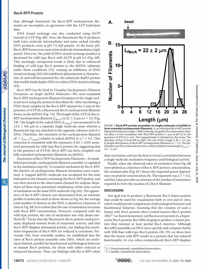
Formation on Single dsDNA Molecules—We next examinedRecA-RFP nucleoprotein filament formation at the single mol-ecule level using the protocol described (8). After incubating aDNA-bead complex in the RecA-RFP channel for 5 min in thepresence of ATP�S, a fluorescent RecA nucleoprotein filamentforms on the dsDNA (Fig. 7A). The length of the ATP�S-RecA-RFP nucleoprotein filament (LRecA) is 22 � 2 �m (n � 21) (Fig.7B). The length of the naked DNA (Lnaked) was measured to be12.8 � 0.6 �m in a separate single molecule assay wherein afluorescent tag was attached to the opposite cohesive end of �DNA. Therefore, the extension of the nucleoprotein filament(R � LRecA/Lnaked) relative to naked dsDNA is 1.7 � 0.2. Thisextension is consistent with the extension (1.65 � 0.05) meas-ured previously for wild-type RecA protein (8), suggesting thatin the presence of ATP�S, RecA-RFP can form complete andfully extended nucleoprotein filaments on dsDNA.Nucleation of RecA-RFPNucleoprotein Filaments—As estab-
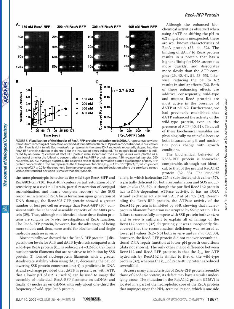
lished previously, nucleoprotein filament assembly is regulatedat the nucleation step (8). To examine nucleation by RecA-RFP,the kinetics of nucleoprotein filament formation were exam-ined. A trapped dsDNA molecule was incubated for the timeindicated in the channel containing the RecA-RFP protein, andwas then moved to the observation channel for analysis. Repe-tition of these steps permitted visualization of the time courseof nucleation on the sameDNAmolecule (Fig. 8A). The appear-ance of RecA-RFP clusters was determined from an intensityprofile of molecules such as those shown in Fig. 8A; the averagetotal number of clusters on the DNA is plotted as a function oftime in Fig. 8B. It is evident that the rate of nucleation increaseswith RecA-RFP concentration. However, compared with thewild-type protein, the rate of nucleation was only about one-third (8).5 Given that the fluorescein-RecA protein used previ-ously displayed normal levels of enzymatic activity, and thatRecA-RFP displays attenuated activity, our finding that nucle-ation frequencies of RecA-RFP are reduced is consistent. Pre-viously (34), from ensemble studies, we established that therates of RecA protein assembly onto DNA, which are nucle-ation-limited, parallel the biochemical and biological behaviorof mutant RecA proteins, for those with either reduced orenhanced functions. Thus, our findings with RecA-RFP refine
that conclusion, and are the first to reveal a correlation betweena single molecule nucleation frequency and biological activity.Finally, when the observed rates of nucleation from Fig. 8B
were plotted as a function of RecA-RFP protein concentration,the resultant plot (Fig. 8C) shows the expected power depend-ence on protein concentration (8). The exponent was 2.7� 0.2,and this value provides an estimate of the number ofmonomersrequired to form the nucleus of a RecA filament.
DISCUSSION
Our goal was to produce a fluorescent RecA fusion proteinthat could be used for visualization both in vivo and in vitro,which would permit comparisons of physiological function andbiochemical behavior. Knowing that the creation of such afusion with RecA protein often created inactive RecA protein(30),6,7 we fusedmonomeric red fluorescent protein to a hyper-active RecA protein, RecA803, hoping to produce a fusion pro-tein that retained at least partial RecA function. AlthoughRecA803 assembles onDNAmore quickly and competes betterwith SSB than wild-type RecA protein (28, 29), we show herethat the fusion with RFP results in a protein that has reducedfunctionality. In vivo, when overproduced, RecA-RFP displays
6 S. C. Kowalczykowski, unpublished observations.7 S. J. Sandler, unpublished observations.
FIGURE 7. RecA-RFP protein assembles on single molecules of dsDNA toform extended nucleoprotein filaments. A, an individual RecA-RFP proteinfilament formed on a single � DNA molecule, imaged in the observation chan-nel after a 5-min incubation with RecA-RFP protein (1 �M) at pH 6.2 in thepresence of ATP�S (1 mM). The trapped bead is indicated by the arrow. Thedirection of flow is from left to right. The size of this image is 28 � 4.2 �m.B, length distribution of RecA-RFP nucleoprotein filaments (n � 21). The dis-tribution was fit to a Gaussian function, which yielded a mean length (LRecA) �22 � 2 �m.
RecA-RFP Protein
18670 JOURNAL OF BIOLOGICAL CHEMISTRY VOLUME 284 • NUMBER 28 • JULY 10, 2009
at University of C
alifornia, Davis on July 31, 2009
ww
w.jbc.org
Dow
nloaded from
the same phenotypic behavior as the wild-type RecA-GFP andRecA803-GFP (30). RecA-RFP confers partial restoration ofUVsensitivity to a recA null strain, partial restoration of conjugalrecombination, and nearly complete recovery of the SOSresponse. In terms of RecA focus formation upon generation ofDNA damage, the RecA803-GFP protein showed a greaternumber of foci per cell on average than RecA-GFP (30), con-sistent with the enhanced assembly capacity of RecA803 pro-tein (29). Thus, although not identical, these three fusion pro-teins are suitable for in vivo investigations of RecA function.The RecA-RFP protein, however, has the advantage of beingmore soluble and, thus, more useful for biochemical and singlemolecule analyses in vitro.Biochemically, we showed that the RecA-RFP protein: 1) dis-
plays lower levels for ATP and dATP hydrolysis compared withwild-type RecA protein (kcat is reduced 2.4–3.2-fold); 2) formsnucleoprotein filaments that are sensitive to inhibition by SSBprotein; 3) formed nucleoprotein filaments with a greatersteady-state stability when using dATP, decreasing the pH, orlowering SSB protein concentrations; 4) is proficient in DNAstrand exchange provided that dATP is present or, with ATP,that a lower pH of 6.2 is used; 5) can be used to image theassembly of individual RecA-RFP filaments on dsDNA; andfinally, 6) nucleates on dsDNA with only about one-third thefrequency of wild-type RecA protein.
Although the enhanced bio-chemical activities observed whenusing dATP or shifting the pH to6.2 might seem unexpected, theseare well known characteristics ofRecA protein (33, 44–52). Thebinding of dATP to RecA proteinresults in a protein that has ahigher affinity for DNA, assemblesmore quickly, and dissociatesmore slowly than the ATP com-plex (26, 40, 41, 51, 53–55). Like-wise, reducing the pH to 6.2results in similar effects (56). Bothof these enhancing effects areadditive; consequently, wild-typeand mutant RecA proteins aremost active in the presence ofdATP at pH 6.2. Furthermore, wehad previously established thatdATP enhanced the activity of thewild-type protein, even in thepresence of ATP (40, 41). Thus, allof these biochemical variables arephysiologically meaningful, becauseboth intracellular pH and nucleo-tide pools change with growthconditions.The biochemical behavior of
RecA-RFP protein is somewhatcomparable, although not identi-cal, to that of the mutant RecA142protein (32, 33). The recA142
allele, in which isoleucine 225 is substituted with valine (57),is partially deficient for both recombination and SOS induc-tion in vivo (58, 59). Although the purified RecA142 proteinhas ssDNA-dependent ATPase activity, it has no DNAstrand exchange activity with ATP at pH 7.5 (32). Resem-bling the RecA-RFP protein, the ATPase activity of theRecA142 protein is inhibited by SSB, showing that nucleo-protein filament formation is disrupted by SSB protein. Thisfailure to successfully compete with SSB protein both in vitroand in vivo is sufficient to explain all of failings of theRecA142 protein (32). Surprisingly, it was subsequently dis-covered that the recombination deficiency was restored atlower pH values (6.2–6.5) both in vitro and in vivo (32, 33);however, the RecA-RFP protein did not recover recombina-tional DNA repair function at lower pH growth conditions(data not shown). The only other major difference betweenRecA142 and RecA-RFP proteins is that the kcat for ATPhydrolysis by RecA142 is similar to that of the wild-typeprotein (32), whereas the kcat of RecA-RFP protein is reducedseveralfold.Because many characteristics of RecA-RFP protein resemble
those of RecA142 protein, its defect may have a similar under-lying cause. The mutation in the RecA142 protein (I225V) islocated in a part of the hydrophobic core of the RecA proteinthat impinges upon the NH2-terminal region, which is one side
FIGURE 8. Visualization of the kinetics of RecA-RFP protein nucleation on dsDNA. A, representative videoframes from recordings of nucleation obtained at four different RecA-RFP protein concentrations in nucleationbuffer. Flow is right to left. Each vertical strip represents the same DNA molecule repeatedly dipped into theRecA-RFP protein solution in channel 3 for the incubation times indicated. The trapped bead position is indi-cated by an arrow. B, clusters of RecA-RFP protein were scored and the average values were plotted as afunction of time for the following concentrations of RecA-RFP protein: squares, 150 nM; inverted triangles, 200nM; circles, 300 nM; triangles, 400 nM. C, the observed rate of cluster formation plotted as a function of RecA-RFPprotein concentration. The line represents the fit to a power function, kobs � 1.2 � 10�8 [RecA]2.7, which yieldedthe value of 2.7 � 0.2 for the exponent. Error bars represent the standard deviation; where the error bars are notvisible, the standard deviation is smaller than the symbols.
RecA-RFP Protein
JULY 10, 2009 • VOLUME 284 • NUMBER 28 JOURNAL OF BIOLOGICAL CHEMISTRY 18671
at University of C
alifornia, Davis on July 31, 2009
ww
w.jbc.org
Dow
nloaded from
of the monomer-monomer interface (33). The NH2 terminuscontains a large �-helix and short �-strand involved in RecAself-assembly (60–62). The fluorescent protein used here wasproduced by fusing RFP to the COOH-terminal end of thehyperactive RecA803 protein, using a short amino acid linker.Although the COOH terminus does not directly contribute tothe monomer-monomer interface (60, 63), nonetheless, it mayocclude this interface and thereby impede the assembly proc-ess. The fact that the rate of nucleation onto DNA is reducedsupports this conclusion. The RecA-RFP protein was hoped tohave activity comparable with the wild-type RecA protein.Instead, the fluorescent fusion protein displays a reduced func-tionality. Nevertheless, the fusion protein can still be useful forin vivo experiments, provided that one bear in mind that it is ahypomorph.We were able to visualize individual filaments of RecA-RFP
forming on single DNA molecules. The length measurementsshowed that the RecA-RFP nucleoprotein filament is extended1.7-fold relative to naked DNA, which is in precise agreementwith our previous measurements using wild-type RecA proteinthat wasmodified with carboxyfluorescein (8), indicating that acomplete nucleoprotein filament is formed. Furthermore, wemeasured directly the rates of filament nucleation by RecA-RFP. Compared with the wild-type protein, the nucleation fre-quencies are reduced. The nucleation of RecA-RFP on dsDNAis slower than the wild-type protein by a factor of �3. Thisfinding is the first report of the nucleation frequency of a RecAprotein with altered functional characteristics. Extensive bio-chemical analysis of mutant RecA proteins, and the correlationwith their phenotypic behavior, led to the conclusion that theability of a RecA protein to compete with SSB protein for DNAbinding is the most predictive biochemical trait that correlatescompletely with its biological function (34). This ability to com-pete with SSB protein, in turn, was related directly to the rate atwhich a RecA protein assembles onto ssDNA. Using dsDNA,we previously showed that assembly of RecA protein is con-trolled by a rate-limiting nucleation step involving 4.5 (� 0.5)monomers. This value is close, but not identical, to the valueobtained with the RecA-RFP fusion protein; although thisapparent difference may reflect real differences between thetwo proteins, we believe that it largely reflects the difficulty ofmeasuring the rates of nucleation at the highest concentrationsof RecA, which disproportionately bias the fit to the powerfunction. Finally, although the absolute rate of nucleation ontossDNA and onto the SSB-ssDNA complex will unquestionablybe different from the rate of nucleation onto dsDNA, if weassume that the rates will parallel one another, then our resultshere show that a 3-fold lower rate of nucleation is sufficient tomanifest reduced function in vivo and in vitro.Finally, even though chemical modification of the RecA pro-
tein yielded a protein that fully retained biochemical activities(8), we note that the fluorescent fusion protein described hasthe advantage that it can be used to analyze quantitatively bothin vivo cellular function and in vitro biochemical activity for thesame protein. Thus, exact correlations of in vivo and in vitrofunction are now possible. Given that the functions of RecAprotein can be altered by physiological factors such as ATP,dATP, pH, concentration of RecA and SSB proteins, volume-
excluding conditions (64), and polyvalent ions (65), compara-tive individual cell analysis in vivo and individualmolecule anal-ysis in vitro will be very revealing.
Acknowledgments—We thank Dr. Ichizo Kobayashi for bacterialstrains and Jarukit Edward Long for technical assistance. We aregrateful to members of the Kowalczykowski laboratory, Petr Cejka,Clarke Conant, Taeho Kim, Katsumi Morimatsu, Jody Plank, andJason Wong for critical reading of the manuscript.
REFERENCES1. Chudakov, D. M., Lukyanov, S., and Lukyanov, K. A. (2005) Trends Bio-
technol. 23, 605–6132. Nie, S., Chiu, D. T., and Zare, R. N. (1994) Science 266, 1018–10213. Funatsu, T., Harada, Y., Tokunaga, M., Saito, K., and Yanagida, T. (1995)
Nature 374, 555–5594. Noji, H., Yasuda, R., Yoshida, M., and Kinosita, K., Jr. (1997) Nature 386,
299–3025. Ha, T., Ting, A. Y., Liang, J., Caldwell, W. B., Deniz, A. A., Chemla, D. S.,
Schultz, P. G., and Weiss, S. (1999) Proc. Natl. Acad. Sci. U.S.A. 96,893–898
6. Amitani, I., Baskin, R. J., and Kowalczykowski, S. C. (2006) Mol. Cell 23,143–148
7. Handa, N., Bianco, P. R., Baskin, R. J., and Kowalczykowski, S. C. (2005)Mol. Cell 17, 745–750
8. Galletto, R., Amitani, I., Baskin, R. J., and Kowalczykowski, S. C. (2006)Nature 443, 875–878
9. Spies,M., and Kowalczykowski, S. C. (2005) inThe Bacterial Chromosome(Higgins, N. P., ed) pp. 389–403, ASM Press, Washington, D.C.
10. Kowalczykowski, S. C., Dixon, D. A., Eggleston, A. K., Lauder, S. D., andRehrauer, W. M. (1994)Microbiol. Rev. 58, 401–465
11. Kuzminov, A. (1999)Microbiol. Mol. Biol. Rev. 63, 751–81312. Bianco, P. R., Tracy, R. B., and Kowalczykowski, S. C. (1998) Front. Biosci.
3, D570-D60313. Lin, Z., Kong, H., Nei, M., and Ma, H. (2006) Proc. Natl. Acad. Sci. U.S.A.
103, 10328–1033314. Kowalczykowski, S. C. (1991) Annu. Rev. Biophys. Biophys. Chem. 20,
539–57515. Egelman, E. H., and Stasiak, A. (1986) J. Mol. Biol. 191, 677–69716. Stasiak, A., and Di Capua, E. (1982) Nature 299, 185–18617. West, S. C. (1996) J. Bacteriol. 178, 1237–124118. Kowalczykowski, S. C., Clow, J., Somani, R., andVarghese, A. (1987) J.Mol.
Biol. 193, 81–9519. Kowalczykowski, S. C., and Krupp, R. A. (1987) J. Mol. Biol. 193, 97–11320. Beernink, H. T., and Morrical, S. W. (1999) Trends Biochem. Sci. 24,
385–38921. Spies, M., and Kowalczykowski, S. C. (2006)Mol. Cell 21, 573–58022. Anderson, D. G., Churchill, J. J., and Kowalczykowski, S. C. (1999) J. Biol.
Chem. 274, 27139–2714423. Umezu, K., Chi, N. W., and Kolodner, R. D. (1993) Proc. Natl. Acad. Sci.
U.S.A. 90, 3875–387924. Umezu, K., and Kolodner, R. D. (1994) J. Biol. Chem. 269, 30005–3001325. Morimatsu, K., andKowalczykowski, S. C. (2003)Mol. Cell 11, 1337–134726. Shan, Q., Bork, J. M., Webb, B. L., Inman, R. B., and Cox, M. M. (1997) J.
Mol. Biol. 265, 519–54027. Volkert, M. R., and Hartke, M. A. (1984) J. Bacteriol. 157, 498–50628. Madiraju, M. V., Templin, A., and Clark, A. J. (1988) Proc. Natl. Acad. Sci.
U.S.A. 85, 6592–659629. Lavery, P. E., and Kowalczykowski, S. C. (1992) J. Biol. Chem. 267,
20648–2065830. Renzette, N., Gumlaw, N., Nordman, J. T., Krieger,M., Yeh, S. P., Long, E.,
Centore, R., Boonsombat, R., and Sandler, S. J. (2005)Mol. Microbiol. 57,1074–1085
31. Campbell, R. E., Tour, O., Palmer, A. E., Steinbach, P. A., Baird, G. S.,Zacharias, D. A., and Tsien, R. Y. (2002) Proc. Natl. Acad. Sci. U.S.A. 99,
RecA-RFP Protein
18672 JOURNAL OF BIOLOGICAL CHEMISTRY VOLUME 284 • NUMBER 28 • JULY 10, 2009
at University of C
alifornia, Davis on July 31, 2009
ww
w.jbc.org
Dow
nloaded from
7877–788232. Kowalczykowski, S. C., Burk, D. L., and Krupp, R. A. (1989) J. Mol. Biol.
207, 719–73333. Zaitsev, E. N., and Kowalczykowski, S. C. (1999)Mol. Microbiol. 34, 1–934. Kowalczykowski, S. C. (1991) Biochimie 73, 289–30435. Bachmann, B. J. (1987) in Escherichia coli and Salmonella typhimurium,
Cellular and Molecular Biology (Neidhardt, F. C., Ingraham, J. L., Low,K. B., Magasanik, B., Schaechter, M., and Umbarger, H. E., eds) pp.1190–1219, American Society for Microbiology, Washington, D.C.
36. McCool, J. D., and Sandler, S. J. (2001) Proc. Natl. Acad. Sci. U.S.A. 98,8203–8210
37. LeBowitz, J. (1985) Biochemical Mechanism of Strand Initiation in Bacte-riophage � DNA Replication, Johns Hopkins University, Baltimore, MD
38. Mirshad, J. K., and Kowalczykowski, S. C. (2003) Biochemistry 42,5945–5954
39. Sandler, S. J., Samra, H. S., and Clark, A. J. (1996) Genetics 143, 5–1340. Menetski, J. P., and Kowalczykowski, S. C. (1989) Biochemistry 28,
5871–588141. Menetski, J. P., and Kowalczykowski, S. C. (1990) J. Mol. Biol. 211,
845–85542. Campbell, M. J., and Davis, R. W. (1999) J. Mol. Biol. 286, 437–44543. Sung, P., and Robberson, D. L. (1995) Cell 82, 453–46144. Kowalczykowski, S. C., Clow, J., and Krupp, R. A. (1987) Proc. Natl. Acad.
Sci. U.S.A. 84, 3127–313145. McEntee, K., Weinstock, G. M., and Lehman, I. R. (1981) J. Biol. Chem.
256, 8835–884446. Karasaki, Y., Hirano, H., andHigashi, K. (1987) Sangyo IkaDaigaku Zasshi
9, 141–147
47. Pugh, B. F., and Cox, M. M. (1987) J. Biol. Chem. 262, 1337–134348. Bryant, F. R. (1988) J. Biol. Chem. 263, 8716–872349. Brenner, S. L., Zlotnick, A., and Stafford,W. F., 3rd (1990) J.Mol. Biol. 216,
949–96450. Muench, K. A., and Bryant, F. R. (1991) J. Biol. Chem. 266, 844–85051. Meah, Y. S., and Bryant, F. R. (1993) J. Biol. Chem. 268, 23991–2399652. Pinsince, J.M.,Muench, K.A., Bryant, F. R., andGriffith, J. D. (1993) J.Mol.
Biol. 233, 59–6653. Kowalczykowski, S. C. (1986) Biochemistry 25, 5872–588154. Zaitsev, E. N., and Kowalczykowski, S. C. (1998) Nucleic Acids Res. 26,
650–65455. Katz, F. S., and Bryant, F. R. (2001) Biochemistry 40, 11082–1108956. Arenson, T. A., Tsodikov, O. V., and Cox, M. M. (1999) J. Mol. Biol. 288,
391–40157. Dutreix, M., Bailone, A., and Devoret, R. (1985) J. Bacteriol. 161,
1080–108558. Clark, A. J. (1973) Annu. Rev. Genet. 7, 67–8659. Horii, Z., and Clark, A. J. (1973) J. Mol. Biol. 80, 327–34460. Story, R. M., Weber, I. T., and Steitz, T. A. (1992) Nature 355, 318–32561. Ogawa, T., Yu, X., Shinohara, A., and Egelman, E. H. (1993) Science 259,
1896–189962. Mikawa, T., Masui, R., Ogawa, T., Ogawa, H., and Kuramitsu, S. (1995) J.
Mol. Biol. 250, 471–48363. Skiba, M. C., and Knight, K. L. (1994) J. Biol. Chem. 269, 3823–382864. Lavery, P. E., and Kowalczykowski, S. C. (1992) J. Biol. Chem. 267,
9307–931465. Lavery, P. E., and Kowalczykowski, S. C. (1990) J. Biol. Chem. 265,
4004–4010
RecA-RFP Protein
JULY 10, 2009 • VOLUME 284 • NUMBER 28 JOURNAL OF BIOLOGICAL CHEMISTRY 18673
at University of C
alifornia, Davis on July 31, 2009
ww
w.jbc.org
Dow
nloaded from