RecBCD Enzyme Switches Lead Motor Subunits in Response...
12
RecBCD Enzyme Switches Lead Motor Subunits in Response to c Recognition Maria Spies, 1,3 Ichiro Amitani, 1 Ronald J. Baskin, 2 and Stephen C. Kowalczykowski 1,2, * 1 Section of Microbiology 2 Section of Molecular and Cellular Biology University of California, Davis, CA 95616-8665, USA 3 Department of Biochemistry, University of Illinois at Urbana-Champaign, Urbana, IL 61801-3602, USA *Correspondence: [email protected] DOI 10.1016/j.cell.2007.09.023 SUMMARY RecBCD is a DNA helicase comprising two motor subunits, RecB and RecD. Recognition of the recombination hotspot, c, causes RecBCD to pause and reduce translocation speed. To understand this control of translocation, we used single-molecule visualization to compare RecBCD to the RecBCD K177Q mutant with a defective RecD motor. RecBCD K177Q paused at c but did not change its translocation velocity. RecBCD K177Q translocated at the same rate as the wild-type post-c enzyme, implicating RecB as the lead motor after c. P1 nuclease treatment eliminated the wild-type enzyme’s velocity changes, revealing a c-containing ssDNA loop preceding c recognition and showing that RecD is the faster motor before c. We conclude that before c, RecD is the lead motor but after c, the slower RecB motor leads, implying a switch in motors at c. We suggest that degradation of foreign DNA needs fast translocation, whereas DNA repair uses slower translocation to coordi- nate RecA loading onto ssDNA. INTRODUCTION The RecBCD helicase/nuclease is needed for recombina- tional DNA repair of double-stranded DNA (dsDNA) breaks in E. coli (for review see Spies and Kowalczykowski, 2005). To initiate repair, RecBCD binds to the broken du- plex DNA end, unwinds the dsDNA for 30 kb on average, and differentially degrades both nascent DNA strands. While translocating, RecBCD can recognize a specific DNA sequence, c (Chi, crossover hotspot instigator; 5 0 -G CTGGTGG-3 0 ), which is a hotspot for homologous recom- bination (Lam et al., 1974). Recognition of c attenuates the nuclease activity of the enzyme (Anderson and Kowalczy- kowski, 1997a; Dixon and Kowalczykowski, 1991, 1993), resulting in production of a long single-stranded DNA (ssDNA) tail with the c sequence at its 3 0 end. RecBCD facilitates loading of the DNA strand exchange protein, RecA, onto this c-containing ssDNA to form the RecA nucleoprotein filament that acts subsequently to pro- mote homologous pairing of DNA (Anderson and Kowalc- zykowski, 1997b). In addition to regulating nucleolytic and RecA-loading activities, c recognition regulates trans- location behavior: RecBCD pauses at c for several sec- onds and subsequently reduces its translocation velocity (Spies et al., 2003). The manner by which c effects these changes in movement is unknown. RecBCD employs two autonomous motor subunits, RecB and RecD, that translocate on opposite strands of the DNA duplex (Dil- lingham et al., 2003; Taylor and Smith, 2003), so we imagined that the c-induced changes in translocation are mediated through one or both of these motor sub- units. Previously, we proposed two models to explain the translocation pause and velocity change elicited by c rec- ognition (Spies et al., 2003). One model posited that the two motor subunits of RecBCD translocate along their respective DNA strands at different rates; in this model, translocation by the motors is uncoupled. Thus, the faster motor would be the ‘‘helicase’’ subunit responsible for strand separation, and the slower motor subunit would be a ‘‘translocase’’ simply translocating along the ssDNA produced by the lead motor. In this model, the unequal translocation rates of two motors would produce a loop of ssDNA between the fast motor and slow motor (Figure 1A). We proposed that when RecBCD recognizes c, the lead motor subunit stops translocating at or near c, resulting in the pause. However, the slower motor con- tinues translocating along its ssDNA track until it catches up to the other paused motor subunit at c. It then becomes the ‘‘helicase’’ motor of the enzyme. The alternative model (Figure 1B) proposed that two motor subunits translocate coordinately at the same velocity before c; in this model, translocation by the motors is concerted. Cooperation of two motors was envisioned as being responsible for the fast enzyme translocation. The pause at c was attributed to the time required for a c-induced conformational change. In this model, the re- duced rate after c is a consequence of inactivation of one of the motors. Thus, in this second model, two motors operate in concerted fashion as the helicase before c, but only one motor functions after c. 694 Cell 131, 694–705, November 16, 2007 ª2007 Elsevier Inc.
Transcript of RecBCD Enzyme Switches Lead Motor Subunits in Response...

RecBCD Enzyme Switches Lead MotorSubunits in Response to c RecognitionMaria Spies,1,3 Ichiro Amitani,1 Ronald J. Baskin,2 and Stephen C. Kowalczykowski1,2,*1Section of Microbiology2Section of Molecular and Cellular Biology
University of California, Davis, CA 95616-8665, USA3Department of Biochemistry, University of Illinois at Urbana-Champaign, Urbana, IL 61801-3602, USA*Correspondence: [email protected]
DOI 10.1016/j.cell.2007.09.023
SUMMARY
RecBCD is a DNA helicase comprising twomotor subunits, RecB and RecD. Recognition ofthe recombination hotspot, c, causes RecBCDto pause and reduce translocation speed. Tounderstand this control of translocation, weused single-molecule visualization to compareRecBCD to the RecBCDK177Q mutant with adefective RecD motor. RecBCDK177Q paused atc but did not change its translocation velocity.RecBCDK177Q translocated at the same rate asthe wild-type post-c enzyme, implicating RecBas the lead motor after c. P1 nuclease treatmenteliminated the wild-type enzyme’s velocitychanges, revealing a c-containing ssDNA looppreceding c recognition and showing that RecDis the faster motor before c. We conclude thatbefore c, RecD is the lead motor but after c,the slower RecB motor leads, implying a switchin motors at c. We suggest that degradation offoreign DNA needs fast translocation, whereasDNA repair uses slower translocation to coordi-nate RecA loading onto ssDNA.
INTRODUCTION
The RecBCD helicase/nuclease is needed for recombina-
tional DNA repair of double-stranded DNA (dsDNA) breaks
in E. coli (for review see Spies and Kowalczykowski,
2005). To initiate repair, RecBCD binds to the broken du-
plex DNA end, unwinds the dsDNA for 30 kb on average,
and differentially degrades both nascent DNA strands.
While translocating, RecBCD can recognize a specific
DNA sequence, c (Chi, crossover hotspot instigator; 50-G
CTGGTGG-30), which is a hotspot for homologous recom-
bination (Lam et al., 1974). Recognition of c attenuates the
nuclease activity of the enzyme (Anderson and Kowalczy-
kowski, 1997a; Dixon and Kowalczykowski, 1991, 1993),
resulting in production of a long single-stranded DNA
(ssDNA) tail with the c sequence at its 30 end. RecBCD
facilitates loading of the DNA strand exchange protein,
694 Cell 131, 694–705, November 16, 2007 ª2007 Elsevier Inc.
RecA, onto this c-containing ssDNA to form the RecA
nucleoprotein filament that acts subsequently to pro-
mote homologous pairing of DNA (Anderson and Kowalc-
zykowski, 1997b). In addition to regulating nucleolytic
and RecA-loading activities, c recognition regulates trans-
location behavior: RecBCD pauses at c for several sec-
onds and subsequently reduces its translocation velocity
(Spies et al., 2003). The manner by which c effects these
changes in movement is unknown. RecBCD employs
two autonomous motor subunits, RecB and RecD, that
translocate on opposite strands of the DNA duplex (Dil-
lingham et al., 2003; Taylor and Smith, 2003), so we
imagined that the c-induced changes in translocation
are mediated through one or both of these motor sub-
units.
Previously, we proposed two models to explain the
translocation pause and velocity change elicited by c rec-
ognition (Spies et al., 2003). One model posited that the
two motor subunits of RecBCD translocate along their
respective DNA strands at different rates; in this model,
translocation by the motors is uncoupled. Thus, the faster
motor would be the ‘‘helicase’’ subunit responsible for
strand separation, and the slower motor subunit would
be a ‘‘translocase’’ simply translocating along the ssDNA
produced by the lead motor. In this model, the unequal
translocation rates of two motors would produce a loop
of ssDNA between the fast motor and slow motor
(Figure 1A). We proposed that when RecBCD recognizes
c, the lead motor subunit stops translocating at or near
c, resulting in the pause. However, the slower motor con-
tinues translocating along its ssDNA track until it catches
up to the other paused motor subunit at c. It then becomes
the ‘‘helicase’’ motor of the enzyme.
The alternative model (Figure 1B) proposed that two
motor subunits translocate coordinately at the same
velocity before c; in this model, translocation by the motors
is concerted. Cooperation of two motors was envisioned
as being responsible for the fast enzyme translocation.
The pause at c was attributed to the time required for
a c-induced conformational change. In this model, the re-
duced rate after c is a consequence of inactivation of one
of the motors. Thus, in this second model, two motors
operate in concerted fashion as the helicase before c, but
only one motor functions after c.

Existing data could be mustered in support of either
model. In our previous single-molecule experiments, we
found that the duration of the pause was not simply related
to the difference in time needed by each motor to travel to
c, based on the rates of the putative fast and slow sub-
units before and after c, respectively (Handa et al., 2005;
Spies et al., 2003). This finding was inconsistent with the
simplest version of the uncoupled motors model and,
therefore, pointed toward the concerted motors model.
However, the uncoupled motors model was consistent
with the existence of the loop-tail unwinding intermediates
that were observed prior to c recognition by electron
microscopy (Taylor and Smith, 1980, 2003). On the other
hand, the behavior of mutant enzymes defective in one
Figure 1. Models of Uncoupled versus Concerted Transloca-
tion by RecBCD prior to c Recognition
(A) In the ‘‘Uncoupled’’ translocation model, the two motor subunits,
RecB and RecD, can translocate independently of one another. In
the illustration, RecD is shown as the faster lead motor subunit and
RecB as the slower motor; faster translocation by RecD will result in
accumulation of ssDNA originating from the 30-terminated strand in
front of RecB.
(B) In the ‘‘Concerted’’ translocation model, the two motor subunits
translocate at equal speeds and work coordinately to move the en-
zyme. Arrows indicate the directions that ssDNA moves through the
motors.
C
or the other motor subunit showed that the dual motor ho-
loenzyme was a faster and more processive helicase than
either mutant, which showed that the holoenzyme was
more than its constituent parts, and which was consistent
with the concerted motors model (Dillingham et al., 2005;
Spies et al., 2005).
Analysis of the mutant RecBCD enzymes, RecBK29QCD
and RecBCDK177Q, wherein the motor functions of RecB
and RecD, respectively, were inactivated by mutagenesis
(Dillingham et al., 2005; Spies et al., 2005), revealed that
a functional RecB motor is required for c recognition
in vitro and for recombination function in vivo. In contrast,
the RecD motor is virtually dispensable, although it did
endow the enzyme with increased translocation speed
and greater processivity. In contrast to the RecBK29QCD
mutant, RecBCDK177Q could recognize and respond to
a recombination hotspot c (Spies et al., 2005). Conse-
quently, here we analyzed the translocation behavior of
just the RecBCDK177Q both before and after an encounter
with c. Because of the intrinsic heterogeneity in transloca-
tion velocities manifest by an ensemble of RecBCD en-
zymes, the only means for detecting the pause and veloc-
ity change at c is the single-molecule assay that we
described previously (Bianco et al., 2001; Handa et al.,
2005; Spies et al., 2003).
Here we use optical trapping of single DNA-RecBCD
complexes to characterize translocation by individual
wild-type and mutant RecBCDK177Q enzymes on c-con-
taining DNA substrates. Our findings show that c recog-
nition by the RecBCD results in an unanticipated switch
in motor usage at c: prior to c, RecD is the lead motor,
but after c, RecB becomes the lead motor.
RESULTS
RecBCDK177Q Pauses at c but Does Not Alter ItsTranslocation Rate after c RecognitionTo determine whether the function of both motor subunits
is required for the c-induced pause and change in trans-
location rate by RecBCD, we analyzed translocation of
individual RecBCDK177Q mutant enzymes. In this mutant,
a lysine-to-glutamine substitution in the Walker A motif
inactivates ATP binding by the RecD subunit and abol-
ishes its ability to translocate along ssDNA (Korangy and
Julin, 1992), leaving the RecB subunit as the only func-
tional motor in this enzyme. For these measurements,
we designed a DNA substrate in which a correctly oriented
c locus was positioned 5.26 kb from the free DNA end.
The substrate is schematically shown to the right of the
graph in Figure 2. Figure 2 also shows a representative
time trace for translocation by the wild-type RecBCD
(blue points and line). As we showed previously (Spies
et al., 2003), the wild-type enzyme paused (4.6 s for the
molecule in Figure 2) in response to c recognition and
then reduced its translocation rate (2.9-fold for this mole-
cule). Analysis of 22 RecBCD molecules (summarized in
Table 1) showed that the average velocity of translocation
for the wild-type enzyme before c was approximately
ell 131, 694–705, November 16, 2007 ª2007 Elsevier Inc. 695

Figure 2. The RecD Motor-Defective
Mutant, RecBCDK177Q, Pauses at c but
Does Not Alter Its Translocation Velocity
after c Recognition
Time courses for DNA unwinding of a single
DNA molecule containing c positioned 5.26 kb
from the DNA end. The DNA substrate is shown
schematically on the right. The arrow above the
graph shows when the RecBCD-DNA-bead
complex was moved into the ATP-containing
channel. The length of the DNA molecule was
measured for each frame of a video and is plot-
ted as a function of time for RecBCD (blue cir-
cles) and RecBCDK177Q (red circles). The rates
of DNA unwinding and both the duration and
the position of the pause were determined by
fitting the data to a continuous 5 segment line
(the fitted lines for wild-type and mutant are
shown in blue and red, respectively). Blue
and red shaded rectangles indicate the re-
spective periods when RecBCD and
RecBCDK177Q are paused. The extent of DNA
unwinding is indicated.
2-fold greater than after c. As reported previously, both
the translocation velocity and the change in velocity for in-
dividual enzymes varied greatly (Figures 3A and 3C). Also
as previously reported (Spies et al., 2003), the transloca-
tion velocity of each wild-type enzyme before c is always
greater, within experimental error, than its velocity after c
(Figure 3A). Furthermore, there is no correlation between
the translocation velocity prior to c and the velocity after
c, illustrating not only the intrinsic heterogeneity, but
696 Cell 131, 694–705, November 16, 2007 ª2007 Elsevier Inc.
also suggesting that different motor forms of the enzyme
might be responsible for translocation before and after c.
In comparison to the wild-type, the behavior of
RecBCDK177Q is different (Figure 2, red circles and line).
Although the frequency of c recognition for RecBCDK177Q
was similar to that of the wild-type (Table 1), the trajectory
shows that, even though this mutant enzyme pauses at c
for 4.5 s, it does not change its translocation velocity
beyond c. The mutant RecBCDK177Q contains only one
Table 1. Summary of Parameters for the Unwinding of c-Containing dsDNA by Wild-Type RecBCD and RecBCDK177Q
Enzymes
RecBCD
RecBCD +
P1 Nuclease RecBCDK177QRecBCDK177Q +
P1 Nuclease
DNA molecules
analyzed
26 28 26 14
Unwinding detected 22 23 21 12
Rate before c, bp/s 629 ± 368 609 ± 274 333 ± 133 415 ± 51
Rate after c, bp/s 379 ± 124 N/A 324 ± 163 402 ± 99
Frequency of c
recognitiona52% N/A 40% 33%
Pause half-life (s) 3.5 ± 0.3 no pause 3.9 ± 0.2 3.2 ± 0.3
Position of thepause (kb)
5.8 ± 1.3 no pause 5.4 ± 0.6 5.2 ± 1.1
Processivityb
(N, kb)
0.999945 ±0.000002 (18.2)
0.999926 ±0.000003 (13.5)
0.99978 ±0.00007 (4.5)
0.99978 ±0.00007 (4.5)
Values for the rates and both the position and duration of the pauses are given as an average value for all molecules ± one standard
deviation unit.a The frequency of c recognition is expressed per c sequence.b Processivity was calculated by plotting the number of enzymes (Y) that unwound at least a given DNA length (grouped in 1 kb bins)
versus that length (X). Processivity, P, was determined by fitting the data to the equation Y = A* PX. The average extent of unwinding,
N, was obtained from P by the equation N = 1/(1 � P). We note that the processivity for wild-type is lower than previous reports(Bianco et al., 2001; Handa et al., 2005; Roman et al., 1992), we suspect, because of imperfections in the DNA substrates that
are introduced by PCR.

functional motor, so the observation of a pause eliminates
the possibility that pausing by the wild-type RecBCD
stems solely from the difference in the translocation rates
for the two motors. Interestingly, the distribution of pause
times is clearly exponential, suggesting that the pause
time reflects a dwell time or kinetic lifetime (Figure 3E).
More interestingly, the exponential distributions of the
pauses for the wild-type and mutant enzymes yield identi-
cal lifetimes (within error) of 3.5 (±0.3) and 3.9 (±0.2) s,
respectively (Table 1). Furthermore, the pause times at c
loci located approximately 7 and 8 kb from the DNA
end, which we reported earlier (Spies et al., 2003), each
also display an exponential distribution with an identical
lifetime (data not shown). Consequently, we now conclude
that the pause can be attributed to a lifetime associated
with c binding and that this lifetime likely reflects the
time needed for the conformational change that occurs
in response to c recognition.
In contrast to wild-type, the RecBCDK177Q mutant shows
no change in the translocation velocity upon c recognition:
both the mean and distribution of translocation velocities
before and after pausing at c were the same within exper-
imental error (333 [±133] versus 324 [±163]) (Figure 3D and
Table 1). Furthermore, for each RecBCDK177Q that paused
at c, the translocation velocity after the pause was identical
to that before c, within error (Figure 3B). These data indi-
cate that the fast translocation behavior prior to c requires
a functioning RecD motor subunit. Suggestively, both the
mean and the distribution of translocation rates for both
RecBCDK177Q and the post-c state of the wild-type en-
zyme (c-modified) were similar: 324 (±163) bp/s and 379
(±124) bp/s (Table 1; Figures 3C and 3D), insinuating that
the RecB motor drives the c-modified enzyme. Thus, the
most economical conclusion from these results is that
RecB subunit is both the lead motor and helicase subunit
after c. However, these findings do not address the ques-
tion of whether the faster translocation seen prior to c for
the wild-type enzyme is due to RecB and RecD acting in
a concerted manner or to either RecB or RecD being the
faster of a pair of uncoupled motors.
P1 Nuclease Treatment Eliminates Both the Pauseand Change in Translocation Velocity of theRecBCD that Is Elicited by c RecognitionIn the uncoupled motors model, if one of the subunits is
the lead motor that is responsible for DNA unwinding prior
to c and the other subunit is the slower motor that lags
behind translocating along the ssDNA, then the wild-
type enzyme should generate a ssDNA loop that grows with
distance. Indeed, such structures have been observed by
electron microscopy and have revealed that the RecD
subunit is the lead motor under those conditions (Braedt
and Smith, 1989; Muskavitch and Linn, 1982; Taylor and
Smith, 1980, 2003). However, given that the kinetic
parameters for DNA unwinding by RecB and RecD are sig-
nificantly affected by solution conditions (e.g., concentra-
tions of ATP and divalent metal ions [Dillingham et al.,
2005; Spies et al., 2005]), we sought to independently
C
determine whether RecD was the faster subunit under
our reaction conditions. If, as depicted in Figure 1A,
RecD is the faster motor subunit, then the expected
ssDNA loop would be in front of the RecB subunit on the
DNA strand that contains the c sequence; in contrast, if
RecB were the faster motor, then the converse would be
true. Consequently, these two scenarios can be distin-
guished by determining whether c is ever revealed as
ssDNA in the presumptive loop. The case of RecD being
the faster subunit is unique because, if the ssDNA in the
loop could be continually degraded by an exogenous
ssDNA-specific endonuclease (Figure 4B), then there
would be no c sequence to recognize; hence, both the
pause and the change in the translocation velocity should
be eliminated. However, if the two motor subunits translo-
cated at the same rate before c (the concerted transloca-
tion model in Figure 1B) or if RecB were the faster motor
prior to c, then no ssDNA loop would form on the c-con-
taining strand, and consequently, an ssDNA-specific nu-
clease would not interfere with the c-induced pause and
change in the translocation rate. To discriminate between
the two models, we added the ssDNA-specific endo/exo-
nuclease, P1, to the reaction channel of the flow cell.
In the presence of P1 nuclease, the translocation behav-
ior of RecBCD prior to c was unaffected (Figure 4A, blue
circles and line; Table 1). However, in stark contrast, P1
nuclease completely eliminated c recognition, as mani-
fested by both the pause and the velocity change. All of the
wild-type RecBCD enzymes (n = 19) that translocated be-
yond c failed to respond to c in the presence of P1 nucle-
ase. These data support our inference and the prior EM ob-
servations (Taylor and Smith, 2003) that a loop of ssDNA is
formed between the two motor subunits. This loop forms
on the 30-terminal DNA strand that contains the c sequence
and is the strand along which the RecB subunit translo-
cates. Hence, RecD is the faster motor subunit prior to c.
P1 Nuclease Activity Does Not Interfere with c
Recognition by the RecBCDK177Q MutantTo eliminate the possibility that P1 nuclease was acting
nonspecifically to block c recognition by RecBCD, we
also tested its effect on RecBCDK177Q. Because RecB is
the only motor subunit of RecBCDK177Q, a loop cannot
form on the c-containing strand. Therefore, if P1 nuclease
were blocking c recognition by degrading the hypothe-
sized ssDNA loop that contains the c sequence, then P1
nuclease should have no effect on this mutant enzyme.
When an identical amount of P1 nuclease was used, the
RecBCDK177Q mutant enzyme was completely unaffected
by its presence (Figure 4A, red circles and line). A repre-
sentative time trace for translocation by the RecBCDK177Q
in the presence of P1 nuclease (Figure 4A, red circles)
clearly shows that this single-motor enzyme pauses at c
and then continues translocating at a similar rate. Both
the average duration of the pause (Figure 3B) and the
translocation rate before and after c (Table 1) were similar
to those observed in the absence of nuclease, confirming
that RecBCDK177Q, whose translocation is supported by
ell 131, 694–705, November 16, 2007 ª2007 Elsevier Inc. 697

Figure 3. RecBCDK177Q Translocates at the Same Velocity Both before and after c Recognition
(A and B) Dot plot for the rate of DNA translocation prior to c versus the rate of translocation after c. The diagonal dotted line denotes the behavior
expected if the rates were identical both before and after c recognition. For (A) and (B), the error bars represent one standard deviation from least-
squares fitting; in the cases where the error bars are not visible, they are smaller than the size of the symbol used for the data point.
(A) For the individual wild-type enzymes, within experimental error, all molecules showed a reduced translocation rate after c recognition; also, there
was no correlation between rates before and after c recognition. The errors for the rates before c are generally larger than for the rate after c because
more data points are used to define the rates after c because of the greater distances traveled beyond c by the wild-type enzyme (see Figure 2).
(B) For RecBCDK177Q, within experimental error, there was no change in the DNA translocation rate after pausing at c. Red symbols represent data
obtained in the absence of P1 nuclease; black symbols represent data obtained in the presence of P1 nuclease.
(C and D) Distribution of the translocation velocities before and after interaction with c for RecBCD (C) and RecBCDK177Q (D). The rates were grouped
in 100 bp/s bins; error bars represent the minimum and maximum boundaries of the observed rates obtained from the least-squares fit.
698 Cell 131, 694–705, November 16, 2007 ª2007 Elsevier Inc.

Figure 4. The ssDNA-Specific Nuclease, P1, Eliminates c Recognition by the Wild-Type RecBCD, but Not by the RecBCDK177Q
Mutant
(A) Representative time courses for unwinding of a single c-containing DNA molecule in the presence of P1 nuclease. The DNA substrate is shown
schematically on the right. The length of each DNA molecules was measured and is plotted as a function of time for RecBCD (blue circles) and
RecBCDK177Q (red circles). The rates of DNA unwinding and both the duration and the position of the pause were determined by fitting the data to
a continuous 5 segment line (the fitted lines for wild-type and mutant are shown in blue and red, respectively).
(B) Cartoon illustrating how, in case of the uncoupled translocation, fast translocation by the leading RecD subunit results in accumulation of P1
nuclease-sensitive ssDNA on the 30-terminated strand in front of RecB subunit. (1) The ssDNA loop formed due to unequal rates of translocation
by RecD and RecB will eventually contain the c sequence; (2) this ssDNA loop is degraded by the endo- and exonucleolytic cleavage activity of
P1 nuclease; (3) continuing translocation by the enzyme results in rethreading of the 30-terminated strand into the RecB subunit; and (4) generation
of a new loop, with degradation repeated.
a single active motor subunit, did not create a P1-suscep-
tible DNA species in front of the RecB subunit.
DISCUSSION
RecBCD has an unusual structural architecture for a DNA
helicase, comprising two motor subunits with opposite
translocation polarities (Dillingham et al., 2003; Taylor
and Smith, 2003). Furthermore, translocation by RecBCD
is regulated by interaction with the recombination hotspot,
c, adding to the list of enzymatic functions that are
controlled by c. Here, we established that c regulates
RecBCD translocation by a novel mechanism: c recogni-
C
tion elicits a switch in lead motor subunit usage. We con-
firmed that RecD is the lead, or faster (Taylor and Smith,
2003), subunit prior to c; however, after c recognition,
we discovered that this responsibility is switched to the
RecB motor subunit. Thus, prior to c, RecD acts as the
helicase subunit that unwinds the dsDNA, and RecB is
simply a translocase that moves behind. However, after
c, RecB becomes the helicase subunit; the functional
fate of RecD is unknown, but it does remain associated
with the holoenzyme (Handa et al., 2005). The capacity
of each motor subunit to function as an autonomous
processive helicase, within the RecBCD heterotrimer, is
fully consistent with the biochemical behavior of the
(E) Distribution of the length of time that RecBCD and RecBCDK177Q pause at c. For each molecule that unwound DNA past c, the duration of the
pause was obtained from a least-squares fit of the data to a multisegment line, and the error bars represent the longest and shortest boundaries
of pause time determined with the standard deviations for the fitted line. The molecules that paused for at least the time indicated were grouped
in 1 s bins. The pause times for wild-type and mutant decay exponentially with the half-times of 3.5 ± 0.3 s and 3.9 ± 0.2 s, respectively.
ell 131, 694–705, November 16, 2007 ª2007 Elsevier Inc. 699

single-motor mutant enzymes (Dillingham et al., 2005;
Spies et al., 2005; Taylor and Smith, 2003).
Our characterization of translocation by the mutant
RecBCDK177Q, where RecB is the only functional motor,
showed that it retains the ability to pause at c, but it
does not alter its velocity after recognizing c. This simple
observation permits two important conclusions. The first
is that the pause at c cannot be explained simply as the
difference in time required for the two uncoupled motor
subunits to translocate to c; rather, the pause measures
the dwell time of RecBCD at c, which likely represents
the time required for the conformational transition induced
by c recognition needed to recommence translocation in
the c-modified form. In addition, we can conclude that
this dwell time is unrelated to the relative translocation
velocities of the two motor subunits and, instead, reflects
only an intrinsic lifetime of the RecBCD-c recognition
event. This conclusion follows most easily from the fact
that the distribution of pause times is the same for both
wild-type and the single-motor mutant (Figure 3E).
The second important conclusion that emerges from
our study of the mutant RecBCDK177Q is that the change
in translocation velocity does, however, require two func-
tioning motor subunits. Given that the average transloca-
tion velocity of the RecBCDK177Q mutant is approximately
2-fold lower than the average velocity of the wild-type en-
zyme before c and, notably, is similar to the average ve-
locity of the wild-type enzyme after c, we conclude that
the RecB motor subunit is driving DNA unwinding after c.
Our real-time single-molecule experiments with P1
nuclease show unequivocally that a loop of ssDNA is
generated prior to c, in complete agreement with earlier
electron microscopic analysis of unwinding intermediates
(Braedt and Smith, 1989; Muskavitch and Linn, 1982;
Taylor and Smith, 1980, 2003). Furthermore, because P1
nuclease eliminated the normal c-dependent responses,
we could conclude that the ssDNA loop is being generated
on the c-containing DNA strand. This result means that the
RecD subunit is the lead motor of RecBCD prior to c and
that the RecB is translocating more slowly. Collectively,
our data show that the c-induced modification of RecBCD
involves switching the designation of the lead motor sub-
unit (Figure 5).
Our experiments cannot determine the size of the
ssDNA loop between RecD and RecB: we can only say
that the amount of ssDNA revealed is sufficient for P1 en-
donucleolytic cleavage. The active site of P1 nuclease
contains a proposed ssDNA binding tunnel about 20 A in
length connecting two nucleotide-binding pockets (Rom-
ier et al., 1998). Therefore, an ssDNA loop of seven nucle-
otides should be sufficient for cleavage by P1 nuclease.
Although carried out under different conditions, electron
microscopy showed that when DNA unwinding occurred
at �370 bp/s, loop growth occurred at �150 nucleo-
tides/s; from these values, we can infer that RecB was
translocating at 220 nucleotides/s (Taylor and Smith,
2003). If both subunits were moving at the same relative
rates in our experiments, this approximately 1.7-fold dif-
700 Cell 131, 694–705, November 16, 2007 ª2007 Elsevier Inc
ference would have created a loop of about 2000 nucleo-
tides when the RecD subunit reached c in our substrate
(Figure 2). However, because the translocation velocities
of the individual motors are sensitive to solution conditions
(Spies et al., 2005), their relative velocities may differ from
those quantified in the electron microscopic study, and
the resulting loop size could be very different. In fact, other
published ensemble experiments, described in the next
paragraph, suggest that the loop size is considerably
smaller.
An explicit feature of the uncoupled translocation model
suggests that RecBCD pauses at the moment when RecB
delivers the c sequence to the recognition site in RecC:
i.e., the holoenzyme pauses by virtue of a conformational
signal transmitted from RecC to RecD and, presumably, to
RecB when RecB and RecC are at c (Figure 5). But
because RecD is a faster motor subunit than RecB, this
means that RecD will be beyond c when RecBCD pauses
at the c sequence. Because the signal in our single-mole-
cule experiments arises from YOYO-1 displacement, we
should see RecBCD pausing not exactly at c, but down-
stream of it. Unfortunately, the typical uncertainty of
500–1000 bp in our measurements does not afford the
precision needed to make this distinction. However, previ-
ous ensemble measurements are consistent with
this model. When c-containing dsDNA is processed by
RecBCD, recognition of c results in both attenuation of nu-
clease activity and a switch in polarity of nuclease activity
(Anderson and Kowalczykowski, 1997a; Dixon and Ko-
walczykowski, 1993). As a consequence, two c-specific
ssDNA fragments are produced: the downstream c-con-
taining fragment and an upstream fragment derived from
the complementary ssDNA. Although degradation of
the c-containing 30-terminated strand is downregulated
precisely at c (Taylor and Smith, 1995), cleavage of the
50-terminated (non-c-containing) DNA strand occurs
approximately 300–500 nucleotides downstream of the
c complement (Anderson and Kowalczykowski, 1997a).
This observation is consistent with the interpretation
that, for this c location that was approximately 3 kb from
the entry site, RecD is 300–500 bp ahead of RecB when
the holoenzyme recognizes c: i.e., the loop size is 300–
500 bp. The pause at c results in a high probability of
cleaving both DNA strands in this loop-tailed configura-
tion, resulting in the observed c-specific ssDNA frag-
ments. Furthermore, in agreement with the other bio-
chemical studies that suggested that the relative
translocation velocities of the motor subunits are sensitive
to solution conditions, the exact cleavage location on this
c-complement-containing strand was sensitive to the free
Mg2+ ion concentration: at the highest tested free Mg2+ ion
concentration (8 mM Mg2+/1 mM ATP), the 50-terminated
strand was cleaved at the c sequence (within 100 bp
accuracy) (Anderson et al., 1997), suggesting that the
loop size is small at this reaction condition. We note that
the unwinding velocity of RecB increases with increasing
free Mg2+ ion concentration (Spies et al., 2005). Thus,
our model and observations are fully consistent with our
.
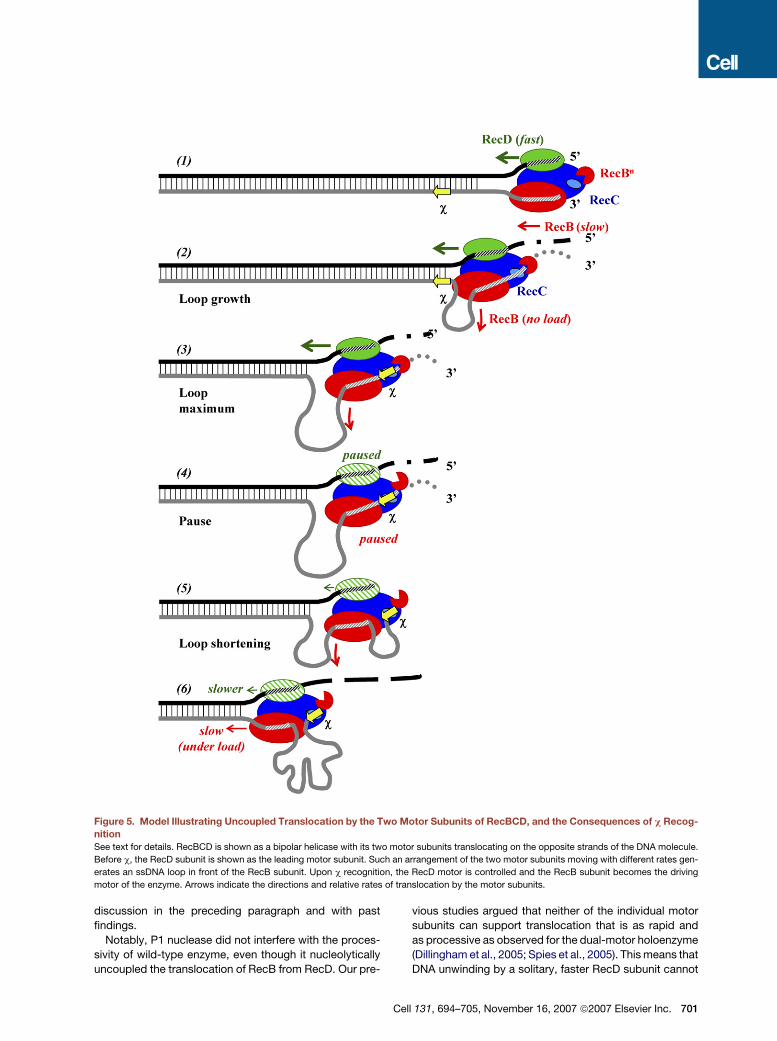
Figure 5. Model Illustrating Uncoupled Translocation by the Two Motor Subunits of RecBCD, and the Consequences of c Recog-
nition
See text for details. RecBCD is shown as a bipolar helicase with its two motor subunits translocating on the opposite strands of the DNA molecule.
Before c, the RecD subunit is shown as the leading motor subunit. Such an arrangement of the two motor subunits moving with different rates gen-
erates an ssDNA loop in front of the RecB subunit. Upon c recognition, the RecD motor is controlled and the RecB subunit becomes the driving
motor of the enzyme. Arrows indicate the directions and relative rates of translocation by the motor subunits.
discussion in the preceding paragraph and with past
findings.
Notably, P1 nuclease did not interfere with the proces-
sivity of wild-type enzyme, even though it nucleolytically
uncoupled the translocation of RecB from RecD. Our pre-
C
vious studies argued that neither of the individual motor
subunits can support translocation that is as rapid and
as processive as observed for the dual-motor holoenzyme
(Dillingham et al., 2005; Spies et al., 2005). This means that
DNA unwinding by a solitary, faster RecD subunit cannot
ell 131, 694–705, November 16, 2007 ª2007 Elsevier Inc. 701
explain the high translocation rate and processivity of
RecBCD. Furthermore, the high processivity in the pres-
ence of P1 nuclease (see Figure 4) implies that, after the
ssDNA loop formed between two subunits is digested
by the nuclease, the 30-terminated strand of the DNA
duplex can be rethreaded into the active site of RecB sub-
unit (Figure 4B). Although our current work does not
further address this issue, this finding is not completely
unexpected because the RecBC enzyme can step across
ssDNA gaps of 22 nucleotides on the 30-terminated strand
and re-engage this discontinuous strand across the gap to
continue unwinding (Bianco and Kowalczykowski, 2000).
Also, the fact that c-modified wild-type enzyme is signifi-
cantly more processive than RecBCDK177Q mutant implies
that, whereas RecB subunit becomes drive motor after c,
the RecD motor is not completely inactivated upon c rec-
ognition and still contributes to processive translocation
(Table 1). This view is consistent with previous single-
molecule work showing that the RecD subunit remains
associated with the c-modified RecBCD (Handa et al.,
2005). Thus, our current data suggest that RecD contrib-
utes to the processive translocation of RecBCD both
before and after c.
The structure of RecBCD bound to dsDNA (Singleton
et al., 2004) reveals the presence of tunnels for each of
the DNA strands after they are separated by a pin struc-
ture in the RecC subunit (see Figure 1A). The 50-terminated
strand passes through the translocation site of the RecD
subunit, whereas the c-containing 30-terminated strand
first passes through the translocation site of the RecB
motor. This strand is then channeled through the enzyme
to the c-recognition site residing in the RecC subunit. Af-
terward, the strands exit the enzyme in proximity to the ac-
tive site of the nuclease domain located in the C terminus
of the RecB subunit. Because the channel for the 30-termi-
nated ssDNA is enclosed within the core of the enzyme,
the RecB motor must actively translocate the c-containing
DNA strand through the channel to the c-recognition site.
The ssDNA loop formed in front of the slower RecB sub-
unit will contain the c sequence at some point during
translocation. Eventually, the c sequence is translocated
into the c-recognition site. We proposed that c sequence
binds to the RecBCD holoenzyme and serves as the allo-
steric effector, which triggers a number of conformational
changes in the enzyme (Kulkarni and Julin, 2004; Single-
ton et al., 2004; Spies et al., 2003; Spies and Kowalczy-
kowski, 2006). One consequence of this conformational
change is a switch in the drive motor of the enzyme—an
unprecedented phenomenon for nucleic acid motor pro-
teins.
Considering the aforementioned discussions, our cur-
rent model for RecBCD function and its control by c
includes elements from previous models and the discov-
eries reported here (Figure 5). In agreement with the
‘‘uncoupled translocation’’ model (Figure 1A), the RecD
motor is responsible for the fast and processive trans-
location of the enzyme before c, resulting in a loop of
ssDNA being formed in front of the RecB subunit (Figure 5,
702 Cell 131, 694–705, November 16, 2007 ª2007 Elsevier Inc
steps 1 and 2; Taylor and Smith, 2003). The size of the loop
and its rate of growth will depend on the velocity of RecB
translocation relative to that of RecD translocation. The
differential sensitivity of these motor subunits to solution
conditions can create loops as large as thousands of nu-
cleotides (Taylor and Smith, 1980, 2003), to inferred sizes
of hundreds of nucleotides, or as little as tens of nucleo-
tides (Anderson et al., 1997; Anderson and Kowalczykow-
ski, 1997a). For the solution conditions employed here,
which are approximately physiological in ATP and Mg2+
concentration, several lines of evidence suggest that the
ssDNA loop is small. First, the duration of pauses are the
same for the wild-type and mutant enzymes, which would
not be the expected result if RecB needed to travel a long
distance to catch up to RecD after the c-induced pause.
Second, as discussed, the loop must be smaller than the
resolution of the single-molecule distance measurement
(i.e., it must be less than �1000 bp) because the pause
occurs at c, within experimental error (Spies et al., 2003).
Third, we have not seen evidence of ssDNA prior to c rec-
ognition in our single-molecule experiments, which would
be expected if there was a significant amount of ssDNA
formed before encountering the nuclease domain in
RecB (Figure 5, step 3; Bianco et al., 2001; Spies et al.,
2003). Finally, the position of the c-induced cleavage on
the strand complementary to the c sequence is consistent
with loop that would range from less than 100 nucleotide
to at most 500 nucleotides (Anderson et al., 1997; Ander-
son and Kowalczykowski, 1997a). Although it is evident
that the size of this ‘‘pre-c’’ ssDNA loop can be regulated,
the precise control of its size is yet to be defined. However,
the size of this loop may be less important than the fact
that its existence means that the unwound ssDNA
strands are not in complementary register when they
exit RecBCD. Thus, spontaneous annealing will be mini-
mized, and the probability of endonucleolytic cleavage
by RecBCD will be maximized. Thus, one simple explana-
tion for the function of the pre-c loop is to enhance endo-
nucleolytic degradation of the ssDNA produced prior to c
recognition by preventing spontaneous renaturation of
DNA strands behind RecBCD.
RecBCD stochastically cleaves the unwound 30-termi-
nated DNA strand endonucleolytically and, less frequently
so, the 50-terminated strand until c is recognized (Figure 5,
step 3; Dixon and Kowalczykowski, 1993, 1995). At this
moment, the enzyme pauses at c, which ensures that
there is a high probability of a cleavage event at the c
sequence; then, the nuclease activity is attenuated overall
and switched to the 50-terminated strand (Figure 5, step
4). At this time, the loop formed prior to c recognition
will have reached its maximum size (Figure 5, step 4).
But the realization is that, depending on the relative veloc-
ities of the motor subunits, the paused position for each
subunit will be at a different location on each DNA strand.
The model shows that the RecB subunit will be paused at
the c sequence but that the RecD subunit will be paused
on the opposite strand some distance downstream of
the c complement; this distance will depend on the size
.

of ssDNA loop, which, in turn, will depend on the relative
translocation velocities of the motor subunits. The model
is fully consistent with biochemical identification of the
‘‘last’’ cleavage event on the c-containing strand being
at c (from 0 to 6 nucleotides upstream of c, depending
on solution conditions) (Dixon and Kowalczykowski,
1993, 1995; Ponticelli et al., 1985; Taylor et al., 1985; Tay-
lor and Smith, 1995) and the ‘‘first’’ cleavage after the po-
larity switch on the complementary strand being at or
several hundred nucleotides downstream of the c-com-
plement sequence (Anderson et al., 1997; Anderson and
Kowalczykowski, 1997a). Binding of the c sequence to
the RecC subunit serves as a cis-acting allosteric modifier
of RecBCD structure (Chedin et al., 2006; Handa et al.,
2005; Singleton et al., 2004; Spies et al., 2003). As a con-
sequence of this structural change, the speed of the RecD
motor is attenuated to be equal to or below that of the
RecB motor, but the change does not involve ejection of
the RecD motor from the holoenzyme at c (Handa et al.,
2005). We suggest that the pause is a measure of the
kinetic lifetime required for this conformational change:
as part of this change, the nuclease domain of RecB is
proposed to undock from RecC, both revealing the cryptic
RecA-loading site (Spies and Kowalczykowski, 2006) and
altering the polarity of DNA degradation (Singleton et al.,
2004; Yu et al., 1998).
After the pause at c, DNA unwinding resumes, but the
c-modified RecBCD is functionally different (Figure 5,
step 5). The 50-terminated strand is now cleaved more fre-
quently than the 30-terminated strand, but the nuclease
activity is attenuated overall (Figure 5, step 6; Anderson
and Kowalczykowski, 1997a). RecB resumes transloca-
tion, and the existing loop between RecB and RecD
shortens as RecB takes up the ssDNA ‘‘slack’’ (Figure 5,
step 5). The c sequence remains bound to the RecC sub-
unit of RecBCD, and therefore, after c, a new ssDNA loop
between the c sequence bound to RecC and the translo-
cating RecB forms and grows with distance traveled
(Spies et al., 2003). The RecB motor now assumes the
responsibility for DNA unwinding, and RecD serves an an-
cillary role that contributes to the enzymes processivity
(Dillingham et al., 2005; Spies et al., 2005). The speed of
RecD translocation relative to RecB is unknown at this
time; however, if the two speeds are identical, then only
the single ssDNA loop shown, involving the c-containing
30-terminated DNA strand, would form; but if the speed
of RecD were slower than that of RecB, then a second
loop would form after c between the RecB and RecD sub-
units on the opposite 50-terminated strand (not illustrated).
This ‘‘post-c’’ ssDNA loop (or loops) is likely a component
of the conformation changes in RecBCD that are elicited
by c recognition (Spies et al., 2003; Spies and Kowalczy-
kowski, 2006). Upon c recognition, the c-containing
ssDNA may begin to exit through an alternative egress
that is created between the RecB and RecC subunits (Sin-
gleton et al., 2004). Minimally, by having the ssDNA
extrude through this new exit, the switched state of
RecBCD is effectively locked until dissociation; further-
C
more, it is possible that the location of this exit facilitates
the loading of RecA by positioning the c-containing
ssDNA near the RecA-loading domain of the RecB sub-
unit. This change in behavior enables the RecB subunit
to begin loading RecA onto the c-containing ssDNA (not
illustrated), thereby allowing the next step of recombina-
tional DNA repair, which is homologous pairing, to ensue
(Anderson and Kowalczykowski, 1997b; Spies and Ko-
walczykowski, 2006). Upon dissociating from the DNA,
the subunits disassemble (Taylor and Smith, 1999), the c
sequence dissociates, and the subunits reassemble to
produce the unmodified RecBCD holoenzyme.
The specific biological reason for this complicated con-
trol of helicase subunit utilization and speed is largely un-
known, but we offer the following observations and spec-
ulations. RecBCD clearly has two seemingly contradictory
biological functions: the destruction of foreign (e.g., phage)
DNA and the repair of chromosomal DNA upon c recogni-
tion. Destruction of foreign dsDNA would be optimal with
a fast helicase coupled to rapid nucleolytic degradation,
whereas repair of chromosomal DNA apparently requires
a slower helicase whose speed is functionally matched to
the relatively slow downstream process of RecA nucleo-
protein filament assembly. Assembly of a RecA nucleo-
protein filament is limited by its nucleation frequency
(Galletto et al., 2006), a step that is facilitated by the c-ac-
tivated RecBCD. Furthermore, because RecBCD must re-
peatedly nucleate RecA filaments for distances as far as
10 Kb downstream of c (Myers et al., 1995), the transloca-
tion velocity of RecBCD must be coordinated to the rates
of RecA nucleoprotein filament nucleation and growth.
Thus, it appears that the slower translocation of the c-
modified RecBCD is a biological requirement of efficient
RecA nucleoprotein filament formation. Furthermore, it is
well established that RecBC, lacking the RecD subunit,
is fully proficient for recombinational DNA repair (Amund-
sen et al., 1986). RecBC does not require c for activation,
being constitutively activated for RecA loading (Churchill
et al., 1999). However, RecBC does not destroy phage
DNA (Chaudhury and Smith, 1984), suggesting that this
simple single-motor enzyme, with little nucleolytic capac-
ity, cannot fulfill the requisite protective function of
RecBCD. Although it would seem that a ‘‘faster’’ version
of RecBC, in conjunction with other cellular nucleases,
could provide this degradative function, it appears instead
that RecBC acquired another motor subunit to provide
both a faster rate of helicase activity and another means
of controlling nuclease activity. Thus, by using the faster
dual motor RecBCD that is also activated for nuclease ac-
tivity, destruction of foreign DNA is assured. However,
through its interaction with a c sequence, both the heli-
case and nuclease activity of RecBCD can be regulated
to switch the enzyme between two seemingly contradic-
tory biological behaviors: the fast, degradative helicase/
nuclease and the slower, DNA-repairing mobile RecA
loader. Thus, we suggest that the biological function of
this motor switch at c is to coordinate the speed of
RecBCD translocation with the loading of RecA onto the
ell 131, 694–705, November 16, 2007 ª2007 Elsevier Inc. 703
c-containing ssDNA. The slower speed would facilitate
a frequency of RecA nucleation onto the c-containing
ssDNA that is sufficiently recurrent to ensure the discon-
tinuous assembly of the RecA nucleoprotein filament
over distances of 10 kb, an assembly that is essential to
complete the recombinational repair of broken chromo-
somal DNA.
EXPERIMENTAL PROCEDURES
Proteins and DNA Substrates
RecBCD and RecBCDK177Q were purified by published protocols (Dil-
lingham et al., 2003; Roman and Kowalczykowski, 1989). P1 nuclease
was from Roche.
Chi-containing dsDNA substrates were produced as described pre-
viously (Spies et al., 2003) with minor modifications. Biotinylated
dsDNA was produced by amplification of a 30 kb region of c-contain-
ing l DNA purified with the Lambda Purification Kit from QIAGEN
(Spies et al., 2003). EXL polymerase (Stratagene) and a biotinylated
(50-bio-AGTATCGGTAAGGCGGTGAC-30) and a nonbiotinylated (50-
GCCCATGACAGGAAGTTGTT-30) primer were used for the PCR
reaction. The PCR product was 30,870 bp in length and contained
a c-recognition locus (three consecutive c sequences spaced 10
nucleotides apart) at 5.26 kb from the nonbiotinylated end. Full-length
dsDNA was separated from shorter PCR products by electrophoresis
in 0.8% agarose and electro-elution with Gene Capsule DNA extrac-
tion kit (Genotech).
DNA Bead Preparation
The protocol for DNA bead preparation was modified from Spies et al.
(2003). The biotinylated DNA (�25 ng at �1 ng/ ml) was incubated with
5 ml of 1 mm, ‘‘ProActive’’ streptavidin-coated microspheres (Bangs
Laboratories) for 1 hr on ice in 80 mM NaHCO3 (pH 8.2). Bead-DNA
complexes were transferred into 1.5 ml of degassed ‘‘sample solution’’
containing 45 mM NaHCO3 (pH 8.2), 20% (w/v) sucrose, 50 mM DTT,
and 100 nM YOYO-1 (Molecular Probes). Immediately before transfer
to the sample syringe, 2 mM magnesium acetate and 50 nM wild-type
or mutant RecBCD enzymes were added. The reaction solution con-
tained 45 mM NaHCO3 (pH 8.2), 20% (w/v) sucrose, 50 mM DTT,
1 mM ATP, 2 mM magnesium acetate, and 20 nM YOYO-1. When in-
dicated, P1 nuclease was added to the reaction solution to a final con-
centration of 25 units/ml.
Optical Trapping and Fluorescence Microscopy
Reactions were performed as described (Bianco et al., 2001; Spies
et al., 2003) but with the following modifications. A new instrument in-
cluded a Nikon TE2000-U inverted microscope, YOYO-1 was exited
with a 488 nm laser, and two neutral density filters combined with
highest sensitivity settings on camera were used to ensure maximum
fluorescence with minimum photobleaching.
Data Analysis
Videos of the enzyme translocation were recorded at 10 frames per
second with Scion Image Software. Every 5 frames were averaged
with an ImageJ plug-in to reduce background and create 2 frames/s
movies. The length of the DNA molecule in each frame was measured
with a plug-in written in this laboratory (B. Liu and S.C.K., unpublished).
The rates before and after c, as well as position and duration of the
pause, were determined by fitting experimental data to a contiguous
five-segment line with GraphPad Prism Software.
Supplemental Data
Four tables are available at http://www.cell.com/cgi/content/full/131/
4/694/DC1/.
704 Cell 131, 694–705, November 16, 2007 ª2007 Elsevier Inc.
ACKNOWLEDGMENTS
We are grateful to Clarke Conant, Petr Cejka, Mark Dillingham, An-
thony Forget, Jovencio Hilario, Ryan Jensen, Bian Liu, Edgar Valen-
cia-Morales, Jody Plank, Behzad Rad, and Jason Wong; to the mem-
bers of Spies lab for their critical reading of the manuscript; and to
Martin Singleton for the structure of RecBCD in Figure 1. This work
was supported by National Institutes of Health grant GM-41347 to
S.C.K. and by American Cancer Society Postdoctoral Fellowship
PF-02-116-01-GMC to M.S.
Received: May 19, 2007
Revised: August 28, 2007
Accepted: September 13, 2007
Published: November 15, 2007
REFERENCES
Amundsen, S.K., Taylor, A.F., Chaudhury, A.M., and Smith, G.R.
(1986). recD: the gene for an essential third subunit of exonuclease
V. Proc. Natl. Acad. Sci. USA 83, 5558–5562.
Anderson, D.G., and Kowalczykowski, S.C. (1997a). The recombina-
tion hot spot c is a regulatory element that switches the polarity of
DNA degradation by the RecBCD enzyme. Genes Dev. 11, 571–581.
Anderson, D.G., and Kowalczykowski, S.C. (1997b). The translocating
RecBCD enzyme stimulates recombination by directing RecA protein
onto ssDNA in a c-regulated manner. Cell 90, 77–86.
Anderson, D.G., Churchill, J.J., and Kowalczykowski, S.C. (1997). Chi-
activated RecBCD enzyme possesses 50/30 nucleolytic activity, but
RecBC enzyme does not: evidence suggesting that the alteration
induced by Chi is not simply ejection of the RecD subunit. Genes Cells
2, 117–128.
Bianco, P.R., and Kowalczykowski, S.C. (2000). Translocation step
size and mechanism of the RecBC DNA helicase. Nature 405, 368–
372.
Bianco, P.R., Brewer, L.R., Corzett, M., Balhorn, R., Yeh, Y., Kowalc-
zykowski, S.C., and Baskin, R.J. (2001). Processive translocation and
DNA unwinding by individual RecBCD enzyme molecules. Nature 409,
374–378.
Braedt, G., and Smith, G.R. (1989). Strand specificity of DNA unwind-
ing by RecBCD enzyme. Proc. Natl. Acad. Sci. USA 86, 871–875.
Chaudhury, A.M., and Smith, G.R. (1984). A new class of Escherichia
coli recBC mutants: implications for the role of RecBC enzyme in ho-
mologous recombination. Proc. Natl. Acad. Sci. USA 81, 7850–7854.
Chedin, F., Handa, N., Dillingham, M.S., and Kowalczykowski, S.C.
(2006). The AddAB helicase/nuclease forms a stable complex with
its cognate c sequence during translocation. J. Biol. Chem. 281,
18610–18617.
Churchill, J.J., Anderson, D.G., and Kowalczykowski, S.C. (1999). The
RecBC enzyme loads RecA protein onto ssDNA asymmetrically and
independently of Chi, resulting in constitutive recombination activa-
tion. Genes Dev. 13, 901–911.
Dillingham, M.S., Spies, M., and Kowalczykowski, S.C. (2003).
RecBCD enzyme is a bipolar DNA helicase. Nature 423, 893–897.
Dillingham, M.S., Webb, M.R., and Kowalczykowski, S.C. (2005). Bi-
polar DNA translocation contributes to highly processive DNA unwind-
ing by RecBCD enzyme. J. Biol. Chem. 280, 37069–37077.
Dixon, D.A., and Kowalczykowski, S.C. (1991). Homologous pairing in
vitro stimulated by the recombination hotspot, Chi. Cell 66, 361–371.
Dixon, D.A., and Kowalczykowski, S.C. (1993). The recombination hot-
spot c is a regulatory sequence that acts by attenuating the nuclease
activity of the E. coli RecBCD enzyme. Cell 73, 87–96.

Dixon, D.A., and Kowalczykowski, S.C. (1995). Role of the Escherichia
coli recombination hotspot, c, in RecABCD-dependent homologous
pairing. J. Biol. Chem. 270, 16360–16370.
Galletto, R., Amitani, I., Baskin, R.J., and Kowalczykowski, S.C. (2006).
Direct observation of individual RecA filaments assembling on single
DNA molecules. Nature 443, 875–878.
Handa, N., Bianco, P.R., Baskin, R.J., and Kowalczykowski, S.C.
(2005). Direct visualization of RecBCD movement reveals cotransloca-
tion of the RecD motor after c recognition. Mol. Cell 17, 745–750.
Korangy, F., and Julin, D.A. (1992). Alteration by site-directed muta-
genesis of the conserved lysine residue in the ATP-binding consensus
sequence of the RecD subunit of the Escherichia coli RecBCD enzyme.
J. Biol. Chem. 267, 1727–1732.
Kulkarni, A., and Julin, D.A. (2004). Specific inhibition of the E. coli
RecBCD enzyme by Chi sequences in single-stranded oligodeoxyribo-
nucleotides. Nucleic Acids Res. 32, 3672–3682.
Lam, S.T., Stahl, M.M., McMilin, K.D., and Stahl, F.W. (1974). Rec-me-
diated recombinational hot spot activity in bacteriophage lambda. II. A
mutation which causes hot spot activity. Genetics 77, 425–433.
Muskavitch, K.M., and Linn, S. (1982). A unified mechanism for the nu-
clease and unwinding activities of the recBC enzyme of Escherichia
coli. J. Biol. Chem. 257, 2641–2648.
Myers, R.S., Stahl, M.M., and Stahl, F.W. (1995). Chi recombination
activity in phage lambda decays as a function of genetic distance.
Genetics 141, 805–812.
Ponticelli, A.S., Schultz, D.W., Taylor, A.F., and Smith, G.R. (1985).
Chi-dependent DNA strand cleavage by RecBC enzyme. Cell 41,
145–151.
Roman, L.J., and Kowalczykowski, S.C. (1989). Characterization of the
helicase activity of the Escherichia coli RecBCD enzyme using a novel
helicase assay. Biochemistry 28, 2863–2873.
Roman, L.J., Eggleston, A.K., and Kowalczykowski, S.C. (1992). Proc-
essivity of the DNA helicase activity of Escherichia coli recBCD
enzyme. J. Biol. Chem. 267, 4207–4214.
Romier, C., Dominguez, R., Lahm, A., Dahl, O., and Suck, D. (1998).
Recognition of single-stranded DNA by nuclease P1: high resolution
crystal structures of complexes with substrate analogs. Proteins 32,
414–424.
Singleton, M.R., Dillingham, M.S., Gaudier, M., Kowalczykowski, S.C.,
and Wigley, D.B. (2004). Crystal structure of RecBCD enzyme reveals
a machine for processing DNA breaks. Nature 432, 187–193.
Spies, M., and Kowalczykowski, S.C. (2005). Homologous recombina-
tion by RecBCD and RecF pathways. In The Bacterial Chromosome,
N.P. Higgins, ed. (Washington, D.C.: ASM Press), pp. 389–403.
Spies, M., and Kowalczykowski, S.C. (2006). The RecA binding locus
of RecBCD is a general domain for recruitment of DNA strand
exchange proteins. Mol. Cell 21, 573–580.
Spies, M., Bianco, P.R., Dillingham, M.S., Handa, N., Baskin, R.J., and
Kowalczykowski, S.C. (2003). A molecular throttle: the recombination
hotspot c controls DNA translocation by the RecBCD helicase. Cell
114, 647–654.
Spies, M., Dillingham, M.S., and Kowalczykowski, S.C. (2005). Trans-
location by the RecB motor Is an absolute requirement for c-recogni-
tion and RecA protein loading by RecBCD enzyme. J. Biol. Chem. 280,
37078–37087.
Taylor, A., and Smith, G.R. (1980). Unwinding and rewinding of DNA by
the RecBC enzyme. Cell 22, 447–457.
Taylor, A.F., and Smith, G.R. (1995). Strand specificity of nicking of
DNA at Chi sites by RecBCD enzyme: modulation by ATP and magne-
sium levels. J. Biol. Chem. 270, 24459–24467.
Taylor, A.F., and Smith, G.R. (1999). Regulation of homologous recom-
bination: Chi inactivates RecBCD enzyme by disassembly of the three
subunits. Genes Dev. 13, 890–900.
Taylor, A.F., and Smith, G.R. (2003). RecBCD enzyme is a DNA heli-
case with fast and slow motors of opposite polarity. Nature 423,
889–893.
Taylor, A.F., Schultz, D.W., Ponticelli, A.S., and Smith, G.R. (1985).
RecBC enzyme nicking at Chi sites during DNA unwinding: location
and orientation-dependence of the cutting. Cell 41, 153–163.
Yu, M., Souaya, J., and Julin, D.A. (1998). The 30-kDa C-terminal do-
main of the RecB protein is critical for the nuclease activity, but not the
helicase activity, of the RecBCD enzyme from Escherichia coli. Proc.
Natl. Acad. Sci. USA 95, 981–986.
Cell 131, 694–705, November 16, 2007 ª2007 Elsevier Inc. 705