Shedding light on the system: studying embryonic ... Lab/Tomer...light on the system: Studying...
8
Available online at www.sciencedirect.com Shedding light on the system: Studying embryonic development with light sheet microscopy Raju Tomer, Khaled Khairy and Philipp J Keller Light sheet-based fluorescence microscopy (LSFM) is emerging as a powerful imaging technique for the life sciences. LSFM provides an exceptionally high imaging speed, high signal-to-noise ratio, low level of photo-bleaching and good optical penetration depth. This unique combination of capabilities makes light sheet-based microscopes highly suitable for live imaging applications. There is an outstanding potential in applying this technology to the quantitative study of embryonic development. Here, we provide an overview of the different basic implementations of LSFM, review recent technical advances in the field and highlight applications in the context of embryonic development. We conclude with a discussion of promising future directions. Address Janelia Farm Research Campus, Howard Hughes Medical Institute, 19700 Helix Drive, Ashburn, VA 20147, USA Corresponding authors: Tomer, Raju ([email protected]), Keller, Philipp J ([email protected]) Current Opinion in Genetics & Development 2011, 21:558–565 This review comes from a themed issue on Developmental mechanisms, patterning and evolution Edited by Sean Megason, Shankar Srinivas, Mary Dickinson and Anna-Katerina Hadjantonakis Available online 19th August 2011 0959-437X/$ – see front matter # 2011 Elsevier Ltd. All rights reserved. DOI 10.1016/j.gde.2011.07.003 Introduction Non-invasive three-dimensional imaging over time is indispensable for a quantitative understanding of bio- logical processes at multiple scales, from molecular inter- actions to tissue morphogenesis. In particular, the in vivo study of early embryogenesis requires state-of-the-art imaging strategies that achieve high spatiotemporal resol- ution without compromising specimen integrity. Progress in sensor technologies, lasers and desktop computing in the past decades has led to a remarkable progress in imaging technologies and capabilities. Specifically, the emergence of Confocal Laser-Scanning Fluorescence Microscopy (CLSM) provided means for non-invasive imaging of fixed as well as live samples for three-dimensional reconstructions and, therefore, confo- cal microscopes have become standard instruments in many laboratories. However, the ever-increasing demand to image for longer periods of time and at higher spatio- temporal resolution is rapidly exposing the limitations of CLSM. The point-scanning implementation of CLSM is inherently slow and causes high levels of photo-bleaching and photo-toxicity, owing to the iterative use of non- selective excitation with high-power beams. Additionally, tissue penetration depth is relatively low. These issues inspired the development of non-linear microscopy, specifically two-photon microscopy, which provided a substantial increase in the penetration depth and reduction in photo-bleaching, albeit, at the expense of spatial resolution and imaging speed. In the past decade, Light Sheet-Based Fluorescence Microscopy (LSFM) has emerged to fill the gap resulting from the inherent limitations of CLSM and point-scanning two-photon microscopy. The key concept behind LSFM is sample illumination in a thin volume section from the side, and fluorescence detection with an independent optical subsystem at a right angle to the illumination axis. This is in contrast to confocal microscopy and conventional wide- field microscopy, which typically use the same objective lens for illumination and detection. By illuminating only the in-focus plane, LSFM provides intrinsic optical sec- tioning and enables simultaneous detection of the fluor- escence signal from an entire plane with highly efficient detectors. Thereby, LSFM combines several crucial prop- erties, including high acquisition speed, high signal-to- noise ratio, minimal levels of photo-bleaching and good penetration depth. Moreover, sample preparation typically involves the use of low-concentration agarose cylinders for sample embedding, which represent a less stressful environment for live biological samples than the tradition- ally employed glass slide/coverslip. LSFM is thus naturally well-suited for studies of early embryogenesis, since it combines high-content dynamic imaging with a more physiological imaging environment. Here, we discuss the various implementations of LSFM, review recent technical advances in the field, and high- light novel applications in the study of early embryogen- esis. Other aspects of light sheet microscopy have been reviewed elsewhere [1–6]. Implementations of light sheet-based fluorescence microscopy LSFM development has accelerated in the past decade. Many different variants with specialized features have emerged. However, the basic concept remains similar to the design of the first light sheet microscope by Current Opinion in Genetics & Development 2011, 21:558–565 www.sciencedirect.com
Transcript of Shedding light on the system: studying embryonic ... Lab/Tomer...light on the system: Studying...
Available online at www.sciencedirect.com
Shedding light on the system: Studying embryonic developmentwith light sheet microscopyRaju Tomer, Khaled Khairy and Philipp J Keller
Light sheet-based fluorescence microscopy (LSFM) is
emerging as a powerful imaging technique for the life sciences.
LSFM provides an exceptionally high imaging speed, high
signal-to-noise ratio, low level of photo-bleaching and good
optical penetration depth. This unique combination of
capabilities makes light sheet-based microscopes highly
suitable for live imaging applications. There is an outstanding
potential in applying this technology to the quantitative study of
embryonic development. Here, we provide an overview of the
different basic implementations of LSFM, review recent
technical advances in the field and highlight applications in the
context of embryonic development. We conclude with a
discussion of promising future directions.
Address
Janelia Farm Research Campus, Howard Hughes Medical Institute,
19700 Helix Drive, Ashburn, VA 20147, USA
Corresponding authors: Tomer, Raju ([email protected]), Keller,
Philipp J ([email protected])
Current Opinion in Genetics & Development 2011, 21:558–565
This review comes from a themed issue on
Developmental mechanisms, patterning and evolution
Edited by Sean Megason, Shankar Srinivas, Mary Dickinson
and Anna-Katerina Hadjantonakis
Available online 19th August 2011
0959-437X/$ – see front matter
# 2011 Elsevier Ltd. All rights reserved.
DOI 10.1016/j.gde.2011.07.003
IntroductionNon-invasive three-dimensional imaging over time is
indispensable for a quantitative understanding of bio-
logical processes at multiple scales, from molecular inter-
actions to tissue morphogenesis. In particular, the in vivostudy of early embryogenesis requires state-of-the-art
imaging strategies that achieve high spatiotemporal resol-
ution without compromising specimen integrity. Progress
in sensor technologies, lasers and desktop computing in
the past decades has led to a remarkable progress in
imaging technologies and capabilities.
Specifically, the emergence of Confocal Laser-Scanning
Fluorescence Microscopy (CLSM) provided means for
non-invasive imaging of fixed as well as live samples for
three-dimensional reconstructions and, therefore, confo-
cal microscopes have become standard instruments in
Current Opinion in Genetics & Development 2011, 21:558–565
many laboratories. However, the ever-increasing demand
to image for longer periods of time and at higher spatio-
temporal resolution is rapidly exposing the limitations of
CLSM. The point-scanning implementation of CLSM is
inherently slow and causes high levels of photo-bleaching
and photo-toxicity, owing to the iterative use of non-
selective excitation with high-power beams. Additionally,
tissue penetration depth is relatively low. These issues
inspired the development of non-linear microscopy,
specifically two-photon microscopy, which provided a
substantial increase in the penetration depth and
reduction in photo-bleaching, albeit, at the expense of
spatial resolution and imaging speed.
In the past decade, Light Sheet-Based Fluorescence
Microscopy (LSFM) has emerged to fill the gap resulting
from the inherent limitations of CLSM and point-scanning
two-photon microscopy. The key concept behind LSFM is
sample illumination in a thin volume section from the side,
and fluorescence detection with an independent optical
subsystem at a right angle to the illumination axis. This is in
contrast to confocal microscopy and conventional wide-
field microscopy, which typically use the same objective
lens for illumination and detection. By illuminating only
the in-focus plane, LSFM provides intrinsic optical sec-
tioning and enables simultaneous detection of the fluor-
escence signal from an entire plane with highly efficient
detectors. Thereby, LSFM combines several crucial prop-
erties, including high acquisition speed, high signal-to-
noise ratio, minimal levels of photo-bleaching and good
penetration depth. Moreover, sample preparation typically
involves the use of low-concentration agarose cylinders for
sample embedding, which represent a less stressful
environment for live biological samples than the tradition-
ally employed glass slide/coverslip. LSFM is thus naturally
well-suited for studies of early embryogenesis, since it
combines high-content dynamic imaging with a more
physiological imaging environment.
Here, we discuss the various implementations of LSFM,
review recent technical advances in the field, and high-
light novel applications in the study of early embryogen-
esis. Other aspects of light sheet microscopy have been
reviewed elsewhere [1–6].
Implementations of light sheet-basedfluorescence microscopyLSFM development has accelerated in the past decade.
Many different variants with specialized features have
emerged. However, the basic concept remains similar
to the design of the first light sheet microscope by
www.sciencedirect.com
Embryonic development and light sheet microscopy Tomer, Khairy and Keller 559
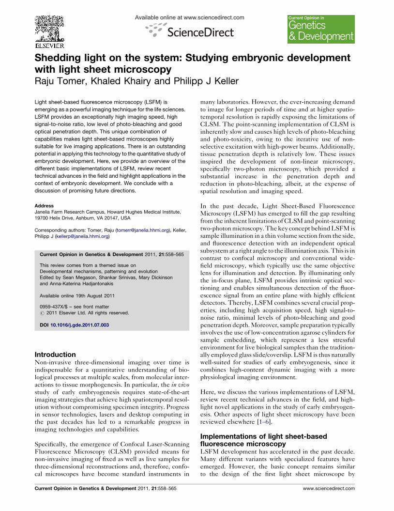
Figure 1
DetectionObjecti ve
Light sheet
Sample
Detection
Illumination
Current Opinion in Genetics & Development
Light sheet-based microscopy. The central concept behind light sheet-based microscopy is specimen illumination in a single plane with a thin sheet of
laser light and recording of the fluorescence emitted by fluorophores in this thin section with a camera-based detection system oriented at a right angle
to the light sheet. The optical subsystems for illumination and detection are decoupled, which allows using separate objectives optimized for low-NA
specimen illumination and high-NA fluorescence detection. The light sheet is typically generated by scanning a pencil beam through the sample or by
focusing a Gaussian beam along one direction into a sheet, using a suitable optical element such as a cylindrical lens.
Siedentopf and Zsigmondy [7]: the sample is illuminated
with a sheet of light that overlaps with the focal plane of
the detection optics (Figure 1).
Almost a century later, Voie et al. developed Orthogonal-
Plane Fluorescence Optical Sectioning (OPFOS) [8].
The authors used a cylindrical lens to create a light sheet
and imaged the internal architecture of the cochlea by
scanning the sample through the sheet, achieving lateral
and axial resolutions of 10 mm and 26 mm, respectively,
for a field-of-view (FOV) of 1.5 mm. The shape of the
light sheet can be well approximated by Gaussian beam
optics and, thus, an intrinsic tradeoff exists between the
central thickness of the light sheet, which directly influ-
ences axial resolution, and the size of the FOV within
which the sheet’s thickness remains sufficiently uniform.
A high-resolution extension of OPFOS (HR-OPFOS) [9]
was developed more recently, in which the authors used a
large aperture lens to generate a thin light sheet. By
moving the (fixed) sample stepwise along the illumina-
tion axis and recording the fluorescence signal from the
thinnest part of the light sheet at each step, large speci-
mens could be recorded with excellent axial resolution.
www.sciencedirect.com
Thin Light Sheet Microscopy (TSLM) was developed to
observe microbes in their natural setting with a milli-
meter-sized FOV [10]. Ultramicroscopy is a similar imple-
mentation optimized for imaging large fixed samples [11].
The sample is placed into a chamber containing the
clearing solution with matched refractive index. Sample
illumination is performed from two sides with aligned
light sheets generated by cylindrical lenses. Both HR-
OPFOS and Ultramicroscopy excel at imaging large fixed
samples, but they are incompatible with the in vivoimaging of dynamic biological samples.
Selective Plane Illumination Microscopy (SPIM) was
developed as an implementation of LSFM for non-inva-
sive live imaging of early embryos [12]. Using fast CCD
cameras and agarose cylinders for sample mounting, Huis-
ken et al. reported long-term imaging of Drosophila embryo-
nic development and high-speed imaging of the beating
heart of a Medaka fish embryo. Since the one-sided illu-
mination used in SPIM was often accompanied by shading
effects resulting in stripe artifacts in the recorded images,
the basic SPIM setup was further extended by multi-
directional illumination (mSPIM) [13].
Current Opinion in Genetics & Development 2011, 21:558–565

560 Developmental mechanisms, patterning and evolution
In addition, multi-view reconstructions and image decon-
volution have been used to improve image quality
[14,15]. In multi-view imaging, different views of the
sample are obtained by rotating the sample. These data
sets can then be combined computationally to achieve
isotropic resolution in transparent samples [14] or to
maximize coverage in partially opaque specimens [16,17].
Keller et al. reported the first beam-scanning implementa-
tion of light sheet-based microscopy (Digital Scanned
Laser Light Sheet Fluorescence Microscopy, DSLM)
[16] and used it to perform fast long-term imaging of
entire developing zebrafish embryos. DSLM provides
uniform intensity levels across the FOV, minimization
of energy losses in the illumination process, a high-quality
implementation of image contrast-enhancing structured
illumination, and the capability to scan the light sheet
through the sample for high-speed three-dimensional
imaging. The beam-scanning approach introduced in
DSLM has also been used as a basis for new LSFM
techniques, such as Bessel plane illumination and effi-
cient two-photon excitation [18��,19��].
Other LSFM implementations include Objective Coupled
Plane Illumination (OCPI) [20], Highly Inclined Lami-
nated Optical Microscopy (HILO) [21] and Oblique Plane
Microscopy (OPM) [22]. In OCPI, the light sheet generat-
ing optics is coupled with the detection objective. The
authors applied their technique to the fast imaging of
neuronal activity in the mouse vomeronasal organ. HILO
uses the same objective for illumination and detection. A
highly inclined sheet of light is created and fluorescence
observation is limited to a small FOV, where the light sheet
and focal plane of the detection system overlap reasonably
well. The basic HILO implementation has been extended
in OPM, which enables an oblique plane in the specimen
to be illuminated as well as imaged with the same objective
lens. This approach allows recording a large FOV using
conventional glass slide-based sample preparation.
Recent advances in light sheet-basedfluorescence microscopyMany aspects of LSFM have been improved over the past
few years, including the development of better illumina-
tion and detection optics, instrument miniaturization,
integration of other modalities, such as Fluorescence Cor-
relation Spectroscopy (FCS), and efforts toward resolution
enhancement and implementation of adaptive optics. Two
particularly noteworthy improvements were recently
reported: first, the use of self-reconstructing Bessel beams
to create thinner light sheets and to reduce image quality
degradation by light scattering, and second, the use of two-
photon excitation in scanned light sheet microscopy.
Bessel beams are created by projecting an annular pattern
at the rear pupil of an illumination objective. The central
peak width is determined by the thickness of the annulus
Current Opinion in Genetics & Development 2011, 21:558–565
and can be decoupled from the Bessel beam’s longitudi-
nal extent. Fahrbach et al. conducted experiments with
holographically shaped Bessel beams to demonstrate the
self-reconstructing properties in three-dimensional scat-
tering tissues, which reduce light scattering-induced im-
age artifacts and increase penetration depth [18��,23].
The authors reported a prototype implementation
(Microscope with Self-Reconstructing Beams; MISERB),
which employs a Spatial Light Modulator (SLM) to
generate scanned light sheets using Bessel beams.
Planchon et al. used an annular mask and an axicon to
generate scanned light sheets from large numerical aper-
ture Bessel beams [19��]. Bessel beams contain a sub-
stantial amount of energy in side lobes, which – if
unaccounted for – lead to reduced axial resolution in
light sheet-based microscopy in comparison to a Gaussian
beam implementation. To address this problem, the
authors used structured illumination and two-photon
excitation, which eliminated the signal contribution of
the side lobes to the recorded fluorescence images. The
overall result is a marked improvement in axial resolution.
Planchon et al. applied their technique to high-speed
volumetric imaging of chromosomes in mitosis, three-
dimensional imaging of protein pairs with isotropic resol-
ution, and the observation of membrane dynamics in live
cells. Photo-bleaching rates in scanned Bessel beam-
based light sheet microscopes are significantly higher
than those obtained with the scanned Gaussian beams
used in DSLM. Thus, further experiments are required to
determine if these principles can be extended to the live
imaging of larger samples, such as entire developing
embryos, over long periods of time.
Two-photon excitation has been implemented in scan-
ning as well as non-scanning light sheet microscopy
[19��,24�]. These implementations combine the intrinsic
advantages of light sheet microscopy with the increased
penetration depth resulting from the use of an infrared
laser beam for fluorescence excitation.
Improvements in axial resolution can also be achieved by
imaging the sample through the thinnest part of a light
sheet created with Gaussian beams. Buytaert et al. used
large aperture cylindrical lenses to generate highly
focused light sheets and moved their sample step by step
along the illumination axis. By stitching the resulting
images, they obtained high axial resolution over a large
FOV (HR-OPFOS) [9,25]. Similarly, Santi et al. reported
TSLIM (Thin-Sheet Laser Imaging Microscope), where
bi-directional light sheet illumination is used and the
sample is moved through the thinnest part of the sheet
to record a large FOV at high axial resolution [26]. Schacht
et al. further improved TSLIM to provide a 70% reduction
in scanning time and a 63% reduction in photo-bleaching
[27]. These approaches are very useful for imaging large
fixed samples at micron axial resolution. However, the
www.sciencedirect.com
Embryonic development and light sheet microscopy Tomer, Khairy and Keller 561
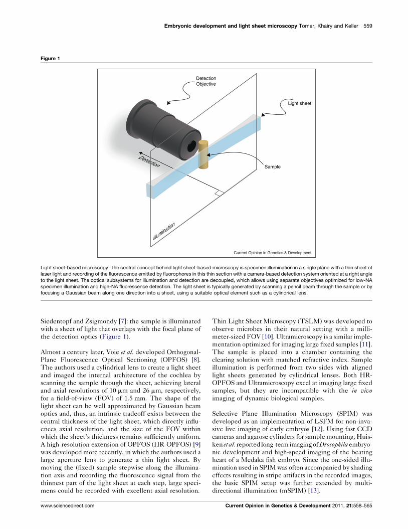
Figure 2
Current Opinion in Genetics & Development
(c)
(d)
(a) (b)
Light sheet-based imaging of gene expression patterns in Drosophila
embryos. Three-dimensional rendering of a late-stage Drosophila
pFlyFos-CG4702 embryo probed with anti-GFP antibody and DRAQ5
nuclear marker. The specimen was imaged with SPIM. Frontal (a),
caudal (b), lateral (c) and ventral (d) views of the same embryo are
shown. Scale bar = 50 mm. Credits: Reprinted from Nature Methods, vol.
6 no. 6, Ejsmont et al., ‘‘A toolkit for high-throughput, cross-species
gene engineering in Drosophila’’, 331–336, Copyright (2009), with
permission from Macmillan Publishers Ltd.
relatively slow acquisition speeds and high rates of photo-
bleaching limit their applicability to live specimens.
Building upon the first implementation of scanned light
sheet microscopy (DSLM) [16], Keller et al. implemented
incoherent structured illumination in DSLM (DSLM-SI)
to eliminate the contribution of scattered light to the
fluorescence signal in partially opaque specimens [28��].The authors demonstrated substantial improvements in
signal-to-noise ratio, image contrast and lateral resolution,
which allowed them to perform high-speed long-term
imaging of zebrafish embryos and highly light-scattering
Drosophila embryos. Similarly, Mertz et al. used scanned
light sheet microscopy and a combination of uniform and
incoherently modulated illumination to reject scattered
signal light in their recordings of mouse brain tissue [29].
Lei et al. used a symmetric four-faceted pyramid to obtain
a coherent three-dimensional structured illumination pat-
tern. Combining this multi-layered light sheet geometry
with the SI reconstruction algorithm introduced by Neil
et al. [30] allowed the authors to discard out-of-focus light
and improve axial resolution [31].
Some applications benefit greatly from miniaturized
implementations of LSFM. Turaga et al. used an uniaxial
gradient-index lens (GRIN), which replaces the cylindrical
lens, to generate a light sheet in a miniaturized imple-
mentation of OCPI [32]. Similarly, Engelbrecht et al. used
single-mode optical fibers, GRINs and a right-angle micro-
prism to generate a light sheet in a miniSPIM setup [33�].
In addition to two-photon excitation, several other con-
cepts have been successfully combined with LSFM.
Friedrich et al. integrated stimulated emission depletion
(STED) in SPIM (STED-SPIM) and achieved a 60%
enhancement in axial resolution [34]. Turaga et al. intro-
duced adaptive optics in light sheet microscopy. The
authors implemented an image-based wavefront sensor
using a variant of generalized phase-diverse imaging
called multi-frame blind deconvolution to calibrate the
deformable mirrors in an OCPI setup [35]. Wohland et al.combined SPIM with Fluorescence Correlation Spec-
troscopy (SPIM-FCS) and reported its in vivo application
by imaging microspheres injected into the blood stream
of a zebrafish embryo [36].
Applications in embryonic developmentLSFM is particularly well suited for fast and long-term
imaging of developmental processes at multiple spatial
and temporal scales. Its applications range from imaging
molecular interactions and gene expression patterns
(Figure 2) to the reconstructions of early developmental
dynamics (Figure 3) and in toto imaging of large fixed
specimens (Figure 4).
Taking advantage of the high imaging speed and excel-
lent signal-to-noise ratio of LSFM, Ritter et al. observed
www.sciencedirect.com
single molecules as deep as 200 mm within salivary
gland tissue of C. tentans larvae [37,38]. The authors
estimated the intra-nuclear viscosity parameters by ima-
ging tracer molecules within the cell nuclei and were
able to visualize the transport of single mRNA mol-
ecules tagged by fluorescently labeled RNA binding
proteins. This study shows the potential of using LSFM
to investigate gene activity in developing embryos at
single molecule resolution.
At the scale of entire embryos, Keller et al. used DSLM
to image early development of wild-type and mutant
Current Opinion in Genetics & Development 2011, 21:558–565
562 Developmental mechanisms, patterning and evolution
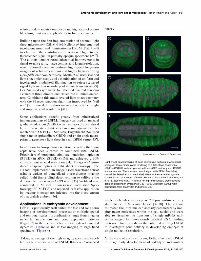
Figure 3
Current Opinion in Genetics & Development
(a) (b)
(d)(c)
Global cell tracking in zebrafish embryos with scanned light sheet microscopy. DSLM microscopy data (right half of embryo: animal view, maximum-
intensity projection) and the subsequently reconstructed ‘digital embryo’ (left half of embryo) with color-encoded cell migration directions. Time points:
289 min post fertilization (mpf) (a), 368 mpf (b), 599 mpf (c), 841 mpf (d). Color code: dorsal migration (cyan), ventral migration (green), toward or away
from body axis (red or yellow), toward yolk (pink). Objective: Carl Zeiss C-Apochromat 10�. Databases and high-resolution movies of the digital
zebrafish embryo are available at http://www.digital-embryo.org.
Credits: Reprinted from Science, vol. 322, Keller et al., ‘‘Reconstruction of Zebrafish Early Embryonic Development by Scanned Light Sheet
Microscopy’’, 1065–1069, Copyright (2008), with permission from AAAS.
zebrafish for 24 h [16]. The authors performed compu-
tational reconstructions of the microscopy data sets to
generate ‘digital embryos’ (www.digital-embryo.org),
which comprise quantitative information on cell coordi-
nates, migratory tracks and cell division patterns
for most of the cells in the developing embryo
(Figure 3). This approach provided a detailed model
of germ layer formation and revealed an initial morpho-
dynamic symmetry break in the patterns of cell division,
which identifies the orientation of the dorso-ventral
body axis. Taking advantage of the capabilities of inco-
Current Opinion in Genetics & Development 2011, 21:558–565
herent structured illumination, Keller et al. extended
DSLM to DSLM-SI, to image and reconstruct early
embryogenesis of partially opaque Drosophila embryos
[28��]. Recently, Swoger et al. used SPIM to image
and quantitatively analyze posterior lateral line (pLL)
organogenesis in zebrafish [39�]. The authors used a
triple-label fluorescence marker strategy for recording
4D data sets of the migration of the primordium and
maturation of proneuromasts. The retrospective track-
ing analysis enabled the identification of hair cell pro-
genitor lineages.
www.sciencedirect.com
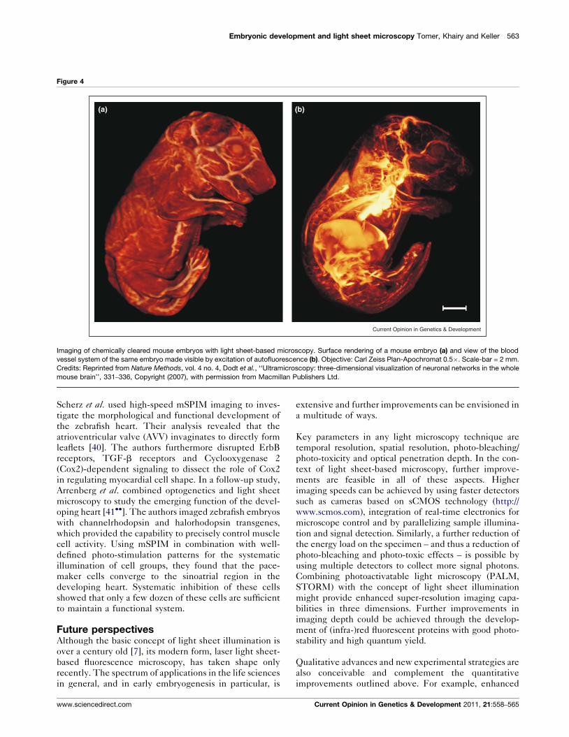
Embryonic development and light sheet microscopy Tomer, Khairy and Keller 563
Figure 4
Current Opinion in Genetics & Development
(a) (b)
Imaging of chemically cleared mouse embryos with light sheet-based microscopy. Surface rendering of a mouse embryo (a) and view of the blood
vessel system of the same embryo made visible by excitation of autofluorescence (b). Objective: Carl Zeiss Plan-Apochromat 0.5�. Scale-bar = 2 mm.
Credits: Reprinted from Nature Methods, vol. 4 no. 4, Dodt et al., ‘‘Ultramicroscopy: three-dimensional visualization of neuronal networks in the whole
mouse brain’’, 331–336, Copyright (2007), with permission from Macmillan Publishers Ltd.
Scherz et al. used high-speed mSPIM imaging to inves-
tigate the morphological and functional development of
the zebrafish heart. Their analysis revealed that the
atrioventricular valve (AVV) invaginates to directly form
leaflets [40]. The authors furthermore disrupted ErbB
receptors, TGF-b receptors and Cyclooxygenase 2
(Cox2)-dependent signaling to dissect the role of Cox2
in regulating myocardial cell shape. In a follow-up study,
Arrenberg et al. combined optogenetics and light sheet
microscopy to study the emerging function of the devel-
oping heart [41��]. The authors imaged zebrafish embryos
with channelrhodopsin and halorhodopsin transgenes,
which provided the capability to precisely control muscle
cell activity. Using mSPIM in combination with well-
defined photo-stimulation patterns for the systematic
illumination of cell groups, they found that the pace-
maker cells converge to the sinoatrial region in the
developing heart. Systematic inhibition of these cells
showed that only a few dozen of these cells are sufficient
to maintain a functional system.
Future perspectivesAlthough the basic concept of light sheet illumination is
over a century old [7], its modern form, laser light sheet-
based fluorescence microscopy, has taken shape only
recently. The spectrum of applications in the life sciences
in general, and in early embryogenesis in particular, is
www.sciencedirect.com
extensive and further improvements can be envisioned in
a multitude of ways.
Key parameters in any light microscopy technique are
temporal resolution, spatial resolution, photo-bleaching/
photo-toxicity and optical penetration depth. In the con-
text of light sheet-based microscopy, further improve-
ments are feasible in all of these aspects. Higher
imaging speeds can be achieved by using faster detectors
such as cameras based on sCMOS technology (http://
www.scmos.com), integration of real-time electronics for
microscope control and by parallelizing sample illumina-
tion and signal detection. Similarly, a further reduction of
the energy load on the specimen – and thus a reduction of
photo-bleaching and photo-toxic effects – is possible by
using multiple detectors to collect more signal photons.
Combining photoactivatable light microscopy (PALM,
STORM) with the concept of light sheet illumination
might provide enhanced super-resolution imaging capa-
bilities in three dimensions. Further improvements in
imaging depth could be achieved through the develop-
ment of (infra-)red fluorescent proteins with good photo-
stability and high quantum yield.
Qualitative advances and new experimental strategies are
also conceivable and complement the quantitative
improvements outlined above. For example, enhanced
Current Opinion in Genetics & Development 2011, 21:558–565
564 Developmental mechanisms, patterning and evolution
capabilities in specimen interaction during imaging can
be realized via optical subsystems for photo-activation,
laser ablation and photo-stimulation. Automation of the
experimental assay will enable the design of entirely new
imaging-based studies, and could be achieved by using
high-throughput approaches to sample loading via micro-
fluidics or robotics and computational approaches to
online data analysis. For example, using a reference
database of embryonic development with single cell
resolution (‘digital embryo’), the control computer of a
light sheet-based microscope could segment and structu-
rally correlate a developing specimen online during image
acquisition, thereby allowing statistical predictions of
cellular or tissue-level identity for targeted laser ablation
in functional studies. New approaches to in vitro tissue/
organ culture may allow time-lapse imaging experiments
in organisms that exhibit challenging optical properties,
whereas new approaches to in vivo sample preparation
may further enhance capabilities in physiological long-
term imaging of entire developing animals.
Finally, it should be noted that large amounts of data
result from high-speed and/or long-term in vivo light
sheet-based microscopy experiments. For steady progress
in the field, it will be imperative to develop advanced
computational strategies to high-throughput data stream-
ing, data management and storage, online processing and
offline data analysis.
AcknowledgementsWe thank the authors of Dodt et al. 2007 and Ejsmont et al. 2009 for kindlysharing their figure materials and permitting us to reprint their figures in thisreview article. This work was funded by the Howard Hughes MedicalInstitute.
References and recommended readingPapers of particular interest, published within the period of review,have been highlighted as:
� of special interest�� of outstanding interest
1. Keller PJ, Pampaloni F, Stelzer EH: Life sciences require thethird dimension. Curr Opin Cell Biol 2006, 18:117-124.
2. Keller PJ, Stelzer EH: Quantitative in vivo imaging of entireembryos with Digital Scanned Laser Light Sheet FluorescenceMicroscopy. Curr Opin Neurobiol 2008, 18:624-632.
3. Huisken J, Stainier DY: Selective plane illumination microscopytechniques in developmental biology. Development 2009,136:1963-1975.
4. Khairy K, Keller PJ: Reconstructing embryonic development.Genesis 2010, 49:488-513.
5. Reynaud EG, Tomancak P: Meeting report: first light sheetbased fluorescence microscopy workshop. Biotechnol J 2010,5:798-804.
6. Santi PA: Light sheet fluorescence microscopy: a review. JHistochem Cytochem 2011, 59:129-138.
7. Siedentopf H, Zsigmondy R: Uber Sichtbarmachung undGroßenbestimmung ultramikroskopischer Teilchen, mitbesonderer Anwendung auf Goldrubinglaser. Ann Phys 1903,315:1-39.
Current Opinion in Genetics & Development 2011, 21:558–565
8. Voie AH, Burns DH, Spelman FA: Orthogonal-planefluorescence optical sectioning: three-dimensional imagingof macroscopic biological specimens. J Microsc 1993,170:229-236.
9. Buytaert JA, Dirckx JJ: Design and quantitative resolutionmeasurements of an optical virtual sectioning three-dimensional imaging technique for biomedical specimens,featuring two-micrometer slicing resolution. J Biomed Opt2007, 12:014039.
10. Fuchs E, Jaffe J, Long R, Azam F: Thin laser light sheetmicroscope for microbial oceanography. Opt Express 2002,10:145-154.
11. Dodt HU, Leischner U, Schierloh A, Jahrling N, Mauch CP,Deininger K, Deussing JM, Eder M, Zieglgansberger W, Becker K:Ultramicroscopy: three-dimensional visualization of neuronalnetworks in the whole mouse brain. Nat Methods 2007,4:331-336.
12. Huisken J, Swoger J, Del Bene F, Wittbrodt J, Stelzer EH: Opticalsectioning deep inside live embryos by selective planeillumination microscopy. Science 2004, 305:1007-1009.
13. Huisken J, Stainier DY: Even fluorescence excitation bymultidirectional selective plane illumination microscopy(mSPIM). Opt Lett 2007, 32:2608-2610.
14. Swoger J, Verveer P, Greger K, Huisken J, Stelzer EH: Multi-viewimage fusion improves resolution in three-dimensionalmicroscopy. Opt Express 2007, 15:8029-8042.
15. Verveer PJ, Swoger J, Pampaloni F, Greger K, Marcello M,Stelzer EH: High-resolution three-dimensional imaging of largespecimens with light sheet-based microscopy. Nat Methods2007, 4:311-313.
16. Keller PJ, Schmidt AD, Wittbrodt J, Stelzer EH: Reconstruction ofzebrafish early embryonic development by scanned lightsheet microscopy. Science 2008, 322:1065-1069.
17. Keller PJ, Stelzer EH: Digital scanned laser light sheetfluorescence microscopy. Cold Spring Harb Protoc 2010, 2010:pdb top78.
18.��
Fahrbach FO, Simon P, Rohrbach A: Microscopy with self-reconstructing beams. Nat Photonics 2010, 4:780-785.
The authors demonstrate self-reconstructing properties of holographi-cally shaped Bessel beams in three dimensional scattering tissues, whichlead to reduced scattering-induced image artifacts and increased opticalpenetration depth.
19.��
Planchon TA, Gao L, Milkie DE, Davidson MW, Galbraith JA,Galbraith CG, Betzig E: Rapid three-dimensional isotropicimaging of living cells using Bessel beam plane illumination.Nat Methods 2011, 8:417-423.
The authors used an annular mask and an axicon to generate scannedlight sheets from high aperture Bessel beams. Using structured illumina-tion and two-photon excitation, the authors achieved a marked improve-ment in axial resolution, which allowed them to perform fast volumetricimaging of living cells with isotropic spatial resolution.
20. Holekamp TF, Turaga D, Holy TE: Fast three-dimensionalfluorescence imaging of activity in neural populations byobjective-coupled planar illumination microscopy. Neuron2008, 57:661-672.
21. Tokunaga M, Imamoto N, Sakata-Sogawa K: Highly inclined thinillumination enables clear single-molecule imaging in cells.Nat Methods 2008, 5:159-161.
22. Dunsby C: Optically sectioned imaging by oblique planemicroscopy. Opt Express 2008, 16:20306-20316.
23. Fahrbach FO, Rohrbach A: A line scanned light-sheetmicroscope with phase shaped self-reconstructing beams.Opt Express 2010, 18:24229-24244.
24.�
Palero J, Santos SI, Artigas D, Loza-Alvarez P: A simple scanlesstwo-photon fluorescence microscope using selective planeillumination. Opt Express 2010, 18:8491-8498.
The authors used near-infrared lasers to achieve two-photon excitationwith light sheets in a SPIM set-up (2p-SPIM). They performed opticalsectioning of C. elegans as a proof-of-principle application of theirimplementation.
www.sciencedirect.com
Embryonic development and light sheet microscopy Tomer, Khairy and Keller 565
25. Buytaert JAN, Dirckx JJJ: Tomographic imaging ofmacroscopic biomedical objects in high resolution and threedimensions using orthogonal-plane fluorescence opticalsectioning. Appl Optics 2009, 48:941-948.
26. Santi PA, Johnson SB, Hillenbrand M, GrandPre PZ, Glass TJ,Leger JR: Thin-sheet laser imaging microscopy for opticalsectioning of thick tissues. Biotechniques 2009, 46:287-294.
27. Schacht P, Johnson SB, Santi PA: Implementation of acontinuous scanning procedure and a line scan camera forthin-sheet laser imaging microscopy. Biomed Opt Express2010, 1:598-609.
28.��
Keller PJ, Schmidt AD, Santella A, Khairy K, Bao Z, Wittbrodt J,Stelzer EH: Fast, high-contrast imaging of animal developmentwith scanned light sheet-based structured-illuminationmicroscopy. Nat Methods 2010, 7:637-642.
The authors introduced incoherent structured illumination to scanned lightsheet microscopy (DSLM-SI) to enhance the image contrast in partiallyopaque specimens and to eliminate image artifacts arising from lightscattering. They applied DSLM-SI to live imaging of zebrafish developmentfor three days and fast imaging of early Drosophila embryogenesis, achiev-ing subcellular resolution. The authors performed a computational recon-struction of the Drosophila recording, creating a digital fly embryo.
29. Mertz J, Kim J: Scanning light-sheet microscopy in the wholemouse brain with HiLo background rejection. J Biomed Opt2010, 15:016027.
30. Neil MA, Juskaitis R, Wilson T: Method of obtaining opticalsectioning by using structured light in a conventionalmicroscope. Opt Lett 1997, 22:1905-1907.
31. Lei M, Zumbusch A: Structured light sheet fluorescencemicroscopy based on four beam interference. Opt Express2010, 18:19232-19241.
32. Turaga D, Holy TE: Miniaturization and defocus correction forobjective-coupled planar illumination microscopy. Opt Lett2008, 33:2302-2304.
33.�
Engelbrecht CJ, Voigt F, Helmchen F: Miniaturized selectiveplane illumination microscopy for high-contrast in vivofluorescence imaging. Opt Lett 2010, 35:1413-1415.
www.sciencedirect.com
Combining graded index lenses (GRIN) and single mode optical fibers,the authors developed a miniaturized version of SPIM (miniSPIM).
34. Friedrich M, Gan Q, Ermolayev V, Harms GS: STED-SPIM:stimulated emission depletion improves sheet illuminationmicroscopy resolution. Biophys J 2011, 100:L43-L45.
35. Turaga D, Holy TE: Image-based calibration of a deformablemirror in wide-field microscopy. Appl Optics 2010, 49:2030-2040.
36. Wohland T, Shi X, Sankaran J, Stelzer EH: Single planeillumination fluorescence correlation spectroscopy (SPIM-FCS) probes inhomogeneous three-dimensionalenvironments. Opt Express 2010, 18:10627-10641.
37. Ritter JG, Spille JH, Kaminski T, Kubitscheck U: A cylindricalzoom lens unit for adjustable optical sectioning in light sheetmicroscopy. Biomed Opt Express 2010, 2:185-193.
38. Ritter JG, Veith R, Siebrasse JP, Kubitscheck U: High-contrastsingle-particle tracking by selective focal plane illuminationmicroscopy. Opt Express 2008, 16:7142-7152.
39.�
Swoger J, Muzzopappa M, Lopez-Schier H, Sharpe J: 4Dretrospective lineage tracing using SPIM for zebrafishorganogenesis studies. J Biophotonics 2011,4:122-134.
The authors applied SPIM to time-lapse imaging of posterior lateral line(pLL) organogenesis in zebrafish. A retrospective tracking analysisenabled the identification of the respective progenitor lineages.
40. Scherz PJ, Huisken J, Sahai-Hernandez P, Stainier DY: High-speedimaging of developing heart valves reveals interplay ofmorphogenesis and function. Development 2008,135:1179-1187.
41.��
Arrenberg AB, Stainier DY, Baier H, Huisken J: Optogeneticcontrol of cardiac function. Science 2010, 330:971-974.
The authors combined transgenic expression of channelrhodopsin/halor-hodopsin and live imaging using mSPIM to precisely control and imagethe activity of muscle cells in the developing zebrafish heart. Using asystematic activation/inhibition approach, the authors showed that thepacemaker cells converge to the sinoatrial region, and that a few dozen ofthese cells are sufficient to maintain a functioning heart.
Current Opinion in Genetics & Development 2011, 21:558–565