Shear stress induces endothelial internalization and ... stress induces endothelial internalization...
115
Shear stress induces endothelial internalization and transcytosis of insulin independently of nitric oxide – potential role of the actin cytoskeleton by Sha Guan A thesis submitted in conformity with the requirements for the degree of Master of Science Institute of Medical Science University of Toronto ©Copyright by Sha Guan 2016
Transcript of Shear stress induces endothelial internalization and ... stress induces endothelial internalization...

Shear stress induces endothelial internalization and transcytosis of insulin independently of nitric oxide –
potential role of the actin cytoskeleton
by
Sha Guan
A thesis submitted in conformity with the requirements for the degree of Master of Science
Institute of Medical Science University of Toronto
©Copyright by Sha Guan 2016

ii
Shear stress induces endothelial internalization and transcytosis of insulin independently of nitric oxide –
potential role of actin cytoskeleton
Sha Guan
Master of Science
Institute of Medical Science University of Toronto
2016
Abstract
Transport of circulating insulin out of the microvasculature to reach muscle
is a potentially rate-limiting step to insulin action. Most evidence suggests that this
occurs by transcytosis through endothelial cells. This study aimed to investigate
the effect of shear stress on insulin transcytosis in endothelial cells. Exposing
HAMEC to two hours of shear stress increased insulin transcytosis and uptake by
approximately two-fold compared to static conditions. Exposure to the NO-donors
sodium nitroprusside and spermine NONOate did not affect insulin uptake;
furthermore, treatment with the nitric oxide synthase inhibitors L-NAME and L-
NNA before and during exposure to shear stress did not ablate the shear-induced
transcytosis. Treatment with cytochalasin D, which induces actin
depolymerization, significantly enhanced shear-induced insulin uptake by
HAMEC, while Jasplakinolide, which promotes actin polymerization, largely

iii
abrogated the shear-induced insulin uptake. Together, these findings suggest a
likely role for actin dynamics in regulating shear-induced insulin uptake by
microvascular endothelial cells.

iv
Acknowledgement I would like to extend my gratitude to my supervisors, Dr. Warren Lee and Dr. Amira Klip, for their support, patience, motivation, enthusiasm, and expert guidance throughout my Master’s degree. Their guidance has helped me tremendously in research as well as the writing of this thesis. I am very grateful for the opportunity to work under their supervision. During my time at the lab, I have had the chance to meet amazing lab members who have helped me tremendously in many ways. I would like to thank Paymon Azizi for taking the time to mentor me at the beginning of my Master’s. I would like to thank Karen Fung, Victoria Tokarz, Hira Raheel, Javier Jaldin Finacti and Michael Sugiyama who have helped me with my experiments, troubleshooting and given me excellent advice during the tough times. I would like to thank Changsen Wang who has provided me a lot of help in my experiments. I would like to thank the undergraduate students who have worked here, Xanita Saayaman and Betty Su who have helped me with data analysis. Furthermore, I would like to thank my committee members Dr. Andras Kapus and Dr. Hoon-Ki Sung for their insightful comments and valuable advice on my project. I would also like to thank my parents for their unwavering support and encouragement throughout my degree. This Master’s would have not been possible without their love and support.

v
Contributions Unless otherwise indicated, all experiments were performed by Sha Guan. Paymon Azizi performed some of the experiments in Figure 1. Changsen Wang performed the experiments in Figure 3. Michael Sugiyama performed the experiments in Figure 3BCD. Javier Jaldin Fincati was responsible for conjugating Alexa Fluorophore 568 to insulin for use in the transcytosis assay. Bryan Heit (Western University) was responsible for writing the mathematical scripts that quantify the number of exocytosis events. Sha Guan, Amira Klip, and Warren Lee were responsible for designing the experiments and directing the project.

vi
Table of contents
Abstract........................................................................................................................ii
Acknowledgement.......................................................................................................iv
Contributions................................................................................................................v
Listofabbreviations..................................................................................................viii
ListofFigures................................................................................................................x
1 Chapter1LiteratureReview...................................................................................11.1 Overview-Type2DiabetesandInsulinResistance.......................................................11.2 Insulin..........................................................................................................................3
1.2.1 Synthesisandsecretion...............................................................................................31.2.2 Physiologicaleffects....................................................................................................41.2.3 InsulinSignaling...........................................................................................................6
1.3 Overviewofthemechanismsofinsulinresistance.....................................................111.3.1 Defectivesignalingintargettissues..........................................................................111.3.2 Defectiveinsulindeliverytotargettissues................................................................121.3.3 Thevasculartree.......................................................................................................13
1.4 Endothelium..............................................................................................................201.4.1 Endothelialcellheterogeneity...................................................................................211.4.2 Endothelialpermeability...........................................................................................25
1.5 VascularWallShearstress..........................................................................................361.5.1 Effectsofshearstressonendothelialcells................................................................371.5.2 Mechanosensorsofshearstress...............................................................................40
2 Chapter2ResearchAims&Hypotheses................................................................45
3 Chapter3Shearstressinducesendothelialinternalizationandtranscytosisofinsulinindependentlyofnitricoxide–potentialroleoftheactincytoskeleton...........47
3.1 Introduction...............................................................................................................473.2 Methods....................................................................................................................50
3.2.1 Cellculture.................................................................................................................503.2.2 Transcytosisassay......................................................................................................503.2.3 Shearstressexperiments...........................................................................................523.2.4 Insulininternalizationassay.......................................................................................533.2.5 NitricOxideMeasurement........................................................................................553.2.6 Actincytoskeletonmodificationandstaining............................................................563.2.7 StatisticalAnalysis......................................................................................................57
3.3 Results.......................................................................................................................583.3.1 Shearstressinducesinsulinuptakeandtranscytosis................................................583.3.2 Nitricoxidedoesnotincreaseinsulinuptakebyadiposemicrovascularendothelialcells 603.3.3 Nitricoxideinducesinsulinuptakebyaorticendothelialcells..................................633.3.4 Shearstressinducesremodellingoftheactincytoskeleton.....................................67
vii
3.4 Discussion..................................................................................................................70
4 Chapter4DiscussionandFutureDirections..........................................................754.1 Conclusions................................................................................................................91
5 References............................................................................................................93

viii
List of abbreviations A568- Alexa Fluor 555 ANG- Angiopoietin ANOVA- Analysis of variance Cav1- Caveolin-1 DAG- Diacylglyceride DN- Dominant-negative DTT- Dithiothreitol ELISA- Enzyme-linked immunosorbent assay endothelial cell eNOS- Endothelial nitric oxide synthase ERK- Extracellular signal-regulated kinases FBS- Fetal bovine serum FFA- Free fatty acids FITC- Fluorescein isothiocyanate GLUT- Glucose transporter HAEC- Human aortic endothelial cell HAMEC- Human adipose microvascular HUVEC- Human umbillical vein endothelial cell ICAM-1- Intercellular adhesion molecule 1 ICAM1- Intercellular adhesion molecule 1 IGF1R- Insulin like growth factor 1 receptor IKK- IκB kinase IR- Insulin receptor IRS- Insulin receptor substrate JNK- c-Jun N-terminal kinase MEK- Mitogen-activated protein kinase molecule 1 NO- Nitric oxide PDK- Phosphoinositide-dependent kinase PECAM1- Platelet endothelial-cell adhesion PHLPP- PH domain and leucine rich repeat protein phosphatases PI3K- Phosphoinositide 3-kinase PIP2- Phosphatidylinositol 4,5-bisphosphate PIP3- Phosphatidylinositol (3,4,5)- PKC- Protein kinase C PP2A- Protein phosphatase 2A PTEN- Phosphatase and tensin homolog PTP1B- Protein-tyrosine phosphatase 1B ROS- Reactive oxygen species siRNA- Small interfering ribonucleic acid
ix
SOS- Son of Sevenless TIRF- Total internal reflection fluorescence TNF- Tumor necrosis factor trisphosphate VCAM- Vascular cell adhesion molecule VEGF- Vascular endothelial growth factor

x
List of Figures Figure 1 Insulin signaling pathways in endothelial cells ................................................ 10 Figure 2 Overview of blood vessels ............................................................................... 16 Figure 3 Exposure to 2 hours of shear stress (0.5 dynes/cm2) induced a two-fold
increase in insulin uptake and transcytosis in HAMEC .......................................... 59 Figure 4 Nitric oxide does not affect insulin uptake and mediate the shear-induced
increase in insulin transcytosis in HAMEC ............................................................. 61 Figure 5 Insulin uptake is increased by the NO-donor spermine NONOate in HAEC but not HAMEC .................................. 65 Figure 6 Remodeling of the actin cytoskeleton is induced by shear stress and may
be involved in the shear-induced insulin uptake ..................................................... 69 Figure 7 Shear stress induces insulin release in HAMEC. ............................................ 82

1
1 Chapter 1 Literature Review
1.1 Overview- Type 2 Diabetes and Insulin Resistance
Diabetes is one of the fastest growing diseases in the world. Every year,
diabetes is responsible for 1.5 million deaths in the world and it is the 7th leading
cause of death in Canada1,2. While diabetes imposes a substantial financial
burden on those living with the disease and their families, it is also a financial
crisis for the healthcare system. It has been estimated by the Canadian Diabetes
Association that in 2020, the cost of diabetes will reach almost $16 billion3.
Diabetes is a metabolic disease in which the body fails to use the glucose
that has been ingested, resulting in an increase in glucose levels in the blood.
This state of high blood glucose, also known as hyperglycemia, if left untreated
can over time have detrimental effects on organs in the body such as the
cardiovascular system, kidney, eyes, and nerves4.Of the people that have
diabetes, 90% have the form known as Type 2 Diabetes (T2D)5.
T2D is characterized by the inability of the body to properly use the insulin
that is being produced by the pancreas, also known as insulin resistance, which
precedes the development of T2D6.The key organs that are involved in the
2
pathophysiology of T2D are the liver, muscle, and fat or adipose tissue7.In insulin
resistant states, the liver is not able to properly control the release of glucose, and
the adipose tissue and muscle are not able to uptake glucose for metabolism,
resulting in increased blood glucose level8.
About 90% of the people who have T2D are overweight or obese9. Obesity
has long been recognized to be associated with T2D, and this association is
mainly due to the ability of obesity to cause insulin resistance9. Obesity is a
condition characterized by an excess amount of fat in the body to an extent that is
harmful for one’s health10. One is considered to be obese when his or her body
mass index (BMI) is over 30kg/m2, and overweight if his or her BMI is between 25
and 29.911. Obesity is a rising issue; in Canada, about 1 in 4 adults and 1 in 10
children are obese12. Since 1980, the worldwide prevalence of obesity has almost
doubled13. As obesity is associated with T2D, this increase in prevalence of
obesity means that the incidence of insulin resistance and T2D is also on the rise.
While there are several options of treatment available currently for
diabetes, they are by no means cures and still pose a huge disturbance to one’s
quality of life. Therefore, there is ongoing research to study insulin resistance and
its molecular mechanisms in an effort to better understand the disease and to
develop improved ways of treating T2D and insulin resistance.

3
1.2 Insulin
Insulin is a peptide hormone produced by the pancreas that regulates the
metabolism of carbohydrates, proteins and fats. Insulin is made up of two peptide
chains: the A- and B- chain, which are linked together by a disulfide bridge. It is
comprised of a total of 51 amino acids and has a molecular weight of 5802
daltons14.
1.2.1 Synthesis and secretion
Insulin is synthesized by the beta cells of the islets of Langerhans in the
pancreas in response to a rise in blood glucose levels15. It is first synthesized as a
single polypeptide chain comprised of the B chain, the A chain, the connecting (C)
peptide, and the signal peptide that together makes up the preproinsulin. The
signal peptide directs the preproinsulin to the endoplasmic reticulum, where the
signal peptide is cleaved and the preproinsulin becomes proinsulin. After folding
into the correct configuration in the ER, the proinsulin then gets transported into
the Golgi apparatus, where the C-peptide will be cleaved off and the insulin will
mature. The mature insulin is stored in secretory granules as zinc-containing
hexamers waiting to be released upon stimulation14.

4
Upon sensing an increase in the level of glucose by the pancreatic beta
cells, the insulin-storing granules are exocytosed releasing the insulin hexamers
from the cell, which will enter the bloodstream through the fenestrated
endothelium of the pancreatic microvasculature. It is in the bloodstream that the
insulin hexamer will dissociate to become insulin monomers, which is the
biologically active form of insulin that can exert its function on target tissues16.
1.2.2 Physiological effects
After taking in a carbohydrate-rich meal, the glucose broken down from the
carbohydrates will be absorbed by the intestines and be delivered into the
bloodstream17. This increase in blood glucose levels is sensed by pancreas,
which will in response secrete insulin to regulate glucose disposal and
metabolism15. The two main physiological effects of insulin are to facilitate of the
uptake of glucose by the adipose tissue and muscle, and to promote glucose
storage in the liver as glycogen18.
When insulin binds to its receptor on the muscle and adipose tissue, the
signaling cascades lead to the translocation of the glucose receptor, GLUT4 that
is stored in intracellular vesicles, to the cell membrane. Once the glucose
transporters reach the cell surface, they are then able to uptake glucose into the
cell decreasing the level of glucose in the blood19. Once glucose is taken up by

5
the muscle and adipose tissue, it will be used towards processes such as
glycolysis and glycogen synthesis, which will be induced by insulin signaling
(which is explained in further detail below)20.
The liver, on the other hand, does not need insulin to stimulate glucose
uptake, as the hepatocytes contain GLUT2, a glucose transporter that is always in
the plasma membrane21. However, it does need insulin to activate the pathways
that will take the glucose to build the glycogen storage. Insulin signaling in the
liver activates hexokinase enzyme, which phosphorylates the glucose keeping it
confined in the cell, as well as inhibits the activity of glucose-6-phosphatase to
enhance the efficiency of this process. Insulin also activates enzymes that are
involved in glycogenesis such as glycogen synthase and phosphofructokinase, to
convert the glucose within the cell into glycogen stores. At the same time, it
inhibits the breakdown of glycogen stores and gluconeogenesis8.
In addition to its effects on glucose metabolism, insulin also regulates the
metabolism of fat and proteins. When there is a high level of glucose, aside from
building glycogen stores, it is also used towards the synthesis of fatty acids. In the
adipose tissue, insulin receptor activation in the adipocytes of the adipose tissue
leads to the activation of the pyruvate dehydrogenase (PDH) enzyme and acetyl-
CoA carboxylase enzyme (ACC)22,23. PDH converts pyruvate into acetyl-CoA,
which is in turn converted into malonyl-CoA, the substrate of fatty acid synthesis,
by ACC22,24. Furthermore, insulin promotes the uptake of glucose by the

6
adipocytes, which are used to make glycerol and subsequently triglycerides, with
the overall effect of increasing triglyceride storage in the adipose tissue25. In the
muscle, insulin receptor activation promotes the transport of amino acids into
muscle promoting protein synthesis and inhibiting the breakdown of protein in
muscle26.
1.2.3 Insulin Signaling
To exert its effects on the target tissues, insulin must first bind to its
receptors, the insulin receptor (IR), and the insulin-like growth factor-1 receptor
(IGF-1R). IR and IGF1-R are composed of two extracellular alpha-subunits and
two transmembrane beta-subunits that are all linked together by disulfide bonds to
form a heterotetrameric complex27. When insulin binds to the extracellular alpha-
subunits, it induces a conformational change in the beta-subunits activating their
kinase activity. This then leads to the autophosphorylation of tyrosine residues on
the receptor, fully activating the receptor. Once activated, the IR and IGF-1R are
now able to phosphorylate the tyrosine residues on downstream substrates such
as those from the insulin receptor substrate family (IRS1, -2, -3, -4), Gab-1, Cbl,
APS, Shc, and signal regulatory protein (SIRP) family members. The best-studied
substrate out of these is the IRS, which is an important mediator of glucose
homeostasis. Phosphorylated IRS will then bind to the p85 regulatory subunit of
the phosphoinositide 3-kinase (PI3-K) activating the enzyme, which will go to the
7
plasma membrane and generate phosphatidylinositol 3,4,5-triphosphosphate
(PIP3) from phosphatidylinositol 4,5-bisphosphate (PIP2). PIP3 will go on to
activate the phosphoinositide-dependent protein kinase 1 (PDK), which activates
a number of downstream kinases. One of which is protein kinase B (Akt), which
mediates most of insulin’s metabolic effects, such as glucose transport, lipid
synthesis, and glycogen synthesis. In addition, Akt also regulates cell proliferation
and survival28.
To mediate the uptake of glucose by target tissues such as muscle and
adipose tissue, Akt phosphorylates the AS160 Rab GTPase-activating protein,
which will associate with and activate Rab10 in adipocytes, and Rab8A and
Rab13 in muscle cells to induce the translocation of vesicles containing the
glucose transporter-4 (GLUT4) to the membrane. After reaching the membrane,
GLUT4 is then able to facilitate the passive transport of insulin into the cell for
metabolism and decrease blood glucose level. Akt also activates downstream
targets to mediate glucose metabolism by indirectly activating the protein and
enzymes that are associated with glycogen synthesis and storage, such as
glycogen synthase, in tissues such as muscle and the liver. Other effectors of Akt
mediate the inhibition of gluconeogenesis in the liver as well as the synthesis of
fatty acids and proteins in the adpose tissue and muscle27.
Another important branch of the insulin-signaling pathway is the mitogenic
PI3K/Akt-independent MAPK signaling pathway, which mediates cell growth and

8
proliferation. The protein that initiates this branch of the signaling pathway is the
Grb2 protein, which binds to phosphorylated IRS and recruit the protein Son of
Sevenless (SOS). SOS is a guanine nucleotide exchange factor (GEF) for Ras; it
activates Ras by converting it into the active GTP-bound form. Activated Ras then
goes and activates the mitogen-activated protein kinase (MEK) which will activate
ERK. ERK can phosphorylate a number of downstream effectors to influence cell
proliferation and survival27.
Another important site of insulin signaling is the endothelial cell. In
endothelial cells, insulin signaling through the PI3K-Akt pathway leads to the
activation of the endothelial nitric oxide synthase (eNOS), which generates nitric
oxide (NO), a potent vasodilator that mediates capillary recruitment and increased
blood flow. Contrarily, signaling through the mitogenic pathway leads to the
expression of the vasoconstrictor endothelin-1. Therefore, insulin signaling in
endothelial cells is mainly responsible for modulating vasomotor tone27.
The insulin-signaling pathway is associated with various regulators that will
activate or inhibit the pathway. Protein tyrosine phosphatase-1B (PTP1B) is
known to reverse the autophosphorylation of IR27, while suppressor of cytokine
signaling-1 (SOCS1) and SOCS3 inhibit the IR tyrosine kinase activity29.
Phosphatase and tensin homology (PTEN) protein converts PIP3 to PIP2 to
downregulate PDK activity30. Protein phosphatase 2A (PP2A) and PH domain
and leucine rich repeat protein phosphatases (PHLPP) dephosphorylate Akt,

9
converting it back into its inactive form31. These proteins act as controls of the
insulin signaling pathway so over-signaling does not occur. As these proteins play
such an important role in controlling the insulin-signaling pathway, any
dysregulation in the activity of these proteins can lead to metabolic disorder.
10
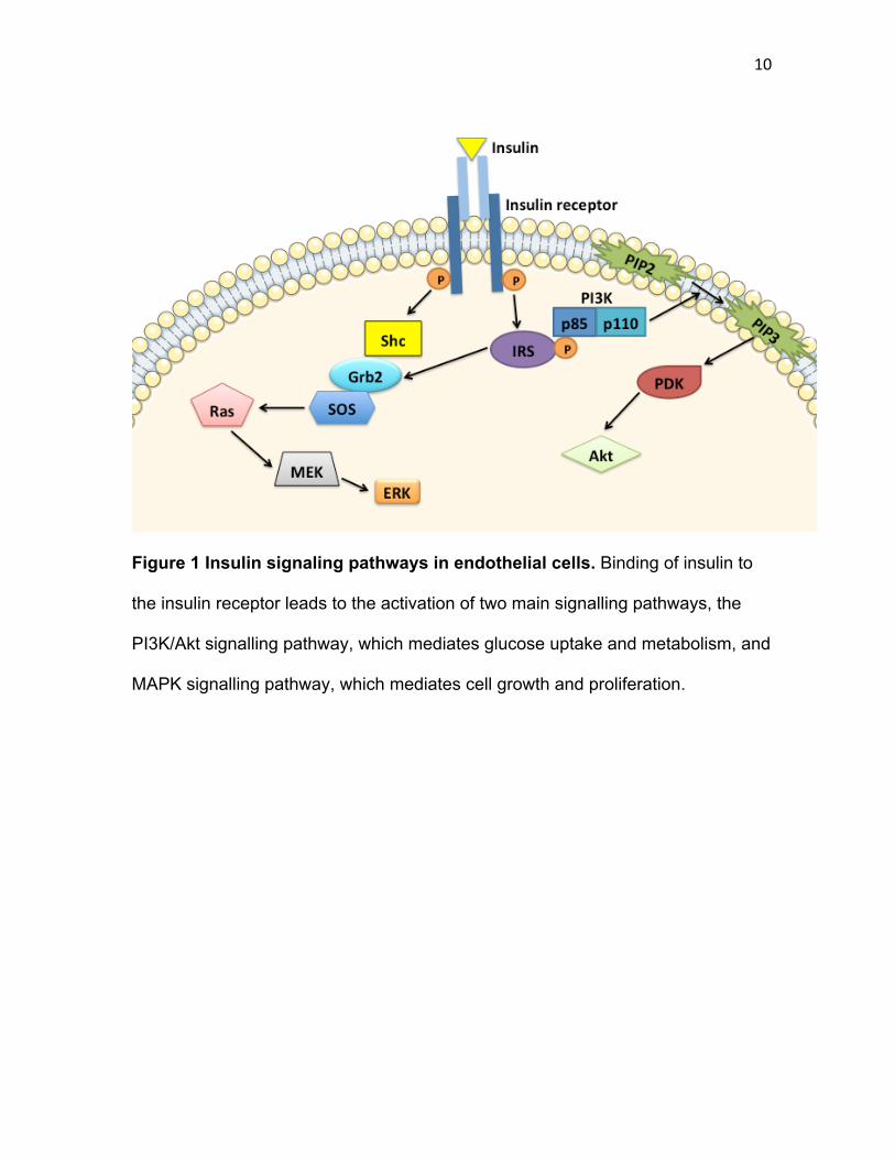
Figure 1 Insulin signaling pathways in endothelial cells. Binding of insulin to
the insulin receptor leads to the activation of two main signalling pathways, the
PI3K/Akt signalling pathway, which mediates glucose uptake and metabolism, and
MAPK signalling pathway, which mediates cell growth and proliferation.

11
1.3 Overview of the mechanisms of insulin resistance
1.3.1 Defective signaling in target tissues
The inability of the body to respond to insulin in insulin resistance is best
known to be caused by defects in the insulin-signaling pathway within target
tissues such as muscle and fat25. Defects have been documented at different
points in the pathway, and have been shown to be caused by the conditions that
lead to and are associated with insulin resistance, such as lipotoxicity and
inflammation32.
A hallmark of insulin resistance is the excess accumulation of lipid
intermediates in non-adipose tissue also known as lipotoxicity32. The increase in
fatty acids has been shown to induce the activation of PKC, which inhibits IRS-1
by serine phosphorylation33. Unlike tyrosine phosphorylation, which activates
proteins such as IR, IRS-1 and Akt, serine and threonine phosphorylation inhibits
them34. The increase in sphingolipid ceramide that is seen in insulin-resistant
subjects inhibits Akt activation by increasing the interaction between Akt and the
protein phosphatase 2A (PP2A) protein, and increasing the threonine
phosphorylation of Akt32. All of these proteins are upstream of the effectors that
regulate GLUT4 translocation and glucose metabolism; thus a defect in their
activation results in a failure to activate these processes32.
12
In addition, insulin resistance is also characterized by a low grade-
inflammatory state. In this state, there is an increase in secretion of cytokines
such as TNF-alpha, IL1B and IL6 by the immune cells and adipocytes, which have
been shown to induce the activation of serine/threonine kinases, and decrease the
expression of IRS-1 and GLUT4 expression32.
In the skeletal muscle, insulin resistance is best known to be caused by the
impairment in the tyrosine phosphorylation of IRS-1. This prevents the activation
of IRS-1 and in turn the activation of PI3K, which is responsible for activating
downstream effectors important for glucose metabolism7.
Many proteins along the insulin-signaling pathway can be dysregulated in
insulin resistance, in addition to the positive and negative modulators that act at
different steps of the pathway. These dysregulations lead to the improper
activation of downstream proteins, resulting in the metabolic syndrome we know
as insulin resistance32.
1.3.2 Defective insulin delivery to target tissues
While insulin resistance is widely known as a condition caused by defects
in the insulin-signaling pathway within target tissues, the role of insulin delivery in

13
the pathogenesis of insulin resistance is less well-studied. Before insulin is able to
exert its action, it must first reach the vascular bed of the target tissue and then
exit out of the circulation to the target tissues by crossing the endothelial
monolayer. Several studies have demonstrated that insulin must cross the
vasculature first in order to exert its effects35,36,37. Therefore, if there is impairment
in the insulin delivery process to the target tissues, this is another way by which
insulin resistance may arise. Indeed there have been several studies suggesting
that an impairment in insulin delivery can contribute to the development of insulin
resistance38,39. They will be discussed below.
1.3.3 The vascular tree
In order to understand the notion of insulin delivery, it is important to
understand the nature of the vascular tree. The vascular system is the
organization of blood vessels in the body that constitutes a part of the circulatory
system. It is important for the delivery of nutrients and oxygen to tissues and the
removal of wastes. The vasculature is comprised of three main types of blood
vessels: the artery, the veins, and the capillaries, which are located in different
parts of the body serving different functions40.
The largest blood vessels in the vascular system are the arteries and veins.
The artery functions to carry blood out of the heart and distribute it to the body

14
through further branching into smaller blood vessels known as arterioles. The
veins carry the blood back into the heart with the help of one-way valves built
within the veins to prevent backward flow. The artery and veins are composed of
three layers: the innermost layer called the tunica intima, which is made of
endothelial cells supported by an internal elastic lamina; the middle layer called
the tunica media, which is made of connective tissues and vascular smooth
muscle; the outer layer called the tunica adventitia, which is made of connective
tissues only. The artery and veins’ structure differ slightly in that the tunica media
is much thicker in the artery than in the veins necessary for withstanding the high
pressure generated from the heart40.
The smallest blood vessel in the vascular system is the capillary. The
capillaries mainly function to exchange nutrients and waste with the target organs
and this is made possible with its unique yet simple structure. Unlike the artery
and veins, the capillary is made up of only one single layer of endothelial cells;
this allows small molecules to easily and quickly get to the target tissue without
having to traverse between layers of smooth muscle and connective tissue. There
are three types of capillaries: continuous, fenestrated or discontinuous.
Continuous capillaries, also the most common type of capillaries, are composed of
endothelial cells that are perfectly aligned forming a tight monolayer, that allows
only the passage of very small molecules such as water and ions through the
intercellular junctions. These capillaries are found in organs such as the muscle
and adipose tissue. Fenestrated capillaries are composed of endothelial cells that

15
have pores allowing small molecules and some proteins to diffuse through. These
capillaries are typically found in the intestines, endocrine glands, kidney and
pancreas. Discontinuous or sinusoidal capillaries are composed of an endothelium
in which the endothelial cells do not form a tight seal, but instead have gaps
between endothelial cells that allow the free diffusion of blood cells and proteins.
Discontinuous capillaries are typically found in the liver, endocrine organs, bone
marrow, spleen and lymphoid tissues, where frequent exchange of materials takes
place41.
16
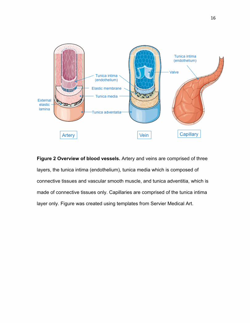
Figure 2 Overview of blood vessels. Artery and veins are comprised of three
layers, the tunica intima (endothelium), tunica media which is composed of
connective tissues and vascular smooth muscle, and tunica adventitia, which is
made of connective tissues only. Capillaries are comprised of the tunica intima
layer only. Figure was created using templates from Servier Medical Art.

17
1.3.3.1 Capillary recruitment and blood flow – role in insulin delivery
Once insulin is secreted into the bloodstream, it is able to enhance its own
delivery to the target tissues by inducing capillary recruitment and increasing
blood flow42. Insulin signaling in endothelial cells leads to the production of nitric
oxide (NO), a vasodilator, which acts on the smooth muscle layer of the blood
vessel to cause the vessel to relax and increase in diameter43. The dilation of
terminal arterioles in turn leads to capillary recruitment or the increase in number
of perfused capillaries. With more capillaries being perfused, more insulin can be
delivered to the target tissues, as the surface area for insulin transport is
increased. The dilation of resistance arterioles, on the other hand, leads to an
increase in blood flow to the limbs, which increases the rate at which insulin is
delivered to the target tissues42. Consequently, any impairment in the insulin-
induced capillary recruitment and increase in blood flow would lead to less insulin
being delivered to the tissues, ultimately affecting glucose metabolism and leading
to the development of insulin resistance. Indeed, there have been a number of
studies showing a decrease in blood flow and capillary recruitment in
obesity/insulin resistance. Increased levels of free fatty acid in the circulation, a
condition that is typical in insulin-resistant obese subjects, have been shown to
significantly decrease the recruitment of capillaries in the muscle and the uptake
of glucose44. This reduction in insulin-stimulated vasodilation has been shown to
be associated with impairments in the pathways that activate the endothelial nitric
oxide synthase (eNOS) in endothelial cells45. It has been shown that

18
hyperlipidemia, a condition that contributes to the development of insulin
resistance, causes a defect in the activation of eNOS, decreasing NO
production46. Inflammatory cytokines, which are increased in the circulation in
insulin resistance, have also been shown to reduce the activation of eNOS in
endothelial cells47. In one study, when TNF-alpha is applied in the rat limb, the
insulin-induced increase in capillary recruitment, blood flow, and glucose uptake
are lost48.
1.3.3.2 Transport of insulin out of the vasculature to the tissues – role of the
endothelium
Once the insulin has reached the blood vessel of the capillary bed, another
potentially rate-limiting step for insulin action is the transport of insulin across the
endothelial barrier. Most evidence to date suggests that the major route by which
insulin is transported across the endothelium is through individual endothelial
cells, a process known as transcytosis35,49,50,51,52,53; this is discussed in detail later
on. The relative importance of this stage of insulin delivery is unclear. For
example, it has been shown that the appearance of insulin in the interstitium is
delayed in obese subjects compared to non-obese subjects during a
hyperinsulinemic euglycemic clamp39,38. Another study has shown that following
an oral glucose tolerance test, the interstitial insulin levels from the adipose tissue
and muscle of non-diabetic obese women were significantly lower than those of

19
the controls38. While these findings indicate that there is an impairment in the
delivery of insulin to the interstitium of the target tissue, it is not known whether
this is contributed wholly by a defect in capillary recruitment and increase in blood
flow or both an impairment in this process and the transcytosis of insulin. It is
difficult to tease out the contribution of the two processes as a defect in either
would result in lower interstitial insulin. As insulin transcytosis is technically
challenging to study, it is not known whether or not this process also contributes to
the defect in insulin delivery that has been observed in insulin resistance. In order
to understand the context and potential role of endothelial transcytosis of insulin,
the next sections will provide important background on endothelial cells and the
determinants of endothelial barrier integrity.
20
1.4 Endothelium
The endothelium is the innermost layer of the blood vessel and it is made
up of a single layer of endothelial cells40. It serves as a selective-barrier between
the lumen of the blood vessel and the tissue, regulating the transport of molecules
between the blood and the tissues54. The endothelial barrier is selective for both
the size and charge of the molecule that pass through55. It is permeable to small
solutes and molecules, and impermeable to larger macromolecules. This size-
selectivity of the endothelial barrier is determined by the interendothelial junctions,
which link the endothelial cells together54. These junctions restrict the passage of
molecules greater than 3-5nm in molecular radius56. The endothelial barrier’s
charge selectivity is determined by the electrical charge on the vascular luminal
surface, which is maintained by a layer of glycoproteins and proteoglycans on top
of the endothelium known as the glycocalyx57. The glycocalyx is between 0.1 to
1um thick, depending on the location along the vascular tree58. It has a net
negative charge due to the negatively charged glycosaminoglycan (GAG) side
chains of the proteoglycans, and this negative charge is dependent on the GAG
side chain sulphation pattern57. Changes in sulphation result in changes in the
charge of the glycocalyx and in turn the permeability of the endothelium. The
glycocalyx repels negatively charged molecules as well as blood cells and
platelets57.

21
Aside from its function as a barrier, the endothelium itself is able to produce
chemicals and proteins to affect other processes, such as the vasomotor tone,
immune and inflammatory responses, and hemostasis59. Endothelial cells produce
vasoactive substances such as nitric oxide and prostacyclin to cause vasodilation
and inhibit platelet aggregation60. The production of these substances by the
endothelial cells is regulated by other chemical stimuli such as bradykinin,
thrombin, as well as changes in blood flow37. Under basal conditions, the
endothelial cells are quiescent, non-thrombogenic, and do not express adhesion
molecules on the cell surface for platelets and leukocytes to adhere61. However,
when the endothelial cells are activated, they enter into a proinflammatory and
procoagulant state, in which the endothelium loses its integrity, starts to express
adhesion molecules to prepare for leukocyte adhesion and extravasation,
changes from an antithrombotic to prothrombotic stage, and produces cytokines to
mediate the inflammatory response62. Clearly, the endothelium is more than just a
structure that makes up a blood vessel; it not only serves as a semi-permeable
barrier to molecules, but also regulates many other processes such as those
related to the inflammatory response.
1.4.1 Endothelial cell heterogeneity
As outlined above, the endothelium from different tissue beds and different
parts of the vascular tree exhibit notable heterogeneity in structure and function.

22
As different tissues and locations of the vasculature have different functions, the
endothelial cells need to be heterogeneous in structure and function in order to
adapt to and accommodate for the differing needs of the tissue and vascular
location41.
In terms of structural heterogeneity, there is a variation in endothelial cell
shape across the vascular tree. Endothelial cells are normally very flat and thin;
however, in the venules, they are thicker and more cuboidal in shape63. The
thickness of endothelial cells also varies depending on which part of the vascular
tree they are derived from; endothelial cells derived from the microvasculature
usually, with some exceptions, have a thickness of less than 0.3 µM, while those
from macrovasculature have a thickness of around 1µM64,65. It is observed that
endothelial cells derived from the arterioles of the mice cremaster muscle are
much longer and larger in surface area than those from the veins. It has also been
found that aortic endothelial cells from rat were more elongated and tapered,
whereas those from the pulmonary veins were much rounder in shape63.
One important function of the endothelium is the regulation of molecule
transport across the vasculature. Molecules move across the endothelium either
paracellularly between endothelial cells or transcellularly through each individual
endothelial cells by a process called transcytosis41. For the transcellular transport
pathway, the internalization step can be mediated by clathrin-mediated
endocytosis, caveolae, macropinocytosis, or phagocytosis66. In general,

23
endothelial cells have more caveolae than clathrin-coated pits, with the exception
of endothelial cells from the liver sinusoids, where more clathrin-mediated
endocytosis occurs63. The density of caveolae is the highest in continuous non-
fenestrated endothelial cells and those derived from the capillaries; the only
exception is the blood brain barrier, where the density of caveolae is particularly
low63. For the paracellular transport pathway, the movement of molecules is
regulated by intercellular junctions such as the tight junction and adherens
junction, which vary in composition and density across the vascular tree and beds.
The tight junctions are dense and well developed in the arteries, as expected, due
to their function of serving as the conduit for blood flow and withstanding the high
pressure from the blood63. The tightness of the junctions then decreases from
arterioles to capillaries to venules, where a laxer paracellular junction is required
for material transport and the modulation of leukocyte extravasation63. As
expected, the junctions are particularly tight in the endothelium of the blood brain
barrier due to its high specificity in regulating molecule transport63.
The expression of certain endothelial adhesion molecules also varies
across the vascular tree, according to where leukocyte trafficking typically takes
place. When there is tissue damage, leukocytes are recruited out of the
bloodstream into the site of damage to fight off infections67. The location in the
vasculature that leukocytes typically exit at is the post-capillary venules68. The
initial step of leukocyte adhesion and rolling is mediated by interaction between
carbohydrate ligands on the leukocyte and glycoproteins expressed by endothelial

24
cells known as selectins, specifically E- and P-selectin69. The expression of E-
selectin and P-selectin has been found to be the highest in the activated
endothelial cells from the postcapillary venules70.
The substances and mechanical forces that the endothelial cells are
exposed to can all affect its phenotype, often induced as result of the activation of
signaling pathways within the cell. Different tissues have a different environment
to which the endothelial cells are exposed63. For example, endothelial cells
derived from the coronary artery are exposed to the mechanical forces of heart
contractions as well as factors released by the cardiomyocytes, whereas
endothelial cells derived from the brain are exposed to factors secreted by brain
cells, which can affect their ability to form a tight blood-brain-barrier63. In addition,
the force that is applied against the endothelium by blood flow, also known as
shear stress, have been shown to induce changes in endothelial cell phenotype
such as the actin cytoskeleton and permeability to certain molecules71; the
specific effects of shear stress on endothelial cells is discussed in detail in a
subsequent section.
Finally, while the location and surroundings of the endothelial cells is one
way of generating and maintaining their heterogeneity, some evidences suggest
that epigenetics may also have a role. Instead of depending on the environment to
maintain its unique properties, the cells may also be epigenetically programmed to
behave in certain ways41. It has been shown by DNA microarray studies that when

25
endothelial cells from the microvasculature and microvasculature are taken into
culture, they exhibit different expression profiles72. Furthermore, when endothelial
cells from the human coronary artery were treated with oxidized LDL, the changes
in expression of genes involved in the development of atherosclerosis such as
adhesion proteins and proliferation were much greater than endothelial cells
derived from the leg vein73. This suggests that the higher susceptibility of arteries
to atherosclerosis than veins is likely due to the arterial endothelial cell itself being
more sensitive to the development of this process than venous endothelial cells.
1.4.2 Endothelial permeability
The endothelium is a major site of material exchange between the blood
and the tissues, serving to transport nutrients to and collecting metabolic waste
from the tissues54. The exchange of these materials can occur by movement
between individual endothelial cells, known as paracellular transport, and through
each individual endothelial cells, also known as transcellular transport or
transcytosis54. These processes are intricately regulated by the junctions between
endothelial cells as well as intracellular vesicle trafficking to maintain a low and
selective permeability across the vasculature54. Below, the regulation of
paracellular and transceullar transport will be discussed.

26
1.4.2.1 Paracellular transport
The paracellular transport pathway is regulated by interendothelial
junctions. There are two types of cell-cell junctions in the endothelium that serve
barrier functions and maintain endothelial permeability; they are the adherens
junction and the tight junctions54.
The adherens junction is comprised of the proteins cadherin, p120-catenin
protein, β-catenin, plakoglobin, and α-catenin75. Cadherins are transmembrane
adhesion proteins that form homodimers in a calcium-dependent manner with the
extracellular domain of other cadherins from adjacent cells75. The p120 protein
binds to the proximal end of the cytoplasmic tail of cadherin and its function is to
regulate the trafficking and turnover of cadherin, as well as cell actin dynamics
through modulating the activities of Rho family GTPases75. β-catenin and
plakoglobin bind to the distal end of the cadherin cytoplasmic tail, and are linked
to the actin cytoskeleton via α-catenin75. The catenin proteins are essential for the
proper formation of the adherens junction as the disruption in interaction between
cadherin and catenin was shown to significantly decrease the strength of the
junction76. Permeability of the paracellular pathway is regulated by changes in the
adherens junction, such as the phosphorylation of the cadherins to induce
cadherin internalization, as well as the promotion of stress fibres formation within
the cell to break apart the adherens junction. Such processes usually occur when
the junction needs to be temporarily disrupted to allow the movement of certain

27
molecules or cells across the endothelial barrier, for example during leukocyte
extravasation64.
The ahderens junction adhesion protein that is exclusively expressed in
endothelial cells is the VE-cadherin (vascular endothelial cadherin). VE-cadherins
are critical in maintaining vascular integrity and permeability75. It has been shown
that disruptions in the interaction between VE-cadherins are seen in a lot of
pathological conditions such as inflammation and acute lung injury77. In addition,
the administration of anti-VE-cadherin antibodies in mice has been shown to
cause a significant increase in permeability, hemorrhages and vascular fragility77.
The other interendothelial junction that regulates barrier integrity is the tight
junction, which forms seals between endothelial cells64. The tight junctions are
formed after adherens junctions, which are built first to establish the architectural
integrity of the endothelium for proper formation of the tight junction78,79. The main
proteins that make up the tight junctions are the claudin and occludin proteins80.
Claudins and occludins are transmembrane proteins that interact with claudin and
occludin proteins from adjacent cells to form a tight seal arranged like a strand
around the cell80. Claudins are important for the assembly of the tight junctions
and regulating the permeability of ions between cells81. It has been shown that
changing the expression of claudin proteins leads to changes in the conductivity of
certain ions82. The claudins that are responsible for forming channels that are
selective to ions are claudins 2, 10, 15 and 1783.There are 27 members in the

28
claudin family84; however, only claudin-5 is exclusively expressed in endothelial
cells64. Claudin is associated with several intracellular adaptor proteins such as
ZO-1, 2, 3, PATJ, and MUPP1, which connect the tight junction to the actin
cytoskeleton80. Occludins are responsible for the stabilization of the tight junction
as well as serving barrier functions80.
Adherens junctions are relatively ubiquitous along the vascular tree
whereas tight junctions are more variable along different segments of the vascular
tree85. In large arteries where the main function is to serve as conduit for blood
delivery, the tight junctions are well developed, strictly restricting the transport of
molecules across the endothelium. However, in the capillaries, where constant
exchange of materials occurs, the tight junctions are much less developed to
facilitate this process. In the blood-brain, where the permeability to molecules is
highly selective, the tight junctions are dense and highly developed64.
1.4.2.2 Transcellular transport (transcytosis)
In addition to paracellular transport, another way by which molecules get
across the endothelium is by moving through (rather than between) individual
endothelial cells. This pathway is usually used for the transport of macromolecules
and involves first vesicle-mediated endocytosis, then trafficking within the cell,
followed by exocytosis at the basal side of the cell86.

29
The most studied pathway for transcytosis in endothelial cells is the
caveolae-mediated transcytosis pathway. Caveolae are small 50-100nm
invaginations in the plasma membrane that are rich in proteins and cholesterol87.
The caveolae-mediated transcytosis is first initiated by the binding of a ligand to its
receptor in the lipid rich domains of the membrane. The caveolin proteins are then
phosphorylated by Src kinase and recruited to the lipid rafts where they will
aggregate and oligomerize to form caveolar endocytic vesicles. Once a bud is
formed, the vesicle is excised from the membrane by the GTPase dynamin,
pinching the vesicle off of the plasma membrane. The vesicles can then fuse with
endosomes within the cell or move to the basolateral membrane, where they will
dock and fuse with to release their contents into the interstitium88. Caveolae are
typically known to mediate the transcytosis of LDL in aortic and umbilical vein
endothelial cells, albumin in rete mirabile microvascular endothelial cells, insulin in
aortic and rete mirabile microsvacular endothelial cells, and chemokines in
venular endothelial cells89,90,91,92,93,94,95. Albumin transcytosis is the most studied
model for transcytosis in endothelial cells; it has been shown that caveolin-1-
deficient endothelial cells are not able to transcytose albumin while wild-type
endothelial cells can96. In addition, the transcytosis of insulin in bovine aortic
endothelial cells (bAECs) has been shown to be mediated by caveolae as well95.
When the expression of caveolin-1 was knocked down by siRNA in bAECs, the
uptake of FITC-insulin was significantly reduced, whereas the overexpression of
caveolin-1 increased insulin uptake95. Similarly, aortic endothelial cells derived

30
from caveolin-1-null mouse did not take up insulin while the cells with caveolin-1
re-expressed did95.
It has recently been found that clathrin also has a role in endothelial cell
transcytosis, specifically the transport of insulin97. Clathrin is a triskelion shaped
protein that is composed of three clathrin heavy chains bound tightly to three light
chains. They are recruited to the membrane by adaptor proteins, which bind to the
cargo on the extracellular side of the cell and clathrin within the cell98. As more
and more clathrins get recruited to the membrane, they start to connect with each
other eventually forming a curved lattice around the pit like a cage98. The GTPase
dynamin then comes in and pinches vesicle from the membrane, allowing the
internalization of the cargo. Soon after scission, the clathrin coat will disassemble,
before the vesicle goes on to fuse with other endosomes or be exocytosed98. It
was shown by Azizi and colleagues that when the clathrin heavy chain is depleted
or inhibited in adipose microvascular endothelial cells, the internalization and
transcytosis of insulin was reduced by 50%, suggesting that clathrin may also
have a role in transendothelial transport97.
31
1.4.2.3 Insulin transcytosis
While some older studies have suggested that insulin may cross the
endothelial barrier by ways other than the transcellular pathway, most evidence to
date supports that insulin crosses the endothelium by transcytosis35,49,50,53,52,53.
The process of insulin transcytosis is mediated by three main steps: the uptake of
insulin into the endothelial cell, traffic within the cell, and exocytosis86. There have
been several studies showing that insulin transport across endothelium is a
receptor-mediated process and that both the insulin receptor and IGF-1 receptor
may be involved. It has been shown that when a blocking antibody to the insulin
receptor (IR) was applied to a retinal endothelial monolayer along with insulin, the
transport of insulin across the monolayer was significantly reduced53. In addition,
fluorescently labeled insulin taken up by aortic endothelial cells has been shown
to colocalize partially with IR and the IGF-1 receptor99.
Furthermore, there has also been in vitro evidence of vesicle-mediated
insulin transport within the endothelial cells. The earliest study showing that insulin
is carried in vesicles within endothelial cells was accomplished through electron
microscopy imaging92. More recently, this has been shown by insulin uptake
experiments, where the uptake is significantly reduced in aortic endothelial cells
with caveolin-1 is knocked down, and enhanced when caveolin-1 is
overexpressed95. Similar results were obtained from endothelial cells isolated from
caveolin-1 knockout mice, where insulin uptake was abolished and only rescued

32
when caveolin-1 was re-expressed in these cells95. Another study has shown in
microvascular endothelial cells that insulin uptake and transcytosis is clathrin-
mediated97. In this study, it was shown that knockdown of clathrin expression
attenuated insulin transcytosis. While there is a discrepancy between the transport
mechanisms of insulin transcytosis, likely due to the different types of endothelial
cells used, it is clear that insulin is transported across the endothelium at least in
vitro by a receptor- and vesicle-mediated process.
The transport of insulin across the endothelium by transcytosis is further
supported by findings from in vivo studies. It has been shown in dogs that during
euglycemic clamp experiments, when they are infused with insulin and inulin,
there are persistently higher plasma levels of insulin than in the lymphatics (a
proxy for hormone’s concentration in the interstitial space), while inulin, a
molecule with a similar molecular weight as insulin known to only cross the
endothelium through the paracellular pathway, was equal in levels from the blood
and the lymph100. Another study by Ader and colleagues also found that when
glucose tolerance test were performed in dogs, there was a persistently higher
plasma level of insulin compared to the lymph, suggesting that the transport of
insulin does not occur by simple diffusion35. Other studies have looked directly at
the saturability of this process in vivo. One study looked at the transport of
radioactive insulin across coronary vessels with and without the presence of
unlabeled insulin, and found that when radioactive insulin was perfused in the
presence of unlabeled insulin, the presence of insulin in the capillary walls and

33
heart parenchyma was much lower than those without unlabeled insulin. When a
modified version of the radioactive insulin that has lost its affinity to bind to the
insulin receptor was applied, much less signal was observed in the capillaries and
muscle; unlabeled insulin also did not result in a reduction of this signal101.
Another study by Herkner and colleagues observed that the interstitial insulin
concentration was much less than that in the plasma during oral glucose tolerance
test and that this ratio of plasma to interstitial levels of insulin is enhanced during a
hyperinsulinemic clamp50. All of these studies suggest that insulin transport across
the microvasculature is a saturable process and also a potentially rate-limiting
step to the initiation of insulin action as the insulin must reach the interstitial space
before it is able to exert its actions.
1.4.2.3.1 Assays for insulin transcytosis
Insulin transcytosis is an important process to study as it has implications
for the development of novel therapies for insulin resistance and T2D; however,
knowledge in the regulation of this process is limited due to technical limitations in
studying insulin transcytosis. Most studies on transcytosis in cell cultures have
made use of the transwell assay, in which transcytosis is quantified by how much
of the added insulin moves across the endothelial monolayer grown on a
permeable membrane. This assay could be used to assess the saturability of
insulin transport across the endothelium by measuring the amount of labeled
34
insulin that crossed the endothelium when in presence of unlabeled insulin.
However, it is not ideal to investigate the specific regulators of this process as this
would involve much harsher manipulations to the cells, such as siRNA, which can
disrupt the endothelial monolayer and induce paracellular gaps. One of the major
caveats of the transwell assay is that it is not able to distinguish between actual
transcytosis and paracellular leak, which can be induced by pharmacological or
molecular manipulation, and this can greatly cofound the measurement of actual
transcytosis102.
Previously, electron microscopy (EM) was also used to study transcytosis.
However, it is mostly useful for studying the vesicular traffic of the molecule within
the cell, such as its targeting to the lysosome and other endosome compartments.
Only fixed cells can be analyzed using EM, which means the amount of molecule
that is transported through the endothelial cells cannot be quantified89. Most of all,
electron microscope is very expensive for the amount of information it is able to
provide to study transcytosis.
More recently, a novel assay for measuring insulin transcytosis by single
cells in a confluent endothelial monolayer was developed by our lab that involves
using total internal reflection fluorescence microscopy or TIRFM and fluorescently
labeled insulin97. TIRF microscopes allow the visualization of a very narrow range
of the cell of approximately 100-150nm. We thus focus on the basolateral
membrane of the cell after fluorescently-tagged insulin has been added. We are
35
then able to see vesicles bearing insulin appear and then disappear as they fuse
with the basolateral membrane. In principle, this dynamic assay eliminates
confounding from paracellular leak, avoids edge effects from transwell assays,
and is not affected by low transfection efficiency of molecular constructs since
single cells are selected102. With this advancement in measuring insulin
transcytosis, it is now easier to study insulin transcytosis and the regulation of this
process. More details on the assay will be discussed in the Methods section.

36
1.5 Vascular Wall Shear stress
Almost all of the in vitro studies described were performed under static
conditions. Yet physiologically, blood vessels are constantly exposed to blood flow;
this exerts a force on the endothelial surface known as shear stress. By definition,
shear stress is the frictional force parallel to the vessel wall at the surface of the
endothelium103. This force is determined by several factors as illustrated in the
equation below:
T = 4ηQ/πr3 104
Where η is blood viscosity, Q is blood flow rate, and r is radius.
Shear stress is directly proportional to blood viscosity and flow rate, while it
is inversely proportional to the cube of the radius of the blood vessel. As these
factors vary along the vasculature, the shear stress that the endothelial cells are
exposed to also varies across the vascular tree. The shear stress that is typically
found in the arteries can range from 10 to 70 dynes/cm2, whereas that found in
the capillaries and veins is much lower, ranging from 0.5 to 5 dynes/cm2104,105.
The application of shear stress to endothelial cells have been shown to induce
various changes to the endothelial phenotype such as changes in gene
expression, activation of signaling pathways within the cells, and changes in cell
morphology, all of which will be discussed in more detail below71.

37
1.5.1 Effects of shear stress on endothelial cells
One of the many effects that shear stress has on the endothelial cells is the
reduction in the rate of endothelial cell proliferation. The exposure to shear stress
increases the number of cells arrested in the G0 and G1 phase of the cell cycle
and has been shown to lower the DNA synthesis rate106,107. The mechanism by
which growth is suppressed in endothelial cells after being exposed to shear
stress has been found to be mediated by the tumour suppressor p53106. P53 is a
transcription factor that induces the expression of a cyclin-dependent kinase (ckd)
inhibitor called p21, which is a protein that inhibits the cdk proteins from promoting
cell cycle and proliferation108. It has been found that shear stress increases the
association of the cdk proteins with the p21 inhibitor repressing their kinase
activity that is required to phosphorylate downstream proteins involved in cell
growth106. Shear stress also induces the phosphorylation of the retinoblastoma
protein, which when hyperphosphorylated, will bind to the transcription factors
required for DNA synthesis, inhibiting DNA transcription and cell growth
toward107,109. In addition to the inhibition of endothelial cell growth and
proliferation, shear stress also inhibits apoptosis. When endothelial cells that have
been exposed to shear are treated with apoptosis inducers such as TNF-alpha
and reactive oxygen species, apoptosis is suppressed110. This is likely mediated
by the increase in expression of inhibitors of apoptosis proteins 1 and 2 (IAP-1, -1)
after exposure to shear stress, which inhibit active caspases and prevent them
from inducing apoptosis111,112.

38
Another effect shear stress has on the endothelial cells is the induction of
cell migration, as shown by studies that have looked at wound healing. These
studies have shown that the exposure to shear stress leads to a higher rate of
endothelial migration to the wound created in the endothelial monolayer compared
to cells under static conditions, resulting in faster wound closure113,114. This
directional migration was shown to be mediated by the formation of protruding
lamellipodia and focal adhesion remodeling, which are regulated by the Rho
family of small GTPases115,116. In addition, focal adhesion kinase (FAK), a protein
that regulates the formation of focal adhesions required for providing traction
during migration, has been shown to be involved in this shear-induced increase in
wound healing, as inhibition of FAK lead to a slower wound healing117,118.
Furthermore, the cytoskeleton is also another effector of shear stress.
Shear stress has been shown to induce the elongation and alignment of
endothelial cells towards the direction of shear stress71. This alignment involves
the remodeling of the cytoskeleton, which is also regulated by the Rho family of
GTPases. The Rho family GTPases is comprised of the GTPases Rac, Rho,
Cdc42, which switch between the inactive GDP-bound form and the active GTP-
bound form119. Rac is activated within 30 minutes of exposure to shear stress and
it has been shown to be important for the alignment of the cytoskeleton towards
the direction of shear stress, as cells transfected with dominant negative (DN)
Rac1 do not become aligned in the direction of shear stress120,121. The speed of

39
migration of endothelial cells has also been shown to be reduced in DN Rac1
cells122. Unlike Rac, shear stress first induces a transient inactivation of Rho
followed by its activation that peaks at 60 minutes and movement to the cell
membrane121. Rho has been shown to also be important for the alignment of the
actin cytoskeleton, as exposing DN Rho-transfected cells to shear stress resulted
in a failure of the cells to align in the direction of shear stress as well as a
decrease in shear induced increase in stress fibres123. Cdc42 is a GTPase known
to be important for mediating the formation of filopodia, finger-like projections at
the cell membrane124,125. Like Rho, it is translocated to the membrane following
exposure to shear stress and it has been shown to regulate the microtubule
cytoskeleton rather than the actin cytoskeleton123,126.
There has been some evidence suggesting that shear stress can also
affect the transport of molecules across the endothelium. It has been shown that
the transcytosis of albumin, the major protein in blood, is increased in response to
shear stress in a dose-dependent manner127. In addition, the internalization of LDL
by aortic endothelial cells, the first step of the transcytosis pathway, has also been
shown to be increased in response to high shear stress128.
The shear stress that is experienced at the level of the macrovasculature
and the level of microvasculature is different. In the aorta, for example, the blood
flow is pulsatile, which results in fluctuations in shear magnitude, while in the
capillaries, the blood flow is continuous, which results in a shear that is temporally
40
and spatially uniform129. Pulsatile flow has been shown to induce significantly
higher phosphorylation of ERK1/2 in bovine aortic endothelial cells as compared
to those exposed to steady continuous flow130. In another study, the change in
endothelial cell shape is less rapid when exposed to pulsatile flow than steady
flow; however, cells are more elongated in shape in the pulsatile flow condition131.
In addition to pulsatile flow, the blood flow in our circulatory system can be either
laminar or turbulent. Laminar flow is when the fluid flows in parallel layers along
the axis of the blood vessel; this occurs mostly at straight parts of the vascular
tree. Turbulent flow is when the fluid flows in irregular patterns; this occurs usually
at branching points of the vascular tree132. Turbulent flow has been shown to
affect the endothelial phenotype differently than laminar flow. Turbulent flow has
been shown to upregulate the expression of genes that promote the development
of atherosclerosis while laminar flow promotes the upregulation of expression of
genes that are atheroprotective133. As the shear stress resulting from different
types of flow affects the endothelial cells differently, the phenotype of the
endothelial cells derived from different locations along the vascular tree is also
expected to be variable.
1.5.2 Mechanosensors of shear stress
The effect that shear stress exerts on endothelial cells is mediated by
mechanosensors in the cell that sense the mechanical force and transduce it into

41
intracellular signaling. There are many molecules that have been suggested to
have the role of being a mechanosensor in endothelial cells; the best-known
mechanosensors are integrins, ion channels, G protein coupled receptors and G
proteins, PECAM-1 and caveolin-171. They will be discussed below.
Integrins are transmembrane proteins that form a link between the cell’s
cytoskeleton and the extracellular matrix (ECM) by binding to specific molecules
associated with the two structures. Integrins are heterodimers that are comprised
of an alpha- and beta-chain134. Tzima and colleagues have found that integrin
αvβ3 is activated in just 5 minutes of exposure to 12 dynes/cm2 shear stress in
bovine aortic endothelial cells, as shown by an increase in WOW-1 (an antibody
that binds to activated integrin αvβ3) staining after exposure to shear stress
compared to static control121. As integrins are activated, they are converted to a
high affinity state, which increases their binding to extracellular matrix (ECM)
proteins such as fibronectin and vitronectin135. The increase in binding to ECM
proteins can then induce various intracellular signaling cascades. Shear stress is
known to activate Extracellular signal-Regulated Kinase-1 (ERK), c-Jun N-terminal
kinases (JNK) and IkappaB (IKB); applying blocking-type anti- αvβ3 to aortic
endothelial cells abolishes the activation of these proteins in response to shear
stress136. Similarly, blocking the activation of β1 integrins attenuated the
previously found shear-induced phosphorylation of downstream proteins Akt, SFK,
and eNOS137. It is well known in the literature that shear stress increases nitric
oxide production in aortic endothelial cells as a result of the activation of eNOS,

42
leading to the vasodilation of blood vessels138. When a synthetic peptide that
inhibits integrin binding to the ECM was applied intraluminally to isolated coronary
arterioles during exposure to shear stress, shear-induced vasodilation is
significantly reduced139. In addition, the application of a blocking-antibody against
the β3 chain of integrin also caused a significant reduction in the shear-induced
vasodilation of the coronary arterioles139. These finding suggest that the shear-
induced increase in nitric oxide and vasodilation is in part mediated by integrins.
Ion channels have also been reported to have a mechanosensory role in
endothelial cells. Studies have shown that shear stress increases the permeability
of the cell membrane to K+ inducing an increase in inward K+ current. In addition,
shear stress has been shown to induce an inward flux of Ca2+ in endothelial cells
as well, which can lead to various Ca2+ dependent cell responses and affect
intracellular signaling and endothelial function71. The ion channels that have been
found to be shear-activated are the TRPV4 and TRPP1/2 channels140.
Shear stress has been shown to activate G proteins on the endothelial cell
membrane. G proteins are molecular switches within the cell that are activated by
the G protein coupled receptor, with the active form being GTP bound and the
inactive form being GDP bound141. Once activated, the G protein can activate
various signalling pathways to change the function of the cell. Ras is a GTPase
that is responsible for regulating cell growth, differentiation and survival; it has
been shown that within 5 seconds of exposing human umbilical vein endothelial
43
cells to flow, Ras activation increased by 10-fold as compared to the static
control142. However, transfecting the cells with antisense Gαq inhibited this
increase. Furthermore, transfecting the cells with constitutively active Gαq did not
enhance the shear-induced Ras activation, but transfecting the cells with the
Gbeta1gamma2 subunit did, suggesting that Galphaq is required to initiate the
mechanosensing of shear stress and that the shear-induced Ras activation is
mediated by Gbeta1gamma2142. As mentioned previously, shear stress activates
the proteins ERK in endothelial cells. To test if G proteins are also important for
mediating the activation of ERK1/2, cells were treated with the pertussis toxin
(PTx), which inactivates Galphai and Galphao proteins, and then ERK activation
was measured by the amount of phosphorylated ERK. Following treatment with
PTx, the shear-induced phosphorylation of ERK1/2 was completely abolished.
These findings provide some evidence for a role of the G proteins in
mechanosensing shear stress in endothelial cells136.
PECAM-1 is a surface adhesion protein located at the junctions between
endothelial cells that is also found in platelets, monocytes, neutrophils, and some
T cell. In endothelial cells, PECAM-1 mediates the migration of leukocytes across
the endothelium, as well as processes such as angiogenesis, thrombosis, and
mechanosensing of shear stress143. PECAM-1 has been found to be
phosphorylated 1 minute after exposure to shear stress144,145. It is known that
shear stress induces the translocation of the tyrosine phosphatase, SHP-2, to the
endothelial junctions within 5 minutes of application of shear stress. However,

44
when PECAM-1 is downregulated in endothelial cells, the translocation of SHP-2
to the cell junctions is lost. In addition, the shear-induced ERK phosphorylation
that is known to occur also decreases after exposure to shear stress when
PECAM-1 expression is decreased146.
Lastly, caveolae/caveolin-1 has also been suggested to have a role in
mechanosensing in endothelial cells. Caveolae are small 50-100nm invaginations
in the plasma membrane that are rich in proteins and cholesterol. Caveolae are
formed with the protein component caveolin-1 and can mediate endocytosis87. It
has been shown that after exposing endothelial cells to shear stress for 24 hours,
more cav-1 and caveolae were observed on the apical side of the cells147. A more
direct evidence for caveolae/caveolin having a role in mechanosensing was
obtained from an in vivo experiment, in which cav-1 knockout mice were
examined148. Shear stress is known to induce the dilation of the blood vessels; it
was found that the shear-induced vasodilation was significantly reduced in the
arteries of the cav-1 KO mice compared to that of the wild type control. However,
this shear-induced vasodilation was restored to similar levels as that of wildtype
when caveolin-1 was re-expressed in the endothelium148. This effect was found to
be due to the reduction in the level of phosphorylated eNOS, as phospho-eNOS
levels were found to be much lower in the carotid arteries from the cav-1 KO mice
as compared to wildtype (WT), and that the reconstitution of cav-1 in endothelial
cells restored the level of p-eNOS to that in WT148. These studies suggest that

45
caveolae/caveolin-1 is another important regulator of mechanotransduction in
endothelial cells.
2 Chapter 2 Research Aims & Hypotheses
Type 2 diabetes is a disease in which the body is unable to metabolize
glucose as a result of insulin resistance149. While insulin resistance is widely
known as a condition caused by defects in the insulin-signaling pathway within
target tissues such as the muscle and fat, the role of the endothelium in the
pathogenesis of insulin resistance is less well-studied25. The transport of insulin
across the microvasculature is a potentially rate-limiting step to the delivery of
insulin and the initiation of its action. It has been shown that insulin appearance in
the interstitial space is delayed in obese insulin-resistant subjects as compared to
healthy subjects39. This finding suggests that an impairment in the transport of
insulin out of the vasculature could also be a contributing factor to insulin
resistance in addition to the impaired insulin-signaling in target tissues.
Understanding how insulin transport across the microvasculature is regulated is
therefore important as it may lead to the identification of novel therapeutic targets
for the treatment of insulin resistance. Most evidence to date suggests that insulin
is transported across the endothelium through individual endothelial cells
(transcytosis)35,49,50,51,52,53; however, little is known about how this occurs.
Most in vitro studies on endothelial cell biology have been done under
46
static conditions; however, in vivo, endothelial cells are constantly exposed to
shear stress, a frictional force at the endothelial surface derived from blood
flow103. This force has been shown to induce various phenotypic and molecular
changes in the endothelial cell, such as the reorganization of the cell cytoskeleton
as well as the change in expression of various genes71. In addition, exposure to
shear stress has been shown to induce transcytosis of molecules such as albumin
through endothelial cells127. However, whether shear stress affects the
transcytosis of insulin in endothelial cells is unknown. Thus, we are interested in
studying insulin transcytosis in endothelial cells under flow or exposed to shear
stress stress, a condition more representative of the physiological milieu.
The purpose of this study was to determine whether physiological shear
stress induced by affects insulin transcytosis by endothelial cells and if so, to
determine the mechanism(s). As shear stress has been shown to induce
transcytosis of certain molecules such as albumin in endothelial cells, we
hypothesize that shear stress also induces the transcytosis of insulin in
endothelial cells.

47
3 Chapter 3 Shear stress induces endothelial internalization and
transcytosis of insulin independently of nitric oxide –
potential role of the actin cytoskeleton
3.1 Introduction
While much research into the pathogenesis of insulin resistance has
focused on signaling within target tissues such as skeletal muscle, there is
growing appreciation of a potential role for the vascular endothelium150. Insulin
delivery to downstream tissues like muscle and fat is regulated by hemodynamic
factors such as capillary blood flow and capillary recruitment. The dilation of
upstream arterioles and precapillary sphincters increases blood flow and recruits
non-perfused capillaries, augmenting the amount of insulin that can ultimately
reach the downstream tissue bed151,152,153. While precapillary sphincters have
been described in the mesentery, it should be noted that it is still controversial
whether it exist in the muscle206,207,208. The significance of this regulation is
suggested by the observation that insulin delivery is impaired in conditions
associated with whole body insulin resistance such as obesity154. Impairment of
insulin delivery has been attributed to decreased endothelial nitric oxide (NO)
production, as NO is a potent endogenous vasodilator153,150.
48
Notwithstanding the hemodynamic factors that regulate insulin delivery, it is
important to note that in tissues like skeletal muscle and fat, the endothelial layer
that lines capillaries is continuous63,155 – without intercellular gaps – thus
potentially constituting a barrier to the diffusion of luminal insulin out of the
capillary microvasculature. This is in contrast to the sinusoidal endothelium of the
liver, which is fenestrated and allows for bidirectional movement of fluids and
solutes between the interstitium and the vascular lumen. Thus in skeletal muscle
and fat, endothelial permeability to insulin may occur via some combination of two
potential routes: by diffusion between adjacent endothelial cells (i.e. paracellular
diffusion) or by movement through the cytoplasm of individual cells in the
microvascular endothelial monolayer, a process known as transcytosis156,157. In
the latter, luminal insulin is internalized at the apical endothelial surface by
endocytosis, moves through the cell by vesicular traffic and is ultimately released
by exocytosis to the interstitial space.
Despite being described over 30 years ago53, the mechanisms and
physiological importance of insulin transcytosis remain unclear. Numerous in vitro
studies have supported the notion that endothelial cells may restrict the diffusion
of insulin158,53,159,95, but the cellular and molecular mechanisms remain elusive.
Some of the uncertainty reflects technical difficulties in distinguishing paracellular
diffusion from transcytosis in cultured cells102 and the underlying heterogeneity of
the endothelium that was studied; for instance, while endothelial cells derived from

49
large vessels (e.g. the aorta) require caveolae for insulin uptake95, endothelial
cells from the microvasculature appear to use clathrin-coated pits instead97,53.
A further limitation of existing in vitro studies is that they are almost
uniformly performed under static conditions. In vivo, endothelial cells are
constantly exposed to shear stress, the force exerted by blood flow which acts in
the direction parallel to the cells. This physiological force can induce significant
changes in cellular behavior, including cytoskeletal remodeling 160 and altered
gene expression. Furthermore, shear stress has been reported to enhance nitric
oxide synthase activity161,162,163 leading to increased nitric oxide levels. NO, in
turn, has been observed to enhance internalization of insulin by endothelial cells
from the aorta164; whether this occurs in endothelial cells from the
microvasculature was not determined.
To date, the effect of shear stress on endothelial internalization and
transcytosis of insulin is unknown. In this study, we applied a defined shear stress
to confluent adipose microvascular endothelial monolayers in culture, determined
that endothelial internalization of insulin was enhanced, and elucidated the
mechanism of this effect.

50
3.2 Methods
3.2.1 Cell culture
Primary human adipose microvascular endothelial cells (HAMEC) were
purchased from ScienCell (Carlsbad, CA), and primary human aortic endothelial
cells (HAEC) were purchased from Lonza (Allendale, NJ); both were grown in
Endothelial Growth Media-MV (Lonza) at 37°C and 5% CO2. HAMEC were
expanded and used for experiments from passage 4 to 8, and the HAECs from
passage 6 to 8. Cells were always seeded on gelatin-coated coverslips or plates
and allowed to grow to confluency before experiments.
3.2.2 Transcytosis assay
To visualize and measure insulin transcytosis through individual endothelial
cells, fluorescently labeled insulin and Total Internal Reflection Fluorescence
Microscopy were used as previously reported97. As FITC-insulin (used in the
insulin internalization assays) photobleaches too quickly for visualization under
TIRFM, Alexa Fluor 568-labeled insulin (A568-insulin) was used instead.
A568-insulin was generated by Dr. Javier Jaldin Fincati by conjugating
human insulin (Sigma-Aldrich, St. Louis, MO) with Alexa Fluor 568-succinimidyl

51
ester using the protocol from the Molecular Probes manual (A20003; Life
Technologies). Unreacted fluorophores were removed by extensive dialysis.
Cells were pulsed with 500nM Alexa Fluor 468-insulin for 10 minutes at 4°C
to allow binding of the insulin to the membrane. After membrane-binding, the cells
were washed twice with 4°C PBS (with Mg2+ and Ca2+) to remove any unbound
insulin. The cells are then visualized under TIRFM on a heated stage in HEPES
containing RMPI 1640 media.
Samples were imaged using an Olympus cell TIRF Motorized Multicolor
TIRF module mounted on an Olympus IX81 microscope (Olympus, Hamburg,
Germany), at 150x magnification. Images were acquired using a laser with a
561nm excitation and a TIRF penetration depth value of 110nm, and acquisition
was performed using Volocity software. Videos of each cell were taken at 10
frames per second for 15 seconds. Transcytosis events were quantified using a
custom-written MATLAB scripts developed by Dr. Bryan Heit from the University
of Western Ontario, as previously reported (7) and based on an approach
described by Steyer et al165. Briefly, the algorithm identifies a vesicle containing
A568-insulin by their expected size (9-36 pixels2) and circularity (>0.2). Once the
vesicles are identified, the algorithm then tracks the moving vesicles and only
analyzes the ones that have stopped, representing cessation of vesicle transport
as it docks at the plasma membrane preparing for cargo release. A transcytosis
event is identified by a sudden loss in fluorescence of the vesicle (i.e. a decrease
52
in fluorescence intensity over the last 2 time points of their tracks equivalent to a
drop of at least 2.5 standard deviations of the vesicular intensity over the entire
period the vesicle has been tracked), as the vesicle fuses with the membrane and
releases the fluorescent insulin.
3.2.3 Shear stress experiments
A parallel flow chamber connected to a pump (Bioptechs FCS2) was used
to apply laminar flow to the HAMEC at 37°C and 5% CO2. The flow chamber
contains a 0.1% gelatin-coated coverslip on which the HAMEC are seeded, and a
0.25mm thick gasket (14mm x 22mm) to separate the cells from the glass
microaqueduct from which the media is delivered. The entrance to the chamber is
attached to the pump and the exit is connected to the reservoir. Media for
perfusion is taken up from the reservoir by the pump forming a closed circuit. The
shear stress applied to the cells was 0.5 dynes/cm2 based on published reports on
the microvasculature. The amount of shear stress is calculated by τ = 6µQ/a2b,
where u is the viscosity of the media, Q is the flow rate (ml/second), w is the width
and h is the height of the flow region. Endothelial Growth Media-MV (Lonza,
Allendale, NJ) was used as the perfusing media.

53
3.2.4 Insulin internalization assay
3.2.4.1 Insulin uptake in response to shear stress
Immediately following exposure to shear stress, HAMEC on a coverslip
were taken out of the flow chamber and incubated with 500nM FITC-insulin
(Sigma-Aldrich) at 37°C for 10 minutes to allow uptake. The cells were then
washed with 4°C PBS (with Mg2+ and Ca2+) and fixed with 4% paraformaldehyde
(PFA) for 30 minutes at room temperature. Following this, the cells were
incubated with 0.15% glycine for 15 minutes, washed with PBS, and mounted
using mounting media (Dako, Carpinteria, CA) supplemented with 1ug/ml DAPI.
Insulin uptake after exposure to shear stress was imaged using an Olympus IX81
confocal spinning disk microscopy at 60x magnification, with the acquisition
settings kept constant for all images captured. 30-50 cells were imaged per
condition. Uptake was quantified using the ImageJ software (NIH, Bethesda, MD).
Background correction was applied to all images.
For insulin transcytosis experiments, the cells were exposed to shear
stress alone or shear stress supplemented with the nitric oxide synthase inhibitor,
Nω-Nitro-L-arginine methyl ester hydrochloride (L-NAME) (Sigma-Aldrich) for 2
hours, followed by the measurement of insulin transcytosis using this assay. In
separate experiments, a second inhibitor of nitric oxide synthase, L-NG-
54
Nitroarginine; NG-nitro-L-Arginine(L-NNA) (Sigma-Aldrich) was tested; cells were
exposed to shear stress alone or in the presence of 800uM of L-NNA followed by
assessment of insulin uptake.
3.2.4.2 Insulin uptake in response to sodium nitroprusside (SNP) and
spermine NONOate
HAMECs were serum-starved for 2 hours before being allowed to uptake
FITC-insulin in the presence of 0, 0.01, 0.3, 0.5, 1uM SNP (Calbiochem) at 37°C
for 30 minutes. In separate experiment, HAMECs were treated with 0, 0.1, 1 and
10uM spermine NONOate in serum-free media for 3 hours before being allowed to
uptake 500nM FITC-insulin at 37°C for 10 minutes. In both cases, cells were then
washed with 4°C PBS (with Mg2+ and Ca2+) and fixed with 4% paraformaldehyde
(PFA) for 30 minutes at room temperature. Following this, the cells were
incubated with 0.15% glycine for 15 minutes, washed with PBS, and mounted
using mounting media (Dako, Carpinteria, CA) supplemented with 1ug/ml DAPI.
Insulin uptake after exposure to shear stress was imaged using an Olympus IX81
confocal spinning disk microscopy at 60x magnification, with the acquisition
settings kept constant for all images captured. 30-50 cells were imaged per
condition. Insulin uptake was quantified by the number of punctas using ImageJ
software. Background correction was applied to all images.

55
3.2.5 Nitric Oxide Measurement
To measure nitric oxide (NO) production in HAMEC and HAEC, we used
DAF-FM diacetate (Thermo Fisher, Eugene, OR), a reagent that is non-
fluorescent until it reacts with NO to form a fluorescent benzotriazole. DAF-FM
diacetate is cell-permeant; once it reaches inside the cell, it is deacetylated by
intracellular esterases to become DAF-FM, which can no longer leave the cell.
Consequently, intracellular NO levels can be measured through the amount of
fluorescence within a cell.
To validate that the assay is indeed able to detect a change in NO levels,
spermine NONOate, an NO donor, was used. The cells were treated with 10uM
spermineNONOate for 3 hours at 37°C. The spermine NONOate was then
removed and the cells were loaded with 5uM DAF-FM in HBSS (with Mg2+ and
Ca2+) for 30mins at 37°C. Following DAF-FM loading, the cells were washed with
HBSS (with Mg2+ and Ca2+) and imaged immediately under the ImageXpress
Micro XLS system. DAF signal was analyzed and quantified using the
ImageXpress Software Molecular Devices.
To validate the effectiveness of L-NG-Nitroarginine; NG-nitro-L-Arginine(L-
NNA) (Sigma-Aldrich) to inhibit nitric oxide production in HAMEC, the cells were
treated with 800uM of L-NNA for 3 hours and then NO levels measured using the
DAF-FM assay.

56
3.2.6 Actin cytoskeleton modification and staining
To modify the actin cytoskeleton, the actin cytoskeleton-modifying drugs,
Cytochalasin D (CD) (Sigma-Aldrich) and Jasplakinolide (Jas) (Sigma-Aldrich),
were used. Cytochalasin D binds to the barbed end of the F-actin, preventing
polymerization, while Jas stimulates actin nucleation and promotes actin
polymerization.
To stain for the actin cytoskeleton after exposure to shear stress, the cells
were fixed immediately after exposure to shear stress with 4% PFA for 30 minutes
at room temperature. Following this, the cells were incubated with 0.15% glycine
for 15 minutes, washed with PBS (without Mg2+ and Ca2+), and permeabilized with
0.1% Triton X-100 for 20 minutes. Following permeabilization, the fixed cells were
incubated with 5% for 1 hour, followed by a wash and incubation with rhodamine-
phalloidin for 30 minutes in the dark. Following staining, the cells were washed
and mounted using mounting media (Dako, Carpinteria, CA) supplemented with
1ug/ml DAPI. The actin cytoskeleton staining was imaged using Olympus IX81
confocal spinning disk microscopy at 20x magnification, with the acquisition
settings kept constant for all images captured.
For the insulin uptake experiments under static and shear stress
conditions, HAMEC were treated with 20nM of CD and 5nM of Jas for 30 minutes
immediately before uptake.

57
3.2.7 Statistical Analysis
All experiments were done at least 3 times, unless otherwise indicated.
Data are presented as mean and standard error, unless otherwise indicated. One-
sample test was performed for the normalized data. Comparison of multiple
conditions was evaluated using one-way ANOVA with a post hoc test. All
statistical analyses were performed using Prism software (Graphpad, La Jolla,
USA).

58
3.3 Results
3.3.1 Shear stress induces insulin uptake and transcytosis
Our experimental system uses primary human adipose microvascular
endothelial cells (HAMEC), which form continuous monolayers in culture that
demonstrate circumferential tight and adherens junctions as well as polarized
behaviour97. Confluent monolayers of these cells were subjected to 2 hours of 0.5
dyn/cm2 of continuous shear force, a level of shear stress similar to what has
been reported to occur in the microvasculature166,167. Afterwards, the cellular
internalization of FITC-conjugated insulin over 10 minutes was then quantified.
Cells that had been exposed to shear stress exhibited a two-fold increase in
internalization of insulin (Figure 3 AB); internalization of dextran, as a control for
non-specific fluid phase uptake, was unaffected (not shown). To determine
whether this increased uptake of insulin translated into an increase in insulin
transcytosis, we used a recently reported assay based on total internal reflection
fluorescence (TIRF) microscopy that measures the exocytosis of insulin-
containing vesicles with the basal membrane of endothelial cells168,97. Similar to
the effect on uptake, shear stress induced a significant increase in insulin
exocytosis events (Figure 3C).

59
Figure 3. Exposure to 2 hours of shear stress (0.5 dynes/cm2) induced a
two-fold increase in insulin uptake and transcytosis in HAMEC. (AB) Cells
treated with FITC-insulin for 10 minutes after exposure to 2 hours of shear stress.
White scale, 10µM. **p<0.01 by one sample t test; data are normalized to control
cells. N=4. (C) Average A568-insulin transcytosis events after exposure to 2 hours
of shear stress. **p<0.01 by one sample t test; data are normalized to control
cells. N=5. Inset shows the number of exocytosis events for each condition and
the variability within one representative experiment.
Static Flow A. B.
C.
Static Flow0.0
0.5
1.0
1.5
2.0
2.5
Insu
lin tr
ansc
ytos
is(n
orm
aliz
ed to
con
trol
)
Static Flow0
1
2
3
Insu
lin u
ptak
e(n
orm
aliz
ed to
con
trol
) **
**
Effect of flow on insulin transcytosis
Static Flow0
20
40
60
80
Exo
cyto
sis
even
ts

60
3.3.2 Nitric oxide does not increase insulin uptake by adipose
microvascular endothelial cells
Given that shear stress is known to induce nitric oxide (NO) production by
endothelial cells, we hypothesized that the effect on insulin uptake was related to
increased NO. We incubated HAMEC monolayers with increasing concentrations
of sodium nitroprusside (Na2[Fe(CN)5NO], a well-established donor of nitric
oxide164 that has been reported to induce insulin uptake by endothelial cells
derived from the aorta. However to our surprise, sodium nitroprusside had no
significant effect on insulin internalization across a broad range of concentrations
(Figure 4AB). Furthermore, inhibition of nitric oxide synthases with the classical
inhibitor L-NAME169had no effect on shear-stress induced insulin transcytosis
(Figure 4C).

61
Figure 4. Nitric oxide does not affect insulin uptake and mediate the shear-
induced increase in insulin transcytosis in HAMEC. Insulin uptake is not
affected by sodium nitroprusside (an NO-donor) in HAMEC and blocking nitric
oxide synthase with L-NAME (a NOS inhibitor) during exposure to shear stress
further enhances shear-induced transcytosis. (A) Images demonstrate cells which
have been treated with insulin and increasing doses of SNP for 30 minutes. (B)
Control 0.01µMSNP
0.3µMSNP
0.5µMSNP
1uMSNP
0.0
0.5
1.0
1.5
2.0
Insu
lin u
ptak
e(n
orm
aliz
ed to
con
trol
)
A.
B.
C. **
Control 0.01μSNP 0.3μSNP
0.5μSNP
1μSNP
62
Quantification of insulin uptake in cells treated with FITC-insulin and SNP for 30
minutes. White scale, 35 µm. N=3 (C) Average A568-insulin transcytosis events
after exposure to 2 hours of shear stress and shear stress supplemented with
100µM L-NAME. **p<0.01 by ANOVA and **p<0.01 compared to control group by
post-hoc analysis. N=4.

63
3.3.3 Nitric oxide induces insulin uptake by aortic endothelial cells
The observation that sodium nitroprusside had no effect on insulin uptake
by HAMEC monolayers was surprising given what has been reported using
endothelial cells from the bovine aorta164. Although classically sodium
nitroprusside is thought to spontaneously release NO, some data have indicated
that an active conversion of the drug into NO is required via an NADH
oxidoreductase enzyme170. Thus, to assess the role of NO on insulin
internalization without this potential confounder, we incubated endothelial cells
from both the aorta and adipose tissue with spermine NONOate, a NO donor that
dissociates spontaneously into NO at physiological pH171. We confirmed the
release of NO by the drug in HAMEC monolayers using DAF-FM Diacetate (4-
Amino-5-Methylamino-2’,7’Difluorescein Diacetate), a highly sensitive and
membrane-permeant fluorescent sensor for NO that is cleaved upon cellular
internalization and then retained inside cells172. As expected, incubation of cells
with spermine NONOate induced a marked (almost 80-fold) increase in DAF-FM
fluorescence (Figure 5A). However, while the drug increased insulin uptake in
confluent human aortic endothelial cells (HAEC) in a dose-dependent manner, as
well as a significant increase in uptake at 10µM spermine NONOate, it had no
effect on HAMEC monolayers (Figure 5BC). Finally, to confirm the data we
obtained with the eNOS inhibitor L-NAME (i.e. Figure 2C), we pre-incubated
HAMEC monolayers with a second endothelial nitric oxide synthase (eNOS)
inhibitor (L-NNA173). After confirming that it reduced NO production (Figure 5D),

64
we determined its effect on shear stress-induced insulin internalization. Similar to
the experiments with L-NAME, L-NNA had no effect on the increase in insulin
uptake induced by shear stress (Figure 5E). Taken together, these data suggest
that the effect of shear stress on the internalization and transcytosis of insulin by
adipose microvascular endothelial cells is independent of nitric oxide.

65
Figure 5. Insulin uptake is increased by the NO-donor spermine NONOate in
HAEC but not HAMEC, and blocking NO production during exposure to
shear sterss does not affect shear-induced insulin uptake. (A) Pre-treatment
of HAMEC with spermine NONOate for 3 hours resulted in a 75-fold increase in
NO production as measured by the DAF-FM assay. N=1. (B) Pre-treatment of
human aortic endothelial cells (HAEC) with different doses of spermine NONOate

66
for 3 hours lead to a dose-dependent increase in FITC-insulin uptake. *p<0.05 by
ANOVA and *p<0.05 compared to control group by post-hoc analysis. N=3 for
control, 0.1 µM and 1 µM spermine NONOate. N=5 for 10µM spermine NONOate.
(C) Pretreatment of HAMEC with different doses of spermine NONOate for 3
hours did not affect insulin uptake. N=3. (D) Pre-treatment of HAMEC with the
nitric oxide synthase-inhibitor L-NNA for 3 hours resulted in a dose-dependent
decrease in NO production as measured by the DAF-FM assay. *p<0.05 by
ANOVA and *p<0.05 compared to control group by post-hoc analysis. N=3 (E)
Exposing HAMEC to 2 hours of shear stress supplemented with the endothelial
nitric oxide synthase inhibitor, L-NNA, did not affect the shear-induced uptake.
N=3.

67
3.3.4 Shear stress induces remodelling of the actin cytoskeleton
We then turned our attention to the actin cytoskeleton, a structure known to
be affected by shear stress and which regulates endocytosis174. We hypothesized
that the effect of shear stress to induce insulin internalization required actin
remodelling. As expected, shear stress induced re-alignment of cells in the
HAMEC monolayer in the direction of shear stress (Figure 6A). To determine
whether actin remodelling was required for shear-induced insulin internalization,
we took advantage of well-characterized pharmacologic agents that act on the
actin cytoskeleton in distinct and opposing ways. Specifically, we used
cytochalasin D, which binds to the barbed ends of microfilaments and blocks actin
polymerisation175, and jasplakinolide, a cyclic peptide that stimulates actin
polymerization176. To focus on the role of shear stress, we first determined the
dose and duration of exposure to cytochalasin D and jasplakinolide that had no
effect on insulin uptake under static conditions (Figure 6BC). We then observed
the effect of these agents under conditions of shear stress. Jasplakinolide
completely prevented the shear-induced internalization of insulin by HAMEC
monolayers, while cytochalasin D had the opposite effect, significantly potentiating
shear-induced insulin internalization (Figure 6DE). Together, these data suggest
that the effect of stress stress to induce insulin internalization by HAMEC
monolayers involves actin remodeling.

68
Static Flow A.
D.
Flow Static
Flow + CD
E. Flow Static
Flow + Jas
5nM Jas Control C.
Control 5nMJas
0.0
0.5
1.0
1.5
Insu
lin u
ptak
e(n
orm
aliz
ed to
con
trol
)
B. 20nM CD Control
Control Flow Flow+
Jas
0
1
2
3
Insu
lin u
ptak
e(n
orm
aliz
ed to
con
trol) * **

69
Figure 6. (see opposite page) Remodeling of the actin cytoskeleton is
induced by shear stress and may be involved in the shear-induced insulin
uptake. (A) Rhodamine-phalloidin staining of the actin cytoskeleton in HAMEC
under static conditions and after exposure to 2 hours of shear stress. The actin
cytoskeleton appears more organized and aligned in the direction of shear stress.
N=1. (B, C) Treatment of HAMEC with 20nM CD and 5nM Jas for 30 minutes
does not affect FITC-insulin uptake under static condition. N=3. (D, E) Exposing
HAMEC to 2 hours of shear stress supplemented with 20nM CD in the last 30
minutes further enhanced the shear-induced increase in insulin uptake while 5nM
Jas ablated the shear-induced effect. N=3. *p<0.05 and **p<0.01 by ANOVA,
*p<0.05 and **p<0.01 compared to control group by post-hoc analysis.

70
3.4 Discussion
Although many mechanisms for insulin resistance have been described177,
most research has focused on the role of impaired signaling of insulin at the level
of skeletal muscle. However for insulin to exert its effects on downstream organs,
it first requires access to the capillaries that perfuse the tissues. The process by
which circulating insulin reaches the tissues is known as insulin delivery.
Pioneering work by Bergman’s group has demonstrated that plasma insulin levels
exceed those in the interstitial fluid or the lymphatics of skeletal muscle by as
much as a factor of 2 to 335,51. In experiments in which insulin is injected into the
vein, there is a delay before any increase in insulin concentration in the muscle
interstitial fluid or muscle glucose uptake35; this delay is obviated if insulin is
instead injected directly into the muscle, bypassing the vasculature52. These data
suggest that insulin delivery is rate-limiting for insulin action.
While the hemodynamic component of insulin delivery – i.e. blood flow and
capillary recruitment – has been well studied, the second half of insulin delivery –
the transit of insulin across the endothelial monolayer – is much less well
understood. Most in vitro studies suggest that this process occurs by endothelial
transcytosis178,53,159, a facilitated process that integrates endocytosis of insulin at
the luminal surface, vesicular traffic and finally exocytosis of insulin-containing
vesicles at the basal membrane of the cells. As with insulin delivery in general, if
insulin transcytosis is rate-limiting for insulin action, developing ways to accelerate

71
insulin transcytosis might constitute a novel and clinically useful approach for
addressing insulin resistance.
Work by Wang and Barrett’s group has implicated caveolae in the
endothelial internalization and transcytosis of insulin. It has been proposed that
pro-inflammatory cytokines such as IL-6 and TNF-α can inhibit caveolin-1
expression and thereby impair insulin transcytosis17916-09-13 2:48 PM; in one
study, TNF-α reduced insulin internalization by aortic endothelial cells and this
was overcome through administration of the NO donor sodium nitroprusside164.
This conceptual framework is attractive given that systemic insulin resistance is
associated with endothelial dysfunction, a broad term encompassing increased
expression of adhesion molecules, cytokine production and diminished production
of NO180. However, endothelial cells are known to be heterogeneous depending
on their tissue bed63,155; furthermore, while large vessels such as the aorta
possess a smooth muscle layer that can respond to NO, most of the
microvasculature is lined only by endothelial cells. Thus, it is not apparent a priori
that microvascular and macrovascular endothelial cells should produce or respond
to NO in the same way.
In this report, we describe an effect of shear stress to enhance
microvascular endothelial uptake and transcytosis of insulin that is independent of
NO. We observed similar results using two different donors of nitric oxide and two
distinct chemical inhibitors of endothelial nitric oxide synthase. Instead, our data

72
implicate a role for remodelling of the actin cytoskeleton in regulating the shear-
induced endocytosis of insulin.
Actin remodelling is a feature of endocytosis in many cell types, with
changes in actin filament dynamics being reported to both stimulate181 and
inhibit182 vesicular internalization. These apparently contradictory roles of actin in
endocytosis reflect a requirement for actin polymerization to generate force and to
drive membrane invagination, but also the potential of a cortical actin
microfilament network to obstruct vesicular internalization. Shear stress is a potent
force for actin remodelling, inducing the depolymerisation of actin filaments within
minutes and reducing cell motility160. Interestingly, the actin polymerizing agent
jasplakinolide has been reported to induce the arrest of clathrin-coated pits by
epithelial cells182, consistent with our data, in which we showed that jasplakinolide
prevented the shear-induced insulin uptake (known to be mediated by clathrin in
HAMEC97).
Actin remodelling is a complex phenomenon regulated by members of the
RhoGTPase family and numerous actin-binding and nucleation proteins, including
formins, Arp2/3 and cofilin174. Future work will be required to delineate the precise
molecular mechanisms of the effect of shear-stress, which we postulate involves
modulation of the activity and subcellular localization of Rac or RhoA. We
speculate that this may involve mechanosensing of shear force by focal
adhesions, which link the extracellular matrix with the cytoskeleton183,184.

73
Transcytosis can be conceptualized as consisting of three phases: ligand
internalization, vectorial traffic of the vesicles, and exocytosis at the basal
membrane. While we have focused on the first step, endocytosis, it is conceivable
that shear stress affects the other stages in different ways. It is therefore
reassuring that our data using total internal reflection fluorescence microscopy as
an integrated measure of transcytosis also demonstrates induction by shear
stress.
Our data are derived from adipose microvascular endothelial cells and
should be confirmed in microvascular endothelial cells from skeletal muscle, since
skeletal muscle is the major determinant of post-prandial glucose uptake185. That
said, our observations reaffirm the important differences between endothelial cells
derived from the microvasculature and those originating from larger
vessels97,186,95. As nutrient exchange takes place at the level of capillaries,
choosing the appropriate cellular system of study is clearly important. Finally,
given that endothelial cells exhibit phenotypic drift in culture, it will be important to
confirm our observations using ex vivo or in vivo approaches.
At a broader level, our data suggest the possibility of NO-independent
strategies for enhancing insulin delivery to tissues like fat and skeletal muscle.
Given that NO-mediated vasodilation is critical for insulin delivery by augmenting
tissue perfusion, it is possible to envisage coupling NO-repletion strategies with

74
distinct downstream, microvascular endothelial-specific mechanisms for
increasing endothelial transcytosis of insulin. Ongoing studies to elucidate the
regulation of insulin transcytosis by the microvascular endothelium may therefore
provide important insights for the management of insulin resistance.
75
4 Chapter 4 Discussion and Future Directions
Type 2 diabetes is a disease in which the body is unable to metabolize
glucose as a result of insulin resistance149. While insulin resistance is widely
known as a condition caused by defects in the insulin-signaling pathway within
target tissues such as the muscle and fat25, the role of the endothelium in the
pathogenesis of insulin resistance is less well-studied. The transport of insulin
across the microvasculature is a potentially rate-limiting step to the delivery of
insulin and the initiation of its action. It has been shown that insulin appearance in
the interstitial space is delayed in obese insulin-resistant subjects as compared to
healthy subjects39. This finding suggests that an impairment in the transport of
insulin out of the vasculature could also be a contributing factor to insulin
resistance in addition to the impaired insulin-signaling in target tissues.
Understanding how insulin transcytosis in endothelial cells is regulated is
thus important, as it will lend insights into whether or not this process also
contributes to insulin resistance. In addition, identifying ways to induce insulin
transcytosis in endothelial cells would have great implications for the development
of novel therapeutics for the treatment of insulin resistance. We are therefore
interested in studying insulin transcytosis and ways by which we can stimulate
and enhance this process.

76
Most in vitro studies on insulin transcytosis have been done under static
conditions. However, physiologically, the endothelial cells are constantly exposed
to a force applied to the surface of the endothelium generated by blood flow called
shear stress103. This force has been shown to induce various changes to the
endothelial phenotype, including the induction of albumin and LDL
transcytosis127,128. We are thus interested in studying insulin transcytosis under
the influence of shear stress in vitro to see if insulin transcytosis is also affected
by this mechanical force, and if so, to determine the mechanism behind it.
Accordingly, we hypothesize that insulin transcytosis is increased after exposure
to shear stress.
We have demonstrated that exposing HAMEC to two hours of flow with a
shear stress of 0.5 dynes/cm2 induced a two-fold increase in transcytosis
compared to the static control. To assess whether shear stress induces insulin
transcytosis using another method and to also explore the mechanism behind the
increase in transcytosis after exposure to shear stress, we looked at insulin
uptake, the first step in transcytosis. From our results, we observed a two-fold
increase in insulin uptake in HAMEC after exposure to shear stress compared to
the static control, just like the increase we saw with insulin transcytosis. This
provides further support that insulin transcytosis may be induced after exposure to
shear stress and that this is in part due to the increase in internalization of insulin.
However, this does not exclude other possible mechanisms for inducing
transcytosis such as the induction of the exocytosis step. Further studies could be

77
done to test whether shear stress also induces exocytosis by allowing HAMEC to
uptake insulin before exposing them to shear stress, and measuring the release of
insulin during and after exposure to shear stress. An increase in release would
indicate that shear stress also has an effect on the exocytosis step of insulin
transit as the uptake was the same between both the shear stress and static
conditions.
To investigate the mechanism behind the shear-induced increase in insulin
uptake, a proxy for transcytosis, we turned to nitric oxide. Wang and colleagues
have recently found that nitric oxide increases insulin uptake and flux in BAEC164.
When they applied SNP, an NO donor, or L-arginine, the substrate of the
endothelial nitric oxide synthase, to BAEC, they observed a significant increase in
insulin uptake164. Similarly, when they applied SNP during a transendothelial
transport assay using transwells, they also saw an increase in the transendothelial
flux of insulin164. This paper received much interest in the field, as this is the first
study to identify a substance that is able to induce insulin uptake and transcytosis
in endothelial cells. It is well known in the literature that shear stress induces the
activation of the eNOS and production of NO137. Since we found that shear stress
induces insulin uptake, the role of nitric oxide in the shear-induced insulin uptake
was investigated.
The most logical first step in approaching this investigation is to measure if
eNOS activation indeed increases after exposure to shear stress in HAMEC.
78
However, due to technical limitations, such as the inability to extract enough
protein from the small 14mm x 22mm flow region to probe for phospho-eNOS, we
were not able measure this and prove that eNOS activation and nitric oxide
production is increased after exposure to shear stress in HAMEC. Therefore,
there is also a possibility that nitric oxide production is not stimulated in response
to shear stress in HAMEC. Either way, we could still go ahead with the
experiments in testing the hypothesis that NO is mediating the shear-induced
increase in insulin uptake.
Following the insulin uptake protocol from Wang and colleagues’ study, we
treated the HAMEC with different doses of SNP, and did not observe a significant
change in insulin uptake at any of the doses compare to control. Similarly,
treatment with spermine NONOate, another NO donor that we verified causes an
increase in nitric oxide using the DAF-FM assay, also did not induce an increase
in insulin uptake. However, like Wang el al.'s study164, spermineNONOate did
induce a dose-dependent increase in insulin uptake in aortic endothelial cells. This
discrepancy in the effect of NO on insulin uptake in aortic and microvascular
endothelial cells is likely due to the fact that these cells were derived from different
tissue beds and locations in the vascular tree. Given that endothelial cells from
different tissue beds display different functional characteristics63,155 and that the
vessels involved in the direct delivery of insulin to the target tissues are not
surrounded by smooth muscle for which sensitivity to nitric oxide would be

79
expected, microvascular endothelial cells might not respond the same way to NO
as the macrovascular endothelial cells.
Taken together, our data suggest that NO is unlikely to account for shear-
induced insulin uptake by microvascular adipose endothelial cells.
Limitations
TIRF assay
While the TIRF assay is a new and improved method of measuring insulin
transcytosis, there are still limitations associated with this assay. It has been
discovered that the transcytosis events that are measured and quantified in this
assay may be confounded by photobleaching events. The algorithm for the TIRF
assay differentiates a transcytosis event from a photobleaching event by looking
for a sudden loss in fluorescent signal indicating the fusion of the vesicle bearing
insulin with the membrane; the loss of fluorescence from fusion typically greatly
exceeds the gradual loss due to photobleaching. However, we have recently
found that when A568-insulin is applied to a glass coverslip with no cells, it
produces signals that are similar to that of transcytosis events; this signal can also
be detected by the algorithm. The fluorescence of a single fluorophore decays
much faster than several fluorophores combined together; thus, the
photobleaching of a single fluorophore may be detected as a transcytosis event.

80
Vesicles usually contain more than one internalized insulin, so the vesicles that
get into the TIRF zone but are not transcytosed are unlikely have a sudden loss in
fluorescence signal profile and be counted as transcytosis. However, A568-insulin
applied to the cells during the membrane binding step could get under the cell,
stick to the coverslip, and the photobleaching of a single A568-insulin molecule
could be counted as a transcytosis event. In addition to the limitations of the
algorithm to distinguish between transcytosis events and photobleaching events,
the A568-insulin used in this experiment were found post-experiment to contain
many free fluorophores, which could also contribute to the transcytosis signal and
confound the true transcytosis events measured. Free fluorophores may be taken
up by the cells by macropinocytosis and transcytosed contributing to a
transcytosis event.
As the TIRF assay has this limitation, it would be wise to seek other means
of confirming the shear-induced increase in insulin transcytosis. One way by
which this can be done is to expose HAMEC to shear stress after they have taken
up insulin, and then measure the amount of insulin released by the cells using an
enzyme-linked immunosorbent assay (ELISA). This experiment will also provide
information as to whether or not the shear-induced increase in transcytosis is a
result of an increase in insulin internalization only or also an increase in the
exocytosis of insulin. If shear stress also induces insulin exocytosis, then a higher
release of insulin would be expected in the shear stress condition. However, if
shear stress only increases the internalization of insulin and thereby the amount of

81
insulin available to be exocytosed, then an increase in insulin release may not
necessarily be observed. We have performed this experiment wherein we
measured the release of insulin under static conditions and in response to shear
stress after uptaking the same amount of insulin, and have shown that after
allowing the cells from the static and shear stress condition to uptake unlabelled
insulin for 5 minutes and then exposing them to 15 minutes of shear stress, the
amount of insulin released by the cells is much greater in the shear stress
condition than the static condition (Figure 7). This provides further support that
shear stress indeed induces insulin transcytosis and that this occurs by both an
increase in internalization and the exocytosis of insulin.

82
Figure 7 Shear stress induces insulin release in HAMEC. Insulin
release is increased upon exposure to shear stress over time.HAMEC were
allowed to uptake insulin for 5 minutes, and then exposed to static
condition or shear stress for 15 minutes. A sample of the supernatant at
time points 0, 5 and 15 minutes after exposure to shear stress was taken.
0
200
400
600
800
1000
1200
1400
0 2 4 6 8 10 12 14 16
Insulin
Con
centraWo
n(pM)
Time(minutes)
Control
Flow

83
Shear stress experiments
The shear stress that is applied in this study is 0.5 dynes/cm2; however,
physiologically, the shear stress that microvascular endothelial cells are exposed
to ranges from 0.5 to 5 dynes/cm2105. It has been shown by several studies that
exposure of endothelial cells to different shear stress actually results in different
endothelial phenotype and behavior71. Furthermore, the duration of exposure to
shear stress has also been shown to affect endothelial cells differently. For
example, after exposing bovine aortic endothelial cells to shear stress for 3 hours,
the cells had an increase in stress fibers, thicker intercellular junctions, and more
cortical actin. After 6 hours, the cells began to have their microtubule organizing
centers and nucleus towards the upstream region of the cell. After 12 hours, the
stress fibers became thicker and longer, and the height of the intercellular
junctions was enhanced205. In this study, we have only explored the effect of
shear stress at one particular shear stress for a specific duration. Whether or not
the exposure to higher shear stress and longer duration of shear stress would
produce the same effect is unknown. Therefore, it would be worth exploring the
effect of different amount of shear stress and duration of exposure on insulin
uptake and transcytosis for the future.
Finally, one experiment that would have strengthened our hypothesis that
the changes in the shear-induced phenotype produced by applying CD and Jas
are indeed mediated by changes in the actin cytoskeleton would be to image the

84
actin cytoskeleton (e.g. phalloidin, GFP-tagged actin) under the different
conditions. This will show more directly what changes occur in the actin
cytoskeleton when the cells are exposed to shear stress and shear stress with
CD/Jas and establish that the inhibitors are working in the way we propose.

85
Future Directions
How does shear alter the cytoskeleton to favor insulin uptake by endothelial
cells?
Our data suggest (but do not prove) that remodeling of the actin
cytoskeleton is behind the effect of shear stress to induce insulin uptake. While it
is not known how changes in the actin cytoskeleton are able to affect insulin
uptake, it is known what regulates actin cytoskeleton dynamics, and the regulators
of this process have been shown to be affected by shear stress. Actin dynamics
are regulated by the Rho family of GTPases, which includes Rac, Rho, and
Cdc42187. These molecules have all been shown to be activated in endothelial
cells after exposure to shear stress188. Rac is activated within 30 minutes of
exposure to shear stress in bovine aortic endothelial cells, and has been shown to
be important for the alignment of the actin cytoskeleton towards the direction of
shear stress120,121. Similarly, Rho GTPase is also activated following exposure to
shear stress, although there is an initial transient inactivation at the start of
exposure to shear stress, and it is translocated to the cell membrane121. Like, Rac,
it also mediates the alignment of the actin cytoskeleton as exposing DN Rho
endothelial cells to shear stress resulted in a failure of the cells to align in the
direction of shear stress123. The role that Cdc42 plays in response to shear stress
is mainly the regulation of the microtubule cytoskeleton rather than the actin
cytoskeleton123,126. As Rac and Rho have been shown to mediate actin
86
cytoskeleton, it is possible that they may be involved in the shear-induced
increase in insulin uptake and transcytosis.
In addition to molecular regulators of the actin cytoskeleton,
mechanosensors in endothelial cells can also be explored. One known
mechanosensor is the integrins, which are proteins in the membrane that form a
link between the extracellular matrix and the cell’s actin cytoskeleton134. They are
known to associate with regulators of the actin cytoskeleton such as parvin,
filamin, alpha-actinin and tensin, and have been shown to recruit actin nucleating
proteins such as arp2/3 complex and formin189,190. Furthermore, they are required
for the formation of stress fibres, a phenotype that is often observed in endothelial
cells after exposure to shear stress191. Integrins have been shown to be activated
within 5 minutes of exposure to shear stress and are known to mediate various
shear-induced effects in endothelial cells192. For example, blocking the activation
of b1 integrins attenuated the shear-induced phosphorylation of downstream
proteins Akt, SFK, and eNOS137. To investigate a possible role for integrins in the
shear-induced insulin effect, a blocking-antibody against integrins may be applied
during shear stress followed by insulin uptake/transcytosis. An ablation in the
shear-induced effect would suggest a role of the integrins in mediating this effect.
Another well-known mechanosensor of shear stress is caveolin-1, a protein
component of caveolae, which are cholesterol-rich invaginations in the plasma
membrane87. Caveolin-1 has been shown to mobilize to the apical side of the cell

87
in response to shear stress and mediate the shear-induced activation of eNOS147.
However, whether or not it mediates the induction of insulin uptake/transcytosis in
endothelial cells after exposure to shear stress is unknown. One can investigate
this by inhibiting the formation of caveolae using cholesterol depletion drugs such
as methyl-B-cyclodextrin (MBCD) and nystatin or knocking down caveolin-1 by
siRNA to look at its effect on the shear-induced insulin uptake and transcytosis.
Transient receptor potential channels (TRP), such as the calcium channels
TRPV4 and TRPP1/2, are also known mechanosensors of shear stress140. The
influx of Ca2+ has been shown to be significantly increased through these
channels immediately following the application of shear stress on endothelial
cells193. Ca2+ is an important signalling molecule that is able to affect various
signalling pathways and also the interaction between the myosin and the actin
filaments194,195. It would be interesting to test whether the increase in
uptake/transcytosis after exposure to shear stress is a result of increased Ca2+ by
applying inhibitors of the TRPV4 and TRPP1/2 channels or Ca2+ chelators during
exposure to shear stress.
Is the effect specific to insulin?
In this study, we have shown that shear stress increases the uptake and
transcytosis of insulin by microvascular endothelial cells; however, whether or not
this effect is specific to insulin only is not known. In endothelial cells derived from

88
large vessels such as the aorta, shear stress has been shown to increase the
uptake and transcytosis of albumin and LDL127,128; thus, it is a possibility that
shear stress induces a general enhancement in the uptake and transcytosis of
molecules in endothelial cells. In fact, one study showed that exposing bovine
aortic endothelial cells to increasing level of shear stress for 1 hour lead to a
shear-dependent increase in pinocytosis196. However, once again, this study was
performed in aortic endothelial cells; whether this finding also applies in
microvascular endothelial cells is unknown. Other molecules such as VLDL, IgG
and transferrin are also recognized to traverse the endothelium by the transcytosis
pathway197. It would be interesting to apply the assays from this study on these
other molecules known to transcytose in the endothelial cells and see if shear
stress also affects their transport.
Does the effect occur in vivo?
As mentioned previously, most in vitro studies of endothelial cells have
been done under static conditions, including those from our group reported above.
However, in vivo, endothelial cells are constantly exposed to shear stress, a
frictional force at the endothelial surface derived from blood flow198. Due to this
reason, we were interested in looking at insulin uptake and transcytosis under
more physiological conditions by applying flow or shear stress to an endothelial
monolayer. While we were able to study insulin transcytosis under a more
physiological condition by applying shear stress to an endothelial monolayer, it is

89
not known if this effect actually occurs in vivo as endothelial cells taken into
culture experience phenotypic drift. For example, the glycocalyx is completely lost
when endothelial cells are taken into culture199, and the number of caveolae
decreases with increasing passages of the cells200. In vivo, endothelial cells are
covered apically by a layer of glycocalyx and surrounded by pericytes and
connective tissues201,202. It is known that the endothelial phenotype and function
can be greatly affected by their environment41. Due to these differences between
the in vitro and in vivo environment, the endothelial cells may not respond the
same way to shear in vivo. A future direction for this study would be to validate our
findings in vivo; one way this can be approached is to perfuse an aorta ex vivo
with a set shear stress, and observe the uptake of fluorescent insulin in the aorta.
Furthermore, it can be studied in vivo where movement of fluorescent insulin
across the microvasculature of the cremaster muscle can be imaged by intravital
microscopy under controlled perfusion rates.
What are the physiological implications of our results?
Our results show that applying shear stress to adipose microvascular
endothelial cells induces the uptake and transcytosis of insulin. In addition, our
preliminary data suggest that shear stress may also stimulate the exocytosis of
insulin. Physiologically, after a carbohydrate-rich meal, insulin is released
stimulating its own delivery to the tissue bed by increasing blood flow and capillary
recruitment to the target tissues42. Our data suggest that this increase in blood

90
flow and capillary perfusion may further increase the delivery of insulin to the
target tissue by enhancing the transcytosis of insulin in endothelial cells. It has
been shown by Azizi and colleagues that after allowing HAMEC to uptake insulin
for 5 minutes, 85% of the insulin is stored97. It would be interesting to speculate
whether the increase in blood flow and capillary perfusion could stimulate the
mobilization of this insulin storage pool inducing its release to the basolateral side.
Furthermore, it is known that blood flow to skeletal muscle is greatly
reduced in insulin resistant states203. This has been attributed to the reduction in
endothelial nitric oxide synthase activation by insulin as a result of an impairment
in the insulin-signaling pathway204. With a decrease in nitric oxide production,
there is less vasodilation of the blood vessels and less increase in blood flow and
capillary recruitment in response. This decrease in blood flow, in turn, further
exacerbates insulin resistance as less insulin is delivered to the tissue bed on top
of the tissue’s insensitivity to insulin. As our data shows that flow or shear stress
induces insulin uptake/transcytosis in endothelial cells, the transport of insulin
across the endothelial barrier may be another process that is affected in insulin
resistance. The reduced blood flow in insulin resistance states may also cause a
reduction in the amount of insulin transported across the endothelium, even
further contributing to the development of insulin resistance.

91
4.1 Conclusions
Much of the existing literature on shear stress and nitric oxide originates
from arteries and arterioles, large vessels that contain smooth muscle for which
sensitivity to nitric oxide would be expected. However, flux of insulin out of the
vasculature to its target organs occurs at the level of the microvasculature (e.g.
capillaries and post-capillary venules), which lacks smooth muscle. While it is
known that the transcytosis of macromolecules such as LDL and albumin in
endothelial cells is able to be induced by shear stress, whether or not the
transcytosis of insulin is affected by shear stress has never been studied. Our
data indicate that shear stress is a potent stimulus for insulin transcytosis by
microvascular endothelial cells, likely by increasing endothelial internalization of
insulin, and that this effect is independent of nitric oxide. Instead, this effect is
likely mediated by changes in the actin cytoskeleton, although the exact
mechanism by which changes in the cytoskeleton can induce this effect is
unknown.
Clearly, much work lies ahead for investigating the key regulators of this
NO-independent pathway of inducing insulin uptake and transcytosis in
microvascular endothelial cells. This work is significant as any regulator of this
effect found may serve as a novel therapeutic target for insulin resistance and
92
Type 2 Diabetes to increase insulin delivery to the target tissues. The molecular
mechanism for increasing insulin transport across the endothelial barrier could be
harnessed as a complementary approach on top of NO-repletion strategies to
further enhance the delivery of insulin in insulin resistance and type 2 diabetes.
93
5 References 1.WHO|Diabetes.Availableat:
http://www.who.int/mediacentre/factsheets/fs312/en/.(Accessed:16thMay2016)2.AmericanDiabetesAssociation.Availableat:http://www.diabetes.org/diabetes-
basics/statistics/.(Accessed:16thMay2016)3.CanadianDiabetesAssociation.Availableat:
https://www.diabetes.ca/CDA/media/documents/publications-and-newsletters/advocacy-reports/canada-at-the-tipping-point-english.pdf.(Accessed:16thMay2016)
4.DiagnosisandClassificationofDiabetesMellitus.DiabetesCare32,S62–S67(2009).5.TypesofDiabetes.CanadianDiabetesAssociationAvailableat:
http://www.diabetes.ca/about-diabetes/types-of-diabetes.(Accessed:16thMay2016)6.Groop,L.C.Insulinresistance:thefundamentaltriggeroftype2diabetes.Diabetes
ObesMetab1Suppl1,S1–7(1999).7.DeFronzo,R.A.&Tripathy,D.SkeletalMuscleInsulinResistanceIsthePrimaryDefect
inType2Diabetes.DiabetesCare32,S157–S163(2009).8.Laville,M.&Nazare,J.-A.Diabetes,insulinresistanceandsugars.ObesRev10Suppl1,
24–33(2009).9.Whitmore,C.Type2diabetesandobesityinadults.BrJNurs19,880,882–886(2010).10. Ofei,F.Obesity-APreventableDisease.GhanaMedJ39,98–101(2005).11. Little,M.,Humphries,S.,Patel,K.&Dewey,C.FactorsassociatedwithBMI,
underweight,overweight,andobesityamongadultsinapopulationofruralsouthIndia:across-sectionalstudy.BMCObes3,(2016).
12. AboutObesityinCanada-CanadianObesityNetwork.Availableat:http://www.obesitynetwork.ca/obesity-in-canada.(Accessed:31stMay2016)
13. WHO|Obesityandoverweight.WHOAvailableat:http://www.who.int/mediacentre/factsheets/fs311/en/.(Accessed:31stMay2016)
14. Weiss,M.,Steiner,D.F.&Philipson,L.H.inEndotext(eds.DeGroot,L.J.etal.)(MDText.com,Inc.,2000).
15. MacDonald,P.E.,Joseph,J.W.&Rorsman,P.Glucose-sensingmechanismsinpancreaticβ-cells.PhilosTransRSocLondBBiolSci360,2211–2225(2005).
16. Fu,Z.,Gilbert,E.R.&Liu,D.RegulationofInsulinSynthesisandSecretionandPancreaticBeta-CellDysfunctioninDiabetes.CurrDiabetesRev9,25–53(2013).
17. Kellett,G.L.Thefacilitatedcomponentofintestinalglucoseabsorption.JPhysiol531,585–595(2001).
18. Newsholme,E.A.&Dimitriadis,G.Integrationofbiochemicalandphysiologiceffectsofinsulinonglucosemetabolism.Exp.Clin.Endocrinol.Diabetes109Suppl2,S122–134(2001).
19. Regulationofglucosetransportbyinsulin:trafficcontrolofGLUT4:Article:NatureReviewsMolecularCellBiology.Availableat:
94
http://www.nature.com/nrm/journal/v13/n6/full/nrm3351.html.(Accessed:31stMay2016)
20. Rask-Madsen,C.&Kahn,C.R.Tissue-specificinsulinsignaling,metabolicsyndromeandcardiovasculardisease.ArteriosclerThrombVascBiol32,2052–2059(2012).
21. Thorens,B.GLUT2,glucosesensingandglucosehomeostasis.Diabetologia58,221–232(2015).
22. Mukherjee,C.&Jungas,R.L.Activationofpyruvatedehydrogenaseinadiposetissuebyinsulin.Evidenceforaneffectofinsulinonpyruvatedehydrogenasephosphatephosphatase.BiochemJ148,229–235(1975).
23. Halestrap,A.P.&Denton,R.M.Insulinandtheregulationofadipose-tissueacetyl-coenzymeAcarboxylase.BiochemJ132,509–517(1973).
24. Berg,J.M.,Tymoczko,J.L.&Stryer,L.AcetylCoenzymeACarboxylasePlaysaKeyRoleinControllingFattyAcidMetabolism.(2002).
25. Wilcox,G.InsulinandInsulinResistance.ClinBiochemRev26,19–39(2005).26. Dimitriadis,G.,Mitrou,P.,Lambadiari,V.,Maratou,E.&Raptis,S.A.Insulin
effectsinmuscleandadiposetissue.DiabetesRes.Clin.Pract.93Suppl1,S52–59(2011).
27. Siddle,K.SignallingbyinsulinandIGFreceptors:supportingactsandnewplayers.JMolEndocrinol47,R1–R10(2011).
28. Lizcano,J.M.&Alessi,D.R.Theinsulinsignallingpathway.Curr.Biol.12,R236–238(2002).
29. Ueki,K.,Kondo,T.&Kahn,C.R.Suppressorofcytokinesignaling1(SOCS-1)andSOCS-3causeinsulinresistancethroughinhibitionoftyrosinephosphorylationofinsulinreceptorsubstrateproteinsbydiscretemechanisms.Mol.Cell.Biol.24,5434–5446(2004).
30. PTENTumorSuppressorNetworkinPI3K-AktPathwayControl.Availableat:http://www.ncbi.nlm.nih.gov/pmc/articles/PMC3092286/.(Accessed:1stJune2016)
31. Kuo,Y.-C.etal.RegulationofPhosphorylationofThr-308ofAkt,CellProliferation,andSurvivalbytheB55αRegulatorySubunitTargetingoftheProteinPhosphatase2AHoloenzymetoAkt.J.Biol.Chem.283,1882–1892(2008).
32. Boucher,J.,Kleinridders,A.&Kahn,C.R.Insulinreceptorsignalinginnormalandinsulin-resistantstates.ColdSpringHarbPerspectBiol6,(2014).
33. Russell,A.P.Lipotoxicity:theobeseandendurance-trainedparadox.Int.J.Obes.Relat.Metab.Disord.28Suppl4,S66–71(2004).
34. Copps,K.D.&White,M.F.Regulationofinsulinsensitivitybyserine/threoninephosphorylationofinsulinreceptorsubstrateproteinsIRS1andIRS2.Diabetologia55,2565–2582(2012).
35. Yang,Y.J.,Hope,I.D.,Ader,M.&Bergman,R.N.Importanceoftranscapillaryinsulintransporttodynamicsofinsulinactionafterintravenousglucose.Am.J.Physiol.266,E17–25(1994).
95
36. Majumdar,S.,Genders,A.J.,Inyard,A.C.,Frison,V.&Barrett,E.J.Insulinentryintomuscleinvolvesasaturableprocessinthevascularendothelium.Diabetologia55,450–456(2012).
37. Kolka,C.M.&Bergman,R.N.Theendotheliumindiabetes:itsroleininsulinaccessanddiabeticcomplications.RevEndocrMetabDisord14,13–19(2013).
38. Sandqvist,M.,Strindberg,L.,Schmelz,M.,Lönnroth,P.&Jansson,P.-A.Impaireddeliveryofinsulintoadiposetissueandskeletalmuscleinobesewomenwithpostprandialhyperglycemia.J.Clin.Endocrinol.Metab.96,E1320–1324(2011).
39. Sjöstrand,M.etal.DelayedTranscapillaryTransportofInsulintoMuscleInterstitialFluidinObeseSubjects.Diabetes51,2742–2748(2002).
40. BloodVesselsandEndothelialCells-MolecularBiologyoftheCell-NCBIBookshelf.Availableat:http://www.ncbi.nlm.nih.gov/books/NBK26848/.(Accessed:2ndJune2016)
41. Aird,W.C.EndothelialCellHeterogeneity.ColdSpringHarbPerspectMed2,a006429(2012).
42. Barrett,E.J.etal.Thevascularactionsofinsulincontrolitsdeliverytomuscleandregulatetherate-limitingstepinskeletalmuscleinsulinaction.Diabetologia52,752–764(2009).
43. Cleland,S.J.,Petrie,J.R.,Ueda,S.,Elliott,H.L.&Connell,J.M.C.Insulin-MediatedVasodilationandGlucoseUptakeAreFunctionallyLinkedinHumans.Hypertension33,554–558(1999).
44. Clerk,L.H.,Rattigan,S.&Clark,M.G.Lipidinfusionimpairsphysiologicinsulin-mediatedcapillaryrecruitmentandmuscleglucoseuptakeinvivo.Diabetes51,1138–1145(2002).
45. Muniyappa,R.,Iantorno,M.&Quon,M.J.AnIntegratedViewofInsulinResistanceandEndothelialDysfunction.EndocrinolMetabClinNorthAm37,685–x(2008).
46. Wang,W.-Q.etal.Adiponectininhibitshyperlipidemia-inducedplateletaggregationviaattenuatingoxidative/nitrativestress.PhysiolRes60,347–354(2011).
47. Dauphinee,S.&Karsan,A.EndothelialDysfunctionandInflammation.(Springer,2010).
48. Youd,J.M.,Rattigan,S.&Clark,M.G.Acuteimpairmentofinsulin-mediatedcapillaryrecruitmentandglucoseuptakeinratskeletalmuscleinvivobyTNF-alpha.Diabetes49,1904–1909(2000).
49. Sjöstrand,M.,Holmäng,A.&Lönnroth,P.Measurementofinterstitialinsulininhumanmuscle.Am.J.Physiol.276,E151–154(1999).
50. Herkner,H.etal.Transcapillaryinsulintransferinhumanskeletalmuscle.Eur.J.Clin.Invest.33,141–146(2003).
51. Yang,Y.J.,Hope,I.D.,Ader,M.&Bergman,R.N.Insulintransportacrosscapillariesisratelimitingforinsulinactionindogs.J.Clin.Invest.84,1620–1628(1989).

96
52. Chiu,J.D.etal.Directadministrationofinsulinintoskeletalmusclerevealsthatthetransportofinsulinacrossthecapillaryendotheliumlimitsthetimecourseofinsulintoactivateglucosedisposal.Diabetes57,828–835(2008).
53. King,G.L.&Johnson,S.M.Receptor-mediatedtransportofinsulinacrossendothelialcells.Science227,1583–1586(1985).
54. Yuan,S.Y.&Rigor,R.R.TheEndothelialBarrier.(Morgan&ClaypoolLifeSciences,2010).
55. Hinsbergh,V.W.M.van.EndothelialPermeabilityforMacromoleculesMechanisticAspectsofPathophysiologicalModulation.ArteriosclerThrombVascBiol17,1018–1023(1997).
56. Michel,C.C.&Curry,F.E.Microvascularpermeability.Physiol.Rev.79,703–761(1999).
57. Alphonsus,C.S.&Rodseth,R.N.Theendothelialglycocalyx:areviewofthevascularbarrier.Anaesthesia69,777–784(2014).
58. Becker,S.&Kuznetsov,A.HeatTransferandFluidFlowinBiologicalProcesses.(AcademicPress,2014).
59. Sumpio,B.E.,Riley,J.T.&Dardik,A.Cellsinfocus:endothelialcell.Int.J.Biochem.CellBiol.34,1508–1512(2002).
60. Mitchell,J.A.,Ali,F.,Bailey,L.,Moreno,L.&Harrington,L.S.Roleofnitricoxideandprostacyclinasvasoactivehormonesreleasedbytheendothelium.Exp.Physiol.93,141–147(2008).
61. Félétou,M.MultipleFunctionsoftheEndothelialCells.(Morgan&ClaypoolLifeSciences,2011).
62. Colman,R.W.HemostasisandThrombosis:BasicPrinciplesandClinicalPractice.(LippincottWilliams&Wilkins,2006).
63. Aird,W.C.PhenotypicHeterogeneityoftheEndotheliumI.Structure,Function,andMechanisms.CircRes100,158–173(2007).
64. Endothelialadherensandtightjunctionsinvascularhomeostasis,inflammationandangiogenesis.Availableat:http://www.sciencedirect.com/science/article/pii/S000527360700346X.(Accessed:3rdJune2016)
65. Theendothelialcell.Availableat:http://www.ncbi.nlm.nih.gov/pmc/articles/PMC1943522/.(Accessed:3rdJune2016)
66. Access:Regulatedportalsofentryintothecell:Nature.Availableat:http://www.nature.com/nature/journal/v422/n6927/full/nature01451.html.(Accessed:3rdJune2016)
67. Muller,W.A.GettingLeukocytestotheSiteofInflammation.VetPathol50,7–22(2013).
68. Muller,W.A.MechanismsofLeukocyteTransendothelialMigration.AnnuRevPathol6,323–344(2011).
69. Etzioni,A.AdhesionMolecules-TheirRoleinHealthandDisease.PediatrRes39,191–198(1996).

97
70. Eppihimer,M.J.,Wolitzky,B.,Anderson,D.C.,Labow,M.A.&Granger,D.N.HeterogeneityofExpressionofE-andP-SelectinsInVivo.CircRes79,560–569(1996).
71. Li,Y.-S.J.,Haga,J.H.&Chien,S.Molecularbasisoftheeffectsofshearstressonvascularendothelialcells.JBiomech38,1949–1971(2005).
72. Chi,J.-T.etal.Endothelialcelldiversityrevealedbyglobalexpressionprofiling.PNAS100,10623–10628(2003).
73. Deng,D.X.-F.etal.Differencesinvascularbeddiseasesusceptibilityreflectdifferencesingeneexpressionresponsetoatherogenicstimuli.Circ.Res.98,200–208(2006).
74. Aird,W.C.MechanismsofEndothelialCellHeterogeneityinHealthandDisease.CircRes98,159–162(2006).
75. Giannotta,M.,Trani,M.&Dejana,E.VE-CadherinandEndothelialAdherensJunctions:ActiveGuardiansofVascularIntegrity.DevelopmentalCell26,441–454(2013).
76. Catenin-dependentand-independentfunctionsofvascularendothelialcadherin.-GoogleSearch.Availableat:https://www.google.ca/search?q=Catenin-dependent+and+-independent+functions+of+vascular+endothelial+cadherin.&ie=utf-8&oe=utf-8&gws_rd=cr&ei=d8RRV-jaMuOUjwStjp1w.(Accessed:3rdJune2016)
77. Dejana,E.,Orsenigo,F.&Lampugnani,M.G.TheroleofadherensjunctionsandVE-cadherininthecontrolofvascularpermeability.JournalofCellScience121,2115–2122(2008).
78. Dejana,E.,Orsenigo,F.,Molendini,C.,Baluk,P.&McDonald,D.M.Organizationandsignalingofendothelialcell-to-celljunctionsinvariousregionsofthebloodandlymphaticvasculartrees.CellTissueRes.335,17–25(2009).
79. Dejana,E.,Tournier-Lasserve,E.&Weinstein,B.M.Thecontrolofvascularintegritybyendothelialcelljunctions:molecularbasisandpathologicalimplications.Dev.Cell16,209–221(2009).
80. Anderson,J.M.&VanItallie,C.M.PhysiologyandFunctionoftheTightJunction.ColdSpringHarbPerspectBiol1,(2009).
81. Findley,M.K.&Koval,M.Regulationandrolesforclaudin-familytightjunctionproteins.IUBMBLife61,431–437(2009).
82. Claudins.(AcademicPress,2010).83. Krug,S.M.etal.Charge-selectiveclaudinchannels.Ann.N.Y.Acad.Sci.1257,20–
28(2012).84. Alshbool,F.Z.&Mohan,S.DifferentialExpressionofClaudinFamilyMembers
duringOsteoblastandOsteoclastDifferentiation:Cldn-1IsaNovelPositiveRegulatorofOsteoblastogenesis.PLoSOne9,(2014).
85. Foundation,N.VascularDevelopment.(JohnWiley&Sons,2007).86. Komarova,Y.&Malik,A.B.RegulationofEndothelialPermeabilityviaParacellular
andTranscellularTransportPathways.AnnualReviewofPhysiology72,463–493(2010).

98
87. Pilch,P.F.,Meshulam,T.,Ding,S.&Liu,L.Caveolaeandlipidtraffickinginadipocytes.ClinLipidol6,49–58(2011).
88. Bastiani,M.&Parton,R.G.Caveolaeataglance.JCellSci123,3831–3836(2010).89. Vasile,E.,Simionescu,M.&Simionescu,N.Visualizationofthebinding,
endocytosis,andtranscytosisoflow-densitylipoproteininthearterialendotheliuminsitu.J.CellBiol.96,1677–1689(1983).
90. Kim,M.J.,Dawes,J.&Jessup,W.Transendothelialtransportofmodifiedlow-densitylipoproteins.Atherosclerosis108,5–17(1994).
91. Ghitescu,L.,Fixman,A.,Simionescu,M.&Simionescu,N.Specificbindingsitesforalbuminrestrictedtoplasmalemmalvesiclesofcontinuouscapillaryendothelium:receptor-mediatedtranscytosis.J.CellBiol.102,1304–1311(1986).
92. Bendayan,M.&Rasio,E.A.Transportofinsulinandalbuminbythemicrovascularendotheliumoftheretemirabile.J.Cell.Sci.109(Pt7),1857–1864(1996).
93. Middleton,J.,Patterson,A.M.,Gardner,L.,Schmutz,C.&Ashton,B.A.Leukocyteextravasation:chemokinetransportandpresentationbytheendothelium.Blood100,3853–3860(2002).
94. Middleton,J.etal.TranscytosisandSurfacePresentationofIL-8byVenularEndothelialCells.Cell91,385–395(1997).
95. Wang,H.,Wang,A.X.&Barrett,E.J.Caveolin-1isrequiredforvascularendothelialinsulinuptake.AmJPhysiolEndocrinolMetab300,E134–E144(2011).
96. Schubert,W.etal.Caveolae-deficientendothelialcellsshowdefectsintheuptakeandtransportofalbumininvivo.J.Biol.Chem.276,48619–48622(2001).
97. Azizi,P.M.etal.Clathrin-dependententryandvesicle-mediatedexocytosisdefineinsulintranscytosisacrossmicrovascularendothelialcells.Mol.Biol.Cell26,740–750(2015).
98. Kirchhausen,T.,Owen,D.&Harrison,S.C.MolecularStructure,Function,andDynamicsofClathrin-MediatedMembraneTraffic.ColdSpringHarbPerspectBiol6,(2014).
99. Lomovskaya,O.etal.IdentificationandcharacterizationofinhibitorsofmultidrugresistanceeffluxpumpsinPseudomonasaeruginosa:novelagentsforcombinationtherapy.Antimicrob.AgentsChemother.45,105–116(2001).
100. Poulin,R.A.,Steil,G.M.,Moore,D.M.,Ader,M.&Bergman,R.N.Dynamicsofglucoseproductionanduptakearemorecloselyrelatedtoinsulininhindlimblymphthaninthoracicductlymph.Diabetes43,180–190(1994).
101. Bar,R.S.,Boes,M.&Sandra,A.Vasculartransportofinsulintoratcardiacmuscle.Centralroleofthecapillaryendothelium.J.Clin.Invest.81,1225–1233(1988).
102. Armstrong,S.M.etal.Co-regulationoftranscellularandparacellularleakacrossmicrovascularendotheliumbydynaminandRac.Am.J.Pathol.180,1308–1323(2012).
103. White,C.R.&Frangos,J.A.Theshearstressofitall:thecellmembraneandmechanochemicaltransduction.PhilosTransRSocLondBBiolSci362,1459–1467(2007).

99
104. Ibrahim,J.&Berk,B.C.Flow-mediatedvascularremodelinginhypertension:relationtohemodyamics.Stroke40,582–590(2009).
105. Lipowsky,H.H.inFlow-DependentRegulationofVascularFunction(eds.M.D,J.A.B.,Kaley,G.&M.D,G.M.R.)28–45(SpringerNewYork,1995).
106. Akimoto,S.,Mitsumata,M.,Sasaguri,T.&Yoshida,Y.Laminarshearstressinhibitsvascularendothelialcellproliferationbyinducingcyclin-dependentkinaseinhibitorp21(Sdi1/Cip1/Waf1).Circ.Res.86,185–190(2000).
107. Lin,K.etal.Molecularmechanismofendothelialgrowtharrestbylaminarshearstress.ProcNatlAcadSciUSA97,9385–9389(2000).
108. Beckerman,R.&Prives,C.TranscriptionalRegulationbyP53.ColdSpringHarbPerspectBiol2,a000935(2010).
109. Nevins,J.R.TowardanunderstandingofthefunctionalcomplexityoftheE2Fandretinoblastomafamilies.CellGrowthDiffer.9,585–593(1998).
110. Dimmeler,S.,Haendeler,J.,Nehls,M.&Zeiher,A.M.Suppressionofapoptosisbynitricoxideviainhibitionofinterleukin-1beta-convertingenzyme(ICE)-likeandcysteineproteaseprotein(CPP)-32-likeproteases.J.Exp.Med.185,601–607(1997).
111. Jin,Z.-G.etal.Ligand-independentactivationofvascularendothelialgrowthfactorreceptor2byfluidshearstressregulatesactivationofendothelialnitricoxidesynthase.Circ.Res.93,354–363(2003).
112. Taba,Y.etal.15-deoxy-delta12,14-prostaglandinJ2andlaminarfluidshearstressstabilizec-IAP1invascularendothelialcells.Am.J.Physiol.HeartCirc.Physiol.285,H38–46(2003).
113. Sprague,E.A.,Luo,J.&Palmaz,J.C.HumanAorticEndothelialCellMigrationontoStentSurfacesunderStaticandFlowConditions.JournalofVascularandInterventionalRadiology8,83–92(1997).
114. Wu,M.H.-D.etal.Effectofdifferentialshearstressonplateletaggregation,surfacethrombosis,andendothelializationofbilateralcarotid-femoralgraftsinthedog.JournalofVascularSurgery22,382–392(1995).
115. Davies,P.F.,Robotewskyj,A.&Griem,M.L.Quantitativestudiesofendothelialcelladhesion.Directionalremodelingoffocaladhesionsitesinresponsetoflowforces.JClinInvest93,2031–2038(1994).
116. Li,S.etal.Theroleofthedynamicsoffocaladhesionkinaseinthemechanotaxisofendothelialcells.PNAS99,3546–3551(2002).
117. Schwartz,M.A.,Schaller,M.D.&Ginsberg,M.H.Integrins:emergingparadigmsofsignaltransduction.Annu.Rev.CellDev.Biol.11,549–599(1995).
118. Romer,L.H.,McLean,N.,Turner,C.E.&Burridge,K.Tyrosinekinaseactivity,cytoskeletalorganization,andmotilityinhumanvascularendothelialcells.Mol.Biol.Cell5,349–361(1994).
119. Etienne-Manneville,S.&Hall,A.RhoGTPasesincellbiology.Nature420,629–635(2002).
100
120. Tzima,E.etal.ActivationofRac1byshearstressinendothelialcellsmediatesbothcytoskeletalreorganizationandeffectsongeneexpression.EMBOJ.21,6791–6800(2002).
121. Tzima,E.,delPozo,M.A.,Shattil,S.J.,Chien,S.&Schwartz,M.A.ActivationofintegrinsinendothelialcellsbyfluidshearstressmediatesRho-dependentcytoskeletalalignment.EMBOJ20,4639–4647(2001).
122. Ridley,A.J.Rhoproteins,PI3-kinases,andmonocyte/macrophagemotility.FEBSLett.498,168–171(2001).
123. Li,S.etal.DistinctrolesforthesmallGTPasesCdc42andRhoinendothelialresponsestoshearstress.JClinInvest103,1141–1150(1999).
124. Nobes,C.D.&Hall,A.Rho,rac,andcdc42GTPasesregulatetheassemblyofmultimolecularfocalcomplexesassociatedwithactinstressfibers,lamellipodia,andfilopodia.Cell81,53–62(1995).
125. Kozma,R.,Ahmed,S.,Best,A.&Lim,L.TheRas-relatedproteinCdc42HsandbradykininpromoteformationofperipheralactinmicrospikesandfilopodiainSwiss3T3fibroblasts.MolCellBiol15,1942–1952(1995).
126. Palazzo,A.F.etal.Cdc42,dynein,anddynactinregulateMTOCreorientationindependentofRho-regulatedmicrotubulestabilization.Curr.Biol.11,1536–1541(2001).
127. Jo,H.,Dull,R.O.,Hollis,T.M.&Tarbell,J.M.Endothelialalbuminpermeabilityissheardependent,timedependent,andreversible.Am.J.Physiol.260,H1992–1996(1991).
128. Sprague,E.A.,Steinbach,B.L.,Nerem,R.M.&Schwartz,C.J.Influenceofalaminarsteady-statefluid-imposedwallshearstressonthebinding,internalization,anddegradationoflow-densitylipoproteinsbyculturedarterialendothelium.Circulation76,648–656(1987).
129. Desai,S.CardiacEmergenciesintheICU,AnIssueofCriticalCareClinics,.(ElsevierHealthSciences,2014).
130. Wittstein,I.S.,Qiu,W.,Ziegelstein,R.C.,Hu,Q.&Kass,D.A.OppositeeffectsofpressurizedsteadyversuspulsatileperfusiononvascularendothelialcellcytosolicpH:roleoftyrosinekinaseandmitogen-activatedproteinkinasesignaling.Circ.Res.86,1230–1236(2000).
131. Helmlinger,G.,Geiger,R.V.,Schreck,S.&Nerem,R.M.Effectsofpulsatileflowonculturedvascularendothelialcellmorphology.JBiomechEng113,123–131(1991).
132. Li,J.K.Laminarandturbulentflowinthemammalianaorta:Reynoldsnumber.J.Theor.Biol.135,409–414(1988).
133. Tarbell,J.M.,Shi,Z.-D.,Dunn,J.&Jo,H.FluidMechanics,ArterialDisease,andGeneExpression.AnnuRevFluidMech46,591–614(2014).
134. Barczyk,M.,Carracedo,S.&Gullberg,D.Integrins.CellTissueRes339,269–280(2010).
135. Banno,A.&Ginsberg,M.H.Integrinactivation.BiochemSocTrans36,229–234(2008).

101
136. Jo,H.etal.Differentialeffectofshearstressonextracellularsignal-regulatedkinaseandN-terminalJunkinaseinendothelialcells.Gi2-andGbeta/gamma-dependentsignalingpathways.J.Biol.Chem.272,1395–1401(1997).
137. Yang,B.&Rizzo,V.ShearStressActivateseNOSattheEndothelialApicalSurfaceThroughβ1ContainingIntegrinsandCaveolae.CellMolBioeng6,346–354(2013).
138. Balligand,J.-L.,Feron,O.&Dessy,C.eNOSActivationbyPhysicalForces:FromShort-TermRegulationofContractiontoChronicRemodelingofCardiovascularTissues.PhysiologicalReviews89,481–534(2009).
139. Muller,J.M.,Chilian,W.M.&Davis,M.J.Integrinsignalingtransducesshearstress--dependentvasodilationofcoronaryarterioles.Circ.Res.80,320–326(1997).
140. Yao,X.&Garland,C.J.RecentDevelopmentsinVascularEndothelialCellTransientReceptorPotentialChannels.CircRes97,853–863(2005).
141. McCudden,C.R.,Hains,M.D.,Kimple,R.J.,Siderovski,D.P.&Willard,F.S.G-proteinsignaling:backtothefuture.CellMolLifeSci62,551–577(2005).
142. Gudi,S.etal.RapidactivationofRasbyfluidflowismediatedbyGalpha(q)andGbetagammasubunitsofheterotrimericGproteinsinhumanendothelialcells.Arterioscler.Thromb.Vasc.Biol.23,994–1000(2003).
143. Woodfin,A.,Voisin,M.-B.&Nourshargh,S.PECAM-1:amulti-functionalmoleculeininflammationandvascularbiology.Arterioscler.Thromb.Vasc.Biol.27,2514–2523(2007).
144. Harada,N.,Masuda,M.&Fujiwara,K.Fluidflowandosmoticstressinducetyrosinephosphorylationofanendothelialcell128kDasurfaceglycoprotein.Biochem.Biophys.Res.Commun.214,69–74(1995).
145. Osawa,M.,Masuda,M.,Harada,N.,Lopes,R.B.&Fujiwara,K.Tyrosinephosphorylationofplateletendothelialcelladhesionmolecule-1(PECAM-1,CD31)inmechanicallystimulatedvascularendothelialcells.Eur.J.CellBiol.72,229–237(1997).
146. Osawa,M.,Masuda,M.,Kusano,K.&Fujiwara,K.Evidenceforaroleofplateletendothelialcelladhesionmolecule-1inendothelialcellmechanosignaltransduction.JCellBiol158,773–785(2002).
147. Zeng,Y.&Tarbell,J.M.TheAdaptiveRemodelingofEndothelialGlycocalyxinResponsetoFluidShearStress.PLOSONE9,e86249(2014).
148. Yu,J.etal.Directevidencefortheroleofcaveolin-1andcaveolaeinmechanotransductionandremodelingofbloodvessels.JClinInvest116,1284–1291(2006).
149. Scheen,A.J.Pathophysiologyoftype2diabetes.ActaClinBelg58,335–341(2003).
150. Muniyappa,R.&Quon,M.J.Insulinactionandinsulinresistanceinvascularendothelium.CurrOpinClinNutrMetabCare10,523–530(2007).
151. Clerk,L.H.etal.Thevasodilatoryactionsofinsulinonresistanceandterminalarteriolesandtheirimpactonmuscleglucoseuptake.DiabetesMetab.Res.Rev.20,3–12(2004).
102
152. Vincent,M.A.etal.Microvascularrecruitmentisanearlyinsulineffectthatregulatesskeletalmuscleglucoseuptakeinvivo.Diabetes53,1418–1423(2004).
153. Zhang,L.etal.Insulinsensitivityofmusclecapillaryrecruitmentinvivo.Diabetes53,447–453(2004).
154. Clerk,L.H.etal.Obesitybluntsinsulin-mediatedmicrovascularrecruitmentinhumanforearmmuscle.Diabetes55,1436–1442(2006).
155. Aird,W.C.PhenotypicHeterogeneityoftheEndotheliumII.RepresentativeVascularBeds.CircRes100,174–190(2007).
156. Tuma,P.L.&Hubbard,A.L.Transcytosis:crossingcellularbarriers.Physiol.Rev.83,871–932(2003).
157. Mehta,D.&Malik,A.B.Signalingmechanismsregulatingendothelialpermeability.Physiol.Rev.86,279–367(2006).
158. Dernovsek,K.D.&Bar,R.S.Processingofcell-boundinsulinbycapillaryandmacrovascularendothelialcellsinculture.Am.J.Physiol.248,E244–251(1985).
159. Solenski,N.J.&Williams,S.K.Insulinbindingandvesicularingestionincapillaryendothelium.J.Cell.Physiol.124,87–95(1985).
160. Osborn,E.A.,Rabodzey,A.,Dewey,C.F.&Hartwig,J.H.Endothelialactincytoskeletonremodelingduringmechanostimulationwithfluidshearstress.Am.J.Physiol.,CellPhysiol.290,C444–452(2006).
161. Lam,C.-F.etal.IncreasedbloodflowcausescoordinatedupregulationofarterialeNOSandbiosynthesisoftetrahydrobiopterin.Am.J.Physiol.HeartCirc.Physiol.290,H786–793(2006).
162. Rizzo,V.,McIntosh,D.P.,Oh,P.&Schnitzer,J.E.Insituflowactivatesendothelialnitricoxidesynthaseinluminalcaveolaeofendotheliumwithrapidcaveolindissociationandcalmodulinassociation.J.Biol.Chem.273,34724–34729(1998).
163. Corson,M.A.etal.Phosphorylationofendothelialnitricoxidesynthaseinresponsetofluidshearstress.Circ.Res.79,984–991(1996).
164. Wang,H.,Wang,A.X.,Aylor,K.&Barrett,E.J.Nitricoxidedirectlypromotesvascularendothelialinsulintransport.Diabetes62,4030–4042(2013).
165. Steyer,J.A.,Horstmann,H.&Almers,W.Transport,dockingandexocytosisofsinglesecretorygranulesinlivechromaffincells.Nature388,474–478(1997).
166. Adams,S.etal.DifferentialbindingofclonalvariantsofPlasmodiumfalciparumtoallelicformsofintracellularadhesionmolecule1determinedbyflowadhesionassay.Infect.Immun.68,264–269(2000).
167. Walmet,P.S.,Eckman,J.R.&Wick,T.M.Inflammatorymediatorspromotestrongsicklecelladherencetoendotheliumundervenularflowconditions.Am.J.Hematol.73,215–224(2003).
168. Armstrong,S.M.etal.AnovelassayuncoversanunexpectedroleforSR-BIinLDLtranscytosis.Cardiovasc.Res.108,268–277(2015).
169. Schubert,W.etal.Microvascularhyperpermeabilityincaveolin-1(-/-)knock-outmice.Treatmentwithaspecificnitric-oxidesynthaseinhibitor,L-NAME,restores

103
normalmicrovascularpermeabilityinCav-1nullmice.J.Biol.Chem.277,40091–40098(2002).
170. Mohazzab-H,K.M.,Kaminski,P.M.,Agarwal,R.&Wolin,M.S.Potentialroleofamembrane-boundNADHoxidoreductaseinnitricoxidereleaseandarterialrelaxationtonitroprusside.Circ.Res.84,220–228(1999).
171. Majumder,S.etal.AcomparativestudyofNONOatebasedNOdonors:spermineNONOateisthebestsuitedNOdonorforangiogenesis.NitricOxide36,76–86(2014).
172. Nakatsubo,N.etal.Directevidenceofnitricoxideproductionfrombovineaorticendothelialcellsusingnewfluorescenceindicators:diaminofluoresceins.FEBSLett.427,263–266(1998).
173. Griffith,O.W.&Kilbourn,R.G.Nitricoxidesynthaseinhibitors:aminoacids.Meth.Enzymol.268,375–392(1996).
174. Mooren,O.L.,Galletta,B.J.&Cooper,J.A.Rolesforactinassemblyinendocytosis.Annu.Rev.Biochem.81,661–686(2012).
175. Wakatsuki,T.,Schwab,B.,Thompson,N.C.&Elson,E.L.EffectsofcytochalasinDandlatrunculinBonmechanicalpropertiesofcells.J.Cell.Sci.114,1025–1036(2001).
176. Bubb,M.R.,Senderowicz,A.M.,Sausville,E.A.,Duncan,K.L.&Korn,E.D.Jasplakinolide,acytotoxicnaturalproduct,inducesactinpolymerizationandcompetitivelyinhibitsthebindingofphalloidintoF-actin.J.Biol.Chem.269,14869–14871(1994).
177. Samuel,V.T.&Shulman,G.I.Mechanismsforinsulinresistance:commonthreadsandmissinglinks.Cell148,852–871(2012).
178. Dernovsek,K.D.,Bar,R.S.,Ginsberg,B.H.&Lioubin,M.N.Rapidtransportofbiologicallyintactinsulinthroughculturedendothelialcells.J.Clin.Endocrinol.Metab.58,761–763(1984).
179. Wang,H.,Wang,A.X.,Liu,Z.&Barrett,E.J.InsulinSignalingStimulatesInsulinTransportbyBovineAorticEndothelialCells.Diabetes57,540–547(2008).
180. Gimbrone,M.A.&García-Cardeña,G.EndothelialCellDysfunctionandthePathobiologyofAtherosclerosis.Circ.Res.118,620–636(2016).
181. Lamaze,C.,Fujimoto,L.M.,Yin,H.L.&Schmid,S.L.Theactincytoskeletonisrequiredforreceptor-mediatedendocytosisinmammaliancells.J.Biol.Chem.272,20332–20335(1997).
182. Boulant,S.,Kural,C.,Zeeh,J.-C.,Ubelmann,F.&Kirchhausen,T.Actindynamicscounteractmembranetensionduringclathrin-mediatedendocytosis.Nat.CellBiol.13,1124–1131(2011).
183. Ren,X.D.etal.FocaladhesionkinasesuppressesRhoactivitytopromotefocaladhesionturnover.J.Cell.Sci.113(Pt20),3673–3678(2000).
184. Deng,Q.,Huo,Y.&Luo,J.Endothelialmechanosensors:thegatekeepersofvascularhomeostasisandadaptationundermechanicalstress.SciChinaLifeSci57,755–762(2014).
185. Abdul-Ghani,M.A.&DeFronzo,R.A.Pathogenesisofinsulinresistanceinskeletalmuscle.J.Biomed.Biotechnol.2010,476279(2010).

104
186. Stitt,A.W.,Anderson,H.R.,Gardiner,T.A.,Bailie,J.R.&Archer,D.B.Receptor-mediatedendocytosisandintracellulartraffickingofinsulinandlow-densitylipoproteinbyretinalvascularendothelialcells.Invest.Ophthalmol.Vis.Sci.35,3384–3392(1994).
187. Spiering,D.&Hodgson,L.DynamicsoftheRho-familysmallGTPasesinactinregulationandmotility.CellAdhMigr5,170–180(2011).
188. Tzima,E.RoleofSmallGTPasesinEndothelialCytoskeletalDynamicsandtheShearStressResponse.CircRes98,176–185(2006).
189. Legate,K.R.&Fässler,R.Mechanismsthatregulateadaptorbindingtoβ-integrincytoplasmictails.JournalofCellScience122,187–198(2009).
190. Blystone,S.D.Integratinganintegrin:adirectroutetoactin.BiochimicaetBiophysicaActa(BBA)-MolecularCellResearch1692,47–54(2004).
191. Kusano,Y.etal.Participationofsyndecan2intheinductionofstressfiberformationincooperationwithintegrinalpha5beta1:structuralcharacteristicsofheparansulfatechainswithaviditytoCOOH-terminalheparin-bindingdomainoffibronectin.Exp.CellRes.256,434–444(2000).
192. Wojciak-Stothard,B.&Ridley,A.J.Shearstress–inducedendothelialcellpolarizationismediatedbyRhoandRacbutnotCdc42orPI3-kinases.JCellBiol161,429–439(2003).
193. Mendoza,S.A.etal.TRPV4-mediatedendothelialCa2+influxandvasodilationinresponsetoshearstress.Am.J.Physiol.HeartCirc.Physiol.298,H466–476(2010).
194. Clapham,D.E.CalciumSignaling.Cell131,1047–1058(2007).195. Moraczewska,J.,Sliwińska,M.&Redowicz,M.J.[Calciumionsintheregulationof
acto-myosininteractions].PostepyBiochem.58,437–451(2012).196. Davies,P.F.,Dewey,C.F.,Bussolari,S.R.,Gordon,E.J.&Gimbrone,M.A.
Influenceofhemodynamicforcesonvascularendothelialfunction.Invitrostudiesofshearstressandpinocytosisinbovineaorticcells.JClinInvest73,1121–1129(1984).
197. Simionescu,M.&Simionescu,N.EndothelialCellBiologyinHealthandDisease.(SpringerScience&BusinessMedia,2013).
198. Papaioannou,T.G.&Stefanadis,C.Vascularwallshearstress:basicprinciplesandmethods.HellenicJCardiol46,9–15(2005).
199. Palmer,G.H.&Azad,A.F.IntracellularPathogensII:Rickettsiales.(AmericanSocietyforMicrobiologyPress,2012).
200. Stan,R.V.Structureofcaveolae.Biochim.Biophys.Acta1746,334–348(2005).201. Reitsma,S.,Slaaf,D.W.,Vink,H.,vanZandvoort,M.A.M.J.&oudeEgbrink,M.
G.A.Theendothelialglycocalyx:composition,functions,andvisualization.PflugersArch454,345–359(2007).
202. Endothelial/PericyteInteractions.Availableat:http://circres.ahajournals.org/content/97/6/512.full.(Accessed:5thJune2016)
203. Baron,A.D.,Brechtel-Hook,G.,Johnson,A.&Hardin,D.Skeletalmusclebloodflow.Apossiblelinkbetweeninsulinresistanceandbloodpressure.Hypertension21,129–135(1993).

105
204. Mauvais-Jarvis,F.NovelLinkBetweenInflammation,EndothelialDysfunction,andMuscleInsulinResistance.Diabetes62,688–690(2013).
205. Galbraith,C.G.,Skalak,R.&Chien,S.Shearstressinducesspatialreorganizationoftheendothelialcellcytoskeleton.CellMotil.Cytoskeleton40,317–330(1998).
206. ErikssonE,GermannG.&MathurA.Microcirculationinmuscle.Annalsofplasticsurgery17,13-16(1986).207. HolleyJA&FahimMA.Scanningelectronmicroscopyofmousemusclemicrovasculature.TheAnatomicalrecord205,109-117(1983).208. SakaiT&HosoyamadaY.Aretheprecapillarysphinctersandmetarteriolesuniversalcomponentsofthemicrocirculation?Anhistoricalreview.JPhysiolSci63,319-331(2013).