p,p’-DDT induces testicular oxidative stress-induced ...
10
RESEARCH Open Access p,p’-DDT induces testicular oxidative stress- induced apoptosis in adult rats Neila Marouani 1 , Dorsaf Hallegue 1 , Mohsen Sakly 1 , Moncef Benkhalifa 2 , Khémais Ben Rhouma 1* and Olfa Tebourbi 1 Abstract Background: The 1,1,1-trichloro-2,2-bis(4-chlorophenyl)ethane (p,p’-DDT) is a known persistent organic pollutant and male reproductive toxicant. The present study is designed to test the hypothesis that oxidative stress mediates p,p’-DDT-induced apoptosis in testis. Methods: Male Wistar rats received an intraperitoneal (ip) injection of the pesticide at doses of 50 and 100mg/kg for 10 consecutive days. The oxidative stress was evaluated by biomarkers such lipid peroxidation (LPO) and metallothioneins (MTs) levels. Antioxidant enzymes activities was assessed by determination of superoxide dismutase (SOD), catalase (CAT) and hydrogen peroxide (H 2 O 2 ) production. In addition, glutathione-dependent enzymes and reducing power in testis was evaluated by glutathione peroxidase (Gpx), glutathione reductase (GR), glutathione S-transferase (GST) activities and reduced and oxidized glutathione (GSH - GSSG) levels. Apoptosis was evaluated by DNA fragmentation detected by agarose gel electrophoresis. Germinal cells apoptosis and the apoptotic index was assessed through the TUNEL assay. Results: After 10 days of treatment, an increase in LPO level and H 2 O 2 production occurred, while MTs level, SOD and CAT activities were decreased. Also, the Gpx, GR, GST, and GSH activities were decreased, whereas GSSG activity was increased. Testicular tissues of treated rats showed pronounced degradation of the DNA into oligonucleotides as seen in the typical electrophoretic DNA ladder pattern. Intense apoptosis was observed in germinal cells of DDT-exposed rats. In addition, the apoptotic index was significantly increased in testis of DDT-treated rats. Conclusions: These results clearly suggest that DDT sub-acute treatment causes oxidative stress in rat testis leading to apoptosis. Keywords: p,p’-DDT, Testis, Oxidative stress, Apoptosis, DNA fragmentation, Rat Background The 1,1,1-trichloro-2,2-bis(4-chlorophenyl)ethane (p,p’- DDT) was commercialized as an agricultural pesticide in 1945. It is the first widely used synthetic organochlorine pesticide introduced all over the world to eliminate unwanted pests, and helped one billion people live free from malaria [1, 2]. It was banned for agricultural use in 1970s–1980s primarily on the basis of ecological consid- eration [1]. When DDT emissions ceased in 1990, about 634 kt DDT were released into the environment [3]. Even though the Stockholm Convention on Persistent Organic Pollutants listed DDT as the “Dirty Dozen” in 2001 for the global community [4], DDT is still currently used in indoor residue spraying in 14 tropical countries and several other countries are preparing to reintroduce it [5]. High levels of DDT (parts per million levels) were always detected in malaria control area. In South Africa, for example, the mean DDT concentration approached 7.3 mg/g in human serum and 240 mg/kg in chicken fat [6]. Also, numerous analytical studies showed higher levels of DDT and its main metabolite 1,1-dichloro-2,2- bis(4-chlorophenyl)ethane (p,p’-DDE) than the allowable daily intake in food [7], adipose tissues [8] and maternal milk [9] all over the world. The toxic effects of direct exposure of DDT in humans have been reviewed [10] and include endocrine disruptions [11], neurological dis- eases [12], cancer [13], reproductive diseases [14] and developmental abnormalities [15]. Studies have also shown that exposure to DDT provoke birth defects in * Correspondence: [email protected] 1 Laboratory of Integrated Physiology, Faculty of Sciences, Carthage University Tunisia, Bizerte, Jarzouna, Tunisia Full list of author information is available at the end of the article © The Author(s). 2017 Open Access This article is distributed under the terms of the Creative Commons Attribution 4.0 International License (http://creativecommons.org/licenses/by/4.0/), which permits unrestricted use, distribution, and reproduction in any medium, provided you give appropriate credit to the original author(s) and the source, provide a link to the Creative Commons license, and indicate if changes were made. The Creative Commons Public Domain Dedication waiver (http://creativecommons.org/publicdomain/zero/1.0/) applies to the data made available in this article, unless otherwise stated. Marouani et al. Reproductive Biology and Endocrinology (2017) 15:40 DOI 10.1186/s12958-017-0259-0
Transcript of p,p’-DDT induces testicular oxidative stress-induced ...
RESEARCH Open Access
p,p’-DDT induces testicular oxidative stress-induced apoptosis in adult ratsNeila Marouani1, Dorsaf Hallegue1, Mohsen Sakly1, Moncef Benkhalifa2, Khémais Ben Rhouma1* and Olfa Tebourbi1
Abstract
Background: The 1,1,1-trichloro-2,2-bis(4-chlorophenyl)ethane (p,p’-DDT) is a known persistent organic pollutantand male reproductive toxicant. The present study is designed to test the hypothesis that oxidative stress mediatesp,p’-DDT-induced apoptosis in testis.
Methods: Male Wistar rats received an intraperitoneal (ip) injection of the pesticide at doses of 50 and 100mg/kgfor 10 consecutive days. The oxidative stress was evaluated by biomarkers such lipid peroxidation (LPO) andmetallothioneins (MTs) levels. Antioxidant enzymes activities was assessed by determination of superoxidedismutase (SOD), catalase (CAT) and hydrogen peroxide (H2O2) production. In addition, glutathione-dependentenzymes and reducing power in testis was evaluated by glutathione peroxidase (Gpx), glutathione reductase (GR),glutathione S-transferase (GST) activities and reduced and oxidized glutathione (GSH - GSSG) levels. Apoptosis wasevaluated by DNA fragmentation detected by agarose gel electrophoresis. Germinal cells apoptosis and theapoptotic index was assessed through the TUNEL assay.
Results: After 10 days of treatment, an increase in LPO level and H2O2 production occurred, while MTs level, SODand CAT activities were decreased. Also, the Gpx, GR, GST, and GSH activities were decreased, whereas GSSG activitywas increased. Testicular tissues of treated rats showed pronounced degradation of the DNA into oligonucleotidesas seen in the typical electrophoretic DNA ladder pattern. Intense apoptosis was observed in germinal cells ofDDT-exposed rats. In addition, the apoptotic index was significantly increased in testis of DDT-treated rats.
Conclusions: These results clearly suggest that DDT sub-acute treatment causes oxidative stress in rat testis leadingto apoptosis.
Keywords: p,p’-DDT, Testis, Oxidative stress, Apoptosis, DNA fragmentation, Rat
BackgroundThe 1,1,1-trichloro-2,2-bis(4-chlorophenyl)ethane (p,p’-DDT) was commercialized as an agricultural pesticide in1945. It is the first widely used synthetic organochlorinepesticide introduced all over the world to eliminateunwanted pests, and helped one billion people live freefrom malaria [1, 2]. It was banned for agricultural use in1970s–1980s primarily on the basis of ecological consid-eration [1]. When DDT emissions ceased in 1990, about634 kt DDT were released into the environment [3].Even though the Stockholm Convention on PersistentOrganic Pollutants listed DDT as the “Dirty Dozen” in2001 for the global community [4], DDT is still currently
used in indoor residue spraying in 14 tropical countriesand several other countries are preparing to reintroduceit [5]. High levels of DDT (parts per million levels) werealways detected in malaria control area. In South Africa,for example, the mean DDT concentration approached7.3 mg/g in human serum and 240 mg/kg in chicken fat[6]. Also, numerous analytical studies showed higherlevels of DDT and its main metabolite 1,1-dichloro-2,2-bis(4-chlorophenyl)ethane (p,p’-DDE) than the allowabledaily intake in food [7], adipose tissues [8] and maternalmilk [9] all over the world. The toxic effects of directexposure of DDT in humans have been reviewed [10]and include endocrine disruptions [11], neurological dis-eases [12], cancer [13], reproductive diseases [14] anddevelopmental abnormalities [15]. Studies have alsoshown that exposure to DDT provoke birth defects in
* Correspondence: [email protected] of Integrated Physiology, Faculty of Sciences, Carthage UniversityTunisia, Bizerte, Jarzouna, TunisiaFull list of author information is available at the end of the article
© The Author(s). 2017 Open Access This article is distributed under the terms of the Creative Commons Attribution 4.0International License (http://creativecommons.org/licenses/by/4.0/), which permits unrestricted use, distribution, andreproduction in any medium, provided you give appropriate credit to the original author(s) and the source, provide a link tothe Creative Commons license, and indicate if changes were made. The Creative Commons Public Domain Dedication waiver(http://creativecommons.org/publicdomain/zero/1.0/) applies to the data made available in this article, unless otherwise stated.
Marouani et al. Reproductive Biology and Endocrinology (2017) 15:40 DOI 10.1186/s12958-017-0259-0

wildlife [16]. A decreased testis weight, sperm cell countand motility as well as increased follicle stimulatinghormone (FSH) and luteinizing hormone (LH) serumconcentrations were observed in rats treated with DDT[17]. Also, a pronounced alteration of spermatogenicprocess with dramatic reduction of spermatozoa pro-duced in the lumen of seminiferous tubule was observedin rats exposed to DDT [17]. On the other hand, it hasbeen reported that oxidative stress can be used as a bio-marker to evaluate damages and a possible mechanismof DDT and DDE toxicity in humans [18, 19]. Further-more, oxidative stress is one of the best known causes ofcellular damage, mostly due to the formation of free rad-icals that damage cell DNA [20]. In the testis, reactiveoxygen species (ROS) generation might play a criticalrole in the initiation of p,p’-DDE-induced apoptosis inrat Sertoli cells through mitochondria-mediated pathway[21]. However, the mechanisms of the reproductive ef-fects of DDT are also poorly understood. In the back-ground of the existing information, it is hypothesizedthat exposure to p,p’-DDT would disrupt testis functionin rat by inducing oxidative stress. Therefore, the aim ofthis work is to investigate the effect of p,p’-DDTsubacute treatment on rat testis and the implication ofoxidative stress and apoptosis in this organ. To this end,the status of the oxidative stress was evaluated by bio-markers such lipid peroxidation (LPO) and metallothio-neins (MTs) levels. Antioxidant enzymes activities weremeasured such as superoxide dismutase (SOD), catalase(CAT) and hydrogen peroxide (H2O2) production. Inaddition, glutathione-dependent enzymes and reducingpower in testes were evaluated. The characteristic DNAmigration patterns of testicular tissues and the detectionof apoptotic cells by TUNEL assay in germinal cells wereaimed to be examined.
MethodsAnimals and reagentsMale Wistar rats (50 days of age) were purchased fromthe Tunisian Company of Pharmaceutical Industries(SIPHAT, Rades, Tunis, Tunisia). The rats were housedunder controlled conditions of temperature (25 °C) witha constant day/night cycle (light from 8:00 to 20:00).Food and water were provided ad libitum. DDT (98%pp’) were purchased from Sigma Chemical (St. Louis,MO, USA). Rats were randomized into three experimen-tal groups of approximately similar weight (n = 8) asfollows: (1) animals received daily an intraperitoneal (ip)injection of DDT diluted with corn oil at a dose of50mg/kg body weight (b.wt) during 10 days, (2) animalswere administered 10 daily injections of 100 mg DDT/kgb.wt, (3) control group received equal daily volumes ofvehicle during the treatment period. The choice of thedosing period and DDT doses was based on the results
of previous studies [17, 22, 23]. Rats were fed and ob-served daily. The body weight of rats was determineddaily through the experiment. After 10 days of treat-ment, all animals were killed by decapitation, the lefttestis were dissected and weighed.
Tissue preparationFractions of testicle (400 mg) from control and treatedgroups were homogenized in phosphate-buffered saline(PBS, pH 7.2). The homogenates were centrifuged at 600x g for 10 min and recentrifuged at 13,000 x g for 20min at +4 °C to obtain a postnuclear homogenate andpostmitochondrial supernatant fraction [24].
Malondialdehyde assayLipid peroxidation was measured in the testicle using thio-barbituric acid reacting substance (TBARS) following themethod of Buege and Aust [25]. Briefly, the stock solutioncontained equal volumes of trichloroacetic acid 15% (w/v)in 0.25N hydrochloric acid and 2-thiobarbituric acid 0.37%(w/v) in 0.25N hydrochloric acid. One volume of the testsample and two volumes of stock reagent were mixed in ascrew-capped centrifuge tube, vortexed and heated for 15min on a boiling water bath. After cooling on ice the pre-cipitate was removed by centrifugation at 1000 x g for 15min and absorbance of the supernatant was measured at532 nM against blank containing all the reagents except testsample. A standard curve was constructed extrapolatingthe amount of commercially bought product malondialde-hyde (MDA) to the measured absorbance. The value isexpressed in μmole of MDA formed per mg protein.
Measurement of metallothioneinsDetermination of MTs was performed according to thetechnique described by Eaton and Cherian [26]. Quanti-fication of MTs was performed by using 109Cd. Briefly, afraction (about 0.5g) of the tissues was homogenized in1ml of a 0.25M sucrose solution. The homogenate werecentrifuged at 10,000g for 10 min at 4°C, the super-natant was stored at−80 °C for analysis of MTs protein.200 μl of 109Cd solution (2 μg/ml) were mixed with 200μl of sample (heat-denatured supernatant) and allowedto incubate at room temperature for 10 min. Then100μl of a 2% bovine haemoglobin solution were addedto the tubes, mixed and heated in a 100°C boiling waterbath for 2min. The tubes were placed on ice for severalminutes, and centrifuged at 10,000g for 2 min in amicrofuge and another 100μl aliquot of 2% haemoglo-bin was added. Heating, cooling and centrifugationwere repeated once again. A 500μl aliquot of the super-natant fraction should be carefully removed. Lastly,aliquots of 500 μl of the supernatant were recoveredcarefully and transferred into clean tubes for countingtheir radioactivity.
Marouani et al. Reproductive Biology and Endocrinology (2017) 15:40 Page 2 of 10

Determination of antioxidant enzymes activitiesSuperoxide dismutase activityThe method described by Murklund and Murklund [27]was used for assay of superoxide dismutase (SOD) activity.Briefly, the assay mixture contained 2.4 ml of 50 mM Tris–HCl buffer containing 1 mM EDTA (pH 7.6), 300 μl of 0.2mM pyrogallol and 300 μl enzyme source. The increase inabsorbance was measured immediately at 420 nm againstblank containing all the components except the enzymeand pyrogallol at 10 s intervals for 3min on a SystronicsSpectrophotometer. The enzyme activity was expressed asnmole pyrogallol oxidized per minute per mg of protein.
Catalase activityThe catalase activity was measured according to themethod of Aebi [28]. Activity was assayed by determin-ing the rate of degradation of H2O2 at 240nm in 10 mMof potassium phosphate buffer (pH 7.0). An extinctioncoefficient of 0.036mM/cm was used for calculations.The enzyme activity was expressed as μmol of H2O2
consumed per minute per mg of protein.
Hydrogen peroxide productionH2O2 generation was assayed by the method of Pick andKeisari [29]. Briefly, the incubation mixture contained1.641 ml phosphate buffer (50 mM, pH 7.6), 54 μl horseradish peroxidase (8.5 units/ml), 30 μl of 0.28 nM phe-nol red, 165 μl of 5.5 nM dextrose, and 100 μl of enzymesource, incubated at 35°C for 30 min. The reaction wasterminated by the addition of 60 μl of 10 N sodiumhydroxide. The absorbance was read at 610 nM against areagent blank on a Systronics Spectrophotometer. Thequantity of H2O2 produced was expressed as nmol ofH2O2 generated per mg of protein.
Glutathione dependant enzymes and reducing powerGlutathione peroxidase activityGpx generation was assayed by the method of Paglia andValentine [30]. The assay mixture contained 1.59 ml ofphosphate buffer (100 mM, pH 7.6), 100μl of EDTA (10mM), 100μl of sodium azide, 50 μl of glutathione reduc-tase, 100μl of reduced glutathione 100μl of NADPH(200 mM), 10 μl of H2O2 and 10 μl enzyme source.Disappearance of NADPH was measured immediately at340 nm against blank containing all the componentsexcept the enzyme at 10s intervals for 3min on a Sys-tronics Spectrophotometer. One unit was defined as1nmole of NADPH oxidized per minute and the specificactivity was reported as units per mg of protein.
Glutathione reductase activityGR activity was determined as described by Calberg andMannervik [31]. In this assay, glutathione oxidized is re-duced by GR at the expense of NADPH consumption,
which is followed at 340 nm. GR activity is proportionalto NADPH decay. GR activity was expressed as units permg of protein.
Glutathione S-transferase activityGST activity was measured using the method of Habiget al. [32]. Briefly, 1 mM of 1-chloro-2,4-dinitrobenzene(CDNB) was added to buffer containing 1 mM GSH andan aliquot of sample to be tested. Upon addition ofCDNB, the change in absorbance at 340 nm was mea-sured as a function of time. The extinction coefficientfor this reaction is 9.6 mM−1cm−1. GST activity wasexpressed as μmol CDNB conjugates/min/mg proteinand was reported as units per mg of protein.
Determination of reduced and oxidized glutathioneThe levels of reduced and oxidized glutathione (GSHand GSSG) were estimated as described by Hissin andHilf [33]. Briefly, GSH in the acid soluble supernatantfraction of testicular cells was reacted with o-phthaldialde-hyde at pH 8.0 to yield a highly fluorescent cyclic product,while GSSG was determined by the same reagent but atpH 12 and in the presence of N-ethylmaleimide. GSH andGSSG contents were expressed as nmol per mg protein,which allowed the calculation of the glutathione redoxratio (GSSG/GSH).
DNA-fragment extract and electrophoresisDNA extraction and electrophoresis have been per-formed according to the method described by Ichimuraet al. [34]. Briefly, Testis were rapidly frozen in liquid ni-trogen and gently homogenized in cell lysis buffer (5mMTris–HCI, 20mM EDTA, 0.5% Triton X100, pH 8.0)with a Polytron homogenizer. The homogenate was cen-trifuged at 2,500 rpm for 10 min. The supernate wassuspended and centrifuged at 13,000 rpm for 10 min toremove high molecular DNA and cellular debris. RNasewas added to the supernatant (10μg/ml) to react at25° C for 30min, then Proteinase K (0.3 mg/ml) to react at55° C for 60 min. This supernatant was mixed with anequal volume of phenol–chloroform–lsoamylalcohol, cen-trifuged at 9000 rpm for 10 min, and the uppermost layerwas collected in a new vial. DNA fragments were collectedfrom this layer, added with sodium acetate (0.3 M) andethanol (70%), and centrifuged at 15,000 rpm for 30 min.DNA concentrations were determined by spectrophoto-metric absorption at 260nm. 4 μg of DNA/sample wasloaded into 1.5% agarose gel in 89mM Tris, 89mM boricacid and 2.5 mM EDTA (pH 8.0), and was electrophoresedat constant current (90mA) for 3 h. The DNA bands werevisualized using UV illumination (260 nm) after ethidiumbromide staining, using a camera equipped with a Polar-oid type 667 film with an orange filter (Kodak).
Marouani et al. Reproductive Biology and Endocrinology (2017) 15:40 Page 3 of 10

Detection of apoptotic cells by TUNEL assayThe testicular tissues were fixed overnight at roomtemperature by direct immersion in 4% paraformalde-hyde in 0.1M phosphate buffer, pH 7.4. The sampleswere dehydrated with ethanol and toluene and embed-ded in paraffin wax. Serial sections (4 μm thick) weremounted on gelatin-coated glass slides and stained withTUNEL (TdT-mediated dUTP-digoxigenin Nick andLabeling). After deparaffinization and rehydratation,tissues sections were incubated with 0.1% (v/v) TritonX-100 for 2min on ice, followed by washing of the slidestwices in PBS (CaCl2 2H2O 0.8mM, KCl 2.6mM, KH2
PO4 1.4mM, MgCl2 6H2O 0.4mM, NaCl 136mM,Na2HPO4 8 mM, pH 7.2). The specimens were then incu-bated one hour at 37°C in a solution consisting of 1mMcobalt chloride, 140 mM sodium cacodylate and terminaldeoxyribonucleotidyl transferase (TdT) at a final concen-tration of 0.1U/μl to insert biotin-16-dUTP at the 3’ –ends of DNA fragments. A streptavidin-peroxydase com-plex and 3-amino-9- ethylcarbazole served as the detec-tion system for biotin. The slides were washed in PBS,developed with 0.05% diaminobenzidine, and stained for15 minutes at room temperature. Sections were lightlycounter-stained with hematoxylin and mounted in gly-cerin jelly. Negative control included omission of TdTfrom the labeling mixture. The positively labeled cellsappear as darkly brown stained. Apoptotic and normalcell numbers in 10 tubules in each slide were countedusing Image-Pro Plus version 4.5 software (Media Cy-bernetics Inc, Silver Spring, MD, USA) at x 400 magni-fication. Apoptotic index was calculated as total TUNELpositive spermatogenetic cell number divided by total nor-mal spermatogenetic cell number [35].
Statistical analysisData were analyzed using Statistica for Windows version5.0 Software. Overall differences in mean values betweencontrol and treatment groups were measured usingone-way analysis of variance (ANOVA) followed byTurkey’s multiple comparison as the post hoc test.The results were expressed as means ± standard errorsof the mean (SEM) and differences were consideredstatistically significant at p < 0.05.
ResultsMDA and MTs levelsExposure of rats to DDT for 10 consecutive days in-duced a significant increase in MDA concentration intestis at the high dose (100 mg of DDT/kg). Thisincrease was about 75.5% compared with the controlgroup (Fig. 1). In contrast, the level of MTs in testis wassignificantly decreased in treated rats compared to con-trol group (Fig. 2). The MTs levels were 209.99 ± 8.41and 159.56 ± 12.56 ng/g, respectively, in rats receiving 50
and 100 mg of DDT/kg compared with 365.56 ± 13.54ng/g in the control group.
Antioxidant enzyme activitiesThe antioxidant enzymes activities in testis are pre-sented in Fig. 3. DDT treatment significantly re-duced SOD (17.74 ± 0.62 and 17.14 ± 0.51 nmol/min/mg protein (Pt) respectively with 50 and 100mg ofDDT/kg versus 21 ± 0.7 nmol/min/mg Pt) and CAT(1.77 ± 0.15 and 1.40 ± 0.10 μmol/H2O2/min/mg Ptrespectively with 50 and 100 mg of DDT/kg versus2.35 ± 0.15 μmol/H2O2/min/mg Pt) activities in adose-dependent fashion (Fig. 3a and b). However,
Fig. 1 Effect of DDT on the concentration of malondialdehyde intestis of adult rat. Each value is the mean ± SEM of 8 determinationsin duplicate per group. DDT treatment was preformed as describedin the methods section. a : p < 0.01 compared with controls (Tukey’smultiple comparison post hoc test)
Fig. 2 Effect of DDT treatment on testis metallothioneinconcentration in adult rat. Each value is the mean of 8determinations with standard error (SEM). DDT treatment waspreformed as described in Methods. a, b : p < 0.001 compared withcontrols (Tukey’s multiple comparison post hoc test)
Marouani et al. Reproductive Biology and Endocrinology (2017) 15:40 Page 4 of 10
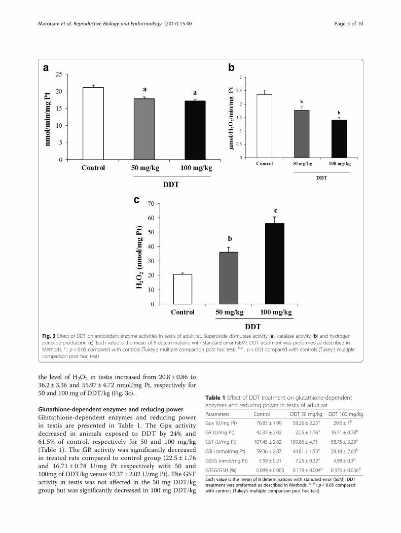
the level of H2O2 in testis increased from 20.8 ± 0.86 to36.2 ± 3.36 and 55.97 ± 4.72 nmol/mg Pt, respectively for50 and 100 mg of DDT/kg (Fig. 3c).
Glutathione-dependent enzymes and reducing powerGlutathione-dependent enzymes and reducing powerin testis are presented in Table 1. The Gpx activitydecreased in animals exposed to DDT by 24% and61.5% of control, respectively for 50 and 100 mg/kg(Table 1). The GR activity was significantly decreasedin treated rats compared to control group (22.5 ± 1.76and 16.71 ± 0.78 U/mg Pt respectively with 50 and100mg of DDT/kg versus 42.37 ± 2.02 U/mg Pt). The GSTactivity in testis was not affected in the 50 mg DDT/kggroup but was significantly decreased in 100 mg DDT/kg
Fig. 3 Effect of DDT on antioxidant enzyme activities in testis of adult rat. Superoxide dismutase activity (a), catalase activity (b) and hydrogenperoxide production (c). Each value is the mean of 8 determinations with standard error (SEM). DDT treatment was preformed as described inMethods. a : p < 0.05 compared with controls (Tukey’s multiple comparison post hoc test). b,c : p < 0.01 compared with controls (Tukey’s multiplecomparison post hoc test)
Table 1 Effect of DDT treatment on glutathione-dependentenzymes and reducing power in testis of adult rat
Parameters Control DDT 50 mg/kg DDT 100 mg/kg
Gpx (U/mg Pt) 76.83 ± 1.99 58.26 ± 2.25a 29.6 ± 1b
GR (U/mg Pt) 42.37 ± 2.02 22.5 ± 1.76a 16.71 ± 0.78b
GST (U/mg Pt) 107.45 ± 2.82 109.86 ± 4.71 58.75 ± 3.29a
GSH (nmol/mg Pt) 59.36 ± 2.87 44.81 ± 1.53a 28.18 ± 2.63b
GSSG (nmol/mg Pt) 5.59 ± 0.21 7.25 ± 0.32a 9.98 ± 0.3b
GSSG/GSH (%) 0.089 ± 0.003 0.178 ± 0.004a 0.376 ± 0.036b
Each value is the mean of 8 determinations with standard error (SEM). DDTtreatment was preformed as described in Methods. a, b : p < 0.05 comparedwith controls (Tukey’s multiple comparison post hoc test)
Marouani et al. Reproductive Biology and Endocrinology (2017) 15:40 Page 5 of 10

group (58.7 ± 3.3 U/mg Pt versus 107.4 ± 2.8 U/mg Pt).DDT treatment induced a dose-dependent increase inGSSG levels (Table 1). This increase reached 29.7% and78.5% of controls for 50 and 100 mg/kg, respectively. Incontrast, the GSH level was significantly decreased intreated rats compared to control group (Table 1). Thisdecrease reached 24.5% and 52.5% of controls for 50 and100 mg of DDT/kg, respectively. The ratio betweenconcentrations of GSSG and GSH is a valuable marker,characterizing cellular redox status. Thus, exposure toDDT significantly increased the ratio GSSG/GSH intreated rats compared to control group (Table 1). Thisincrease reached 100% and 322.5% of controls for 50 and100 mg/kg, respectively.
Fragmentation of DNA induced by DDTEvidence of DNA fragmentation in rat testis treated withDDT was obtained by ethidium-bromide agarose gelelectrophoresis (Fig. 4). DNA isolated from the testiculartissues of rats after administration of DDT to animalsfor 10 days showed degradation into oligonucleotidefragments forming a clear laddering pattern of apoptosiswhen separated by 1.5% agarose gel electrophoresis(Fig. 4, lane b and c), whereas DNA fragmentation isnegligible in testis for control (Fig. 4, lane a).
TUNEL assayApoptosis was characterized by a TUNEL technique thatspecifically detects apoptotic cells in testes. Untreatedrats showed no apoptotic cells in the seminiferoustubule (Fig. 5, Photo a), whereas positive staining ingerm cells was found after treated with 50mg of DDT/kg(Fig. 5, Photo b). With 100 mg of DDT/kg, strong posi-tive staining was observed in germ cells (Fig. 5, Photo c).The apoptotic index grew 8.2 fold (p < 0.01) and 23.2
Fig. 4 In vivo DDT apoptotic DNA laddering in testis. Testicular DNAwas isolated, subjected to agarose gel electrophoresis and stainedwith ethidium bromide as described in the text. Each lane contained4μg of DNA. Lanes right to left: a control, no treatment; b treatedrats with 50mg of DDT/kg; c treated rats with 100mg of DDT/kg.Lane M: 1kb molecular weight DNA ladder marker
Fig. 5 Detection of apoptotic cells in the testis of rats as revealed byTUNEL assay. Seminiferous tubule from control showed no apoptoticcells (Photo a). Seminiferous tubules from DDT- treated rats with 50 mg/kg (Photo B) and 100 mg/kg (Photo c) showed TUNEL-positive germcells. Rats received an ip injection of 50 or 100 mg/kg body weight ofDDT during 10 days. TS, Seminiferous tubule; SP, spermatozoa; S,spermatocyte cells; L, leydig cells. Magnification: ×400 (a, b, c)
Marouani et al. Reproductive Biology and Endocrinology (2017) 15:40 Page 6 of 10

fold (p <0.01) in treated rats with 50 and 100 mg ofDDT/kg, respectively compared to control (Table 2).
DiscussionThe purpose of this study was to investigate how the p,p’-DDT treatment induced oxidative stress and the mecha-nisms involved in DDT-induced apoptosis in testis. Lipidperoxidation is an identified cell damage mechanism inplants and animals, and it is used as an indicator of oxida-tive stress in cells and tissues. Our results showed thatexposure of rats to 100mg of DDT/kg b.wt, during 10 con-secutive days, significantly increased MDA level in thetestis. Our finding was in accordance with another studycarried out in rats and which have also reported a highlevel of lipid peroxidation in the testis of DDE- treated rats[36]. It was reported that DDT induced reactive oxygenspecies (ROS) generation in different animal tissues, in-cluding human cells [19, 37]. It is well documented thatmale reproductive organs are particularly susceptible tothe deleterious effects of ROS and lipid peroxidation,which ultimately lead to impaired fertility [38]. IncreasedLPO during spermatogenesis leads to tissue damage [39],impaired membrane function, decreased membrane fluid-ity, altered structural integrity and inactivation of severalmembrane bound enzymes [40]. Previous studies reportedthe enhanced production of ROS in testis exposed to p,p’-DDE [21, 41]. MTs are members of a family of lowmolecular weight proteins rich in cysteine that play a keyrole in transport of essential heavy metals, detoxificationof toxic metals and protection of cells against oxidationstress. Our result showed that the levels of MTs decreasedin a dose-dependent manner in testis of DDT-treated rats.The inhibited level of MTs is closely associated with in-creased formation of ROS and reactive nitrogen species,respectively. Excessive production of these harmful sub-stances along with a reduction in anti-oxidants couldreduce the level of MTs in testis [42]. Antioxidant en-zymes, such as SOD and CAT, are essential parts in thecellular defense against free radical–mediated tissue orcellular damage. Our results showed a decrease in thelevel of SOD and CAT activities while H2O2 productionincreased in the testis of DDT-treated rats. Similarly, re-cent studies showed that exposure to p,p’-DDE for 10consecutive days decreased SOD activity in testis [36, 41].
The increase of hydrogen peroxide levels may be due toreduced SOD and CAT activities in testis. This conditioncould be favorable to hydroxyl radical formation whichmay lead to lipid peroxidation [43]. Besides, SOD isknown to catalyse the dismutation of superoxide anions toH2O2 and molecular oxygen, while CAT has been shownto be responsible for the detoxification of H2O2 [44]. Thereduction in CAT activity may reflect less capacity oftesticular mitochondria and microsomes to eliminateH2O2 produced in response to DDT [45]. It is also knownthat CAT protect SOD against inactivation by H2O2 andthat SOD, in turn, protects CAT against superoxideanions. The balance of these enzyme systems may beessential to testicular health. Hence, the significant reduc-tion in enzyme activities, accompanied by marked increaselipid peroxidation, may reflect adverse effects of DDT onthe antioxidant system [41]. Therefore, the decrease inSOD and CAT activities may explain the early-elevatedROS levels, since it was a crucial enzyme involved in thedetoxification of ROS. Moreover, our results showed thatp-p’-DDT administration decreased testicular Gpx, GR,GST and GSH activities while increased the ratio GSSG/GSH, which led to the production of free radicals andcausing LPO [46]. GSH is one of the most important non-enzymatic antioxidant against cellular damage produced byROS [47]. GSH supplementation has been shown to have aprotective action against seminal plasma lipid peroxidation,and it has been implicated in the treatment of male infertil-ity [48]. The decreased glutathione concentration may beexplained by the adverse effect of ROS which can decreasesynthesis and/or reduce transport into the testis (because itis well known that most GSH is synthesized by liver), oraccelerate degradation or enhance export of oxidized form[49]. It cannot be excluded that the system(s) of synthesisand transport of GSH could be affected by exposure toDDT. GR is involved in the supplementation of GSH tospermatogenic cells [50]. GSH is also GST co-substrat. GSTcatalyzes the conjugation of reduced glutathione with a var-iety of endogenous compounds and xenobiotics [51].Therefore a depression in GSH levels together with GSTactivity makes the cells more susceptible to the attack bytoxic compounds [52]. In the present study, the decrease inGPx, GST and GSH activities, accompanied by the increaseof GSSG/GSH ratio and MDA levels, supports that oxida-tive stress is produced due to DDT administration. Apop-tosis is a genetically regulated cellular suicide mechanism inwhich multiple signaling pathways are implicated [53].Among them, oxidative stress is an important event whichmay affect different macromolecules and components ofthe cells, triggering the activation of several antioxidantresponse genes and mechanisms [53]. The oxidative stresscould be associated with severe damage to DNA. Previousstudy revealed that exposure to DDT induced DNA single-strand breaks [54]. Recently, it was reported that exposure
Table 2 Effect of DDT treatment on apoptotic index in testis ofadult rat
Apoptotic index (%)
Control 0.55 ± 0.09
DDT 50 mg/kg 4.5 ± 0.92a
DDT 100 mg/kg 12.78 ± 1.54b
Each value is the mean of 8 determinations with standard error (SEM). DDTtreatment was preformed as described in Methods. a,b : p < 0.05 comparedwith controls (Tukey’s multiple comparison post hoc test)
Marouani et al. Reproductive Biology and Endocrinology (2017) 15:40 Page 7 of 10

to p,p’-DDE induced DNA damage in Sertoli cells, whichmight account for subsequent development of apoptosis[21, 36, 41]. In this study, the DNA isolated from testiculartissues of DDT-treated rats showed degradation into oligo-nucleotide fragments forming a clear ladder pattern. Inaddition, histological examination of testicular tissue by theTUNEL method showed that apoptosis cells occurred inthe germ cells of DDT-treated rats. Also, the apoptoticindex was significantly increased in testis of DDT-treatedrats. Apoptosis is a complex event regulated by a well-tuned balance of inducer and repressor factors, such as theBcl-2 family, which is a pivotal integrator of survival anddeath signal. In addition, the Fas system is a widely recog-nized apoptosis signal transduction pathway in which aligand-receptor interaction triggers the cell death pathway[55]. Fas is a surface receptor that triggers apoptotic celldeath when cross-linked by FasL [56]. Ligation of FasL toFas in the cell membrane triggers activation of caspase-8.Once activated, caspase-8 transduces a signal to effectorcaspases, including caspases 3, 6, and 7, and eventuallyleads to the hydrolysis of cytosolic and nuclear substrates[57]. Previous studies showed that p,p’-DDE could induceapoptosis of Sertoli cells through a FasL-dependent path-way including nuclear translocation of NF-κB, increase ofthe FasL expression, and activation of the caspase 8 and 3[21, 36]. Recently, it was reported that p,p’-DDT activatedNF-κB/FasL pathway and mitochondrial pathway in humanliver cells which were mediated by ROS. Moreover, it hasbeen demonstrated that the excess or deprivation of hor-mones such as FSH and testosterone can lead to cellularapoptosis in the testis [58]. It has been shown that bothextrinsic and intrinsic apoptotic death pathways are opera-tive in the germ cells following decrease in FSH and testos-terone levels; therefore, FSH and testosterone maintainspermatogenic homeostasis by inhibiting death signals forthe germ cells [59]. In our earlier study, serum FSH and LHlevels were significantly increased and testosterone levelswere decreased in rats exposed to 50 and 100mg of DDT/kg for 10 days [17]. It is possible that the decreased testos-terone levels and the increased FSH levels in response toDDT exposure stimulates caspase activity and producesDNA fragmentation in germ cells [60]. Fewer study eluci-dated the mechanism of DDT-induced apoptosis in testis.So, in this study, we have shown for the first time thatp,p’-DDT treatment induced apoptosis in germ cells.These findings suggested that p,p’-DDT-induced apoptosisof germ cells through mitochondria-mediated and FasL-dependent pathway. It is possible that different stimuli suchas DNA damage or increased ROS level caused by DDTmight trigger Bax activation via acting diverse moleculessuch as p53, and Fas system. Activation of Bax protein leadsto the formation of pores in the mitochondria and resultsin the collapse of the electro chemical gradient across themitochondrial membrane, then cytochrome c is released
into cytoplasm where it is associated with procaspase-9/Apaf-1. This complex, in turn, activates a downstreamcaspase program that ultimately leads to apoptotic celldeath [61].
ConclusionsIn conclusion, the results obtained from the present studydemonstrate that the sub-acute treatment of p,p’-DDTcauses DNA fragmentation and apoptotic cell death intestis probably mediated by oxidative stress which leads tothe adverse toxic effects of DDT on male reproduction ofrats. Further studies are needed to elucidate the expres-sion and/or activity of pro and anti-apoptotic proteins intesticular cells after exposure to DDT.
AbbreviationsCAT: Catalase; GR: Glutathione reductase; GSH: Reduced glutathione;GSSG: Oxidized glutathione; GST: Glutathione S-transferase; H2O2:Hydrogen peroxide; LPO: Lipid peroxidation; MDA: Malondialdehyde;MTs: Metallothioneins; PBS: Phosphate-buffered saline; ROS: Reactiveoxygen species; SOD: Superoxide dismutase
AcknowledgmentsThis work was supported by the Tunisian Ministry of Higher Education,Scientific Research and Technology and Carthage University. The authorsthank B. Azib for his excellent technical assistance.
FundingThis work was supported by the Tunisian Ministry of higher Education andScientific Research, Carthage University.
Availability of data and materialsPlease contact author for raw data requests.
Authors’ contributionsN M, D H, M S, M B, K BR and O T analyzed and interpreted data, drafted orrevised the manuscript, read and approved the final manuscript, and agreedto be accountable for all aspects of the work.
Competing interestsThe authors declare that they have no competing interests.
Consent for publicationNot applicable.
Ethics approval and consent to participateAnimals were cared for in compliance with the code of practice for the Careand Use of Animals for Scientific Purposes. Approval for these experimentswas obtained from the Medical Ethical Committee for the Care and Use ofLaboratory Animals of Pasteur Institute of Tunis (approval number: LNFP/Pro152012). The experimental protocols were approved by the Faculty EthicsCommittee (Faculté des Sciences de Bizerte, Tunisia).
Publisher’s NoteSpringer Nature remains neutral with regard to jurisdictional claims inpublished maps and institutional affiliations.
Author details1Laboratory of Integrated Physiology, Faculty of Sciences, Carthage UniversityTunisia, Bizerte, Jarzouna, Tunisia. 2Reproductive Medicine and MedicalCytogenetics Department, Regional University Hospital and School ofMedicine, Picardie University Jules Verne, Amiens, France.
Marouani et al. Reproductive Biology and Endocrinology (2017) 15:40 Page 8 of 10

Received: 9 March 2017 Accepted: 9 May 2017
References1. Leber ER, Benya TJ. Chlorinated hydrocarbon insecticides. In: Clayton GD,
Clayton FE, editors. Patty’s Industrial Hygiene and Toxicology, Vol. 2, Part B.New York: Wiley; 1994. p. 1503–6.
2. Rogan WJ, Chen A. Health risks and benefits of bis (4-chlorophenyl)-1,1, 1-trichloroethane (DDT). Lancet. 2005;366:763–73.
3. Stemmler I, Lammel G. Cycling of DDT in the global environment 1950–2002:World ocean returns the pollutant. Geophys Res Lett. 2009;36:L24602–6.
4. UNEP. Stockholm convention on persistent organic pollutants (POPs).Geneva: United Nations Environment Programme; 2002.
5. van den Berg H. Global status of DDT and its alternatives for use in vectorcontrol to prevent disease. Environ Health Perspect. 2009;117:1656–63.
6. van Dyk JC, Bouwman H, Barnhoorn IE, Bornman MS. DDT contaminationfrom indoor residual spraying for malaria control. Sci Total Environ. 2010;408:2745–52.
7. Muralidharan S, Dhananjayan V, Jayanthi P. Organochlorine pesticides incommercial marine fishes of Coimbatore, India and their suitability forhuman consumption. Environ Res. 2009;109:15–21.
8. Aulakh RS, Bedi JS, Gill JPS, Joia BS, Pooni PA, Sharma JK. Occurrence ofDDT and HCH insecticide residues in human biopsy adipose tissues inPunjab. India Bull Environ Contam Toxicology. 2007;78:330–4.
9. Malarvannan G, Kunisue T, Isobe T, Sudaryanto A, Takahashi S, Prudente M,Subramanian A, Tanabe S. Organohalogen compounds in human breast milkfrom mothers living in Payatas and Malate, the Philippines: levels,accumulation kinetics and infant health risk. Environ Pollut. 2009;157:1924–32.
10. Guimaraes RM, Asmus CI, Meyer A. DDT reintroduction for malaria control: thecost-benefit debate for public health. Cad Saude Publica. 2007;23:2835–44.
11. Brucker-Davis F. Effects of environmental synthetic chemicals on thyroidfunction. Thyroid. 1998;8:827–56.
12. ATSDR: Agency for Toxic Substances and Diseases Registry. ToxicologicalProfile for 4, 4’-DDT, 4, 4’-DDE, 4, 4’-DDD. (Final report, ATSDR/TP-93/05).Department of Health and Human Services Atlanta GA: Public HealthService; 1994. p. 192.
13. Jaga K, Brosius D. Pesticide exposure: human cancers on the horizon. RevEnviron Health. 1999;14:39–50.
14. Hauser R, Singh NP, Chen Z, Pothier L, Altshul L. Lack of an associationbetween environmental exposure to polychlorinated biphenyls and p, p’-DDE and DNA damage in human sperm measured using the neutral cometassay. Hum Reprod. 2003;18:2525–33.
15. Longnecker MP, Klebanoff MA, Zhou H, Brock JW. Association betweenmaternal serum concentration of the DDT metabolite DDE and preterm andsmall-for-gestational-age babies at birth. Lancet. 2001;358:110–4.
16. Hamlin HJ, Guillette LJJ. Birth defects in wildlife: the role of environmentalcontaminants as inducers of reproductive and developmental dysfunction.Syst Biol Reprod Med. 2010;56:113–21.
17. Ben Rhouma K, Tebourbi O, Krichah R, Sakly M. Reproductive toxicity of DDTin adult male rats. Hum Exp Toxicol. 2001;20:393–7.
18. Tebourbi O, Sakly M, Rhouma KB. Molecular mechanisms of pesticidetoxicity. Pesticides in the Modern World–Pests Control and PesticidesExposure and Toxicity Assessment, Dr. Margarita Stoytcheva (Ed.), InTech.2011;Chapter 15:297–332.
19. Jin XT, Song L, Zhao JY, Li ZY, Zhao MR, Liu WP.Dichlorodiphenyltrichloroethane exposure induces the growth ofhepatocellular carcinoma via Wnt/b-catenin pathway. Toxicol Lett.2014;225:158–66.
20. Wu CC, Bratton SB. Regulation of the intrinsic apoptosis pathway byreactive oxygen species. Antioxid Redox Signal. 2013;19:546–58.
21. Song Y, Liang X, Hu Y, Wang Y, Yu H, Yang K. p, p’-DDE inducesmitochondria-mediated apoptosis of cultured rat Sertoli cells. Toxicology.2008;253(1–3):53–61.
22. Harada TS, Yamaguchi R, Ohtsuka M, Takeda H, Fujisawa T, Yoshida A,Enomoto A, Chiba Y, Fukumori J, Kojima S, Tomiyama N, Saka M, Ozaki M,Maita K. Mechanisms of promotion and progression of preneoplastic lesions inhepatocarcinogenesis by DDT in F344 rats. Toxicol Pathol. 2003;31(1):87–98.
23. Tebourbi O, Hallègue D, Yacoubi MT, Sakly M, Ben RK. Subacute toxicity ofp, p’-DDT on rat thyroid: Hormonal and histopathological changes. EnvironToxicol Pharmacol. 2010;29:271–9.
24. Beytut E, Aksakal M. Effects of dietary vitamin E and selenium on oxidativedefense mechanisms in the liver of rats treated with high doses ofglucocorticoid. Biol Trace Elem Res. 2003;91:231–41.
25. Buege JA, Aust SD. Lactoperoxidase catalyzed lipid peroxidation of microsome-rich and artificial membranes. Biochim Biophys Acta. 1976;444:192–201.
26. Eaton DL, Cherian MG. determination of metallothionein in tissues bycadmium-hemoglobin affinity assay. Methods Enzymol. 1991;205:83–8.
27. Marklund S, Marklund G. Involvement of the superoxide anion radical in theautoxidation of pyrogallol and a convenient assay for superoxide dismutase.Eur J Biochem. 1974;47(3):469–74.
28. Aebi H. Catalase in vitro. Methods Enzymol. 1984;105:121–6.29. Pick E, Keisari Y. Superoxide anion and hydrogen peroxide production by
chemically elicited peritoneal macrophages–induction by multiplenonphagocytic stimuli. Cell Immunol. 1981;59(2):301–18.
30. Paglia DE, Valentine WN. Studies on the quantitative and qualitativecharacterization of erythrocyte glutathione peroxidase. J Lab Clin Med.1967;70(1):158–69.
31. Calberg I, Mannervik B. Glutathione reductase. Methods Enzymol. 1985;113:484–90.
32. Habig WH, Pabst MJ, Jakoby WB. Glutathione S-transferases. The first enzymaticstep in mercapturic acid formation. J Biol Chem. 1974;249:7130–9.
33. Hissin PJ, Hilf R. Fluorometric method for determination of oxidized andreduced glutathione in tissues. Anal Biochem. 1976;74:214–26.
34. Ichimura T, Kawamura M, Mitani A. Co-localized expression of FasL, Fas,Caspase-3 and apoptotic DNA fragmentation in mouse testis after oralexposure to di (2-ethylhexyl) phthalate. Toxicology. 2003;194:35–42.
35. Karagüzel E, Kutlu Ö, Yuluģ E, Mungan S, Kazaz IO, Tok DS, Özgür GK.Comparison of the protective effect of dipyridamole and acetylsalicylic acidon long-term histologic damage in a rat model of testicular ischemia-reperfusion injury. J Pediatr Surg. 2012;47:1716–23.
36. Shi YQ, Wang YP, Song Y, Li HW, Liu CJ, Wu ZG, Yang KD. p, p’-DDE inducestesticular apoptosis in prepubertal rats via the Fas/FasL pathway. ToxicolLett. 2010;193(1):79–85.
37. Perez-Maldonado IN, Herrera C, Batres LE, Gonzalez-Amaro R, Diaz-Barriga F,Yanez L. DDT-induced oxidative damage in human blood mononuclearcells. Environ Res. 2005;98:177–84.
38. Williams K, Frayne J, McLaughlin EA, Hall L. Expression of extracellular superoxidedismutase in the human male reproductive tract, detected using antisera raisedagainst a recombinant protein. Mol Hum Reprod. 1998;4(3):235–42.
39. Mylonas C, Kouretas D. lipid peroxidation and tissue damage. In Vivo. 1999;13:295–309.
40. Gutteridge JM, Halliwell B. Free radicals and antioxidants in the year 2000: ahistorical look to the future. Ann N Y Acad Sci. 2000;899:136–47.
41. Shi YQ, Li HW, Wang YP, Liu CJ, Yang KD. p, p’-DDE induces apoptosis andmRNA expression of apoptosis-associated genes in testes of pubertal rats.Environ Toxicol. 2013;28:31–41.
42. Chin JL, Banerjee D, Kadhim SA, Kontozoglou TE, Chauvin PJ, Cherian MG.Metallothionein in testicular germ cell tumors and drug resistance. Cancer.1993;72:3029–35.
43. Lissi EA. Ca’ceres T, Llesuy S, Solari L, Boveris A, Videla LA. On thecharacteristics of the visible chemiluminescence following free radical lipidperoxidation. Free Radic Res Commun. 1989;6(5):293–301.
44. Linares V, Sánchez DJ, Bellés M, Albina L, Gómez M, Domingo JL.Pro-oxidant effects in the brain of rats concurrently exposed touranium and stress. Toxicology. 2007;236:82–91.
45. Pigolet E, Corbisier P, Houbion A, Lambert D, Michiels C, Raes M, ZacharyMD, Remacle J. Glutathione peroxidase, superoxide dismutase and catalaseinactivation by peroxides and oxygen derived free radicals. Mech AgeingDev. 1990;51:283–90.
46. Nehru LB, Bansal MP. Effect of selenium supplementation on theglutathione redox system in the kidney of mice after chronic cadmiumexposures. J App Toxicol. 1997;17(1):81–4.
47. Luberda Z. The role of glutathione in mammalian gametes. Reprod Biol.2005;5(1):5–17.
48. Irvine DS. Glutathione as a treatment for male infertility. Rev Reprod.1996;1:1–12.
49. Lushchak OV, Kubrak OI, Torous IM, Nazarchuk TY, Storey KB, Lushchak VI.Trivalent chromium induces oxidative stress in goldfish brain. Chemosphere.2009;75(1):56–62.
50. Kaneko T, Iuchi Y, Kobayashi T, Fujii T, Saito H, Kurachi H, Fujii J. Theexpression of glutathione reductase in the male reproductive system of rats
Marouani et al. Reproductive Biology and Endocrinology (2017) 15:40 Page 9 of 10

supports the enzymatic basis of glutathione function in spermatogenesis.Eur J Biochem. 2002;269:1570–8.
51. Romeu M, Mulero M, Giralt M, Folch J, Nogués MR, Torres A, Fortuño A,Sureda FX, Cabré M, Paternáin JL, Mallol J. Parameters related to freeradicals in erythrocytes, plasma and epidermis of the hairless rat. Life Sci.2002;71:1739–49.
52. Boesch-Saadatmandi C, Loboda A, Jozkowicz A, Huebbe P, Blank R,Wolffram S, Dulak J, Rimbach G. Effect of ochratoxin A on redox-regulatedtranscription factors, antioxidant enzymes and glutathione-S-transferase incultured kidney tubulus cells. Food Chem Toxicol. 2008;46:2665–71.
53. Kiechle FL, Zhang X. Apoptosis: biochemical aspects and clinicalimplications. Clin Chim Acta. 2002;326:27–45.
54. Hassoun E, Bagchi M, Bagchi D, Stohs SJ. Comparative studies on lipidperoxidation and DNA-single strand breaks induced by lindane, DDT,chlordane and endrin in rats. Comp Biochem Physiol C. 1993;104(3):427–31.
55. Feng H, Zeng Y, Graner WM, Whitesell L, Katsanis E. Evidence for a novel,caspase-8-independent. Fas death domain-mediated apoptotic pathwayJ Biomed Biotechnol. 2004;2004:41–51.
56. Nagata S. Apoptosis by death factor. Cell. 1997;88:355–65.57. De Maria R, Lenti L, Malisan F, d’Agostino F, Tomassini B, Zeuner A, Rippo
MR, Testi R. Requirement for GD3 ganglioside in CD95- and ceramide-induced apoptosis. Science. 1997;277:1652–5.
58. Shaha C, Tripathi R, Mishra DP. Male germ cell apoptosis: regulation andbiology. Phil Trans R Soc B. 2010;365:1501–15.
59. Pareek TK, Joshi AR, Sanyal A, Dighe RR. Insights into male germ cellapoptosis due to depletion of gonadotropins caused by GnRH antagonists.Apoptosis. 2007;12:1085–100.
60. Tesarik J, Martinez F, Rienzi L, Iacobelli M, Ubaldi F, Mendoza C, Greco E.In-vitro effects of FSH and testosterone withdrawal on caspase activationand DNA fragmentation in different cell types of human seminiferousepithelium. Hum Reprod. 2002;17:1811–9.
61. Kuwana T, Newmeyerm DD. Bcl-2-family proteins and the role ofmitochondria in apoptosis. Curr Opin Cell Biol. 2003;15:691–9.
• We accept pre-submission inquiries
• Our selector tool helps you to find the most relevant journal
• We provide round the clock customer support
• Convenient online submission
• Thorough peer review
• Inclusion in PubMed and all major indexing services
• Maximum visibility for your research
Submit your manuscript atwww.biomedcentral.com/submit
Submit your next manuscript to BioMed Central and we will help you at every step:
Marouani et al. Reproductive Biology and Endocrinology (2017) 15:40 Page 10 of 10


![DDT - PCD.go.thinfofile.pcd.go.th/haz/25-DDT.pdf · 8 “√Õ—πµ√“¬ - o,p/-DDT ¡’™ ËÕ‡§¡’«à“ 1-chloro-2-[2,2,2-trichloro-1-(4-chloro-phenyl)ethyl] benzene](https://static.fdocuments.us/doc/165x107/5f37beca69f3641abe2ca19a/ddt-pcdgo-8-aoeaaaaoe-op-ddt-aa-aaaoe.jpg)