Separation and Analysis of Honeybee Venom Components Levi Blazer Liz Denning Laura Rhodes Juniata...
15
Separation and Analysis of Honeybee Venom Components Levi Blazer Liz Denning Laura Rhodes Juniata College Research Advisors: Dr. Lorraine Mulfinger and Dr. Michael Boyle
-
date post
20-Dec-2015 -
Category
Documents
-
view
215 -
download
1
Transcript of Separation and Analysis of Honeybee Venom Components Levi Blazer Liz Denning Laura Rhodes Juniata...
Separation and Analysis of Honeybee Venom Components
Levi BlazerLiz DenningLaura RhodesJuniata CollegeResearch Advisors: Dr. Lorraine Mulfinger and Dr. Michael Boyle
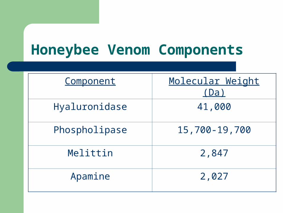
Honeybee Venom Components
Component Molecular Weight (Da)
Hyaluronidase 41,000
Phospholipase 15,700-19,700
Melittin 2,847
Apamine 2,027
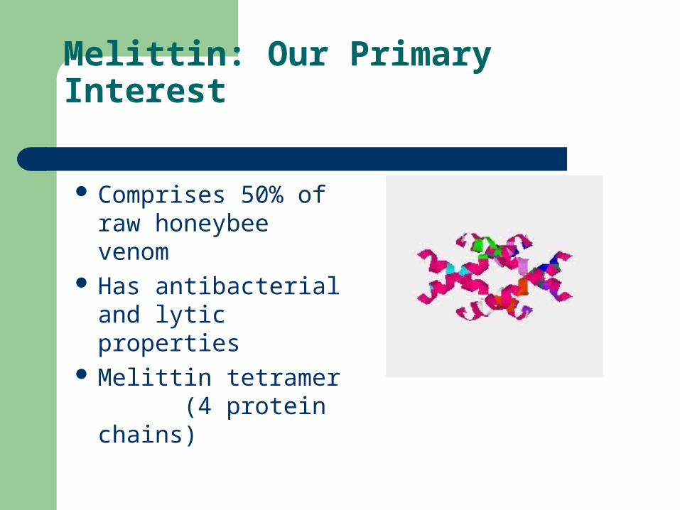
Melittin: Our Primary Interest
Comprises 50% of raw honeybee venom
Has antibacterial and lytic properties
Melittin tetramer (4 protein chains)
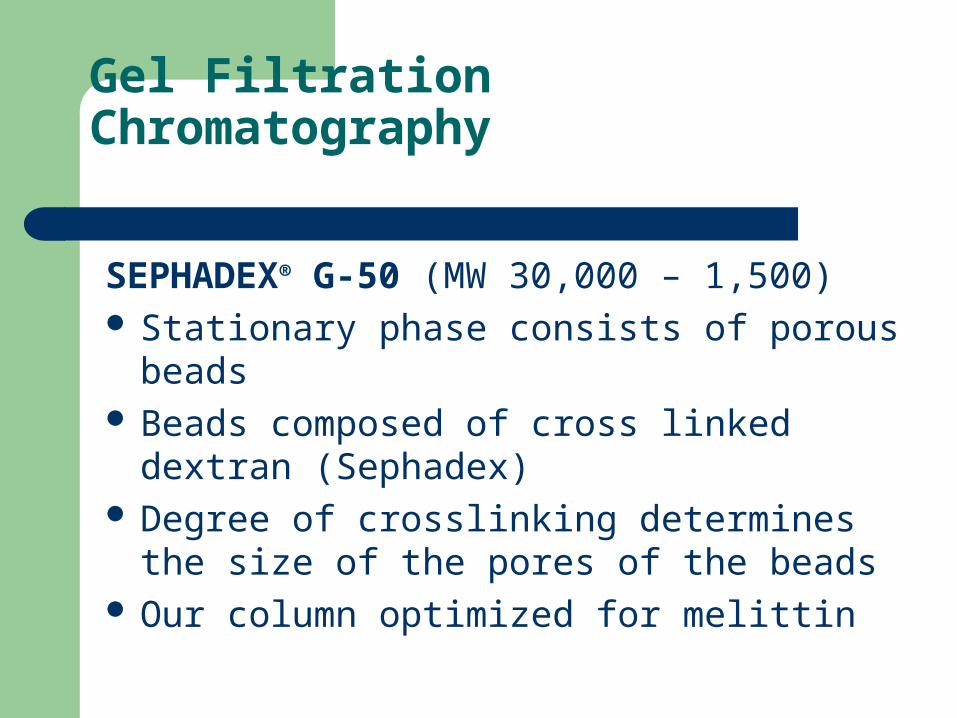
Gel Filtration Chromatography
SEPHADEX® G-50 (MW 30,000 – 1,500) Stationary phase consists of porous beads Beads composed of cross linked dextran
(Sephadex) Degree of crosslinking determines the size of
the pores of the beads Our column optimized for melittin
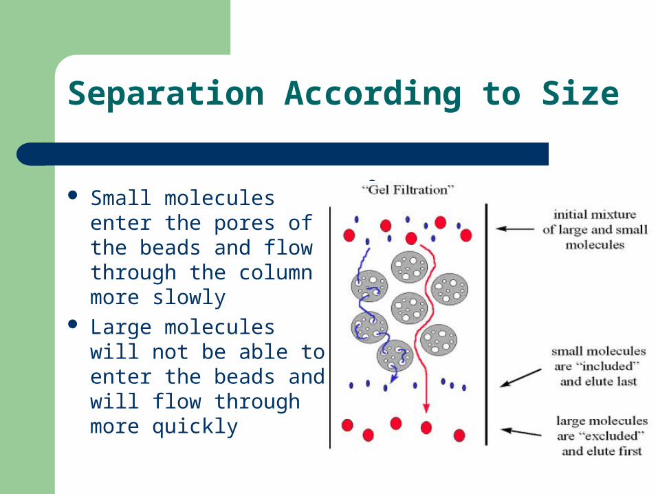
Separation According to Size
Small molecules enter the pores of the beads and flow through the column more slowly
Large molecules will not be able to enter the beads and will flow through more quickly
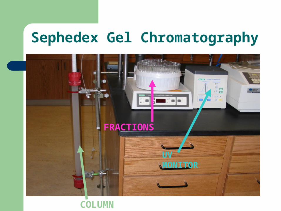
Sephedex Gel Chromatography
UV MONITOR
COLUMN
FRACTIONS
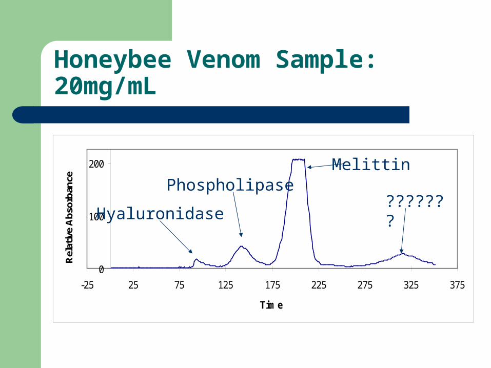
Honeybee Venom Sample: 20mg/mL
0
100
200
-25 25 75 125 175 225 275 325 375
Time
Rel
ativ
e A
bsor
banc
e
Melittin
???????Phospholipase
Hyaluronidase
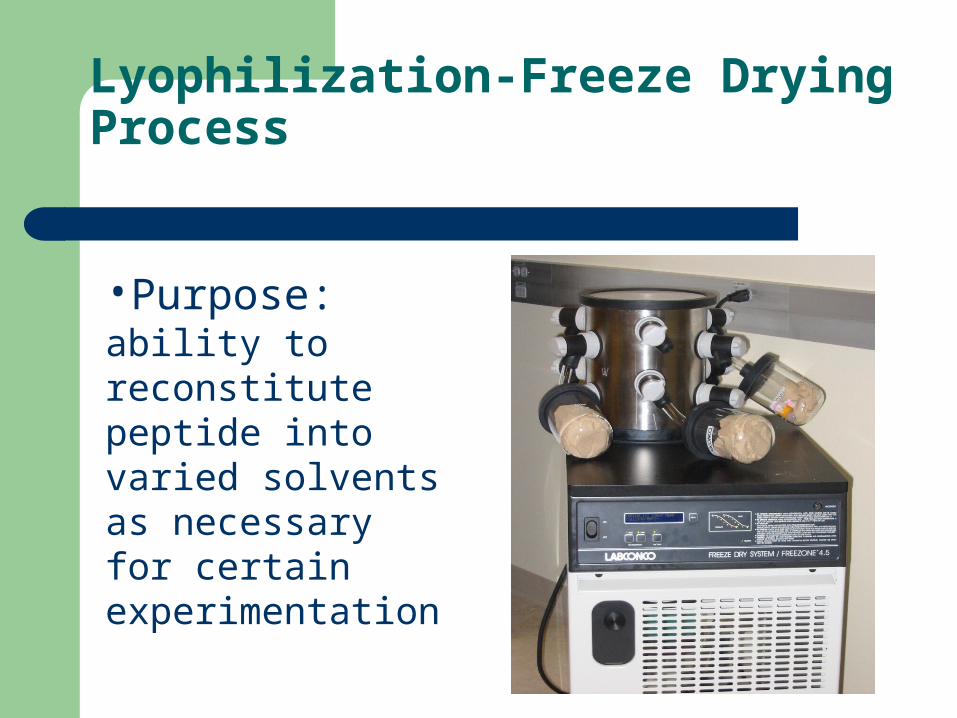
Lyophilization-Freeze Drying Process
•Purpose: ability to reconstitute peptide into varied solvents as necessary for certain experimentation
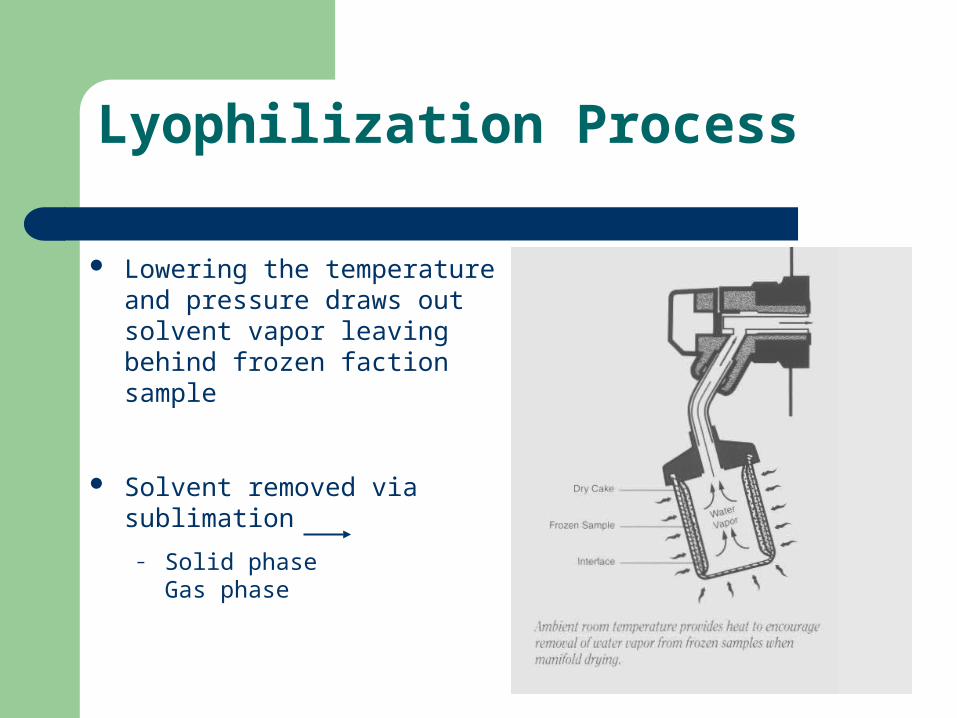
Lyophilization Process
Lowering the temperature and pressure draws out solvent vapor leaving behind frozen faction sample
Solvent removed via sublimation
– Solid phase Gas phase
Analysis of Column Fractions
Gel Electrophoresis
SELDI-TOF Mass Spectrometry
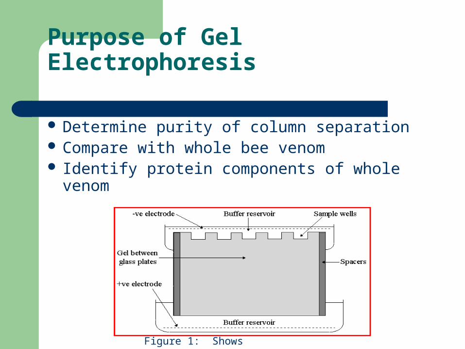
Purpose of Gel Electrophoresis
Determine purity of column separation Compare with whole bee venom Identify protein components of whole
venom
Figure 1: Shows electrophoresis components
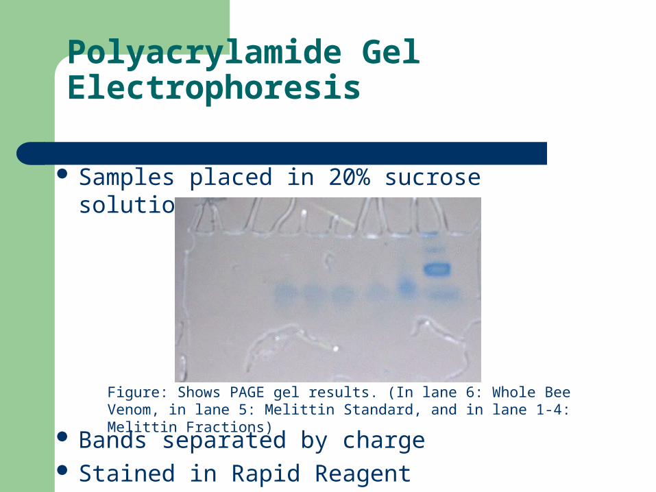
Polyacrylamide Gel Electrophoresis
Samples placed in 20% sucrose solution
Bands separated by charge Stained in Rapid Reagent
Figure: Shows PAGE gel results. (In lane 6: Whole Bee Venom, in lane 5: Melittin Standard, and in lane 1-4: Melittin Fractions)
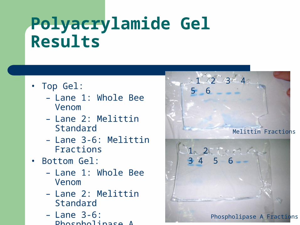
Polyacrylamide Gel Results
• Top Gel:– Lane 1: Whole Bee
Venom– Lane 2: Melittin Standard– Lane 3-6: Melittin
Fractions• Bottom Gel:
– Lane 1: Whole Bee Venom
– Lane 2: Melittin Standard– Lane 3-6: Phospholipase
A Fractions
Melittin Fractions
Phospholipase A Fractions
1 2 3 4 5 6
1 2 3 4 5 6
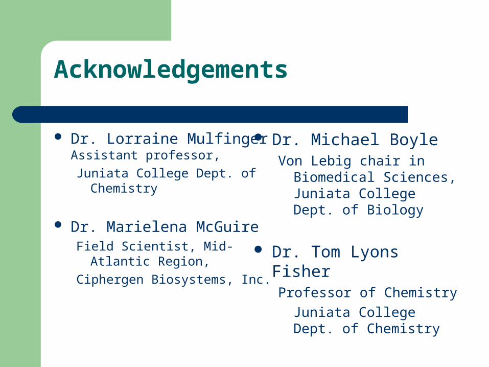
Acknowledgements
Dr. Lorraine MulfingerAssistant professor,
Juniata College Dept. of Chemistry
Dr. Marielena McGuireField Scientist, Mid-Atlantic
Region,
Ciphergen Biosystems, Inc.
Dr. Michael BoyleVon Lebig chair in Biomedical
Sciences, Juniata College Dept. of Biology
Dr. Tom Lyons FisherProfessor of Chemistry
Juniata College Dept. of Chemistry
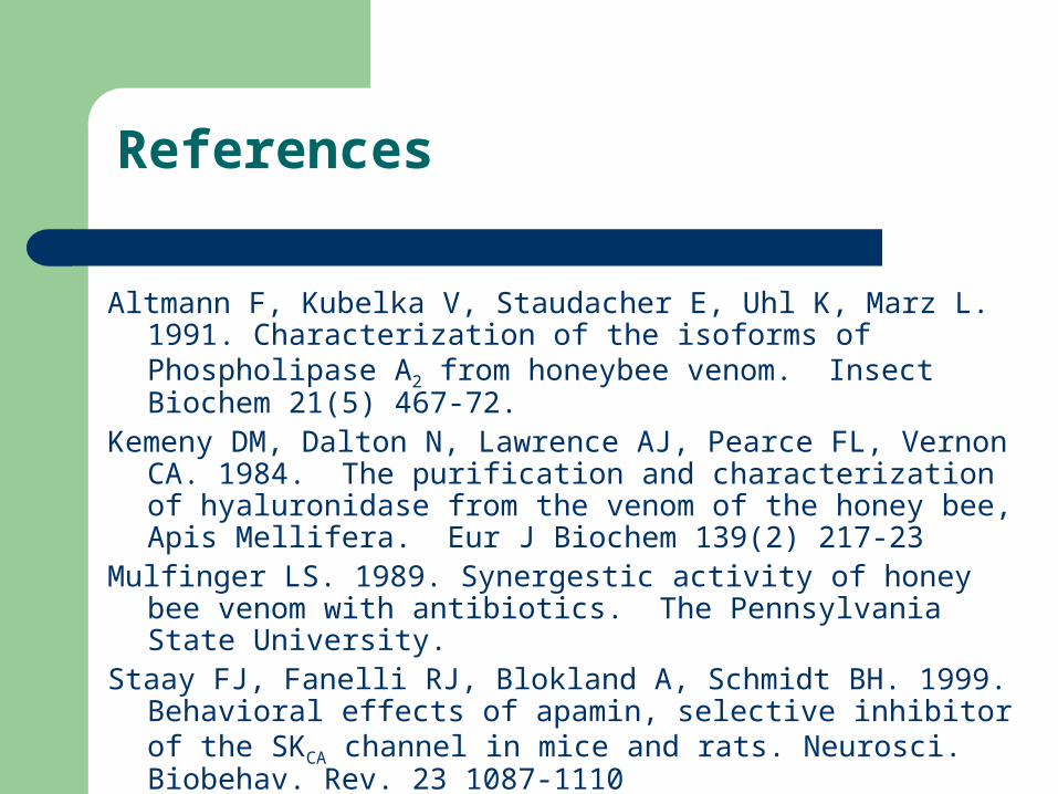
References
Altmann F, Kubelka V, Staudacher E, Uhl K, Marz L. 1991. Characterization of the isoforms of Phospholipase A2 from honeybee venom. Insect Biochem 21(5) 467-72.
Kemeny DM, Dalton N, Lawrence AJ, Pearce FL, Vernon CA. 1984. The purification and characterization of hyaluronidase from the venom of the honey bee, Apis Mellifera. Eur J Biochem 139(2) 217-23
Mulfinger LS. 1989. Synergestic activity of honey bee venom with antibiotics. The Pennsylvania State University.
Staay FJ, Fanelli RJ, Blokland A, Schmidt BH. 1999. Behavioral effects of apamin, selective inhibitor of the SKCA channel in mice and rats. Neurosci. Biobehav. Rev. 23 1087-1110