Conformation Dependence of Diphenylalanine Self-Assembly ...

1
Self-assembly of diphenylalanine with
preclick components as capping groups
Andrea Gemma,1,
Enric Mayans,1,2,
Gema Ballano,3 Juan Torras,
1
Angélica Díaz,1,2
Ana I. Jiménez,3 Jordi Puiggalí,
1,2,* Carlos Cativiela,
3,*
and Carlos Alemán1,2,*
1 Departament d’Enginyeria Química, EEBE, Universitat Politècnica de Catalunya,
Edifici I.2, C/ Eduard Maristany, 10-14, 08019, Barcelona, Spain
2 Research Center for Multiscale Science and Engineering, Universitat Politècnica de
Catalunya, C/ Eduard Maristany, 10-14, 08019, Barcelona, Spain
3 Departmento de Química Orgánica and Instituto de Síntesis Quimica y Catalisis
Homogenea (ISQCH), Universidad de Zaragoza–CSIC, 50009 Zaragoza, Spain
These authors contributed equally to this work.
* [email protected], [email protected] and [email protected]
2
ABSTRACT
Alkyne and azide, which are commonly used in the cycloaddition reaction
recognized as “click chemistry”, have been used as capping groups of two engineered
diphenylalanine (FF) derivatives due to their capacity to form weak intermolecular
interactions (i.e. dipole- and - stacking). In Poc-FF-N3 the alkyne and azide act as
N- and C-terminal capping groups, respectively, while such positions are exchanged in
N3-FF-OPrp. The self-assembly of such two synthesized peptides have been extensively
studied in their “pre-click” state, considering the influence of three different factors: the
peptide concentration, the polarity of the medium, and the nature of the substrate. Poc-
FF-N3 assembles into microfibers that, depending on the medium and the substrate, can
aggregate hierarchically in supramolecular structures with different morphologies. The
most distinctive one corresponds to very stable birefringent dendritic-like
microstructures, which are derived from the ordered agglomeration of microfibers.
These branched supramolecular structures, which are observed in a variety of
conditions, are relatively uncommon in short FF sequences. At the molecular level, Poc-
FF-N3 organizes in antiparallel -sheets stabilized by N–H···O intermolecular hydrogen
bonds and re-enforced by weak interactions between the azide and alkyne groups of
neighbouring molecules. In opposition, N3-FF-OPrp exhibits a very poor tendency to
organize into structures with well-defined morphology. Theoretical calculations on
model complexes indicate that the tendency of the latter peptide to organize into small
amorphous agglomerates is due to its poor ability to form specific intermolecular
interactions in comparison with Poc-FF-N3. The implications of the weak interactions
induced by the alkyne and azide groups, which strengthen peptide···peptide hydrogen
bonds and -ladders due to the stacked aromatic phenyl side groups, are discussed.
3
INTRODUCTION
In the self-assembly process, disordered molecules organize into ordered
supramolecular architectures due to the synergistic combination of non-covalent forces,
such as hydrogen bonding, electrostatic, - stacking, hydrophobic, van der Waals and
dipole-dipole interactions.1-5
As these interactions are ubiquitous in biological systems,
nature has inspired artificial building blocks amenable to self-assemble into
supramolecular structures.6-9
With respect to biological building blocks, peptides are of
great interest for materials science due to their structural simplicity, functional
versatility, cost effectiveness, and widespread applications.10-12
As one of the simplest peptide building blocks, diphenylalanine (FF), a core
recognition motif of Alzheimer’s-amyloid polypeptide,13
has been demonstrated to be
an exciting tool for the development of new, environmental benign, functional
materials. The FF peptide self-assembles into hollow nanotubes with outer diameters of
100-150 nm and micrometrical length,13
which are thermally and chemically stable,14,15
and exhibit excellent mechanical properties.16
Due to these characteristics, extensive
research efforts have been devoted to expand this class of peptide-based materials.17,18
More specifically, a large number of FF-based biomaterials has been engineered
considering, individually or in combination, the following general approaches: (1)
increment the number of F residues in the peptide sequence (i.e. growing from FF to
FFF or FFFF);19-22
(2) add N- and/or C-terminal capping groups to the homopeptide
sequence;23-27
and (3) introduce chemical modifications at the own F residues.28
Among peptide capping groups, the fluorenylmethoxycarbonyl (Fmoc) has been
widely studied because of its ability to form strong aromatic interactions, which
frequently drive the peptide self-assembly into nanofibers or nanotubes.17,29-32
In peptide
sequences with aromatic residues, as F-homopeptides, the incorporation of the Fmoc

4
capping group at the N-terminus enhances the already important role played by -
stacking interactions. Specifically, Fmoc-FF forms peptide fibrils24
and very stable
hydrogels23,33,34
due to the - stacking interaction between Fmoc groups and between
phenyl side groups. The ability of Fmoc aromatic moiety to facilitate gelation, which is
significantly higher than that of other non-aromatic hydrophobic groups (e.g. tert-
butoxycarbonyl), was unambiguously demonstrated studying series of functionalized
dipeptides and amino acids.35
In a recent work we examined the self-assembly of phenylalanine-based peptides
capped with Fmoc and 9-fluorenylmethyl ester (OFm) as N- and C-terminal aromatic
components, respectively.25
These systems exhibit a very high concentration of
aromatic groups, which results from the complete elimination of both the normally free
basic (N-terminus) and acidic (C-terminus) ends. Accordingly, supramolecular self-
assembled organizations were expected to be reached through - stacking interactions.
Polymorphic structures with stacked-braids, dendritic and microtubular morphologies
were found for Fmoc-FF-OFm depending on the solvents used to promote the molecular
self-assembly.25
Furthermore, theoretical calculations on small model complexes
predicted an antiparallel -structure, which was in good agreement with FTIR spectra.
The most important characteristics associated with such -sheet disposition were: (i) the
existence of aromatic -ladders due to the stacked aromatic rings; and (ii) the formation
of two strong intermolecular hydrogen bonds per pair of interacting molecules.
Furthermore, the self-assembly of such highly aromatic peptides was regulated by
controlling both the surface roughness and the hydrophilicity / lipophilicity of the
substrate used to collect the samples.36
In this work, we examine the influence of the strength of intermolecular - stacking
interactions in the self-assembly and supramolecular organization of FF-derivatives. For
5
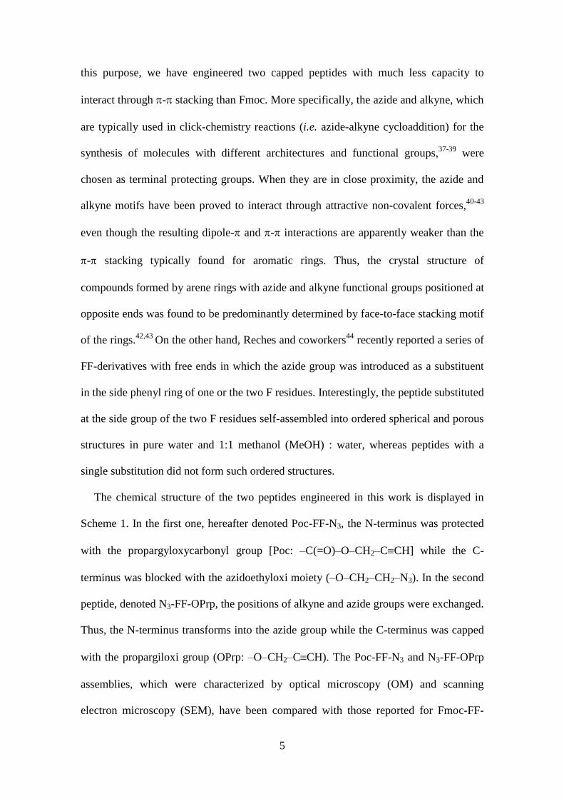
this purpose, we have engineered two capped peptides with much less capacity to
interact through - stacking than Fmoc. More specifically, the azide and alkyne, which
are typically used in click-chemistry reactions (i.e. azide-alkyne cycloaddition) for the
synthesis of molecules with different architectures and functional groups,37-39
were
chosen as terminal protecting groups. When they are in close proximity, the azide and
alkyne motifs have been proved to interact through attractive non-covalent forces,40-43
even though the resulting dipole- and - interactions are apparently weaker than the
- stacking typically found for aromatic rings. Thus, the crystal structure of
compounds formed by arene rings with azide and alkyne functional groups positioned at
opposite ends was found to be predominantly determined by face-to-face stacking motif
of the rings.42,43
On the other hand, Reches and coworkers44
recently reported a series of
FF-derivatives with free ends in which the azide group was introduced as a substituent
in the side phenyl ring of one or the two F residues. Interestingly, the peptide substituted
at the side group of the two F residues self-assembled into ordered spherical and porous
structures in pure water and 1:1 methanol (MeOH) : water, whereas peptides with a
single substitution did not form such ordered structures.
The chemical structure of the two peptides engineered in this work is displayed in
Scheme 1. In the first one, hereafter denoted Poc-FF-N3, the N-terminus was protected
with the propargyloxycarbonyl group [Poc: –C(=O)–O–CH2–CCH] while the C-
terminus was blocked with the azidoethyloxi moiety (–O–CH2–CH2–N3). In the second
peptide, denoted N3-FF-OPrp, the positions of alkyne and azide groups were exchanged.
Thus, the N-terminus transforms into the azide group while the C-terminus was capped
with the propargiloxi group (OPrp: –O–CH2–CCH). The Poc-FF-N3 and N3-FF-OPrp
assemblies, which were characterized by optical microscopy (OM) and scanning
electron microscopy (SEM), have been compared with those reported for Fmoc-FF-

6
OFm, Fmoc-FF and FF. Moreover, in order to ascertain details about the interactions
between peptide molecules, structural analyses were complemented with both
spectroscopic studies and Density Functional Theory (DFT) calculations. It should be
emphasized that, as our main objective was to apply the alkyne and azide moieties as
weak -interacting capping groups, the conduction of all experiments have been
performed maintaining their “preclick” state.
Scheme 1
RESULTS AND DISCUSSION
Peptide Synthesis
The preparation of Poc-FF-N3 and N3-FF-OPrp was carried out following standard
procedures of peptide synthesis in solution starting from the corresponding
phenylalanine derivative and using the Boc, N3 or Poc group as protection for the amino
moieties. A general scheme for the coupling reactions is given in Figures 1 and 2. A
detailed description of the synthetic procedure as well as the characterization of all
intermediates and final peptides are supplied in the Electronic Supporting Information.
Poc-FF-N3
Self-assembly onto glass substrates: influence of the solvents mixtures
N
O
N
Poc-L-Phe-L-Phe-O-(CH2)2-N3
HO
OO
O
H
N3
N3
O
N
N3-L-Phe-L-Phe-OPrp
HO
O
Poc-FF-N3 N3-FF-OPrp
7
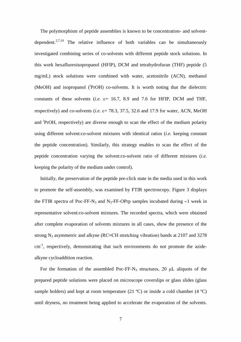
The polymorphism of peptide assemblies is known to be concentration- and solvent-
dependent.2,7,10
The relative influence of both variables can be simultaneously
investigated combining series of co-solvents with different peptide stock solutions. In
this work hexafluoroisopropanol (HFIP), DCM and tetrahydrofuran (THF) peptide (5
mg/mL) stock solutions were combined with water, acetonitrile (ACN), methanol
(MeOH) and isopropanol (iPrOH) co-solvents. It is worth noting that the dielectric
constants of these solvents (i.e. = 16.7, 8.9 and 7.6 for HFIP, DCM and THF,
respectively) and co-solvents (i.e. = 78.3, 37.5, 32.6 and 17.9 for water, ACN, MeOH
and iPrOH, respectively) are diverse enough to scan the effect of the medium polarity
using different solvent:co-solvent mixtures with identical ratios (i.e. keeping constant
the peptide concentration). Similarly, this strategy enables to scan the effect of the
peptide concentration varying the solvent:co-solvent ratio of different mixtures (i.e.
keeping the polarity of the medium under control).
Initially, the preservation of the peptide pre-click state in the media used in this work
to promote the self-assembly, was examined by FTIR spectroscopy. Figure 3 displays
the FTIR spectra of Poc-FF-N3 and N3-FF-OPrp samples incubated during 1 week in
representative solvent:co-solvent mixtures. The recorded spectra, which were obtained
after complete evaporation of solvents mixtures in all cases, show the presence of the
strong N3 asymmetric and alkyne (RC≡CH stretching vibration) bands at 2107 and 3278
cm-1
, respectively, demonstrating that such environments do not promote the azide-
alkyne cycloaddition reaction.
For the formation of the assembled Poc-FF-N3 structures, 20 μL aliquots of the
prepared peptide solutions were placed on microscope coverslips or glass slides (glass
sample holders) and kept at room temperature (21 ºC) or inside a cold chamber (4 ºC)
until dryness, no treatment being applied to accelerate the evaporation of the solvents.

8
The humidity was kept constant in both laboratories at 50%. In spite of the huge number
of examined conditions, the structures obtained from all them were carefully examined
by OM. However, SEM studies were restricted to structures that fulfilled the following
requirements: i) to present a clearly defined morphology (i.e. structured morphology);
ii) to be systematically observed when the same conditions are used in different and
independent experiments (i.e. reproducibility); and iii) to remain formed upon
manipulation for OM and/or SEM observations (i.e. stability).
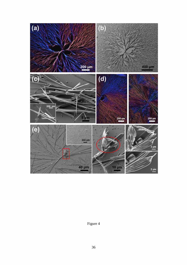
From concentrated 4:1 DCM:MeOH solutions (4 g/mL), Poc-FF-N3 was shown to
form birefringent dendritic-like microstructures at room temperature (Figure 4a). Each
supramolecular domain extends over hundreds of micrometres (Figure 4b) and arises
from the directional grouping of fibres of nano- and micrometrical thickness, as is
reflected by the high resolution SEM micrographs displayed in Figure 4c. Moreover, the
hierarchic dendritic-like microstructures obtained in such conditions exhibit some
morphological diversity. This is evidenced in Figure 4d, which displays OM
micrographs of other birefringent dendritic-like microstructures obtained using the same
experimental conditions than those used in Figure 4a. Dendritic-like structures arise
systematically from DCM:MeOH solutions with peptide concentrations comprised
between 4 and 2 mg/mL (i.e. 4:1 and 2:3 DCM:MeOH ratios), even though the density
of branches rapidly decreases with the concentration (Figure 4e). Besides, Figure 4e
includes high resolution SEM micrographs of the core region that nucleates the primary
frameworks of the branched dendritic structure. As it can be seen, the nucleus is
constituted by groups of densely staked fibres forming compact 2D aggregates. This
morphology is frequently observed in the nucleus of Poc-FF-N3 dendritic-like
microstructures, independently of the peptide concentration.
9
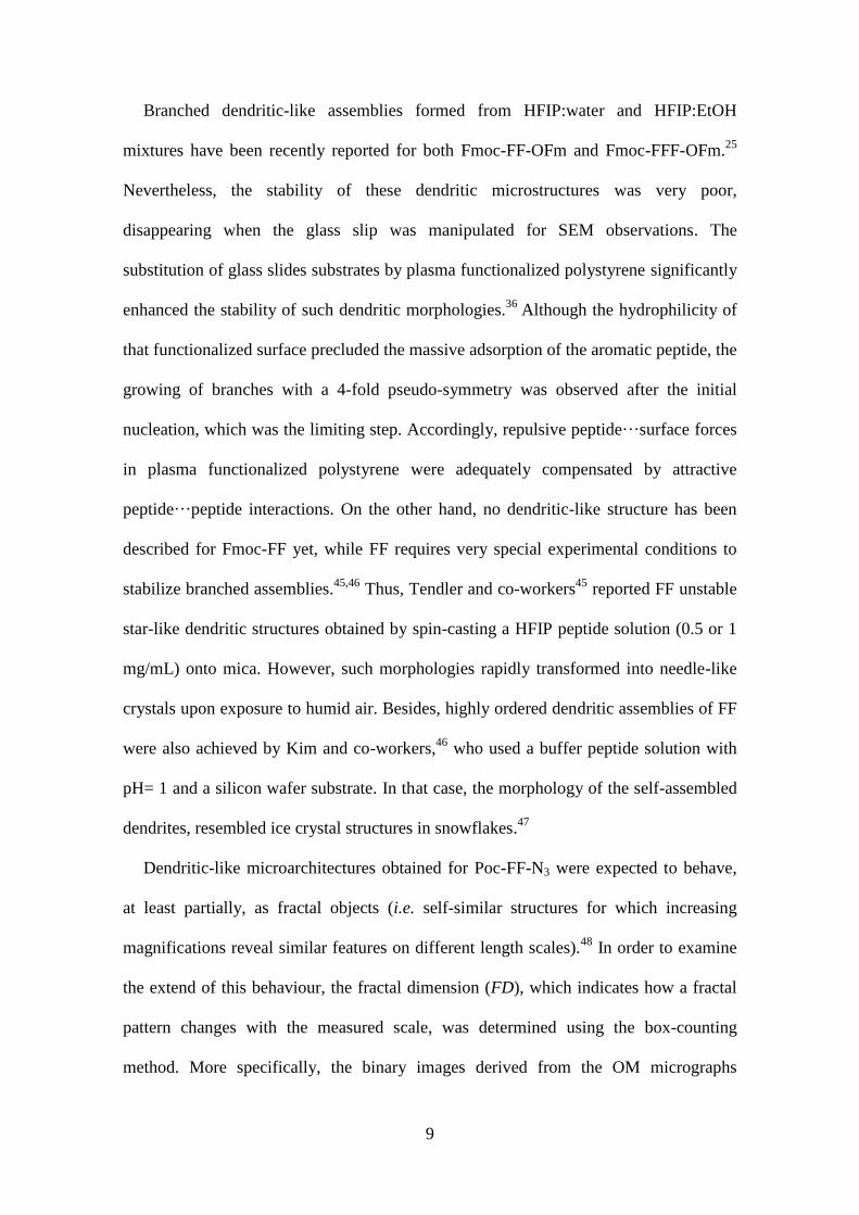
Branched dendritic-like assemblies formed from HFIP:water and HFIP:EtOH
mixtures have been recently reported for both Fmoc-FF-OFm and Fmoc-FFF-OFm.25
Nevertheless, the stability of these dendritic microstructures was very poor,
disappearing when the glass slip was manipulated for SEM observations. The
substitution of glass slides substrates by plasma functionalized polystyrene significantly
enhanced the stability of such dendritic morphologies.36
Although the hydrophilicity of
that functionalized surface precluded the massive adsorption of the aromatic peptide, the
growing of branches with a 4-fold pseudo-symmetry was observed after the initial
nucleation, which was the limiting step. Accordingly, repulsive peptide···surface forces
in plasma functionalized polystyrene were adequately compensated by attractive
peptide···peptide interactions. On the other hand, no dendritic-like structure has been
described for Fmoc-FF yet, while FF requires very special experimental conditions to
stabilize branched assemblies.45,46
Thus, Tendler and co-workers45
reported FF unstable
star-like dendritic structures obtained by spin-casting a HFIP peptide solution (0.5 or 1
mg/mL) onto mica. However, such morphologies rapidly transformed into needle-like
crystals upon exposure to humid air. Besides, highly ordered dendritic assemblies of FF
were also achieved by Kim and co-workers,46
who used a buffer peptide solution with
pH= 1 and a silicon wafer substrate. In that case, the morphology of the self-assembled
dendrites, resembled ice crystal structures in snowflakes.47
Dendritic-like microarchitectures obtained for Poc-FF-N3 were expected to behave,
at least partially, as fractal objects (i.e. self-similar structures for which increasing
magnifications reveal similar features on different length scales).48
In order to examine
the extend of this behaviour, the fractal dimension (FD), which indicates how a fractal
pattern changes with the measured scale, was determined using the box-counting
method. More specifically, the binary images derived from the OM micrographs

10
displayed in Figures 4a and 4d, which can be considered as representative among the
branched-like organization found for Poc-FF-N3, were divided in squares boxes of side
length L and the number of squares boxes that occupy at least part of the dendritic-like
microstructure (N) was counted.
Figure 5, which represents logarithm plots of N as a function of L, uses the scaling
relationship N(L) L-FD
to relate FD with slope in the logarithmic scale (m): FD = –m.
Although the excellent linear behaviour displayed in Figure 5 indicates that the
dendritic-like structures formed by Poc-FF-N3 exhibit dimensional consistency at
different length scales, the slopes reflect poor self-similarity. Thus, the obtained FD
values, which range from 1.75 (Figure 4d-right) to 1.84 (Figure 4a), deviate from the
ideal value of 1.67 expected for microstructures generated by diffusion limited
aggregation (DLA) onto a 2D substrate surface.49,50
Poc-FF-N3 molecules diffuse by
Brownian motion and a random walk process occurs until they contact and adhere to
another one. This process gives place to the formation of clusters through attractive
short-range interactions (i.e. hydrogen bonds and - stacking), which in turn interact
among them forming dendritic-like microstructures. These peptide···peptide forces are
weaker for Poc-FF-N3 than for peptides with a larger F-segment (e.g. Fmoc-FFFF-
OFm25
). We attribute this feature not only the lower number of hydrogen bonding
capability of the former but also to poor stacking ability of the alkyne and azide
moieties. These features combined with the steric shielding of the internal regions of the
clusters, which depends on the size of the N- and C-terminus protecting groups, are
responsible of the degree of fractality of the resulting microstructures.
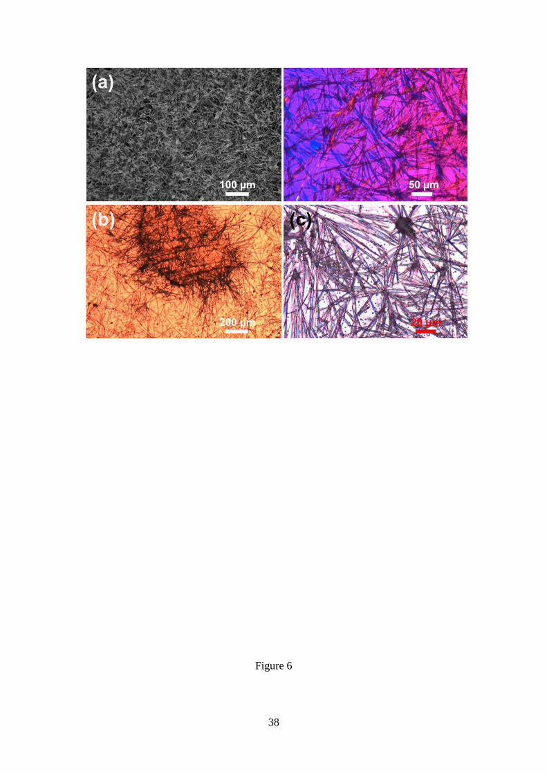
Replacement of the 4:1 DCM:MeOH mixture by 4:1 THF:water (i.e. maintaining the
4 mg/mL peptide concentration) results in a completely different microstructure. This
consists in abundant, individual and birefringent microfibers with a very low degree of

11
branching and a “star-burst” morphology, where several microfibers are grown from a
common nucleus (Figures 6a). In comparison with Figure 4, the disposition of these
microfibers is very disordered. This has been attributed to the presence of a higher
concentration of nuclei, as is reflected in Figure 6a (micrograph at the right side).
Similarly, disordered agglomerates of relatively long microfibers (Figures 6b-c) were
formed in HFIP:water at 1-2 mg/mL peptide concentrations (i.e. solvent:co-solvent
ratios comprised between 4:1 and 2:3).
The important changes obtained in THF:water and HFIP:water with respect to
DCM:MeOH solutions may be attributed to the variation of both the polarity (i.e.
DCM:MeOH is less polar than THF:water and HFIP:water) and/or the evaporation rate
(i.e. DCM:MeOH evaporates faster than THF:water and HFIP:water at identical
experimental conditions – temperature, exposed surface area and humidity).
Comparison of the results displayed in Figures 4 and 6 suggests that, in this case, the
crystallization rate is mainly controlled by the polarity of the medium. Thus,
considering the low polarity of the Poc-FF-N3 molecule, peptide-peptide interactions
predominate over peptide-solvent interactions in THF:water and HFIP:water increasing
the concentration of nuclei, while the opposite occurs in DCM:MeOH. Accordingly, for
the higher concentration of nuclei obtained in THF:water and HFIP:water, abundant and
disordered microfibers are observed, while the few nuclei obtained in DCM:MeOH
cause the unrestricted growth of branched supramolecular structures ideally formed for
well aligned fibres.
Self-assembly from HFIP:MeOH solutions: influence of the substrate
Although the surface is known to play a noticeable control in the self-assembly of
compounds,36,45,51-53
studies about the influence of solid substrate on the assembly of F-

12
derivatives are relatively scarce. More specifically, this phenomenon has been only
reported for the parent FF dipeptide45,51
and the highly aromatic Fmoc-FFF-OFm.36
In
this sub-section, we examine the surface mediated hierarchical assembly of Poc-FF-N3
from HFIP:MeOH solutions considering glass, silanized glass, plasma-functionalized
polystyrene and stainless steel AISI 316.
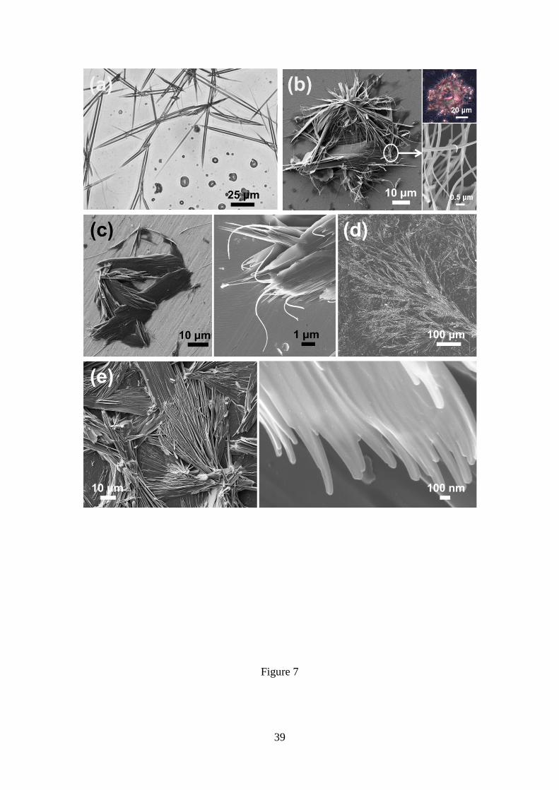
Glass promotes the formation of well-defined microfibers (Figure 7a), which are
aggregated to a lesser extent than those formed in HFIP:MeOH (Figures 6c-d). In
silanized glass the mineral surface is treated with Cl2SiMe2 to increase its
hydrophobicity (i.e. the contact angle increases from 58º±2º to 76º±2º) without alter the
very low surface roughness (i.e. 1.5±0.5 and 3.5±0.8 nm for glass and silanized glass,
respectively). Drop-cast of Poc-FF-N3 solutions in HFIP:MeOH onto silanized resulted
in the formation of micro- and nanofibers that hierarchically aggregate in reproducible
doughnut-like supramolecular structures like those displayed in Figures 7b and S1.
Although the size of such rounded-like supramolecular structures increases with the
peptide concentration (Figure S1), they are relatively isolated. Thus, for low
concentrations the peptide is not observed in large areas of the surface, as is illustrated
in Figures S1b-c, suggesting that fibre···fibre interactions predominate over
fibre···surface ones. Deposition of the peptide dissolved onto steel AISI 316 results in
the spontaneous formation of similar hollow rounded-like supramolecular structures
(Figure 7c). This should be attributed to the fact that the properties of the latter substrate
(i.e. contact angle and roughness of 74º±7º and 14.4±3.2 Å, respectively) and silanized
glass are very similar.
Plasma-treatment of polystyrene is the method of choice for routinely incorporate
oxygen-containing moieties in bioactive polystyrene surfaces to increase their
hydrophilicity and promote cell-polymer interactions.54
The morphology of the
13
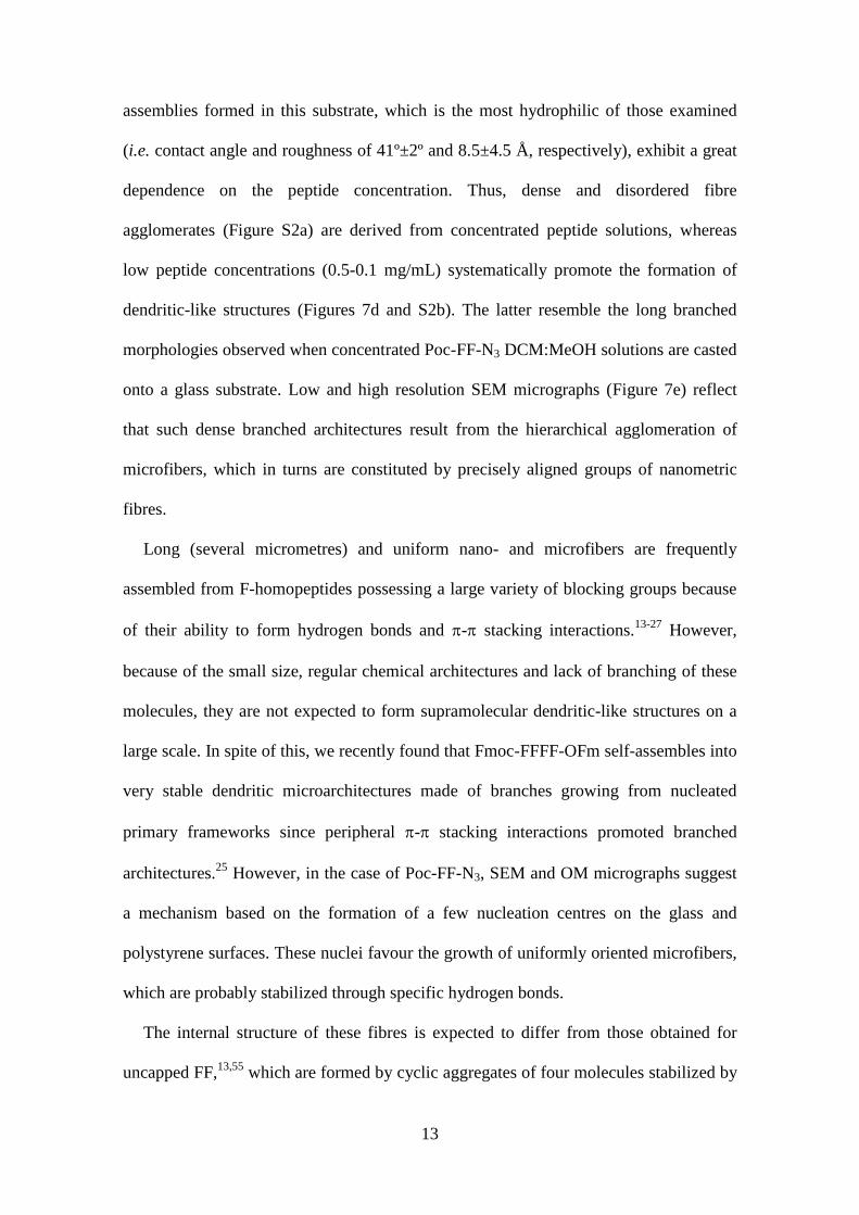
assemblies formed in this substrate, which is the most hydrophilic of those examined
(i.e. contact angle and roughness of 41º±2º and 8.5±4.5 Å, respectively), exhibit a great
dependence on the peptide concentration. Thus, dense and disordered fibre
agglomerates (Figure S2a) are derived from concentrated peptide solutions, whereas
low peptide concentrations (0.5-0.1 mg/mL) systematically promote the formation of
dendritic-like structures (Figures 7d and S2b). The latter resemble the long branched
morphologies observed when concentrated Poc-FF-N3 DCM:MeOH solutions are casted
onto a glass substrate. Low and high resolution SEM micrographs (Figure 7e) reflect
that such dense branched architectures result from the hierarchical agglomeration of
microfibers, which in turns are constituted by precisely aligned groups of nanometric
fibres.
Long (several micrometres) and uniform nano- and microfibers are frequently
assembled from F-homopeptides possessing a large variety of blocking groups because
of their ability to form hydrogen bonds and - stacking interactions.13-27
However,
because of the small size, regular chemical architectures and lack of branching of these
molecules, they are not expected to form supramolecular dendritic-like structures on a
large scale. In spite of this, we recently found that Fmoc-FFFF-OFm self-assembles into
very stable dendritic microarchitectures made of branches growing from nucleated
primary frameworks since peripheral - stacking interactions promoted branched
architectures.25
However, in the case of Poc-FF-N3, SEM and OM micrographs suggest
a mechanism based on the formation of a few nucleation centres on the glass and
polystyrene surfaces. These nuclei favour the growth of uniformly oriented microfibers,
which are probably stabilized through specific hydrogen bonds.
The internal structure of these fibres is expected to differ from those obtained for
uncapped FF,13,55
which are formed by cyclic aggregates of four molecules stabilized by

14
head-to-tail hydrogen bonds. The cycles stack in sheets forming hydrophobic tubes that,
in turn, self-associate forming fibres. For Poc-FF-N3 fibres we hypothesize the
formation of alkyne···azide interactions, which have been observed in both aromatic
organic compounds and biomolecules blocked with azide and alkyl groups,56-59
and the
the participation of stronger intermolecular interactions involving the peptide and/or
phenyl groups. The structure of both the sheets and the tubes has been examined in the
next sub-section.
Table 1 compares the different supramolecular structures observed for Poc-FF-N3
under different experimental conditions.
Structure of the sheets and intermolecular interactions
FTIR spectroscopy has revealed that the presence of -sheets can be determined by
analysing the amide I bands, which occur in the wavenumber range from 1600 cm-1
to
1700 cm-1
, and arise primarily from stretching vibrations of main chain carbonyl
groups. Early investigations suggested that FTIR spectroscopy might be able to
distinguish between parallel and antiparallel -sheets.60-63
Figure 3a displays the amide I regions of the FTIR spectra recorded for the structures
derived from 4 and 1 mg/mL Poc-FF-N3 solutions in DCM:MeOH and HFIP:water,
respectively. The spectra, which were recorded for peptide structures formed after
solvent evaporation, show two bands in the amide I region. The most intense band
appears at 1655 cm-1
, while the least intense one is at 1699 cm-1
. This feature is
consistent with an antiparallel disposition of the -sheets. However, the 1699 / 1650
ratio is below one (i.e. around 0.75), suggesting that the percentage of antiparallel
arrangement is relatively low. On the other hand, the fact that the major component
appears in all cases, independently of the solvent mixture and peptide concentration, at

15
the same position (1655 cm-1
) indicates the absence of excitonic coupling effects in the
frequency position.64
On the other hand, the amide II and C=O stretching bands appear
at 1541 and 1734 cm-1
, respectively, while the amide A at around 3310 cm-1
partially
overlaps the alkyne band at 3278 cm-1
.
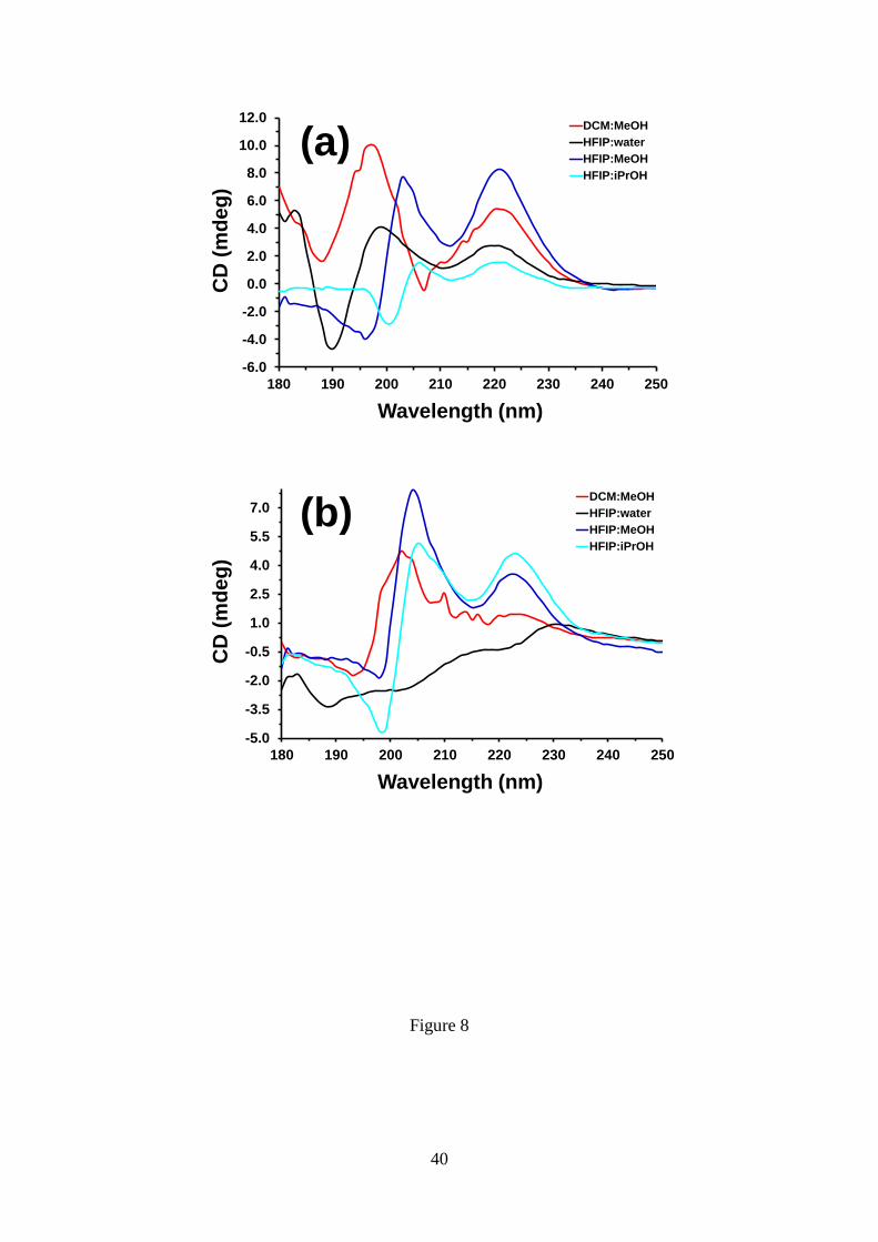
In order to obtain information about the structure of the aggregates in the first stages
of the assembly process, circular dichroism (CD) spectra were recorded at very low
peptide concentration (0.01 mg/mL) in different environments (i.e. 1:9 DCM:MeOH,
HFIP:water, HFIP:MeOH and HFIP:iPrOH mixtures coming from 1 mg/mL DCM or
HFIP stock solutions). The CD spectra recorded at 30 ºC for the different mixtures are
compared in Figure 8a. In HFIP:MeOH, Poc-FF-N3 molecules are predominantly in
random coil conformation, which is proved by the negative band at 196 nm. In contrast,
the spectrum obtained in DCM:MeOH, which exhibits a positive band at 197 nm and a
negative band at 208 nm, suggests the dominance of a -sheet structure. The positive
maxima at around 200 and 220 nm indicate the presence of - stacking of aromatic
units in dilute HFIP:water and HFIP:iPrOH solutions. This interaction is frequently
detected in the CD profiles for biomolecular self-assembly with -turn
conformation.65,66
In all cases these structural characteristics are preserved between -50
ºC and +60 ºC, as is demonstrated by the spectra acquired at different temperatures
within such interval (Figure S3).
Density functional theory (DFT) calculations were carried out considering different
structural models. First, the relative stability of the antiparallel and parallel -sheet
dispositions was examined using mode complexes involving three -strands. In an
attempt to optimize the geometry of the inter-strand interactions (i.e. hydrogen bonds
and - stacking), different starting arrangements were constructed for each disposition.
Geometry optimizations were performed using the M06L, M06L-D3 and BLYP-D3
16
correlated functionals combined with the 6-31G(d) basis set. Results indicated that the
antiparallel disposition is the most stable, independently of the computational method,
which is agreement with the FTIR observations. Table 2, which lists the relative
energies (E) obtained using the different theoretical levels, indicates that the parallel
disposition was disfavoured by around 0.9-3.7 kcal/mol, depending on the functional.
The antiparallel disposition was proposed by Smith et al.31
for Fmoc-FF and by
Mayans et al.25
for Fmoc-FF-OFm. Comparison of the models reported for the parallel
and antiparallel -sheets of such Fmoc-containing dipeptides reveals that intermolecular
interactions are characterized by the following trends:
(i) In the parallel disposition the phenyl side groups of all strands are perfectly
packed forming aromatic -ladders, while the phenyl groups of consecutive
strands point in opposite directions in the antiparallel arrangement.
(ii) Fmoc-FF-OFm exhibits similar Fmoc···Fmoc interactions for both the
parallel and antiparallel dispositions, whereas in the case of Fmoc-FF such
interactions are much stronger for the parallel assembly than for the
antiparallel one.
(iii) Hydrogen bonding parameters, especially the H···O distance, are more
favourable for the antiparallel than for the parallel assembly.
These features reflect the predominant role played by hydrogen bonds in the packing
of Fmoc-containing FF-derivatives. Results from the analysis of the intermolecular
interactions in the antiparallel and parallel dispositions modelled for Poc-FF-N3 -
strands, which are displayed in Figures 9a-b and S4, are compared in Table 2. As it can
be seen in Figures 9b, S4b and S4d (for geometries optimized at the B3LYP-D3/6-
31G(d), M06L-D3/6-31G(d) and M06L/6-31G(d) level, respectively), the parallel

17
disposition of Poc-FF-N3 molecules shows the same alignment of the phenyl side
groups reported for Fmoc-FF-OFm.25
This attractive interaction, which originates the
formation of aromatic -ladders, is practically absent in the antiparallel disposition
(Figures 9a, S4a and S4c). On the other hand, hydrogen bonding parameters also favour
the parallel disposition in comparison with the antiparallel one (Table 2). The stability
of the antiparallel disposition with respect to the parallel is explained by comparing the
alkyne···azide interactions detected for the former with the azide···azide and
alkyne···alkyne interactions of the latter. Thus, although the distances between the -
stacked groups are, in general, slightly shorter for the parallel disposition than for the
antiparallel one (Table 2), -dipole interactions are only possible for the former
arrangement (Figures 9a, S4a and S4c). According to these DFT calculations and the
FTIR spectra, -dipole interactions play a crucial role in the assembly of Poc-FF-N3
molecules, which is fully consistent with the crystal X-ray analyses of compounds
containing azide and alkyne motifs.40-43
Thus, in such compounds the azide and alkyne
groups of adjacent molecules are arranged in a head-to-tail fashion, which is equivalent
to the antiparallel disposition of Poc-FF-N3 strands, favouring the formation of dipole-
interactions in addition of - interactions.
On the other hand, by analogy with the structures reported for FF55
and FFFF22
nanotubes, cycles of dimers, trimers and tetramers (i.e. 2, 3 and 4 Poc-FF-N3 molecules
disposed forming a ring with a central channel) were constructed. Thus, the stacking
and lateral packing of such cycles should result in the growing of fibres. In the starting
cyclic complexes, the alkyne and azide groups of neighbouring peptide molecules were
arranged to form head-to-tail - stacking interactions, which surrounded the inner core
of the rings, while phenyl side chains emanated from the rings forming the hydrophobic
external side. Geometry optimizations at the M06L/6-31G(d) level led to the minimum

18
structures displayed in Figure 10. As it can be seen, complexes with molecules arranged
in irregular dispositions were obtained in all cases, even though the disorganization of
the molecules increases with the size of the complex. The formation during the
optimization of both N–H···O hydrogen bonds (i.e. complexes with 3 and 4 molecules;
Figures 10b-c) and N-H··· interactions (i.e. complex with 4 molecules; Figure 109c),
explains the loss of the starting cyclic disposition. Thus, the strength of hydrogen bonds
is significantly higher than that of alkyne···azide head-to-tail interactions. Therefore,
hydrogen bonds governed the re-organization of the molecules in the complexes,
inducing the loss of the starting cyclic geometries during the optimization. This fact
explains that Poc-FF-N3 fibres are much less defined and ordered than the FF fibers,
which adopt a hexagonal-like shape due to the high stability of the head-to-tail NH3+···
–
OOC hydrogen bonds in FF cyclic hexamers.55
N3-FF-OPrp
The “preclick” state of the peptide after evaporation of the solvent is corroborated in
the FTIR spectra displayed in Figure 3b. The strong N3 asymmetric band appears at
2113 cm-1
, while the alkyne stretching is contained in the broad band centred at 3296
cm-1
, which also includes the amide A vibration. Also, some bands attributed to trapped
solvent molecules are observed in the region of 2350 cm-1
.
The experimental conditions (i.e. solvent:co-solvent mixtures and substrates) used in
the previous section for Poc-FF-N3, were applied to explore the assembly behaviour of
N3-FF-OPrp. Amazingly, after many attempts at room temperature, we concluded that
the latter peptide hardly forms nano- or microstructures with well-defined
morphologies. Thus, N3-FF-OPrp tends to organize into small amorphous agglomerates
completely irregular or with short-range regularity, independently of the experimental

19
conditions at room temperature. This is illustrated in Figure S5, which displays
representative micrographs of the disordered assemblies obtained using different
solvent:co-solvents and substrates.
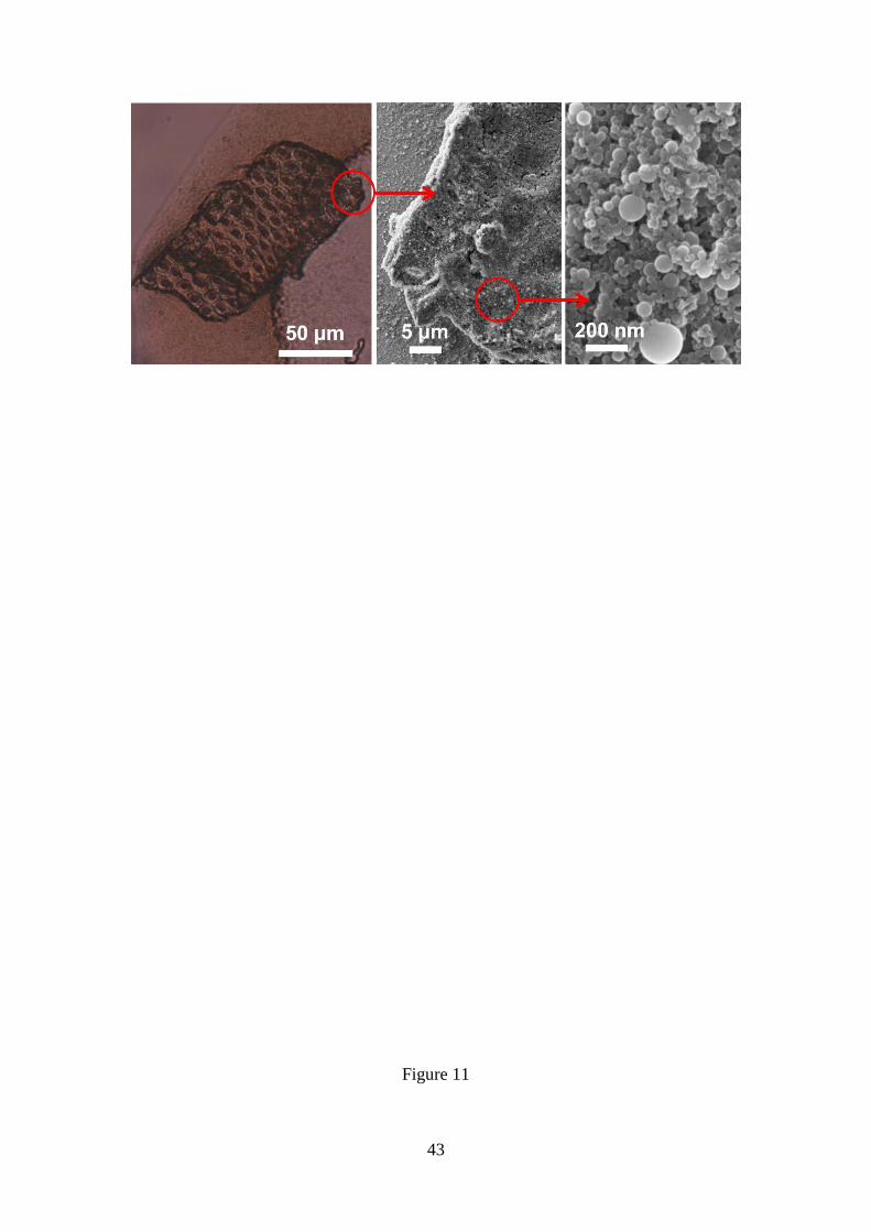
In order to expand this search of regular N3-FF-OPrp assemblies, new sets of
experiments were performed keeping the samples inside a cold chamber (4 ºC) during
the solvent drying process. In this case, an exception to the general behaviour observed
for N3-FF-OPrp was obtained for diluted (< 1 mg/mL) HFIP:water solutions deposited
onto glass coverslips, which systematically resulted in the formation regular assemblies,
as it is reflected in Figure 11. In such particular experimental conditions the peptide
forms well-defined spheres of heterogeneous sizes (i.e. the diameter ranged from 20 to
200 nm), which in turn aggregate forming micrometric structures with a honeycomb
morphology. This behaviour has been attributed to the hydrophobicity of the N3-FF-
OPrp, which forces the refolding of the structures to protect many molecules from the
aqueous medium. Thus, the initial organization of the peptide in nanospheres and their
subsequent aggregation in microstructures are consequence of the repulsive interaction
with the polar environment (i.e. diluted peptide HFIP:water solutions present a high
content of co-solvent).
CD spectra of 0.01 mg/mL N3-FF-OPrp solutions in 1:9 DCM:MeOH, HFIP:water,
HFIP:MeOH and HFIP:iPrOH mixtures are shown in Figure 8b. In HFIP:MeOH and,
especially, HFIP:iPrOH the spectra are appreciably contributed by the random coil
conformation, as is evidenced by the negative band at 197-198 nm. Besides, the spectra
obtained in DCM:MeOH and HFIP:water do not suggest any conformational
preference. These results, which are independent of the temperature (Figure S6), support
that N3-FF-OPrp agglomerates start from small aggregates of unstructured peptide
molecules.
20
The amide I band of the FTIR spectra recorded for N3-FF-OPrp aggregates after
evaporation of the solvent (Figure 3b) reflects important changes in comparison to those
obtained for Poc-FF-N3 (Figure 3a). More specifically, the two narrow and strong peaks
observed for the latter at 1655 and 1699 cm-1
transforms into a single and relatively
broad peak centred at 1678 cm-1
. The position of this band is relatively far from that
typically observed for parallel -sheets (i.e. 1650 cm-1
). This feature suggests that the
N3-FF-OPrp molecules tend to poorly organize in distorted or irregular parallel sheets.
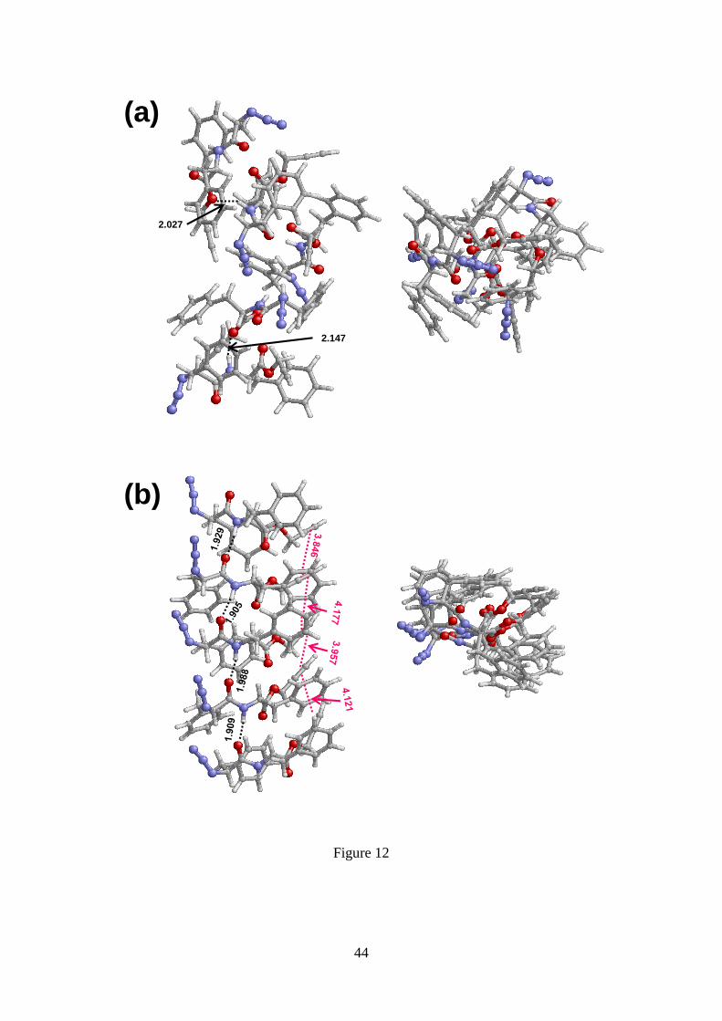
These results was supported by B3LYP-D3/6-31G(d) calculations on complexes formed
by three or five molecules arranged in antiparallel and parallel -sheets (Figures S7 and
12).
Geometry optimizations of the antiparallel -sheet resulted in the formation of
unstructured complexes (Figures S7a and 12a), in which characteristic alignment of the
molecules is lost. Moreover, comparison of the geometries obtained for complexes with
three and five N3-FF-OPrp strands evidence that the stability of the antiparallel -sheet
does not increase with the number of interacting molecules. On the other hand, the
parallel disposition, which is energetically favoured with respect to the antiparallel one
(i.e. 8.1 and 13.9 kcal/mol for complexes with 3 and 5 N3-FF-OPrp molecules,
respectively), preserves the -sheet structure (Figures S7b and 12b). These results are
explained by examining the capacity of N3-FF-OPrp to form intermolecular interactions.
Thus, due to the single peptide bond of its chemical structure (Scheme 1), each N3-FF-
OPrp molecule only participates in two intermolecular hydrogen bonds: one as acceptor
and one as donor. This represents a significant reduction with respect to Poc-FF-N3
(Figures 9a and S4), in which peptide molecules used their two peptide bonds to interact
forming four hydrogen bonds per molecule (i.e. two as acceptor and two as donor).
Accordingly, the possible formation of two hydrogen bonds per N3-FF-OPrp molecule

21
is not enough to stabilize the antiparallel -sheet disposition, which disrupts and gives
place to a disordered aggregation of molecules. In the case of the parallel -sheet
disposition, the two hydrogen bonds are strengthened by the existence of aromatic -
ladders formed by stacked phenyl rings. Interactions between aromatic rings, which are
not possible in the antiparallel disposition, are more stabilizing than alkyne···azide
interactions,40-43
explaining the cohesion of the parallel disposition and the unsteadiness
of the antiparallel one.
On the other hand, alkyne···alkyne are the only - interactions detected in the
parallel model of N3-FF-OPrp, the distance between the azide groups of adjacent
molecules (> 5 Å in all cases) hampering the formation of azide···azide interactions.
This has been attributed to the fact that rest of interactions found in such organization
determine the molecular conformation because of their higher strength. Overall, the
distortions and irregularities suggested by FTIR in the -sheets of N3-FF-OPrp can been
attributed to the relatively poor interaction pattern of the latter compound with respect
to that of Poc-FF-N3.
CONCLUSIONS
We have successfully synthesized and study the self-assembly of FF-derivatives
capped at the N- and C-terminus by azide and alkyne groups, which have been
maintained in the preclick state. Results prove that despite intermolecular azide···alkyne
interactions are significantly weaker than hydrogen bonding in FF and Fmoc···Fmoc -
interactions in Fmoc-FF-OFm, Poc-FF-N3 molecules hierarchically aggregate forming
well-defined supramolecular structures. Furthermore, the morphology of such structures
can be regulated through the peptide concentration, the polarity of the medium and/or
the hydrophilicity of substrate. In particular, the formation of stable dendritic-like
22
structures is very striking since, although this kind of branched structures was recently
detected in homopeptides with a higher number of phenylalanine residues, they were
found to be highly unstable for other FF-derivatives (e.g. Fmoc-FF-Fmoc25
). Theoretical
calculations and FTIR spectra probe that Poc-FF-N3 assemblies prefer antiparallel -
sheets. This has been attributed to the strength of azide···alkyne interactions, which
exhibit two components: - stacking and -dipole. The sum of these contributions is
more stabilizing than the - stacking of the azide···azide and alkyne···alkyne
interactions found in the parallel -sheet.
The similar peptide in which the position of the alkyne and azide groups was
exchanged, N3-FF-OPrp, exhibited much less tendency to form ordered structures under
the same experimental conditions. Indeed, the only supramolecular assembly identified
for N3-FF-OPrp was observed in very polar environments, in which peptide molecules
organize in nanospheres that subsequently aggregate into honeycomb supramolecular
structures. Furthermore, the parallel is the only stable -sheet disposition found for this
peptide, molecules arranged in the antiparallel disposition evolving towards completely
disordered structures during geometry optimizations. The assembly of N3-FF-OPrp
molecules is governed by the formation of phenyl -ladders due to their restricted
hydrogen bonding capacity. Overall, results obtained in this work reflect that, although
the role of the interactions involving alkyne and azide groups is much less decisive than
the one played by hydrogen bonds and aromatic - stacking interactions, the formers
allow modulation of the assembly stabilizing microstructures that are usually unstable.
ACKNOWLEDGEMENTS
Authors thank supports from MINECO and FEDER (MAT2015-69367-R,
MAT2015-69547-R and CTQ2013-40855-R) and Gobierno de Aragón - FEDER
23
(research group E40). Support for the research of C.A. was received through the prize
“ICREA Academia” for excellence in research funded by the Generalitat de Catalunya.
REFERENCES
1. A. Ciesielski, M. El-Garah, S. Masiero and P. Samori,Small, 2016, 12, 83-95.
2. C. H. Cai, J. P. Lin, Y. Q. Lu, Q. Zhang and L. Q. Wang, Chem. Soc. Rev., 2016, 45,
5985-6012.
3. Y. Liu, B. Liua and Z. Nie, Nano Today, 2015, 10, 278-300.
4. A. Groschel and A. H. E. Mueller, Nanoscale, 2015, 7, 11841-11876.
5. S. Scanna, D. J. Pine and G.-R. Yi, Soft Matter, 2013, 9, 8096-8106.
6. L. Z. Zhao, R. Qu, A. Li, R. J. Ma and L. Q. Shi, Chem. Commun., 2016, 52, 13543-
13555.
7. G. Fichman and E. Gazit, Acta Biomater., 2014, 10, 1671-1682.
8. Y. Zhao, F. Sakai, L. Su, Y. J. Liu, K. C. Wei, G. S. Chen and M. Jiang, Adv.
Mater., 2013, 25, 5215-5256.
9. K. H. Smith, E. Tejeda-Montes, M. Poch and A. Mata, Chem. Soc. Rev., 2011, 40,
4563-4577.
10. K. Tao, A. Levin, L. Adler-Abramovich and E. Gazit, Chem. Soc. Rev., 2016, 45,
3935-3953.
11. B. E. I. Ramakers, J. C. M. van Hest and D. W. P. M. Lowik, Chem. Soc. Rev.,
2014, 43, 2743-2756.
12. Peptide Materials: From Nanostructures to applications, C. Alemán, A. Bianco and
M. Venanzi (Eds), Wiley, 2013.
13. M. Reches and E. Gazit, Science, 2003, 300, 625-627.

24
14. V. L. Sedman, L. Adler-Abramovich, S. Allen, E. Gazit and S. J. B. Tendler, J. Am.
Chem. Soc., 2006, 128, 6903-6908.
15. L. Adler-Abramovich, M. Reches, V. L. Sedman, S. Allen and S. J. B. Tendler,
Langmuir, 2006, 22, 1313-1320.
16. N. Kol, L. Adler-Abramovich, D. Barlam, R. Z. Shneck, E. Gazit and I. Rousso,
Nano Lett., 2005, 5, 1343-1346.
17. X. Yan, P. Zhu and J. Li, Chem. Soc. Rev., 2010, 39, 1877-1890.
18. S. Fleming and R. V. Ulijn, Chem. Soc. Rev., 2014, 43, 8150-8177.
19. P. Tamamis, L. Adler-Abramovich, M. Reches, K. Marshall, P. Sikorski, L. Serpell,
E. Gazit and G. Archontis, Biophys. J., 2009, 96, 5020-5029.
20. C. Guo, Z. A. Arnon, R. Qi, Q. Zhang, L. Adler-Abramovich, E. Gazit and G. Wei,
ACS Nano, 2016, 10, 8316-8324.
21. C. Guo, Y. Luo, R. Zhou and G. Wei, Nanoscale, 2014, 6, 2800-2811.
22. E. Mayans, G. Ballano, J. Casanovas, A. Díaz, M. M. Pérez-Madrigal, F. Estrany, J.
Puiggalí, C. Cativiela and C. Alemán, Chem. Eur. J., 2015, 21, 16895-16905.
23. V. Jayawarna, M. Ali, T. A. Jowitt, A. F. Miller, A. Saiani, J. E. Gough and R. V.
Ulijn, Adv. Mater., 2006, 18, 611-614.
24. N. Amdursky, E. Gazit and G. Rosenman, Adv. Mater., 2010, 22, 2311-2315.
25. E. Mayans, G. Ballano, J. Casanovas, L. J. del Valle, M. M. Pérez-Madrigal, F.
Estrany, A. I. Jiménez, J. Puiggalí, C. Cativiela and C. Alemán, C, Soft Matter,
2016, 12, 5475-5488.
26. N. Amdursky, M. Molotskii, E. Gazit and G. Rosenman, Appl. Phys. Lett., 2009, 94,
261907.
27. L. Adler-Abramovich, N. Kol, I. Yanai, D. Barlam, R. Z. Shneck, E. Gazit and I.
Rousso, Angew. Chem. Int. Ed., 2010, 49, 9939-9942.
25
28. M. Reches and E. Gazit, Phys. Biol. 2006, 3, S10-S19.
29. S. Flemming and R. V. Ulijn, Chem. Soc. Rev., 2014, 43, 8150-8177.
30. S. Bai, C. Pappas, S. Debnath, P. W. J. M. Frederix, J. Leckie, S. Fleming and R. V.
Ulijn, ACS Nano, 2014, 8, 7005-7013.
31. A. M. Smith, R. J. Williams, C. Tang, P. Coppo, R. F. Collins, M. L. Turner, A.
Saiani and R. V. Ulijn, Adv. Mater., 2008, 20, 37-41.
32. A. K. Das, R. Collins and R. V. Ulijn, Small, 2008, 4, 279-287.
33. A. Mahler, M. Reches, M. Rechter, S. Cohen and E. Gazit, Adv. Mater., 2006, 18,
1365-1370.
34. J. Raeburn, C. Mendoza-Cuenca, B. N. Cattoz, M. A. Little, A. E. Terry, A. Z.
Cardoso, P. C. Griffiths and D. J. Adams, Soft Matter, 2015, 11, 927-935.
35. S. Debnath, A. Shome, D. Das and P. K. Das, J. Phys. Chem. B, 2010, 114, 4407-
4415.
36. E. Mayans, G. Fabregat, R. Juárez, C. Cativiela, J. Puiggalí and C. Alemán, Chem.
Select, 2017, 22, 1133-1139.
37. F. Amblard, J. H. Cho and R. F. Schinazi, Chem. Rev., 2009, 109, 4207-4220.
38. P. L. Golas and K. Matyjaszewski, Chem. Soc. Rev., 2010, 39, 1338-1354.
39. W. Tang and M. L. Becker, Chem. Soc. Rev., 2014, 43, 7013-7039.
40. A. Pathigoolla, R. G. Gonnade and K. M. Sureshan, Angew. Chem. Int. Ed., 2012,
51, 4362-4366.
41. W. Li, X. Li, W. Zhu, C. Li, D. Xu, Y. Ju and G. Li, Chem. Commun., 2011, 47,
7728-7730.
42. .B.-B. Ni, C. Wang, H. Wu, J. Pei and Y. Ma, Chem. Commun., 2010, 46, 782–784.
43. A. Pathigoolla and K. M. Sureshan, Angew. Chem. Int. Ed., 2014, 53, 9522-9525.
44. S. Yuran, Y. Razvag, P. Das and M. Reches, J. Pept. Sci., 2014, 20, 479-486.

26
45. V. V. Korolkov, S. Allen, C. J. Roberts and S. J. B. Tendler, Faraday Discuss.,
2013, 166, 257-267.
46. T. H. Han, J. K. Oh, G.-J. Lee, S. II Pyun and S. O. Kim, Colloids Surf. B, 2010, 79,
440-445.
47. L. Chronopoulou, S. Sennato, F. Bordi, D. Giannella, A. Di Nitto, A. Barbetta, M.
Dentini, A. R. Togna, G. I. Togna, S. Moschini and C. Palocci, Soft Matter, 2014,
10, 1944-1952.
48. B. B. Mandelbrot, The Fractal Geometry of Nature, Freeman, San Francisco, CA,
USA, 1982.
49. T. A. Witten and L. M. Sander, Phys. Rev. Lett., 1981, 47, 1400-1403.
50. P. Meakin, Phys. Rev. A, 1983, 27, 604-607.
51. P. Kumaraswamy, R. Lakshmanan, S. Sethuraman and U. M. Krishnan, Soft Matter,
2011, 7, 2744-2754.
52. L. Klosterman, J. K. Riley, C. J. Bettinger, Langmuir, 2015, 31, 3451-3458.
53. X. B. Mao, C. X. Wang, X. K. Wu, X. J. Ma, L. Liu, L. Zhang, L. Niu, Y. Y. Guo, .
H. Li, Y L. Yang and C. Wang. Proc. Natl. Acad. Sci. USA, 2011, 108, 19605-
19610.
54. T. G. van Kooten, H. T. Spijker and H. J. Busscher, Biomaterials, 2004, 25, 1735-
1747.
55. C. H. Görbitz, Chem. Eur. J., 2001, 7, 5153-5159.
56. B.-B. Ni, C. Wang, H. Wu, J. Pei and Y. Ma, Chem. Commun., 2010, 46, 782–784.
57. T. Hirose, N. Maita, H. Gouda, J. Koseki, T. Yamamoto, A. Sugarawa, H. Nakano,
S. Hirono, K. Shiomi, T. Watanable, H. Taniguchi, K. B. Sharpless, S. Omura and
T. Sunazuka, Proc. Natl. Acad. Sci., 2013, 110, 15892–15897.
58. A. Pathigoolla and K. M. Sureshan, Angew. Chem. Int. Ed., 2014, 53, 9522-9525.

27
59. A. Pathigoolla and K. M. Sureshan, Chem.Commun., 2016, 52, 886-888.
60. C. Toniolo and M. Palumbo, Biopolymers, 1977, 16, 219-224.
61. S. Krimm and J. Bandekar, Adv. Protein Chem., 1986, 38, 181-364.
62. R. Khurana and A. L. Fink, Biophys. J., 2000, 78, 994-1000.
63. E. Goormaghtigh, V. Cabiaux and J.-M. Ruysschaert, Subcell. Biochem., 1994, 23,
329-362.
64. L. Z. Polzi, A. Amadei, M. Aschi and I. Daidone, J. Am. Chem. Soc., 2011, 133,
11414-11417.
65. M. Gupta, A. Bagaria, A. Mishra, P. Mathur, A. Basu, S. Ramakumar and V. S.
Chauhan, Adv. Mater., 2007, 19, 858-861
66. X. Yan, Y. Cui, Q. He, K. Wang and J. Li, Chem. Mater., 2008, 20, 1522-1526.

28
CAPTIONS TO FIGURES
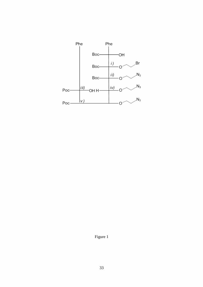
Figure 1. Scheme of the coupling reactions used to obtain Poc-FF-N3. i) Boc-L-Phe-
OH, N-[3-(dimethylamino)-propyl]-N’-ethylcarbodiimide hydrochloride (EDC·HCl)/1-
hydroxy-7-azabenzotriazole (HOBt), 2-bromoethanol, 4-dimethylaminopyridine
(DMAP), DCM. ii) NaN3, DMF. iii) H-L-Phe-OH, NaHCO3 1N (aqueous), NaOH 4N
(aqueous), propargyl chloroformate. iv) TFA/DCM 1/1. v) Poc-L-Phe-OH, N-[3-
(dimethylamino)-propyl]-N’-ethylcarbodiimide hydrochloride (EDC·HCl)/1-hydroxy-7-
azabenzotriazole (HOBt), N-methylmorpholine (NMM; to keep pH 8), DCM.
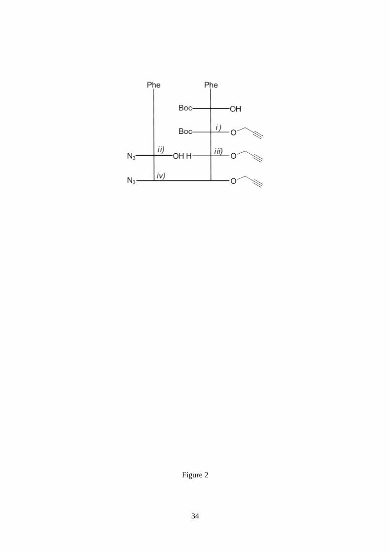
Figure 2. Scheme of the coupling reactions used to obtain N3-FF-Prp. i) Boc-L-Phe-
OH, N-[3-(dimethylamino)-propyl]-N’-ethylcarbodiimide hydrochloride (EDC·HCl)/1-
hydroxy-7-azabenzotriazole (HOBt), propargyl alcohol, 4-dimethylaminopyridine
(DMAP), DCM. ii) H-L-Phe-OH, triethylamine (NEt3), 1H-benzotriazole-1-sulfonyl
azide, CuSO4·5H2O, MeCN/H2O (1/1). iii) TFA/DCM 1/1. iv) N3-L-Phe-OH, N-[3-
(dimethylamino)-propyl]-N’-ethylcarbodiimide hydrochloride (EDC·HCl)/1-hydroxy-7-
azabenzotriazole (HOBt), N-methylmorpholine (NMM; to keep pH 8), DCM.
Figure 3. FTIR spectra of (a) Poc-FF-N3 and (b) N3-FF-OPrp samples from 4:1
DCM:MeOH and 1:4 HFIP:water solutions (4 and 1 mg/mL peptide concentration,
respectively).
Figure 4. Microstructures obtained by self-assembly from Poc-FF-N3 solutions in
DCM:MeOH mixtures at room temperature. (a,b) OM micrographs of representative
birefringent dendritic-like structures derived from 4 mg/mL peptide solutions (4:1
DCM:MeOH). (c) High resolution SEM micrographs of fibres hierarchically aligned to
form the dendritic-like structures displayed in (a) and (b). (d) OM micrographs of other
frequently observed birefringent dendritic-like morphologies derived from 4 mg/mL
peptide solutions. (e) OM micrographs (left) of representative birefringent dendritic-like
29
structures derived from 2 mg/mL peptide solutions (2:3 DCM:MeOH) and high
resolution SEM micrographs (centre and right) of the nucleus, which is indicated by the
red dashed rectangle in the left micrograph. Micrographs displayed at the right show
details of the region marked by the red dashed circle.
Figure 5. Analysis of the fractal dimension using the box-counting method for the
Poc-FF-N3 dendritic-like microstructures displayed in Figures 4a and 4d (both left and
right). The fractal dimension is related to the slope of the adjusted equations (see text).
Figure 6. (a) Disordered birefringent microfibers obtained from 4 mg/mL Poc-FF-N3
solutions in 4:1 THF:water. Disordered agglomerates of microfibers formed from (b) 1
and (c) 2 mg/mL Poc-FF-N3 solutions in 1:4 and 2:3 HFIP:water, respectively.
Figure 7. Optical and/or SEM representative micrographs of Poc-FF-N3 assemblies
formed onto (a) glass coverslip, (b) silanized glass, (c) steel AISI 316 and (d-e) plasma
treated polystyrene using (a) 0.5 mg/mL (1:9), (b) 0.1 mg/mL (1:49), (c) 4 mg/mL (4:1),
(d-e) 0.5 mg/mL (1:9) HFIP:MeOH solutions (solvent:co-solvent ratio indicated in
parenthesis).
Figure 8. CD spectra of (a) Poc-FF-N3 and (b) N3-FF-OPrp in different environments
(1:9 solvent:co-solvent) at 0.01 mg/mL peptide concentration.
Figure 9. Lateral and top views of the (a) antiparallel and the (b) parallel -sheet
assemblies obtained for a complex with three Poc-FF-N3 strands. Geometries were
optimized at the B3LYP-D3/6-31G(d) level. Intermolecular hydrogen bonds are
represented by black dashed lines, the (N–)H···O distances (in Å) being displayed.
Intermolecular azide···alkyne, azide···azide, and alkyne···alkyne interactions are
represented by pink dashed lines. These three -stacking interactions have been
considered to occur when the distance between the two motifs is lower than 4.5 Å
(values are provided in the graphic). Azide···alkyne distances in the antiparallel

30
disposition, have been determined considering the central nitrogen atom of the azide
group and each of the two carbons of the alkyne group. Azide···azide and
alkyne···alkyne distances in the parallel disposition have been determined considering
the central nitrogen atom of each azide group and the geometric centre of each CC
bond, respectively.
Figure 10. Lateral and top views of complexes formed by (a) two, (b) three and (c)
four Poc-FF-N3 molecules after geometry optimization at the B3LYP-D3/6-31G(d)
level. In all cases molecules were initially arranged forming cycles with alkyne···azide
head-to-tail interactions. Intermolecular hydrogen bonds are represented by black
dashed lines, the (N–)H···O distances (in Å) being displayed. Intermolecular
alkyne···azide interactions are represented by pink dashed lines. The latter interaction
has been considered to occur when the distance between the two motifs is lower than
4.5 Å (values are provided in the graphic).
Figure 11. Representative optical and/or SEM micrographs of N3-FF-OPrp
assemblies formed at 4 ºC onto glass coverslips using a 0.5 mg/mL peptide solution
in1:9 HFIP:water.
Figure 12. Lateral and top views of the geometries obtained after optimization of the
(a) antiparallel and the (b) parallel -sheet assemblies constructed using five N3-FF-
OPrp strands. Geometries were optimized at the B3LYP-D3/6-31G(d) level.
Intermolecular hydrogen bonds are represented by black dashed lines, the (N–)H···O
distances (in Å) being displayed. Intermolecular alkyne···alkyne interactions are
represented by pink dashed lines. These interactions have been considered to occur
when the distance between the geometric centre of two adjacent alkyne motifs is lower
than 4.5 Å (values are provided in the graphic). Azide···azide distances were larger than
5 Å.
31
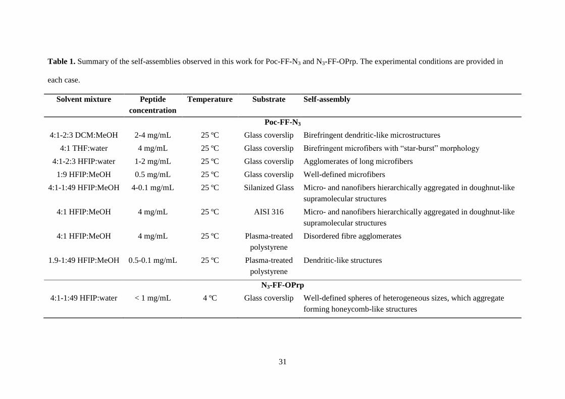
Table 1. Summary of the self-assemblies observed in this work for Poc-FF-N3 and N3-FF-OPrp. The experimental conditions are provided in
each case.
Solvent mixture Peptide
concentration
Temperature Substrate Self-assembly
Poc-FF-N3
4:1-2:3 DCM:MeOH 2-4 mg/mL 25 ºC Glass coverslip Birefringent dendritic-like microstructures
4:1 THF:water 4 mg/mL 25 ºC Glass coverslip Birefringent microfibers with “star-burst” morphology
4:1-2:3 HFIP:water 1-2 mg/mL 25 ºC Glass coverslip Agglomerates of long microfibers
1:9 HFIP:MeOH 0.5 mg/mL 25 ºC Glass coverslip Well-defined microfibers
4:1-1:49 HFIP:MeOH 4-0.1 mg/mL 25 ºC Silanized Glass Micro- and nanofibers hierarchically aggregated in doughnut-like
supramolecular structures
4:1 HFIP:MeOH 4 mg/mL 25 ºC AISI 316 Micro- and nanofibers hierarchically aggregated in doughnut-like
supramolecular structures
4:1 HFIP:MeOH 4 mg/mL 25 ºC Plasma-treated
polystyrene
Disordered fibre agglomerates
1.9-1:49 HFIP:MeOH 0.5-0.1 mg/mL 25 ºC Plasma-treated
polystyrene
Dendritic-like structures
N3-FF-OPrp
4:1-1:49 HFIP:water < 1 mg/mL 4 ºC Glass coverslip Well-defined spheres of heterogeneous sizes, which aggregate
forming honeycomb-like structures
32
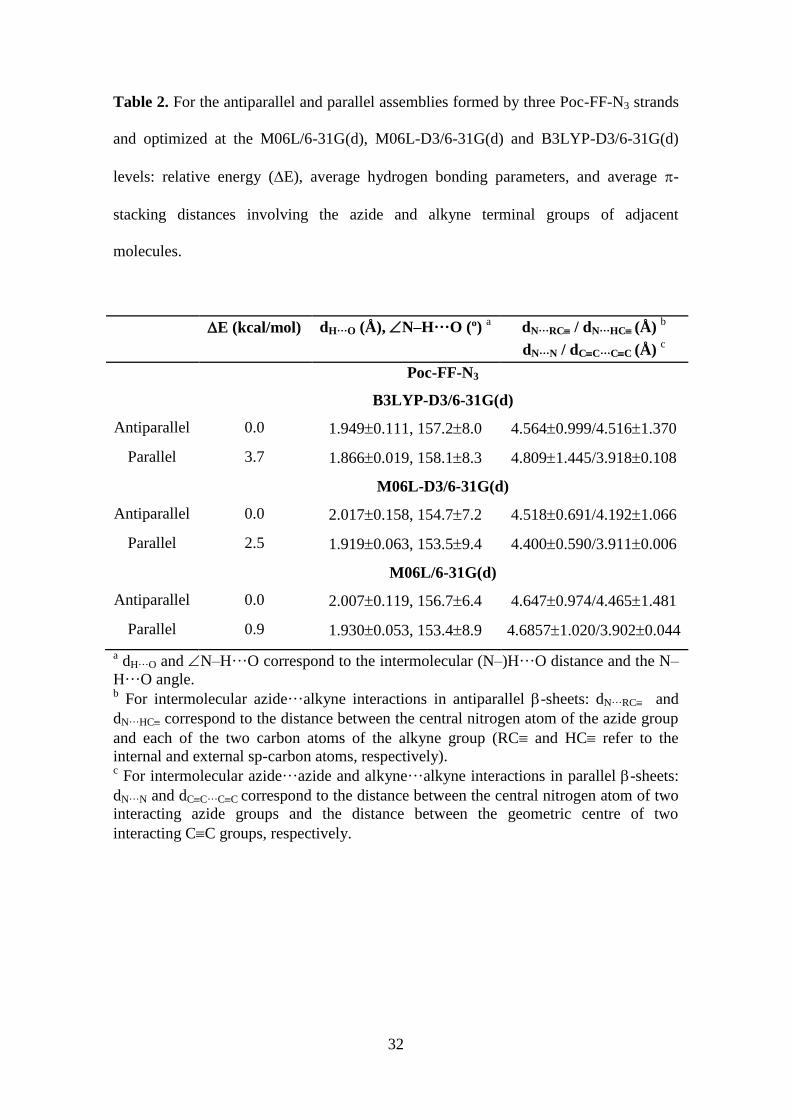
Table 2. For the antiparallel and parallel assemblies formed by three Poc-FF-N3 strands
and optimized at the M06L/6-31G(d), M06L-D3/6-31G(d) and B3LYP-D3/6-31G(d)
levels: relative energy (E), average hydrogen bonding parameters, and average -
stacking distances involving the azide and alkyne terminal groups of adjacent
molecules.
E (kcal/mol) dH···O (Å), N–H···O (º) a
dN···RC / dN···HC (Å) b
dN···N / dCC···CC (Å) c
Poc-FF-N3
B3LYP-D3/6-31G(d)
Antiparallel 0.0 1.9490.111, 157.28.0 4.5640.999/4.5161.370
Parallel 3.7 1.8660.019, 158.18.3 4.8091.445/3.9180.108
M06L-D3/6-31G(d)
Antiparallel 0.0 2.0170.158, 154.77.2 4.5180.691/4.1921.066
Parallel 2.5 1.9190.063, 153.59.4 4.4000.590/3.9110.006
M06L/6-31G(d)
Antiparallel 0.0 2.0070.119, 156.76.4 4.6470.974/4.4651.481
Parallel 0.9 1.9300.053, 153.48.9 4.68571.020/3.9020.044
a dH···O and N–H···O correspond to the intermolecular (N–)H···O distance and the N–
H···O angle.
b For intermolecular azide···alkyne interactions in antiparallel -sheets: dN···RC and
dN···HC correspond to the distance between the central nitrogen atom of the azide group
and each of the two carbon atoms of the alkyne group (RC and HC refer to the
internal and external sp-carbon atoms, respectively). c For intermolecular azide···azide and alkyne···alkyne interactions in parallel -sheets:
dN···N and dCC···CC correspond to the distance between the central nitrogen atom of two
interacting azide groups and the distance between the geometric centre of two
interacting CC groups, respectively.
33
Figure 1
34
Figure 2

35
Figure 3
36
Figure 4

37
Figure 5
38
Figure 6
39
Figure 7
40
Figure 8
-6.0
-4.0
-2.0
0.0
2.0
4.0
6.0
8.0
10.0
12.0
180 190 200 210 220 230 240 250
DCM:MeOH
HFIP:water
HFIP:MeOH
HFIP:iPrOH
-5.0
-3.5
-2.0
-0.5
1.0
2.5
4.0
5.5
7.0
180 190 200 210 220 230 240 250
DCM:MeOH
HFIP:water
HFIP:MeOH
HFIP:iPrOH
CD
(m
deg
)
Wavelength (nm)
(a)
CD
(m
deg
)
Wavelength (nm)
(b)

41
Figure 9

42
Figure 10
43
Figure 11
44
Figure 12
2.147
2.027
(a)
(b)

45
Graphical Abstract