Hospira, Inc. QUELICIN- succinylcholine chloride injection ...

JBUR-4882; No. of Pages 8
Case report
Second-degree burns with six etiologies treated withautologous noncultured cell-spray grafting
Roger Esteban-Vives a, Myung S. Choi b, Matthew T. Young a, Patrick Over a,Jenny Ziembicki c, Alain Corcos c, Jorg C. Gerlach a,*aDepartment of Surgery and Bioengineering, McGowan Institute for Regenerative Medicine, University of Pittsburgh,
Pittsburgh, PA, USAb School of Medicine, University of Pittsburgh, Pittsburgh, PA, USAcThe University of Pittsburgh Medical Center, UPMC Mercy Hospital Trauma Services and Burn Center, Pittsburgh, PA, USA
b u r n s x x x ( 2 0 1 6 ) x x x . e 1 – x x x . e 8
a r t i c l e i n f o
Article history:
Accepted 18 February 2016
Keywords:
Burns
Keratinocytes
Cell-spray grafting
Re-epithelialization
Wound healing
a b s t r a c t
Partial and deep partial-thickness burn wounds present a difficult diagnosis and prognosis
that makes the planning for a conservative treatment versus mesh grafting problematic. A
non-invasive treatment strategy avoiding mesh grafting is often chosen by practitioners
based on their clinical and empirical evidence. However, a delayed re-epithelialization
after conservative treatment may extend the patient’s hospitalization period, increase the
risk of infection, and lead to poor functional and aesthetic outcome. Early spray grafting,
using non-cultured autologous cells, is under discussion for partial and deep partial-
thickness wounds to accelerate the re-epithelialization process, reducing the healing time
in the hospital, and minimizing complications. To address planning for future clinical
studies on this technology, suitable indications will be interesting. We present case
information on severe second-degree injuries after gas, chemical, electrical, gasoline,
hot water, and tar scalding burns showing one patient per indication. The treatment
results with autologous non-cultured cells, support rapid, uncomplicated re-epithelializa-
tion with aesthetically and functionally satisfying outcomes. Hospital stays averaged
7.6 � 1.6 days. Early autologous cell-spray grafting does not preclude or prevent simulta-
neous or subsequent traditional mesh autografting when indicated on defined areas of full-
thickness injury.
# 2016 Elsevier Ltd and ISBI. All rights reserved.
* Correspondence to: McGowan Institute for Regenerative Medicine, University of Pittsburgh, 3025 East Carson Street, Pittsburgh, PA 15203,USA. Tel.: +1 412 383 7640; fax: +1 412 383 9460.
E-mail address: [email protected] (J.C. Gerlach).
Abbreviations: ABSI, abbreviated burn severity risk index; BMI, body mass index; HD, hospital day; POD, post-operative day; LOS, lengthof stay; TBSA, total body surface area; IRB, Institutional Review Board; FDA, Federal Drug Administration USA; UPMC, University ofPittsburgh Medical Center PA USA.
Available online at www.sciencedirect.com
ScienceDirect
journal homepage: www.elsevier.com/locate/burns
Please cite this article in press as: Esteban-Vives R, et al. Second-degree burns with six etiologies treated with autologous noncultured cell-spraygrafting. Burns (2016), http://dx.doi.org/10.1016/j.burns.2016.02.020
http://dx.doi.org/10.1016/j.burns.2016.02.0200305-4179/# 2016 Elsevier Ltd and ISBI. All rights reserved.

JBUR-4882; No. of Pages 8
b u r n s x x x ( 2 0 1 6 ) x x x . e 1 – x x x . e 8e2
1. Introduction
The initial clinical diagnosis of a burn wound depth usually
determines the treatment, however, intermediate partial
thickness burns can be difficult to classify accurately with
an early evaluation [1]. Deeper partial-thickness injuries may
need to undergo surgical treatment, including excision and
optional split-skin mesh grafting [1–3]. Conservative treat-
ment of extensive, deep partial-thickness wounds avoids early
mesh grafting at the risk of a delay in wound closure, which
may result in infection and poor aesthetic and functional
outcomes. Possible complications of this therapeutic ap-
proach include hypertrophic scarring, contracture, and poor
functional and aesthetic outcomes that could result in a
reduced range of motion and unsatisfactory psychosocial
adjustment [4]. Thus, in this borderline indication, an early
autologous cell-spray grafting of extensive, deep partial-
thickness wounds could be an interesting therapeutic option
[5]. In addition, an enlargement of the donor-to-graft-area
ratio from a routine 3:1 has its typical clinical limitation at 6:1,
while using cell-spray grafting the ratio is between 20:1 and
80:1 [6,7].
Skin regeneration is a dynamic process that involves
different cell lineages and cell signaling, which leads pro-
genitors cells to restore the tissue structure and function [8].
Epidermal burn-wound regeneration starts from the edge of
the wound where the epidermal structures remain. The
adjacent epidermis contains all different functional structures
including the hair follicle (HF), inter follicular epidermis (IFE),
and the sebaceous glands that are involved in the healing
process [9–11]. Epidermal homeostasis and regeneration are
enabled by quiescent epidermal stem cells (Fig. 1A), some of
which can be activated and proliferate as transient amplified
keratinocytes in the Stratum basale [12,13]. Through post-
mitotic differentiation and migration, cells from the basal
layer can regenerate the entirely stratified epidermis [10,14–
17]. In deep partial-thickness burn wounds, mesh and cell-
spray grafting aim to distribute these cells over the center of
the wound and speed up central re-epithelialization.
Various cell-spray grafting methods have been introduced
and are thought to provide a fast re-epithelialization and then
reduce the healing time and minimize complications [5,18,19].
This innovative technique is still under clinical evaluation and
Fig. 1 – Single cell-spray therapy proof of concept. (A) The basal la
proliferative keratinocytes that we included for spray-grafting o
Epidermal–dermal separation is performed by dispase and follo
collagenase digestion on the dermis. (C) Isolated cells are combi
Please cite this article in press as: Esteban-Vives R, et al. Second-degree bugrafting. Burns (2016), http://dx.doi.org/10.1016/j.burns.2016.02.020
to address planned future clinical studies, suitable indications
are of interest. Since 2008, skin-cell-spray grafting, using
isolated non-cultured autologous keratinocytes (Fig. 1B), has
been used at our center as a treatment option for 45 partial-
thickness burn patients. Our chosen regulatory Innovative
Practice Institutional Review Board (IRB) approach precludes a
study with controls. At times, the procedure has been used in
combination with mesh grafting for patients with combined
second- and third-degree burn wounds. Here, we present six
second-degree burn patients and their treatments, showing
different burn etiologies: gas, chemical, electrical, gasoline,
hot water, and tar (Table 1). In all indications, the results, after
early autologous cell-spray grafting shows a fast re-epitheli-
alization and an aesthetic and functionally satisfying outcome
with no major complications. We suggest considering these
indications for future clinical studies in this new field.
2. Material and methods
2.1. Patient criteria for study inclusion
The Institutional Review Board (IRB) from UPMC Mercy
Hospital, through its Technology and Innovative Practice
Assessment Committee, approved the cell-based grafting
procedures under an innovative practice approach. Therefore,
performing a clinical study with controls was not possible.
Patient data collection for this retrospective analysis was
performed under an authorization from the Institutional
Review Board (IRB# PRO14010023, 23-01). Exclusion criteria for
the treatment consisted of age <18 years, pre-existing local
and systemic infections, hypersensitivity to trypsin or other
enzymatic wound treatments, and the risks associated with
anesthesia. For inclusion, the team of surgeons at UPMC Mercy
Trauma and Burn Centers (Pittsburgh, PA) decided that the
treatments should be limited to deep, relatively extensive
partial-thickness burn wound patients. The decision to offer a
patient treatment was based on clinical judgment of the
surgeon in charge, and consultation with the burn service as a
whole. Consideration typically involved the wound appear-
ance after the first debridement, and at 24 and 48 h. Before cell-
spray therapy, a consent form was obtained from each patient
after the alternative conservative or invasive treatment was
explained in detail. The physicians explained to the patient
yer contains epidermal stem cells and transient amplifying
n the wound. (B) Three-enzymatic step isolation process.
wed by trypsin digestion of the epidermis and, a
ned and seeded onto wound through cell-spray deposition.
rns with six etiologies treated with autologous noncultured cell-spray
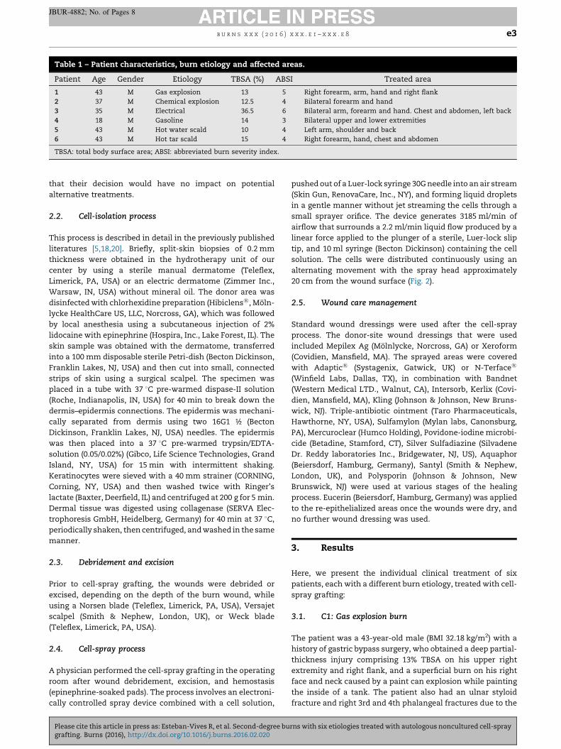
Table 1 – Patient characteristics, burn etiology and affected areas.
Patient Age Gender Etiology TBSA (%) ABSI Treated area
1 43 M Gas explosion 13 5 Right forearm, arm, hand and right flank
2 37 M Chemical explosion 12.5 4 Bilateral forearm and hand
3 35 M Electrical 36.5 6 Bilateral arm, forearm and hand. Chest and abdomen, left back
4 18 M Gasoline 14 3 Bilateral upper and lower extremities
5 43 M Hot water scald 10 4 Left arm, shoulder and back
6 43 M Hot tar scald 15 4 Right forearm, hand, chest and abdomen
TBSA: total body surface area; ABSI: abbreviated burn severity index.
b u r n s x x x ( 2 0 1 6 ) x x x . e 1 – x x x . e 8 e3
JBUR-4882; No. of Pages 8
that their decision would have no impact on potential
alternative treatments.
2.2. Cell-isolation process
This process is described in detail in the previously published
literatures [5,18,20]. Briefly, split-skin biopsies of 0.2 mm
thickness were obtained in the hydrotherapy unit of our
center by using a sterile manual dermatome (Teleflex,
Limerick, PA, USA) or an electric dermatome (Zimmer Inc.,
Warsaw, IN, USA) without mineral oil. The donor area was
disinfected with chlorhexidine preparation (Hibiclens1, Moln-
lycke HealthCare US, LLC, Norcross, GA), which was followed
by local anesthesia using a subcutaneous injection of 2%
lidocaine with epinephrine (Hospira, Inc., Lake Forest, IL). The
skin sample was obtained with the dermatome, transferred
into a 100 mm disposable sterile Petri-dish (Becton Dickinson,
Franklin Lakes, NJ, USA) and then cut into small, connected
strips of skin using a surgical scalpel. The specimen was
placed in a tube with 37 8C pre-warmed dispase-II solution
(Roche, Indianapolis, IN, USA) for 40 min to break down the
dermis–epidermis connections. The epidermis was mechani-
cally separated from dermis using two 16G1 ½ (Becton
Dickinson, Franklin Lakes, NJ, USA) needles. The epidermis
was then placed into a 37 8C pre-warmed trypsin/EDTA-
solution (0.05/0.02%) (Gibco, Life Science Technologies, Grand
Island, NY, USA) for 15 min with intermittent shaking.
Keratinocytes were sieved with a 40 mm strainer (CORNING,
Corning, NY, USA) and then washed twice with Ringer’s
lactate (Baxter, Deerfield, IL) and centrifuged at 200 g for 5 min.
Dermal tissue was digested using collagenase (SERVA Elec-
trophoresis GmbH, Heidelberg, Germany) for 40 min at 37 8C,
periodically shaken, then centrifuged, and washed in the same
manner.
2.3. Debridement and excision
Prior to cell-spray grafting, the wounds were debrided or
excised, depending on the depth of the burn wound, while
using a Norsen blade (Teleflex, Limerick, PA, USA), Versajet
scalpel (Smith & Nephew, London, UK), or Weck blade
(Teleflex, Limerick, PA, USA).
2.4. Cell-spray process
A physician performed the cell-spray grafting in the operating
room after wound debridement, excision, and hemostasis
(epinephrine-soaked pads). The process involves an electroni-
cally controlled spray device combined with a cell solution,
Please cite this article in press as: Esteban-Vives R, et al. Second-degree bugrafting. Burns (2016), http://dx.doi.org/10.1016/j.burns.2016.02.020
pushed out of a Luer-lock syringe 30G needle into an air stream
(Skin Gun, RenovaCare, Inc., NY), and forming liquid droplets
in a gentle manner without jet streaming the cells through a
small sprayer orifice. The device generates 3185 ml/min of
airflow that surrounds a 2.2 ml/min liquid flow produced by a
linear force applied to the plunger of a sterile, Luer-lock slip
tip, and 10 ml syringe (Becton Dickinson) containing the cell
solution. The cells were distributed continuously using an
alternating movement with the spray head approximately
20 cm from the wound surface (Fig. 2).
2.5. Wound care management
Standard wound dressings were used after the cell-spray
process. The donor-site wound dressings that were used
included Mepilex Ag (Molnlycke, Norcross, GA) or Xeroform
(Covidien, Mansfield, MA). The sprayed areas were covered
with Adaptic1 (Systagenix, Gatwick, UK) or N-Terface1
(Winfield Labs, Dallas, TX), in combination with Bandnet
(Western Medical LTD., Walnut, CA), Intersorb, Kerlix (Covi-
dien, Mansfield, MA), Kling (Johnson & Johnson, New Bruns-
wick, NJ). Triple-antibiotic ointment (Taro Pharmaceuticals,
Hawthorne, NY, USA), Sulfamylon (Mylan labs, Canonsburg,
PA), Mercuroclear (Humco Holding), Povidone-iodine microbi-
cide (Betadine, Stamford, CT), Silver Sulfadiazine (Silvadene
Dr. Reddy laboratories Inc., Bridgewater, NJ, US), Aquaphor
(Beiersdorf, Hamburg, Germany), Santyl (Smith & Nephew,
London, UK), and Polysporin (Johnson & Johnson, New
Brunswick, NJ) were used at various stages of the healing
process. Eucerin (Beiersdorf, Hamburg, Germany) was applied
to the re-epithelialized areas once the wounds were dry, and
no further wound dressing was used.
3. Results
Here, we present the individual clinical treatment of six
patients, each with a different burn etiology, treated with cell-
spray grafting:
3.1. C1: Gas explosion burn
The patient was a 43-year-old male (BMI 32.18 kg/m2) with a
history of gastric bypass surgery, who obtained a deep partial-
thickness injury comprising 13% TBSA on his upper right
extremity and right flank, and a superficial burn on his right
face and neck caused by a paint can explosion while painting
the inside of a tank. The patient also had an ulnar styloid
fracture and right 3rd and 4th phalangeal fractures due to the
rns with six etiologies treated with autologous noncultured cell-spray
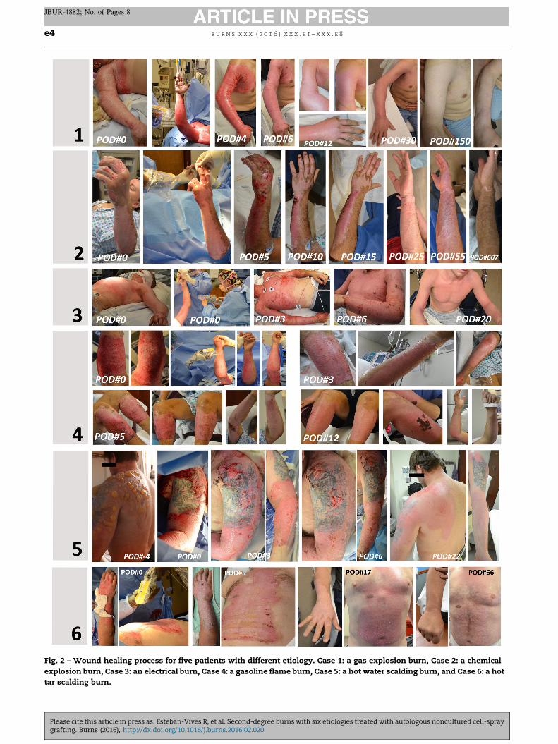
Fig. 2 – Wound healing process for five patients with different etiology. Case 1: a gas explosion burn, Case 2: a chemical
explosion burn, Case 3: an electrical burn, Case 4: a gasoline flame burn, Case 5: a hot water scalding burn, and Case 6: a hot
tar scalding burn.
b u r n s x x x ( 2 0 1 6 ) x x x . e 1 – x x x . e 8e4
JBUR-4882; No. of Pages 8
Please cite this article in press as: Esteban-Vives R, et al. Second-degree burns with six etiologies treated with autologous noncultured cell-spraygrafting. Burns (2016), http://dx.doi.org/10.1016/j.burns.2016.02.020
b u r n s x x x ( 2 0 1 6 ) x x x . e 1 – x x x . e 8 e5
JBUR-4882; No. of Pages 8
explosion. The right upper extremity was initially treated
with Silvadene, and the right face was treated with Triple
Antibiotics with Sulfamylon to the right ear. The patient was
prepared for wound debridement and cell isolation on HD#3.
The donor skin sample area was about 32.2 cm2, with a 0.2 mm
thickness obtained with an electric dermatome, and covered
with Mepilex Ag. The treated areas were the face, right hand,
right forearm, right arm, and the right flank, with a total
sprayed area of 2646 cm2. After the cell isolation, 20 million
cells were obtained to cover the burned surface with a density
of 7559 cells/cm2. The spray area was covered with Adaptic1
combined with triple-antibiotic ointment and wrapped with
N-Terface1, Kerlix, and Bandnet. The dressings were changed
on POD#4 and then daily. Small re-epithelialized areas were
observed on POD#4 on the wound edges of the right flank,
anterior forearm, and hand. The wound became dry on POD#6
on the right flank, forearm, arm, and hand and the patient
were discharged that day. The patient had to undergo multiple
orthopedic surgeries after discharge for his wrist injury and
chronic shoulder problems. On his follow-up visit, complete
epithelialization and some hypopigmentation were noted on
POD#14. A full range of motion in the right hand with some
limitations in wrist motion was noted on POD#30. At his 5-
month follow-up, minimal hyperpigmentation on the right
side of his face and very slight hypopigmentation on the right
shoulder and upper extremity were noted. There was no
evidence of hypertrophic scarring throughout the prior burn
area, and his only functional impairment, at this point, was
due to his wrist injury.
3.2. C2: Chemical explosion burn
The patient was a 37-year-old male (BMI 24.07 kg/m2) who
presented with 12.5% TBSA deep partial-thickness injury on
his bilateral upper extremities and hands caused by a
potassium nitrate explosion. The burn wounds were initially
covered with Mepilex and the patient underwent daily
hydrotherapy. On HD#3, the patient was prepared for
debridement, and a 14 cm2 skin donor skin sample of
0.2 mm thickness was obtained using a manual dermatome
from the left hip. The donor site was covered with Mepilex Ag,
and 9.4 million cells were isolated and used to spray the
entirely burned surface with a density of 6356 cells/cm2 on the
debrided burn wound. The treated areas were the left arm,
dorsum of the left hand, and all left-hand digits with a total left
upper extremity area of 331.25 cm2; also, the right forearm,
dorsum of the right hand, and all the right-hand digits with a
total right upper extremity area of 1148 cm2. The total spray
area (1479.25 cm2) was covered with Xeroform, triple-antibi-
otic ointment Intersorb, Kerlix, and Bandnet. The dressings
were changed on POD#3, and it was noted that the wounds
were still open. On POD#5, all wound areas were mostly dry
except for some open areas, especially on the right hand
where no sign of re-epithelialization was detected. On POD#7,
the patient underwent an additional split-thickness skin
grafting on the right hand, right middle finger, right ring
finger, right small finger, and right lateral wrist. The patient
was discharged on POD#9, and during his follow-up visit on
POD#14, it was noted that there were some small open areas
primarily on the left forearm (<5 mm in maximal dimension)
Please cite this article in press as: Esteban-Vives R, et al. Second-degree bugrafting. Burns (2016), http://dx.doi.org/10.1016/j.burns.2016.02.020
and they were treated with Mercuroclear (Humco Holding
Group, Texarkana, TX) multiple times a day. The difficulty
with full flexion of R fingers was also noted on POD#14;
however, on POD#25 full range of motion was observed. On
POD#55, minimal hypertrophic scarring on a few of his right
fingers was noted. On POD#607, the areas of autografts were
noted to be almost indiscernible with the normal skin and no
hypertrophic scarring or contractures were noted. The patient
maintained a full range of motion in all extremities without
restriction.
3.3. C3: Electrical burn
The patient was a 35-year-old male (BMI 22.72 kg/m2) with a
20-year history of cigarette smoking, narcotics, and recrea-
tional drug abuse who presented with 36.5% TBSA electrical
burn after grabbing a live wire. The patient had a deep partial-
thickness burn injury on the head, chest (more superficial in
nature), abdomen, bilateral upper extremities, and back (the
left back was more superficial in nature), and full-thickness
injuries on the right hand and foot. Silver Sulfadiazine was
initially used for his wounds. The patient was prepared on
HD#4 for wound debridement, tangential excision of deep
wounds on forearms and hands with a Weck blade and
excision of more superficial wounds on the left back and chest
with a Norsen blade before spray grafting. A 30-cm2 donor skin
sample was taken from the right anterior thigh using a 0.2 mm
manual Weck blade at the hydrotherapy unit and it was
covered with Mepilex Ag. The 23.8 million cells taken were
isolated and used to spray the entire partial-thickness surface
with a density of 4162 cells/cm2 on the excised and debrided
burn wounds. The cell-sprayed areas included the right hand,
right forearm, right arm, the left hand, left forearm, the left
arm, chest, abdomen, and left back with a total area of
5719 cm2. Concurrently, split-thickness autografts were iso-
lated from the donor skin sample with a 0.3 mm Zimmer blade
from the right anterior thigh and then ‘‘pie-crusted’’ and
applied on the right hand and foot. The treated wounds were
covered with Adaptic1, triple-antibiotic ointment, Intersorb,
Kerlix, and Bandnet. On POD#4, re-epithelialization of the
chest and the arms were observed with few open areas. On
POD#5, the arms were noted as ‘‘healed’’, and hands were still
noted as open. Silver Sulfadiazene and topical emollients,
such as Aquaphor and Mercuroclear, were applied to the open
areas and Santyl/Polysporin was used to lift an eschar. On
POD#6, the right hand and foot were still open and Adaptic was
applied, but the left hand was healed. On POD#13, an eschar
was noted on the right arm, and it was treated with Santyl/
Polysporin, and the left foot was treated with wet-to-dry
dressing changes. On POD#20, all of the areas treated with cell-
spray grafting were noted as completely healed and re-
epithelialized. There was no evidence of hypertrophic scars or
wound contracture, and the patient had a functional range of
motion in all extremities.
3.4. C4: Gasoline flame burn
The patient was an 18-year-old male (BMI 30.91 kg/m2) with a
history of poor wound healing in the right foot with split-
thickness skin grafting about 3 months prior to presentation.
rns with six etiologies treated with autologous noncultured cell-spray

b u r n s x x x ( 2 0 1 6 ) x x x . e 1 – x x x . e 8e6
JBUR-4882; No. of Pages 8
This prior wound had been healing well by the time the
patient presented with a new 14% TBSA gasoline flame burn
injury. He had superficial partial-thickness injury to bilateral
upper extremities and deep partial thickness injury to lower
extremities. His wounds were initially treated with Sulfamy-
lon, and his left arm wound was placed in Mepilex. The
patient was prepared on HD#5 for wound debridement with a
Norsen debrider. Approximately 36 cm2 of skin with 0.2 mm
thickness was obtained with a manual dermatome Weck
blade from left upper thigh and the donor skin sample area
was covered with Xeroform. Forty-five million cells were
obtained and used to spray the entire burn wound surface
with a density of 15,198 cells/cm2. The treated area included
right hand, right forearm, right upper arm, right lower leg, left
forearm, and left lower leg. The total 2961 cm2 sprayed area
was covered with N-terface1, Sulfamylon, Kerlix, and
Bandnet. Wound areas started showing signs of re-epitheli-
alization on POD#3 and dressings were changed to Adaptic
dry dressing for healing areas (bilateral lower extremities),
and only Eucerin for healed areas (bilateral upper extremi-
ties). The majority of the wounds were healed by POD#6. The
donor site developed an eschar that resolved with Santyl/
Polysporin. Wounds were completely healed by POD#13 and
there was no evidence of hypertrophic scarring or contrac-
tures, and the patient demonstrated a full range of motion in
all extremities.
3.5. C5: Hot water scalding
The patient was a 43-year-old male (BMI 35.37 kg/m2) who
presented with a 10% TBSA burn wound caused by a hot water
scalding. He was initially treated at an outside facility with
Bacitracin and presented to our center the day after when his
wounds appeared worse. The patient had a partial-thickness
burn on the left upper extremity, shoulder, and back. The
wounds were dressed in Sulfamylon first and the patient
underwent daily dressing changes at hydrotherapy. The
patient was prepared on HD#5 for wound debridement with
Versajet before cell spraying. Approximately, 30 cm2 of skin
with 0.2 mm thickness was obtained with a manual derma-
tome Weck blade from the left thigh. The donor area was
covered with Xeroform, triple-antibiotic ointment, Kerlix, and
Bandnet. Seventeen million epidermal cells were sprayed on
the wound surface with a density of 8695 cells/cm2 on the
excised burn wounds. The area treated with the cell-spray
grafting included the left shoulder, arm, forearm, and back
with a total sprayed area of 1955 cm2. The cell-sprayed
wounds were covered with Adaptic1 and triple-antibiotic
ointment and were wrapped with Intersorb, Kerlix, Kling, and
Bandnet. The dressings were changed on POD#3, and the areas
of cell-spray grafting were noted to have good re-epitheliali-
zation with only small scattered open areas. Adaptic and dry
dressing were applied and the wound was rechecked on
POD#6. A 100% re-epithelialization was noted and the patient
was discharged that day with instructions to apply Eucerin
moisturizer to the wound. On POD#46, the patient was also
noted to have a full range of motion in his extremities. On
POD#136, a very small area of hypertrophic scarring was noted
on the left lateral humerus just inferior to the deltoid.
However, the area was very small and was not causing any
Please cite this article in press as: Esteban-Vives R, et al. Second-degree bugrafting. Burns (2016), http://dx.doi.org/10.1016/j.burns.2016.02.020
functional deficits. He was noted to have an excellent
aesthetic outcome.
3.6. C6: Hot tar scalding
The patient was a 43-year-old male (BMI 28.53 kg/m2) who
presented with 15% TBSA burn wounds caused by hot tar
scalding. The patient had a partial-thickness burn on the right
arm, right hand, and anterior trunk. At hydrotherapy, the
affected areas were cleaned and blisters were debrided. On
HD#5, the burn areas were tangentially excised with #10 Weck
blade, a Norsen blade, and a VersaJet (Smith & Nephew) before
cell spraying. Approximately 42 cm2 of skin with 0.2 mm
thickness was obtained with a manual dermatome Weck
blade from the right thigh. The donor area was covered with
Xeroform, triple-antibiotic ointment, Kerlix, and Bandnet.
Twenty-seven million epidermal and dermal cells were
sprayed on the wound surface with a density of 15,607 cells/
cm2 on the excised burn wounds. The area treated with cell-
spray grafting included the right arm and hand, and anterior
trunk with a total sprayed area of 1730 cm2. The cell-sprayed
wounds were covered with Adaptic1, and triple-antibiotic
ointment and wrapped with Intersorb, Kerlix, Kling, and
Bandnet. The dressings were changed on POD#3, and patient’s
right arm and hand were noted to be nearly healed. The
anterior trunk was healing with some spotty open areas. The
wound areas were placed back in Adaptic and dry dressing and
rechecked on POD#5 when re-epithelialization was noted in all
areas except for small areas to the central chest and abdomen,
which remained minimally open. With physiotherapy and
Occupational therapy, the patient was noted to have a full
range of motion without restrictions due to his burn wounds at
this point. By POD#7, all areas were noted to be healed and re-
epithelialized and the patient was discharged. On POD#14, he
was noted to have complete re-epithelialization with hyper-
pigmentation, but without hypertrophic scarring. On POD#66,
his skin pigmentation appeared to be returning to normal and
he was still without any hypertrophic scarring or contractures.
4. Discussion
The clinical results of burn trauma, depending on the burn
area, are often devastating and may be complicated or fatal
[21]. In the USA, more than 1.25 million burns are reported per
year. The NIH spends about 6.5 million dollars annually on
patients wound healing projects and an excess of 25 billion is
spent annually on wound treatment [22,23]. Although cultured
epidermal autografts (CEA) have been available in burn
therapy for some time, the waiting time associated with the
process can be extensive and the results unsatisfactory [24].
We are promoting the use of non-cultured autologous cells
[5,18] using a patient’s own wound as a ‘‘bioreactor’’ for cell
expansion.
Departing from the use of cultured cells [20] we are
currently using a three-step enzymatic-cell-isolation process
[5,18] that allows a tissue-specific enzymatic digestion. The
process starts with a dispase-mediated epidermal/dermal
junction separation, and subsequently a trypsin-mediated
epidermal cell isolation of the exposed basal layer, together
rns with six etiologies treated with autologous noncultured cell-spray
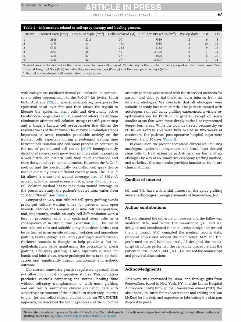
Table 2 – Information related to cell-spray therapy and healing process.
Patient Treated area (cm2) Donor sample (cm2) Cells isolated (M) Cell density (cells/cm2) Pre-op days POD LOS
1 2646 32.2 20 7559 2 6 8
2 1479 14 9.4 6355 2 10 12
3 5719 30 23.8 4162 3 9 12
4 2961 36 45 15,198 4 7 11
5 1955 30 17 8696 4 6 10
6a 1730 42 27 15,607 4 7 11
Treated area is the defined as the burned area that was cell sprayed. Cell density is the number of cells sprayed on the treated area. The
Hospital Length of Stay (LOS) includes the preoperative days (Pre-op) and the postoperative days (POD).a Dermal and epidermal cell combination for cell spray.
b u r n s x x x ( 2 0 1 6 ) x x x . e 1 – x x x . e 8 e7
JBUR-4882; No. of Pages 8
with collagenase-mediated dermal cell isolation. In compari-
son to other approaches, like the ReCell1 kit (Avita, South
Perth, Australia) [19], our specific isolation regime exposes the
epidermal basal layer first and then allows the trypsin to
liberate the epidermal stem cells and divisionally active
keratinocyte progenitors [25]. Our method allows the enzyme
elimination after the cell isolation, using a centrifugation step
and a Ringer’s Lactate cell re-suspension that dilutes the
residual traces of the enzyme. The enzyme elimination step is
important to avoid extended protolithic activity on the
isolated cells especially during a prolonged waiting time
between cell isolation and cell spray process. In contrast, to
the use of pre-cultured cell sheets [26,27] homogeneously
distributed sprayed cells grow from multiple starting points in
a well-distributed pattern until they reach confluence and
close the wound by re-epithelialization. However, the ReCell1
method and the electronically controlled cell spray device
used in our study have a different coverage area. The ReCell1
kit allows a maximum wound coverage area of 320 cm2,
according to the manufacturer’s instructions [19], while our
cell isolation method has no maximum wound coverage. In
the presented study, the patient’s treated area varies from
1500 to 5700 cm2 (see Table 2).
Compared to CEA, non-cultured cell-spray grafting avoids
prolonged culture waiting times for patients with open
wounds, reduces the amount of in vitro cell manipulation,
and, importantly, avoids an early cell differentiation with a
loss of progenitor cells and epidermal stem cells as a
consequence of in vitro culture expansion [25]. The use of
non-cultured cells and suitable spray deposition devices can
be performed in an on-site setting of isolation and immediate
grafting. Early autologous cell-spray grafting of severe partial-
thickness wounds is thought to help provide a fast re-
epithelialization while maintaining the possibility of mesh
grafting. Cell-spray grafting is also especially suitable for
hands and joint areas, where prolonged times to re-epitheli-
zation may significantly impact functionality and esthetic
outcome.
Our current innovative practice regulatory approach does
not allow for clinical comparative studies. This limitation
precludes controls comparing the normal healing time
without cell-spray transplantation or with mesh grafting,
and our results summarize clinical evaluation data with
subjective assessments of the initial burn depth only. In order
to plan for controlled clinical studies under an FDA IDE/IRB
approach, we described the healing process and the outcomes
Please cite this article in press as: Esteban-Vives R, et al. Second-degree bugrafting. Burns (2016), http://dx.doi.org/10.1016/j.burns.2016.02.020
after six patients were treated with the described methods for
partial- and deep-partial-thickness burn injuries from six
different etiologies. We conclude that all etiologies were
suitable as study inclusion criteria. The patients treated with
autologous skin cell spray grafting experienced a visible re-
epithelialization by POD#3-6 in general, except on some
smaller areas that were more deeply excised or represented
deeper burn areas. While the wounds treated became dry on
POD#6 on average and were fully healed in two weeks at
maximum, the patients’ post-operative hospital stays were
between 6 and 10 days (Table 2).
In conclusion, we present acceptable clinical results using
autologous epidermal progenitors and basal layer derived
stem cells to treat extensive partial-thickness burns of six
etiologies by way of an innovative cell-spray grafting method,
and we believe that our results provide a foundation for future
clinical studies.
Conflict of interest
J.G. and R.E. have a financial interest in the spray-grafting
device technologies through payments of RenovaCare, NY.
Author contributions
R.E. coordinated the cell-isolation process and the follow-up,
analyzed data, and wrote the manuscript. J.G. and R.E
designed and coordinated the manuscript design and revised
the manuscript. M.C. compiled the medical records data,
provided advice and revised the manuscript. M.Y. and P.O.
performed the cell isolations. A.C., J.Z designed the manu-
script structure, performed the cell-spray procedure and the
patient follow-up. M.Y., M.C., A.C., J.Z. revised the manuscript
and provided discussions.
Acknowledgments
This work was sponsored by UPMC and through gifts from
RenovaCare, based in New York, NY, and the Ladies Hospital
Aid Society (LHAS) through their Innovation Award (2013). We
also thank Jim Harris for text corrections and editing and Dan
McKeel for his help and expertise in fabricating the skin gun
disposable parts.
rns with six etiologies treated with autologous noncultured cell-spray

b u r n s x x x ( 2 0 1 6 ) x x x . e 1 – x x x . e 8e8
JBUR-4882; No. of Pages 8
r e f e r e n c e s
[1] Johnson RM, Richard R. Partial-thickness burns:identification and management. Adv Skin Wound Care2003;16:178–87 [quiz 88-9].
[2] Struz˙yna J, Krajewski A. History of necrotic burn wounddebridement. Polish J Surg 2010;82:317–23.
[3] Jackson D, Topley E, Cason JS, Lowbury EJ. Primary excisionand grafting of large burns. Ann Surg 1960;152:167–89.
[4] Balakrishnan C, Hashim M, Gao D. The effect of partial-thickness facial burns on social functioning. J Burn CareRehabil 1999;20:224–5.
[5] Gerlach JC, Johnen C, McCoy E, Brautigam K, Plettig J,Corcos A. Autologous skin cell spray-transplantation for adeep dermal burn patient in an ambulant treatment roomsetting. Burns 2011;37:e19–23.
[6] Wood FM, Stoner ML, Fowler BV, Fear MW. The use of anon-cultured autologous cell suspension and Integra1
dermal regeneration template to repair full-thickness skinwounds in a porcine model: a one-step process. Burns2007;33:693–700.
[7] Tanner Jr JC, Shea Jr PC, Bradley WH, Vandeput JJ. Large-mesh skin grafts. Plast Reconstr Surg 1969;44:504–6.
[8] Martin P. Wound healing—aiming for perfect skinregeneration. Science 1997;276:75–81.
[9] Commo S, Gaillard O, Bernard BA. The human hair folliclecontains two distinct K19 positive compartments in theouter root sheath: a unifying hypothesis for stem cellreservoir. Differentiation 2000;66:157–64.
[10] Webb A, Li A, Kaur P. Location and phenotype of humanadult keratinocyte stem cells of the skin. Differentiation2004;72:387–95.
[11] Braun KM, Niemann C, Jensen UB, Sundberg JP, Silva-Vargas V, Watt FM. Manipulation of stem cell proliferationand lineage commitment: visualisation of label-retainingcells in wholemounts of mouse epidermis. Development2003;130:5241–55.
[12] Potten CS. The epidermal proliferative unit: the possiblerole of the central basal cell. Cell Prolifer 1974;7:77–88.
[13] Potten CS, Loeffler M. Stem cells: attributes, cycles, spirals,pitfalls and uncertainties. Lessons for and from the crypt.Development 1990;110:1001–20.
[14] Jaks V, Barker N, Kasper M, van Es JH, Snippert HJ, CleversH, et al. Lgr5 marks cycling, yet long-lived, hair follicle stemcells. Nat Genet 2008;40:1291–9.
[15] Kloepper JE, Tiede S, Brinckmann J, Reinhardt DP, Meyer W,Faessler R, et al. Immunophenotyping of the human bulge
Please cite this article in press as: Esteban-Vives R, et al. Second-degree bugrafting. Burns (2016), http://dx.doi.org/10.1016/j.burns.2016.02.020
region: the quest to define useful in situ markers for humanepithelial hair follicle stem cells and their niche. ExpDermatol 2008;17:592–609.
[16] Liu Y, Lyle S, Yang Z, Cotsarelis G. Keratin 15 promotertargets putative epithelial stem cells in the hair folliclebulge. J Investig Dermatol 2003;121:963–8.
[17] Lyle S, Christofidou-Solomidou M, Liu Y, Elder DE, AlbeldaS, Cotsarelis G. The C8/144B monoclonal antibodyrecognizes cytokeratin 15 and defines the location ofhuman hair follicle stem cells. J Cell Sci 1998;111:3179–88.
[18] Gerlach JC, Johnen C, Ottoman C, Brautigam K, Plettig J,Belfekroun C, et al. Method for autologous single skin cellisolation for regenerative cell spray transplantation withnon-cultured cells. Int J Artif Organs 2011;34:271.
[19] Gravante G, Di Fede MC, Araco A, Grimaldi M, De Angelis B,Arpino A, et al. A randomized trial comparing ReCellsystem of epidermal cells delivery versus classic skin graftsfor the treatment of deep partial thickness burns. Burns2007;33:966–72.
[20] Hartmann B, Ekkernkamp A, Johnen C, Gerlach JC,Belfekroun C, Kontscher MV. Sprayed cultured epithelialautografts for deep dermal burns of the face and neck. AnnPlast Surg 2007;58:70–3. http://dx.doi.org/10.1097/01.sap.0000250647.39784.bb.
[21] Brigham PA, McLoughlin E. Burn incidence and medicalcare use in the United States: estimates, trends, and datasources. J Burn Care Res 1996;17:95–107.
[22] Medical Data International. U.S. Markets for WoundManagement Products. Irvine, CA: Medical DataInternational; 1997.
[23] Sen CK, Gordillo GM, Roy S, Kirsner R, Lambert L, Hunt TK,et al. Human skin wounds: a major and snowballing threatto public health and the economy. Wound Repair Regen2009;17:763–71.
[24] Chester DL, Balderson DS, Papini RPG. A review ofkeratinocyte delivery to the wound bed. J Burn Care Rehabil2004;25:266–75.
[25] Esteban-Vives R, Young M, Over P, Schmelzer E, Corcos A,Ziembicki J, et al. In vitro keratinocyte expansion for celltransplantation therapy is associated with differentiationand loss of basal layer derived progenitor population.Differentiation 2015;89:137–45.
[26] Rheinwald JG, Green H. Serial cultivation of strains ofhuman epidermal keratinocytes: the formation ofkeratinizing colonies from single cells. Cell 1975;6:331–43.
[27] Rheinwald JG, Green H. Formation of a keratinizingepithelium in culture by a cloned cell line derived from ateratoma. Cell 1975;6:317–30.
rns with six etiologies treated with autologous noncultured cell-spray