Sea Urchin Fertilization Lab - University of Hawaii System
17
1 Sea Urchin Fertilization Lab Hawai‘i Institute of Marine Biology Education Program Clyde Tamaru, Ph.D. Malia Rivera, Ph.D. Roxanne Haverkort Kelvin Gorospe Part I: Pre-activities for the classroom Science background Production of gametes (eggs and sperm) is a fundamental characteristic of sexually reproducing organisms. Evolutionarily, sea urchins are on the same lineage that led to mammals, and the size and shape of sea urchin eggs and sperm are similar to our own. Because they are easily studied, urchin eggs and sperm provide valuable information on fertilization and development that applies to many organisms, from jelly fish to humans. In this sense, urchin gametes provide excellent model embryos for a unique understanding of development and sexual reproduction. Spawning is a term to describe the release of eggs and sperm into the water column. Under a very special set of circumstances, spawning is followed by the uniting of two gametes in a process called fertilization. A successfully fertilized egg is called a zygote, and is the first step in the creation of a new individual. As you might expect, the beginnings of such an important event is quite complex and involves a suite of processes that all must take place sequentially in order to be successful. By studying the eggs and sperm of sea urchins, scientists have begun to understand fertilization at both the cellular and molecular levels. Sperm Egg Interactions: Sea urchins release their gametes into the environment which may be as small as a tide pool or as large as an ocean. Immediately following release from the adult, two challenges confront the potential fertilization: 1) the unlikely chance of sperm and eggs meeting in the water in such a dilute concentration, and 2) the sperm being prevented from fertilizing eggs of another species that may also be in the water column at the same time. The high local abundance of adults and the shear quantity of sperm and eggs that are produced by individual sea urchins is one strategy to increase the odds of gametes making contact with each other in the water column. Many species have further evolved a mechanism where the egg emits a chemical that attracts the sperm towards the egg in a process called “chemotaxis” (Figure 1). What is even more astonishing is the sperm is only attracted to the eggs of its own species; in other words Figure 1: One second photomicrogaph exposures showing the response of sea urchin A. punctulata sperm to A) control, B- D) 20, 40, 90 second exposure to one nanoliter of a 10-nM solution of resact, a molecule involved in chemotaxis (see text). Note clumping of sperm cells at point of entry of resact. From Ward et al. 1985.
Transcript of Sea Urchin Fertilization Lab - University of Hawaii System
1
Sea Urchin Fertilization Lab Hawai‘i Institute of Marine Biology
Education Program Clyde Tamaru, Ph.D. Malia Rivera, Ph.D.
Roxanne Haverkort
Kelvin Gorospe
Part I: Pre-activities for the classroom
Science background
Production of gametes (eggs and sperm) is a fundamental characteristic of sexually reproducing
organisms. Evolutionarily, sea urchins are on the same lineage that led to mammals, and the size
and shape of sea urchin eggs and sperm are similar to our own. Because they are easily studied,
urchin eggs and sperm provide valuable information on fertilization and development that
applies to many organisms, from jelly fish to humans. In this sense, urchin gametes provide
excellent model embryos for a unique understanding of development and sexual reproduction.
Spawning is a term to describe the release of eggs and sperm into the water column. Under a
very special set of circumstances, spawning is followed by the uniting of two gametes in a
process called fertilization. A successfully fertilized egg is called a zygote, and is the first step in
the creation of a new individual. As you might expect, the beginnings of such an important
event is quite complex and involves a suite of processes that all must take place sequentially in
order to be successful. By studying the eggs and sperm of sea urchins, scientists have begun to
understand fertilization at both the cellular and molecular levels.
Sperm Egg Interactions: Sea urchins release their
gametes into the environment which may be as small as
a tide pool or as large as an ocean. Immediately
following release from the adult, two challenges
confront the potential fertilization: 1) the unlikely
chance of sperm and eggs meeting in the water in such
a dilute concentration, and 2) the sperm being prevented
from fertilizing eggs of another species that may also be
in the water column at the same time. The high local
abundance of adults and the shear quantity of sperm and
eggs that are produced by individual sea urchins is one
strategy to increase the odds of gametes making contact
with each other in the water column.
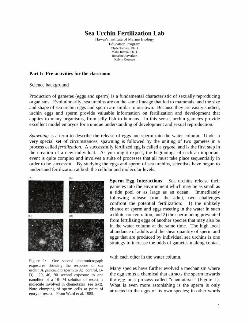
Many species have further evolved a mechanism where
the egg emits a chemical that attracts the sperm towards
the egg in a process called “chemotaxis” (Figure 1).
What is even more astonishing is the sperm is only
attracted to the eggs of its own species; in other words
Figure 1: One second photomicrogaph
exposures showing the response of sea
urchin A. punctulata sperm to A) control, B-
D) 20, 40, 90 second exposure to one
nanoliter of a 10-nM solution of resact, a
molecule involved in chemotaxis (see text).
Note clumping of sperm cells at point of
entry of resact. From Ward et al. 1985.
2
the chemical attraction is species-specific. At least one chemotactic molecule, a 14-amino acid
peptide called resact, has been isolated from the sea urchin Arbacia punctulata. Eggs produced
by A. punctulata are surrounded by a jelly coat that contains resact, while sperm produced by the
same species have receptors on their surface (plasma membrane) that bind resact. In recent
studies, the binding of resact to these receptors has been shown to activate the machinery that
controls movement in the sperm’s tail, propelling the sperm through the egg’s jelly coat (Ward et
al, 1985).
Sperm Motility: The scientific term for the tail of the sperm is flagellum, which is the
mechanism by which sperm move. Sperm motility is an important factor in successful
fertilization, since sperm must travel through the water column to reach the egg, and then
penetrate the egg to unite the male and female gametes. The flagella beat in a rapid undulating
motion, propelling the sperm in a forward direction. Under a microscope this movement is
easily observed, and in healthy sperm looks like an almost random and rapid movement of small
particles.
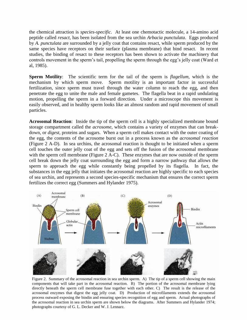
Acrosomal Reaction: Inside the tip of the sperm cell is a highly specialized membrane bound
storage compartment called the acrosome, which contains a variety of enzymes that can break-
down, or digest, proteins and sugars. When a sperm cell makes contact with the outer coating of
the egg, the contents of the acrosome burst out in a process known as the acrosomal reaction
(Figure 2 A-D). In sea urchins, the acrosomal reaction is thought to be initiated when a sperm
cell touches the outer jelly coat of the egg and sets off the fusion of the acrosomal membrane
with the sperm cell membrane (Figure 2 A-C). These enzymes that are now outside of the sperm
cell break down the jelly coat surrounding the egg and form a narrow pathway that allows the
sperm to approach the egg while constantly being propelled by its flagella. In fact, the
substances in the egg jelly that initiates the acrosomal reaction are highly specific to each species
of sea urchin, and represents a second species-specific mechanism that ensures the correct sperm
fertilizes the correct egg (Summers and Hylander 1975).
Figure 2. Summary of the acrosomal reaction in sea urchin sperm. A) The tip of a sperm cell showing the main
components that will take part in the acrosomal reaction. B) The portion of the acrosomal membrane lying
directly beneath the sperm cell membrane fuse together with each other. C) The result is the release of the
acrosomal enzymes that digest the egg jelly coat. D) Production of microfilaments extends the acrosomal
process outward exposing the bindin and ensuring species recognition of egg and sperm. Actual photographs of
the acrosomal reaction in sea urchin sperm are shown below the diagrams. After Summers and Hylander 1974;
photographs courtesy of G. L. Decker and W. J. Lennarz.
3
Sperm and Egg Fusion: A third mechanism that ensures a species-specific fusion of the sperm
with the egg involves a protein called bindin. Bindin has been found to be closely associated
with the acrosomal elements (Figure 2D) that are present in the head of the sperm, and only
becomes exposed at the end of the acrosomal reaction. Scientists have isolated bindin from the
acrosome and found it to be capable of binding to eggs of only the same species of sea urchin.
Just like how a lock and key works, receptors (e.g., the ‘lock’) on the egg cell surface only
recognize the bindin protein (e.g., the ‘key’) originating from sperm of the same species.
Recognition of the bindin protein allows for the initiation of the fusion of both egg and sperm
membranes, resulting in the next phase of the fertilization process.
Blocks to polyspermy: Polyspermy is a term used to describe
more than one sperm penetrating the egg membrane, a case that
would result in a non-viable zygote. Mechanisms that prevent
this from occurring are called blocks to polyspermy. A rapid, or
fast block to polypsermy occurs when the fusion of sperm and
egg initiates a sudden (e.g., one to three seconds) change in the
sodium ions in the egg that spreads over the cell membrane in an
electrical wave of activity. This electrical event provides short-
term prevention against additional sperm entering the egg. In
addition to this ‘fast block’, the sperm and egg fusion also
initiates a process called the cortical reaction, which is a series
of reactions that ultimately modifies a protein coat on the outside
of the plasma membrane (the vitelline layer), causing it to be
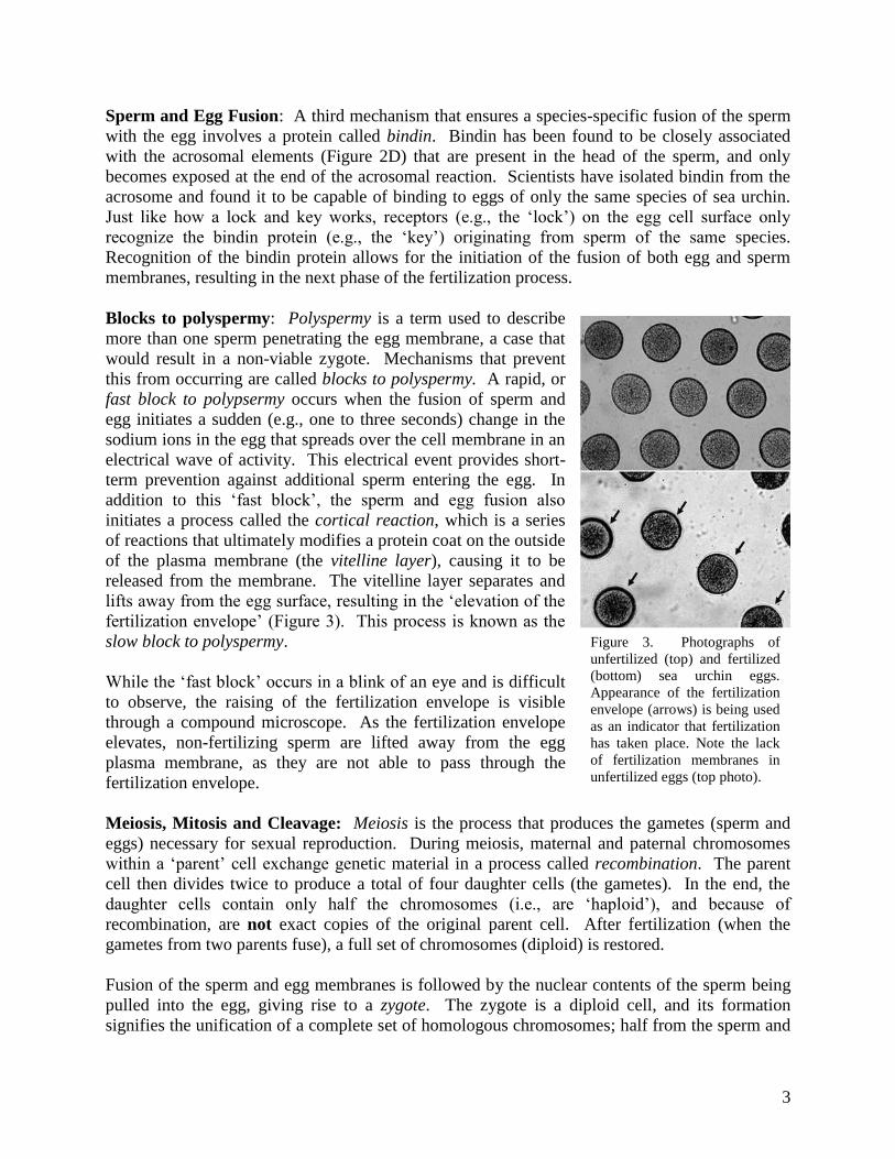
released from the membrane. The vitelline layer separates and
lifts away from the egg surface, resulting in the ‘elevation of the
fertilization envelope’ (Figure 3). This process is known as the
slow block to polyspermy.
While the ‘fast block’ occurs in a blink of an eye and is difficult
to observe, the raising of the fertilization envelope is visible
through a compound microscope. As the fertilization envelope
elevates, non-fertilizing sperm are lifted away from the egg
plasma membrane, as they are not able to pass through the
fertilization envelope.
Meiosis, Mitosis and Cleavage: Meiosis is the process that produces the gametes (sperm and
eggs) necessary for sexual reproduction. During meiosis, maternal and paternal chromosomes
within a ‘parent’ cell exchange genetic material in a process called recombination. The parent
cell then divides twice to produce a total of four daughter cells (the gametes). In the end, the
daughter cells contain only half the chromosomes (i.e., are ‘haploid’), and because of
recombination, are not exact copies of the original parent cell. After fertilization (when the
gametes from two parents fuse), a full set of chromosomes (diploid) is restored.
Fusion of the sperm and egg membranes is followed by the nuclear contents of the sperm being
pulled into the egg, giving rise to a zygote. The zygote is a diploid cell, and its formation
signifies the unification of a complete set of homologous chromosomes; half from the sperm and
Figure 3. Photographs of
unfertilized (top) and fertilized
(bottom) sea urchin eggs.
Appearance of the fertilization
envelope (arrows) is being used
as an indicator that fertilization
has taken place. Note the lack
of fertilization membranes in
unfertilized eggs (top photo).
4
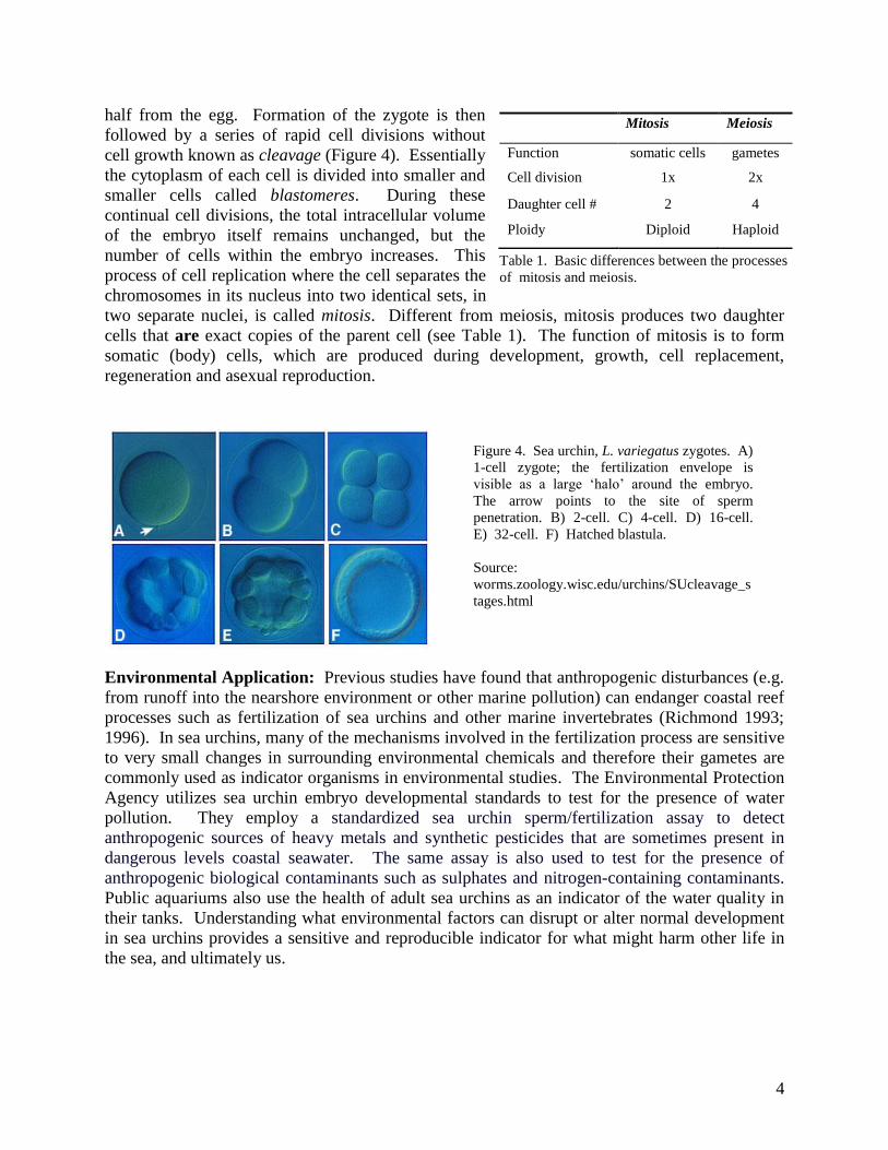
Figure 4. Sea urchin, L. variegatus zygotes. A)
1-cell zygote; the fertilization envelope is
visible as a large ‘halo’ around the embryo.
The arrow points to the site of sperm
penetration. B) 2-cell. C) 4-cell. D) 16-cell.
E) 32-cell. F) Hatched blastula.
Source:
worms.zoology.wisc.edu/urchins/SUcleavage_s
tages.html
half from the egg. Formation of the zygote is then
followed by a series of rapid cell divisions without
cell growth known as cleavage (Figure 4). Essentially
the cytoplasm of each cell is divided into smaller and
smaller cells called blastomeres. During these
continual cell divisions, the total intracellular volume
of the embryo itself remains unchanged, but the
number of cells within the embryo increases. This
process of cell replication where the cell separates the
chromosomes in its nucleus into two identical sets, in
two separate nuclei, is called mitosis. Different from meiosis, mitosis produces two daughter
cells that are exact copies of the parent cell (see Table 1). The function of mitosis is to form
somatic (body) cells, which are produced during development, growth, cell replacement,
regeneration and asexual reproduction.
Environmental Application: Previous studies have found that anthropogenic disturbances (e.g.
from runoff into the nearshore environment or other marine pollution) can endanger coastal reef
processes such as fertilization of sea urchins and other marine invertebrates (Richmond 1993;
1996). In sea urchins, many of the mechanisms involved in the fertilization process are sensitive
to very small changes in surrounding environmental chemicals and therefore their gametes are
commonly used as indicator organisms in environmental studies. The Environmental Protection
Agency utilizes sea urchin embryo developmental standards to test for the presence of water
pollution. They employ a standardized sea urchin sperm/fertilization assay to detect
anthropogenic sources of heavy metals and synthetic pesticides that are sometimes present in
dangerous levels coastal seawater. The same assay is also used to test for the presence of
anthropogenic biological contaminants such as sulphates and nitrogen-containing contaminants. Public aquariums also use the health of adult sea urchins as an indicator of the water quality in
their tanks. Understanding what environmental factors can disrupt or alter normal development
in sea urchins provides a sensitive and reproducible indicator for what might harm other life in
the sea, and ultimately us.
Mitosis Meiosis
Function somatic cells gametes
Cell division 1x 2x
Daughter cell # 2 4
Ploidy Diploid Haploid
Table 1. Basic differences between the processes
of mitosis and meiosis.
5
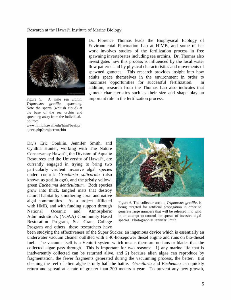
Figure 5. A male sea urchin,
Tripneustes gratilla, spawning.
Note the sperm (whitish cloud) at
the base of the sea urchin and
spreading away from the individual.
Source:
www.himb.hawaii.edu/html/beef/pr
ojects.php?project=urchin
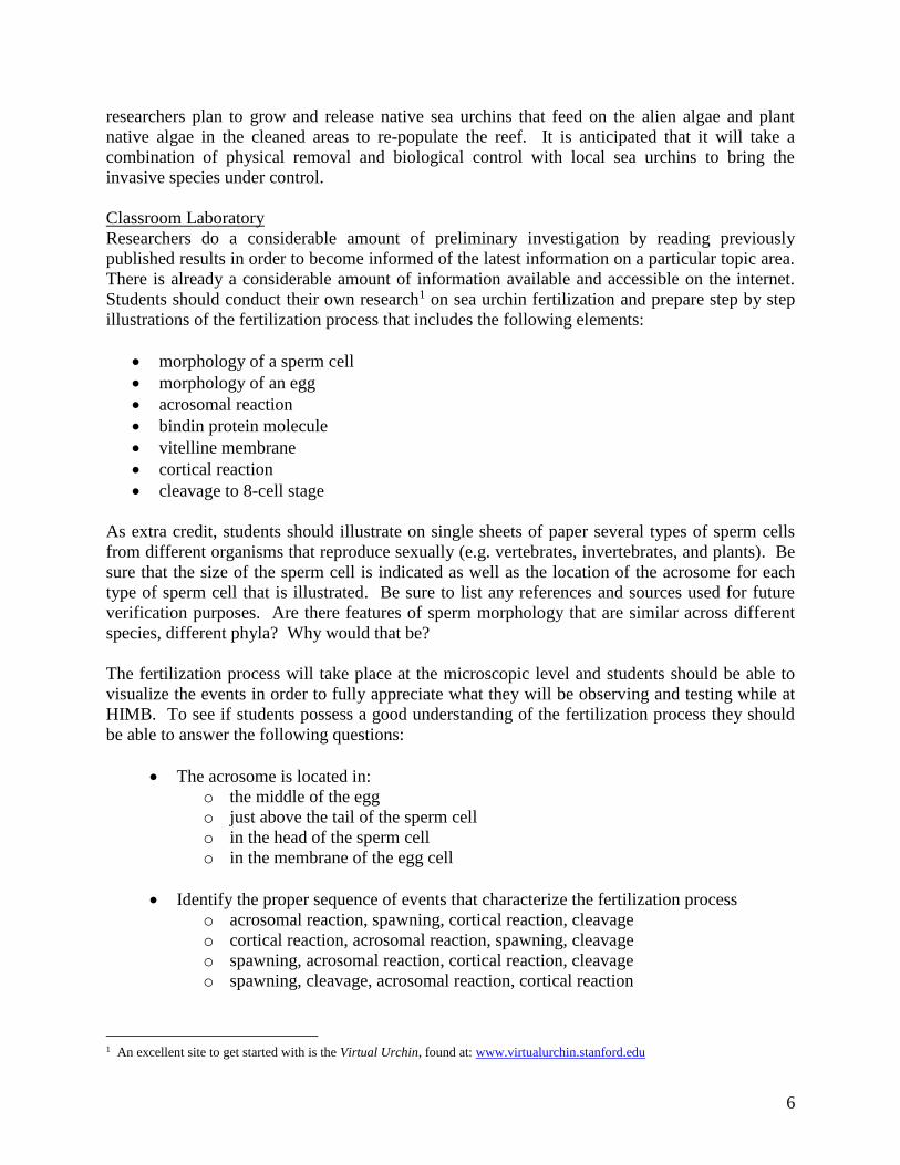
Figure 6. The collector urchin, Tripnuestes gratilla, is
being targeted for artificial propagation in order to
generate large numbers that will be released into wild
in an attempt to control the spread of invasive algal
species. Photograph © Jennifer Smith.
Research at the Hawai‘i Institute of Marine Biology
Dr. Florence Thomas leads the Biophysical Ecology of
Environmental Fluctuation Lab at HIMB, and some of her
work involves studies of the fertilization process in free
spawning invertebrates including sea urchins. Dr. Thomas also
investigates how this process is influenced by the local water
flow patterns and by physical characteristics and movements of
spawned gametes. This research provides insight into how
adults space themselves in the environment in order to
maximize opportunities for successful fertilization. In
addition, research from the Thomas Lab also indicates that
gamete characteristics such as their size and shape play an
important role in the fertilization process.
Dr.’s Eric Conklin, Jennifer Smith, and
Cynthia Hunter, working with The Nature
Conservancy Hawai‘i, the Division of Aquatic
Resources and the University of Hawai‘i, are
currently engaged in trying to bring two
particularly virulent invasive algal species
under control: Gracilaria salicornia (also
known as gorilla ogo), and the gristly yellow-
green Eucheuma denticulatum. Both species
grow into thick, tangled mats that destroy
natural habitat by smothering coral and native
algal communities. As a project affiliated
with HMB, and with funding support through
National Oceanic and Atmospheric
Administration’s (NOAA) Community Based
Restoration Program, Sea Grant College
Program and others, these researchers have
been studying the effectiveness of the Super Sucker, an ingenious device which is essentially an
underwater vacuum cleaner outfitted with a 40-horsepower diesel engine and runs on bio-diesel
fuel. The vacuum itself is a Venturi system which means there are no fans or blades that the
collected algae pass through. This is important for two reasons: 1) any marine life that is
inadvertently collected can be returned alive, and 2) because alien algae can reproduce by
fragmentation, the fewer fragments generated during the vacuuming process, the better. But
cleaning the reef of alien algae is only half the battle. Gracilaria and Eucheuma can quickly
return and spread at a rate of greater than 300 meters a year. To prevent any new growth,
6
researchers plan to grow and release native sea urchins that feed on the alien algae and plant
native algae in the cleaned areas to re-populate the reef. It is anticipated that it will take a
combination of physical removal and biological control with local sea urchins to bring the
invasive species under control.
Classroom Laboratory
Researchers do a considerable amount of preliminary investigation by reading previously
published results in order to become informed of the latest information on a particular topic area.
There is already a considerable amount of information available and accessible on the internet.
Students should conduct their own research1 on sea urchin fertilization and prepare step by step
illustrations of the fertilization process that includes the following elements:
• morphology of a sperm cell
• morphology of an egg
• acrosomal reaction
• bindin protein molecule
• vitelline membrane
• cortical reaction
• cleavage to 8-cell stage
As extra credit, students should illustrate on single sheets of paper several types of sperm cells
from different organisms that reproduce sexually (e.g. vertebrates, invertebrates, and plants). Be
sure that the size of the sperm cell is indicated as well as the location of the acrosome for each
type of sperm cell that is illustrated. Be sure to list any references and sources used for future
verification purposes. Are there features of sperm morphology that are similar across different
species, different phyla? Why would that be?
The fertilization process will take place at the microscopic level and students should be able to
visualize the events in order to fully appreciate what they will be observing and testing while at
HIMB. To see if students possess a good understanding of the fertilization process they should
be able to answer the following questions:
• The acrosome is located in:
o the middle of the egg
o just above the tail of the sperm cell
o in the head of the sperm cell
o in the membrane of the egg cell
• Identify the proper sequence of events that characterize the fertilization process
o acrosomal reaction, spawning, cortical reaction, cleavage
o cortical reaction, acrosomal reaction, spawning, cleavage
o spawning, acrosomal reaction, cortical reaction, cleavage
o spawning, cleavage, acrosomal reaction, cortical reaction
1 An excellent site to get started with is the Virtual Urchin, found at: www.virtualurchin.stanford.edu
7
• The bindin molecule is responsible for:
o the egg to survive in the water column
o recognition of sperm and egg being from the same species
o initiation of the acrosomal reaction
o initiation of sperm motility
• The fertilization envelope is found:
o just after cleavage
o just before cleavage
o just before the cortical reaction
o just after the cortical reaction
What to expect during the field trip day
During your visit to HIMB you will observe how sea urchins are spawned in order to obtain their
gametes in a controlled fashion. In this manner, you will be able to conduct a series of
observations at both the macroscopic level (means using just your eyes) and at the microscopic
level (using a compound microscope). Using these basic tools you will be conducting both
observations as well as hypothesis driven experiments in order to gain insight into the
fertilization process. Students are also encouraged to bring products (e.g., laundry detergent,
dish soap, household cleaning products, bleach, vinegar, etc.) from home to investigate impacts
that household agents have on gametes and/or the fertilization process.
8
Part II: Field trip day at HIMB
Introduction
A) To investigate fertilization requires that we control the availability of gametes and how they
interact with each other. For that reason, the first activity in the laboratory will focus on the
controlled acquisition of sperm and eggs from the sea urchins. During this initial phase the
instructors will be artificially inducing male and female sea urchins to spawn, and the students
may be able to observe the various steps that are undertaken.
B) During the next phase, students will expose the sperm and eggs to various treatments in order
to simulate situations that occur naturally (e.g., rainfall) or are man-made (e.g., non-point source
pollution). By conducting specific experiments you will be able to test some hypotheses
regarding impacts these events have on the fertilization process in sea urchins. This is possible
because we can visually see some of the impacts on the sperm (e.g., sperm motility) resulting
from the various treatments. For eggs, we will be using the presence or absence of the
fertilization membrane as an indicator of treatment effects. While the focal point of the activities
at HIMB will be sea urchins, the results you obtain should also provide insights as to the
potential impacts that natural or anthropogenic events have on other marine organisms.
C) The last phase of the activity at HIMB will be focused on observing the culminating event of
the fertilization process which is the production of a zygote, or the beginning of a unique
individual. Confirmation that this has successfully taken place is recognized when the fertilized
egg begins to cleave. We can say this because watching cleavage taking place also means that
incorporation of genetic material from both the egg and the sperm has successfully occurred.
To assist you in your science based inquiry of the fertilization process, there are a number of
guiding questions that you should think about in developing the hypotheses you want to test
while at HIMB. The listed questions are to assist you in developing testable hypotheses, which
forms the basis for the next set of activities. An example of how a null hypothesis is derived and
how to test it is available in the Appendix.
Guiding Questions
• What conditions (natural or man made) might affect fertilization in sea urchins?
• What kinds of things might have a negative effect on sea urchin gametes? On other
marine organisms?
• How much (e.g., concentration) of a particular substance will impact sperm motility?
• How much of a particular substance will affect an egg from being fertilized?
• What conditions might affect cleavage from taking place?
• What conditions may affect the rate at which cleavage and embryonic development takes
place.
9
Tools available
Motic digital compound microscope
Cordless digital compound microscopes
Materials available:
• 0.5M potassium chloride (KCl) solution (3.73g of KCl in 100mL of distilled water)
• Filtered sea water
• Petri dishes
• Depression slides
• Glass slides and cover slips
• Small syringes
• Plastic pipettes
• 100 mL glass beakers
• 250 mL glass beakers
• 500 mL glass beakers
• Rulers
• 10 mL test tubes
• Test tube racks
• Urchins to induce spawning of gametes
• Sea salt solutions
• Miracle Grow
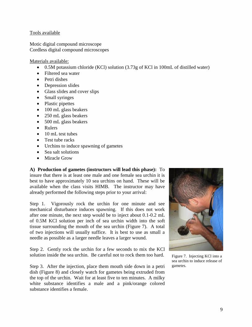
A) Production of gametes (instructors will lead this phase): To
insure that there is at least one male and one female sea urchin it is
best to have approximately 10 sea urchins on hand. These will be
available when the class visits HIMB. The instructor may have
already performed the following steps prior to your arrival:
Step 1. Vigorously rock the urchin for one minute and see
mechanical disturbance induces spawning. If this does not work
after one minute, the next step would be to inject about 0.1-0.2 mL
of 0.5M KCl solution per inch of sea urchin width into the soft
tissue surrounding the mouth of the sea urchin (Figure 7). A total
of two injections will usually suffice. It is best to use as small a
needle as possible as a larger needle leaves a larger wound.
Step 2. Gently rock the urchin for a few seconds to mix the KCl
solution inside the sea urchin. Be careful not to rock them too hard.
Step 3. After the injection, place them mouth side down in a petri
dish (Figure 8) and closely watch for gametes being extruded from
the top of the urchin. Wait for at least five to ten minutes. A milky
white substance identifies a male and a pink/orange colored
substance identifies a female.
Figure 7. Injecting KCl into a
sea urchin to induce release of
gametes.
10
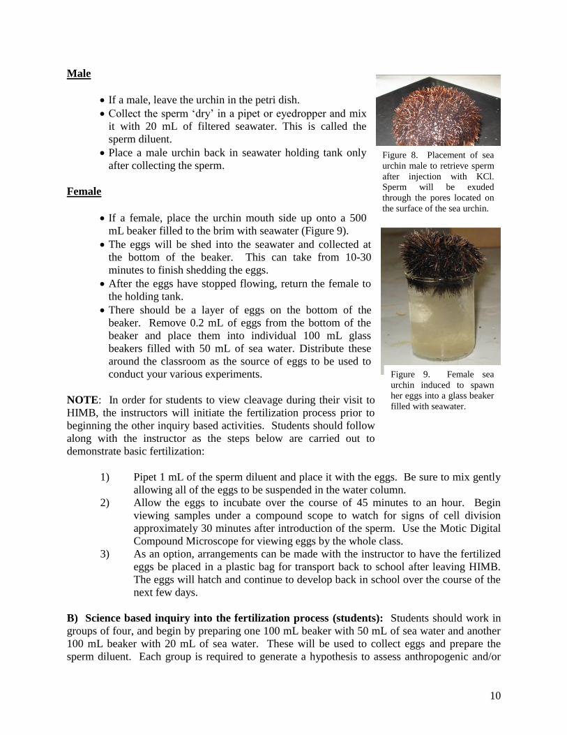
Male
• If a male, leave the urchin in the petri dish.
• Collect the sperm ‘dry’ in a pipet or eyedropper and mix
it with 20 mL of filtered seawater. This is called the
sperm diluent.
• Place a male urchin back in seawater holding tank only
after collecting the sperm.
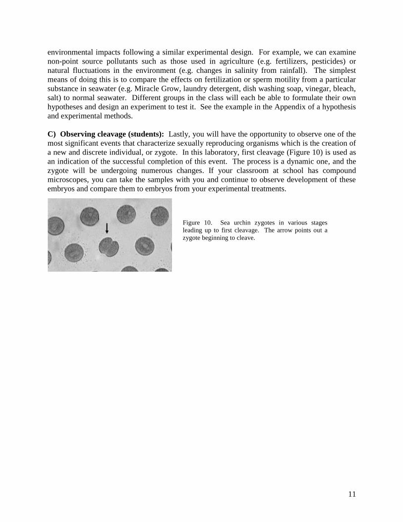
Female
• If a female, place the urchin mouth side up onto a 500
mL beaker filled to the brim with seawater (Figure 9).
• The eggs will be shed into the seawater and collected at
the bottom of the beaker. This can take from 10-30
minutes to finish shedding the eggs.
• After the eggs have stopped flowing, return the female to
the holding tank.
• There should be a layer of eggs on the bottom of the
beaker. Remove 0.2 mL of eggs from the bottom of the
beaker and place them into individual 100 mL glass
beakers filled with 50 mL of sea water. Distribute these
around the classroom as the source of eggs to be used to
conduct your various experiments.
NOTE: In order for students to view cleavage during their visit to
HIMB, the instructors will initiate the fertilization process prior to
beginning the other inquiry based activities. Students should follow
along with the instructor as the steps below are carried out to
demonstrate basic fertilization:
1) Pipet 1 mL of the sperm diluent and place it with the eggs. Be sure to mix gently
allowing all of the eggs to be suspended in the water column.
2) Allow the eggs to incubate over the course of 45 minutes to an hour. Begin
viewing samples under a compound scope to watch for signs of cell division
approximately 30 minutes after introduction of the sperm. Use the Motic Digital
Compound Microscope for viewing eggs by the whole class.
3) As an option, arrangements can be made with the instructor to have the fertilized
eggs be placed in a plastic bag for transport back to school after leaving HIMB.
The eggs will hatch and continue to develop back in school over the course of the
next few days.
B) Science based inquiry into the fertilization process (students): Students should work in
groups of four, and begin by preparing one 100 mL beaker with 50 mL of sea water and another
100 mL beaker with 20 mL of sea water. These will be used to collect eggs and prepare the
sperm diluent. Each group is required to generate a hypothesis to assess anthropogenic and/or
Figure 9. Female sea
urchin induced to spawn
her eggs into a glass beaker
filled with seawater.
Figure 8. Placement of sea
urchin male to retrieve sperm
after injection with KCl.
Sperm will be exuded
through the pores located on
the surface of the sea urchin.
11
environmental impacts following a similar experimental design. For example, we can examine
non-point source pollutants such as those used in agriculture (e.g. fertilizers, pesticides) or
natural fluctuations in the environment (e.g. changes in salinity from rainfall). The simplest
means of doing this is to compare the effects on fertilization or sperm motility from a particular
substance in seawater (e.g. Miracle Grow, laundry detergent, dish washing soap, vinegar, bleach,
salt) to normal seawater. Different groups in the class will each be able to formulate their own
hypotheses and design an experiment to test it. See the example in the Appendix of a hypothesis
and experimental methods.
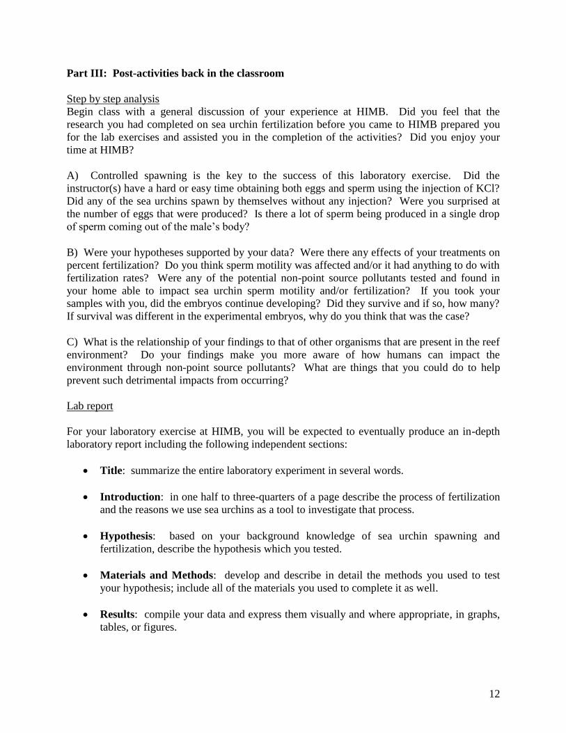
C) Observing cleavage (students): Lastly, you will have the opportunity to observe one of the
most significant events that characterize sexually reproducing organisms which is the creation of
a new and discrete individual, or zygote. In this laboratory, first cleavage (Figure 10) is used as
an indication of the successful completion of this event. The process is a dynamic one, and the
zygote will be undergoing numerous changes. If your classroom at school has compound
microscopes, you can take the samples with you and continue to observe development of these
embryos and compare them to embryos from your experimental treatments.
Figure 10. Sea urchin zygotes in various stages
leading up to first cleavage. The arrow points out a
zygote beginning to cleave.
12
Part III: Post-activities back in the classroom
Step by step analysis
Begin class with a general discussion of your experience at HIMB. Did you feel that the
research you had completed on sea urchin fertilization before you came to HIMB prepared you
for the lab exercises and assisted you in the completion of the activities? Did you enjoy your
time at HIMB?
A) Controlled spawning is the key to the success of this laboratory exercise. Did the
instructor(s) have a hard or easy time obtaining both eggs and sperm using the injection of KCl?
Did any of the sea urchins spawn by themselves without any injection? Were you surprised at
the number of eggs that were produced? Is there a lot of sperm being produced in a single drop
of sperm coming out of the male’s body?
B) Were your hypotheses supported by your data? Were there any effects of your treatments on
percent fertilization? Do you think sperm motility was affected and/or it had anything to do with
fertilization rates? Were any of the potential non-point source pollutants tested and found in
your home able to impact sea urchin sperm motility and/or fertilization? If you took your
samples with you, did the embryos continue developing? Did they survive and if so, how many?
If survival was different in the experimental embryos, why do you think that was the case?
C) What is the relationship of your findings to that of other organisms that are present in the reef
environment? Do your findings make you more aware of how humans can impact the
environment through non-point source pollutants? What are things that you could do to help
prevent such detrimental impacts from occurring?
Lab report
For your laboratory exercise at HIMB, you will be expected to eventually produce an in-depth
laboratory report including the following independent sections:
• Title: summarize the entire laboratory experiment in several words.
• Introduction: in one half to three-quarters of a page describe the process of fertilization
and the reasons we use sea urchins as a tool to investigate that process.
• Hypothesis: based on your background knowledge of sea urchin spawning and
fertilization, describe the hypothesis which you tested.
• Materials and Methods: develop and describe in detail the methods you used to test
your hypothesis; include all of the materials you used to complete it as well.
• Results: compile your data and express them visually and where appropriate, in graphs,
tables, or figures.
13
• Discussion: analyze your data in essay form; discuss the results and emphasize what the
results obtained mean to you and your group. You should also propose a new experiment
which may help explain the results or test a new hypothesis you had developed during
your discussion.
• Conclusion: in a paragraph or so, summarize your results and make concise conclusions
about them. Also include a sentence or two conveying your general conclusions about
your results in the context of how the information obtained by studying sea urchins has
value in understanding processes taking place in other organisms, including humans.
References
Science background information condensed and/or compiled from the following sources:
1) Leland Stanford Junior University (2008). Sea Urchin Embryology, Core Lab. Retrieved October 20, 2009 from http://www.stanford.edu/group/Urchin/first.htm
2) Gilbert, Scott, F. 2000. Developmental Biology, 6th Edition, Chapter 7. Sinauer and Associates. Retrieved October 19, 2009 from: http://www.ncbi.nlm.nih.gov/bookshelf/br.fcgi?book=dbio&part=A1359
3) Ward, G.E., C.J. Brokaw, D.L. Garbers, and V.D. Vacquier. 1985. Chemotaxis of Arbacia punctulata spermatozoa to resact, a peptide from the egg jelly layer. J. Cell Biol. 101:2324–2329.
4) Student Website for Life: The Science of Biology (2004). Seventh Edition, by William K. Purves, David Sadava, Gordon H. Orians, and H. Craig Heller. Retrieved on October 20, 2009 from http://bcs.whfreeman.com/thelifewire/content/chp43/4302001.html
5) Campbell, N. A & Reece, J.B. (2005). Biology 7th Edition. San Francisco: Pearson Benjamin Cummings.
6) Richmond, R. H. (1993). Coral Reefs: Present Problems and Future Concerns Resulting from Anthropogenic Disturbance. Amer. Zool. 33 (6): 524-536.
7) Richmond, R. H. (1996). Effects of coastal runoff on coral reproduction. Biological Conservation Volume 76, Number 2, 1996 , pp. 211-211(1)
Acknowledgements
The HIMB Education Program would like to thank Karen Brittain and Mark Heckman for caring for urchins used during the development of this lab. We would also like to thank Dr. Bradley ‘Kai’ Fox, the HIMB Community Education Program volunteers, and graduate student assistants
Kelvin Gorospe and Roxanne Haverkort for providing additional comments and suggestions.
14
Relevant Next Generation Science Standards
Science and Engineering Practices
1. Scientific Knowledge is Open to Revision in Light of New Evidence
a. Most scientific knowledge is quite durable, but is, in principle, subject to change
based on new evidence and/or reinterpretation of existing evidence.
2. Engaging in Argument from Evidence
a. Evaluate the claims, evidence, and reasoning behind currently accepted
explanations or solutions to determine the merits of arguments.
3. Planning and Carrying Out Investigations
a. Plan and conduct an investigation individually and collaboratively to produce data
to serve as the basis for evidence, and in the design: decide on types, how much,
and accuracy of data needed to produce reliable measurements and consider
limitations on the precision of the data (e.g., number of trials, cost, risk, time), and
refine the design accordingly.
- - - - - - - - - - - - - - - - - - - - - - - - - - - - - - - - - - - -
Connections to Nature of Science
4. Scientific Investigations Use a Variety of Methods
a. Scientific inquiry is characterized by a common set of values that include: logical
thinking, precision, open-mindedness, objectivity, skepticism, replicability of
results, and honest and ethical reporting of findings.
5. Analyzing and Interpreting Data
a. Apply concepts of statistics and probability (including determining function fits to
data, slope, intercept, and correlation coefficient for linear fits) to scientific and
engineering questions and problems, using digital tools when feasible.
6. Obtaining, Evaluating, and Communicating Information
a. Communicate scientific information (e.g., about phenomena and/or the process of
development and the design and performance of a proposed process or system) in
multiple formats (including orally, graphically, textually, and mathematically).
Disciplinary Core Ideas
1. LS1.B: Growth and Development of Organisms
a. In multicellular organisms individual cells grow and then divide via a process
called mitosis, thereby allowing the organism to grow. The organism begins as a
single cell (fertilized egg) that divides successively to produce many cells, with
each parent cell passing identical genetic material (two variants of each
chromosome pair) to both daughter cells. Cellular division and differentiation
produce and maintain a complex organism, composed of systems of tissues and
organs that work together to meet the needs of the whole organism.
2. LS2.C: Ecosystem Dynamics, Functioning, and Resilience
a. A complex set of interactions within an ecosystem can keep its numbers and types
of organisms relatively constant over long periods of time under stable conditions.
If a modest biological or physical disturbance to an ecosystem occurs, it may
return to its more or less original status (i.e., the ecosystem is resilient), as
opposed to becoming a very different ecosystem. Extreme fluctuations in
15
conditions or the size of any population, however, can challenge the functioning
of ecosystems in terms of resources and habitat availability.
3. LS4.D: Biodiversity and Humans
a. Biodiversity is increased by the formation of new species (speciation) and
decreased by the loss of species (extinction). (secondary)
b. Humans depend on the living world for the resources and other benefits provided
by biodiversity. But human activity is also having adverse impacts on biodiversity
through overpopulation, overexploitation, habitat destruction, pollution,
introduction of invasive species, and climate change. Thus sustaining biodiversity
so that ecosystem functioning and productivity are maintained is essential to
supporting and enhancing life on Earth. Sustaining biodiversity also aids
humanity by preserving landscapes of recreational or inspirational value.
(secondary) (Note: This Disciplinary Core Idea is also addressed by HS-LS4-6.)
4. LS2.D: Social Interactions and Group Behavior
a. Group behavior has evolved because membership can increase the chances of
survival for individuals and their genetic relatives.
5. LS1.A: Structure and Function
a. Systems of specialized cells within organisms help them perform the essential
functions of life.
6. HS-LS1-4 From Molecules to Organisms: Structures and Processes
a. Use a model to illustrate the role of cellular division (mitosis) and differentiation
in producing and maintaining complex organisms.
7. HS-LS2-6 Ecosystems: Interactions, Energy, and Dynamics
a. Evaluate the claims, evidence, and reasoning that the complex interactions in
ecosystems maintain relatively consistent numbers and types of organisms in
stable conditions, but changing conditions may result in a new ecosystem.
8. HS-LS2-7 Ecosystems: Interactions, Energy, and Dynamics
a. Design, evaluate, and refine a solution for reducing the impacts of human
activities on the environment and biodiversity.*
9. HS-LS2-8 Ecosystems: Interactions, Energy, and Dynamics
a. Evaluate the evidence for the role of group behavior on individual and species’
chances to survive and reproduce.
Crosscutting Concepts
1. Stability and Change
a. Much of science deals with constructing explanations of how things change and
how they remain stable.
2. Cause and Effect
a. Empirical evidence is required to differentiate between cause and correlation and
make claims about specific causes and effects.
Common Core State Standards Connections:
ELA/Literacy -
1. SL.11-12.5
a. Make strategic use of digital media (e.g., textual, graphical, audio, visual, and
interactive elements) in presentations to enhance understanding of findings,
reasoning, and evidence and to add interest. (HS-LS1-4)
16
2. RST.11-12.1
a. Cite specific textual evidence to support analysis of science and technical texts,
attending to important distinctions the author makes and to any gaps or
inconsistencies in the account. (HS-LS2-2)
3. WHST.9-12.2
a. Write informative/explanatory texts, including the narration of historical events,
scientific procedures/ experiments, or technical processes. (HS-LS2-2)
4. RST.11-12.7
a. Integrate and evaluate multiple sources of information presented in diverse
formats and media (e.g., quantitative data, video, multimedia) in order to address
a question or solve a problem. (HS-LS2-6)
5. WHST.9-12.7
a. Conduct short as well as more sustained research projects to answer a question
(including a self-generated question) or solve a problem; narrow or broaden the
inquiry when appropriate; synthesize multiple sources on the subject,
demonstrating understanding of the subject under investigation. (HS-LS2-7)
6. WHST.9-12.9
a. Draw evidence from informational texts to support analysis, reflection, and
research. (HS-LS4-4)
Mathematics -
1. MP.2
a. Reason abstractly and quantitatively. (HS-LS2-2
2. HSN.Q.A.3
a. Choose a level of accuracy appropriate to limitations on measurement when
reporting quantities. (HS-LS2-2)
17
Appendix:
The following section describes an EXAMPLE of a possible hypothesis and experiment you
could conduct at HIMB. By reading through this example, you will also get a better sense of
how to use the tools available in the lab. Your experimental design must be DIFFERENT from
this activity, and must directly address your hypothesis. The techniques and tools (i.e., the sea
urchin eggs and sperm, glass slides, seawater, microscopes, etc.) described in this experiment are
the same ones you will be using, but you must come up with your own question and your own
way to answer it.
Hypothesis (EXAMPLE):
Does salinity affect the fertilization success of sea urchin eggs? In this case, the null hypothesis
to be tested is that increased salinity will have no impact on the fertilization rate of sea urchin
eggs. To test the hypothesis, the following experiment was carried out:
1. I prepared the following solutions in test tubes: a) 10 mL of filtered sea water (FSW) and b) 9
mL of FSW combined with 1 mL of a salt saturated solution (provided by instructors).
2. I then added 1 mL of FSW containing a concentrated amount of eggs to each individual
solution, such that each of the solutions now contained approximately the same number of eggs.
I recorded all of this information to be used in my lab report.
3. The eggs in each solution of varying salinity were allowed to incubate for 5 minutes. After
the 5 minute incubation had elapsed, I slowly decanted and discarded the water from each of the
test tubes, being sure to carefully leave the eggs behind. I then immediately refilled each tube
with fresh FSW.
4. Next, I prepared a fresh sperm diluent (provided by the instructor) and fertilized the eggs in
each test tube using the same amount of sperm per tube.
5. I allowed the eggs to incubate with the sperm for a further 1 minute. Then, using a pipette, I
collected the eggs from the bottom of each tube and placed them in individual depression slides
(one depression slide per solution tested).
6. I viewed the eggs under a compound microscope using the 10X objective, and counted the
number of eggs having fertilization membranes versus those that did not (see Figure 3 for
examples of fertilized verses un-fertilized sea urchin eggs). I recorded this data in data table
format.
7. To determine the percent fertilization observed in each of the solutions, I divided the number
of eggs having fertilization membranes by the total number of eggs (i.e. with and without
fertilization membranes) and multiplied by 100.