
Scientific Posters Poster 1 5 MM TROCARS FOR THE · PDF file5 mm trocars for the treatment of...
82
Scientific Posters Poster 1 5 MM TROCARS FOR THE TREATMENT OF TENSION PNEUMOTHORAX: A SUPERIOR ALTERNATIVE TO NEEDLE DECOMPRESSION Quinton M. Hatch, MD, Mia Debarros, MD, Kelly S. Blair, MD, Eric K. Johnson, MD, Kenji Inaba, MD, Matthew J. Martin, MD* Madigan Army Medical Center Presenter: Quinton M. Hatch, MD Objectives: Needle thoracostomy (NT) is a commonly taught intervention for tension pneumothorax (TPTX) but has a high failure rate. We hypothesize that standard 5mm laparoscopic trocars may be a safe and more effective alternative. Methods: 30 episodes of TPTX and 27 episodes of tension‐induced pulseless electrical activity (PEA) were induced in 5 adult swine using thoracic CO2 insufflation via balloon trocar. Tension was defined as a 50% decrease in cardiac output. Chest decompression was performed with 5 mm laparoscopic trocars for the treatment of both TPTX with hemodynamic compromise and tension‐induced PEA. The lungs and heart were inspected and graded at necropsy for trocar‐related injury. Results were also compared to success rates with NT in the same model. Results: The placement of a 5mm trocar rapidly and immediately relieved tension physiology in 100% of cases. Mean arterial pressure, cardiac output, central venous pressure, and pulmonary capillary wedge pressure all returned to baseline within 1 minute of trocar placement. Adequate perfusion was restored in 100% of tension‐induced PEA cases within 30 seconds of trocar placement. There was no evidence of trocar‐related heart or lung damage in any of the experimental animals at necropsy (mean injury scores = 0 for both). The 5mm trocars significantly outperformed standard NT for both TPTX and for tension‐ induced PEA arrest (Image 1). Conclusions: Tension pneumothorax and tension‐induced pulseless electrical activity can be safely and effectively treated with chest decompression using 5 mm laparoscopic trocars. This technique may serve as a more rapid and reliable alternative to needle decompression.
Transcript of Scientific Posters Poster 1 5 MM TROCARS FOR THE · PDF file5 mm trocars for the treatment of...
Scientific Posters
Poster 1
5 MM TROCARS FOR THE TREATMENT OF TENSION PNEUMOTHORAX: A SUPERIOR ALTERNATIVE TO NEEDLE DECOMPRESSION
Quinton M. Hatch, MD, Mia Debarros, MD, Kelly S. Blair, MD, Eric K. Johnson, MD, Kenji Inaba, MD, Matthew J. Martin, MD*
Madigan Army Medical Center
Presenter: Quinton M. Hatch, MD
Objectives: Needle thoracostomy (NT) is a commonly taught intervention for tension pneumothorax (TPTX) but has a high failure rate. We hypothesize that standard 5mm laparoscopic trocars may be a safe and more effective alternative.
Methods: 30 episodes of TPTX and 27 episodes of tension‐induced pulseless electrical activity (PEA) were induced in 5 adult swine using thoracic CO2 insufflation via balloon trocar. Tension was defined as a 50% decrease in cardiac output. Chest decompression was performed with 5 mm laparoscopic trocars for the treatment of both TPTX with hemodynamic compromise and tension‐induced PEA. The lungs and heart were inspected and graded at necropsy for trocar‐related injury. Results were also compared to success rates with NT in the same model.
Results: The placement of a 5mm trocar rapidly and immediately relieved tension physiology in 100% of cases. Mean arterial pressure, cardiac output, central venous pressure, and pulmonary capillary wedge pressure all returned to baseline within 1 minute of trocar placement. Adequate perfusion was restored in 100% of tension‐induced PEA cases within 30 seconds of trocar placement. There was no evidence of trocar‐related heart or lung damage in any of the experimental animals at necropsy (mean injury scores = 0 for both). The 5mm trocars significantly outperformed standard NT for both TPTX and for tension‐induced PEA arrest (Image 1).
Conclusions: Tension pneumothorax and tension‐induced pulseless electrical activity can be safely and effectively treated with chest decompression using 5 mm laparoscopic trocars. This technique may serve as a more rapid and reliable alternative to needle decompression.

Comparison of needltensio
e decompreson pneumoth
sion to 5 mmorax and tens
m laparoscpoicsion‐induced
c trocar thorapulseless ele
acostomy for ectrical activit
the treatmenty.
nt of

Scientific Posters
Poster 2
THE USE OF LIMITED ECHOCARDIOGRAM IN THE TRAUMA BAY AS A TOOL TO ASSESS CARDIAC ACTIVITY IN TRAUMATIC ARREST IS ASSOCIATED WITH A LOWER RATE OF NON THERAPEUTIC
THORACOTOMY
Paula Ferrada, MD*, Rahul J. Anand, MD*, Poornima Vanguri, MD, James Whelan, MD, Julie A. Mayglothling, MD*, Therese M. Duane, MD, FACS*, Kristen A Wong, Bachelor of Science,
Michel Aboutanos, MD, MPH*, Ajai K. Malhotra, MD* Virginia Commonwealth University
Presenter: Paula Ferrada, MD
Objectives: Limited echocardiogram (LTTE) has been introduced as a tool to evaluate cardiac anatomy, physiology and fluid status in trauma and acute care surgery patients.The aim of this study was to evaluate the utility of LTTE during the evaluation of non surviving patients who presented to the trauma bay (TB) in traumatic cardiac arrest (TCA)
Methods: Approval by the Institutional Review Board (IRB) at the Virginia Commonwealth University was obtained. All non‐surviving patients with TCA who reached the TB where evaluated retrospectively for a period of one year. Comparison between the patients who had an LTTE performed (LTTEp), or not performed (non‐LTTE) as part of the resuscitation effort was completed.
Results: From January 2012 to January 2013, 37 patients did not survive TCA while in the TB. 14 patients were found to be in the LTTEp group, while 23 were found to be in non‐LTTE. When comparing LTTEp group to non‐LTTE, both were similar in gender distribution ( LTTEp: 86% males, non‐LTTE: 74% males p=0.68), age (34.8 vs. 24.1 years, p=0.55), Injury Severity Score ( 41 vs. 38.2, p=0.48), and percent penetrating trauma (21.6% vs. 21.7% p=0.29).Compared with the non‐LTTE group, the LTTEp group spent significantly less time in the trauma bay (13.7 min vs. 37.9 min, p=0.01), received significantly less blood products (7.1% vs. 31.2%), and were less likely to undergo non therapeutic thoracotomy in the emergency room (7.14% vs. 39.1%, p<0.05).
Conclusions: In this study, image‐guided resuscitation with LTTE decreased the time in the trauma bay, use of blood products, and avoided non‐therapeutic thoracotomy in non‐surviving trauma patients. LTTE could improve the utilization of health care resources in patients suffering a traumatic cardiac arrest.

Notes
Scientific Posters
Poster 3
THE PUBLIC HEALTH BURDEN OF EMERGENCY GENERAL SURGERY ‐ ANALYSIS OF THE NATIONWIDE INPATIENT SAMPLE: 2001‐2010
Stephen C. Gale, MD*, Shahid Shafi, MD, MPH*, Viktor Dombrovskiy, MD, PhD, MPH, Dena Arumugam, MD, Gregory L. Peck, DO*, Vicente H. Gracias, MD*
Robert Wood Johnson Medical School
Presenter: Stephen C. Gale, MD
Objectives: Emergency General Surgery (EGS) represents illnesses of very diverse pathology related only by their urgent nature. The growth of Acute Care Surgery has emphasized this public health problem, yet the true burden of disease remains unknown. Building on efforts by the AAST to standardize an EGS definition we sought to describe the Burden of Disease for EGS in the US. We hypothesize that EGS patients represent a large, diverse and challenging cohort and that the Burden is increasing.
Methods: The study population was selected from the Nationwide Inpatient Sample, 2001‐2010, using the AAST EGS ICD‐9 codes selecting all EGS patients age≥18 with urgent/emergent admission status. Rates for operations, mortality and sepsis were compiled along with hospital type, length of stay, insurance and demographic data. Chi‐square, t‐test, and Cochran‐Armitage trend test were used; p<0.05 was significant.
Results: From 2001‐2010 there were 27,668,807 EGS admissions: 7.1% of all hospitalizations. The population‐adjusted case rate for 2010 was 1290 admits per 100000 people [95%CI:1288.9‐1291.8]. Mean age was 58.7 yrs; most had comorbidities. 7,979,578 patients (28.8%) required surgery. Over ten years, admissions increased by 27.5%, operations by 32.3% and sepsis cases by 15%**. Mortality and length of stay both decreased**. Medicaid and uninsured rates increased by a combined 38.1%**. Nearly 85% were treated in urban hospitals and nearly 40% in teaching hospitals; both increased over time**.(** = p<0.0001 for all)
Conclusions: The EGS Burden of Disease is substantial and is increasing. Its annual case rate (1290/100000) is higher than the sum of all new cancer diagnoses (all ages/types): 650 per 100000 [95%CI:370.1‐371.7], yet the public health implications remain largely unstudied. These data can be used to guide future research into improved access to care, resource allocation and quality improvement efforts.

Notes

Scientific Posters
Poster 4
DISSECTING THE MECHANICAL BREATH: A NOVEL ANALYSIS TO IDENTIFY PROTECTIVE MECHANICAL VENTILATION
Bryanna M. Emr, MD, Louis Gatto, PhD, Qinghe Meng, MD, Josh Satalin, BS, Kathleen Snyder, BS, Michaela C. Kollisch‐Singule, MD, Penny L. Andrews, RN, BSN,
Nader M. Habashi, MD, William H. Marx, DO, FACS*, Gary Nieman, BS SUNY Upstate
Presenter: Bryanna M. Emr, MD
Objectives: Inappropriate tidal volume(Vt), plateau pressure or positive end expiratory pressure(PEEP) can cause Acute Respiratory Distress Syndrome(ARDS). However, the mechanism for injury is still unknown and there are many components of the mechanical breath that may cause ARDS. We have identified 10 components that comprise the Mechanical Breath Profile (MBP). We studied the impact of MBP on the development of ARDS in rats subject to trauma/hemorrhagic shock (T/HS). We hypothesize that Time, a component of MBP, is as important as pressure or volume in development of ARDS.
Methods: Rats (n=6) were subjected to T/HS at mean arterial pressure 25‐30mmHg for 60 min then resuscitated. Rats were ventilated for 6h with: 1) CMV (Controlled Mechanical Ventilation) (Vt 10cc/kg, PEEP 2cmH2O), or 2) APRV (Airway Pressure Release Ventilation). Parameters measured in MBP: Time at Inspiration(TI), Pressure at Inspiration(PI), Time at Expiration(TE), Pressure at Expiration(PE), Transition Time from PE to PI(∆TI), Transition Time from PI to PE(∆TE), Respiratory Rate(RR), Vt, Inspiratory Flow(Qi), and Expiratory Flow(QE). Correlation Efficiency analysis was done to correlate each component with PaO2/FiO2 ratio(P/F).
Results: APRV prevented ARDS (P/F: APRV=466.95±51.45 vs. CMV=148.00±93.70, p<0.05) and reduced histopathologic lung damage (Fig 1). Six breath components correlated with improved P/F and lung protection (Table 1). APRV breath had optimal values of TI, TE, PI, QE, and Vt that correlated strongly with lung protection. Standardized coefficiency showed extended TI to be the strongest indicator of lung protection, which was highest in APRV.
Conclusions: Preemptive application of APRV can prevent development of ARDS in a Rat T/HS model. These data demonstrate that the component of time is critical in lung protection with the time at inspiration (TI) being the most important factor in APRV‐induced lung protection.
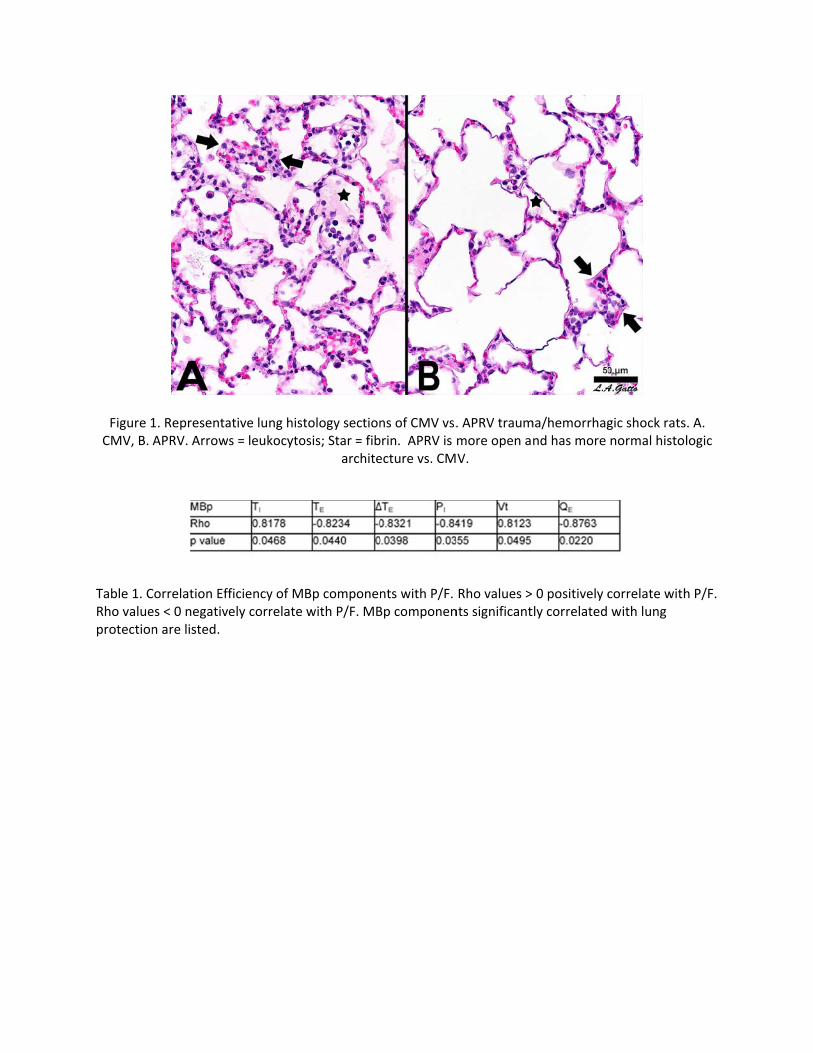
Figure 1CMV, B.
Table 1. CRho valueprotection
1. RepresentaAPRV. Arrow
Correlation Effes < 0 negativn are listed.
ative lung hists = leukocyto
ficiency of Mely correlate
tology sectionosis; Star = fib
archite
Bp componenwith P/F. MB
ns of CMV vs.brin. APRV is ecture vs. CM
nts with P/F. Bp componen
. APRV traummore open aV.
Rho values >nts significant
ma/hemorrhagnd has more
0 positively ctly correlated
gic shock ratsnormal histo
correlate withwith lung
s. A. logic
h P/F.
Scientific Posters
Poster 5
RESIDENT LEADERSHIP SKILLS DURING TRAUMA RESUSCITATIONS: AN EVALUATION USING IN‐SITU LOW FIDELITY SIMULATION
Benjamin H. Schnapp, MD, Kaushal H. Shah, MD*, Suzanne Bentley, MD The Mount Sinai Hospital
Presenter: Benjamin H. Schnapp, MD
Objectives: While leadership skills may come naturally to some residents, others may be less proficient. In a chaotic trauma resuscitation, feedback on these skills can be a low priority. We hypothesize that simulation and feedback in an in‐situ, low fidelity simulated trauma scenario will highlight areas for leadership skills improvement.
Methods: At an urban trauma center, we conducted a prospective observational study. The trauma team consisted of a third year resident leading two junior residents. The team was asked to proceed with the volunteer simulated MVC patient as with an actual patient, with study staff indicating the outcomes of procedures that were impossible to perform (e.g. IV placement, head CT). All medical staff present then evaluated the trauma leader on a 7‐point Likert scale for several leadership attributes.
Results: The simulation was run 7 times, with 7 different leaders. 64 evaluations were collected; not all evaluations were complete. Cases lasted approximately 20 minutes. Leaders rated an average of 5.39, 95% CI [5.01, 5.77] on “Identifies self as leader.” They rated 5.63, 95% CI [5.38, 5.89] on “Prioritizes acute issues.” On “Maintains a bird’s‐eye view,” they were rated 5.27, 95% CI [4.95, 5.60]. They scored 4.81, 95% CI [4.48, 5.13] on “Uses closed‐loop communication.” “Effective communication” was rated a 5.78, 95% CI [5.49, 6.06], while “Delegates tasks” was rated a 4.73, 95% CI [4.40, 5.05]. “Discusses differential” was rated 5.32, 95% CI [5.00, 5.64] and “Encourages safe environment” was scored a 5.02, 95% CI [4.71, 5.32].
Conclusions: Residents performed well in areas that residents already perform in the emergency department, such as “Prioritizes acute issues.” They performed weakest in skills unique to a leadership scenario, such as “Delegates tasks appropriately.” We will use these results to guide development of a tailored leadership curriculum for residents.

Notes
Scientific Posters
Poster 6
LEADERSHIP AND GENDER DURING ACUTE TRAUMA RESUSCITATION
Maureen McCunn, MD, MIPP, FCCM*, Breah L Paciotti, MPH, Mayur Narayan, MD, MPH, MBA, Jonathan Ziegert, PhD, Fran Barg, PhD
Hospital of the University of Pennsylvania
Presenter: Maureen McCunn, MD, MIPP, FCCM
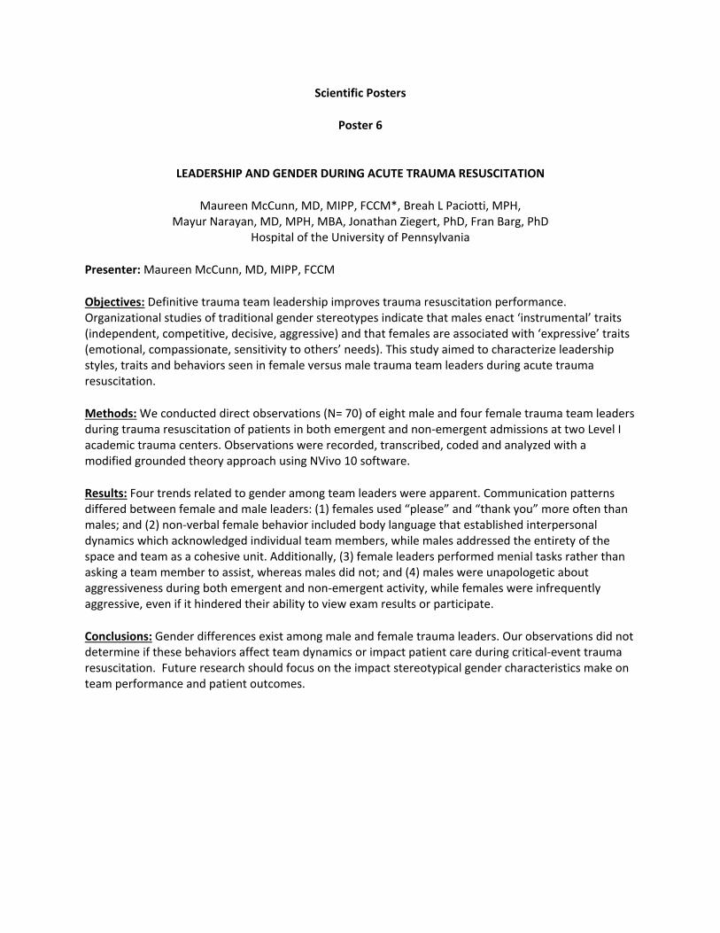
Objectives: Definitive trauma team leadership improves trauma resuscitation performance. Organizational studies of traditional gender stereotypes indicate that males enact ‘instrumental’ traits (independent, competitive, decisive, aggressive) and that females are associated with ‘expressive’ traits (emotional, compassionate, sensitivity to others’ needs). This study aimed to characterize leadership styles, traits and behaviors seen in female versus male trauma team leaders during acute trauma resuscitation.
Methods: We conducted direct observations (N= 70) of eight male and four female trauma team leaders during trauma resuscitation of patients in both emergent and non‐emergent admissions at two Level I academic trauma centers. Observations were recorded, transcribed, coded and analyzed with a modified grounded theory approach using NVivo 10 software.
Results: Four trends related to gender among team leaders were apparent. Communication patterns differed between female and male leaders: (1) females used “please” and “thank you” more often than males; and (2) non‐verbal female behavior included body language that established interpersonal dynamics which acknowledged individual team members, while males addressed the entirety of the space and team as a cohesive unit. Additionally, (3) female leaders performed menial tasks rather than asking a team member to assist, whereas males did not; and (4) males were unapologetic about aggressiveness during both emergent and non‐emergent activity, while females were infrequently aggressive, even if it hindered their ability to view exam results or participate.
Conclusions: Gender differences exist among male and female trauma leaders. Our observations did not determine if these behaviors affect team dynamics or impact patient care during critical‐event trauma resuscitation. Future research should focus on the impact stereotypical gender characteristics make on team performance and patient outcomes.

Notes
Scientific Posters
Poster 7
PREDICTIVE VALUE OF ADMISSION LACTATE IN PEDIATRIC TRAUMA
Rajesh Ramanathan, MD, Joseph Hartwich, MD, Jeffrey H. Haynes, MD Virginia Commonwealth University
Presenter: Rajesh Ramanathan, MD
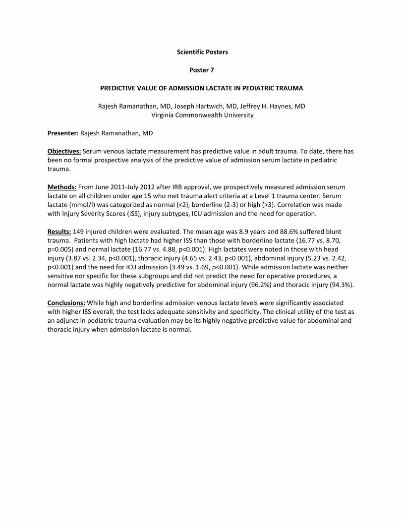
Objectives: Serum venous lactate measurement has predictive value in adult trauma. To date, there has been no formal prospective analysis of the predictive value of admission serum lactate in pediatric trauma.
Methods: From June 2011‐July 2012 after IRB approval, we prospectively measured admission serum lactate on all children under age 15 who met trauma alert criteria at a Level 1 trauma center. Serum lactate (mmol/l) was categorized as normal (<2), borderline (2‐3) or high (>3). Correlation was made with Injury Severity Scores (ISS), injury subtypes, ICU admission and the need for operation.
Results: 149 injured children were evaluated. The mean age was 8.9 years and 88.6% suffered blunt trauma. Patients with high lactate had higher ISS than those with borderline lactate (16.77 vs. 8.70, p=0.005) and normal lactate (16.77 vs. 4.88, p<0.001). High lactates were noted in those with head injury (3.87 vs. 2.34, p<0.001), thoracic injury (4.65 vs. 2.43, p<0.001), abdominal injury (5.23 vs. 2.42, p<0.001) and the need for ICU admission (3.49 vs. 1.69, p<0.001). While admission lactate was neither sensitive nor specific for these subgroups and did not predict the need for operative procedures, a normal lactate was highly negatively predictive for abdominal injury (96.2%) and thoracic injury (94.3%).
Conclusions: While high and borderline admission venous lactate levels were significantly associated with higher ISS overall, the test lacks adequate sensitivity and specificity. The clinical utility of the test as an adjunct in pediatric trauma evaluation may be its highly negative predictive value for abdominal and thoracic injury when admission lactate is normal.

Notes

Scientific Posters
Poster 8
SCHEDULING OF MIDLEVEL PROVIDERS AT LEVEL I TRAUMA CENTERS
Robert A. Myers, MSE, Pratik Parikh, PhD, Akpofure Peter Ekeh, MD*, Elizabeth Denlinger, RN, BSN, MSA, Mary McCarthy, MD, FACS
Wright State University
Presenter: Robert A. Myers, MSE
Objectives: Midlevel providers (MLPs), nurse practitioners and physician assistants, are essential to the provision of trauma care services, particularly in the wake of residency hour restrictions. The demand for these providers fluctuates with cyclical patient arrivals, however, most trauma teams continue to staff MLPs in a linear fashion. Failure to plan for variable arrivals may contribute to excessive patient wait times and emergency department overcrowding. The purpose of this study was to compare qualitative (e.g., visual) and quantitative (e.g., computer model) approaches to MLP staffing, and the impact on patient wait times and MLP cost.
Methods: A computer simulation model was developed to model the flow of trauma patients (Figure 1) and the associated availability of MLPs. The model was validated using 2010 data from a US Level I Trauma Center and interviews with center managers. The model was run for baseline, two “what‐if” schedules, and two additional schedules to minimize patient time in the emergency department.
Results: A visual overlay of MLP staffing on patient arrivals during 2010 indicated substantial times of resource mismatch, which prompted the trauma manager to add an additional MLP during weekday evenings. This resulted in a 14.8% increase in MLP work hours, and a 27% reduction in patient wait times. Using a simulation analysis of a schedule implemented in 2012 yielded a 10.5% increase in MLP hours, with a 73% reduction in wait times. We also delineated two additional schedule options resulting in a 78% reduction in wait time with no additional increase in MLP work hours (Figure 2).
Conclusions: Staffing of highly‐skilled and highly‐paid MLPs must be synchronized with cyclic trauma patient arrivals. Evaluating alternate shift times and assignments with the same staffing via quantitative methods, such as a computer simulation model, to best match patient arrival patterns can provide substantial benefits over visual approaches.

Figure 2Patient
2. Two New St Wait Time R
F
Schedules (BLReductions (D
Figure 1: Trau
L‐ReA and FS‐Dark line is ML
uma Patient F
Cov) with NoLP staffing lev
Flow Data
o Additional Avels, overlaid
ACP Hours and over hourly p
d Correspondpatient arriva
ding als)
Scientific Posters
Poster 9
INCIDENCE OF VITAMIN D DEFICIENCY IN THE ICU TRAUMA POPULATION
Joseph Ibrahim, MD*, Karen Safcsak, RN, Jennifer Rickerds, BS, Staci L. Belcher, Michael L. Cheatham, MD, FACS, FCCM*
Orlando Regional Medical Center
Presenter: Joseph Ibrahim, MD
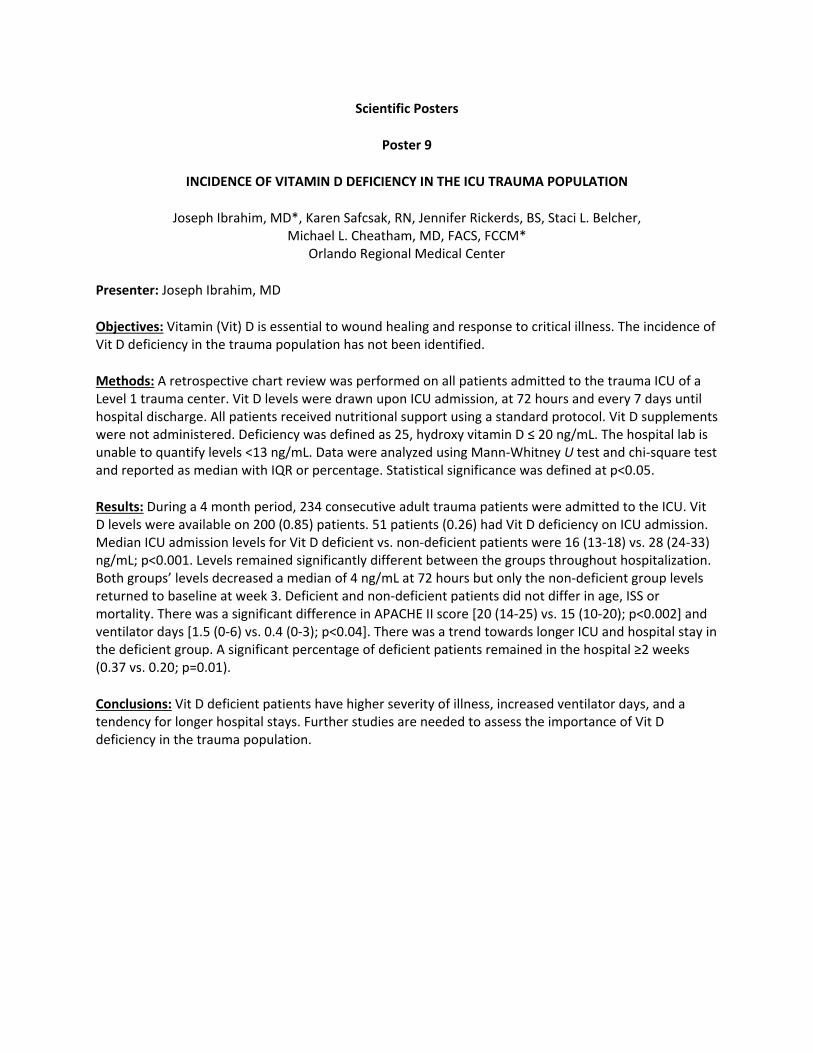
Objectives: Vitamin (Vit) D is essential to wound healing and response to critical illness. The incidence of Vit D deficiency in the trauma population has not been identified.
Methods: A retrospective chart review was performed on all patients admitted to the trauma ICU of a Level 1 trauma center. Vit D levels were drawn upon ICU admission, at 72 hours and every 7 days until hospital discharge. All patients received nutritional support using a standard protocol. Vit D supplements were not administered. Deficiency was defined as 25, hydroxy vitamin D ≤ 20 ng/mL. The hospital lab is unable to quantify levels <13 ng/mL. Data were analyzed using Mann‐Whitney U test and chi‐square test and reported as median with IQR or percentage. Statistical significance was defined at p<0.05.
Results: During a 4 month period, 234 consecutive adult trauma patients were admitted to the ICU. Vit D levels were available on 200 (0.85) patients. 51 patients (0.26) had Vit D deficiency on ICU admission. Median ICU admission levels for Vit D deficient vs. non‐deficient patients were 16 (13‐18) vs. 28 (24‐33) ng/mL; p<0.001. Levels remained significantly different between the groups throughout hospitalization. Both groups’ levels decreased a median of 4 ng/mL at 72 hours but only the non‐deficient group levels returned to baseline at week 3. Deficient and non‐deficient patients did not differ in age, ISS or mortality. There was a significant difference in APACHE II score [20 (14‐25) vs. 15 (10‐20); p<0.002] and ventilator days [1.5 (0‐6) vs. 0.4 (0‐3); p<0.04]. There was a trend towards longer ICU and hospital stay in the deficient group. A significant percentage of deficient patients remained in the hospital ≥2 weeks (0.37 vs. 0.20; p=0.01).
Conclusions: Vit D deficient patients have higher severity of illness, increased ventilator days, and a tendency for longer hospital stays. Further studies are needed to assess the importance of Vit D deficiency in the trauma population.

Notes

Scientific Posters
Poster 10
AGE‐RELATED OUTCOME FOLLOWING BLUNT TRAUMA: HOW OLD IS "OLD"?
Haniee Chung, MD, Stephanie Bonne, MD*, Julie L. Nash, RN, Douglas J.E. Schuerer, MD, FACS*, Grant V. Bochicchio, MD,MPH*, Robert D. Winfield, MD*
Washington University School of Medicine
Presenter: Haniee Chung, MD
Objectives: Current EAST practice management guidelines have established age 65 or greater as the definition of an elderly trauma patient; however, this age criterion is based on primary sources which contain varying age criteria for identification of the elderly patient. For purposes of triage and the provision of ongoing care to the geriatric trauma patient, we hypothesized that an age could be identified at which point outcome following trauma worsens, regardless of mechanism or comorbidities.
Methods: We evaluated all patients between the ages of 18 and 89 years admitted to our Level I Trauma Center presenting with a blunt mechanism of injury via either same level fall or motor vehicle collision between January 1, 2008 and December 31, 2012. Demographics, comorbidities, injury pattern and severity, interventions, and outcomes were reviewed. Patients were grouped by age for univariate analyses and multinomial logistic regression; a p value <0.05 was considered significant in all evaluations.
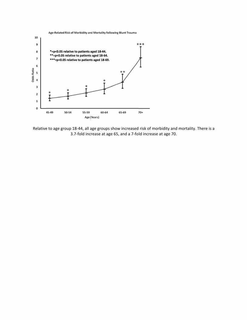
Results: 5,431 patients presented to our institution after sustaining blunt injury. Falls increased as a mechanism as patients aged, but injury severity scores remained similar across age groups. In our final logistic regression, we evaluated the effect of age on outcome, demonstrating that relative to blunt trauma victims between the ages of 18 and 44 years, all patient groups show an increased risk of morbidity and mortality. There was a 3.7‐fold increase in risk of morbidity and mortality at age 65 (p<0.001), and a 7‐fold increase at age 70 (p<0.001). Resource utilization measures were not significantly associated with age. Survival curves generated for the different age groups studied showed an increased mortality in the age>70 group with both early and late deaths.
Conclusions: Our data show that at age 65, there is a dramatic increase in mortality after blunt trauma, and support the use of age 65 as the definition of the elderly trauma patient.
Relative
e to age groupp 18‐44, all ag3.7‐fold incr
ge groups shoease at age 6
ow increased65, and a 7‐fo
risk of morbld increase at
idity and mort age 70.
rtality. There is a
Scientific Posters
Poster 11
ULTRASOUND MEASURED LEAN MUSCLE MASS LOSS IN SEVERE SEPSIS
Erin Vanzant, MD, Frederick Moore, MD, Rohit Patel, MD, Ruth J. Davis, RN, Jennifer Lanz, MSN, Lori F. Gentile, MD, Anatole Martin, PhD, PT, Tezcan Ozrazgat‐Baslanti, PhD, Azra Bihorac, MD, MS,
Christiaan Leeuwenburgh, PhD, Lawrence Lottenberg, MD*, Lyle L. Moldawer, PhD, Philip Efron, MD* University of Florida
Presenter: Erin Vanzant, MD
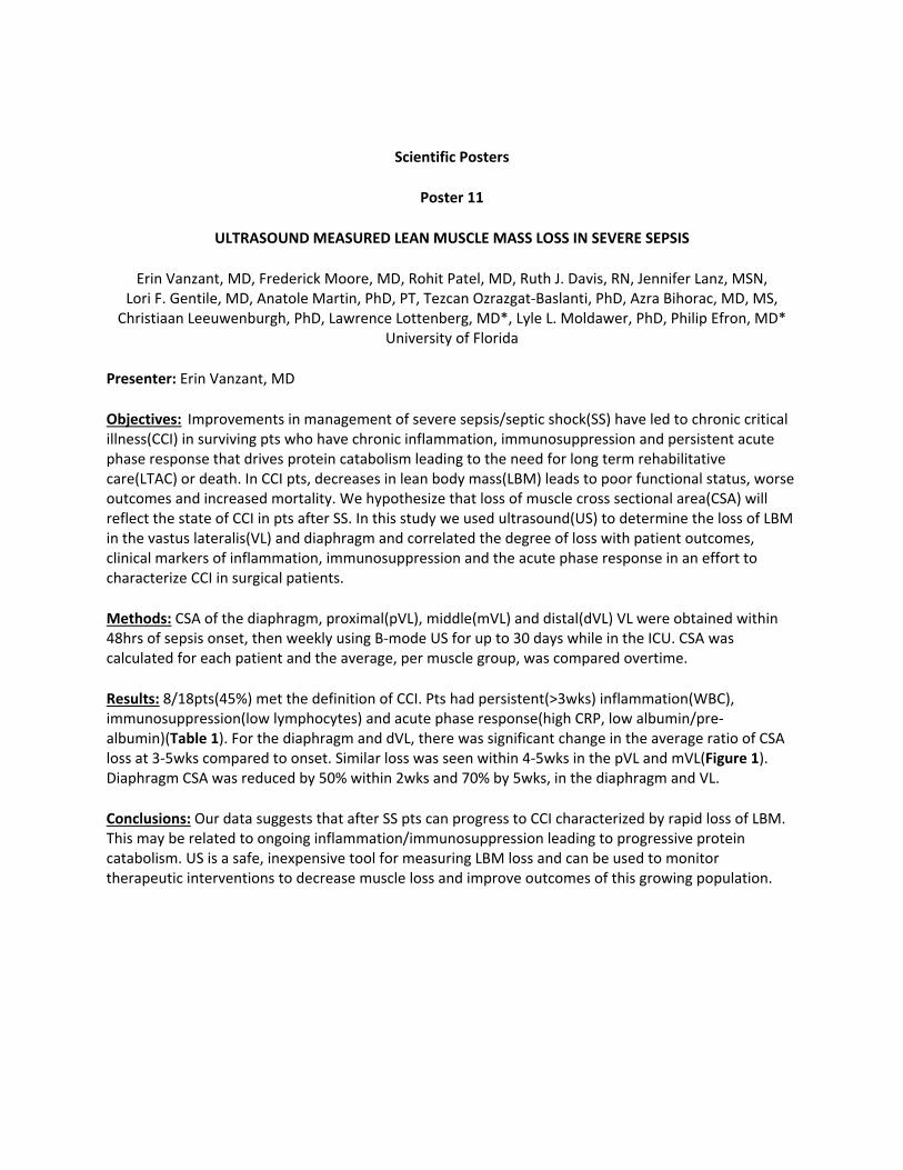
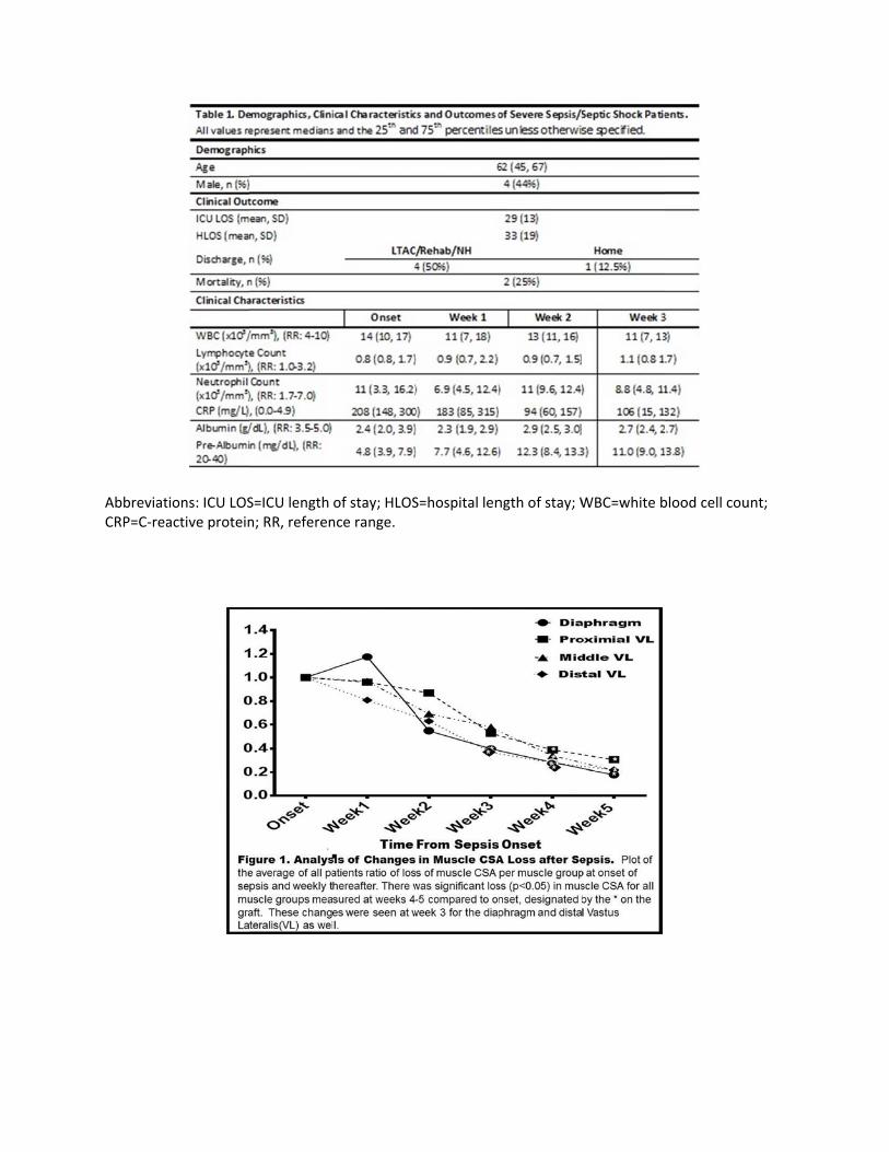
Objectives: Improvements in management of severe sepsis/septic shock(SS) have led to chronic critical illness(CCI) in surviving pts who have chronic inflammation, immunosuppression and persistent acute phase response that drives protein catabolism leading to the need for long term rehabilitative care(LTAC) or death. In CCI pts, decreases in lean body mass(LBM) leads to poor functional status, worse outcomes and increased mortality. We hypothesize that loss of muscle cross sectional area(CSA) will reflect the state of CCI in pts after SS. In this study we used ultrasound(US) to determine the loss of LBM in the vastus lateralis(VL) and diaphragm and correlated the degree of loss with patient outcomes, clinical markers of inflammation, immunosuppression and the acute phase response in an effort to characterize CCI in surgical patients.
Methods: CSA of the diaphragm, proximal(pVL), middle(mVL) and distal(dVL) VL were obtained within 48hrs of sepsis onset, then weekly using B‐mode US for up to 30 days while in the ICU. CSA was calculated for each patient and the average, per muscle group, was compared overtime.
Results: 8/18pts(45%) met the definition of CCI. Pts had persistent(>3wks) inflammation(WBC), immunosuppression(low lymphocytes) and acute phase response(high CRP, low albumin/pre‐albumin)(Table 1). For the diaphragm and dVL, there was significant change in the average ratio of CSA loss at 3‐5wks compared to onset. Similar loss was seen within 4‐5wks in the pVL and mVL(Figure 1). Diaphragm CSA was reduced by 50% within 2wks and 70% by 5wks, in the diaphragm and VL.
Conclusions: Our data suggests that after SS pts can progress to CCI characterized by rapid loss of LBM. This may be related to ongoing inflammation/immunosuppression leading to progressive protein catabolism. US is a safe, inexpensive tool for measuring LBM loss and can be used to monitor therapeutic interventions to decrease muscle loss and improve outcomes of this growing population.
AbbreviatCRP=C‐rea
tions: ICU LOSactive protein
S=ICU length n; RR, referen
of stay; HLOSnce range.
S=hospital len
ngth of stay; WWBC=white b
blood cell couunt;

Scientific Posters
Poster 12
YOU CAN’T GO HOME: ROUTINE CONCUSSION EVALUATION IS NOT ENOUGH
M. Chance Spalding, DO, PhD, Jennifer L. Hartwell, MD, Brian Fletcher, MS, RN, ACNP‐BC Grant Medical Center
Presenter: M. Chance Spalding, DO, PhD
Objectives: Traumatic brain injury affects almost 2 million people in the US each year with 75% classified as mild traumatic brain injury (MTBI), or concussion. Traditional care is to allow these patients to go home from the Emergency Department (ED) if their GCS is 15 and they have a normal head CT. However, this does not address short‐term deficits or the benefits of early screening and treatment. Our hypothesis is that a notable percentage of patients with concussive symptoms will need outpatient neurocognitive therapy despite a reassuring initial evaluation.
Methods: This is a single institution retrospective review of patients suffering from MTBI at an urban level 1 trauma center between August 2010 and December 2011. Inclusion criteria were age 14 years or older with a diagnosis of MTBI, GCS of 15, negative head CT, a neurocognitive evaluation within 48 hours, blunt mechanism, and no confounding psychiatric comorbidities.
Results: 6032 trauma patients were seen during the study period. 905 patients had MTBI and a negative CT head. 396 met inclusion criteria. Average age was 38 years (range 14‐93), 64% male, with a mean ISS of 8.2. 41% were cleared for discharge without any follow up or supervision. 25% required outpatient therapy. 3% were unsafe for discharge home. Of those cleared for discharge home, 88% had positive or questionable loss of consciousness (LOC) and 81% requiring additional therapy had positive or questionable LOC (p=0.20). Age, gender, ISS, and alcohol use were compared between the groups and were found to not be significantly different.
Conclusions: A surprisingly high percentage (28%) of our patients who would have met traditional criteria for discharge from the ED after MTBI required ongoing therapy. LOC was an unreliable predictor of clearance for discharge home. We provide evidence to suggest that premature discharge places patients with MTBI at risk for not being referred for appropriate neurocognitive therapy.

Resuults Summary
y

Scientific Posters
Poster 13
PROMPT AND RAPID ENDOVASCULAR STRATEGIES IN TRAUMA OCCASIONS (PRESTO): INTERVENTIONAL RADIOLOGY FOR HEMODYNAMICALLY UNSTABLE PATIENTS
Motoyuki Miykata, Kohei Morimoto, MD,PhD, Brandon Lohman, Junichi Matsumoto, MD, Yasuhiko Taira, MD,PhD
National Hospital Organization Disaster Medical Center
Presenter: Motoyuki Miykata
Objectives: Application of interventional radiology (IR) in hemodynamically unstable trauma patients have been limited in spite of the fact that there were many papers about utility of IR for bleeding in trauma. This is because of lack of time‐conscious endovascular strategies and poor availability of IR team in time, and appropriate strategy enables to extend application of IR even in hemodynamically unstable patients. The purpose of this presentation is to evaluate utility of aggressive application of IR with time‐conscious strategy; Prompt and Rapid Endovascular Strategy in Trauma Occasions (‘PRESTO’) in unstable patients.
Methods: We retrospectively identified hemodynamiccally unstable blunt trauma patients (2010 to 2012) who underwent IR for multiple hemorrhages. All subjects underwent the PRESTO protocol: IR team activated with pre‐hospital information, immediate CT scanning, Pre‐Procedural‐Planning with MDCT data, femoral sheath insertion in the ER, and embolization within a tentative embolization time per artery, which all fall under our institution’s new concept of damage control interventional radiology.
Results: 17 patients with severe blunt trauma underwent IR were retrospectively reviewed. Subjects mean age was 35.2 years and all indicated a Shock Index >1.0 with a mean ISS of 36.17. The average time to control bleeding was 50.53 minutes. The average number of embolized arteries and time for a single artery embolization was 5.1 arteries and 10.72 minutes respectively. Three subjects died within 24 hours from severe brain damage (GCS<8 on admission). None of them died from IR failure.
Conclusions: This study demonstrates that when delivered aggressively and in a predefined fashion, the novel paradigm of PRESTO is effective to control traumatic bleeding in hemodynamically unstable patients.
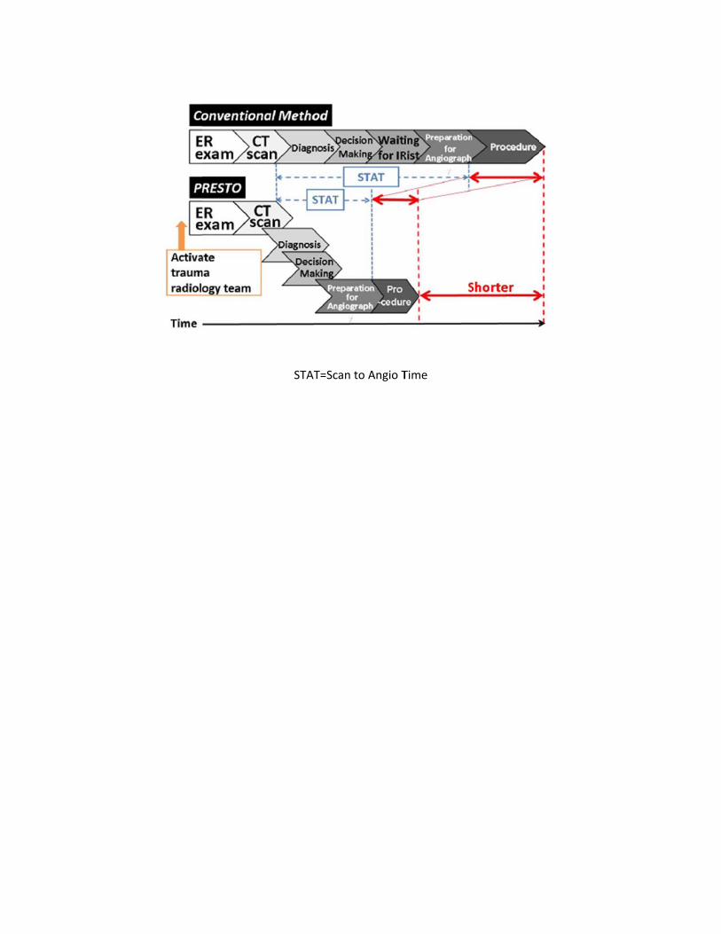
STAT=Sccan to Angio T
Time

Scientific Posters
Poster 14
FACTORS IMPACTING TIME TO INITIATE TRANSFER IN PEDIATRIC TRAUMA
Thomas J. Desmarais, BS, Michael Wallendorf, PhD, Adam M. Vogel, MD*, Pamela Choi, MD, Martin Keller, MD*
Washington University School of Medicine
Presenter: Thomas J. Desmarais, BS
Objectives: We sought to determine factors influencing time to initiate Level I Pediatric Trauma Center (PTC) transfer to identify targets for educational outreach and trauma system improvement.
Methods: Records of all children (≤17 years) transferred to our PTC over a 2.5 year period requiring trauma activation were reviewed. All children met CDC and state triage criteria for transfer to the highest level of trauma care. Linear regression and analysis of variance were used to assess the relationship between the time to initiate transfer and patient age, race, insurance, mechanism, referring hospital (RH) designation, transport mode, distance (RH to PTC), pediatric trauma score, time to obtain computed tomography (CT) at the RH, and injury severity score (ISS). Variables determined to be significant (p < 0.05) were included in a stepwise multiple regression to control for confounding. R2 was used to assess quality of model.
Results: Between 1/1/10 and 6/30/12, 101 patients were transferred and met CDC criteria, most frequently with Glasgow coma scale ≤13. Median age was 13 (IQR 5.5 ‐ 16), median ISS 10 (IQR 4 ‐ 21), and median time to initiate transfer 28 minutes (IQR 28.5 ‐ 91.5). Age, distance from the RH to the PTC, time to obtain CT at the RH, and ISS all were significantly related to the time to initiate transfer and included in the multivariate model. Only age and time to obtain CT at the RH remained significant after inclusion. The adjusted R2 was 0.621, indicating 62.1% of the variability in the time to initiate transfer was explained by the model. Linear regression with time to obtain CT at the RH as the sole variable had R2 of .593.
Conclusions: Delays in obtaining CT at RHs had the greatest influence on time to initiate transfer for children meeting criteria on presentation. Further educational activities directed at helping health care professionals recognize roles and improve trauma system throughput is warranted.

Notes
Scientific Posters
Poster 15
OUTCOMES OF COMPLETE VERSUS PARTIAL FLAIL CHEST STABILIZATION
Terry P. Nickerson, MD, Brian D. Kim, MD*, Henry J. Schiller, MD*, Martin D. Zielinski, MD, FACS*, Donald H. Jenkins, MD*
Mayo Clinic
Presenter: Terry P. Nickerson, MD
Objectives: There has been increasing acceptance of surgical rib fracture stabilization for flail chest. We attempt to repair all fractures associated with a flail segment. Difficulty in accessing anatomically remote portions of flail segments due to overlying bony and muscular tissue, however, can prevent full rib stabilization leaving some fractures unaddressed. We hypothesized partial flail segment stabilization (PFS) had similar outcomes compared to complete flail segment stabilization (CFS).
Methods: A review of patients who underwent surgical rib stabilization for flail chest from Aug. 2009–Feb. 2013 was performed. PFS was defined as one or more flail segments that were partially repaired while CFS was defined by all flail segments undergoing full fixation. A p–value < 0.05 was considered statistically significant.
Results: Forty three patients were identified (median 61 years [30‐85]) with a median follow up of 189 days [9‐472]. 84% of patients were followed beyond six months. We found no difference in age, sex, operating time, ISS, ICU or hospital duration of stay, pneumonia, or narcotic utilization at one month between (table). Total Lung Capacity (TLC) was significantly improved in the CFS group at 3 months (90% predicted vs 72% predicted, p=0.02) and 6 months (94% predicted vs 75% predicted, p=0.038) but with no difference in other measures of pulmonary function at 3, 6 and 12 months. No patient had a significant chest wall deformity. No reoperations were required for incomplete repair in the PFS patient cohort.
Conclusions: Despite advances in surgical technique, not all flail segment rib fractures are accessible for repair via standard thoracotomy. There were no differences in clinical outcomes in patients who underwent PFS versus CFS save for temporally limited TLC. Our data suggest that PFS is acceptable and that extending or creating additional incisions for exposure for CFS is unwarranted.
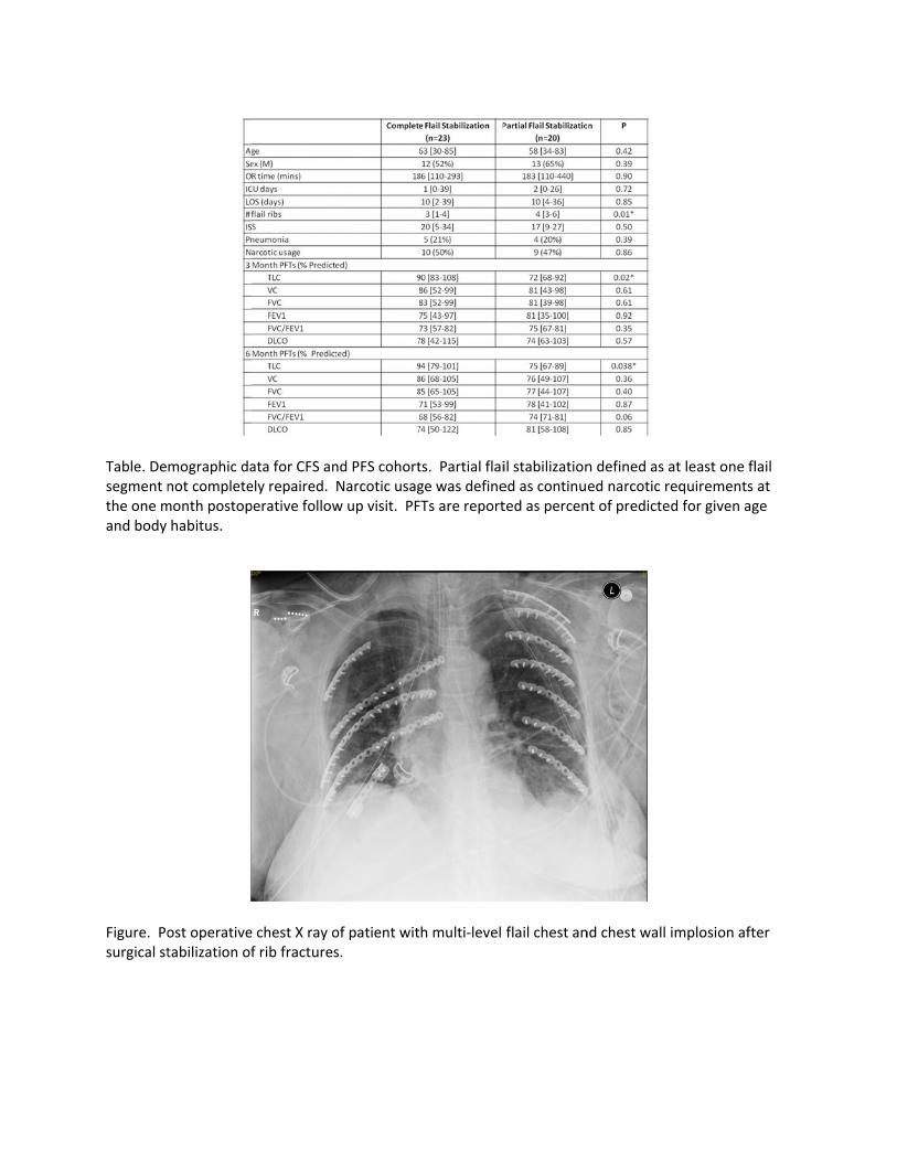
Table. Desegment nthe one mand body
Figure. Posurgical st
mographic danot completemonth postophabitus.
ost operativetabilization of
ata for CFS anely repaired. perative follow
e chest X ray of rib fractures
nd PFS cohortNarcotic usagw up visit. PF
of patient wits.
ts. Partial flaige was defineFTs are report
h multi‐level
il stabilizationed as continuted as percen
flail chest an
n defined as aed narcotic rent of predicted
nd chest wall i
at least one flequirements d for given ag
implosion aft
lail at ge
ter
Scientific Posters
Poster 16
ANTICOAGULATION AND TRAUMA – A NEMESIS FOR TRAUMA SURGEONS AND A PROTOCOL TO COMBAT IT: THE ACT ALERT
Amelia Rogers, BS, Michael Horst, PhD, Elizabeth H. Clark, Katherine Bupp, Katelyn Rittenhouse, BS,
William Adams, MD, Weston Shertzer, Frederick Rogers, MD, MS, FACS* Lancaster General Health
Presenter: Frederick Rogers, MD, MS, FACS
Objectives: In busy EDs, elderly patients on anticoagulation (AC) who sustain low impact falls can be relegated to a lower priority for evaluation with potential catastrophic outcomes. We sought to determine if an ED activation system would prioritize workup and improve outcome.
Methods: In a PA‐verified Level II trauma center with>107,000 ED admissions/yr, the ACT Alert (AntiCoagulation and Trauma) was implemented in Mar2012. Triage parameters include: age>65, AC/antiplatelet agents, GCS≥13, possible LOC, and fall≤24 hrs. ACT alerts are announced overhead in the ED and must be assessed by an ED physician, nurse, and phlebotomist in≤15min. They must have a Point of Care INR in≤20min and head CT scan in≤30min. Positive CT findings mandate trauma service be contacted. Univariate analysis was used to compare ACT Alert patients from Mar‐Dec2012 to patients>65yrs on anticoagulants with the same chief complaints as ACT admitted to the ED from Jun2011‐Feb2012 (control). A p‐value≤0.05 was considered significant.
Results: Of 860 study patients, 426 (49.5%) were ACT and 434 (50.5%) were control. There was no significant difference in mean age between groups (ACT 81.6yrs vs. Control 82.0yrs; p=0.48). Of admitted patients, ACT had significantly fewer with a hospital length of stay (LOS)>5days (ACT 38.1% vs. Control 51.8%; p<0.001). Significantly more ACT patients were discharged directly from ED (ACT 55.6% vs. Control 30.2%; p<0.001). Of patients not admitted, ACT were discharged significantly more rapidly than control (p=0.001).
Conclusions: The ACT Alert improves ED throughput and hospital LOS while identifying at‐risk, mild head injured geriatric patients on AC. There was a trend towards improved time to definitive care and survival. A method of rapid ED evaluation for minimally head injured patients without the involvement of trauma service decreases both undertriage and overtriage in this high risk population.

Univaariate Model of ED Outcommes of ACT Al
lert vs. Controol Population
ns
Scientific Posters
Poster 17
NATURAL HISTORY & CLINICAL IMPLICATIONS OF NONDEPRESSED SKULL FRACTURE IN YOUNG CHILDREN
Saif F. Hassan, MD, , Stephen Cohn, MD*, Oliver Nunez‐Cantu, MD, Yousef Arar, John Myers, MD, Daniel Dent, MD, Brian J. Eastridge, MD*, Deborah Mueller, MD, Mark Gunst, MD,
Ramon Cestero, MD, Helen Markowski, MD, Lillian Liao, MD, MPH* University of Texas Health Science Center San Antonio
Presenter: Saif F. Hassan, MD
Objectives: Head injury is the most common cause of neurologic disability & mortality in children. Prior studies have demonstrated that depressed skull fractures represent 15‐25% of all skull fractures in children & approximately 7‐10% of total hospital admissions after head injury. In the current analysis, we hypothesized that nondepressed skull fractures in the pediatric population were not associated with adverse neurologic outcomes
Methods: We reviewed the medical records of all children ≤ 5 years old with skull fracture (SF) who presented to our level I trauma center from 2007 through 2010. Data collected included patient demographics, GCS upon admission, level of consciousness at the time of injury, type of SF depressed (DSF) vs nondepressed (NDSF), magnitude of depression of DSF (0‐0.5; 0.5‐1.0; >1.0cm), evidence of neurologic deficit, & the requirement for neurosurgical intervention
Results: 1546 injured children were evaluated during the study period. From this cohort, 563 had isolated head injury and 223 of them had only SF. Of the SF group, 163 (73%) were NDSFs of which 128 (78%) presented with GCS of 15. None of the NDSF GCS 15 patients required neurosurgical intervention or developed any neurodeficit. 60 (27%) children had DSFs, of which 35 presented with GCS of 15 (58%). Likewise, none of DSF GCS 15 developed any long‐term neurodeficit. Neurologic deficit was common in DSF with GCS <15 at presentation & was associated with magnitude of depression (0‐0.5 n=5/43 [12%]; 0.5‐1.0 n=2/8 [25%]; >1.0cm n=9/9 [100%]) In this group, 42% required neurosurgical intervention & depression >1.0 cm was associated with a 55% mortality rate
Conclusions: Children ≤5 years old with nondepressed skull fractures & normal neurologic examination on admission do not develop neurologic deterioration. Children with isolated nondepressed skull fractures can be safely discharged from the emergency room after a period of observation

Notes

Scientific Posters
Poster 18
ALPHA GLUTATHIONE S‐TRANSFERASE (ΑGST): A NOVEL END POINT SERUM MARKER IN TRAUMATIC SHOCK?
Morgan P. McMonagle, MB, BCh, BAO, MD, FRCSI*, Daniel N. Holena, MD*, Nikolai S. Tolstoy, BS, Patrick M. Reilly, MD*, C. William Schwab, MD*, Carrie A. Sims, MD*
Hospital of the University of Pennsylvania
Presenter: Morgan P. McMonagle, MB, BCh, BAO, MD, FRCSI
Objectives: GST belongs to a family of cytosolic proteins involved in cell protection.Having previously described the potential use of aGST as an endpoint in the management of decompensated shock and resuscitation in an animal model, we tested its utility in a clinical setting of trauma.
Methods: longitudinal identification of patients in the trauma bay (TB) and followed over 48hrs and data collected prospectively including other endpoints of resuscitation (BP, pulse, shock index (SI), lactate, base excess (BE), pH and Hb), blood and fluid requirements, ISS scores as well as clinical outcomes (mortality, length of stay, days ventilated, morbidities). Serum aGST was measured using a commercially available enzyme immunoassay (ELISA) kit at specific time points (TB, ICU, 8, 16 and 24 and 48 hours). Data was analyzed using Mann‐Whitney and Kruskal‐Wallis tests for non‐parametric data and Pearson correlation with a significance set at 5% throughout.
Results: 33 (88% male) patients were recruited with a mean age 33.2 (SD 12.8) of which 82% had penetrating injury. There were no deaths in our study. aGST levels were highest for the most severely injured patients displaying a significant correlation with ISS (p=0.03). aGST seemed to correlate best in the first 24 hours after trauma and in particular levels measured in the TB, ICU and at 8 hours post‐admission. Although aGST levels were not strongly correlated with HR and BP, higher levels were associated with higher transfusion requirements (P=0.001), higher crystalloid requirements (P=0.005) suggesting that perhaps it is a more sensitive marker of shock and adequacy of resuscitation.
Conclusions: aGST is a sensitive marker of haemorrhagic ischemia with higher levels reflecting injury severity and may be useful endpoint resuscitation in conjunction with other biochemical markers. Further research is needed to further elucidate its role in the management of hemorrhagic shock.

Notes
Scientific Posters
Poster 19
RE‐INITATION OF WARFARIN IN MEDICARE BENEFICIARIES SURVIVING TRAUMATIC INTRACRANIAL HEMORRHAGE
Courtney E. Collins, MD, Heena P. Santry, MD, MS, BA*, Elan R. Witkowski, MD, MS, Julie Flahive, M.S., Fred Anderson Jr., PhD
University of Massachusetts
Presenter: Courtney E. Collins, MD
Objectives: To determine the rate and timing of warfarin re‐initiation among older Americans who suffered intra‐cranial hemorrhage (ICH) while on warfarin
Methods: We reviewed a 5% random sample of Medicare beneficiaries (2009‐2010). Patients treated for ICH while on warfarin were identified by ICD‐9 diagnosis codes (Part A claims) and >2 warfarin prescriptions filled <60 days before the index hospitalization for head injury (Part D prescription drug claims). Post discharge warfarin (PDW) was defined a warfarin prescription filled at any point in the study period after discharge. We compared demographic characteristics, co‐morbidities, injury patterns, and index hospitalization outcomes (length of stay (LOS), ICU‐LOS, ICU admission) between patients treated with and without PDW using univariate tests of association. Multivariable models were used to determine predictors of PDW.
Results: We identified 388 patients suffering traumatic ICH while on warfarin during our study period. 345 (88%) survived to discharge. 85 survivors (25%) were started on PDW an average of 55 days (SD 68) after discharge with 41% of prescriptions filled within 30 days of discharge. Age (~82 years), gender (~70% female), and Elixhauser index (~4) were similar across all patients. Patients prescribed PDW had longer index hospital LOS (7 vs. 5 days, p<0.01) and higher rates of ICU care (44% vs. 32% p value 0.05) compared to non‐PDW patients. In multivariable analyses only long bone fracture was a significant predictor of PDW. PDW patients were more likely to be admitted for recurrent injury (35% vs. 19%, p<0.01).
Conclusions: Age and co‐morbidities do not appear to be considerations for warfarin re‐initiation after traumatic ICH even though higher rates of recurrent trauma after re‐initiation may yield additional adverse outcomes. Prescribing warfarin after ICH should be approached cautiously.

Notes
Scientific Posters
Poster 20
TRANSFORMING HEMOGLOBIN MEASUREMENT IN TRAUMA PATIENTS: NON‐INVASIVE SPOT CHECK HEMOGLOBIN
Bellal Joseph, MD*, Hassan Aziz, MD, Viraj Pandit, MD, Narong Kulvatunyou, MD*, Andrew L. Tang, MD*, Julie L. Wynne, MD, MPH*, Terence O'Keeffe, MD, MSPH*,
Gary A. Vercruysse, MD*, Randall S. Friese, MD*, Peter Rhee, MD, MPH* The University of Arizona
Presenter: Bellal Joseph, MD
Objectives: Studies have reported poor correlation between continuous non‐invasive hemoglobin(Hgb) devices with invasive Hgb in trauma patients. Advancements in technology have allowed for a Spot‐check non‐invasive Hgb measurement. The aim of our study was to assess non‐invasive Spot‐check Hgb measurement using the Pronto‐7® Pulse Co‐Oximeter in trauma patients.
Methods: We performed a prospective cohort analysis of all trauma patients presenting to our level 1 trauma center. Invasive(IHgb) and Spot‐check hemoglobin measurements were obtained simultaneously upon presentation of the patient. Spot‐check was measured three times for each invasive Hgb value. We defined normal Hgb as >8mg/dL. Spearmen and Bland‐Altman correlation plot analysis was performed.
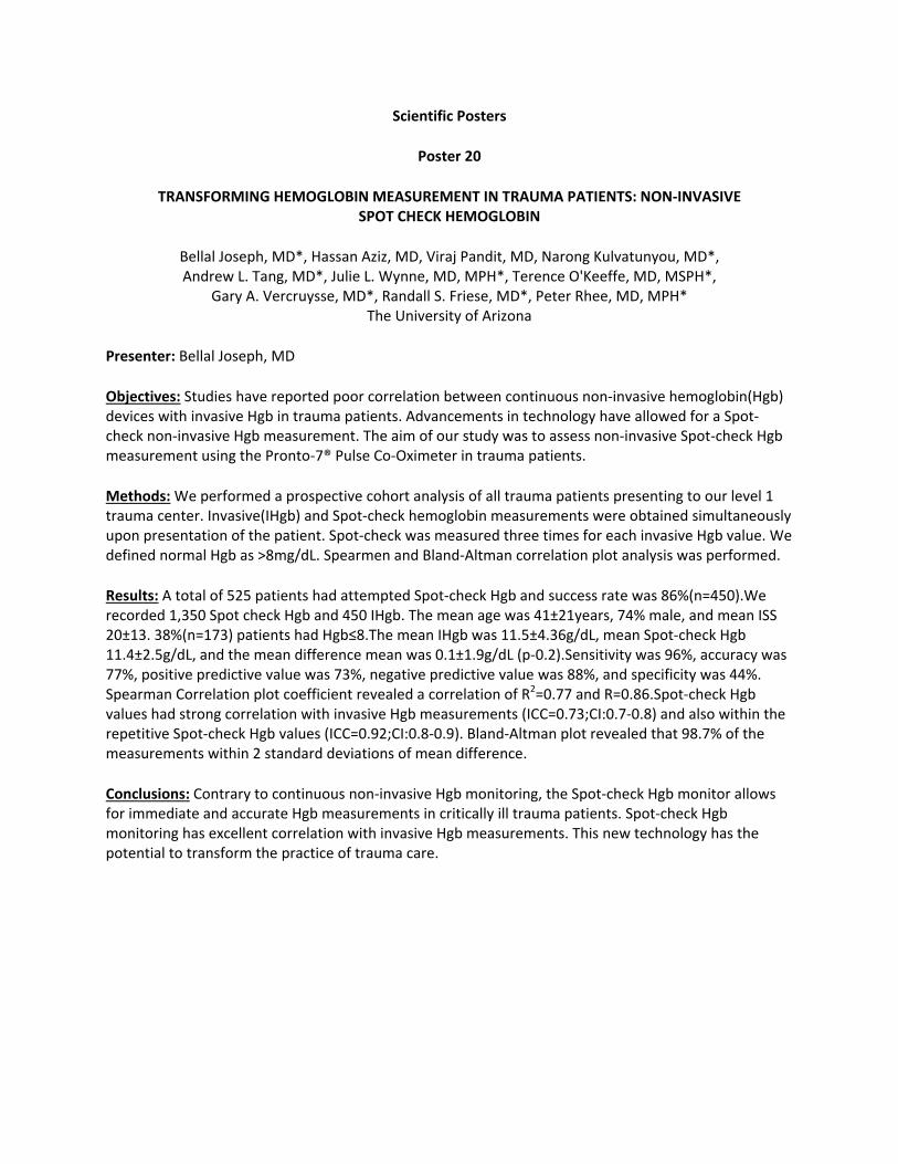
Results: A total of 525 patients had attempted Spot‐check Hgb and success rate was 86%(n=450).We recorded 1,350 Spot check Hgb and 450 IHgb. The mean age was 41±21years, 74% male, and mean ISS 20±13. 38%(n=173) patients had Hgb≤8.The mean IHgb was 11.5±4.36g/dL, mean Spot‐check Hgb 11.4±2.5g/dL, and the mean difference mean was 0.1±1.9g/dL (p‐0.2).Sensitivity was 96%, accuracy was 77%, positive predictive value was 73%, negative predictive value was 88%, and specificity was 44%. Spearman Correlation plot coefficient revealed a correlation of R2=0.77 and R=0.86.Spot‐check Hgb values had strong correlation with invasive Hgb measurements (ICC=0.73;CI:0.7‐0.8) and also within the repetitive Spot‐check Hgb values (ICC=0.92;CI:0.8‐0.9). Bland‐Altman plot revealed that 98.7% of the measurements within 2 standard deviations of mean difference.
Conclusions: Contrary to continuous non‐invasive Hgb monitoring, the Spot‐check Hgb monitor allows for immediate and accurate Hgb measurements in critically ill trauma patients. Spot‐check Hgb monitoring has excellent correlation with invasive Hgb measurements. This new technology has the potential to transform the practice of trauma care.
Spearman Correlation PPlot: Invasive
and Spot che
eck Hgb
Scientific Posters
Poster 21
INTERVAL CHOLECYSTECTOMY AFTER CHOLECYSTOSTOMY TUBE PLACEMENT: FACTORS INFLUENCING OUTCOME
Mohammad A. Khasawneh, MBBS, Andrea Shamp, PA, Stephanie Heller, MD*, Martin D. Zielinski, MD, FACS*, Donald H. Jenkins, MD*, John B. Osborn, M.Sc.*, David S. Morris, MD*
Mayo Clinic
Presenter: Mohammad A. Khasawneh, MBBS
Objectives: Interval cholecystectomy (CC) after percutaneous cholecystostomy tube (PCT) placement is the definitive treatment for cholecystitis for patients who are operative candidates after optimization of medical comorbidities. It is not clear, however, which patients will be able to have a laparoscopic CC after PCT placement. We aimed to identify factors associated with successful laparoscopic CC in these patients.
Methods: Review of patients who had a PCT from 2009 to 2011. Patient’s baseline demographics, clinical data, and outcomes were analyzed. Univariable and multivariable comparisons were performed between patients who did and did not undergo interval CC. A subgroup analysis of patients who had laparoscopic CC and open CC was performed. Data is presented as percentages, medians with interquartile ranges, or odds ratios with 95% C.I. as appropriate.
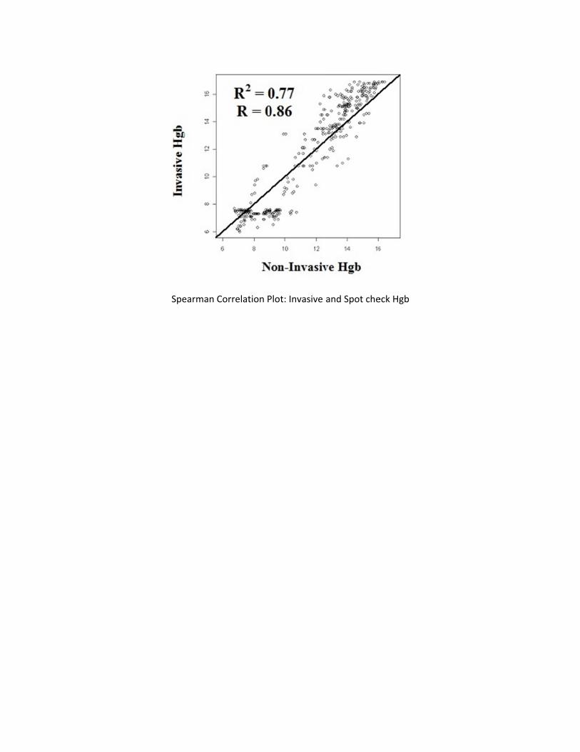
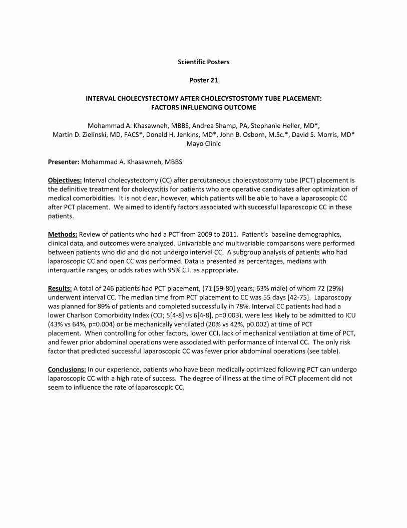
Results: A total of 246 patients had PCT placement, (71 [59‐80] years; 63% male) of whom 72 (29%) underwent interval CC. The median time from PCT placement to CC was 55 days [42‐75]. Laparoscopy was planned for 89% of patients and completed successfully in 78%. Interval CC patients had had a lower Charlson Comorbidity Index (CCI; 5[4‐8] vs 6[4‐8], p=0.003), were less likely to be admitted to ICU (43% vs 64%, p=0.004) or be mechanically ventilated (20% vs 42%, p0.002) at time of PCT placement. When controlling for other factors, lower CCI, lack of mechanical ventilation at time of PCT, and fewer prior abdominal operations were associated with performance of interval CC. The only risk factor that predicted successful laparoscopic CC was fewer prior abdominal operations (see table).
Conclusions: In our experience, patients who have been medically optimized following PCT can undergo laparoscopic CC with a high rate of success. The degree of illness at the time of PCT placement did not seem to influence the rate of laparoscopic CC.
Table. Mucholecyst
ultivariable reectomy
egression: Feaatures associaated with cho
olecystectomyy and laparos
scopic
Scientific Posters
Poster 22
GENDER DISPARITY IN VENTILATOR‐ASSOCIATED PNEUMONIA FOLLOWING TRAUMA: IDENTIFYING RISK FACTORS FOR MORTALITY
John P. Sharpe, MD, MS, Louis J. Magnotti, MD*, Jordan A. Weinberg, MD*, Thomas J. Schroeppel, MD*, Ben L. Zarzaur, MD, MPH*, Timothy C. Fabian, MD*, Martin A. Croce, MD*
University of Tennessee Health Science Center ‐ Memphis
Presenter: John P. Sharpe, MD, MS
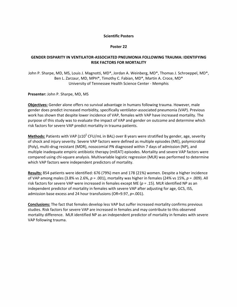
Objectives: Gender alone offers no survival advantage in humans following trauma. However, male gender does predict increased morbidity, specifically ventilator‐associated pneumonia (VAP). Previous work has shown that despite lower incidence of VAP, females with VAP have increased mortality. The purpose of this study was to evaluate the impact of VAP and gender on outcome and determine which risk factors for severe VAP predict mortality in trauma patients.
Methods: Patients with VAP (≥105 CFU/mL in BAL) over 8 years were stratified by gender, age, severity of shock and injury severity. Severe VAP factors were defined as multiple episodes (ME), polymicrobial (Poly), multi‐drug resistant (MDR), nosocomial PN diagnosed within 7 days of admission (NP), and multiple inadequate empiric antibiotic therapy (mIEAT) episodes. Mortality and severe VAP factors were compared using chi‐square analysis. Multivariable logistic regression (MLR) was performed to determine which VAP factors were independent predictors of mortality.
Results: 854 patients were identified: 676 (79%) men and 178 (21%) women. Despite a higher incidence of VAP among males (3.8% vs 2.6%, p = .001), mortality was higher in females (24% vs 15%, p = .009). All risk factors for severe VAP were increased in females except ME (p = .15). MLR identified NP as an independent predictor of mortality in females with severe VAP after adjusting for age, GCS, ISS, admission base excess and 24 hour transfusions (OR=9.97, p=.001).
Conclusions: The fact that females develop less VAP but suffer increased mortality confirms previous studies. Risk factors for severe VAP are increased in females and may contribute to this observed mortality difference. MLR identified NP as an independent predictor of mortality in females with severe VAP following trauma.

Notes
Scientific Posters
Poster 23
THE CLINICAL EVALUATION OF ALCOHOL INTOXICATION IS INACCURATE IN TRAUMA PATIENTS
Travis L. Holloway, MD, Ashwini Kumar, MD, Gregory G. Goodwiler, MPAS, PA‐C, Kristin Schlather, MPAS, PA‐C, Rick Sambucini, RN, Rachelle B. Jonas, RN, BSN, Stephen Cohn, MD*
University of Texas Health Science Center San Antonio
Presenter: Travis L. Holloway, MD
Discussant:
Objectives: Many patients brought to trauma centers are legally intoxicated (LI) and many do not have injuries that warrant admission. There is a wide range of alcohol clearance rates reported and centers vary in their discharge protocol of determining sobriety prior to safe hospital discharge. We hypothesized that a hand‐held alcohol breath analyzer would provide an accurate and easy method of determining legal sobriety in a trauma setting.
Methods: Twenty LI patients brought to our trauma center were prospectively enrolled. Serial breath samples ($350 initial, 40 cents/sample) were obtained using an Intoximeter® AlcoSensor FST as a surrogate measure of repeated blood alcohol levels (BALs). A clinical exam (nystagmus, one‐leg balance, heel‐toe walk) was performed prior to each breath sampling.
Results: The enrollees were 85% male, 30±9.6 (range 19‐51) years old, with BMI of 28.9±7.1. The average initial BAL was 245±61.2 (range 162‐370) mg/dL. Based on breath samples, the alcohol elimination rates varied from 21.5 mg/dL/hr to 45.7 mg/dL/hr (average 28.5 mg/dL/hr). There were no significant differences in alcohol elimination rate by gender, age, or BMI. The clinical exam also varied widely among patients; only 7 of 16 (44%) LI patients demonstrated horizontal nystagmus (suggesting sobriety when actually LI) and the majority of the LI patients (66%) were able to complete the balance tasks (suggesting sobriety).
Conclusions: Given the wide range of alcohol elimination rates and inaccuracy of clinical exam, a portable alcohol breath analyzer has the potential to easily and inexpensively establish legal sobriety in the trauma population.

Notes

Scientific Posters
Poster 24
UTILITY OF TEG/PLATELET MAPPING IN SDH PATIENTS
Alex Axelrad, MD, FACS,FCCM*, Gabriel E. Ryb, MD, MPH, FACS*, Jeremy D. Glick, BA Lincoln Medical Center
Presenter: Alex Axelrad, MD, FACS,FCCM
Objectives: Trauma centers are confronted with a large number of patients receiving anti‐platelet (AP) therapy (tx) [i.e. aspirin(ASA) and clopidogrel(Plavix)]. The purpose of this study is to assess the benefit of TEG/Platelet Mapping in subdural hematoma (SDH) patients.
Methods: SDH cases assessed with TEG/Platelet Mapping assay (TEG 5000 Thromboelastograph Hemostasis System, Haemoscope) were selected from the registry of a regional trauma center. Demographic, AP tx, injury and outcome data were obtained. Maximum Amplitude (MA) Arachidonic Acid (AA), MA Adenosine diphosphate (ADP), AA inhibition (INH) (%) and ADP INH (%) were analyzed in relation to AP tx, mortality and therapies using Chi‐ square and student T test (α= 0.05).
Results: 118 cases of SDH were studied (mean age 74.6 y, mean ISS 19.2 and mean GCS 12.3). Pre‐injury AP tx was present in 48% of the cases [5% Plavix only, 30% ASA only, and 13% both]. Mortality was 12.7%. Those receiving both ASA and PLAVIX experienced a non‐significant higher mortality (28% vs. 11%, p=0.08). ASA tx was linked to higher mean AA INH and lower mean MA AA. Plavix tx revealed no significant differences in mean ADP INH and mean MA ADP (table 1). Compared to survivors, those dying experienced higher ADP INH and lower MA ADP, but no differences in relation to MA(AA) and AA INH. Plavix tx was linked to Platelet transfusions, but ASA therapy did not influence the likelihood of receiving platelet transfusions or DDAVP. Higher ADP INH was linked to both PLT transfusion and DDAVP tx, and a higher AA INH was linked to platelet transfusions only.
Conclusions: Among SDH patients, AA INH seems to be linked to ASA tx. ADP INH, however, appears not to be linked to Plavix tx. ADP INH and low MA ADP are sensitive mortality markers in patients who sustain SDH. TEG/Platelet Mapping is a valuable diagnostic and predictive tool and seems to influence tx choices. Further studies with larger samples are needed to corroborate these findings.
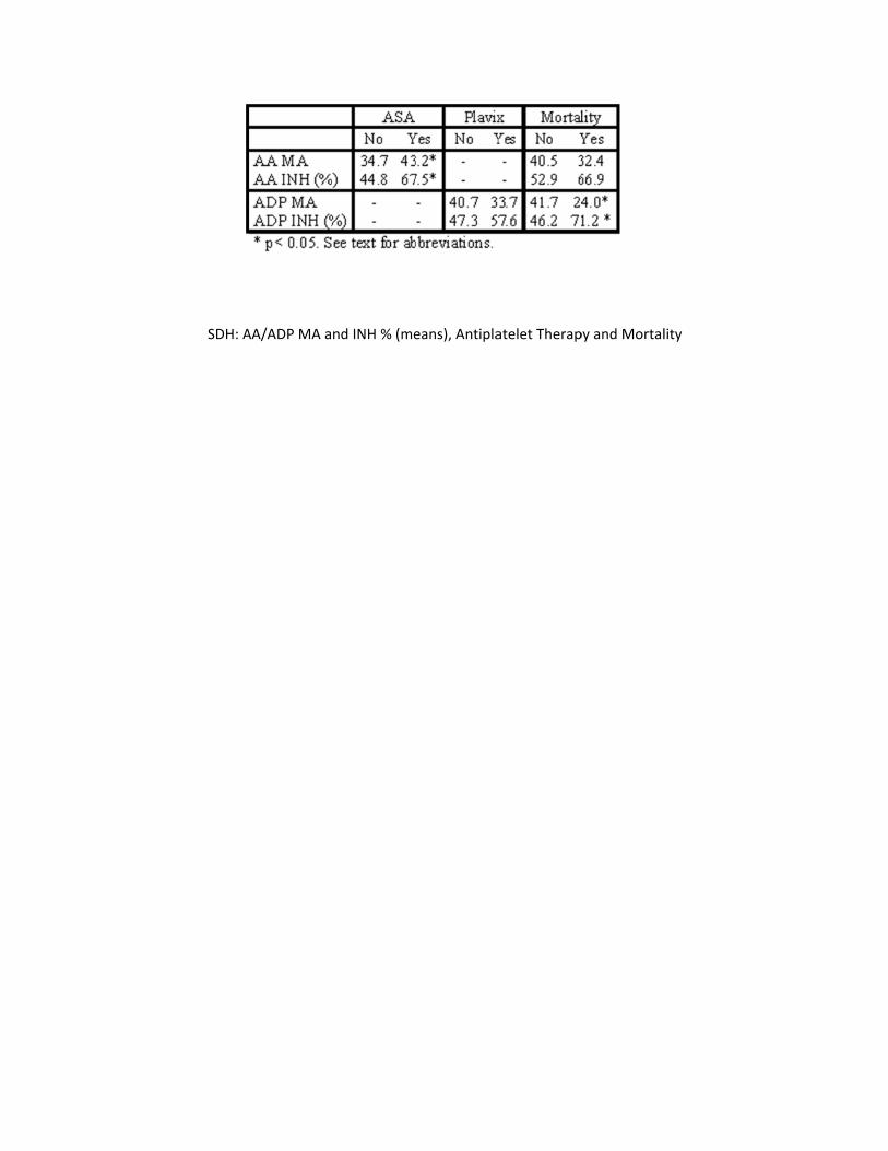
SDH: AAA/ADP MA and INH % (me
eans), Antipla
atelet Therap
py and Mortallity
Scientific Posters
Poster 25
THE TRAUMA SPECIFIC VASCULAR INJURY SHUNT (TS‐VIS): PROOF OF CONCEPT AND FUNCTION
Robert Houston, MD, Kyle Sokol, MD, Jonathan J. Morrison, MRCS, J. Devin B. Watson, MD, Jerry R. Spencer, BS, Todd Rasmussen, MD
San Antonio Military Medical Center
Presenter: Robert Houston, MD
Objectives: Temporary vascular shunts are a valuable adjunct to restore perfusion in the setting of extremity vascular injury. To date commercially available shunts were designed for to facilitate operations on age related cerebrovascular disease and not trauma. The objective of this study is to report the feasibility and capabilities of a novel Trauma Specific‐Vascular Injury Shunt (TS‐VIS).
Methods: Swine (80‐90kg) underwent isolation of the right iliac artery through a retroperitoneal incision. The TS‐VIS was inserted into the iliac artery position and animals placed into one of two groups, both of which had shunt patency and hind limb perfusion assessed for 6 hours. In Group 1 (n=9) shunt patency was tested without the use of heparin and Group 2 (n=8) had a continuous infusion of heparin through the novel infusion side port. Shunt patency and hind limb perfusion were evaluated using femoral artery duplex ultrasound velocity, side port arteriography and measuring circulating markers of inflammation.
Results: Group 1 had an 89% patency rate with distal thrombosis in one animal at the 4‐hour mark; all shunts remained patent in the second group. The continuous infusion in Group 2 was performed successfully, without obstruction or shunt displacement in all animals as well as completion angiogram demonstrating patency of the superficial and deep femoral systems. No difference was identified between the shunted extremity and the control extremity on histological analysis of neuromuscular injury (Group 1, p=0.083; Group 2, p=0.330).
Conclusions: The TS‐VIS is a novel device with features designed to facilitate use in the setting of vascular trauma. The TS‐VIS is a standalone vascular shunt with multiple utilities of the injection port. Further studies are needed to evaluate the TS‐VIS to commercially available temporary vascular shunts. Future generations of the TS‐VIS will include a shorter 3‐5cm shunt permitting “in‐line” placement.
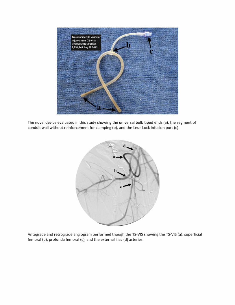
The novelconduit w
Antegradefemoral (b
l device evaluwall without re
e and retrogrb), profunda f
uated in this seinforcement
rade angiografemoral (c), a
study showingt for clamping
am performedand the extern
g the universag (b), and the
d though the nal iliac (d) ar
al bulb tiped e Leur‐Lock in
TS‐VIS showirteries.
ends (a), the fusion port (c
ng the TS‐VIS
segment of c).
S (a), superficcial
Scientific Posters
Poster 26
IMPACT OF MEAN INFUSION RATES OF FRESH FROZEN PLASMA (FFP), PACKED RED BLOOD CELLS (PRBC), AND PLATELETS (PLTS) ON THE INCIDENCE OF ACUTE RESPIRATORY DISTRESS SYNDROME
(ARDS) IN PATIENTS WITH SEVERE HEMORRHAGE
Dietric Hennings, MD, Eric R. Simms, MD, Adam Hauch, MD, MBA, Kira Long, Norman E. McSwain, Jr., MD, FACS, NREMT‐P*, Peter Meade, MD, MPH*,
Juan C. Duchesne, MD, FACS, FCCP, FCCM* Tulane University School of Medicine
Presenter: Dietric Hennings, MD
Objectives: Massive transfusion protocol (MTP) with FFP and PRBCs in a 1:1 ratio has been widely adopted for resuscitation in patients with severe hemorrhage. Use of FFP has been linked to increased risk of ARDS. To date, the impact of FFP infusion rates on risk of developing ARDS has not been analyzed. We hypothesize no correlation in incidence of ARDS based on rate of infusion.
Methods: This was a 24‐month retrospective analysis of MTP patients surviving >30 minutes and undergoing DCS at an urban Level 1 trauma center. Mean Infusion Rates (MIR, in ml/min) of PRBCs, FFP, and Platelets (Plts) were calculated for Length Of Intervention (LOI, ED time + OR time) and compared between patients with ARDS and without ARDS. Student’s t‐tests were performed to analyze differences in MIR for PRBC, FFP, or Plts between groups. Logistic regression was performed to analyze the impact of FFP in the development of ARDS.
Results: There were no statistically significant differences between patient demographics. 151 patients met inclusion criteria, 18 (11.9%) developed ARDS (MIR for PRBC 57.86 ml/min, FFP 112.2 ml/min, Plts 3.79 ml/min) vs. 133(88.1%) who did not develop ARDS (MIR for PRBC 68.24 ml/min, FFP 76.12 ml/min, Plts 2.87 ml/min) p=0.72, 0.22, 0.44 respectively. In our logistic regression FFP MIR did not increase the likelihood for ARDS 1.0(0.99‐1.00) p=0.96.
Conclusions: To the best of our knowledge, this is the first study to analyze MIR in DCS and the incidence of ARDS. Although the volume of blood products was statistical higher in the ARDS group, there was no correlation between incidence of ARDS and MIR for PRBC, FFP and Plts in massively transfused patients undergoing DCS. Better understanding of kinetics between LOI and total volume of infusion as potential precursors of ARDS needs further investigation.
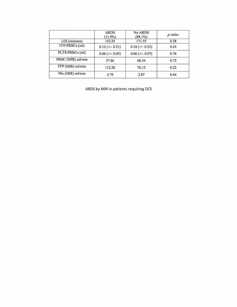
ARRDS by MIR inn patients req
quiring DCS
Scientific Posters
Poster 27
OVERUTILIZATION OF HEAD CTS IN PATIENTS WITH A GCS OF 15: A LOT FOR A LITTLE
Robert T. Dahlquist, MD, Peter E. Fischer, MD, MS*, Harsh Desai, BS, A. Britton Christmas, MD, FACS*, Michael Gibbs, MD*, Ronald F. Sing, DO*
Carolinas Medical Center
Presenter: Robert T. Dahlquist, MD
Objectives: Patients with a GCS of 15 after trauma frequently receive head CT scans based on a variety of clinical decision rules that have shown the ability to predict the probability of abnormal CT scan. The purpose of this study was to assess the rate of intracranial injuries in patients with a GCS of 15 and any subsequent neurosurgical intervention required to treat these injuries.
Methods: A retrospective review of patients sustaining a blunt head injury with a documented GCS of 15 on presentation to the emergency department to a Level I trauma center between 2008 and 2010 was conducted. Demographics, physiologic and anatomic data were reviewed. Radiologic findings and any subsequent neurosurgical intervention was also recorded.
Results: 1,242 patients were identified of which 170 (13.7%) were pediatric (age<18). 579 patients (46.6%) had a documented loss of consciousness and 60 (4.8%) had a clinically‐suspected skull fracture. 442 (35.6%) had a fracture or intracranial abnormality noted on CT scan. Ultimately, 17 (1.4%; 95% confidence interval [CI] 0.9% to 2.1%) required neurosurgical intervention. Of these 17 patients, 13 had conspicuous findings during the history and physical examination including skull fractures, penetrating injury, focal neurologic deficit, or history of anticoagulation. Of the remaining four patients, three were pediatric patients (age <9) who presented with persistent vomiting and one was an elderly patient (age >65) with an acute‐on‐chronic subdural hematoma.
Conclusions: Intracranial injury requiring neurosurgical intervention is exceedingly rare in those who incur blunt head injury with a GCS of 15, despite abnormal CT. Head CT utilization in these patients can be used more judiciously to decrease radiation exposure and healthcare expenditure. Elderly patients, anti‐coagulated patients, and pediatric patients with emesis are at higher risk of significant intracranial injury requiring intervention.

Notes
Scientific Posters
Poster 28
REDUCTION OF REPEAT IMAGING IN PATIENTS TRANSFERRED TO A LEVEL ONE URBAN TRAUMA CENTER DECREASES COST AND RADIATION EXPOSURE
Mayur Narayan, MD, MPH, MBA, Damon Clark, MD, Samuel M Galvagno, DO, PhD*, Thomas M. Scalea, MD, FACS, FCCM*, Deborah M. Stein, MD*
R Adams Cowley Shock Trauma Center
Presenter: Mayur Narayan, MD, MPH, MBA
Objectives: Repeat imaging in trauma patients transferred within a regional trauma system to a tertiary care referral center leads to unnecessary delays in management, radiation exposure, and increased costs. Poor image quality, incompatible imaging software, and misplaced imaging discs are well‐established reasons for repeat imaging. We hypothesized that implementation of a secure web‐based network would reduce inefficiency, remove unnecessary cost, and limit radiation exposure for patients transferred to our Level I center.
Methods: Prospective data was analyzed on patients transferred pre‐lifeIMAGE (PRE) between January 1 and June 30, 2011, and post‐lifeIMAGE (POST) between January 1 and June 30, 2012. Cost (dollars) and radiation exposure (mSv) were evaluated. Outcomes included length of stay (LOS), time in trauma unit (TRU), ISS, and survival to hospital discharge.
Results: 1,950 patients transferred to our trauma center were included. There was no difference in ISS and other demographics between groups. There was a decrease in number of patients who underwent repeat imaging after lifeIMAGE was implemented (62% PRE vs. 47% POST; p< 0.01). Cost of imaging per patient significantly decreased by 18.7% ($413.77 PRE vs. $333.77 POST; p < 0.01). Total cost of imaging decreased from PRE $401,765.97 to POST $326,756.79. Radiation exposure decreased by 19.2% (8.39 mSv PRE vs. 7.23 mSv POST; p < 0.01). There was decreased Hospital LOS after the lifeIMAGE system was implemented (4.4 days vs. 3.8 days; p = 0.07). No difference in in‐hospital mortality was seen (3.8% PRE vs. 4.4% POST; p = 0.52).
Conclusions: Repeating radiology studies on transferred trauma patients to a tertiary care referral center continues to be a significant problem for regional trauma systems. Implementation of a secure web‐based network reduces costs, radiation exposure, and improves efficiencies of care.

Notes

Scientific Posters
Poster 29
INCREASED MAP GOALS AND EPISODES OF RELATIVE HYPOTENSION DO NOT AFFECT IN‐HOSPITAL FUNCTIONAL OUTCOMES AFTER ACUTE SPINAL CORD INJURY
Niels D. Martin, MD*, Christopher K Kepler, MD, MBA, Muhammed H Zubair, MD, Amirali Sayadipour, MD, Murray J. Cohen, MD*, Michael S. Weinstein, MD, FACS*
Thomas Jefferson University
Presenter: Niels D. Martin, MD
Objectives: Acute spinal cord injury (SCI) is often treated with induced hypertension to enhance spinal cord perfusion. The optimal mean arterial pressure (MAP) likely varies from patient to patient. Arbitrary goals are often set, frequently requiring vasopressors to achieve, with no clear evidence supporting this practice. We hypothesize that increased MAP goals and episodes of relative hypotension do not affect hospital outcome.
Methods: All cervical and thoracic SCI patients treated at a level one trauma & regional spinal cord injury center over at 2.5 year period (2006‐2009) were retrospectively reviewed. Lowest and average hourly MAP was recorded for the first 72 hours of hospitalization, allowing for quantification of mean MAP and total number of episodic relative hypotensive events. These data were further compared to daily American Spinal Injury Association Motor Score (AMS), which was used to determine severity of SCI and improvement/decline during hospitalization.
Results: There were 148 SCI patients during the study period. 16 were excluded for missing data. 69.7% required vasopressors. When using a MAP of 65mmHg as acceptable, there was no difference in admission or change in AMS during hospitalization for those averaging above or below 65. However, when MAP >85 is used, increasing episodes of relative hypotension correlated with lower admission AMS (<10 episodes, AMS 66.2; >50 episodes, 22.0; p<0.001) and the need for vasopressors (p<0.03) but showed no statistical change in AMS by hospital discharge.
Conclusions: Episodes of relative hypotension are progressively more frequent with more severe SCI as denoted by lower admission AMS. Episodes of hypotension did not affect the change in AMS during the acute hospitalization, regardless of MAP goal level. Arbitrary MAP goals may not be efficacious but further prospective study with long‐term follow‐up is needed.

Notes
Scientific Posters
Poster 30
DOES EMPIRIC ANTIBIOTICS THERAPY LEAD TO ANTIBIOTIC RESISTANCE IN SICU?
Colleen Stoeppel, MD, Michael Cripps, MD, Evert Eriksson, MD*, Kenneth Hawkins, BSRRT, Joseph P. Minei, MD, FACS*, Christian T. Minshall, MD, PhD*
University of Texas Southwestern Medical Center
Presenter: Colleen Stoeppel, MD
Objectives: As part of our quality improvement process, we reviewed the antibiotic treatment of ventilator‐associated pneumonia (VAP) in the surgical intensive care unit (SICU). We measured our compliance with the national healthcare safety network (NHSN) VAP criteria and looked at the development of subsequent antibiotic resistant organisms in these patients.
Methods: We prospectively tracked all SICU patients treated for VAP from 9/12 to 5/13. We evaluated compliance with NHSN indicators for diagnosing probable VAP: white blood cell count (WBC) >12 or <4, temperature >38°C or <36°, purulent sputum >25 neutrophils per hpf and quantitative culture >105 CFU of pathogenic organisms on bronchoalveolar lavage (BAL). We also evaluated duration of antibiotic therapy and subsequent nosocomial infections with antibiotic resistant organisms.
Results: 291 patients remained on the ventilator >48 hours and 87 (30%) patients were treated with antibiotics for presumed VAP; 87% (76/87) met 3 or more of the NHSN criteria. Fifty‐two percent (45/87) were treated with empiric antibiotics for an unrelated indication prior to starting treatment. In all patients treated for VAP, there were 77 subsequent positive cultures. Thirty‐seven of these cultures (49%) demonstrated resistance to at least one class of antibiotics. The empiric antibiotic treatment for VAP in patients who had <3 NHSN criteria led to the development of significantly fewer resistant organisms (16%, 6/37) when compared to patients treated empirically for other indications (66%, 25/37; p < 0.007).
Conclusions: Our SICU is 87% compliant with initiating antibiotics when 3 or more of the NHSN criteria for VAP are present. Empiric antibiotic therapy appears to play a significant role in the development of resistant organisms in our SICU. Future studies are needed to determine appropriate duration and roles for empiric and definitive antibiotic therapy in this patient population to minimize resistance.

Notes

Scientific Posters
Poster 31
TRAUMA WRIST BANDS: BINDING NEW SCENE PROTOCOLS TO HOSPITAL OUTCOMES
Aman Banerjee, MD, Michael Nowak, PhD, Debra Allen, BSN, RN, Jeffrey Claridge, MD MetroHealth Medical Center
Presenter: Aman Banerjee, MD
Objectives: Using a unique identifying number on a trauma wrist band (TWB) we prospectively evaluated the implementation of a revised pre‐hospital triage protocol with a primary goal to reduce the transport of minimally injured patients to designated trauma centers (TC).
Methods: A revised EMS trauma scene triage protocol was implemented on November 2012, which also included applying a TWB. This protocol identifies patients as Red, Yellow, or Green based on injury mechanism and physiologic derangement. Red and Yellow patients were most likely to be severely injured and transport to a TC was recommended. Green patients were those with a low likelihood of injury and the new protocol recommends transport to the nearest emergency department (ED) for treatment, instead of a trauma center. EMS and hospital records from December 2012 through March 2013 were linked using the TWB number. Protocol compliance was evaluated through an audit of EMS triage checklists for accurate completion and transport to appropriate hospital. Patients treated at non‐trauma hospitals (NTH) were evaluated for under‐triage, defined as the need for admission, operation, TC transfer, or death.
Results: Over 95% of patients had a TWB placed, which identified 536 consecutive patients. Of these 77 (14%) were Red, 229 (43%) were Yellow, and 230 (43%) were Green. Of the 114 patients transported to NTH, 1 (1%) was designated Red, 5 (4%) were Yellow and 108 (95%) were Green. Overall protocol compliance was 87%. EMS accurately completed the checklist in 89% of patients and transported patients to the appropriate hospital in 93% of these patients. There were 29 (5.4%) patients who were under‐triaged; of these 26 were admitted, 11 patients had an operation, 5 required an ICU admission, and 2 were referred to a TC. There were no deaths in the Green group.
Conclusions: Utilization of TWB is an effective tool, and EMS providers can safely transfer minimally injured patients to NTH.

Notes

Scientific Posters
Poster 32
POINT OF CARE TESTING DURING TRAUMA RESUSCITATION, A REVIEW OF 4,482 PATIENTS
Emily F. Cantrell, MD, Lawrence Lottenberg, MD*, Azra Bihorac, MD, MS, Tezcan Ozrazgat‐Baslanti, PhD University of Florida
Presenter: Emily F. Cantrell, MD
Objectives: Point of care (POC) testing is now a widely used tool during the initial trauma resuscitation. It is unclear if these values and subsequent actions based upon these values are predictive of outcomes. The aim of this study is to determine the accuracy of POC values and predictability of mortality and massive transfusion (MT) events.
Methods: This is a single institution retrospective cohort study from a state designated level 1 trauma center of 4,842 patients over an 8 year period (2005‐2012). POC data using iSTAT was collected from patients during their initial trauma resuscitation in the ED. Multivariate logistic regression methods were used to compare ability of models to predict mortality and MT events using values from POC and central lab adjusting for demographic and clinical risk factors. Area under the receiver operating characteristics curve (AUC) values and 95% confidence intervals (CI) were used to assess model discrimination.
Results: For mortality model using POC values (AUC 0.94, 95%CI 0.93‐0.95) had slightly better discrimination that central lab values (AUC 0.93, 95%CI 0.92‐0.95), with age, ISS, GCS, and lactate as the most significant predictors. The model for MT using central lab values had slightly higher AUC (AUC 0.93, 95%CI 0.91‐0.96) than the model using POC values (AUC 0.92, 95%CI 0.88‐0.95) with age, ISS, heart rate, HCT and lactate as the most significant predictors. When the POC values for lactate, HCT and base deficit were compared with samples ran in the central lab, differences and 95%CI between these values were found to be 1.16(1.03, 1.30), 1.04(0.96, 1.11) and 0.04(0.16, 0.24).
Conclusions: Based on this trauma population, POC testing values can be used to predict mortality and MT events. In this study, the POC testing value most predictive of death was lactate while lactate and hematocrit were most predictive of MT events.

Notes
Scientific Posters
Poster 33
CHARACTERIZATION OF “AGGRESSIVE” VS. “DORMANT” ACUTE APPENDICITIS
Matt Apel, MD, Narong Kulvatunyou, MD, Lynn Gries, Randall S. Friese, MD*, Bellal Joseph, MD*, Terence O'Keeffe, MD, MSPH*, Donald Green, MD, Andrew L. Tang, MD*,
Julie L. Wynne, MD, MPH*, Peter Rhee, MD, MPH* The University of Arizona
Presenter: Matt Apel, MD
Objectives: The etiology of acute appendicitis (AA) and reason for its perforation remain unknown. The notion that many cases of AA can be treated with antibiotics while some fails suggests that there may be a different types or pathway of AA. The goal of this study is to compare “aggressive” AA (early perforation, < 24 hours) to those “dormant” AA (delay without perforation, > 48 hours).
Methods: From October 2009 through September 2012, we performed a retrospective chart review on patients with AA seen by our acute care surgery service. We collected demographic data, Alvarado score, body mass index, presence of fecaliths, patient time (PT) (i.e., time from abdominal symptom onset to time of arrival in the emergency department [ED]), hospital time (HT) (i.e., time from arrival in the ED to time of surgical incision), and total time (TT) (i.e., PT + HT). We defined “aggressive” AA (AG‐AA) if AA was gangrenous or perforated and the TT < 24hours, and “dormant” AA (DO‐AA) if AA was not perforated and TT > 48 hours. We performed the group comparison and logistic regression analysis to identify predictors of AG‐AA.
Results: During a 3‐year period, 564 patients were seen by our service for possible AA. There were 24 AG‐AA and 90 DO‐AA. Table 1 (figure 1) showed group comparison. After a multiple logistic regression analysis, age, male gender, and elevated WBC were significantly associated with AG‐AA. Interestingly, the incidence of fecaliths between AG‐AA and DO‐AA was not different.
Conclusions: In this preliminary study, we found AG‐AA patients to be male, older, with elevation of WBC despite a very short time of presentation. The incidence of fecalith was not different, so fecalith was not the cause of perforation.
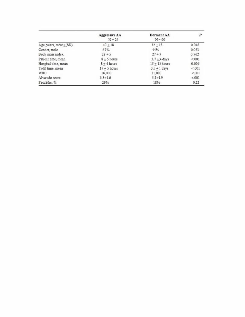

Scientific Posters
Poster 34
EARLY SCREENING AND TREATMENT ELIMINATES STROKE FROM BLUNT CERVICAL VASCULAR INJURY
Salim Lala, MD, BMedSci, Michael Cripps, MD, Herb A. Phelan III, MD, FACS*, Brian H. Williams, MD, FACS*, Christian T. Minshall, MD, PhD*,
Joseph P. Minei, MD, FACS*, Alexander L. Eastman, MD, MPH, FACS* University of Texas Southwestern Medical Center
Presenter: Salim Lala, MD, BMedSci
Objectives: Early screening and treatment are advocated to reduce the rate of BCVI‐related stroke. However, the effectiveness of these programs has not been well studied. The goal of this study was to assess the effectiveness of a BCVI screening and treatment program in reducing the stroke rate following BCVI.
Methods: From January 1, 2011 to January 31, 2012, all trauma patients at a Level I center with BCVI identified using a CTA‐only screening program following the modified Denver criteria were studied. Protocols provided for antiplatelet or anticoagulation therapy. Data collected included injury grade, concomitant injuries and demographics. Outcomes were assessed by chart review to determine resolution or progression of BCVI and incidence of stroke. Stroke was diagnosed by comparing arrival CTA with those at one week and one month after injury, along with clinic records.
Results: During the study period, 3,120 patients were admitted; 269 patients met screening criteria for BCVI. The overall incidence of BCVI was 1.9%, median±SD age was 42.0±18.6 years, 77% were male (n=208), and the median±SD ISS score for all patients with BCVI was 22.0±13.4. Of 60 BCVIs, 56% (n=34) were grade 1, 12% (n=7) grade 2, 17% (n=10) grade 3 and 15% (n=9) grade 4. Among all patients with BCVI, 87% received appropriate antiplatelet or anticoagulation therapy. 13% (n=8) of patients did not present for follow up imaging and/or clinic visit, of which 62% (n=5) were low grade injuries. There were no ischemic strokes on one week or one month imaging. The mortality rate among all patients diagnosed with BCVI was 6.7% (n=4), however none were due to stroke.
Conclusions: Early screening and treatment eliminates stroke‐related morbidity and mortality traditionally associated with BCVI. Further, a large multi‐center study is required to reset current expectations for post‐BCVI stroke‐rate at trauma centers as present rates are likely too high.

Notes
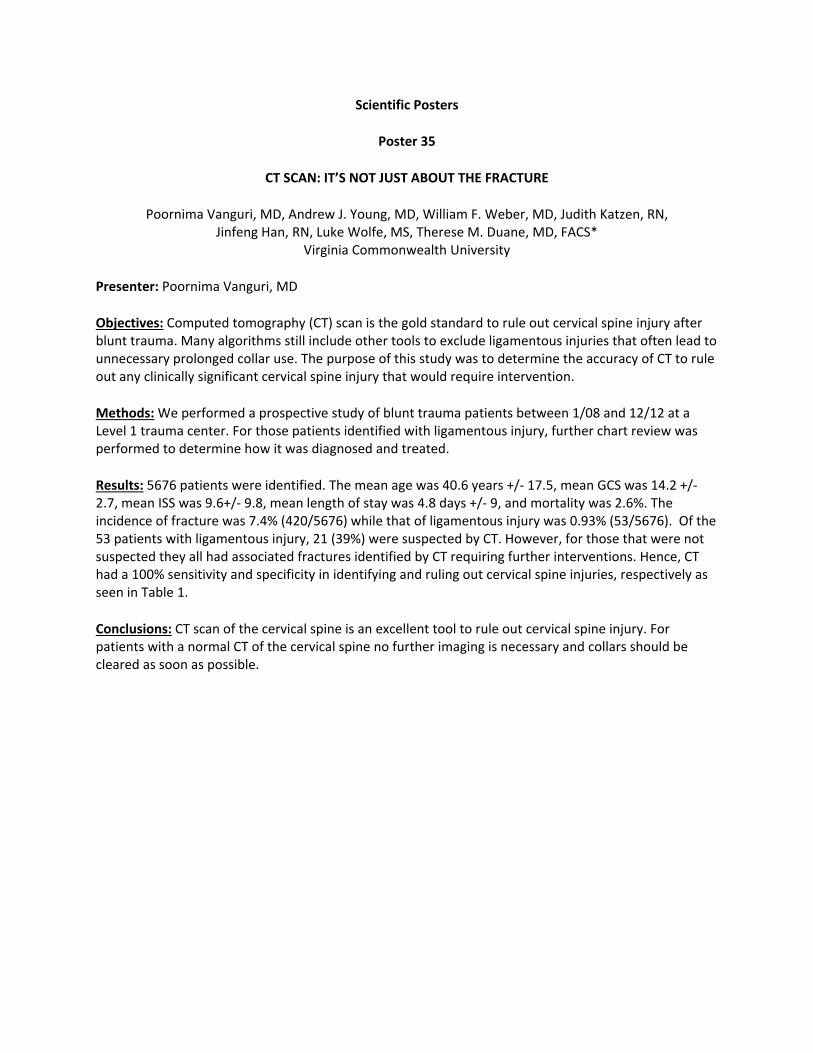
Scientific Posters
Poster 35
CT SCAN: IT’S NOT JUST ABOUT THE FRACTURE
Poornima Vanguri, MD, Andrew J. Young, MD, William F. Weber, MD, Judith Katzen, RN, Jinfeng Han, RN, Luke Wolfe, MS, Therese M. Duane, MD, FACS*
Virginia Commonwealth University
Presenter: Poornima Vanguri, MD
Objectives: Computed tomography (CT) scan is the gold standard to rule out cervical spine injury after blunt trauma. Many algorithms still include other tools to exclude ligamentous injuries that often lead to unnecessary prolonged collar use. The purpose of this study was to determine the accuracy of CT to rule out any clinically significant cervical spine injury that would require intervention.
Methods: We performed a prospective study of blunt trauma patients between 1/08 and 12/12 at a Level 1 trauma center. For those patients identified with ligamentous injury, further chart review was performed to determine how it was diagnosed and treated.
Results: 5676 patients were identified. The mean age was 40.6 years +/‐ 17.5, mean GCS was 14.2 +/‐ 2.7, mean ISS was 9.6+/‐ 9.8, mean length of stay was 4.8 days +/‐ 9, and mortality was 2.6%. The incidence of fracture was 7.4% (420/5676) while that of ligamentous injury was 0.93% (53/5676). Of the 53 patients with ligamentous injury, 21 (39%) were suspected by CT. However, for those that were not suspected they all had associated fractures identified by CT requiring further interventions. Hence, CT had a 100% sensitivity and specificity in identifying and ruling out cervical spine injuries, respectively as seen in Table 1.
Conclusions: CT scan of the cervical spine is an excellent tool to rule out cervical spine injury. For patients with a normal CT of the cervical spine no further imaging is necessary and collars should be cleared as soon as possible.
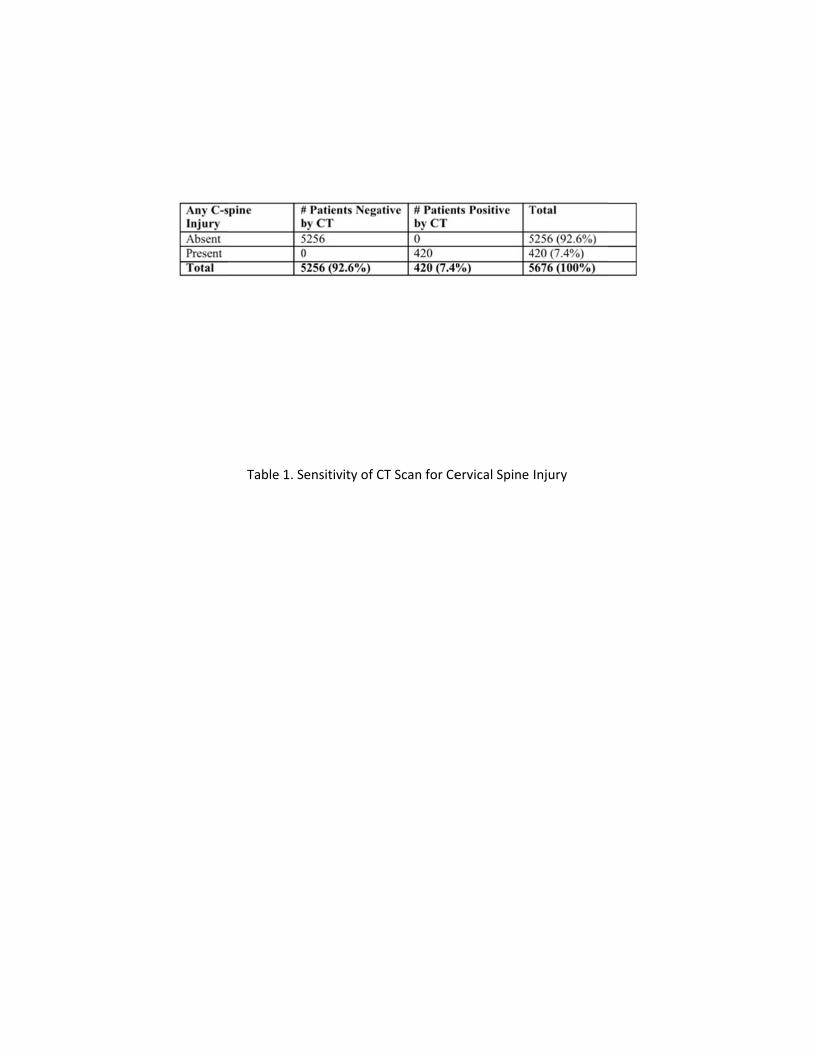
Table 1. Sensitivity of CCT Scan for Ce
ervical Spine Injury

Scientific Posters
Poster 36
PHASE I DEVELOPMENT OF GERIATRIC SPECIFIC TRAUMA SCORE
Eric H. Bradburn, DO MS FACS*, Bryan Fletcher, MD, Bryan R. Collier, DO FACS*, Mark E. Hamill, MD FACS*, Katherine Shaver, MS,
Frederick Rogers, MD, MS, FACS*, Christopher C. Baker, MD FACS Virginia Tech Carilion School of Medicine
Presenter: Eric H. Bradburn, DO MS FACS
Objectives: Trauma scoring systems are poor at predicting mortality in the geriatric population. We have previously demonstrated that the pre‐trauma modified Functional Independence Measure (FIM) score, specifically the expression component, was predictive of survival in the geriatric trauma victim. The purpose of this study was to create statistical models with the goal of developing a geriatric specific trauma score.
Methods: Retrospective analysis of the trauma registry patients age 65 and older admitted to a Level I trauma center from July 1, 2006 to July 1, 2012. 2630 patients were split into two equal random samples for regression modeling. Age, gender, race, BMI, ISS, GCS, RTS, Shock Index (SI) and Pre‐ trauma FIM scores (12 point scale) were studied. The primary outcome variable was survival. Stepwise logistic regression was used in one of the datasets to identify the best predictors of survival. The final model used the predictors identified in the stepwise procedure and was validated by multiple logistic regression analysis in the other dataset with statistical significance reached with p‐value < .05. Adjusted odds ratios were reported with confidence intervals.
Results: The mean age of patients was 78(SD±8.2) and 52% were female. The mean ISS was 13(SD±8.7) and a mean BMI of 26. Overall mortality was 11%, and 58% of patients who died were male. Table 1 contains the odds ratios for the predictor variables in each of three regression models using significant predictors from the stepwise regression analysis. Female gender, GCS, FIM expression were consistently validated in each of our regression models.
Conclusions: GCS, Gender, and Pre‐trauma FIM expression scores were validated as important predictors of survival in the geriatric trauma patient. We plan to utilize this data in the development of a multi institutional study to validate a geriatric specific trauma scoring system.

TTable 1: Regrression Modeels and OR forr Geriatric Tra
auma Scoring System Deve
elopment
Scientific Posters
Poster 37
PARAMEDICS AND SURGEONS PERCEIVE SURGICAL READINESS DIFFERENTLY‐A CRITICAL FACTOR IN IMPLEMENTATION OF CDC GUIDELINES FOR FIELD TRIAGE
Andrew C. Bernard, MD *, Catherine P. Starnes, BS, Jerome K. Manson, BS, Kristin Long, MD, Chris Culpepper, MD
University of Kentucky
Presenter: Andrew C. Bernard, MD
Objectives: CDC Guidelines for Field Triage of Injured Patients are predicated upon: 1) proper implementation by medics and 2) surgeon willingness to operate. We hypothesized that in a state with an immature trauma system and few trauma centers that a significant discrepancy exists between medic and surgeon perception of trauma surgical readiness resulting in heterogeneous implementation of the CDC Guidelines.
Methods: A survey was conducted among all licensed medics and all FACS general/thoracic surgeons in our state. Implementation of destination protocols for trauma, trauma center location, opinion of surgeon willingness (by medics), interest in trauma surgery (by surgeons) and approach to a standard clinical trauma scenario were assessed.
Results: 83% of medics work under a protocol for destination determination. 79% (38/48) of surgeon respondents indicate that they would provide surgery for an exsanguinating trauma victim if transfer elsewhere was not an option. 51% (313/618) of medics indicated that their local surgeon would definitely not or not likely operate in the same scenario. Given a scenario in which the patient clearly meets criteria for trauma center admission but no local trauma center exists, 21% (132/619) of medics will transport to the nearest geographic center regardless of surgeon availability, contrary to CDC Guidelines.
Conclusions: Most responding surgeons indicate willingness to operate if lives were in danger, but medics are confident of such surgeon willingness only half the time‐resulting in inconsistent application of the Guidelines. Though most medics work under protocols for destination determination, nearly ¼ of exsanguinating trauma victims are still arriving at geographically closest centers that may have no surgeon at all. Further efforts at building medic‐surgeon dialogue & working partnership are necessary for CDC Guidelines to be effective.

Notes
Scientific Posters
Poster 38
TRAUMA RECIDIVISM IN A SUBURBAN COHORT
Jerry A. Rubano, MD*, Jane E. McCormack, RN, BSN*, James A. Vosswinkel, MD* Stony Brook University Medical Center
Presenter: Jerry A. Rubano, MD
Objectives: Prevention of trauma may best be achieved by risk factor modification. Identifying the risk factors is paramount. Trauma recidivists—those with two or more traumatic injuries—can serve to identify these risk factors. Previous work has identified differences between urban and rural recidivists. This investigation seeks to identify recidivists within a suburban trauma system.
Methods: A retrospective analysis of countywide population‐based trauma registry was performed. A suburban county of 1.5 million residents was reviewed. Unique to our study, recidivism was not defined as multiple admissions to a single institution, but rather multiple unrelated trauma admissions to any institution.
All trauma admissions from January 2001 through December 2009 were considered for inclusion. Deaths during the first admission were excluded.
Measures included patient characteristics (age, sex, comorbidities), injury characteristics (mechanism, GCS, ISS). Outcome measures included disposition on discharge, mortality, and length of stay. Multivariant logistic regression was utilized to identify independent risk factors.
Results: 17247 patients met study criteria. There were 307 recidivists (1.81%). The rate of recidivism was highest during the first 6 months (27.4%) and age greater than 64, pre‐existing diagnosis of diabetes, hypertension, bleeding disorders, psychiatric diagnoses and a history of alcohol or drug abuse were all more common in the recidivist group (p < 0.001). Multivariant logistic regression revealed age over 64 and alcohol/drug abuse to be independent predictors of second injury. Race, sex, and mechanism of injury were not predictive. The second injury was associated with a significantly higher mortality (10.75%, p <0.001).
Conclusions: Recidivism within our suburban trauma system is rare, but the second injury is associated with a significantly higher mortality. Strategies to identify at risk individuals and prevent the second injury may be life saving.
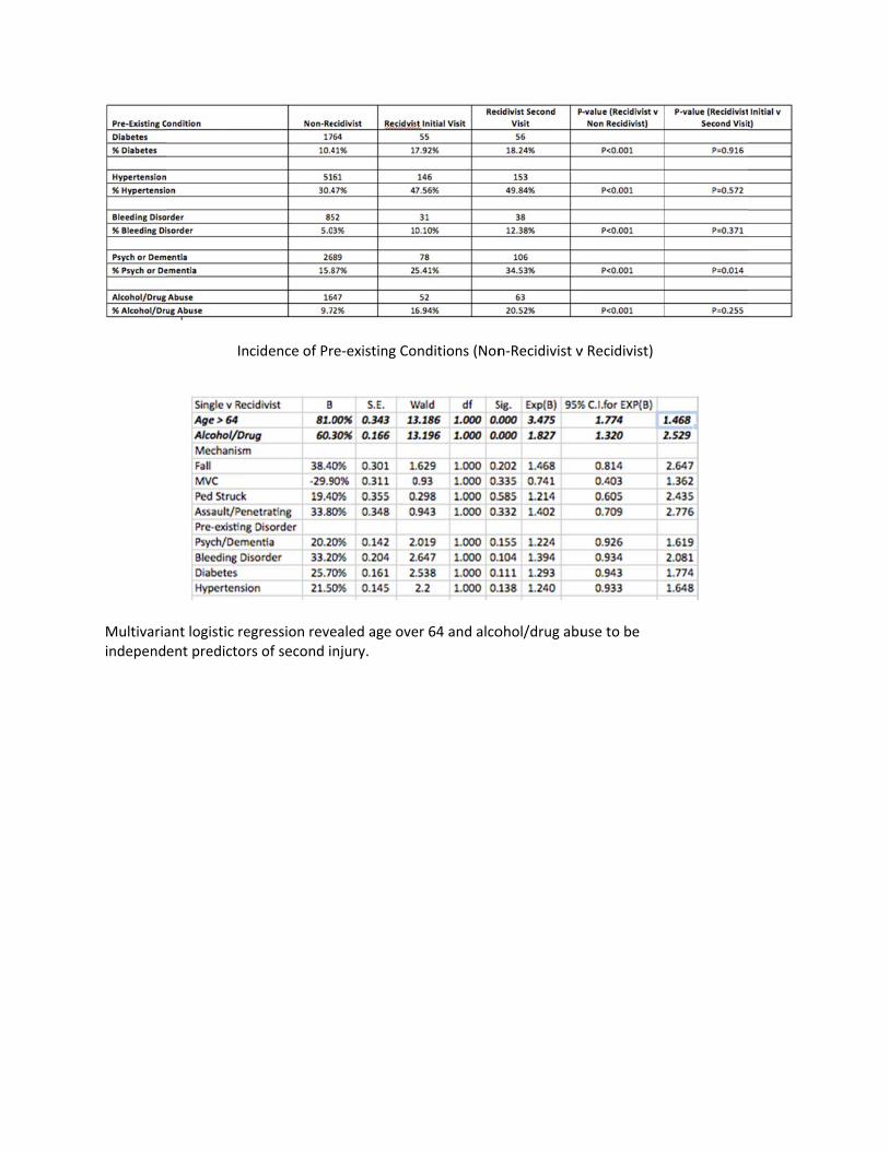
Multivariaindepend
In
ant logistic reent predictor
cidence of Pr
egression revers of second i
re‐existing Co
ealed age ovenjury.
onditions (Non
er 64 and alco
n‐Recidivist v
ohol/drug abu
v Recidivist)
use to be

Scientific Posters
Poster 39
PREHOSPITAL TOURNIQUET USE IN TRAUMA PATIENTS: AN UNDERUTILIZED MODALITY
Glenn Miller, Patricia M. Byers, MD, FACS*, Punam Parikh, BS, Enrique Ginzburg, MD* University of Miami Miller School of Medicine
Presenter: Glenn Miller
Objectives: Brachial artery injury requires selective use of tourniquets in order to prevent exsanguination yet preserve function. It is unknown whether or not tourniquet use for brachial artery injury affects patient outcome or if tourniquet‐related complications occur.
Methods: Retrospective review of patients in an urban trauma center from 2000‐2012 with traumatic brachial artery injury was performed. Patient records were evaluated for tourniquet use prior to trauma center arrival and stratified according to whether or not they presented in shock.
Results: One‐hundred and one patients were admitted with brachial artery injury and required operative intervention. There were fourteen (13.8%) patients who arrived with a tourniquet in place and 87 (86.1%) patients arrived with no tourniquet. Penetrating injury was the mechanism in all 14 tourniqueted patients and only 68% of patients without a tourniquet (p=0.35). Eleven of fourteen (79%) patients arriving with a tourniquet were in shock, contrasting with the twenty‐eight (32%) patients in the non‐tourniquet group arriving in shock (p=0.001). There was no significant difference in ISS (13.0±6.1 vs. 16.7±10.2, p=0.230) between those presenting in shock with or without a tourniquet. The length of stay (LOS) in the tourniquet group was significantly shorter than those patients without a tourniquet (7.1±3.4days vs. 12.2±9.3days, p=0.03). There were no complications attributable to tourniquet use. The non‐tourniquet group had a single mortality due to multisystem organ failure (3.6%).
Conclusions: Tourniquets are underutilized for upper extremity injury despite an organized urban trauma system familiar with their use. Liberal use of tourniquets in patients presenting with shock may reduce hospital LOS and appears to confer no additional morbidity. We recommend institution of tourniquet protocols for all EMS providers and monitoring to ensure proper utilization.

Notes

Scientific Posters
Poster 40
TRAUMATIC CARDIOPULMONARY ARREST: ORGAN DONATION CHALLENGES PARAMETERS FOR RESUSCITATION
Katie M. Love, MD*, Joshua B. Brown, MD*, Brian G. Harbrecht, MD*, Glen A. Franklin, MD* University of Louisville
Presenter: Katie M. Love, MD
Objectives: Survival after traumatic cardiopulmonary arrest (TCPA) is rare. The study aims were to identify predictors of survival and organ donation after TCPA resuscitation, estimate cost of these outcomes, and determine whether organ donation as an outcome is cost‐effective.
Methods: Patients at a level I trauma center were retrospectively reviewed over 36 months. Data was obtained from medical records, hospital accounting data, and the local organ procurement agency. Outcomes included survival to discharge, neurologic function, and organ donor eligibility. Logistic regression was used to identify predictors of these outcomes, p<0.05 significant. Incremental cost effectiveness ratio (ICER) was calculated to determine additional cost per life saved when organ donation is included as an endpoint of TCPA resuscitation.
Results: Over the study period 8,932 subjects were evaluated. TCPA occurred in 237 (3%) patients. The mortality rate was 97%. Independent predictors of survival included emergency department (ED) disposition to the operating room (p=0.02) and reactive pupils (p<0.01). Of 7 survivors, 4 were discharged neurologically intact. 5% of TCPA patients were eligible for organ donation with a procurement rate of 2%. Organ donor eligibility was independently associated with arrest after arrival to the ED (p<0.01) and transfusion of fresh frozen plasma (FFP) (p=0.02). The cost of TCPA resuscitation per survivor was $1.8 million; cost per survivor or life saved by donation was $538,000. The ICER was $875,000 per additional life saved using donation as an outcome.
Conclusions: TCPA survival and organ donor eligibility have distinct clinical predictors. The decision to pursue resuscitation should depend on duration of arrest and pupil reactivity. Early FFP transfusion may increase successful organ donation. The financial burden of TCPA resuscitation may be mitigated by expanding endpoints to include organ donation.

Notes
Scientific Posters
Poster 41
PRE‐HOSPITAL ULTRASONOGRAPHY FOR THE EN ROUTE IDENTIFICATION OF CORRECTABLE THORACIC PATHOLOGY
Jeffrey Coughenour, MD*, Christopher Nelson, MD*, Salman Ahmad, MD*, Stephen L. Barnes, MD* University of Missouri
Presenter: Jeffrey Coughenour, MD
Objectives: Ultrasound is a standard adjunct to the primary survey of the injured patient, and its pre‐hospital use continues to gain acceptance. We sought to evaluate the utility of thoracic ultrasound in the pre‐hospital aeromedical environment, where ambient noise and vibration render the diagnosis of pneumothorax or mainstem intubation extraordinarily difficult.
Methods: Non‐physician providers were trained in the performance and interpretation of thoracic ultrasound. Examinations were performed on all adult trauma patients and only medical patients requiring intubation. A repeat exam occurred upon emergency department (ED) arrival. A questionnaire was completed by each sonographer to document findings. Pre‐hospital images were reviewed by two faculty members for quality and accuracy. Findings were compared to chest x‐ray and CT when performed.
Results: 149 patients (136 trauma/12 medical) met inclusion criteria. Mean age 44.4 (18‐94) years, 69% male. Mean ISS 17.68 (1‐75) and mean chest AIS 2.93 (0‐6) in the injured group. Twenty pneumothoraces and one mainstem intubation were identified. Sixteen were correctly identified, 3 were missed and 1 was seen but misinterpreted in the field. The mainstem intubation was seen but misinterpreted. When compared to chest CT (n=116) pre‐hospital ultrasound had a sensitivity of 68% (95% CI 46‐85%), a specificity of 96% (95% CI 90‐98%), and an overall accuracy of 91% (95% CI 85‐95%). In comparison, ED ultrasound had a sensitivity of 84% (95% CI 62‐94%), specificity of 98% (95% CI 93‐99%), and an accuracy of 96% (95% CI 90‐98%). The AUC was 82% and 91% respectively.
Conclusions: Pre‐hospital and ED ultrasound are comparable in the identification of thoracic pathology. Given the unique characteristics of the aeromedical environment, thoracic ultrasound improves the diagnostic capabilities of pre‐hospital providers and should be routinely utilized in flight.

Pre‐hospital vs. ED PPneumothora
ax Identificatiion


![Untitled-1 [ ] · PDF fileErgonomics, Hand instruments and trocars Sterlization of laparoscopic instruments ... Gynaecological emergencies for surgeons VATS I Thoracoscopic surgery](https://static.fdocuments.us/doc/165x107/5aaa8cee7f8b9a9a188e5eed/untitled-1-hand-instruments-and-trocars-sterlization-of-laparoscopic-instruments.jpg)


