SCAI expert consensus statement for infrapopliteal ... · Original Studies SCAI Expert Consensus...
13
Original Studies SCAI Expert Consensus Statement for Infrapopliteal Arterial Intervention Appropriate Use Bruce H. Gray, 1 * DO, Larry. J. Diaz-Sandoval, 2 MD, Robert S. Dieter, 3 MD, Michael R. Jaff, 4 DO, and Christopher J. White, 5 MD Intervention to treat infrapopliteal arterial disease can be challenging because the patients’ comorbidities, the anatomic variables, and the limitations of our techniques. Clinical scenarios based on anatomic and clinical variables are presented. Recommen- dations regarding intervention (appropriate care, may be appropriate care, rarely appropriate care) are made based on best evidence. V C 2014 Wiley Periodicals, Inc. Key words: peripheral intervention; appropriate use; peripheral arterial disease INTRODUCTION Infrapopliteal (IP) arterial disease or “below the knee” arterial disease is commonly seen in patients with long-standing diabetes mellitus, chronic kidney disease, or the elderly. The most concerning manifesta- tion of peripheral arterial disease (PAD) in these high risk patients is the development of critical limb ische- mia (CLI: ischemic rest pain or ischemic ulcers). This arterial bed consists of relatively small caliber vessels, which are often calcified and associated with diffuse, multilevel disease. Due to the complexity of this patient population, their co-morbidities, and severity of vascular disease, there is a paucity of scientific evi- dence for the generalizability of percutaneous revascu- larization. In general, non-ambulatory patients with a short life expectancy and extensive necrosis or gangrene should undergo primary amputation. Ambulatory patients who are acceptable surgical candidates, expected to survive more than two years with a patent IP artery that pro- vides direct flow to the foot (considered to be a good distal target), and an adequate autologous venous con- duit should be considered for surgical bypass. Patients with significant medical co-morbidities that limit life expectancy, those at increased risk for surgery, those without an adequate distal target for bypass, or with poor venous conduit should be considered for an endovascular-first approach. This document was developed to guide physicians in clinical decision-making in the modern practical appli- cation of endovascular intervention for patients with IP arterial disease. ANATOMIC CONSIDERATIONS Patients with CLI typically have disease involving mul- tiple levels (i.e., aorto-iliac, femoropopliteal (FP) and IP), but less than 10% of patients with CLI have hemodynami- cally significant disease in all three levels [1–3] (Table I). Infrainguinal disease (FP and IP) can be further subdivided 1 University of South Carolina School of Medicine/Greenville, Greenville, South Carolina 2 Metro Heart & Vascular Center; Metro Health Hospital, Wyoming, Michigan 3 Loyola University, Maywood, IL and Hines VA, Hines, Illinois 4 MGH Institute for Heart, Vascular and Stroke Care, Harvard University, Boston, Massachusetts 5 John Ochsner Heart & Vascular institute, Ochsner Medical Center, New Orleans, Louisiana *Correspondence to: Bruce H. Gray, D.O., 200 Patewood Drive, Suite 300, Greenville, South Carolina 29681. E-mail: [email protected] Received 31 December 2013; Revision accepted 1 January 2014 DOI: 10.1002/ccd.25395 Published online 00 Month 2014 in Wiley Online Library (wileyonlinelibrary.com) V C 2014 Wiley Periodicals, Inc. Catheterization and Cardiovascular Interventions 00:00–00 (2014)
Transcript of SCAI expert consensus statement for infrapopliteal ... · Original Studies SCAI Expert Consensus...

Original Studies
SCAI Expert Consensus Statement for InfrapoplitealArterial Intervention Appropriate Use
Bruce H. Gray,1* DO, Larry. J. Diaz-Sandoval,2 MD, Robert S. Dieter,3 MD,Michael R. Jaff,4 DO, and Christopher J. White,5 MD
Intervention to treat infrapopliteal arterial disease can be challenging because the
patients’ comorbidities, the anatomic variables, and the limitations of our techniques.
Clinical scenarios based on anatomic and clinical variables are presented. Recommen-
dations regarding intervention (appropriate care, may be appropriate care, rarely
appropriate care) are made based on best evidence. VC 2014 Wiley Periodicals, Inc.
Key words: peripheral intervention; appropriate use; peripheral arterial disease
INTRODUCTION
Infrapopliteal (IP) arterial disease or “below theknee” arterial disease is commonly seen in patientswith long-standing diabetes mellitus, chronic kidneydisease, or the elderly. The most concerning manifesta-tion of peripheral arterial disease (PAD) in these highrisk patients is the development of critical limb ische-mia (CLI: ischemic rest pain or ischemic ulcers). Thisarterial bed consists of relatively small caliber vessels,which are often calcified and associated with diffuse,multilevel disease. Due to the complexity of thispatient population, their co-morbidities, and severity ofvascular disease, there is a paucity of scientific evi-dence for the generalizability of percutaneous revascu-larization.
In general, non-ambulatory patients with a short lifeexpectancy and extensive necrosis or gangrene shouldundergo primary amputation. Ambulatory patients whoare acceptable surgical candidates, expected to survivemore than two years with a patent IP artery that pro-vides direct flow to the foot (considered to be a gooddistal target), and an adequate autologous venous con-duit should be considered for surgical bypass. Patientswith significant medical co-morbidities that limit lifeexpectancy, those at increased risk for surgery, thosewithout an adequate distal target for bypass, or withpoor venous conduit should be considered for anendovascular-first approach.
This document was developed to guide physicians inclinical decision-making in the modern practical appli-cation of endovascular intervention for patients with IParterial disease.
ANATOMIC CONSIDERATIONS
Patients with CLI typically have disease involving mul-tiple levels (i.e., aorto-iliac, femoropopliteal (FP) and IP),but less than 10% of patients with CLI have hemodynami-cally significant disease in all three levels [1–3] (Table I).Infrainguinal disease (FP and IP) can be further subdivided
1University of South Carolina School of Medicine/Greenville,Greenville, South Carolina2Metro Heart & Vascular Center; Metro Health Hospital,
Wyoming, Michigan3Loyola University, Maywood, IL and Hines VA, Hines, Illinois4MGH Institute for Heart, Vascular and Stroke Care, HarvardUniversity, Boston, Massachusetts5John Ochsner Heart & Vascular institute, Ochsner Medical
Center, New Orleans, Louisiana
*Correspondence to: Bruce H. Gray, D.O., 200 Patewood Drive,Suite 300, Greenville, South Carolina 29681. E-mail: [email protected]
Received 31 December 2013; Revision accepted 1 January 2014
DOI: 10.1002/ccd.25395Published online 00 Month 2014 in Wiley Online Library(wileyonlinelibrary.com)
VC 2014 Wiley Periodicals, Inc.
Catheterization and Cardiovascular Interventions 00:00–00 (2014)

into those with predominantly isolated IP disease (�33%)and those with both FP and IP disease (�67%) [4–7]. Iso-lated IP disease is mainly seen in the elderly (>80-yearsold), diabetic, or dialysis-dependent patients [5]. These
patients are at higher risk for amputation and have ashorter amputation-free survival compared to those withFP and IP disease (median amputation-free survival:17 months (95% CI¼ 9–24 m) versus 37 months (95%CI¼ 28–44 m), P¼ 0.001) [6]. Clinical and non-invasivecriteria have been used to determine which CLI patientswould benefit from revascularization. These include Ruth-erford categories 4–6, systolic ankle pressure< 50 mmHg, non-pulsatile plethysmographic tracing, and/or trans-cutaneous oxygen pressure <30 mm Hg [8,9].
The candidacy of patients with extensive IP disease forsurgical bypass is often compromised by inadequate auto-logous vein, poor skin integrity, significant medical co-morbidities, and calcified or diseased target arteries. Withthe development and evolution of catheter-based technol-ogy, the TASC II document states that “there is increasingevidence to support a recommendation for angioplasty inpatients with CLI and IP artery occlusion.” [10].
TABLE I. Rutherford Classifications for Peripheral Arterial
Disease
Classification 0 Asymptomatic
Classification 1 Mild Claudication (calf pain climbing more
than two flights of stairs)
Classification 2 Moderate Claudication (calf pain climbing less
than two flights of stairs)
Classification 3 Severe Claudication (calf pain climbing less
than one flight of stairs)
Classification 4 Ischemic Rest Pain (foot pain due to inadequate
perfusion that improves with placing the foot
in a dependent position)
Classification 5 Minor Tissue Loss (cutaneous ischemic ulceration)
Classification 6 Major Tissue Loss (skin necrosis and gangrene)
Fig. 1. An arteriogram in the anterior–posterior (A) and lateral projection (B) to identify per-
fusion to the right foot. Notice there is plantar arch occlusion. In the absence of CLI, inter-
vention would rarely be indicated (RAC).
2 Gray et al.
Catheterization and Cardiovascular Interventions DOI 10.1002/ccd.Published on behalf of The Society for Cardiovascular Angiography and Interventions (SCAI).
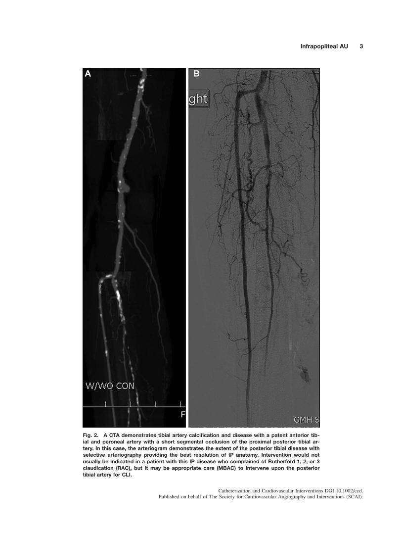
Fig. 2. A CTA demonstrates tibial artery calcification and disease with a patent anterior tib-
ial and peroneal artery with a short segmental occlusion of the proximal posterior tibial ar-
tery. In this case, the arteriogram demonstrates the extent of the posterior tibial disease with
selective arteriography providing the best resolution of IP anatomy. Intervention would not
usually be indicated in a patient with this IP disease who complained of Rutherford 1, 2, or 3
claudication (RAC), but it may be appropriate care (MBAC) to intervene upon the posterior
tibial artery for CLI.
Infrapopliteal AU 3
Catheterization and Cardiovascular Interventions DOI 10.1002/ccd.Published on behalf of The Society for Cardiovascular Angiography and Interventions (SCAI).

Procedural success is defined as the re-establishmentof direct “in-line” pulsatile flow to the foot through atleast one IP artery. It is currently unknownwhether heal-ing rates are improved when in-line flow to the foot isestablished through more than one IP artery, but maxi-mizing blood flow through more than one artery is partic-ularly attractive in patients with inadequate collaterals,plantar arch vessels, or limb-threatening ischemia. Con-sequently, the size, location, and extent of the necrosismay impact the necessity for multi-vessel revasculariza-
tion. Furthermore, revascularization of the angiosome (athree-dimensional vascular territory supplied by specificsource artery), has been shown to improve healing rates,using either endovascular or surgical therapies, whencompared to revascularization of the non-angiosome ter-ritory [11–13]. However, realistically the angiosome-based revascularization strategy may be limited by thelength and/or complexity of disease, the extent of collat-eralization, and the anatomic variability among patients,including anatomic anomalies [5].
Fig. 3. A 67-year-old patient with right toe necrosis, diabetes with non-compressible tibial
vessels by ABI and toe pressure of <20 mm Hg. Diffuse disease in the posterior tibial and
plantar arch that is appropriate for endovascular therapy given CLI (AC).
4 Gray et al.
Catheterization and Cardiovascular Interventions DOI 10.1002/ccd.Published on behalf of The Society for Cardiovascular Angiography and Interventions (SCAI).
CLINICAL CONSIDERATIONS
The goals of therapy for patients with IP arterial dis-ease and CLI (Rutherford 4–6) include: relief of pain,healing of ulcerations, preservation of the limb frommajor amputation, improvement in the patient’s qualityof life and functionality, and prolonging survival.
Arteriographically severe IP stenosis is defined as aluminal reduction of 70–99% or occlusion in at leastone IP artery [6,14,15]. Moderate IP stenoses isdefined as a luminal reduction of 50–69% and mild IPstenosis is defined as a luminal reduction of <50%.Obstructive disease in the below-knee popliteal arterylimits blood flow to the three tibial vessels (anterior,posterior and peroneal) and is equivalent to three ves-sel disease, while narrowing of the tibioperoneal trunkaffects two tibial arteries (peroneal and posterior tibial)and is considered two-vessel disease. Tibial arteryocclusions can be classified according to length asshort (<2 cm), medium (2–4 cm), or long (>4 cm)[10]. Prior to considering IP intervention, all hemody-namically significant inflow disease (aortoiliac and/orFP) should be treated to normalize inflow to the IP cir-culation. Then, if deemed clinically necessary, onemay proceed with management of the IP disease(Figs. 1–8).
Table II lists the appropriate use for IP interventionsand provides a management strategy of IP disease.Only moderate to severe IP arterial obstructions shouldbe considered for treatment, since the evidence forbenefit for treatment of milder obstructions is unclear.The clinical scenarios in Table II assume that inflowdisease has been revascularized. At the present time,patients with IP disease and claudication should bepreferentially treated pharmacologically and a walkingprogram before considering any revascularization pro-cedure and the clinical scenarios in Table II assumethat life-style limiting claudication has been refractoryto pharmacologic and exercise therapy. The categoriesof Appropriate Care (AC), May Be Appropriate Care(MBAC), and Rarely Appropriate Care (RAC) wereassigned by consensus based on the best available evi-dence (Table II).
TECHNICAL CONSIDERATIONS
Techniques for IP intervention vary widely and it isbeyond the scope of this manuscript to detail each one.However, it is important to describe some fundamentalelements of procedural strategy. Ideally the re-establishment of pulsatile flow to the limb shouldrestore pedal pulses. Non-invasive studies should beperformed during longitudinal follow-up with the goalof normalizing the ankle:brachial index, improving toe
perfusion pressure (>30 mm Hg), and/or transcutane-ous oxygen pressure (>40 Torr) [8,9].
Access
The ideal arterial access site for IP interventionshould maximize access to the lesion and allow visual-ization throughout the procedure, with limited use ofremote contrast injections. The most common site ofaccess is either the contralateral, retrograde, commonfemoral artery with a cross over technique, or the ipsi-lateral, antegrade, common femoral artery approach.Other less common access strategies include: Ipsilateralantegrade superficial femoral artery, ipsilateral
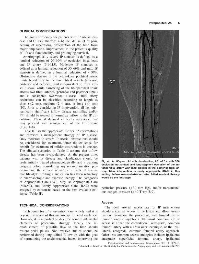
Fig. 4. An 86-year old with claudication, ABI of 0.4 with SFA
occlusion (not shown) and long-segment occlusion of the an-
terior tibial artery with mild disease in the posterior tibial ar-
tery. Tibial intervention is rarely appropriate (RAC) in this
setting (Inflow revascularization after failed medical therapy
would be the first step).
Infrapopliteal AU 5
Catheterization and Cardiovascular Interventions DOI 10.1002/ccd.Published on behalf of The Society for Cardiovascular Angiography and Interventions (SCAI).

antegrade popliteal artery, and retrograde tibio-pedalaccess. These are usually facilitated with fluoroscopic,angiographic, or ultrasound guidance.
Each approach has advantages and disadvantages, andthe success rate is largely dependent upon physician expe-rience. Ipsilateral, antegrade common femoral arteryaccess allows for easier guidewire control and device
pushability/deliverability compared to a contralateralapproach. Antegrade access can be challenging in obesepatients, those with a high takeoff of the superficial femo-ral artery, and those with extensive scarring of the groin.Retrograde tibio-pedal access is generally reserved forlesions that cannot be crossed from an antegradeapproach. Access can be obtained with visualization using
Fig. 5. A left leg tibial arteriogram done with the catheter placed in the aorta (A) and then in
the ipsilateral SFA (B). Medical decision making for tibial intervention requires selective imag-
ing, often with oblique imaging. This is an example of no direct flow to the calf or foot due to
disease affecting all three tibial arteries. Intervention of the tibioperoneal trunk would be indi-
cated (AC) in a symptomatic patient.
6 Gray et al.
Catheterization and Cardiovascular Interventions DOI 10.1002/ccd.Published on behalf of The Society for Cardiovascular Angiography and Interventions (SCAI).

ultrasound or angiographic visualization after injectingcontrast from the primary access site and inserting theneedle directly into the contrast column.
Percutaneous Transluminal Angioplasty
Percutaneous transluminal angioplasty (PTA) or bal-loon angioplasty is the current standard of therapy, with atechnically successful result (<30% residual stenosis) inthe majority of cases (77–100%), but is limited by highrestenosis rates [16,17]. Flexible, low profile, balloons
have enhanced the ability to cross, and successfully treat,focal, multi-focal, diffuse, and occlusive lesions.
Balloon catheters are available in lengths that can treatthe entire IP artery with one or two inflations. Clinicalstudies are limited by the heterogeneity of disease, lack ofrandomized trials, multiplicity of techniques used, exclu-sion of early treatment failures, and crossover to openbypass during follow-up [17,18].
Restenosis after IP balloon angioplasty is commonwithin the first year and is dependent upon the size of theartery and length of diseased segment. Negative
Fig. 6. A 78-year old with ischemic ulceration of the left anterior shin and arteriographic find-
ings of SFA stenosis (not shown) with long segment occlusion of posterior tibial (PT) artery,
long segment occlusion of the peroneal artery, and short segmental occlusion of the anterior
tibial artery that reconstitutes distally. Intervention is indicated to treat the SFA and anterior tib-
ial artery (AC), leaving the PTand peroneal for patients with incomplete healing (MBAC).
Infrapopliteal AU 7
Catheterization and Cardiovascular Interventions DOI 10.1002/ccd.Published on behalf of The Society for Cardiovascular Angiography and Interventions (SCAI).

predictors include: the presence of diabetes mellitus,
chronic kidney disease, non-ambulatory status, and
increasing severity of ischemia at the time of presenta-
tion [19].
Cutting Balloon Angioplasty
Balloon angioplasty has been the subject of mostclinical studies, but several modified balloons havebeen used, i.e., cryoplasty or cutting/scoring balloons.
Fig. 7. An arteriogram of right leg (A, B) shows long segment occlusion of anterior tibial ar-
tery and posterior tibial artery (arrow) distally with a poorly collateralizing peroneal artery. In
the presence of inflow disease (iliac or SFA) in a patient with claudication, there is no indica-
tion (RAC) to treat the tibial disease. However, in a patient with foot ulceration, attempting to
treat the posterior tibial artery may be appropriate (MBAC). [Color figure can be viewed in
the online issue, which is available at wileyonlinelibrary.com.]
8 Gray et al.
Catheterization and Cardiovascular Interventions DOI 10.1002/ccd.Published on behalf of The Society for Cardiovascular Angiography and Interventions (SCAI).

There are no comparative data for these newer modal-ities versus balloon angioplasty for IP arteries,
although studies in other vascular territories have been
negative [20,21]. Cutting balloon angioplasty (Boston
Scientific, Natick, MA) may be appropriate for lesions
resistant to plain balloon angioplasty that are fibrous
or calcified, but the added cost, and lack of compara-
tive evidence, makes these balloons not ideal for rou-
tine use.
Drug-Coated Balloons
Drug-coated balloons deliver anti-proliferativedrugs to the arterial wall at the time of dilation. Withinflation times of 30–60 sec, anti-proliferative drug(paclitaxel, sirolimus, and everolimus) levels have beenshown to reach the adventitia of the artery, and remaindetectable for weeks. The inhibition of the intimalhyperplastic response to arterial injury can potentiallylimit the amount of clinical restenosis [22]. This
Fig. 8. An arteriogram of right leg (A5anterior–posterior pro-
jection and B5 lateral) in a 59-year-old male with ischemic ulcer-
ation of the right foot, and no SFA or iliac disease of significance.
There is long segment disease of the tibioperoneal trunk (TPT)
with reconstitution of peroneal artery and excellent visualization
of the distal anterior tibial artery above the ankle. In a low surgi-
cal risk patient likely to survive two years with good quality auto-
logous vein, bypass is the preferred treatment. In a high surgical
risk patient, with extensive comorbidities, endovascular treat-
ment of TPTand peroneal artery may be indicated (MBAC).
Infrapopliteal AU 9
Catheterization and Cardiovascular Interventions DOI 10.1002/ccd.Published on behalf of The Society for Cardiovascular Angiography and Interventions (SCAI).

promising technology is not yet approved for use in theUnited States.
Atherectomy
Atherectomy devices help modify arterial compli-ance and/or remove plaque in order to facilitate lesion
crossing, and to debulk lesions prior to balloon angio-plasty. Devices are designed to ablate the atheroma
(Excimer laser, Spectranetics, Colorado Springs, CO),longitudinally excise the atheroma (TurboHawk/Sil-verHawk, ev3 Inc., Plymouth, MN), or rotationally
excise the atheroma (Jetstream G3, Pathway Medical,Kirkland WA and Orbital Atherectomy System CSI,St. Paul, MN) [23,24]. The clinical efficacy of thesedevices remains to be proven in randomized con-trolled clinical trials. Currently, there is no compara-tive evidence to support the use of any atherectomydevices in the IP arteries to improve clinicaloutcomes.
Infrapopliteal Stenting
Stenting of recalcitrant, restenotic, short or long IPlesions have been done with self-expanding nitinolstents, bare-metal stents (BMS), and drug-eluting stents(DES). [14,15,25–31]. These devices are associatedwith significantly better primary patency, primary-
assisted patency, and fewer secondary interventions at3-year follow-up than balloon angioplasty. DES seemsto enhance this effect over BMS. The Drug-Elutingstents in The Critically Ischemic Lower Leg Trial(DESTINY Trial), a prospective randomized trial of
140 patients with IP lesions >50% with a reference di-ameter of 2.0–3.5 mm and length< 4 cm, wererandomized to either BMS (Multi-Link Vision) or DES(Xience V, everolimus eluting). Primary patency (85%vs. 54%), late lumen loss, mean in-stent diameter ste-
nosis, and target lesion revascularization were all sig-nificantly in favor of DES treatment. However, neitherfinal Rutherford class 0 or 1 at 1 year, nor 1-year sur-vival, was different between the groups [14]. Limita-tions of balloon expandable stents include: their lackof significant flexibility, the potential for fracture or
compression from external trauma (particularly in thedistal anterior and posterior tibial artery), and theirhigh-cost expense (particularly when using multiplestents for long lesions).
The prospective randomized multicenter compari-son of balloon angioplasty and IP stenting, with thesirolimus-eluting stent in patients with ischemic pe-ripheral arterial disease (ACHILLES Trial), comparedpercutaneous balloon angioplasty to sirolimus-elutingstents (SES) for symptomatic IP artery disease. Twohundred patients (mean age 73 years; 64% diabetic)were randomized with the primary endpoint of arte-riographic patency at one year. The authors foundthat SES significantly lowered angiographic restenosis(22.4% vs. 41.9%, P¼ 0.019) and improved vesselpatency (75.0% vs. 57.1%, P¼ 0.025). There were no
TABLE II. Clinical Scenarios in Which Treatment of Infrapo-
pliteal Artery Disease May Be Considered
Appropriate Care � Moderate–severe claudication (RC 2–3) with
two, or three-vessel IP disease (if the arterial
target lesion is focal)
� Ischemic rest pain (RC4) with two, or three-
vessel IP disease (to provide direct flow to
the plantar arch and to maximize volume
flow to foot)
� Minor tissue loss (RC 5) with two, or three-
vessel IP disease (to provide direct flow to
the plantar arch and to maximize volume
flow to foot)
� Major tissue loss (RC 6) with two, or three-
vessel IP disease (to prevent major amputa-
tiona and to facilitate healing a minor ampu-
tationb)
May Be
Appropriate Care
� Moderate–severe claudication (RC 2–3) with
two, or three-vessel IP disease (occlusion or
diffuse disease)
� Ischemic rest pain (RC 4) with one, or two-
vessel IP disease (to provide direct flow to
the plantar arch and in two-vessel, to maxi-
mize volume flow to foot)
� Minor tissue loss (RC 5) with one-vessel IP
disease (to provide direct flow to the plantar
arch and to maximize volume flow to foot)
Rarely Appropriate
Care
� Mild claudication (RC 1) with, one, two, or
three-vessel IP disease
� Moderate–severe (RC 2–3) claudication
symptoms with one-vessel IP disease
� Major tissue loss (RC 6) with one-vessel IP
disease
RC¼Rutherford Classifications for chronic limb ischemia. One-vessel
infrapopliteal disease implies that two tibial arteries are without hemody-
namically significant stenosis or occlusion; two-vessel infrapopliteal dis-
ease implies that one tibial artery is without hemodynamically
significant stenosis or occlusion; three-vessel infrapopliteal disease
implies that all three tibial arteries have hemodynamically significant
stenosis and/or occlusion; no significant infrapopliteal disease implies
that all three tibial arteries are without hemodynamically significant ste-
nosis or occlusion; Severe stenosis¼ luminal narrowing 70–99%; Moder-
ate stenosis¼ luminal narrowing 50–69%; Mild stenosis¼ luminal
narrowing <50%; Occlusion¼No flow through the arterial segment.
Tibioperoneal trunk disease affects both the posterior tibial and peroneal
arteries so would be consistent with two-vessel disease. Focal infrapopli-
teal lesion¼ discrete area of narrowing that can be treated with a single
15 mm long balloon/stent; Multiple lesions¼more than one focal lesion
in non-contiguous arterial segments; Diffuse lesion¼ a continuous seg-
ment of disease treated with> 15 mm long balloon/stent.aMajor amputation¼ removal of leg either above, or below the knee but
above the ankle.bMinor amputation¼ removal of the foot or portions of it [i.e., isolated
toe(s).]
10 Gray et al.
Catheterization and Cardiovascular Interventions DOI 10.1002/ccd.Published on behalf of The Society for Cardiovascular Angiography and Interventions (SCAI).
differences in death, amputation, target vessel revas-cularization, or functional improvement (Rutherfordcategory) rates between SES and PTA [31]. Thesedata support the perception that stents improve pat-ency over PTA, even for short lesions in the tibialarteries.
The XCELL trial, a study to evaluate the safety andperformance of the Xpert TM stent in treating below-the-knee lesions in patients undergoing percutaneousintervention for chronic CLI, screened over 700patients and enrolled 120 CLI patients to evaluate therole of nitinol stents for IP disease [15]. The majorityof these patients also required inflow artery treatment.Overall, the mean lesion length was 4.76 4.2 cm withstenoses averaging 3.66 3.5 cm, and occlusions aver-aging 7.16 4.5 cm. The angiographic binary restenosisrate was 68.5%, with a 6-month ulcer-healing rate of43%. These data provide important lessons: promptwound healing may require more than improved perfu-sion (<50% healing at six months), limb salvage rateswere comparable to historical surgical rates, restenosisrates were higher for IP intervention, and the stent didnot prevent restenosis in the majority of patients. Con-sequently, the use of stents in the IP arteries is stillunder scrutiny and is felt to be most effective in focal,proximal lesions that fail primary therapy with balloonangioplasty. Furthermore, the patency advantage ofstenting may be more applicable to Rutherford 3 and 4than Rutherford 5 and 6 where stenting has not beenconsistently shown to affect limb salvage rates.
Unresolved Issues
There are many unanswered questions regarding therole of endovascular intervention for the treatment ofCLI in patients with IP atherosclerotic disease. Therole of bio-absorbable stents, or the combination ofdrug-coated balloon following atherectomy or stentingin the IP vessels, remains to be determined [32].
CONCLUSION
CLI is the predominant clinical indication for treat-ment of IP arterial disease and occurs when arterialperfusion is reduced below a critical level resulting inischemic pain and/or skin breakdown. Prompt revascu-larization is aimed at symptom relief with improvedlimb salvage and ulcer healing. Multilevel disease ismore common than isolated IP disease, and a system-atic approach to achieve straight-line flow from theiliac to pedal arch with complete revascularization isnecessary to optimize outcomes.
PTA is the current standard for endovascular therapyfor clinically significant IP disease. Bailout bare metal
and drug eluting stents in the tibial arteries should beconsidered for failures of balloon angioplasty. Studiesare currently enrolling patients to address the use ofcombined strategies (i.e., atherectomy and drug-coatedballoons). Further data are needed regarding the utilityof atherectomy devices, drug-coated balloons, DES,and bioabsorbable stents in IP interventions. However,until these results are available, given the increasedcosts of other modalities (e.g., cutting balloons, cryo-plasty, laser, orbital, rotational, and directional atherec-tomy catheters), and the lack of comparative data tosupport their efficacy, balloon angioplasty shouldremain the initial endovascular therapy for most IPdisease.
ACKNOWLEDGMENT
The authors thank Allyson Hale for her editorialassistance.
REFERENCES
1. European Working Group on Critical Limb Ischaemia. SecondEuropean Consensus Document on chronic critical leg ischae-
mia. Eur J Vasc Surg 1992;6 (Suppl A):1–32.2. Wolfe JH, Wyatt MG. Critical and subcritical ischaemia. Eur J
Vasc Endovasc Surg 1997;13:578–582.3. Aboyans V, Desormais I, Lacroix P, Salazar J, Criqui MH,
Laskar M. The general prognosis of patients with peripheral ar-
terial disease differs according to the disease localization. JACC2010;55:898–903.
4. Graziani L, Silvestro A, Bertone V, Manara E, Andreini R,Sigala A, Mingardi R, De Giglio R. Vascular involvement in di-abetic subjects with ischemic foot ulcer: A new morphologic
categorization of disease severity. Eur J Vasc Endovasc Surg2007;33:453–460.
5. Gray BH, Grant AA, Kalbaugh CA, Blackhurst D, Langan EMIII, Taylor SA, Cull DL. The impact of isolated tibial disease onoutcomes in the critical limb ischemic population. Ann Vasc
Surg 2010;24:349–359.6. Sadek M, Ellozy SH, Turnbull IC, et al. Improved outcomes are
associated with multilevel endovascular intervention involving
the tibial vessels compared with isolated tibial intervention.J Vasc Surg 2009;49:638–644.
7. Bradbury AW, et al. Bypass versus angioplasty in severe ische-mia of the leg (BASIL): Multicenter, randomized controlledtrial. Lancet 2005;366:1925–1934.
8. Fernandez N, McEnaney R, Marone LK, et al. Predictors of fail-ure and success of tibial interventions for critical limb ischemia.
J Vasc Surg 2010;52:834–842.9. Karnabatidis D, et al. Incidence, anatomical location, and clinical
significance of compressions and fractures in infrapopliteal
balloon-expandable metal stents. J Endovasc Ther 2009;16:15–22.10. Norgren L, Hiatt WR, Dormandy JA, Nehler MR, Harris KA,
Fowkes FG, TASC II Working Group. Inter-Society Consensusfor the Management of Peripheral Arterial Disease (TASC II).J Vasc Surg 2007;45 (Suppl S):S5–S67.
11. Neville RF, Attinger CE, Bulan EJ, Ducic I, Thomassen M,Sidawy AN. Revascularization of a specific angiosome for limbsalvage: Does the target artery matter: Ann Vasc Surg 2009;23:
367–373.
Infrapopliteal AU 11
Catheterization and Cardiovascular Interventions DOI 10.1002/ccd.Published on behalf of The Society for Cardiovascular Angiography and Interventions (SCAI).

12. Iida O, Soga Y, Hirano K, Kawasaki D, Suzudi K, et al. Long-term results of direct and indirect endovascular revascularization
based on the angiosome concept in patients with critical limbischemia presenting with isolated below-the-knee lesions. J VascSurg 2012;55:363–370.
13. Alexndrescu V, Vincent G, Azdad K, et al. A reliableapproach to diabetic neuroischemic foot wounds: Below-the-
knee angiosome-oriented angioplasty. J Endovasc Ther 2011;18:376–387.
14. Bosiers M, Scheinert D, Peeters P, Torsello G, Zeller T,
Deloose K, Schmidt A, Tessarek J, Vinck E, Schwartz LB.Randomized comparison of everolimus-eluting versus bare-
metal stents in patients with critical limb ischemia and infrapo-pliteal arterial occlusive disease. J Vasc Surg 2012;55:390–398.
15. Rocha-Singh KJ, Jaff M, Joye J, Laird J, Ansel G, Schneider P;
on behalf of the VIVA Physicians. Major adverse limb eventsand wound healing following infrapopliteal artery stent implan-tation in patients with critical limb ischemia: The XCELL trial.
Catheter Cardiovasc Interv 2012;80:1042–1051.16. Romiti M, Albers M, Brochado-Neto FC, Durazzo AE, Pereira
CA, DeLuccia N. Meta-analysis of infrapopliteal angioplastyfor chronic critical limb ischemia. J Vasc Surg 2008;47:975–981.
17. Jaffery Z, Grant AG, White CJ. Critical limb ischemia: doeslong-term patency matter? Vasc Med 2010;15:439–441.
18. Ihnat DM, Mills JL. Current assessment of endovascular therapyfor infrainguinal arterial occlusive disease in patients with dia-betes. J Vasc Surg. 2010;52:92S–95S.
19. Taylor SM, York JW, Cull DL, Kalbaugh CA, Cass AL, LanganEL III, Clinical success using patient-oriented outcome measures
after lower extremity bypass and endovascular intervention forischemic tissue loss. J Vasc Surg 2009;50:534–541.
20. Das TS, McNamara T, Gray BH, Sedillo GJ, Turley BR,
Kollmeyer K, Rogoff M, Aruny JE. Primary Cryoplasty therapyprovides durable support limb salvage in critical limbischemia patients with infrapopliteal lesions: 12-month follow-
up results from the BTK Chill Trial. J Endovasc Ther 2009;16:II19–II30.
21. Ansel GM, Sample NS, Botti III CF Jr, Tracy AJ, Silver MJ,Marshall BJ, George BS. Cutting balloon angioplasty of thepopliteal and infrapopliteal vessels for symptomatic limb ische-
mia. Catheter and Cardiovasc Interv 2004;61:1–4.22. Schmidt A, Piorkowski M, Werner M, et al. First experience
with drug-eluting balloons in infrapopliteal arteries: restenosisrate and clinical outcome. J Am Coll Cardiol 2011;58:1105–1109.
23. Zeller T, Sixt S, Schwarzwalder U, Schwarz T, Frank U,Burgelin K, Pochert V, Muller C, Noory E, Krankenberg H,
Hauswald K, Neumann FJ, Rastan A. Two-year results after
directional Atherectomy of infrapopliteal arteries with the Sil-verHawk device. J Endovasc Ther 2007;14:232–240.
24. Laird JR, Zeller T, Gray BH, Scheinert D, Vranic M, Reiser C,Biamino G; LACI Investigators. Limb salvage following laser-assisted angioplasty for critical limb ischemia: results of the
LACI multicenter trial. J Endovasc Ther 2006;13:1–11.25. Kickuth R, Keo HH, Triller J, et al. Initial clinical experience
with the 4-F self-expanding XPERT stent system for infrapopli-teal treatment of patients with severe claudication and criticallimb ischemia. J Vasc Interv Radiol 2007;18:703–708.
26. Feiring AJ, Wesolowski AA, Lade S. Primary stent-supportedangioplasty for treatment of below-knee critical limb ischemia
and severe claudication: Early and one-year outcomes. J AmColl Cardio 2004;44:2307–2314.
27. Siablis D, Karnabatidis D, Katsanos K, Diamantopoulos A,
Spiliopoulos S, Kagadis GC, Tsolakis J. Infrapopliteal applica-tion of sirolimus-eluting versus bare metal stents for criticallimb ischemia: Analysis of long-term angiographic and clinical
outcome. J Vasc Interv Radiol 2009;20:1141–1150.28. Rastan A, Tepe G, Krankenberg H, Zahorsky R, Beschorner U,
Noory E, Sixt S, Schwarz T, Brechtel K, B€ohme C, NeumannFJ, Zeller T. Sirolimus-eluting stents vs. bare-metal stents fortreatment of focal lesions in infrapopliteal arteries: A double-
blind, multi-centre, randomized clinical trial. Eur Heart J 2011;32:2274–2281.
29. Karnabatidis D, Spiliopoulos S, Diamantopoulos A, Katsanos K,Kagadis GC, Kakkos S, Siablis D. Primary everolimus-elutingstenting versus balloon angioplasty with bailout bare metal
stenting of long infrapopliteal lesions for treatment of criticallimb ischemia. J Endovasc Ther 2011;18:1–12.
30. Biondi-Zoccai GG, Sangiorgi G, Lotrionte M, Feiring A,Commeau P, Fusaro M, Agostoni P, Bosiers M, Peregrin J,Rosales O, Cotroneo AR, Rand T, Sheiban I. Infragenicular
stent implantation for below-the-knee atherosclerotic disease:Clinical evidence from an international collaborative meta-analysis on 640 patients. J Endovasc Ther 2009;16:251–260.
31. Scheinert D, Katsanos K, Zeller T, Koppensteiner R, et al. Aprospective randomized multicenter comparison of balloon
angioplasty and infrapopliteal stenting with the Sirolimus-eluting stent in patients with ischemic peripheral arterial disease:1-year results from the ACHILLES Trial. J Am Coll Cardiol
2012;60:2290–2295.32. Bosiers M, Peeters P, D’Archambeau O, Hendriks J, Pilger E,
D€uber C, Zeller T, Gussmann A, Lohle PN, Minar E, ScheinertD, Hausegger K, Schulte KL, Verbist J, Deloose K, Lammer J;AMS INSIGHT Investigators. AMS INSIGHT—Absorbable
metal stent implantation for treatment of below-the-knee criticallimb ischemia: 6-month analysis. Cardiovasc Intervent Radiol
2009;32:424–435.
12 Gray et al.
Catheterization and Cardiovascular Interventions DOI 10.1002/ccd.Published on behalf of The Society for Cardiovascular Angiography and Interventions (SCAI).

AuthorRelationshipswithIndustryandOtherEntities(C
omprehensive)—Appropriate
UseforInfrapoplitealArterialIntervention:AnExpert
ConsensusDocumentfrom
theSociety
forCardiovascularAngiographyandIntervention(SCAI)
CommitteeMem
ber
Advisory
Board/Board
Mem
ber
Consultant
Speaker’s
Bureau
Ownership/
Partnership/
Principal
Personal
Research
Institutional,Organizational
orOther
Financial
Benefit
Expert
Witness
Larry
Diaz-Sandoval,MD
None
CSI,Inc.*
None
None
None
None
None
RobertDieter,MD
None
None
None
None
None
None
None
Bruce
Gray,MD
None
None
None
None
None
None
None
Kam
alGupta,MD
None
None
None
None
BARD*
None
None
MichaelJaff,DO
AbbottVascular*
American
Genomics
Astra
Zeneca
Biomet
Biologicals
BostonScientific*
Cordis*
Covidien*
EkosCorporation
Medtronic*
Micell,Inc.
Primacea
None
PQ
Bypass
None
VIV
APhysicians,a501c3not-for-profit
educationandresearch
organization—
Boardmem
ber
None
Christopher
White,
MD
None
None
None
None
None
None
None
This
table
represents
allhealthcare
relationshipsofcommitteemem
berswithindustry
andother
entities
that
werereported
byauthors,includingthose
notdeemed
toberelevantto
this
document,
atthetimethis
documentwas
under
development.Thetable
does
notnecessarily
reflectrelationshipswithindustry
atthetimeofpublication.A
personis
deemed
tohaveasignificantinterest
in
abusinessiftheinterest
represents
ownership
of�5%
ofthevotingstock
orshareofthebusinessentity,orownership
of�$10000ofthefairmarket
valueofthebusinessentity;oriffunds
received
bythepersonfrom
thebusinessentity
exceed
5%
oftheperson’s
gross
incomeforthepreviousyear.Relationshipsthat
existwithnofinancial
benefitarealso
included
forthepurpose
oftransparency.Relationshipsin
thistable
aremodestunless
otherwisenoted.Pleasereferto
http://www.cardiosource.org/Science-A
nd-Q
uality/Practice-Guidelines-and-Q
uality-Standards/Relation-
ships-With-Industry-Policy.aspxfordefinitionsofdisclosure
categories
oradditional
inform
ationabouttheACCFDisclosure
Policy
forWritingCommittees.
*Nofinancial
benefit.
Infrapopliteal AU 13
Catheterization and Cardiovascular Interventions DOI 10.1002/ccd.Published on behalf of The Society for Cardiovascular Angiography and Interventions (SCAI).