SBES Fall Newsletter - 2008
16
What is Regenerative Medicine? The main thrust of regenerative medicine is to harness the natural healing process by helping cells to grow, divide and dif- ferentiate outside of the body before implantation, or by stimulating progenitor cells to repair tissues in the body. Why Regenerative Medicine? Since the first organ transplant in 1954, there have been few clinical advances. Organ transplantation was a major advance in medicine in the twentieth century but demand for transplantable organs consistently outstrips supply. Every 11 minutes, a name is added to the national transplant wait- ing list, and more than 97,014 people cur- rently await transplants. An aver- age of 18 people die each day while waiting for organs, according to statistics A tissue engineering scaffold made from liver is injected with a red dye. Cells are removed from the liver but the ul- trastructure of the extracellular matrix is left intact, so much so that the vascular network is visible as the dye travels through the vessels. This scaffold can be re-populated with cells and new tissue created with a built-in vascular supply. SBES Is At The Forefront Of Regenerative Medicine from United Network for Organ Sharing, 4/29/08. The prevalence of obesity, hypertension and diabetes, and the growing numbers among the aging population will likely increase the need for organs for years to come. Then there are the complica- tions of the actual transplant; rejections and medications. Immuno- suppressing medications become a lifelong need and carry both short and long-term side effects that can reduce the quality and life span of patients. Where will the organs come from? Regenerative medicine can bypass the organ shortage and the transplant complications by making the donor and the recipient the same. A biopsy from the patient yields cells that are nurtured in the laboratory to form functional tissues and organs which can then be reimplanted into the patient. The use of a patient’s own cells eliminates the risk of rejection that accompanies traditional organ transplantation so that immune system-altering drugs are not necessary. Wake Forest’s role in new technology commercialization Children and teenagers, who have received laboratory grown bladders using their own cells, have experienced success. Dr. Anthony Atala, at Wake Forest Institute for Regenerative Medicine (WFIRM), implanted the first laboratory-grown bladder in 1999, and in April 2006 released a report discussing the long-term results of seven patients who had the surgery. Tengion is a regenerative medicine company which is commercializing this technology and has already entered phase II clinical trials. Where discovery and hope meet In January 2007 scientists from the WFIRM and Harvard Medical School discovered a new source of stem cells and have used them to create muscle, bone, fat, blood vessel, nerve and liver cells in the laboratory. Atala and colleagues discovered a small amount of stem cells in the amniotic fluid — estimated at one percent — which can give rise to many of the specialized cell types found in the human body. These stem cells are called amniotic fluid-derived stem (AFS) cells, and it took seven years of work to determine if they were true stem cells. These cells were harvested from amniotic fluid taken during routine amniocentesis procedures and similar cells were isolated from the placenta or “afterbirth.” This breakthrough may provide alternative cells to those isolated from embryos. Functional tests of AFS cells transplanted into mice have been successful, such as injection of neural cells created from AFS into mice with degenerative brain disease. The cells grew and “re-populated” the diseased areas. Many scientists believe stem cells have the potential to replace damaged cells and tissue in conditions such as spinal cord injuries, diabetes, and Alzheimer’s. BRIDGING THE GAP ENGINEERING, SCIENCE, & MEDICINE SBES NEWS VIRGINIA TECH WAKE FOREST UNIVERSITY School of Biomedical Engineering and Sciences See Forefront, page 6 S U M M E R / F A L L 2 0 0 8 • W W W . S B E S . V T . E D U
-
Upload
alexparrish -
Category
Documents
-
view
222 -
download
0
description
Virginia Tech - Wake Foreset School of Biomedical Engineering and Sciences Fall Newsletter - 2008
Transcript of SBES Fall Newsletter - 2008
What is Regenerative Medicine? The main thrust of regenerative medicine is to harness the
natural healing process by helping cells to grow, divide and dif-ferentiate outside of the body before implantation, or by stimulating progenitor cells to repair tissues in the body.
Why Regenerative Medicine?Since the first organ transplant in 1954, there have been few
clinical advances. Organ transplantation was a major advance in medicine in the twentieth century but demand for transplantable
organs consistently outstrips supply. Every11 minutes, a name is added to the
national transplant wait-ing list, and more
than 97,014people cur-
rently awaittransplants.
An aver-age of 18people die each day while waiting for organs,
accordingto statistics
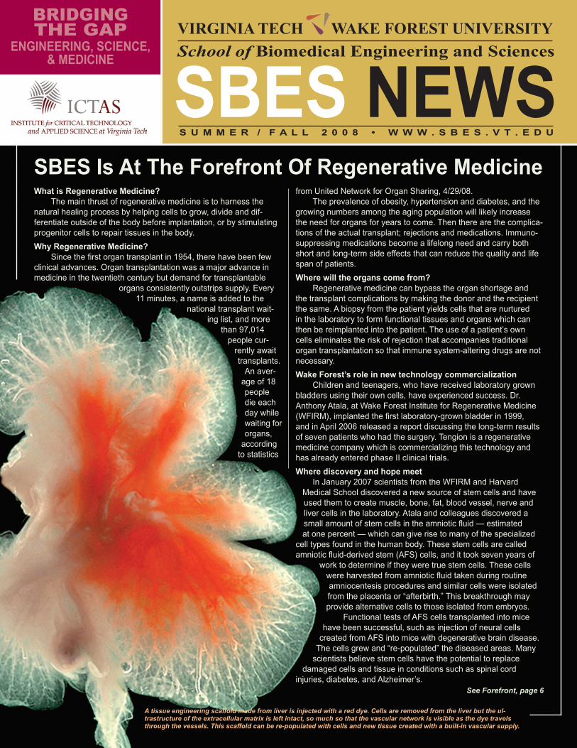
A tissue engineering scaffold made from liver is injected with a red dye. Cells are removed from the liver but the ul-trastructure of the extracellular matrix is left intact, so much so that the vascular network is visible as the dye travels through the vessels. This scaffold can be re-populated with cells and new tissue created with a built-in vascular supply.
SBES Is At The Forefront Of Regenerative Medicinefrom United Network for Organ Sharing, 4/29/08.
The prevalence of obesity, hypertension and diabetes, and the growing numbers among the aging population will likely increase the need for organs for years to come. Then there are the complica-tions of the actual transplant; rejections and medications. Immuno-suppressing medications become a lifelong need and carry both short and long-term side effects that can reduce the quality and life span of patients.
Where will the organs come from? Regenerative medicine can bypass the organ shortage and
the transplant complications by making the donor and the recipient the same. A biopsy from the patient yields cells that are nurtured in the laboratory to form functional tissues and organs which can then be reimplanted into the patient. The use of a patient’s own cells eliminates the risk of rejection that accompanies traditional organ transplantation so that immune system-altering drugs are not necessary.
Wake Forest’s role in new technology commercializationChildren and teenagers, who have received laboratory grown
bladders using their own cells, have experienced success. Dr. Anthony Atala, at Wake Forest Institute for Regenerative Medicine (WFIRM), implanted the first laboratory-grown bladder in 1999, and in April 2006 released a report discussing the long-term results of seven patients who had the surgery. Tengion is a regenerative medicine company which is commercializing this technology and has already entered phase II clinical trials.
Where discovery and hope meetIn January 2007 scientists from the WFIRM and Harvard
Medical School discovered a new source of stem cells and haveused them to create muscle, bone, fat, blood vessel, nerve and liver cells in the laboratory. Atala and colleagues discovered asmall amount of stem cells in the amniotic fluid — estimatedat one percent — which can give rise to many of the specialized
cell types found in the human body. These stem cells are called amniotic fluid-derived stem (AFS) cells, and it took seven years of
work to determine if they were true stem cells. These cellswere harvested from amniotic fluid taken during routineamniocentesis procedures and similar cells were isolatedfrom the placenta or “afterbirth.” This breakthrough mayprovide alternative cells to those isolated from embryos.
Functional tests of AFS cells transplanted into micehave been successful, such as injection of neural cells
created from AFS into mice with degenerative brain disease.The cells grew and “re-populated” the diseased areas. Many
scientists believe stem cells have the potential to replacedamaged cells and tissue in conditions such as spinal cord
injuries, diabetes, and Alzheimer’s.
BRIDGINGTHE GAP
ENGINEERING, SCIENCE,& MEDICINE
SBES NEWSVIRGINIA TECH WAKE FOREST UNIVERSITYSchool of Biomedical Engineering and Sciences
See Forefront, page 6
S U M M E R / F A L L 2 0 0 8 • W W W . S B E S . v T . E d u
2
T he Virginia Tech – Wake Forest School of Biomedical Engineering and Sciences (SBES) strives to meet today’s technological and ac-ademic needs by preparing students to anticipate the future of the
biomedical field. This unique school was established by bringing together the Wake Forest University School of Medicine, the Virginia Tech College of Engineering, and the Virginia – Maryland Regional College of Veteri-nary Medicine, combining the resources and leveraging the strengths of these founding members to produce an environment that fosters graduate education and outstanding interdisciplinary research. Each college is in-ternationally recognized, and SBES combines researchers and resources into a coherent group.
According to Dean Richard Benson of the Virginia Tech College of Engineering, “The creation of SBES — a dynamic and collegial depart-ment that resides in three colleges at two universities — is the most interesting academic partnership that I have witnessed in three decades of university life. Our partnership with the Wake Forest University School of Medicine and the Virginia – Maryland Regional College of Veterinary Medicine has brought tremendous opportunities for biomedical engineer-ing research and education to the Virginia Tech College of Engineering.”
The school has made significant achievements in academics, in re-search with increased funding, and in professional growth by securing bril-liant faculty members who are leading us into the future. Ge Wang, Ph.D., is an excellent example. He is the Samuel Reynolds Pritchard Professor of Engineering, the director of the SBES Biomedical Imaging Division, and a joint faculty member at Virginia Tech and Wake Forest. Among his many honors and contributions, Dr. Wang and his collaborators published the first papers on spiral cone-beam CT, biolumi-nescence tomography, and interior tomography, each of which represents a frontier of biomedical imaging.
As I review the short-term success of SBES, I believe we are now strategically posi-tioned to be a leader in a unique academic program and to continue to de-velop as an internationally recognized educational and research biomedical engineering program. William Applegate, Ph.D., interim president of Wake Forest University Health Sciences and dean of the School of Medicine, states that “SBES is the “Jewel in the Crown” of
Producing the Next Generation of Biomedical Engineersour academic research program.” SBES has brought talented individuals/researchers together, and we are seeing their work now as stepping-stones for future innovation. For example, in April 2008 successful clinical trials were completed in irreversible electroporation (IRE), based on a paper published in 2005 by Rafael Davalos, Ph.D. IRE is a proven method for tumor ablation that creates no secondary thermal effects and therefore preserves the extracellular matrix, microvasculature, and nerves.
The combination of a public university from Virginia with recognized excellence in engineering and veterinary medicine and a private univer-sity in North Carolina with recognized excellence in human medicine has resulted in a program that is much more than the sum of its parts. Re-cently the Global Human Body Modeling Consortium awarded the Center for Injury Biomechanics (CIB) $4.9 million, and the Wake Forest Institute for Regenerative Medicine (WFIRM), where many of our students and faculty are involved in tissue engineering, has been awarded $42.5 mil-lion from the U.S. Department of Defense as part of a joint consortium. I invite you to read about these and many other initiatives and success stories in this newsletter. I think you will agree with Dr. Wayne Meredith, director of the Division of Surgical Sciences at Wake Forest University School of Medicine, that “SBES is one of the most exciting and stimulat-ing ventures of our schools. I am constantly amazed and inspired by the unique inquisitive environment it creates and the productivity of the research that results.” It is with this attitude that we will continue to chart our course.~ by Wally Grant, Ph.d., Head, virginia Tech - Wake Forest university
School of Biomedical Engineering and Sciences (SBES)
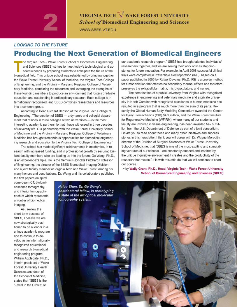
Haiou Shen, Dr. Ge Wang’s postdoctoral fellow, is prototyping a state of the art optical molecular tomography system.
VIRGINIA TECH WAKE FOREST UNIVERSITYSchool of Biomedical Engineering and Sciences
WWW.SBES.VT.EDU
LOOKING TO THE FUTURE
3
Over 70 million people in the United States with high blood pressure are at risk for left ventricular diastolic dysfunction (LVDD), and numerous studies have shown a link be-tween LVDD and heart failure. However, due to compensatory mechanisms, early stage dysfunction can be difficult to diagnose, and despite numerous advances in clinical modali-ties the prognosis and diagnosis of LVDD has remained unchanged over the past 20 years.
The Advanced Experimental Thermo-Flu-ids Engineering Research (AEThER) fluid me-chanics laboratory at Virginia Tech, in conjunc-tion with a team of cardiologists headed by Dr. William Little from Wake Forest University Baptist Medical Center, has been investigat-ing the role of hydrodynamics in LVDD. This investigation involves the examination and analysis of clinical imaging of the left ventricle.
Anonymous patient data was routinely di-agnosed by cardiologists and then sent to Vir-ginia Tech for hydrodynamic analysis. Through a series of image processing techniques and the implementation of fluid dynamics equa-tions, Color M-mode echocardiogram and phase contrast magnetic resonance images are being used to non-invasively collect propa-gation velocities and pressure distributions within the left ventricle. Propagation velocity is the wave speed at which the left ventricle is filling, as fluid moves towards the left ventricu-lar apex. Current results show an improved correlation of decreasing propagation velocity with decreasing diastolic function over cur-rently used clinical methods of calculating propagation velocities. The pressure gradi-ents and velocities calculated by the newly developed automated algorithms allow for the combination of variables to define dimension-less scaling parameters to characterize the effectiveness of the left ventricular filling.
This research will augment the under-standing of the causal relationship between
the left ventricular filling hydrodynamics and diastolic heart failure. It will enable the devel-opment of novel and more reliable diagnostic tools. This work will be presented at the 2008 ASME Summer Bio-engineering Confer-ence. Also, a portion of this work will con-tribute to Kelly Stew-art’s master’s thesis and will be part of the Ph.D. dissertation for John Charonko.
The AEThER Laboratory is focus-ing on other health issues such as, the hemodynamics of commercial coronary stents and magnetic drug targeting. Using a powerful laboratory-based measurement technique called Digital Particle Image Ve-locimetry (DPIV), a team of SBES graduate
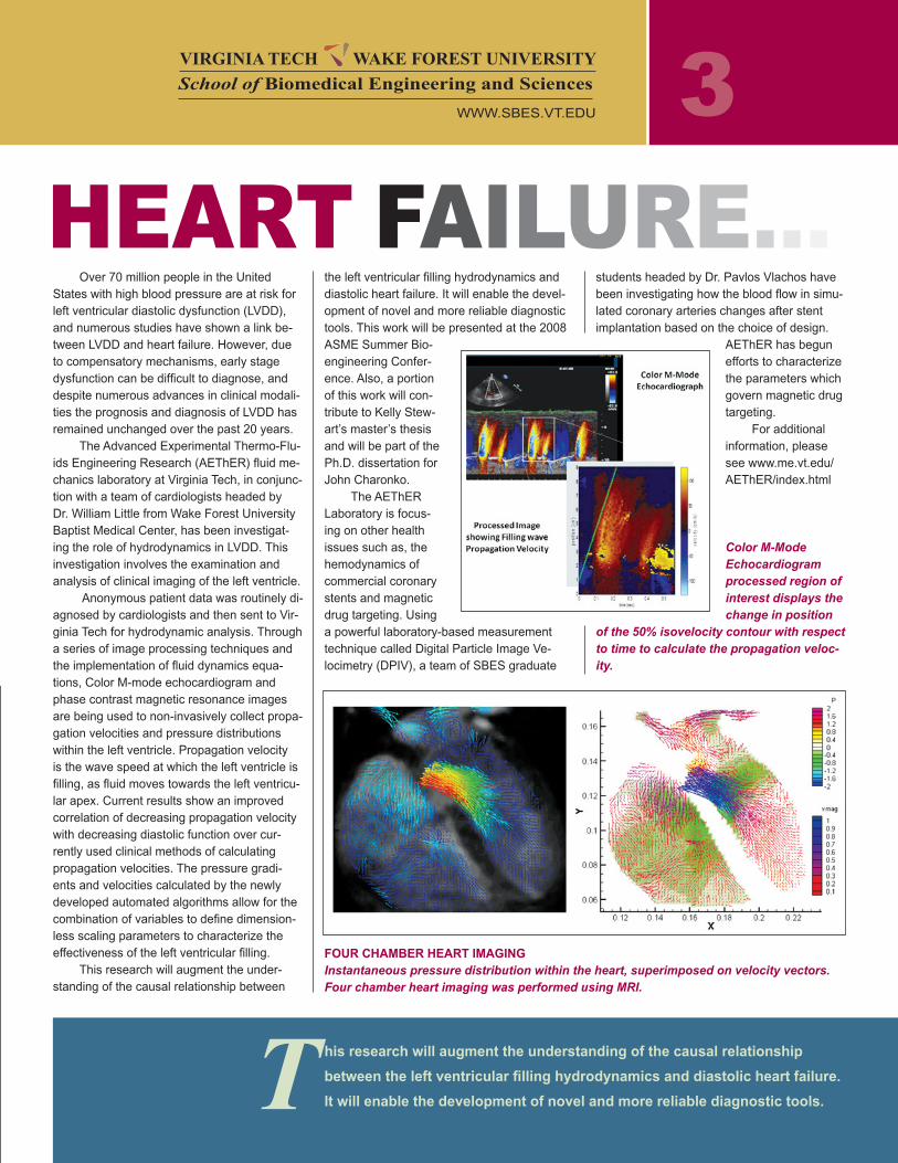
Color M-Mode Echocardiogram processed region of interest displays the change in position
of the 50% isovelocity contour with respect to time to calculate the propagation veloc-ity.
students headed by Dr. Pavlos Vlachos have been investigating how the blood flow in simu-lated coronary arteries changes after stent implantation based on the choice of design.
AEThER has begun efforts to characterize the parameters which govern magnetic drug targeting.
For additional information, please see www.me.vt.edu/AEThER/index.html
FOuR CHAMBER HEART IMAGINGInstantaneous pressure distribution within the heart, superimposed on velocity vectors. Four chamber heart imaging was performed using MRI.
HEART FAILURE...
his research will augment the understanding of the causal relationship
between the left ventricular filling hydrodynamics and diastolic heart failure.
It will enable the development of novel and more reliable diagnostic tools.T
VIRGINIA TECH WAKE FOREST UNIVERSITYSchool of Biomedical Engineering and Sciences
WWW.SBES.VT.EDU
4
dr. Stefan duma, of Virginia Tech, leads a team whose focus is to quantify the head accelera-tions of football players and has developed a unique sensor that consists of 12 accelerometers and a wireless transmitter designed for integration into existing football helmets. The data are collected in real time and downloaded to the sideline computer during all prac-tices and games. The Virginia Tech football team helmets have been instrumented with these sensors for five seasons, and over 50,000 head impacts have been recorded that provide linear and rotational accel-eration traces for each hit. The data are being used to develop brain injury criteria to validate advanced computational models of the human brain.
dr. Warren Hardy, also of Vir-ginia Tech, examines brain displace-ment and deformation during impact using high-speed biplane x-ray, and neutral density targets (NDTs) im-planted in the brain. Relative motion, maximum principal strain, maximum shear strain, and intracranial pressure are measured in human cadaver head and neck specimens. This research has shown that during impact, local brain tissue tends to keep its position and shape with respect to the inertial frame, resulting in relative motion between the brain and skull and deformation of the brain. A comple-mentary, multidisciplinary research effort involves the study of diffuse axonal injury (DAI) development after head impact and the associated injury cascades. This study is designed to obtain the direct relationship between focal neuronal damage as determined via classical pathology immunohis-tochemistry techniques, and magnetic resonance imaging (MRI) using a por-cine in-vivo model. The tissue staining is performed at the Virginia-Maryland Regional College of Veterinary Medi-cine, and the MRI scans are con-ducted using the Bruker 7T scanner of the Wake Forest University Center for Biomolecular Imaging. This study will quantify the location and extent of neuronal damage as it develops over time using spectral and diffusion-weighted MR scans.
dr. Joel Stitzel’s group is work-ing with the National Highway Traffic Safety Administration to develop geo-metric shape and size scaling factors for the pediatric brain and skull. The goal is to generate size and shape appropriate finite element models of the human brain for injury prediction. Kerry Danelson, a Ph.D. student, is working with Dr. Carol Geer, a neuro-surgeon and interventional radiologist, and Dr. Dennis Slice, an anthropolo-gist from the University of Vienna. Danelson has digitized landmarks from CT and MRI scans of normal pe-diatric brains and is using these land-marks with advanced shape analysis techniques to assess size and shape changes quantitatively in the brain with age. The scaling relationships will allow the prediction of mild traumatic brain injury in any age individual. In the future, the team will be extending their work to the aged population and looking at the effects of shape chang-es on the injury metrics resulting from similar impacts for predicting clinically relevant injuries.
dr. Clay Gabler’s and dr. Warren Hardy’s teams at Virginia Tech are examining traumatic brain injury (TBI) at the cellular level. The project is being conducted in collaboration with Dr. Beverly Rzi-galinski at the Edward Via Virginia College of Osteopathic Medicine (VCOM, located near the Virginia Tech campus), and Dr. Warren Hardy. TBI can occur from high-rate, high-magnitude mechanical loads to the head from events such as car crashes or explosive blasts. Their approach is to study the injury response of cell cultures of neural cells to these mechanical loads both through in-vitro experiments and through computational model-ing. The group has developed an Advanced Cell Deformation System (ACDS), which can provide pulse durations ranging from 20 to 100 ms, independent control of strain and strain rate, and arbitrary pres-sure pulse shape. When combined with finite element models of these experiments, these systems should lead to a fundamental understand-ing of the complex biochemical response of neural tissues to impact and blast loading.
Approximately 50,000 people die annu-ally from Traumatic Brain Injury (TBI) in the United States, representing more than 33 percent of all injury-related deaths (Centers for Disease Control and Prevention, 2002). The leading causes of TBI death are violence, mo-tor vehicle accidents, and falls.
While the Virginia Tech – Wake Forest
THE FACuLTY
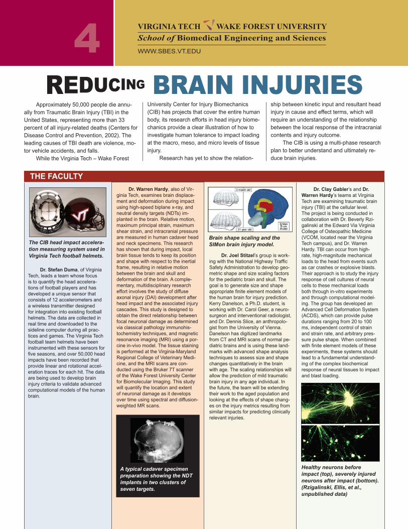
The CIB head impact accelera-tion measuring system used in Virginia Tech football helmets.
A typical cadaver specimen preparation showing the NDT implants in two clusters of seven targets.
University Center for Injury Biomechanics (CIB) has projects that cover the entire human body, its research efforts in head injury biome-chanics provide a clear illustration of how to investigate human tolerance to impact loading at the macro, meso, and micro levels of tissue injury.
Research has yet to show the relation-
ship between kinetic input and resultant head injury in cause and effect terms, which will require an understanding of the relationship between the local response of the intracranial contents and injury outcome.
The CIB is using a multi-phase research plan to better understand and ultimately re-duce brain injuries.
REDUCING BRAIN INJURIES
Brain shape scaling and the SIMon brain injury model.
Healthy neurons before impact (top), severely injured neurons after impact (bottom). (Rzigalinski, Ellis, et al., unpublished data)
VIRGINIA TECH WAKE FOREST UNIVERSITYSchool of Biomedical Engineering and Sciences
WWW.SBES.VT.EDU
Researchers at the Virginia Tech – Wake Forest University
Center for Injury Biomechanics (CIB) strive to reduce fatali-
ties and injuries as a result of traumatic impacts. The CIB
has over 40 researchers working on projects with applications in
automobile safety, sports biomechanics, military restraints and con-
sumer products. With over 15,000 sq. ft. of research space, the CIB
is equipped to perform everything from large scale sled crash tests
to the smallest cellular biomechanics study. CIB research projects
are supported through research awards from the NIH, CDC, NSF,
DOT, DOD as well as a range of industrial sponsors. Since its incep-
tion in 2003, the CIB has been awarded over $25 million in research
funding. www.cib.vt.edu
CIB Facts
5
In order to develop a unified computer model, eleven interna-tional car manufacturers and sup-pliers joined together and formed the Global Human Body Models Consortium, LLC (GHBMC).
Through a competitive pro-posal process from universities throughout the world, the GHB-MC selected the CIB as the over-all Integration Center, led by Dr. Joel Stitzel and in collaboration with Hongik University in Korea.
The CIB was also selected as the Center of Expertise (COE) for the abdomen portion of the model, led by Dr. Warren Hardy, in collaboration with the French National Institute for Transporta-tion and Safety Research (IN-RETS).
The CIB’s success in obtain-ing these contracts speaks to the quality of the students and faculty in the program.
Initially, four sizes of individ-uals will be modeled to cover the maximum range of normal sizes in the world population. A fifth and 50th percentile female and a 50th and 95th percentile male model will be developed. These models will match the industry standard dummies in use today.
The GHBMC effort began June 2008 and involves an initial 3.5 year effort to develop the baseline models. The GHBMC will then, over the next 4-12 years, undertake the develop-ment of scalable models to repre-sent other shapes and sizes fol-
lowed by models to represent children and the elderly.
The Virginia Tech – Wake For-est University Cen-ter for Injury Bio-mechanics will be centrally involved in this effort, along with numerous members of the School of Biomedi-cal Engineering and Sciences.
For the abdo-men model, the CIB is collaborating with Dr. Philippe Beillas of INRETS in Lyon-Bron, and Dr. Philippe Vezin, the head of the Laboratory of Biomechanics and Impact Mechanics (LBMC).
The research
Center for Injury Biomechanics (CIB) Awarded $4.9 Million by the Global Human Body Models Consortium
approach involves empirical and numerical components at multiple scales. For the development of an improved finite element tool for the evaluation of local abdom-inal injury, material properties, tolerance of tissues and systems, and the local structural respons-
es during impact are needed, and will be obtained throughout the course of this project.
The CIB is conducting the majority of the empirical work, and INRETS is conducting the majority of the numerical work for the abdomen Center of Expertise.
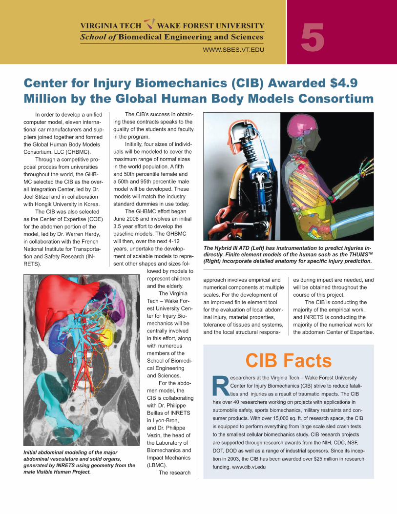
The Hybrid III ATD (Left) has instrumentation to predict injuries in-directly. Finite element models of the human such as the THUMSTM (Right) incorporate detailed anatomy for specific injury prediction.
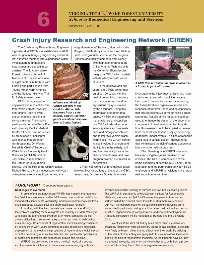
Initial abdominal modeling of the major abdominal vasculature and solid organs, generated by INRETS using geometry from the male Visible Human Project.
VIRGINIA TECH WAKE FOREST UNIVERSITYSchool of Biomedical Engineering and Sciences
WWW.SBES.VT.EDU
6The Crash Injury Research and Engineer-
ing Network (CIREN) was established in 2006 with the goal of bringing engineering and med-ical expertise together with experienced crash investigators to understand how injuries are caused in car crashes. The Toyota – Wake Forest University School of Medicine CIREN center is one of eight centers in the U.S. with funding and participation from Toyota Motor North America and the National Highway Traf-fic Safety Administration.
CIREN brings together engineers and medical doctors from Wake Forest University and Virginia Tech to evalu-ate car crashes resulting in serious injuries. The study’s participants come to Wake For-est University Baptist Medical Center’s Level I Trauma center by ambulance or helicopter with injuries that are often life-threatening. Dr. Wayne Meredith, Chief of Surgery at Wake Forest University Health Sciences (WFUHS), and Dr. Joel Stitzel, a researcher in the Center for Injury Biome-chanics, are the PI’s of the CIREN center. Michael Burke, a crash investigator with years of experience reconstructing crashes, is an
Crash Injury Research and Engineering Network (CIREN)integral member of the team, along with Katie Morgan, CIREN study coordinator and Kathryn Loftis, lead graduate student on the program. Students and faculty members work closely
with their counterparts at the CIB at Virginia Tech and with the Center for Biomolecular Imaging at WFU, which assists with detailed reconstructions of injuries.
In the past two and half years, the CIREN center has enrolled 100 cases with the goal of determining the injury mechanism for each and ev-ery serious injury sustained by the occupant. Using this information and other data-bases, NHTSA and automobile manufacturers and suppliers use CIREN to develop better safety systems such as seat-belts and airbags for vehicles, and to improve vehicle crash-worthiness. The CIREN center is also involved in understand-ing injuries in the elderly, soft and hard tissue injuries in the chest and abdomen, and how pregnant women are injured in car crashes.
CIREN has worked with numerous cases involving liver lacerations and one of the CIB’s researchers, Dr. Jessica Sparks, is actively
investigating the injury mechanisms and injury criteria associated with blunt liver trauma. Her current projects focus on characterizing the tissue-level and organ-level mechanical response of the liver under loading conditions that are representative of motor vehicle crash scenarios. Results of this research could be used to enhance the design of the abdominal component of crash test dummies. In addi-tion, this research could be applied to develop finite element simulations of injury-producing abdominal impact events. This line of research could lead to vehicle design improvements that will mitigate the risk of serious abdominal injury in motor vehicle crashes.
The ultimate goal of CIREN is to save lives and mitigate injuries received in car crashes. The CIREN center is one of the prime examples of how the SBES and CIB col-laboration and the partnership between SBES engineers and WFUHS physicians have had a real impact on saving lives.
Injuries sustained by CIREN patients in car crashes. Above: Rib fractures from a side impact. Below: Posterior pelvis acetabular fracture from a frontal impact.
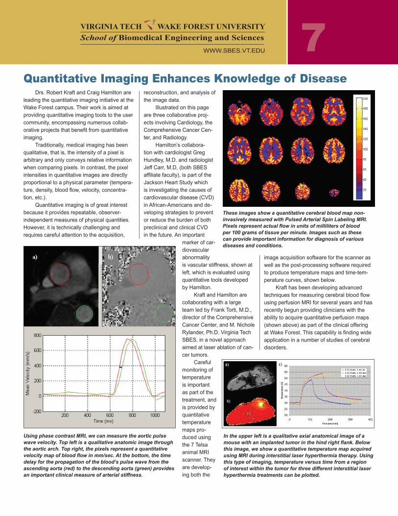
A CIREN case vehicle that was involved in a frontal impact with a tree.
Challenges to overcomeIn spite of the great advances WFIRM has made in the regenera-
tive field, there are many hurdles left to overcome, such as: inability to expand cells, inadequate vascularity, inadequate biomaterials/scaffolds, and inadequate psysiological and pharmacological function.
In working with the liver, the cells are painted on a scaffold, but the problem is getting them to migrate and multiply. Dr. Mark Van Dyke, who leads the Biomaterials Program at WFIRM, compares the cell growth difficulties of some cell types to a human trying to walk without arms and legs. Components of regenerative medicine being considered by engineers at WFIRM are controlled release of bioactive molecules, assessment of the mechanical properties of regenerative medicine prod-ucts, the processing of novel biomaterials, and parameter optimization of scaffolding materials that support cell growth and function.
WFIRM has envisioned the future medical needs of a society and the research is intended to encompass ever-changing technical
advancements while seeking to harness our own body’s healing power. The WFIRM, in partnership with McGowan Institute for Regenerative Medicine, was awarded $42.5 million over five years to form a con-sortium called the Armed Forces Institute of Regenerative Medicine (AFIRM). Its research focus will be battlefield injuries including burns, wound healing without scarring, craniofacial reconstruction, limb recon-struction, regeneration or transplantation, and compartment syndrome. A second consortium will be managed by Rutgers and the Cleveland Clinic.
Scientists at the WFIRM, led by Atala, have taken a creative ap-proach by focusing on inter-disciplinary teams of investigators. Scientists work freely with each other sharing all points of their work. By building on the ideas of others, they are giving birth to new innovative ideas and leading the field of regenerative medicine. As the students develop, they are producing results, and when they leave they take with them a proven approach to solving the problems of regenerative medicine.
FOREFRONT (Continued from page 1)
VIRGINIA TECH WAKE FOREST UNIVERSITYSchool of Biomedical Engineering and Sciences
WWW.SBES.VT.EDU
7Drs. Robert Kraft and Craig Hamilton are
leading the quantitative imaging initiative at the Wake Forest campus. Their work is aimed at providing quantitative imaging tools to the user community, encompassing numerous collab-orative projects that benefit from quantitative imaging.
Traditionally, medical imaging has been qualitative, that is, the intensity of a pixel is arbitrary and only conveys relative information when comparing pixels. In contrast, the pixel intensities in quantitative images are directly proportional to a physical parameter (tempera-ture, density, blood flow, velocity, concentra-tion, etc.).
Quantitative imaging is of great interest because it provides repeatable, observer-independent measures of physical quantities. However, it is technically challenging and requires careful attention to the acquisition,
Quantitative Imaging Enhances Knowledge of Diseasereconstruction, and analysis of the image data.
Illustrated on this page are three collaborative proj-ects involving Cardiology, the Comprehensive Cancer Cen-ter, and Radiology.
Hamilton’s collabora-tion with cardiologist Greg Hundley, M.D. and radiologist Jeff Carr, M.D, (both SBES affiliate faculty), is part of the Jackson Heart Study which is investigating the causes of cardiovascular disease (CVD) in African-Americans and de-veloping strategies to prevent or reduce the burden of both preclinical and clinical CVD in the future. An important
marker of car-diovascular abnormality is vascular stiffness, shown at left, which is evaluated using quantitative tools developed by Hamilton.
Kraft and Hamilton are collaborating with a large team led by Frank Torti, M.D., director of the Comprehensive Cancer Center, and M. Nichole Rylander, Ph.D. Virginia Tech SBES, in a novel approach aimed at laser ablation of can-cer tumors.
Careful monitoring of temperature is important as part of the treatment, and is provided by quantitative temperature maps pro-duced using the 7 Telsa animal MRI scanner. They are develop-ing both the
Using phase contrast MRI, we can measure the aortic pulse wave velocity. Top left is a qualitative anatomic image through the aortic arch. Top right, the pixels represent a quantitative velocity map of blood flow in mm/sec. At the bottom, the time delay for the propagation of the blood’s pulse wave from the ascending aorta (red) to the descending aorta (green) provides an important clinical measure of arterial stiffness.
In the upper left is a qualitative axial anatomical image of a mouse with an implanted tumor in the hind right flank. Below this image, we show a quantitative temperature map acquired using MRI during interstitial laser hyperthermia therapy. Using this type of imaging, temperature versus time from a region of interest within the tumor for three different interstitial laser hyperthermia treatments can be plotted.
These images show a quantitative cerebral blood map non-invasively measured with Pulsed Arterial Spin Labeling MRI. Pixels represent actual flow in units of milliliters of blood per 100 grams of tissue per minute. Images such as these can provide important information for diagnosis of various diseases and conditions.
image acquisition software for the scanner as well as the post-processing software required to produce temperature maps and time-tem-perature curves, shown below.
Kraft has been developing advanced techniques for measuring cerebral blood flow using perfusion MRI for several years and has recently begun providing clinicians with the ability to acquire quantitative perfusion maps (shown above) as part of the clinical offering at Wake Forest. This capability is finding wide application in a number of studies of cerebral disorders.
VIRGINIA TECH WAKE FOREST UNIVERSITYSchool of Biomedical Engineering and Sciences
WWW.SBES.VT.EDU
8
he National Cancer Institute es-timates over 1.4 million new cases of cancer will be diagnosed and over 550,000 deaths from cancer occurred in the united States alone in 2007.
Cancer treatment presently, remains based primarily on treatment approaches developed over a quarter-century ago. Non-specific and highly toxic chemother-apy treatment, aggressive radiation ther-apy, and invasive surgical resection are a patient’s primary means of recourse against this deadly disease. Conse-quently, cancer patients today battle both the disease and the cure. Inadequacies in the ability to administer therapeutic moieties to selectively reach the desired targets with marginal or no collateral damage has largely accounted for this discrepancy. Alternative, more effective imaging and treatment protocols are
needed for all forms of cancer. Faculty from the School of Biomedical Engineering and Sciences (SBES) in collaboration with Wake Forest University Comprehensive Cancer Center are developing advanced methods for cancer therapy utilizing nanotechnology, microfluidics, electroporation, laser optics, heat/mass transfer, computational model-ing, and tissue engineering.
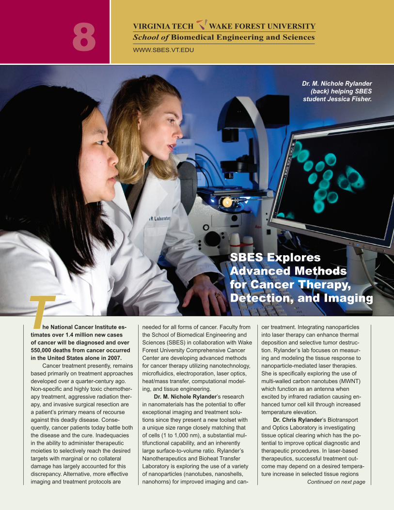
dr. M. Nichole Rylander’s research in nanomaterials has the potential to offer exceptional imaging and treatment solu-tions since they present a new toolset with a unique size range closely matching that of cells (1 to 1,000 nm), a substantial mul-tifunctional capability, and an inherently large surface-to-volume ratio. Rylander’s Nanotherapeutics and Bioheat Transfer Laboratory is exploring the use of a variety of nanoparticles (nanotubes, nanoshells, nanohorns) for improved imaging and can-
cer treatment. Integrating nanoparticles into laser therapy can enhance thermal deposition and selective tumor destruc-tion. Rylander’s lab focuses on measur-ing and modeling the tissue response to nanoparticle-mediated laser therapies. She is specifically exploring the use of multi-walled carbon nanotubes (MWNT) which function as an antenna when excited by infrared radiation causing en-hanced tumor cell kill through increased temperature elevation.
dr. Chris Rylander’s Biotransport and Optics Laboratory is investigating tissue optical clearing which has the po-tential to improve optical diagnostic and therapeutic procedures. In laser-based therapeutics, successful treatment out-come may depend on a desired tempera-ture increase in selected tissue regions
Continued on next page
VIRGINIA TECH WAKE FOREST UNIVERSITYSchool of Biomedical Engineering and Sciences
WWW.SBES.VT.EDU
T
Dr. M. Nichole Rylander (back) helping SBES
student Jessica Fisher.
SBES Explores Advanced Methods for Cancer Therapy, Detection, and Imaging
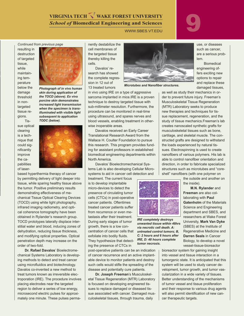
9resulting in destruction of targeted tissue, while maintain-ing tem-perature below the damage threshold in non-targeted tissue re-gions.
Tis-sue optical clearing is a tech-nique that could sig-nificantly improve the ca-pabilities of laser-based hyperthermia therapy of cancer by permitting delivery of light deeper into tissue, while sparing healthy tissue above the tumor. Positive preliminary results demonstrating effectiveness of me-chanical Tissue Optical Clearing Devices (TOCD) using white light photography, infrared imaging radiometry, and opti-cal coherence tomography have been obtained in Rylander’s research group. TOCD prototypes laterally displace inter-stitial water and blood, inducing zones of dehydration, reducing tissue thickness, and modifying optical properties. Optical penetration depth may increase on the order of two-fold.
dr. Rafael davalos’ Bioelectrome-chanical Systems Laboratory is develop-ing methods to detect and treat cancer using microfluidics and biotechnology. Davalos co-invented a new method to treat tumors known as irreversible elec-troporation (IRE). The procedure involves placing electrodes near the targeted region to deliver a series of low energy, microsecond electric pulses for approxi-mately one minute. These pulses perma-
nently destabilize the cell membranes of the targeted tissue thereby killing the cells.
Davalos’ re-search has showed the complete regres-sion in 12 out of 13 treated tumors in vivo using IRE on a type of aggressive sarcoma implanted in mice.IRE is a proven technique to destroy targeted tissue with sub-millimeter resolution. Furthermore, the procedure can be monitored in real-time using ultrasound, and spares nerves and blood vessels, enabling treatment in other-wise inoperable areas.
Davalos received an Early Career Translational Research Award from the Wallace H. Coulter Foundation to pursue this research. This program provides fund-ing for assistant professors in established biomedical engineering departments within North America.
Davalos’ Bioelectromechanical Sys-tems Lab is also developing Cellular Micro-systems to aid in cancer cell detection and treatment. The current focus is to develop implantable micro-devices to detect the presence of circulating tumor cells (CTCs) in post-operative cancer patients. Oftentimes treated cancer patients suffer from recurrence or even me-tastasis after their treatment. During early stages of tumor growth, there is a low con-centration of cancer cells that exfoliate into bodily fluids. They hypothesize that detect-ing the presence of CTCs in post-operative patients can be an indication of cancer recurrence and an active implant-able device to monitor patients and destroy such cells would stifle the spreading of the disease and potentially cure patients.
dr. Joseph Freeman’s Musculoskel-etal Tissue Regeneration (MTR) Laboratory is focused on developing engineered tis-sues to replace damaged or diseased tis-sue associated with cancer. Damaged mus-culoskeletal tissues, through trauma, daily
use, or diseases such as cancer, are a serious prob-lem.
Biomedical engineering of-fers exciting new options to repair and replace these damaged tissues,
as well as study their mechanics in or-der to prevent future injury. Freeman’s Musculoskeletal Tissue Regeneration (MTR) Laboratory seeks to produce new therapies and techniques for tis-sue replacement, regeneration, and the study of tissue mechanics.Freeman’s lab creates nanoscaled synthetic grafts for musculoskeletal tissues such as bone, cartilage, and skeletal muscle. The con-structed grafts are designed to withstand the loads experienced by natural tis-sues. Electrospinning is used to create nanofibers of various polymers. His lab is able to control nanofiber orientation and direction, in order to fabricate specialized structures such as microtubes and “core-shell” nanofibers (with one polymer on
the outside and another on the inside).
M.N. Rylander and Freeman are also col-laborating with Paul Gatenholm of the Materials Science and Engineering department and SBES, and researchers at Wake Forest University, Mark van dyke (SBES) at the Institute of Regenerative Medicine and darren Seals in Cancer Biology, to develop a novel vessel-tissue-bioreactor
bioreactor system for gaining insight into vessel and tissue interaction in a tumorgenic state. It is anticipated that this system will be used to study cancer de-velopment, tumor growth, and tumor vas-cularization in a wide variety of tissues. Better understanding of the mechanisms of tumor vessel and tissue proliferation and their response to various drug agents will also permit identification of new can-cer therapeutic targets.
Microtubes and Nanofiber structures.Photograph of in vivo human skin during application of the TOCD (above). Ex vivo porcine skin demonstrates increased light transmission when the specimen is trans-illuminated with visible light subsequent to application TODC (below).
IRE completely destroys unwanted tissue within 48hrs via necrotic cell death. A: untreated control tumors; B, C: 2 hours and 6 hours after IRE, D: 48 hours complete tumor necrosis.
Continued from previous page
VIRGINIA TECH WAKE FOREST UNIVERSITYSchool of Biomedical Engineering and Sciences
WWW.SBES.VT.EDU
10SBES Collaboration Creates an Engineered Bone Graft
Dr. Aaron Goldstein, at Virginia Tech – in collaboration with Dr Brian Love at the University of Michigan, Dr Jeff Hollinger at Carnegie Mellon University, and Dr Scott Guelcher at Vanderbilt University – has been developing methods to form engi-neered tissue by combining adult stem cells, a resorbable biomaterial scaffold, and a perfusion bioreactor.
The scaffold serves as a carrier and support structure for delivering cells and bioactive factors, and perfusion bioreactor is being used to condition the adult stem cells and induce them to secrete growth factors that will accelerate healing in the patient.
Goldstein has three main research thrusts. They are the synthesis of novel biomaterial scaffolds, molecular analysis
of mechanotransduction – the mechanism by which mechanical stimuli affect cell be-havior (e.g., gene expression, formation of bone tissue) – and perfusion culture of adult stem cells in porous scaffolds.
One biomaterial project is the incor-poration of amorphous calcium phosphate (ACP) into conventional biomaterial scaf-folds. ACP is a bioactive ceramic similar to hydroxyapatite, but because of the absence of crystallinity, it dissolves readily in aque-ous environments and raises the local cal-cium and phosphate concentrations. These ions can reprecipitate in and around tissues to facilitate osteoblast differentiation and the formation of new bone tissue.
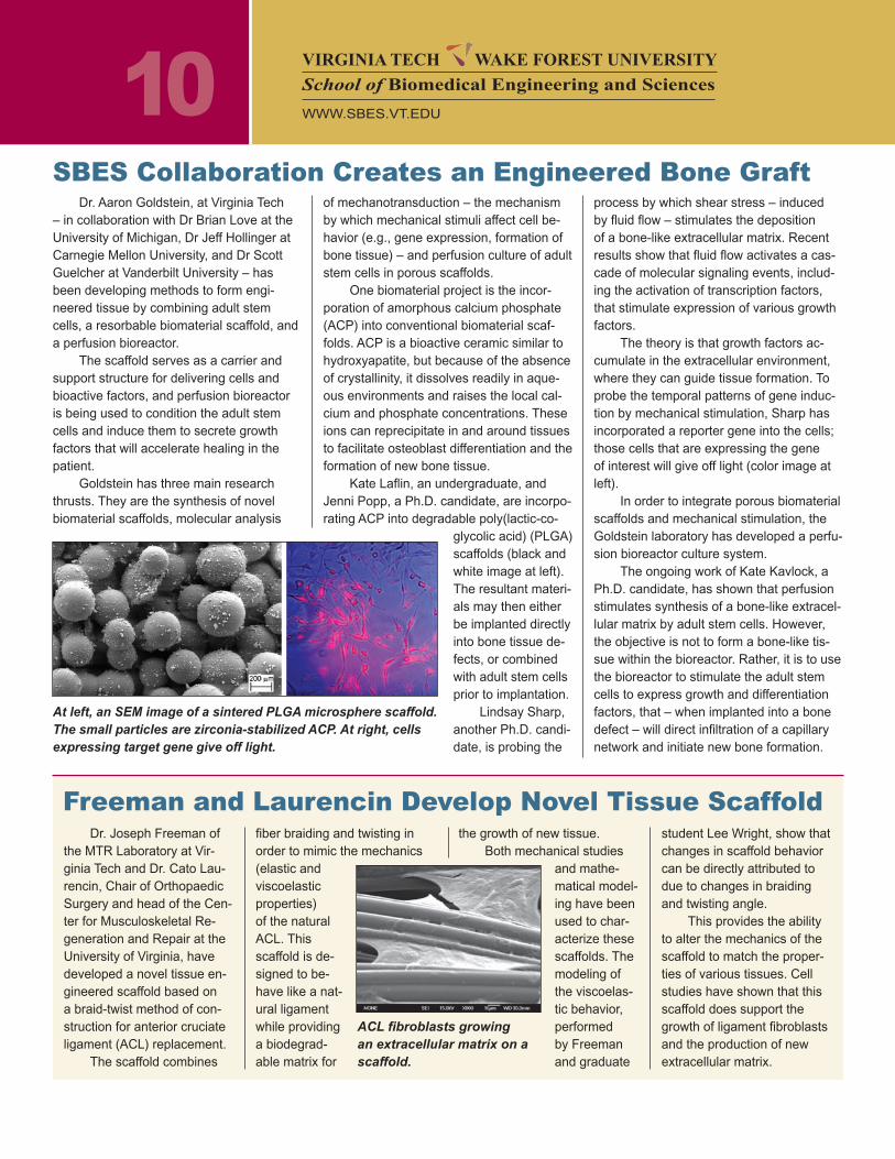
Kate Laflin, an undergraduate, and Jenni Popp, a Ph.D. candidate, are incorpo-rating ACP into degradable poly(lactic-co-
glycolic acid) (PLGA) scaffolds (black and white image at left). The resultant materi-als may then either be implanted directly into bone tissue de-fects, or combined with adult stem cells prior to implantation.
Lindsay Sharp, another Ph.D. candi-date, is probing the
process by which shear stress – induced by fluid flow – stimulates the deposition of a bone-like extracellular matrix. Recent results show that fluid flow activates a cas-cade of molecular signaling events, includ-ing the activation of transcription factors, that stimulate expression of various growth factors.
The theory is that growth factors ac-cumulate in the extracellular environment, where they can guide tissue formation. To probe the temporal patterns of gene induc-tion by mechanical stimulation, Sharp has incorporated a reporter gene into the cells; those cells that are expressing the gene of interest will give off light (color image at left).
In order to integrate porous biomaterial scaffolds and mechanical stimulation, the Goldstein laboratory has developed a perfu-sion bioreactor culture system.
The ongoing work of Kate Kavlock, a Ph.D. candidate, has shown that perfusion stimulates synthesis of a bone-like extracel-lular matrix by adult stem cells. However, the objective is not to form a bone-like tis-sue within the bioreactor. Rather, it is to use the bioreactor to stimulate the adult stem cells to express growth and differentiation factors, that – when implanted into a bone defect – will direct infiltration of a capillary network and initiate new bone formation.
At left, an SEM image of a sintered PLGA microsphere scaffold. The small particles are zirconia-stabilized ACP. At right, cells expressing target gene give off light.
VIRGINIA TECH WAKE FOREST UNIVERSITYSchool of Biomedical Engineering and Sciences
WWW.SBES.VT.EDU
Dr. Joseph Freeman of the MTR Laboratory at Vir-ginia Tech and Dr. Cato Lau-rencin, Chair of Orthopaedic Surgery and head of the Cen-ter for Musculoskeletal Re-generation and Repair at the University of Virginia, have developed a novel tissue en-gineered scaffold based on a braid-twist method of con-struction for anterior cruciate ligament (ACL) replacement.
The scaffold combines
Freeman and Laurencin Develop Novel Tissue Scaffoldfiber braiding and twisting in order to mimic the mechanics (elastic and viscoelastic properties) of the natural ACL. This scaffold is de-signed to be-have like a nat-ural ligament while providing a biodegrad-able matrix for
the growth of new tissue.Both mechanical studies
and mathe-matical model-ing have been used to char-acterize these scaffolds. The modeling of the viscoelas-tic behavior, performed by Freeman and graduate
student Lee Wright, show that changes in scaffold behavior can be directly attributed to due to changes in braiding and twisting angle.
This provides the ability to alter the mechanics of the scaffold to match the proper-ties of various tissues. Cell studies have shown that this scaffold does support the growth of ligament fibroblasts and the production of new extracellular matrix.
ACL fibroblasts growing an extracellular matrix on a scaffold.
11
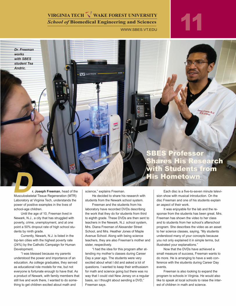
r. Joseph Freeman, head of the Musculoskeletal Tissue Regeneration (MTR) Laboratory at Virginia Tech, understands the power of positive examples in the lives of school-age children.
Until the age of 10, Freeman lived in Newark, N.J., a city that has struggled with poverty, crime, unemployment, and at one point a 50% dropout rate of high school stu-dents by ninth grade.
Currently, Newark, N.J. is listed in the top-ten cities with the highest poverty rate (24%) by the Catholic Campaign for Human Development.
“I was blessed because my parents understood the power and importance of an education. As college graduates, they served as educational role models for me, but not everyone is fortunate enough to have that. As a product of Newark, with family members that still live and work there, I wanted to do some-thing to get children excited about math and
science,” explains Freeman.He decided to share his research with
students from the Newark school system.Freeman and the students from his
laboratory have recorded DVDs describing the work that they do for students from third to eighth grade. These DVDs are then sent to teachers in the Newark, N.J. school system, Mrs. Diana Freeman of Alexander Street School, and Mrs. Heather Jones of Maple Avenue School. Along with being science teachers, they are also Freeman’s mother and sister, respectively.
“I had the idea for this program after at-tending my mother’s classes during Career Day a year ago. The students were very excited about what I did and asked a lot of questions. I wanted to keep their enthusiasm for math and science going but there was no way that I could visit New Jersey on a regular basis, so I thought about sending a DVD,” Freeman says.
Each disc is a five-to-seven minute televi-sion show with musical introduction. On the disc Freeman and one of his students explain an aspect of their work.
It was enjoyable for the lab and the re-sponse from the students has been great. Mrs. Freeman has shown the video to her class and to students from her school’s afterschool program. She describes the video as an asset to her science classes, saying, “My students understood many of your concepts because you not only explained it in simple terms, but illustrated your explanations.”
Now that the DVDs have achieved a small measure of success, Freeman wants to do more. He is arranging to have a web con-ference with the students during Career Day events.
Freeman is also looking to expand the program to schools in Virginia. He would also like to speak at local schools to raise the inter-est of children in math and science.
SBES Professor Shares His Researchwith Students from His HometownD
Dr. Freeman works with SBES student Tea Andric.
VIRGINIA TECH WAKE FOREST UNIVERSITYSchool of Biomedical Engineering and Sciences
WWW.SBES.VT.EDU
12The Virginia Tech —
Wake Forest University School of Biomedical Engineering and Sci-ences sponsors a clinical rotation for engineering students in the Ph.D. program. Its purpose is to provide engineering students with real ex-perience in the medical arena in order to better understand how their biomedical research projects relate to clinical practice.
The rotation con-sists of four weeks during which the students take part in gross anatomy, patient simulation, and clinical situations under
Educational Opportunities with SBES…
Virginia Tech is one of 13 universities chosen to participate in a new di-rected venture to promote bioengineering and bioin-formatics related careers and graduate education. Participating are Virginia Tech-Wake Forest School of Biomedical Engineering and Sciences (SBES) and the Virginia Bioinformatics Institute (VBI).
The Bioengineering & Bioinformatics Sum-mer Institute (BBSI) is a nationwide effort funded by the National Science Foundation (NSF) and the National Institute of Biomedical Imaging and Bioengineering (NIBIB) of the National Institutes of Health (NIH).
As a collaborative effort between SBES and VBI, the BBSI program provides undergradu-
the mentorship of physicians and other medical personnel.
While many possibilities exist, one scenario is that under the mentorship of a physician, the student completes a pre-en-counter assignment, attends pro-cedures, goes on rounds, meets with the physician to discuss the procedures and related engineer-ing issues, and completes post-encounter assignments. The na-ture of involvement beyond this is at the physician’s discretion. Fol-lowing the rotation completion, students can meet with the physi-cian to discuss their assignments, experience, and other issues of interest to the mentor.
The key is that students can bring as much to the process as the physician. The Clinical Rota-
ate students with a quantitative and integrated bioengineering/bioinformatics related educational and research experiences. The program is also designed to mo-tivate these students to pursue graduate degrees and careers in biomedical engineering and bioin-formatics related fields.
BBSI emphasizes three major thrust areas: computational systems biology, computational bio-imaging and computational
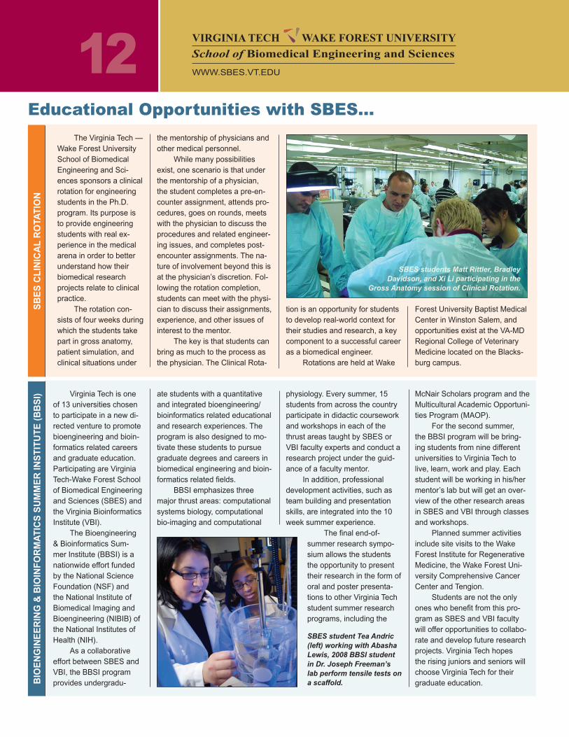
SBES student Tea Andric (left) working with Abasha Lewis, 2008 BBSI student in Dr. Joseph Freeman’s lab perform tensile tests on a scaffold.
tion is an opportunity for students to develop real-world context for their studies and research, a key component to a successful career as a biomedical engineer.
Rotations are held at Wake
Forest University Baptist Medical Center in Winston Salem, and opportunities exist at the VA-MD Regional College of Veterinary Medicine located on the Blacks-burg campus.
physiology. Every summer, 15 students from across the country participate in didactic coursework and workshops in each of the thrust areas taught by SBES or VBI faculty experts and conduct a research project under the guid-ance of a faculty mentor.
In addition, professional development activities, such as team building and presentation skills, are integrated into the 10 week summer experience.
The final end-of-summer research sympo-sium allows the students the opportunity to present their research in the form of oral and poster presenta-tions to other Virginia Tech student summer research programs, including the
McNair Scholars program and the Multicultural Academic Opportuni-ties Program (MAOP).
For the second summer, the BBSI program will be bring-ing students from nine different universities to Virginia Tech to live, learn, work and play. Each student will be working in his/her mentor’s lab but will get an over-view of the other research areas in SBES and VBI through classes and workshops.
Planned summer activities include site visits to the Wake Forest Institute for Regenerative Medicine, the Wake Forest Uni-versity Comprehensive Cancer Center and Tengion.
Students are not the only ones who benefit from this pro-gram as SBES and VBI faculty will offer opportunities to collabo-rate and develop future research projects. Virginia Tech hopes the rising juniors and seniors will choose Virginia Tech for their graduate education.
SB
ES
CL
INIC
AL
RO
TAT
ION
BIO
EN
GIN
EE
RIN
G &
BIO
INF
OR
MA
TIC
S S
uM
ME
R IN
ST
ITu
TE
(B
BS
I)VIRGINIA TECH WAKE FOREST UNIVERSITYSchool of Biomedical Engineering and Sciences
WWW.SBES.VT.EDU
SBES students Matt Rittler, Bradley Davidson, and Xi Li participating in the
Gross Anatomy session of Clinical Rotation.
13
Determination is a key characteristic in describing Martin Tanaka, a husband and the father of four boys. After a 12-year break from academics, Martin came back to Virginia Tech to earn his Ph.D. in biomedical engineering. Martin left Virginia Tech in 1993 with an M.S. and joined industry. Seizing an opportunity to relocate to Blacksburg, Martin pursued his dream. Martin’s wife, Dana, returned to work while he left his job to concentrate on his stud-ies.
Challenges abounded due to the time between his master’s degree and his entry into the SBES doctoral program. While taking a mammalian physiology class, Martin real-ized the last biology class he had taken was in high school in 1984. He faced similar chal-lenges throughout the program. For instance, Matlab, a popular computer program used for mathematical modeling, had to be mastered while he was taking courses. FORTRAN, the programming language, had become obsolete.
There are numerous tracks to choose in the SBES program, some of which include other departments. Martin followed the biome-chanics track. He was advised by one of the top five researchers in the country for move-ment dynamics in cerebral palsy, Dr. Kevin Granata, a professor in the Department of Engineering Science and Mechanics (ESM). One reason Martin chose Granata as an advi-sor was his understanding and support for students who had a family life. Granata always said, “Think big, and don’t try to do something incrementally better than the other person; be risky and make a significant contribution.” Mar-tin has taken Granata’s advice to heart.
Martin learned that the class that Gra-nata had recommended for Fall ’07, taught by Dr. Ishwar Puri, ESM Department Head was canceled due to the aftermath of the April 16th tragedy at Virginia Tech when Granata was killed. Martin asked Puri about the possibility of taking the course as an independent study. Puri readily agreed. The course consisted of a weekly meeting and an on-line lecture. Ac-cording to Puri, “Martin took this very serious-ly.” He completed the work in half a semester. This led Puri to a perplexing question, “What
to do with Martin for the remainder of the se-mester?” He decided to offer Martin a project on brain tumors, specifically gliomas, which are highly invasive with a poor prognosis, de-spite medical intervention.
Since he was still working on his biome-chanics research and writing his dissertation, Martin found this a challenge not only to his expertise area, but also to his time. Puri be-lieves a key strength of the SBES program is in producing students who thrive in an inter-disciplinary environment. Martin turned to Dr. Pete Santago, SBES Associate Head at the Wake Forest campus, for suggestions regard-ing a co-advisor for the oncology expertise he would have to acquire. Martin said that “due to the SBES program, connections and resourc-es were readily available, and Wake was very responsive and supportive.” Dr. Waldemar Debinski, director of the Brain Tumor Center of Excellence at Wake Forest Medical Center, agreed to help with the project.
Gliomas are often not diagnosed until they become large and begin to affect brain and bodily functions. These tumors could be described as having long hair-like projections from a main tumor body. These projections of migratory cells are the culprits that invade un-affected healthy brain tissue and can’t be sur-gically removed. Martin focused his research
on these migratory cells. Current mathematical methods are generally based on processes that are relevant to the main tumor body and not migrating cells.After reviewing the litera-ture on mathematical modeling, Martin pro-posed a new hybrid model that includes both deterministic and stochastic methods, which Puri calls “quite innovative.” His hybrid model takes a deterministic approach for the inside of the tumor and uses a stochastic method for its outside migrating cells, while looking at cell populations and their spatial distributions. Although Martin’s hybrid model will still require supportive clinical and experimental data, this could be a major advance in modeling for gliomas or others cancerous tumors that might also rapidly metastasize.
Martin allotted his time between the glioma project and defending his biomechan-ics dissertation. A recent SBES graduate, Martin will focus on his biomechanics work at Wake Forest, but has been so intrigued by the glioma project that he plans to continue working on it in his spare time. Puri will also continue with the project. He and Tanaka have submitted a research paper for publication. Martin Tanaka recalled Kevin Granata’s advice as he pursued his doctorate. Dr. Tanaka was definitely “thinking big” on this project due to Granata’s influence.
Despite Challenges, Tanaka Pursues Dreams
S T u d E N T P R O F I L E S
SBES is an affiliate with the Institute for Critical Technology and Applied Science (ICTAS) at Virginia
Tech. ICTAS supports and promotes cutting-edge research at the intersection of engineering, science
and medicine. The partnership between SBES and ICTAS was forged due to the dynamic nature of
visionary research shared by both organizations. Working with ICTAS enables SBES to utilize emerg-
ing technologies. As a result of aligning with ICTAS, SBES research will advance through the use of its
premier laboratories and facilities. In October 2008, SBES will join with ICTAS in a new
100,000-square-feet building. For more information please see:
http://www.ictas.vt.edu
VIRGINIA TECH WAKE FOREST UNIVERSITY Biomedical Engineering and Sciences
WWW.SBES.VT.EDU
14
“Moving Ahead”
S T u d E N T P R O F I L E S
Saami Yazdani came to Virginia Tech to pursue biomechanics in the Engineering Sci-ence & Mechanics (ESM) Department. A self-declared “late bloomer,” it wasn’t until his fourth year as an undergraduate that everything clicked. Now you could use the words “flourish-ing” to describe him.
In 2006, he co-authored a manuscript that pointed to his future direction in research. “En-gineering of Blood Vessels from Acellular Col-lagen Matrices Coated with Human Endothelial Cells.” In 2007, with Dr. George Christ, Wake Forest Institute for Regenerative Medicine (WFIRM) and SBES faculty, Yazdani co-au-thored a chapter entitled, “Tissue Engineering of Large Diameter Vessels” in Principles of Regenerative Medicine.
Yazdani is affectionately known as “Yaz” to his many friends.
Karen Watson, administrative assistant at Wake Forest, describes “Yaz” as one of
those truly friendly and kind people that you enjoy knowing. Dr. Joel Berry, his Ph.D. advi-sor, agrees and is amazed at the people ‘Yaz” knows, everyone from the custodians to the surgeons.
“Yaz represents the kind of graduate for which SBES was intended to produce, a true biomedical engineer,” Berry says.
Upon completion of his undergraduate degree, Yaz continued with his master’s in ESM with Dr. Demetri Telionis, working in the fluid mechanics laboratory. His focus was on fluid dynamics and pulsatile flow in tubes (rep-licating arteries) and stented tubes (replicating stented arteries).
Yaz states, “It’s basic engineering; if blood flows through it, it excites me!”
During this time he met Dr. Joel Berry, a member of the Virginia Tech – Wake Forest School of Biomedical Engineering and Sci-ences (SBES) faculty from the Wake Forest
campus, and they began working on arterial stents. This led him to take full advantage of the Virginia Tech – Wake Forest SBES pro-gram and he chose to work at the Wake Forest campus.
Yaz believes “the SBES program has the best of both worlds, where you get to work in engineering and a clinical setting.”
In the first year of the SBES program at Wake Forest, Yaz and Berry performed finite element modeling of arteries and stented ar-teries, reviewing computer modeling stress in the artery wall and relating it to stent design and mechanical properties. During this time, they began performing research with Dr. Shay Soker at the WFIRM, which has a close affili-ation with SBES. Together, they began with developing bioreactors for tissue engineered arteries; by his second year, Yaz was asked to work at WFIRM.
Yaz gained beneficial knowledge concern-ing the cells of blood vessels from Soker and continued the development of bioreactors to improve engineered blood vessels. This led Yaz from tubes and computer modeling to actually developing new methods for growing cells and seeding tissue engineered arteries.
Working with Dr. Randolph Geary and Bryan Tilman, vascular surgeons at Wake Forest University School of Medicine and Dr. James Yoo from the WFIRM, the research was taken from the laboratory to animal implant testing, enabling them to study installed bioen-gineered arteries preliminary to human use.
As Yaz leaves as a 2008 Ph.D. graduate in biomedical engineering, he will take the next steps of progression in truly understanding the complete cardiovascular system.
Joining CV Path Institute in Maryland, a non-profit organization, he will be a research scientist working with Dr. Renu Virmani, regard-ed as the world’s expert in vascular pathology.
Expounding on his artery research, he will now be able to study the effects of the stent in the human body. In particular, he will investi-gate the pathological remodeling of arteries to stress and strain associated with stents and other devices.
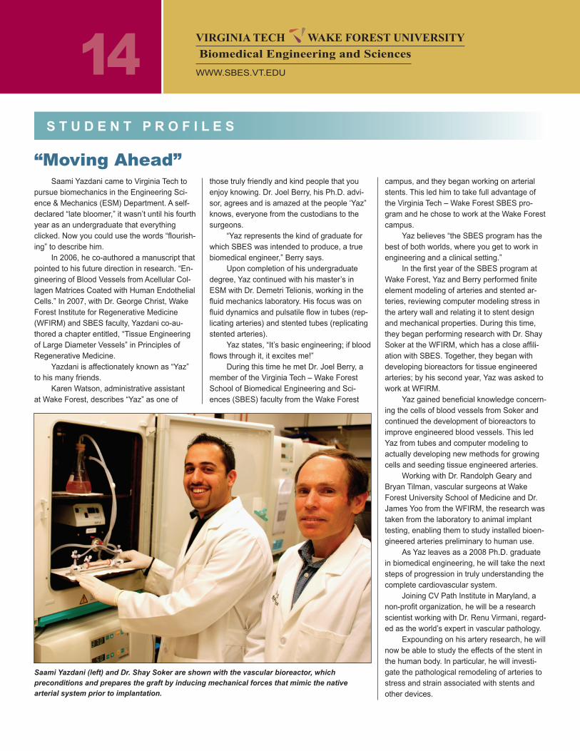
Saami Yazdani (left) and Dr. Shay Soker are shown with the vascular bioreactor, which preconditions and prepares the graft by inducing mechanical forces that mimic the native arterial system prior to implantation.
VIRGINIA TECH WAKE FOREST UNIVERSITY Biomedical Engineering and Sciences
WWW.SBES.VT.EDU
15
Matt Rittler was a member of the first small group of students to enter the School of Biomedical Engineering and Sciences pro-gram when it began in 2003.
Matt came to Virginia Tech with a double B.S. degree in biochemistry and exercise physiology from McDaniel College where he graduated summa cum laude, and was in-ducted into Phi Beta Kappa and the Beta Beta Beta Biological Honor Society.
A Maryland Distinguished Scholar, he began work in 2000 on an M.S. degree in human nutrition, foods, and exercise. His pro-gram focus was in biochemistry but even then biomedical engineering was the field he really wanted. He completed his Ph.D. in biomedical engineering in 2008.
While in HNFE he met Dr. William Huckle in the VA-MD Regional College of Veterinary Medicine who became his advisor. He applied for the Ph.D. in veterinary medical sciences for Fall 2002 (pre-SBES).
Talk of the formation of SBES had al-ready begun earlier that year, and while it didn’t exist yet, Matt knew he wanted to be part of the new program which was expected to begin admitting students in the Fall of 2003.
SBES was officially approved by SCHEV in March of 2003, and the following fall Matt entered the SBES program as one of the origi-nal “pilot class” of students.
Even though his background was not in engineering, the idea of biomedical engineer-ing was very appealing to him.
“Biomedical engineering sounded sexy,” says Matt with a smile. He liked the idea that it was so interdisciplinary and multi-directional; that it could open so many doors.
He explains that he initially experienced a little discomfort feeling like an “outsider” (refer-ring to his attachment to the Vet School). Engi-neering was a bit like a “clique” in high school – a club he wasn’t in. “I knew I’d just have to get over it,” he says.
The big issue for him was his math defi-cit, and once he’d caught up on that, the rest
S T u d E N T P R O F I L E S
“Pilot Group” Graduate MakesSignificant Mark in the SBES Story
was fairly easy. He had considerable strength in physiology and cell and tissue knowledge.
His research is in angiogenesis -- the behavior of vascular endothelial growth factor (VEGF) which binds to receptor sites on cell surfaces to stimulate blood vessel growth. Over-expression of VEGF has been linked to several pathological conditions including the vascularization of cancer tumors. Of special interest is a secreted form of one receptor which can compete with VEGF for “binding space” and thus inhibit angiogenesis. If blood vessel growth in cancer tumors can be inhib-ited, the cancer can hopefully be defeated.
Matt’s research involves building math-ematical models of how this binding and inhibition occurs, so that the mechanisms of the process can be predicted, tested, and ma-nipulated to an advantage. The ultimate goal is to simulate experiments computationally that couldn’t otherwise be done in a laboratory setting.
Beyond the degree pursuitBeyond his academics, Matt made sig-
nificant contributions to SBES. When it was decided that mammalian physiology would be a requirement for the new program’s degree, Matt was asked if he would be the graduate assistant for the course due to his extensive anatomy/physiology background.
He says, “They weren’t exactly sure what that would mean in the beginning or what [I] would do.” But as things developed, weekly review sessions were created to be part of the course which Matt would run.
The first year was a challenge. There was no video-broadcast setup for his sessions so he met with the few Virginia Tech students in a Norris classroom, and “did the best he could” helping the Wake Forest students by e-mail.
He ran the recitations for three years and was also asked to be a co-instructor for the BMVS 4064 Intro to Medical Physiology course when it was created.
In Fall 2005 he created and taught a short-course in anatomy to the new SBES in-
coming students as preparation for the mamma-lian class. His outstanding per-formance and solid depend-ability led to yet another chance to assist.
In the Fall of 2006, Rittler
and another SBES graduate student teamed to run a new Internship program called the Bioengineering and Bioinformatics Summer In-stitute (BBSI), a collaboration between SBES and the Virginia Bioinformatics Institute de-signed to offer summer research opportunities to rising juniors and seniors.
Matt inherited the program completely in January 2007 and continued developing it, serving as its coordinator through the summer of 2007 and into the Fall. In October of 2007, he was sent to the BMES conference in Hol-lywood to give a platform presentation about the BBSI program.
During his time here, Matt also contrib-uted greatly to the SBES recruiting effort. He was always a faithful student volunteer during the spring College of Engineering Graduate Recruiting Weekend. He also represented SBES at outside events such as the College of Engineering Career Fair at N.C. State.
Dr. Wally Grant was once heard to joke, “What can we do to prevent Matt from gradu-ating?”
Matt’s immediate future is in the Wash-ington area where he will take a post-doc position in the National Cancer Institute, a sub-division of NIH, where he will work again in angiogenesis.
Eventually, he says, he imagines himself gravitating back to the academic world. He loves teaching and thinks he’d like to blend it with doing research in a university setting.
~ by T. Sentelle
Matt Rittler
VIRGINIA TECH WAKE FOREST UNIVERSITY Biomedical Engineering and Sciences
WWW.SBES.VT.EDU
dr. Rafael v. davalosAssistant Professor• NASA Tech Briefs “2007: The Year in Technology”
Listed Irreversible Electroporation as a top break-through of 2007.
• 2008 Early Career Translational Research Award in Biomedical Engineering from the Wallace H. Coulter Foundation
dr. Stefan dumaProfessor, Mechanical Engineering• Best Paper, 2007 Association for the Advancement of
Automotive Medicine
dr. Joseph FreemanAssistant Professor• 2008 Early Career Translational Research Award in
Biomedical Engineering from the Wallace H. Coulter Foundation
dr. H. Clay GablerAssociate Professor• 2007 Ralph H. Isbrandt Automotive Safety Engineer-
ing Award• 2008 Lloyd L. Withrow Distinguished Speaker Award
dr. Warren HardyAssociate Professor, Mechanical Engineering• Best Paper, 2007 Stapp Car Crash Journal
U.S. Postage
PA I dPermit No. 28
Blacksburg VA
24060
SBES Newscourtesy of the Institute for Critical Technology and Applied Science (ICTAS)
SBES is bridging the gap between engineering, science, and medicine
Wally Grant, Ph.d.Head
Pete Santago, Ph.d.Associate Head
Pamela StiffEditor
david SimpkinsDesigner
Please send comments and address corrections to Pamela Stiff,[email protected]
virginia Tech - Wake Forest universitySchool of Biomedical Engineering and Sciences114 v Randolph Hall (0298) Blacksburg, virginia 24061
www.sbes.vt.edu
virginia Tech does not discriminate against employees, students, or applicants on the basis of race, color, sex, sexual orientation, disability, age, veteran status, national origin, religion, or political affiliation. Anyone having questions concerning discrimination or accessibility should contact the Office for Equal Opportunity, 540/231-7500.
S B E S F A C u L T Y H O N O R S / A W A R d S
dr. Y.W. LeeAssistant Professor• November 2007: Excellent Presentation Award, Ko-
rean Nutrition Society International Conference
dr. Mark PaulAssistant Professor, Mechanical Engineering• NSF Career Award, Spatiotemporal Chaos in Fluid
Convection: New Physical Insights From Numerics
dr. M. Nichole RylanderAssistant Professor• Recipient of the 2008 Outstanding New Assistant
Professor Award
dr. Pavlos P. vlachosAssistant Professor, Mechanical Engineering• NSF Career Award, Arterial Flow Dynamics Effects of
Pusatility, Compliance, and Curvature
dr. Ge WangVirginia Tech & Wake Forest University Faculty• Feature article in Radiological Society of North Amer-
ica, November 2007, reporting the pioneering work on bioluminescence tomography
• Fellow of the International Society for Optical Engi-neering (effective 1/1/2007 for specific achievements in bioluminescence tomography and x-ray computed tomography
Scott Gayzik• First Place, Best Student Paper Award, Stapp Car
Crash Journal 2007
Greg Webster• John D. States Best Student Paper Award, Associa-
tion of Advancement of Automotive Medicine, 2007 in Melbourne, Australia
Jill Bisplinghoff, Steven Rowson, doug Gabauer, Kerry danelson, F. Scott Gayzik, Sarah Manoo-gian, and Andrew Kemper
• Won eight of 12 awards at the Biomedical Sciences
S B E S S T u d E N T A C H I E v E M E N T S / A W A R d S
Instrumentation Conference, April 4-6, 2008, Copper Mt., Colorado
Scott Gayzik, Kerry danelson, and Amber Bonivtch• First Place, Enhanced Safety of Vehicles 2nd In-
ternational Collegiate Student Safety Technology Design Competition, Enhanced Safety of Vehicles Conference, 2007
Amber Bonivtch• American Society for Bone and Mineral Research
Sun Valley Workshop on Skeletal Tissue Biology, Alice L. Jee Memorial Young Investigator Award Re-cipient: 2008
VIRGINIA TECH WAKE FOREST UNIVERSITY Biomedical Engineering and Sciences
WWW.SBES.VT.EDU