Risk Factors, Clinical Presentation, and Neuroimaging...
8
2115 A dvances in neuroimaging techniques have greatly improved the detection and understanding of neona- tal stroke. 1 Reported incidence of neonatal stroke in term newborns ranges from 1 in 2300 to 1 in 5900. 1–3 Benders et al 4 reported an incidence of 7 in 1000 preterm admissions. In most cases of neonatal stroke, the middle cerebral artery (MCA) is involved. For each of the cerebral arteries, main branch (cortical or pial) or perforator branch involvement can be distinguished. 5 So far, studies on neonatal stroke have mainly focused on cortical stroke. Perforator stroke is appar- ently still under-recognized and little is known about risk fac- tors and clinical presentation. To gain more insight into risk factors, clinical presentation, and neuroimaging findings of neonatal perforator stroke, we report the largest cohort of neonates to date diagnosed with perforator stroke. Patients and Methods All infants admitted to our tertiary (neonatal) intensive care unit between August 1999 and April 2011 and in whom perforator stroke was diagnosed by postnatal cranial ultrasound (CUS) or MRI were enrolled. The study was approved by the Medical Ethics Review Committee of the Erasmus Medical Center, Rotterdam, The Netherlands. Perforator stroke involves the perforators of the anterior choroi- dal artery, anterior cerebral artery, MCA, posterior cerebral artery, and posterior communicating artery, supplying among others thala- mus, striatum, posterior limb of the internal capsule, and centrum semiovale (Table 1). 6 The anatomy of basal ganglia perforators is described in detail in previous studies. 5,6 Perforator stroke was de- fined as a well-delineated hyperechoic lesion in thalamus or stria- tum on CUS (Figure). Isolated lesions in centrum semiovale were not included because these cannot only be caused by focal arterial infarction in a terminal lateral striatal MCA branch. An alternative explanation could be by stroke of a ventriculopetal cortical arteri- al branch of the MCA with occlusion away from the surface as it courses in white matter. In addition, isolated lesions in the centrum semiovale can be confused with punctuate white matter lesions. On MRI, perforator stroke was defined as a well-lineated lesion, hypoin- tense on T1-weighed imaging, hyperintense in T2-weighed imaging, hypointense on apparent diffusion coefficient maps, and hyperintense on diffusion-weighted imaging. All preterm infants had been screened by standard local CUS pro- tocol, as a matter of clinical routine. This entailed ≥2 ultrasound stud- ies in the first week of life, followed by weekly ultrasound studies until discharge. Term infants at risk of brain damage were screened with CUS at the discretion of the attending physician. Sonograms were obtained using a Sequoia (Siemens, Mountain View, CA) or an Esaote system (MyLab 70, Genova, Italy). MRI scanning was Background and Purpose—To date, studies on neonatal stroke have mainly focused on cortical stroke. We have focused on perforator strokes, noncortical strokes in the arterial vascular perforator area. We sought to identify risk factors and evaluate clinical presentation and neuroimaging findings for neonatal perforator stroke, which seems to be under-recognized. Methods—All infants admitted to our tertiary intensive care unit in ≈12 years, whose perforator stroke was diagnosed with postnatal brain imaging, were enrolled in this study. Demographic, perinatal, and postnatal data were evaluated. Results—Seventy-nine perforator strokes were detected in 55 patients (28 boys), with a median gestational age of 37 1/7 weeks (range 24 1/7 to 42 1/7 weeks, 25 preterm). Perforator stroke was asymptomatic in most patients (58%). Initial diagnosis was predominantly made with cranial ultrasound (80%) in the first week of life (60%). Risk factors for stroke were present in all cases: maternal, fetal, and perinatal. Likely pathogenic mechanisms were prolonged birth asphyxia (16%), hypoxia or hypotension (15%), embolism (15%), infection (15%), acute blood loss (9%), and birth trauma (9%). Conclusions—Previously described risk factors for developing neonatal main artery stroke are probably also associated with neonatal perforator stroke. Perforator stroke is often asymptomatic, but cranial ultrasound is a reliable diagnostic tool in diagnosing perforator stroke. (Stroke. 2013;44:2115-2120.) Key Words: neonatal stroke ■ risk factors ■ ultrasound Risk Factors, Clinical Presentation, and Neuroimaging Findings of Neonatal Perforator Stroke Ginette M. Ecury-Goossen, MD; Marlou M.A. Raets, MD; Maarten Lequin, MD, PhD; Monique Feijen-Roon; Paul Govaert, MD, PhD; Jeroen Dudink, MD, PhD Received February 7, 2013; accepted April 23, 2013. From the Department of Pediatrics, Division of Neonatology (G.M.E.-G., M.M.A.R., M.F.-R., P.G., J.D.) and Department of Radiology (M.L., J.D.), Erasmus MC-Sophia Children’s Hospital, Rotterdam, The Netherlands; and Department of Pediatrics, Koningin Paola Children’s Hospital, Antwerp, Belgium (P.G.). The online-only Data Supplement is available with this article at http://stroke.ahajournals.org/lookup/suppl/doi:10.1161/STROKEAHA. 113.001064/-/DC1. Correspondence to Jeroen Dudink, MD, PhD, Department of Pediatrics, Division of Neonatology, Erasmus MC-Sophia Children’s Hospital, Dr Molewaterplein 60, 3015 GJ Rotterdam, Rotterdam, The Netherlands. E-mail [email protected] © 2013 American Heart Association, Inc. Stroke is available at http://stroke.ahajournals.org DOI: 10.1161/STROKEAHA.113.001064 by guest on July 4, 2018 http://stroke.ahajournals.org/ Downloaded from by guest on July 4, 2018 http://stroke.ahajournals.org/ Downloaded from by guest on July 4, 2018 http://stroke.ahajournals.org/ Downloaded from
Transcript of Risk Factors, Clinical Presentation, and Neuroimaging...
2115
Advances in neuroimaging techniques have greatly improved the detection and understanding of neona-
tal stroke.1 Reported incidence of neonatal stroke in term newborns ranges from 1 in 2300 to 1 in 5900.1–3 Benders et al4 reported an incidence of 7 in 1000 preterm admissions. In most cases of neonatal stroke, the middle cerebral artery (MCA) is involved. For each of the cerebral arteries, main branch (cortical or pial) or perforator branch involvement can be distinguished.5 So far, studies on neonatal stroke have mainly focused on cortical stroke. Perforator stroke is appar-ently still under-recognized and little is known about risk fac-tors and clinical presentation.
To gain more insight into risk factors, clinical presentation, and neuroimaging findings of neonatal perforator stroke, we report the largest cohort of neonates to date diagnosed with perforator stroke.
Patients and MethodsAll infants admitted to our tertiary (neonatal) intensive care unit between August 1999 and April 2011 and in whom perforator stroke was diagnosed by postnatal cranial ultrasound (CUS) or MRI were enrolled. The study was approved by the Medical Ethics
Review Committee of the Erasmus Medical Center, Rotterdam, The Netherlands.
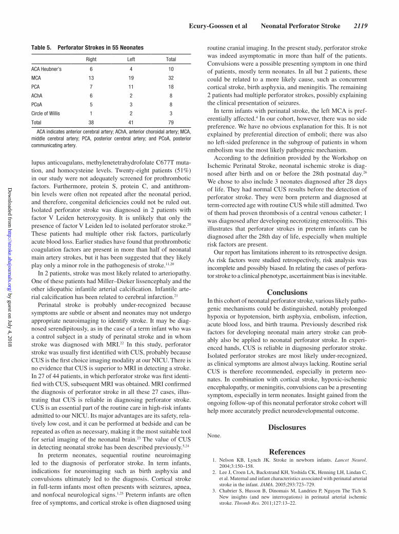
Perforator stroke involves the perforators of the anterior choroi-dal artery, anterior cerebral artery, MCA, posterior cerebral artery, and posterior communicating artery, supplying among others thala-mus, striatum, posterior limb of the internal capsule, and centrum semiovale (Table 1).6 The anatomy of basal ganglia perforators is described in detail in previous studies.5,6 Perforator stroke was de-fined as a well-delineated hyperechoic lesion in thalamus or stria-tum on CUS (Figure). Isolated lesions in centrum semiovale were not included because these cannot only be caused by focal arterial infarction in a terminal lateral striatal MCA branch. An alternative explanation could be by stroke of a ventriculopetal cortical arteri-al branch of the MCA with occlusion away from the surface as it courses in white matter. In addition, isolated lesions in the centrum semiovale can be confused with punctuate white matter lesions. On MRI, perforator stroke was defined as a well-lineated lesion, hypoin-tense on T1-weighed imaging, hyperintense in T2-weighed imaging, hypointense on apparent diffusion coefficient maps, and hyperintense on diffusion-weighted imaging.
All preterm infants had been screened by standard local CUS pro-tocol, as a matter of clinical routine. This entailed ≥2 ultrasound stud-ies in the first week of life, followed by weekly ultrasound studies until discharge. Term infants at risk of brain damage were screened with CUS at the discretion of the attending physician. Sonograms were obtained using a Sequoia (Siemens, Mountain View, CA) or an Esaote system (MyLab 70, Genova, Italy). MRI scanning was
Background and Purpose—To date, studies on neonatal stroke have mainly focused on cortical stroke. We have focused on perforator strokes, noncortical strokes in the arterial vascular perforator area. We sought to identify risk factors and evaluate clinical presentation and neuroimaging findings for neonatal perforator stroke, which seems to be under-recognized.
Methods—All infants admitted to our tertiary intensive care unit in ≈12 years, whose perforator stroke was diagnosed with postnatal brain imaging, were enrolled in this study. Demographic, perinatal, and postnatal data were evaluated.
Results—Seventy-nine perforator strokes were detected in 55 patients (28 boys), with a median gestational age of 37 1/7 weeks (range 24 1/7 to 42 1/7 weeks, 25 preterm). Perforator stroke was asymptomatic in most patients (58%). Initial diagnosis was predominantly made with cranial ultrasound (80%) in the first week of life (60%). Risk factors for stroke were present in all cases: maternal, fetal, and perinatal. Likely pathogenic mechanisms were prolonged birth asphyxia (16%), hypoxia or hypotension (15%), embolism (15%), infection (15%), acute blood loss (9%), and birth trauma (9%).
Conclusions—Previously described risk factors for developing neonatal main artery stroke are probably also associated with neonatal perforator stroke. Perforator stroke is often asymptomatic, but cranial ultrasound is a reliable diagnostic tool in diagnosing perforator stroke. (Stroke. 2013;44:2115-2120.)
Key Words: neonatal stroke ■ risk factors ■ ultrasound
Risk Factors, Clinical Presentation, and Neuroimaging Findings of Neonatal Perforator Stroke
Ginette M. Ecury-Goossen, MD; Marlou M.A. Raets, MD; Maarten Lequin, MD, PhD; Monique Feijen-Roon; Paul Govaert, MD, PhD; Jeroen Dudink, MD, PhD
Received February 7, 2013; accepted April 23, 2013.From the Department of Pediatrics, Division of Neonatology (G.M.E.-G., M.M.A.R., M.F.-R., P.G., J.D.) and Department of Radiology (M.L., J.D.),
Erasmus MC-Sophia Children’s Hospital, Rotterdam, The Netherlands; and Department of Pediatrics, Koningin Paola Children’s Hospital, Antwerp, Belgium (P.G.).
The online-only Data Supplement is available with this article at http://stroke.ahajournals.org/lookup/suppl/doi:10.1161/STROKEAHA. 113.001064/-/DC1.
Correspondence to Jeroen Dudink, MD, PhD, Department of Pediatrics, Division of Neonatology, Erasmus MC-Sophia Children’s Hospital, Dr Molewaterplein 60, 3015 GJ Rotterdam, Rotterdam, The Netherlands. E-mail [email protected]
© 2013 American Heart Association, Inc.
Stroke is available at http://stroke.ahajournals.org DOI: 10.1161/STROKEAHA.113.001064
by guest on July 4, 2018http://stroke.ahajournals.org/
Dow
nloaded from
by guest on July 4, 2018http://stroke.ahajournals.org/
Dow
nloaded from
by guest on July 4, 2018http://stroke.ahajournals.org/
Dow
nloaded from

2116 Stroke August 2013
performed on a 1.5-T GE EchoSpeed scanner (GE Medical Systems, Milwaukee, WI), using an MR-compatible incubator provided with a specialized high-sensitivity neonatal head coil. Perforator strokes, first diagnosed with MRI (Dr Lequin), were reviewed by 2 neona-tologists (Dr Dudink and Dr Govaert) who also reviewed all CUS and remaining MRI studies independently and sought consensus for questionable imaging findings.
Demographic, perinatal, postnatal, and short-term follow-up data were retrieved from the medical records. Perinatal data included delivery, gestational age, sex, birth weight, Apgar score, and umbilical cord pH. Postnatal data included respiratory support, Clinical Risk Index for Babies score,7 the presence of central venous catheters, necrotizing enterocolitis, hypoglycemia, sepsis, and congenital heart disease. Clinical symptoms and neuroimaging findings preceding the radiological diagnosis of perforator stroke were reviewed.Clinical phenotypes for perinatal stroke were classified by etiologic mechanisms of neonatal stroke: infection, birth trauma, embolism, arteriopathy, blood loss, extracorporeal membrane oxygenation, asphyxia, prothrombotic condition, or unclassifiable.8
ResultsPatient Characteristics and Risk FactorsFifty-five patients were included in this study, 0.7% of all 7713 patients admitted to our neonatal intensive care unit (NICU) during the study period, 0.5% admitted were preterm infants, and 0.6% admitted were very-low-birth-weight infants (birth weight <1500 g). Patient characteristics are shown in Table 2. Twenty-five patients were born preterm (<37 weeks postmenstrual age), 17 of whom were born before 32 weeks’ postmenstrual age. Eight mothers had received antenatal beta-methasone. Vacuum extractor was used in 5 cases and was unsuccessful in 1, in which emergency cesarean delivery was then needed. Sixteen other patients were delivered by emer-gency cesarean section because of suspected fetal distress. Birth asphyxia was diagnosed in 18 patients according to Levene’s criteria.9
Risk factors for developing stroke are summarized in Table 3. Data on placental histology were recorded in only 15 cases. These data could not be retrieved for the 39 out-born children. In 8 of 15 cases, the placenta was classified as
abnormal (Table 3). There were no cases reported on perinatal stroke in a sibling. In 2 cases, family history was positive for stroke. In both, multiple family members had a stroke before the age of 50 years.
Thirteen children had culture-proven sepsis and 5 had culture-proven meningitis (1 Listeria monocytogenes, 1 Escherichia coli, and 3 Group B Streptococcus). Forty-five children had ≥1 central venous catheters before the diag-nosis of perforator stroke. Diagnosis of perforator stroke was followed by ultrasound evaluation of the catheter and major veins or heart in 24 cases. This revealed thrombo-sis in 5 (n=2 thrombosis around the tip of the catheter and n=3 venous thrombosis) and multiple air configurations in the liver in 1 patient (with an umbilical vein catheter). Prothrombotic screening was performed in 27 patients, revealing heterozygosity for factor V Leiden in 2 cases. Etiologic mechanisms leading to the perforator stroke are summarized in Table 4.
NeuroimagingNeuroimaging revealed 79 perforator strokes in 55 patients (Table 5): right-sided in 21, left-sided in 20, and bilateral in 14. These 14 patients had 2 to 4 perforator strokes each. Of these 14, none had symmetrical deep gray matter injury and birth asphyxia was diagnosed in only 1 according to Levene’s criteria.10
In 44 patients (80%), the stroke was first identified with CUS. In 27 of them, additional MRI was obtained, confirming the diagnosis of perforator stroke. In 10 patients (18%), the stroke was first detected by MRI. In 1 patient, the stroke was first diagnosed on a computed tomographic scan made else-where because of convulsions and later confirmed with MRI in our hospital.
In 33 patients (60%), the perforator stroke was diagnosed in the first week of life. Perforator stroke was diagnosed with routine CUS in 3 patients with prior normal CUS findings after the age of 28 days. They were born preterm (between 28 and 31 weeks gestational age) and were diagnosed at term equivalent age.
Thirteen patients had concomitant cortical stroke. In none of these patients, perforator stroke could be explained solely by the concomitant cortical stroke. Table VI in the online-only Data Supplement gives an overview of all associated intrace-rebral lesions in this cohort.
Presenting Symptoms and Clinical CoursePerforator stroke was asymptomatic in 32 patients (58%), who were diagnosed with routine neuroimaging. Twenty other patients (36%) had clinical seizures before the diagnosis of perforator stroke. Conditions that are known to cause seizures, such as cortical stroke, birth asphyxia, and meningitis, were diagnosed in 18 of these 20 patients. Eight patients presented with apnea, which was probably related to prematurity, not to perforator stroke. One patient with meningitis presented with hypertonia. One patient presented with unexplained diminished arousal response, and multiple perforator strokes (right MCA lateral striate stroke, left circle of Willis, and left posterior cere-bral artery thalamic stroke) were later diagnosed in that patient.
Table 1. Perforator Stroke Classification
VesselArterial Perforator Vessel Territory
ACA Heubner’s Rostroventral and lateral part of caudate headLateral part of anterior limb of the internal capsule
MCA Lenticulostriate Posterior part of caudate headLateral part of globus pallidusPutamen
PCA Lateral, inferior, and paramedian parts of thalamus
AChA Medial part of globus pallidusPosterior limb of the internal capsuleAmygdaloid nuclei
PCoA Superoanterior part of medial thalamic nuclei
Circle of Willis
Hypothalamus
Thalamus Superolateral parts of thalamus
ACA indicates anterior cerebral artery; AChA, anterior choroidal artery; MCA, middle cerebral artery; PCA, posterior cerebral artery; and PCoA, posterior communicating artery.
by guest on July 4, 2018http://stroke.ahajournals.org/
Dow
nloaded from

Ecury-Goossen et al Neonatal Perforator Stroke 2117
None of the patients in this cohort received thrombolytic therapy. Six patients died before the age of 1 month. None of these deaths was related to perforator stroke, but they were
rather related to cardiopulmonary insufficiency because of other neonatal complications.
DiscussionTo our knowledge, this is the largest cohort of newborns with perforator stroke studied. Both preterm and term infants were included. We found that perforator stroke was asymptomatic in most patients (58%). Most strokes were first diagnosed using CUS (80%), predominantly in the first week of life (60%). Still, 40% were diagnosed after the first week of life and 5% were diagnosed with routine CUS after the age of 28 days. These numbers illustrate the importance of routine serial CUS screening in infants admitted to a NICU. Right-sided lesions occurred as frequently as left-sided lesions. Various likely pathogenic mechanisms for the development of perforator stroke could be distinguished, most often birth asphyxia, prolonged hypoxia or hypotension, embolism, and infection. It seems likely that previously described risk factors for developing neonatal main artery stroke can also be applied to neonatal perforator stroke. Maternal, fetal, or perinatal risk factors were present in all cases.
Figure. Axial and parasagittal perforator templates with illustrative cranial ultrasound (US) and MRI. aa/a indicates artery; ACA, anterior cerebral artery; AChA, anterior choroidal artery; BAS, basilar artery; DWMR, diffusion weighted magnetic resonance imaging; ICA, internal carotid artery; MCA, middle cerebral artery; PCA, posterior cerebral artery; PCoA, posterior communicating artery; and OR, optic radiation.
Table 2. Patient Characteristics
No. (%) or Median (Range)
Gestational age, wk 37 1/7 (24 1/7–42 1/7)
Birth weight, g 2640 (550–4440)
Boys 28 (51)
Singleton 48 (87)
SGA 12 (22)
Apgar score 1 min (n=53) 5 (0–10)
Apgar score 5 min (n=54) 8 (2–10)n=14, Apgar score≤5
Umbilical cord pH (n=31) 7.18 (6.73−7.39)n=10, pH≤7.05
CRIB score (n=54) 2 (0–13)
Age at diagnosis of perforator stroke, d 6 (1–82)
CRIB indicates Clinical Risk Index for Babies; and SGA, small for gestational age.
by guest on July 4, 2018http://stroke.ahajournals.org/
Dow
nloaded from

2118 Stroke August 2013
Pregnancy is considered to be a natural prothrombotic state. Thrombosis on the fetal side of the placenta can potentially lead to embolism in the fetal brain as a result of right-to-left direction of blood flow in the fetal ductus arteriosus and patency of the foramen ovale. Placental disorders may be under-recognized in neonatal stroke because placentas are often not adequately examined or have been discarded before stroke becomes apparent.11 We had data on placental abnormalities for only 15 mothers. More than half of them had placental abnormalities that could be regarded as a risk factor for developing stroke, specifically placental infarction, chorioamnionitis, and placental abruption.12–14
In 5 patients, birth trauma was considered responsible for the occurrence of perforator stroke. Breech or forceps deliv-ery can lead to indirect arterial injury by traction–elongation–torsion. Subdural bleeding after a complicated delivery can lead to compression and spasm of the MCA or its branches, thus leading to stroke. In 1 case, birth trauma led to tentorium tear and uncal herniation, presumably leading to compression of the posterior cerebral artery and thus to posterior cerebral artery stroke.15
Perinatal arterial ischemic stroke has been reported to coin-cide with hypoxic-ischemic encephalopathy; hypoxic-isch-emic encephalopathy has been suggested to be a risk factor for perinatal stroke.16 Birth asphyxia can lead to congestion, endothelial injury, and intravascular coagulation, thus lead-ing to stroke.17 Hypoxic-ischemic encephalopathy is more often present in full-term infants than in preterm infants with stroke.18 This was also the case in this study.
In this cohort, 5 perforator strokes were most likely related to meningitis. The perforator arteries course through the infected meninges to reach the brain parenchyma, and sub-arachnoid inflammation may encompass the major vessels of the circle of Willis. It has been suggested that local vasculop-athy induced by infection and inflammation leads to thrombo-sis, resulting in occlusion of the arteries.19
Embolism was the most likely mechanism of stroke in 15% of patients in this cohort. In 2 cases, embolism was sus-pected, but not proven by ultrasound imaging: in one case from a femoral vein catheter used for exchange transfusion for jaundice and in another case from a suspected throm-bus in a patient with an indwelling femoral vein catheter and abnormal anatomy of the inferior vena cava. In 5 cases, proven thrombosis of a venous catheter or proven venous thrombosis most likely led to perforator stroke. In 1 patient, abdominal ultrasound imaging revealed multiple air con-figurations in the liver, which probably led to air embolism, causing stroke. These events could have been prevented by avoiding the use of central venous catheters. However, this is not always feasible, especially in an NICU setting. We rec-ommend to evaluate critically the necessity of maintaining a central venous catheter.
In a study on risk factors for perinatal arterial stroke in preterm infants, hypoglycemia was the only independent risk factor identified in the neonatal period.18 In this study, hypo-glycemia was present in 8 patients, 7 of whom were preterm. In the subgroup of preterm infants, 7 of 25 (28%) had hypogly-cemia preceding the diagnosis of perforator stroke, compared with 1 of 33 term infants. It is not certain whether hypoglyce-mia did indeed precede perforator stroke, in view of the delay in detecting ultrasound abnormalities in infants with stroke.18
Prothrombotic screening at our institution has evolved over the years. Currently, it entails antithrombin, protein S and pro-tein C levels, factor V Leiden mutation, and factor II G20210A mutation. In some cases, screening is broadened to include
Table 4. Etiologic Mechanisms Leading to the Perforator Stroke
Term (n=30) Preterm (n=25) Total
Infection
Sepsis 1 2 3
Meningitis 4 1 5
Birth trauma 2 3 5
Embolism
Proven thrombosis of catheter 1 1 2
Proven venous thrombosis 2 1 3
Suspected thrombosis 2 0 2
Suspected air embolism 0 1 1
Arteriopathy 1 1 2
Acute blood loss 3 2 5
Birth asphyxia 6 3 9
Prothrombotic condition 0 0 0
Other
Prolonged hypoxia/hypotension 3 5 8
Unclassifiable 5 5 10
Table 3. Risk Factors for Stroke
No. (%)
Maternal
Preeclampsia or HELPP syndrome 6 (11)
Pregnancyinduced or preexisting diabetes mellitus
3 (5)
Placental abnormalities 8/15 (53) n=3 chorioamnionitis, n=4 infarction, n=1 placental abruption
Perinatal
Complicated delivery* 21 (38)
Perinatal asphyxia 18 (33)
Fetomaternal transfusion 6 (11)
Neonatal
Sepsis or meningitis 18 (33)
Patent ductus arteriosus 12 (22)
Congenital heart defect 3 (5)
ECMO 0 (0)
Central venous catheter 45 (82)
Hypoglycemia (<2 mmol/L) 8 (15)
Polycythemia (hematocrit>65%) 8 (15)
Prothrombotic risk factors 2/27 (7)
NEC 6 (11)
ECMO indicates extracorporeal membrane oxygenation; HELPP, hemolytic anemia, elevated liver enzymes, and low platelet count; and NEC, necrotizing enterocolitis.
*Forceps, vacuum extraction, or emergency cesarean section.
by guest on July 4, 2018http://stroke.ahajournals.org/
Dow
nloaded from
Ecury-Goossen et al Neonatal Perforator Stroke 2119
lupus anticoagulans, methylenetetrahydrofolate C677T muta-tion, and homocysteine levels. Twenty-eight patients (51%) in our study were not adequately screened for prothrombotic factors. Furthermore, protein S, protein C, and antithrom-bin levels were often not repeated after the neonatal period, and therefore, congenital deficiencies could not be ruled out. Isolated perforator stroke was diagnosed in 2 patients with factor V Leiden heterozygosity. It is unlikely that only the presence of factor V Leiden led to isolated perforator stroke.20 These patients had multiple other risk factors, particularly acute blood loss. Earlier studies have found that prothrombotic coagulation factors are present in more than half of neonatal main artery strokes, but it has been suggested that they likely play only a minor role in the pathogenesis of stroke.11,20
In 2 patients, stroke was most likely related to arteriopathy. One of these patients had Miller–Dieker lissencephaly and the other idiopathic infantile arterial calcification. Infantile arte-rial calcification has been related to cerebral infarction.21
Perinatal stroke is probably under-recognized because symptoms are subtle or absent and neonates may not undergo appropriate neuroimaging to identify stroke. It may be diag-nosed serendipitously, as in the case of a term infant who was a control subject in a study of perinatal stroke and in whom stroke was diagnosed with MRI.22 In this study, perforator stroke was usually first identified with CUS, probably because CUS is the first choice imaging modality at our NICU. There is no evidence that CUS is superior to MRI in detecting a stroke. In 27 of 44 patients, in which perforator stroke was first identi-fied with CUS, subsequent MRI was obtained. MRI confirmed the diagnosis of perforator stroke in all these 27 cases, illus-trating that CUS is reliable in diagnosing perforator stroke. CUS is an essential part of the routine care in high-risk infants admitted to our NICU. Its major advantages are its safety, rela-tively low cost, and it can be performed at bedside and can be repeated as often as necessary, making it the most suitable tool for serial imaging of the neonatal brain.23 The value of CUS in detecting neonatal stroke has been described previously.5,24
In preterm neonates, sequential routine neuroimaging led to the diagnosis of perforator stroke. In term infants, indications for neuroimaging such as birth asphyxia and convulsions ultimately led to the diagnosis. Cortical stroke in full-term infants most often presents with seizures, apnea, and nonfocal neurological signs.1,25 Preterm infants are often free of symptoms, and cortical stroke is often diagnosed using
routine cranial imaging. In the present study, perforator stroke was indeed asymptomatic in more than half of the patients. Convulsions were a possible presenting symptom in one third of patients, mostly term neonates. In all but 2 patients, these could be related to a more likely cause, such as concurrent cortical stroke, birth asphyxia, and meningitis. The remaining 2 patients had multiple perforator strokes, possibly explaining the clinical presentation of seizures.
In term infants with perinatal stroke, the left MCA is pref-erentially affected.4 In our cohort, however, there was no side preference. We have no obvious explanation for this. It is not explained by preferential direction of emboli; there was also no left-sided preference in the subgroup of patients in whom embolism was the most likely pathogenic mechanism.
According to the definition provided by the Workshop on Ischemic Perinatal Stroke, neonatal ischemic stroke is diag-nosed after birth and on or before the 28th postnatal day.26 We chose to also include 3 neonates diagnosed after 28 days of life. They had normal CUS results before the detection of perforator stroke. They were born preterm and diagnosed at term-corrected age with routine CUS while still admitted. Two of them had proven thrombosis of a central venous catheter; 1 was diagnosed after developing necrotizing enterocolitis. This illustrates that perforator strokes in preterm infants can be diagnosed after the 28th day of life, especially when multiple risk factors are present.
Our report has limitations inherent to its retrospective design. As risk factors were studied retrospectively, risk analysis was incomplete and possibly biased. In relating the cases of perfora-tor stroke to a clinical phenotype, ascertainment bias is inevitable.
ConclusionsIn this cohort of neonatal perforator stroke, various likely patho-genic mechanisms could be distinguished, notably prolonged hypoxia or hypotension, birth asphyxia, embolism, infection, acute blood loss, and birth trauma. Previously described risk factors for developing neonatal main artery stroke can prob-ably also be applied to neonatal perforator stroke. In experi-enced hands, CUS is reliable in diagnosing perforator stroke. Isolated perforator strokes are most likely under-recognized, as clinical symptoms are almost always lacking. Routine serial CUS is therefore recommended, especially in preterm neo-nates. In combination with cortical stroke, hypoxic-ischemic encephalopathy, or meningitis, convulsions can be a presenting symptom, especially in term neonates. Insight gained from the ongoing follow-up of this neonatal perforator stroke cohort will help more accurately predict neurodevelopmental outcome.
DisclosuresNone.
References 1. Nelson KB, Lynch JK. Stroke in newborn infants. Lancet Neurol.
2004;3:150–158. 2. Lee J, Croen LA, Backstrand KH, Yoshida CK, Henning LH, Lindan C,
et al. Maternal and infant characteristics associated with perinatal arterial stroke in the infant. JAMA. 2005;293:723–729.
3. Chabrier S, Husson B, Dinomais M, Landrieu P, Nguyen The Tich S. New insights (and new interrogations) in perinatal arterial ischemic stroke. Thromb Res. 2011;127:13–22.
Table 5. Perforator Strokes in 55 Neonates
Right Left Total
ACA Heubner’s 6 4 10
MCA 13 19 32
PCA 7 11 18
AChA 6 2 8
PCoA 5 3 8
Circle of Willis 1 2 3
Total 38 41 79
ACA indicates anterior cerebral artery; AChA, anterior choroidal artery; MCA, middle cerebral artery; PCA, posterior cerebral artery; and PCoA, posterior communicating artery.
by guest on July 4, 2018http://stroke.ahajournals.org/
Dow
nloaded from

2120 Stroke August 2013
4. Benders MJ, Groenendaal F, De Vries LS. Preterm arterial ischemic stroke. Semin Fetal Neonatal Med. 2009;14:272–277.
5. Abels L, Lequin M, Govaert P. Sonographic templates of newborn perfo-rator stroke. Pediatr Radiol. 2006;36:663–669.
6. Govaert P. Sonographic stroke templates. Semin Fetal Neonatal Med. 2009;14:284–298.
7. The International Neonatal Network. The CRIB (clinical risk index for babies) score: a tool for assessing initial neonatal risk and comparing performance of neonatal intensive care units. Lancet. 1993;342:193–198.
8. Govaert P, Ramenghi L, Taal R, Dudink J, Lequin M. Diagnosis of perinatal stroke II: mechanisms and clinical phenotypes. Acta Paediatr. 2009;98:1720–1726.
9. Levene MI. The asphyxiated newborn infant. In: Levene MI, Lilford RJ, eds. Fetal and Neonatal Neurology and Neurosurgery. Edinburgh: Churchil Livingstone; 1995:405–426.
10. Swarte R, Lequin M, Cherian P, Zecic A, van Goudoever J, Govaert P. Imaging patterns of brain injury in term-birth asphyxia. Acta Paediatr. 2009;98:586–592.
11. Rutherford MA, Ramenghi LA, Cowan FM. Neonatal stroke. Arch Dis Child Fetal Neonatal Ed. 2012;97:F377–F384.
12. Cheong JL, Cowan FM. Neonatal arterial ischaemic stroke: obstetric issues. Semin Fetal Neonatal Med. 2009;14:267–271.
13. Elbers J, Viero S, MacGregor D, DeVeber G, Moore AM. Placental pathology in neonatal stroke. Pediatrics. 2011;127:e722–e729.
14. Lynch JK. Epidemiology and classification of perinatal stroke. Semin Fetal Neonatal Med. 2009;14:245–249.
15. Govaert P, Vanhaesebrouck P, de Praeter C. Traumatic neonatal intracra-nial bleeding and stroke. Arch Dis Child. 1992;67(7 Spec):840–845.
16. Ramaswamy V, Miller SP, Barkovich AJ, Partridge JC, Ferriero DM. Perinatal stroke in term infants with neonatal encephalopathy. Neurology. 2004;62:2088–2091.
17. Voorhies TM, Lipper EG, Lee BC, Vannucci RC, Auld PA. Occlusive vas-cular disease in asphyxiated newborn infants. J Pediatr. 1984;105:92–96.
18. Benders MJ, Groenendaal F, Uiterwaal CS, Nikkels PG, Bruinse HW, Nievelstein RA, et al. Maternal and infant characteristics associated with perinatal arterial stroke in the preterm infant. Stroke. 2007;38:1759–1765.
19. Hernández MI, Sandoval CC, Tapia JL, Mesa T, Escobar R, Huete I, et al. Stroke patterns in neonatal group B streptococcal meningitis. Pediatr Neurol. 2011;44:282–288.
20. Cnossen MH, van Ommen CH, Appel IM. Etiology and treatment of perinatal stroke; a role for prothrombotic coagulation factors? Semin Fetal Neonatal Med. 2009;14:311–317.
21. van der Sluis IM, Boot AM, Vernooij M, Meradji M, Kroon AA. Idiopathic infantile arterial calcification: clinical presentation, therapy and long-term follow-up. Eur J Pediatr. 2006;165:590–593.
22. Mercuri E, Cowan F, Gupte G, Manning R, Laffan M, Rutherford M, et al. Prothrombotic disorders and abnormal neurodevelopmental outcome in infants with neonatal cerebral infarction. Pediatrics. 2001;107:1400–1404.
23. van Wezel-Meijler G, Steggerda SJ, Leijser LM. Cranial ultrasonography in neonates: role and limitations. Semin Perinatol. 2010;34:28–38.
24. Cowan F, Mercuri E, Groenendaal F, Bassi L, Ricci D, Rutherford M, et al. Does cranial ultrasound imaging identify arterial cerebral infarction in term neonates? Arch Dis Child Fetal Neonatal Ed. 2005;90:F252–F256.
25. Kirton A, Armstrong-Wells J, Chang T, Deveber G, Rivkin MJ, Hernandez M, et al; International Pediatric Stroke Study Investigators. Symptomatic neonatal arterial ischemic stroke: the International Pediatric Stroke Study. Pediatrics. 2011;128:e1402–e1410.
26. Raju TN, Nelson KB, Ferriero D, Lynch JK; NICHD-NINDS Perinatal Stroke Workshop Participants. Ischemic perinatal stroke: summary of a workshop sponsored by the National Institute of Child Health and Human Development and the National Institute of Neurological Disorders and Stroke. Pediatrics. 2007;120:609–616.
by guest on July 4, 2018http://stroke.ahajournals.org/
Dow
nloaded from

Govaert and Jeroen DudinkGinette M. Ecury-Goossen, Marlou M.A. Raets, Maarten Lequin, Monique Feijen-Roon, Paul
StrokeRisk Factors, Clinical Presentation, and Neuroimaging Findings of Neonatal Perforator
Print ISSN: 0039-2499. Online ISSN: 1524-4628 Copyright © 2013 American Heart Association, Inc. All rights reserved.
is published by the American Heart Association, 7272 Greenville Avenue, Dallas, TX 75231Stroke doi: 10.1161/STROKEAHA.113.001064
2013;44:2115-2120; originally published online May 30, 2013;Stroke.
http://stroke.ahajournals.org/content/44/8/2115World Wide Web at:
The online version of this article, along with updated information and services, is located on the
http://stroke.ahajournals.org/content/suppl/2013/05/30/STROKEAHA.113.001064.DC1Data Supplement (unedited) at:
http://stroke.ahajournals.org//subscriptions/
is online at: Stroke Information about subscribing to Subscriptions:
http://www.lww.com/reprints Information about reprints can be found online at: Reprints:
document. Permissions and Rights Question and Answer process is available in the
Request Permissions in the middle column of the Web page under Services. Further information about thisOnce the online version of the published article for which permission is being requested is located, click
can be obtained via RightsLink, a service of the Copyright Clearance Center, not the Editorial Office.Strokein Requests for permissions to reproduce figures, tables, or portions of articles originally publishedPermissions:
by guest on July 4, 2018http://stroke.ahajournals.org/
Dow
nloaded from

SUPPLEMENTAL MATERIAL
Table 6. Associated intracerebral lesions
No.
Cortical MCA stroke 11
Cortical PCA stroke 2
Watershed lesions 4
Grade II IVH and watershed lesions 1
Germinal matrix hemorrhage 2
Hydrocephalus 2
Cerebellar hemorrhage 1
Lissencephaly 1
MCA= middle cerebral artery
PCA= posterior cerebral artery