divisions of pulmonary suppuration, i.e., endobronchial and ...

RHINOGENIC AND OTOTICINTRACRANIAL SUPPURATION
Dr H. BOODHOO
F.C.SConsultant Neurosurgeon

CASE PRESENTATION

PATIENT PROFILE
Age: 8 yrsSex: maleAddress: VacoasMother: self-employedFather: carpenterSibling: 5yr a&w

HISTORYReferred from private clinic on 24/06/08Initially attended JH with:
FeverVomitingAbdominal painNo headacheNo fitsNo visual complaints
Duration: 3 days

HISTORY (cont.)Admitted? Early GEMother signed DAMAAdmitted in ClinicPersisting complaintsNext day: neck stiffnessIntravenous antibiotic therapyInvestigation: ↑↑ WCCSpecial investigation: CT Brain ±Contrast

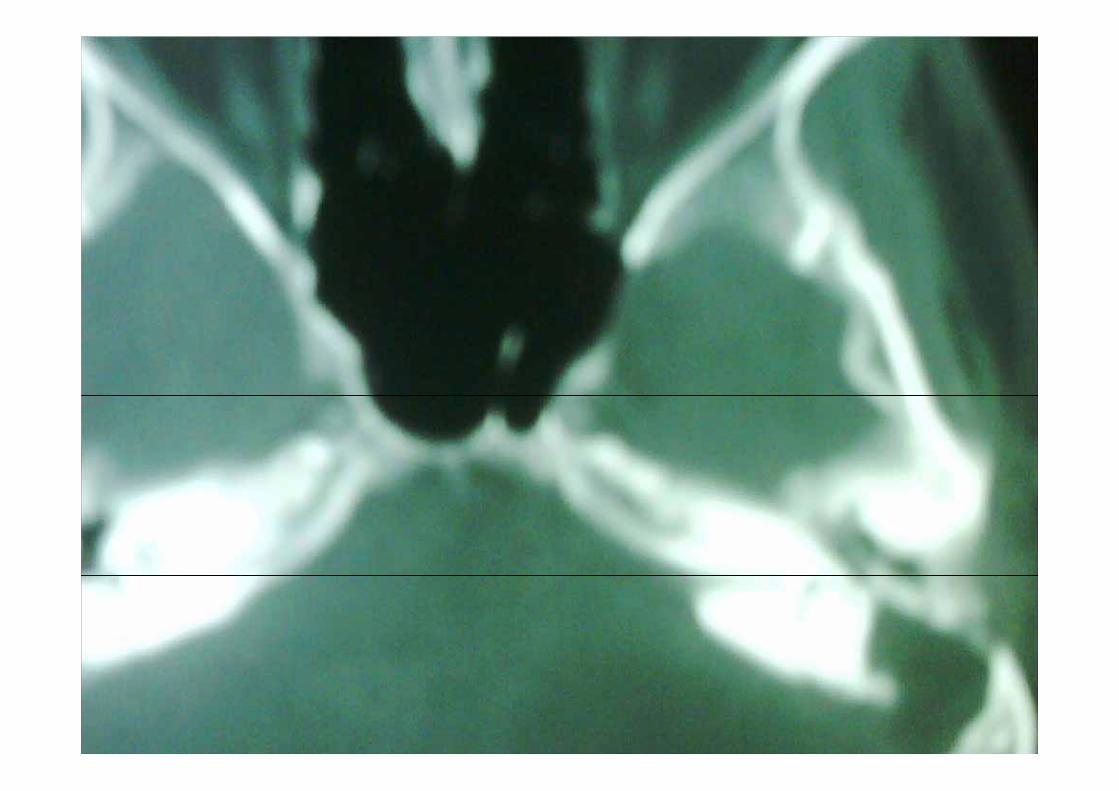
CT BRAINPansinusitisRight frontal brain abscessRight fronto temporo parietal subdural empyemaReferred urgently to neurosurgical unit,
Victoria Hospital

PAST HISTORYh/o fall from stairs 3yrs back- had a lacerated wound on right foreheadPMHPSHDrug historyAllergic historyImmunisation historySocial history

On ExaminationGeneral physical examination
Sick lookingExtremely thinUnusually quietWt. 16kgsP: 92/min T/°C: 37.6 RR: 14/minNo pallor, no jaundiceNo clubbingNo lymphadenopathyENT: nasal secretions ++ rt.>lt.

On Examination (cont)
Systemic examinationCVS: Normal HS, no murmurRS: chest was clear, trachea centrally located, no adventitious soundsAbdomen: scaphoid, no organomegaly, mild RUQ tendernessGenitals: normal

On Examination (cont)
CNS examinationGCS: E4M5V6Higher mental functionsMild neck stiffnessNo cerebellar signsNo photophobiaMoving all limbsFundoscopy: no papilledemaCranial nerve examination: normal

INVESTIGATION
Hematological: ↑↑ WCCBiochemistry: normalLFT: normalSpecial investigation- CT BRAIN ±C

MANAGEMENTAdmittedContinued i.v. antibiotic therapyi.v. fluid therapyConditioned stabilisedUrgent referral to E.N.T Hospital-admittedBAWO- Pus +++ right maxillary sinusBack to VH next day

MANAGEMENT (cont)
25/06/08: Cranial surgery1. Right small frontal craniotomy for
drainage of brain abscess2. Wide temporoparietal craniotomy for
evacuation of subdural empyemaNursed in ICUI.v antibiotics/ i.v phenytoin

POST-OP
Marked improvement in clinical conditionUncomplicated recovery phaseLab culture report: sterileDrains removed after 48 hrsReferred to nutritionist- high protein dietProgress CT brain showed good evacuation of brain abscess & empyema, no features of infarct or ↑ ICPContinued on i.v antibiotics for two weeks

POST-OPStill having RUQ painUltrasound abdomen
1. Gall bladder filled with calculi
2. Small rt. Renal calculus
Surgical opinionPediatric opinionStill under investigationReview with surgeon

REVIEW
With NeurosurgeonOral antibioticsOral AEDRepeat CT of brain

Thank you!!

4/18/2011
Patient Profile 2
16 years MaleComores Islandc/o Chronic discharge Left earHeadache, confusion, feverGCS10/15 (E3M5V2)Spastic, neck stiffness


Emergency combined surgical treatment
Radical mastoidectomy and posterior fossa craniectomy

4/18/2011 30

4/18/2011 31

CAVERNOUS SINUS
2 cm x 1 cm
Located on each of sella turcica and body of sphenoid bone
Superior orbital fissure to apex of petrous bone
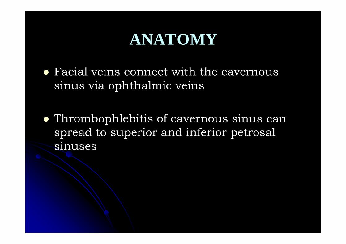
ANATOMY
Facial veins connect with the cavernous sinus via ophthalmic veins
Thrombophlebitis of cavernous sinus can spread to superior and inferior petrosal sinuses

ANATOMY
Posterior intercavernous sinus superior and inferior petrosal sinuses
Receive blood from superior and inferior ophthalmic vein
They drain posteriorly and inferiorly through the superior and inferior petrosal sinuses and pterygoid plexuses

SPREAD
Infections of
Face, nose, orbit, tonsils, soft palate, pharynx, air sinuses, middle ear and mastoid can all spread to cavernous sinuses
Sphenoid and posterior ethmoid sinuses
Jaw –tooth extraction, maxillary surgery via (pterygoid plexuses)

SYMPTOMS & SIGNS
Fever
Ptosis/chemosis
Oculomotor palsies (III, IV, VI)
Contralateral hemiparesis (thrombosis ICA)

CT brain
Irregular filling defect
Convex bulging of the lateral wall
Dilatation of superior opthalmic vein
Thickening of extra ocular muscles and periorbital edema

TREATMENT
Antibiotics (high doses)
(Staph aureus, Strep pneumonia, Haemophilus influenzae
Anticoagulant (no evidence of cortical venous infarct)
Surgery- sphenoid sinus sepsis
100 % mortality to 30 %


RHINOGENIC INTRACRANIAL SEPSIS

Leading neurological manifestation
Fever 96%Seizures 70%Neurological signs 58%


Epidemiology
Most common in malesSeasonal variation

EtiologySpread
Direct- Erosion of Tegmen tympaniErosion of posterior wall of frontal sinusRetrograde septic thrombophlebitisFacial or scalp infectionDental sepsisMeningitisCranial surgery e.g. depressed fracture
Infection at distant sites

Etiology
Otorhinolaryngeal infection- 40-70 %Paranasal sinusitisOtitis mediaMastoiditis
Cranial trauma- 6-30%

Predisposing factors
Diabetes MellitusAlcoholismChest infectionSepsisHIVImmunodepression- steroids, cytotoxic drugsPoor nutrition, poor hygiene, delayed treatment

“Frequent use of broad spectrum antibiotics may contribute to subdural empyema”

Most common pathogens
Strep pneumoniae- 16%Group B strep- 13%H. Influenzae- 13%Salmonella spp- 13%E. coli- 10%Pseudomonas aeruginosa- 10%

Pathogens
Pus- sterile in 40%Use of broad spectrum antibiotics
NTSO- non typhoidal salmonella organisms have been reported in the setting of advanced AIDS infection

Diagnosis
Difficult to clinically differentiate between meningitis and SDEDiagnosis is based on strong clinical suspicionTriad of- fever
sinusitisneurological deficit

Investigation
Infants: brain sonographyCT Bain with contrast, brain and paranasal sinuses, posterior fossa cuts

Investigation
CT Brain (contrast)Thin rim of fluid, slightly hyperdense to CSF with surrounding enhancement, adjacent disproportionate cortical edema and effacement of cortical sulciCranial ultrasound can substitute CT in infantsLP must be avoided

Management
Timing of surgerySimultaneous neurosurgical and ENT intervention
SDE requires surgical evacuation of infected material, irrespective of its volume

Management
Craniotomy was determined to be the surgical procedure of choice in SDEAllows complete evacuationDecompression of cerebral hemisphere


Prognosis
Early diagnosis and treatmentHigh degree of suspicion
“Prolonged fever, seizures, neurological signs”

Prognostic factors
AgeGCSTiming/ aggressiveness of treatmentProgression of disease

Outcome
Mortality- 100% before advent of antibiotics & CTDecreased to 40% after CT Scan10-12% presently

Intracranial subdural empyema is a neurosurgical emergency
It is rapidly fatal if not recognised early and managed promptly

Early drainage, simultaneous eradication of the primary source of sepsis and intravenous administration of high doses of appropriate antibiotics agents represents the mainstay of treatment

Spread
1. Direct spreadErosion through the postwall of
frontal sinus which has one-half the
thickness of the anterior wall
2. Indirect mechanismsRetrograde thrombophlebitis
i l i

Lumbar Puncture L.P
L.P performed in the presence of clinical features of raised ICP and focal neurological signs are extremely dangerous

Disparity between CT imaging and clinical findings
-integrity of arachnoid membrane-prevent spread
-improve blood brain barrier
-Wide cerebral decomposition via a wide craniotomy

MUST HAVE CT SCAN BRAIN & PNS
Infective sinustisPeriorbital swellingPurulent dural dischargePositive Neurosurgical signs
DIAGNOSIS

Role of Non Operative treatment
Fully concious patient, with small EDE (no radiological mass effect) with no neurological deficit, signs of clinical improvement (temperature ; ESR ; WCC May be treated with intravenous antibiotics and prophylatic antiepileptic provided the primary source of sepsis has been surgically eradicated

Unlike SDE, EDE is a disease that should be managed without morbidity or death

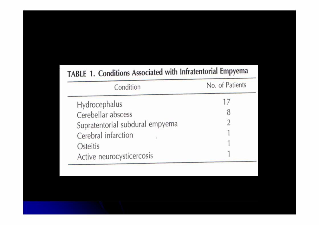
INFRATENTORIAL EMPYEMARare, highly lethal form of intracranial suppuration
Lumbar puncture
Cereballar abcess
Hydrocephalus
Extension of pus to cerebello pontine angle

INFRATENTORIAL EMPYEMATREATMENT
Early aggressive surgical drainage and decompression of the cerebellum by a wide posterior fossa craniectomy , eradication of the primary source of infection (usually mastoiditis) treatment of concomittant hydrocephalus high dose intravenous antibiotics

THANK YOU


![Stab wound in the Craniovertebral spine:Case report and ......stab wound case, as well as delayed neurologic deficits [12] and intra spinal suppuration [13]. A posterior approach to](https://static.fdocuments.us/doc/165x107/5f43397ce7f220415049fd53/stab-wound-in-the-craniovertebral-spinecase-report-and-stab-wound-case.jpg)


