REVIEW OF LITERATURE - Shodhgangashodhganga.inflibnet.ac.in/bitstream/10603/34370/28/ii review...
24
6 REVIEW OF LITERATURE India ranks first in the world in milk production and dairying in India are a classic example of production by masses rather than mass production. Due to changes in human food consumption patterns, demands for fruits, vegetables, milk and milk products, meat, poultry and fisheries have been increasing over the period in recent years. Among the different food sectors, the growth in dairy sector has been commendable (National Academy of Agricultural Sciences, 2013). The rate of growth in milk production in India is also substantially higher (3.6 per cent) than the world average of 1.5 per cent. However, the total projected demand of milk by the year 2030 would be about 200 million tonnes, depending on assumptions about income, population, urban growth, and expenditure elasticity parameters, which would imply an annual increase of around 4 million tonnes during the next two decades (N.D.R.I, 2011). At the existing rate of growth in milk production, supply is likely to fall short of the demand in next ten years. Among the several barriers in achieving the production targets, mastitis continues to remain as a challenging impediment, since the affected quarters may have 30 per cent less productivity and cow may lose about 15 per cent production (Radistitis et al., 2000). Mastitis in dairy animals is considered as one of the most important economic diseases resulting into huge economic loss to the country. Globally, mastitis accounts for about 38 per cent of the total direct costs of the common production diseases (Kossaibati and Esslemont, 1997). In India, the economic losses due to mastitis have increased about 115 folds in last five decades (Dua, 2001). Lack of awareness, delay in detection of sub-clinical mastitis, lack of markers for detecting ensuing mastitis, unhygienic milking practices, diverse production systems, inadequate treatment etc. are some of the important contributing factors in higher incidence of mastitis.
Transcript of REVIEW OF LITERATURE - Shodhgangashodhganga.inflibnet.ac.in/bitstream/10603/34370/28/ii review...

6
REVIEW OF LITERATURE
India ranks first in the world in milk production and dairying in India are a
classic example of production by masses rather than mass production. Due to changes
in human food consumption patterns, demands for fruits, vegetables, milk and milk
products, meat, poultry and fisheries have been increasing over the period in recent
years. Among the different food sectors, the growth in dairy sector has been
commendable (National Academy of Agricultural Sciences, 2013). The rate of growth
in milk production in India is also substantially higher (3.6 per cent) than the world
average of 1.5 per cent. However, the total projected demand of milk by the year 2030
would be about 200 million tonnes, depending on assumptions about income,
population, urban growth, and expenditure elasticity parameters, which would imply
an annual increase of around 4 million tonnes during the next two decades (N.D.R.I,
2011). At the existing rate of growth in milk production, supply is likely to fall short
of the demand in next ten years. Among the several barriers in achieving the
production targets, mastitis continues to remain as a challenging impediment, since
the affected quarters may have 30 per cent less productivity and cow may lose about
15 per cent production (Radistitis et al., 2000). Mastitis in dairy animals is considered
as one of the most important economic diseases resulting into huge economic loss to
the country. Globally, mastitis accounts for about 38 per cent of the total direct costs
of the common production diseases (Kossaibati and Esslemont, 1997). In India, the
economic losses due to mastitis have increased about 115 folds in last five decades
(Dua, 2001). Lack of awareness, delay in detection of sub-clinical mastitis, lack of
markers for detecting ensuing mastitis, unhygienic milking practices, diverse
production systems, inadequate treatment etc. are some of the important contributing
factors in higher incidence of mastitis.

7
Epidemiological status of bovine mastitis (Global vis-à-vis India)
The lactation process has been remarkably successful since the earliest
mammals, allowing them to occupy a vast range of ecological niches. However,
lactation is seriously impacted by the development of mastitis among most, if not all,
mammalian species (Michie et al., 2003). This condition alters milk composition and
reduces milk secretion, facts that impair infant/offspring growth and development. In
food animal species, it is one of the diseases with highest economic impact and a
major animal welfare concern. A broad definition of mastitis is inflammation of the
mammary gland, including not only intramammary tissues but also related anatomical
structures such as nipples, mammary areolas, milk ducts, etc. In veterinary medicine,
mastitis is referred to an intramammary inflammatory reaction caused by an infectious
agent (Fetherston, 2001).
With the increase in milk production, the incidence of mastitis has also
increased. Surveys on the prevalence of mastitis in most of the countries, irrespective
of the cause, show a comparable figure of 50 per cent among dairy cows (Radistitis et
al., 200). Subclinical mastitis which is believed to be more prevalent rather than
clinical in most countries ranged from 19 to 78 per cent (Tuteja et al., 1993).
Although controlled studies involving large sample sizes are very few in the country,
the available reports suggest same pattern. Analysis of 513, 1707 and 1115 lactation
records of Sahiwal and crossbred cows, and Murrah buffaloes, respectively in an
organized farm in northern India over a period of 9 years revealed that overall
incidence of mastitis was 13 per cent with significant difference between the breeds.
Sahiwal cows had higher incidence (20.66 per cent) compared to crossbred cows
(14.18 per cent) or Murrah buffaloes (7.44 per cent). An influence of season on

8
disease incidence was also observed in both cows and buffaloes in the same study. In
other studies, it has been shown that the incidence was the highest among pure-breed
Holsteins and Jerseys but the lowest in local cattle and buffaloes. In Haryana and
Rajasthan, the prevalence has been reported to be 36.69 per cent and 60.25 per cent,
respectively (Sudhan and Neelesh, 2010). In several studies, it has been reported that
subclinical mastitis was 15 to 40 times more prevalent than the clinical form and was
of longer duration, difficult to detect, adversely affected milk quality and quantity. It
constitutes a reservoir of microorganisms that lead to cross-infection of other animals
within the herd. Based on the published reports, it is evident that the average
prevalence of mastitis in 1960s to early 1990s, was not more than 30 per cent but
increased afterwards to even more than 60 per cent (Sharma et al., 2012). Two
decades ago, the mean incidence of clinical mastitis in India was 1-10 per cent with
subclinical mastitis ranging from 10 50 per cent in cows and 5-20 per cent in
buffaloes, while recent studies showed higher incidence of subclinical mastitis
ranging from 20 to 83 per cent in cows and 45 per cent in buffaloes (Sharma, 2007).
Analysis of the data from more than 100 recent studies spread over 21 States of India
indicate that the overall prevalence of mastitis ranged from 25 to 97 per cent with a
mean prevalence of about 50 per cent (Sharma, 2007). This clearly indicates the
drastic increase in the prevalence of mastitis especially the subclinical form of the
disease, which is an alarming situation for the dairy sector in the country.
Incidence of mastitis in dairy cows varies depending on geographical location
and housing environment. In North America and Europe, the incidence of clinical
mastitis in dairy cows ranges from 7 to 30 cases per 100 cows-years at risk (Erskine
et al., 1988; Olde et al., 2008; Barkema et al., 1998). In grazing herds from New

9
Zealand, a lower incidence rate of 10% and 19% was reported, but in one of the
studies only cows with less than 100 days after parturition were evaluated
(McDougall et al., 1999; Petrovski et al., 2009). In the New South Wales region of
Australia clinical mastitis incidence was 16% (Stevenson et al., 2000). Reports on
clinical mastitis incidence and prevalence from other countries are scarce. In Africa
for example, a study from Tanzania reported clinical mastitis incidence of 43 cases
per 100 cow-years at risk (Dohoo et al., 2011). Subclinical mastitis prevalence is
reported to be 31% in USA, 20 to 40% in Western Europe, and 29% in Australia
(Piepers et al., 2007; Rodrigues et al., 2005; Barkema et al., 1997; Roesch et al.,
2007; Plozza et al., 2011). In other countries with developing dairy industries,
subclinical mastitis prevalence is much higher. A survey from a dairy province in
China reported 54% prevalence at the cow level and 28% at the quarter level (Li et
al., 2009). Similarly, cow level prevalence in Brazil was 47% (Costa et al., 1998). All
these reported values should be considered historical, since the incidence and
prevalence of mastitis in dairy cows is largely determined by conditions in each
geographical region and individual herd and are constantly evolving. For example,
some studies demonstrated a direct relation between herds‟ bulk tank SCC and
clinical mastitis incidence. This approach reveals a higher incidence of clinical cases
or herds with lower SCC than those with high counts, possibly due to a reduction in
the total amount of immune cells in the mammary gland (Barkema et al., 1998).
10
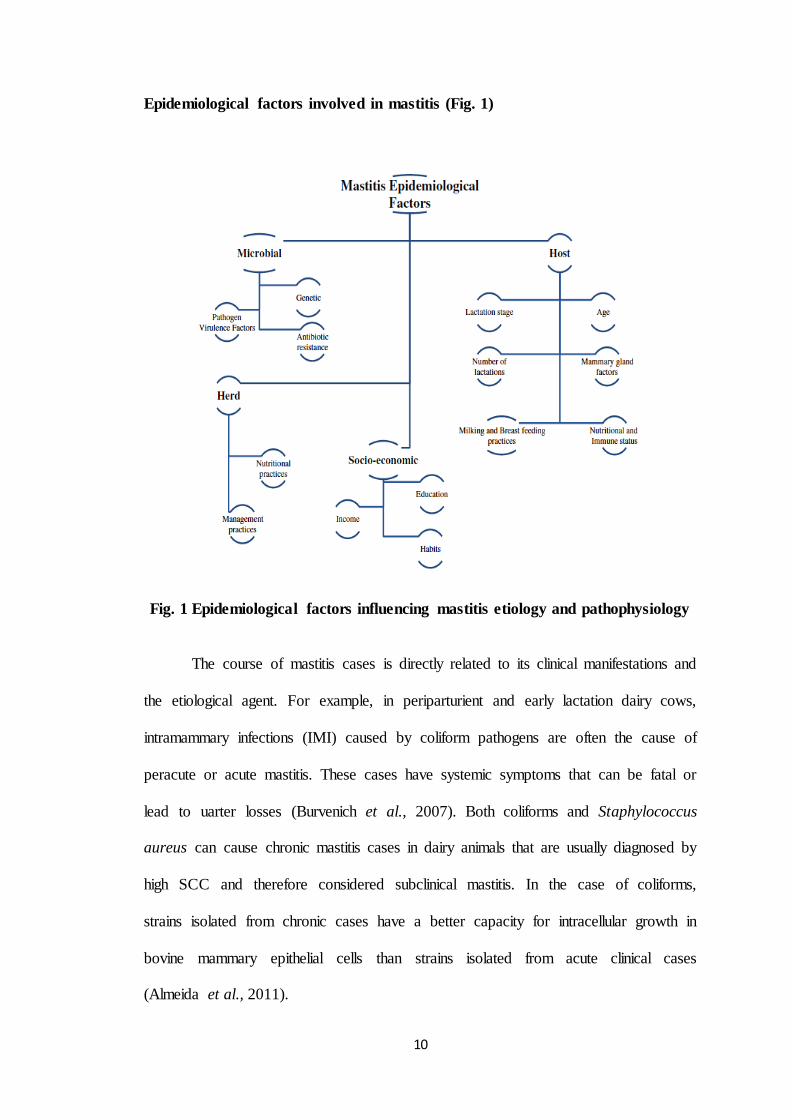
Epidemiological factors involved in mastitis (Fig. 1)
Fig. 1 Epidemiological factors influencing mastitis etiology and pathophysiology
The course of mastitis cases is directly related to its clinical manifestations and
the etiological agent. For example, in periparturient and early lactation dairy cows,
intramammary infections (IMI) caused by coliform pathogens are often the cause of
peracute or acute mastitis. These cases have systemic symptoms that can be fatal or
lead to uarter losses (Burvenich et al., 2007). Both coliforms and Staphylococcus
aureus can cause chronic mastitis cases in dairy animals that are usually diagnosed by
high SCC and therefore considered subclinical mastitis. In the case of coliforms,
strains isolated from chronic cases have a better capacity for intracellular growth in
bovine mammary epithelial cells than strains isolated from acute clinical cases
(Almeida et al., 2011).

11
Mastitis may lead to clinical symptoms and, as a consequence, it is often
diagnosed directly by visual assessment of breast/udder inflammation or by changes
in milk‟s organoleptic properties. In addition, there are several ancillary tests that are
used to detect both clinical and especially subclinical mastitis and these include: SCC,
which is considered the standard method, milk microbiological cultures, pH, lactose
content, electrical conductivity, SFMT, flow measurements, and quantification of
acute phase proteins (Pyorala, 2003). The choice of one or more of these procedures
usually depends on the availability, proximity to a diagnosis laboratory, personal
experience and/or technical skills. Microbiological analysis of milk is the only method
that allows for an etiological diagnosis of mastitis, however, there are relevant factors
that have to be considered when using this diagnostic tool. These factors include: use
of standard protocols for milk sample collection, correct identification and
quantification of bacterial isolates, and the relevance of clinical symptoms in the
animal (Dohoo et al., 2011). The collection of a representative sample for microbial
analysis is of outmost importance for good etiological diagnosis. In food animal
medicine, there are standard protocols for milk sample collection proposed by the
National Mastitis Council (NMC) and the International Dairy Federation (IDF)
(Hogan et al., 1999; Goodridge et al., 2004).
Epidemiological Aspects of Mastitis udder health depends on a balanced
interaction between host and its microbiota, which may contain microorganisms
ranging from probiotic to potentially infectious. Obviously, there are relevant
differences among mammals regarding the number, size, position and structure of the
mammary glands. In addition, mammals (even within a same species) differ widely in
their ecosystems, management and use (e.g., milk producing versus meat-producing
12
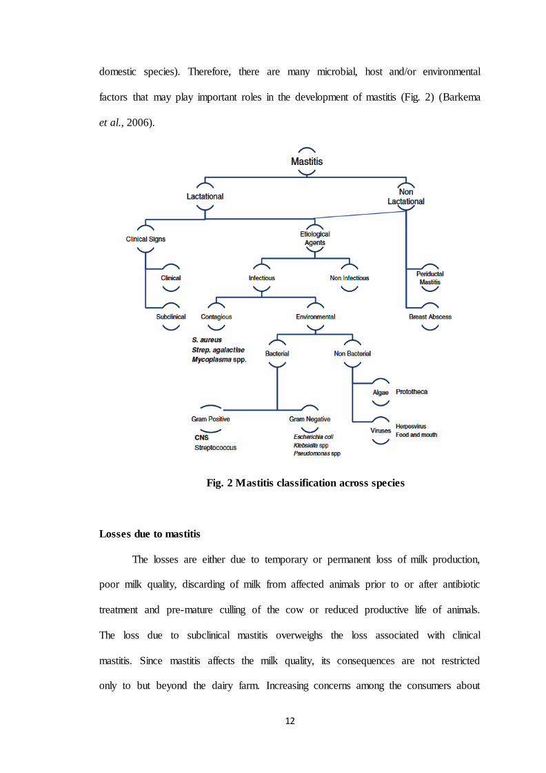
domestic species). Therefore, there are many microbial, host and/or environmental
factors that may play important roles in the development of mastitis (Fig. 2) (Barkema
et al., 2006).
Fig. 2 Mastitis classification across species
Losses due to mastitis
The losses are either due to temporary or permanent loss of milk production,
poor milk quality, discarding of milk from affected animals prior to or after antibiotic
treatment and pre-mature culling of the cow or reduced productive life of animals.
The loss due to subclinical mastitis overweighs the loss associated with clinical
mastitis. Since mastitis affects the milk quality, its consequences are not restricted
only to but beyond the dairy farm. Increasing concerns among the consumers about
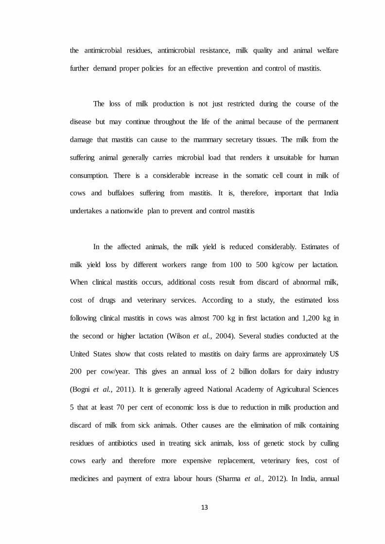
13
the antimicrobial residues, antimicrobial resistance, milk quality and animal welfare
further demand proper policies for an effective prevention and control of mastitis.
The loss of milk production is not just restricted during the course of the
disease but may continue throughout the life of the animal because of the permanent
damage that mastitis can cause to the mammary secretary tissues. The milk from the
suffering animal generally carries microbial load that renders it unsuitable for human
consumption. There is a considerable increase in the somatic cell count in milk of
cows and buffaloes suffering from mastitis. It is, therefore, important that India
undertakes a nationwide plan to prevent and control mastitis
In the affected animals, the milk yield is reduced considerably. Estimates of
milk yield loss by different workers range from 100 to 500 kg/cow per lactation.
When clinical mastitis occurs, additional costs result from discard of abnormal milk,
cost of drugs and veterinary services. According to a study, the estimated loss
following clinical mastitis in cows was almost 700 kg in first lactation and 1,200 kg in
the second or higher lactation (Wilson et al., 2004). Several studies conducted at the
United States show that costs related to mastitis on dairy farms are approximately U$
200 per cow/year. This gives an annual loss of 2 billion dollars for dairy industry
(Bogni et al., 2011). It is generally agreed National Academy of Agricultural Sciences
5 that at least 70 per cent of economic loss is due to reduction in milk production and
discard of milk from sick animals. Other causes are the elimination of milk containing
residues of antibiotics used in treating sick animals, loss of genetic stock by culling
cows early and therefore more expensive replacement, veterinary fees, cost of
medicines and payment of extra labour hours (Sharma et al., 2012). In India, annual

14
economic loss incurred by dairy industry on account of udder infections is estimated
to be Rs. 6053.21 crores and out of which loss of Rs. 4365.32 crore (70 - 80 per cent)
has been attributed to sub-clinical mastitis (Dua, K., 2001). In another report from
India, the annual economic loss due to mastitis has been calculated to be Rs. 7165.51
crores; losses being almost same for cows (3649.56 crores) and buffaloes (3515.95
crores). Subclinical mastitis has been estimated to account for 57.93 per cent (4151.16
crores) of the total economic loss due to mastitis (PDADMAS, 2011).
Etiology of Mastitis
Mastitis is the outcome of interaction of various factors associated with the
host, pathogen(s) and the environment. Association of some host and managerial and
housing determinants with mastitis is well established. At least 137 species of
microorganisms from a broad phylogenetic spectrum, including bacteria, yeast, fungi
and algae, are able to cause bovine mastitis. However, amongst these, only 5 species
of bacteria account for the bulk of bovine mastitis cases (Rinaldia et al., 2010) but
dominant causal agents may have some geographical signatures, as the distribution of
pathogenic bacteria displays a substantial geographic variation. Causal pathogens can
be divided into two groups based on their source: environmental pathogens and
contagious pathogens. Coliform organisms (Escherichia coli, Klebsiella sp etc.) and
Streptococcal organisms (Streptococcus uberis, S. bovis and S. dysgalactiae) are the
important environmental pathogens.
Environmental mastitis is caused by potential pathogens found generally in the
digestive tract (referred to as “coliforms”) of cattle or their surroundings such as
faeces, soil, bedding material and manure (Jones, 2006). These microorganisms

15
generally proliferate substantially in bedding (approximately 1,000,000 or more cells
per gram of bedding). This increases the probability of infection of mammary glands
leading to clinical mastitis (Bradley and Green, 1997). There is a positive correlation
between the number of coliforms present in the bedding material and the bacterial
load on the teat ends as well as the occurrence rates of clinical mastitis (Hogan et al.,
1989). Coliforms- particularly Escherichia coli, Enterobacter aerogenes, Klebsiella
pneumonia, Serratia marcescens and a Streptococcus spp., Streptococcus uberis are
the chief organisms found to cause environmental mastitis. Environmental mastitis
has previously constituted less than 10% of total mastitis cases, but more recently
there has been an increase in the incidence of environmental mastitis [Bradley and
Green, 1997; Boyer, 1997) particularly associated with S. uberis infection. This
pathogen is most often associated with chronic mastitis, which does not respond to
antibiotic treatment (Jones GM, 2006).
Contagious mastitis is caused by bacterial pathogens that thrive on the udder
skin and lesions of teat. They cannot survive for long periods in the environment and
generally are transmitted from one cow to another by the milking machine, the hands
of milkers, milk contaminated fomites or the sponge used while milking (Harmon,
1994; APHIS, 2008). The pathogens mainly associated with contagious mastitis are
Staphylococcus aureus and Streptococcus agalactiae (Smith and Hogan, 1995;
Barkema et al., 2006; Monecke et al., 2007). Although Streptococcus dysgalactiae is
considered as an environmental pathogen, there is evidence of its transmission from
cow to cow as a contagious pathogen causing mastitis (Smith and Hogan, 1995).
Mycoplasma species also cause contagious mastitis. Mycoplasma bovis is the
predominant species sometimes leading to severe problems like sudden onset, rapid
16
transmission and reduction in milk yield and lack of response to treatment (Harmon,
1994). However, the most recognised pathogen in the majority of clinical and
subclinical mastitis cases in most countries is Staphylococcus aureus [Workineh et
al., 2002; Barrett et al., 2005; Middleton, 2008). These bacteria are of immense
importance, causing over 25% of intra-mammary infections and adversely affecting
the quality of milk in a large number of clinical cases (Owens et al., 1988; Haveri,
2008). They are also considered the emerging pathogens causing bovine mastitis since
they are the most commonly isolated bacterial pathogens.
In addition to Staphylococcus spp., Corynebacterium spp. constitutes some of
the emerging pathogens causing bovine mastitis. Corynebacterium bovis is frequently
isolated from milk in many dairy farms and causes moderate inflammation of the
mammary gland (Hommez et al., 1999; Haltia et al., 2006). These infections result in
a slight increase in bulk tank somatic cell counts, changes in the composition of milk,
sudden reduction in milk production and clinical mastitis (Harmon, 1994). Four
species of non-lipophilic Corynebacteria found to cause clinical and sub-clinical
mastitis are C. amycolatum, C. mulcerans, C. pseudotuberculosis, and C.
minutissimum (Hommez et al., 1999). Other species of Corynbacterium isolated from
cases of clinical mastitis in sheep are C. mastiditis and C. camporealensis (Fernandez
et al., 1997).
Recent studies have revealed that coagulase negative staphylococci (CNS)
isolated from teat skin and teat canal, as well as from the coat and the nostril
comprises a major interest area of mastitis causal organisms (Pitkälä et al., 2004;
Tenhagen et al., 2006). Mastitis in heifers at calving is mainly caused by CNS. More
17
than 50 species and subspecies are included in this group (Pyörälä and Taponen,
2009). Staphylococcus epidermidis, Staphylococcus simulans, Staphylococcus
saprophyticus, Staphylococcus hyicus, Staphylococcus warneri, Staphylococcus
chromogenes, Staphylococcus sciuri and Staphylococcus xylosus are the commonly
encountered species of CNS in bovine mastitis (Rupp et al., 2000). The various
species of CNS isolated from bovine mastitis cases show varied pathogenicity,
antimicrobial susceptibility and virulence factors (Zadoks and Schukken, 2006;
Taponen and Pyöröla, 2009).
The major pathogens involved in mastitis are Streptococcus agalactiae,
Staphylococcus aureus, Corynebacterium bovis and Mycoplasma spp. The
distribution of pathogens varies among countries and even within country, production
systems, farms and individual animals. For example, Staphylococcus aureus is most
frequently encountered in clinical mastitis, followed by Streptococcus dysgalactiae in
Norway (Waage et al., 1999) while in Midwestern United States, coliforms are the
most frequently isolated bacteria. In Europe, clinical Klebsiella mastitis occurs less
frequently than clinical E. coli mastitis. In contrast, coliforms are less important and
Streptococcus uberis is the main concern in both clinical and subclinical mastitis in
New Zealand (Zadoks et al., 2011).
In India, Staphylococcus spp. have been reported to be the main etiological
agents of mastitis in cattle and buffaloes. However, there are no studies on nationwide
distribution of mastitis-causing bacteria in India. Apart from regional differences,
cows in tie-stalls have higher incidence of Staphylococcus aureus, Streptococcus
uberis, coagulase-negative staphylococci and other streptococcal infections compared

18
to those in free-stalls, where Klebsiella sp. and E. coli are main concerns (Olde et al.,
2008). Collectively, it suggests that distribution of organisms may vary between
regions and husbandry systems and it is important to pre-ascertain the
epidemiological pattern of mastitis pathogens in the implementation of management
strategies. In recent years, there have been changes in the relative and absolute
importance of different pathogens. In UK, during 1960s, it was observed that
Staphylococcus aureus was the most common organism in mastitis, but in 1980s, E.
coli was most commonly isolated from the milk of mastitis affected cows and the
same trend was also continued in 1990s. In several countries, S. aureus continues to
be the major cause of sub-clinical mastitis and the pathogens previously considered to
be purely environmental may also be capable of causing persistent infection. The
major objectives of the epidemiological investigations include the identification of
risk factors at farm level, major pathogens involved and the susceptibility of the host.
India being a large and diversified country with different farming systems and agro
climatic conditions, the prevalence of mastitis and the pathogens involved are likely
to vary with places and herds. Hence, obtaining the ground situation of the disease
and characterizations of epidemiological parameters to be intervened are of vital
importance.
In animals, clinical mastitis is characterized by signs of inflammation in the
mammary gland including hyperemia, pain, and increased gland size and density.
These symptoms may be accompanied or not by systemic signs, such as fever or
depression (Kemper and Gerjets, 2009).

19
Globally, staphylococci (S. aureus and CNS) are the most common mastitis-
causing agents in cows, buffaloes, ewes, does, dromedaries, rabbits, dolphins and
women (Rowan et al., 1996; Guliye et al., 2002; Moroni et al., 2006; Segura et al.,
20070. They are closely followed by Streptococci and E. coli which in some species
or settings may have a similar or higher prevalence than that of staphylococcal
mastitis. Less commonly, other Gram-positive (Actinomyces spp.,Corynebacterium
spp., Bacillus spp., Mycobacterium spp., Enterococcus spp., Clostridium spp.) and
Gram-negative (Klebsiella spp., Enterobacter spp., Citrobacter spp., Serratia spp.,
Proteus spp. and Pasteurella spp.) In dairy animals, mastitis causing pathogens are
classified depending on their epidemiological behaviour in contagious and
environmental (Eberhart et al., 1987). Among contagious pathogens S. aureus,
Streptococcus agalactiae, and Mycoplasma spp. are included. Contagious pathogens‟
main reservoir is the mammary gland; consequently, these bacteria are spread from
cow to cow or between quarters/ halfs of the same animal during the milking process.
In contrast, environmental pathogen reservoirs include bedding, pasture, and other
unanimated objects. These bacteria are isolated from the skin, teat ends, and teat
canals where they can gain access to the gland causing IMI. Streptococcus other than
agalactiae, Enterococcus sp, CNS, and coliform bacteria including Escherichia coli
and Klebsiella spp. are included in this group. It is important to note that certain
microbial, host and/or environmental factors may induce environmental agents to
behave as contagious pathogens or vice versa (Zadoks et al., 2001; Pyörälä et al.,
2009)
The mammalian ecosystem is hospitable, or at least receptive, to many
microorganisms including most of the bacterial groups that have potential to cause

20
mastitis. The evolutionary process has led to state of mutual acceptance or tolerance.
However, upon disturbance localized or disseminated invasive infections can occur.
Unfortunately, the exact causal events leading to the transition from colonization to
infection still are inaccurate in vivo (van et al., 2009). The ability to colonize and,
eventually, infect a host depends on several microbial characteristics, including the
expression of several virulence factors, e.g. the formation of biofilms, expression of
super antigens, and antimicrobial resistance. At the species level, pathogen-specific
transcriptomic disruptions are observed in the mammary gland as well as in peripheral
organs (Rinaldi et al., 2010). As an example, two major mastitis pathogens, such as
E.coli and S. aureus, elicit differential innate immune responses following IMI in
cows, which may determine the course and severity of the disease (Bannerman et al.,
2004). In relation to different Staphylococcal species, genomic analysis reveals that S.
aureus is more suited to develop acute infections than CNS. Conversely, CNS have a
tendency to cause sub-acute, subclinical or chronic infections. As an example,
members of the enterotoxin and exotoxin families, which function as super antigens
and inducers of a pro-inflammatory cytokine response, are unique to S. aureus and
have not been identified in characterized isolates of S. epidermidis or other CNS.
Several virulence factors or toxins are included in certain pathogenicity genomic
islands. In S. aureus seven islands carry approximately one-half of the virulence
factors that have been identified. Variations in the pathogenic potential of strains
belonging to this species depends largely on allelic variation of the virulence genes,
on the presence or absence of individual and on the presence of single nucleotide
polymorphisms (SNPs) in key genes. Such differences are the most significant factor
contributing to observed variations in antibiotic resistance and virulence among S.
aureus strains (Gill et al., 2005).

21
Pathogenesis
A comprehensive understanding of the pathogenicity of mastitis is key for the
development of appropriate detection techniques. The primary cause of mastitis is a
wide spectrum of bacterial strains; however, incidences of viral, algal and fungal-
related mastitis were also reported (Pyorala, 2003). Normally, the teat canal is tightly
closed by sphincter muscles, preventing the entry of pathogens. It is lined with
keratin, a waxy material derived from stratified squamous epithelium that obstructs
the migration of bacteria and contains antimicrobial agents, such as long-chain fatty
acids, that assist in combating the infection. However, the efficiency of keratin is
restricted (Craven and Williams, 1985; Paulrud, 2005). Fluid accumulates within the
mammary gland as parturition approaches, resulting in increased intramammary
pressure (Paulrud, 2005) and mammary gland vulnerability caused by the dilation of
the teat canal and leakage of mammary secretions (Sordillo and Streicher, 2002).
Additionally, during milking, the keratin is flushed out and there is distention of the
teat canal (Rainard and Riollet, 2006). The sphincter requires nearly 2 hrs to return
back to the contracted position. Once inside the teat, bacteria must also elude the
cellular and humoral defence mechanisms of the udder (Sordillo and Streicher, 2002).
If they are not eliminated, they start multiplying in the mammary gland (Fig. 3). They
liberate toxins and induce leukocytes and epithelial cells to release chemo-attractants.
If the infection persists, internal swelling within the mammary epithelium, not
normally detectable by an external examination, can occur. The mammary gland
alveoli become damaged and start losing anatomical integrity (Fig. 3). The blood–
milk barrier is breached, causing extracellular fluid components, such as chloride,
sodium, hydrogen, potassium and hydroxide ions, to enter the gland and mix with the

22
milk (Zhao and Lacasse, 2008). When extensive damage to the blood–milk barrier has
occurred, blood might be detected in the milk. This leads to visible changes on the
udder, such as enhanced external swelling and reddening of the gland. Changes also
occur in the milk, including increased conductivity, increased pH, raised water
content and the presence of visible clots and flakes (Zhao and Lacasse, 2008; Kitchen,
1981). This marks the initial stage of clinical symptoms, and the most severe
infections might ultimately result in the death of the animal.
Fig. 3 Schematic representation of mastitis development in an infected udder.
Molecular identification of the mastitic pathogens
A reliable etiological analysis must identify the causing organism(s) at least at
the species level. Many phenotypic assays traditionally used for taxonomical
purposes, such as the carbohydrate fermentation pattern, are no longer valid due to

23
low specificity or non-conclusive results. Recently, the introduction of molecular
microbiology techniques such as PCR and matrix-assisted laser desorption/ionization
time-of-flight mass spectrometry (MALDI-TOF MS) have improved the sensitivity,
specificity, and processing time of bacterial identification procedures (Barreiro et al.,
2010). Furthermore, realtime PCR assays performed directly on milk samples
(without culturing) have benefits over conventional culture, including higher speed,
automated interpretation of results, and increased sensitivity (Taponen et al., 2009;
Koskinen et al., 2010). However, implementation of this technique must take into
account that, in addition to live bacteria, milk contains a high quantity of dead
bacteria and free bacterial DNA, thus reducing its specificity (Perez et al., 2007). As a
consequence, bacterial cultures are still required for routine etiological diagnosis of
mastitis. There is no doubt that ongoing and future improvements on the application
of Omics in mastitis diagnosis will provide powerful tools with high specificity and
sensitivity.
More recently, microbial identification techniques based on pyro-sequencing
of the 16S ribosomal RNA gene were used to obtain a description of the milk
microbes (Hunt et al., 2011). Results indicated that milk bacterial communities are
more complex than expected, with several genera representing greater than 5% of the
relative community abundance. Staphylococci (particularly Staphylococcus
epidermidis) and Streptococci were the dominant genera. Bacterial populations were
usually, although not always, stable over time within the same individual. These
findings open a new concept regarding the role of mammary microbiota in
maintaining mammary health in mammalian species.

24
Role of antibiotics in the treatment of bovine mastitis
The success of bovine mastitis therapy depends on the aetiology, clinical
presentation, and antimicrobial susceptibility of the aetiological agent among other
factors (Miltenburg et al., 1996). Therapy failure in the management of mastitis could
result from pathological changes that occur in the udder, aetiology related factors,
pharmacokinetic properties of the antimicrobial drugs, poor animal husbandry and
inadequate veterinary services. However, the control of mastitis has been successfully
achieved through the establishment of effective herd health control programs (Erskine
et al., 2002). Antimicrobial agents are the main therapeutic tools for the treatment and
control of mastitis. Among the main reasons of low efficacy of antibiotic treatment of
mastitis cases is the resistance of the bacteria to antimicrobials. Recently, several
studies have been conducted to determine the antibacterial susceptibility patterns of
mastitis pathogens isolated from clinical studies or submitted to diagnostic
laboratories (Brown and Scasserra, 1990; Gitau et al., 2003; Haile, 2004).
Antimicrobial agents are widely used for the treatment of bovine mastitis,
respiratory tract infections and diarrhoea in cattle. During acute infections and
outbreaks of infectious disease in groups or herds it is important to use an effective
antimicrobial treatment as early as possible. This empirical treatment is generally
based on knowledge of the resistance pattern of the different bacterial pathogens to
antimicrobial agents used in the particular animal species. Antimicrobial resistance is
an increasingly important problem among several bacterial species causing infection
in animals and humans in recent year. The problem for some bacterial species is so
critical that there is few treatment options left (Aarestrup et al., 1999; Levy et al.,
2001). The initial treatment of animals is commonly based on the experience

25
regarding the expected resistance of the infectious agent. The fact that occurrence of
antimicrobial resistance varies between countries and regions has the potential to
complicate that matter. Furthermore, knowledge of expected resistance is limited by
the small proportion of different bacterial pathogens from infected animals that
actually are investigated for their antimicrobial resistance pattern.
The evolution of antibiotic resistance in S. aureus strains is a serious cause of
concern in dairy animals (Wang et al., 2008). Strains of S. aureus resistant to β-
lactam antibiotics are known as methicillin-resistant S. aureus (MRSA). These strains
in intra-mammary dissemination often produce incurable severe intra-herd infections
(Moon et al., 2007; Kumar et al., 2010). MRSA strains have been observed to be
multi-drug resistant, such as aminoglycosides, macrolides, lincosamides,
streptogramins, tetracyclines, etc., which are often used in the treatment of mastitis
(Wang et al., 2008; Kumar et al., 2010). The transmission of bovine MRSA to
humans is possible and may contribute to outbreaks in animal and human populations
(Lee, 2003). Hence, it is necessary to know which endemic strains of S. aureus in
dairy cattle populations are highly pathogenic and methicillin-resistant
Milk
Milk may be defined as the whole, fresh, clean lacteal secretion obtained by
the complete milking of one or more healthy milchy animals, excluding that obtained
within 15 days before or 5 days after calving or such periods as may be necessary to
render the milk practically colostrum free, and containing the minimum prescribed
percentage of milk fat and milk-solids not fat. In India, the term „milk‟ when
unqualified, refers to cow or buffalo milk or a combination of the two.

26
Milk is one of the most important foods of human beings. It is universally
recognized as a complete diet due to its essential components and it is directly
available for consumption (Battaglia et al., 2007; Grimaud et al., 2009). Its role is to
nourish and provide immunological protection (Hemalatha et al., 2010). Milk has
distinct physical, chemical and biological characteristics, which justifies its high
quality for consumption. These characteristics present a favourable environment for
the multiplication of various bacteria. It is well known that freshly obtained milk
contains some bacteria and somatic cells, which represent the biological constituent of
the milk (Hemalatha et al., 2010). The biological constituents easily change
depending on production conditions, the health status of the cattle, hygiene practices
during milking, keeping and transportation of milk (Turner et al., 1990).
Many constituents of cows‟ milk can be broadly categorised according to their
physical properties or/and physiological functions. In this complex biological fluid,
minerals occur in chemical equilibrium between the free ions and complexes with
various components, such as protein, lipids, carbohydrates and low molecular weight
ligands like citrate and amino acids (Vegarud et al., 2000). Mineral and trace element
concentrations in raw cows‟ milk are not constant but mainly vary according to two
kinds of factors, those related with secretion from the mammary gland, such as the
lactation state, animal species and health status, and extrinsic factors, such as season,
dairy cattle ration (nutritional status of cow), environment (nature of soil and locality
of the farm). In this respect, several studies have been carried out to assess mineral
content of cows‟ milk from different areas (Licata et al., 2004; Muñiz-Naveiro et al.,
2005), as well as to evaluate preliminary correlations between animal feeding,

27
manufacturing process and elemental profile in cows‟ milk and dairy products (Coni,
et al., 1995).
The world-wide contamination of milk with undesirable substances via animal
feeds, heavy metals, mycotoxins, dioxins and similar pollutants is considered to be of
great concern to public health due to their toxic effects on humans and wildlife. Milk
products are very important human nutrients and their consumption has increased in
recent years. It is known as an excellent source of calcium, magnesium and zinc and
hence supply very small amount of Fe and Cu (Ali et al., 2011)
Genetic diversity of bacteria
Several studies suggest that there are differences between strains isolated from
bovine and human hosts (Musser and Kapur, 1992; Kapur et al., 1995). Different
epidemiological studies of isolates from human and animals have been based on
phenotypic techniques, such as biotyping, bacteriophage typing and antibiotic
sensitivity testing (Tenover et al., 1994). Biotyping has been used as a simplified
method to differentiate isolates from humans and animals into host-specific (human,
bovine, poultry and ovine) or non-host specific biotypes (Devriese, 1984). Recently,
molecular DNA typing methods have been developed on the basis of genotype
characterization (Van et al., 1995). Random amplification of polymorphic DNA
(RAPD-PCR) has been applied extensively to distinguish different isolates (Van et
al., 1995). RAPD is an accessible and sensitive method based on the use of arbitrary
primers to amplify polymorphic segments of DNA. This technique has been widely
used in recent years for detection of diversity among isolates (Welsh et al., 1990;
Maslow et al., 1993). The modified polymerase chain reaction (PCR) with single

28
primers of arbitrary nucleotide sequence and requiring no prior sequence information
have proved useful in detecting intraspecific polymorphism among organisms (Welsh
and Mc Clelland, 1990; Williams et al., 1990). This amplification technique
(arbitrarily primed PCR on RAPD) can generate specific DNA fragments useful for
genome mapping, identification of isolates and application in molecular biology
(Hadrys et. al., 1992).
Molecular characterization of mastitic bacteria
Biofilm formation is another important virulence factor. The number of strains
carrying the biofilm related ica A and D gene and showing resistance to oxacillin,
erythromycin, clindamycin and mupirocin was significantly higher among mastitis
isolates (Delgado et al., 2009). Resistance to diverse antibiotics and a higher ability to
form biofilms found among mastitis specific strains may explain the chronic and
recurrent nature of this infectious condition. There are several other virulence factors
that modify bacterial ability to colonize and cause IMI but the combination of genes
associated to pathogenicity is strain-specific (Barkema et al., 2006; Monecke et al.,
2007, Wolf et al., 2011). In veterinary medicine, this could possibly lead to the use of
bacterial genetic characterization as a management tool. For instance, animal culling
or segregation could be determined on the bases of infectious status with strains
expressing specific virulence factors.
Although multiple bacterial and external factors influence attachment and
accumulation leading to biofilm formation (Gotz, 2002), the production of a
polysaccharide intercellular adhesin (PIA) or polymeric N-acetyl-glucosamine
(PNAG) by intercellular gene cluster adhesion (ica) operon-encoded enzymes (Mack

29
et al., 1996; Maira et al., 2002) is currently the best understood mechanism of biofilm
formation in staphylococci in vitro and in vivo (Rupp et al., 1999; Mack et al., 2004).
A great variety of Staphylococcus aureus strains carry the ica cluster, and some of
them constitute biofilm. Loss of the ica locus results in an incapacity to produce
polysaccharidic intercellular adhesin and to develop biofilms (Cramton et al., 2000).
Staphylococcal infections produced by ica carriers can be more problematic due to the
presence of methicillin and mupirocin resistance genes (Jones et al., 2001; Stewart
and Costerton, 2001). The rapid detection of the ica locus in Staphylococcal isolates,
will allow the development of prevention methods to reduce the bacterial capacity in
mastitis.
Therefore, based on the review of literature the present study was designed to
do survey of Bovine Mastitis in North Karnataka, isolation, identification and
antimicrobial susceptibility / resistance of bacterial isolates, effects on biochemical
parameters of the milk inoculated by mastitic pathogens and to do genetic diversity
and molecular characterization of the isolated mastitic bacteria.