Review Article Nephrolithiasis: Molecular Mechanism of ...
22
Hindawi Publishing Corporation BioMed Research International Volume 2013, Article ID 292953, 21 pages http://dx.doi.org/10.1155/2013/292953 Review Article Nephrolithiasis: Molecular Mechanism of Renal Stone Formation and the Critical Role Played by Modulators Kanu Priya Aggarwal, 1 Shifa Narula, 1 Monica Kakkar, 2 and Chanderdeep Tandon 1 1 Department of Biotechnology and Bioinformatics, Jaypee University of Information Technology, Waknaghat, Solan, Himachal Pradesh 173234, India 2 Department of Biochemistry, Himalyan Institute Hospital Trust, Swami Ram Nagar, Dehradun, Uttrakhand 248140, India Correspondence should be addressed to Chanderdeep Tandon; [email protected] Received 29 April 2013; Accepted 26 July 2013 Academic Editor: Beatrice Charreau Copyright © 2013 Kanu Priya Aggarwal et al. is is an open access article distributed under the Creative Commons Attribution License, which permits unrestricted use, distribution, and reproduction in any medium, provided the original work is properly cited. Urinary stone disease is an ailment that has afflicted human kind for many centuries. Nephrolithiasis is a significant clinical problem in everyday practice with a subsequent burden for the health system. Nephrolithiasis remains a chronic disease and our fundamental understanding of the pathogenesis of stones as well as their prevention and cure still remains rudimentary. Regardless of the fact that supersaturation of stone-forming salts in urine is essential, abundance of these salts by itself will not always result in stone formation. e pathogenesis of calcium oxalate stone formation is a multistep process and essentially includes nucleation, crystal growth, crystal aggregation, and crystal retention. Various substances in the body have an effect on one or more of the above stone- forming processes, thereby influencing a person’s ability to promote or prevent stone formation. Promoters facilitate the stone formation while inhibitors prevent it. Besides low urine volume and low urine pH, high calcium, sodium, oxalate and urate are also known to promote calcium oxalate stone formation. Many inorganic (citrate, magnesium) and organic substances (nephrocalcin, urinary prothrombin fragment-1, osteopontin) are known to inhibit stone formation. is review presents a comprehensive account of the mechanism of renal stone formation and the role of inhibitors/promoters in calcium oxalate crystallisation. 1. Introduction Renal stones have afflicted humans for millennia. Many researchers are attempting to elucidate the mechanism of CaOx renal stone formation. Archeological findings give profound evidence that humans have suffered from kidney and bladder stones for centuries [1]. e risk of developing urolithiasis in adults appears to be higher in the western hemisphere (5–9% in Europe, 12% in Canada, and 13–15% in the USA) than in the eastern hemisphere (1–5%), although the highest risks have been reported in some Asian countries such as Saudi Arabia (20.1%) with lifetime recurrence rates of upto 50% [2]. e interval between recurrences is variable, with approximately 10% within one year, 35% in five years, and 50% by 10 years [3]. However, approximately 75% of stones are primarily calcium oxalate, but up to 50% of these include calcium hydroxyl phosphate (brushite or calcium hydroxyapatite) in trace or greater amounts; 10–20% are composed of magnesium ammonium phosphate (struvite or triple phosphate); 5% are composed of urate; and 1-2% are composed of cystine [4, 5]. With its multifactor etiology and high rate of recurrences, urinary tract stone disease provides a medical challenge [6]. ere is thus a pressing need to prevent this disease and its recurrence. e physiochemical mechanism of stone formation via precipitation, growth, aggregation, and concretion of various modulators in urine is represented in Figure 1. In addition, some researchers have recently emphasized that the interaction between crystals and renal tubular epithelial cells, including the adhesion or endocytosis of crystals by cells, is an important factor in stone formation [7, 8]. Long-standing interest in the possible role of macromolecules in nephrolithiasis stems from the observation that all human kidney stones consist of a complex amalgam of mineral and organic material [9]. e study of stone matrix has come a long way in recent years, but the wealth of knowledge we have gained has been offset
Transcript of Review Article Nephrolithiasis: Molecular Mechanism of ...
Hindawi Publishing CorporationBioMed Research InternationalVolume 2013, Article ID 292953, 21 pageshttp://dx.doi.org/10.1155/2013/292953
Review ArticleNephrolithiasis: Molecular Mechanism of Renal StoneFormation and the Critical Role Played by Modulators
Kanu Priya Aggarwal,1 Shifa Narula,1 Monica Kakkar,2 and Chanderdeep Tandon1
1 Department of Biotechnology and Bioinformatics, Jaypee University of Information Technology, Waknaghat, Solan,Himachal Pradesh 173234, India
2Department of Biochemistry, Himalyan Institute Hospital Trust, Swami Ram Nagar, Dehradun, Uttrakhand 248140, India
Correspondence should be addressed to Chanderdeep Tandon; [email protected]
Received 29 April 2013; Accepted 26 July 2013
Academic Editor: Beatrice Charreau
Copyright © 2013 Kanu Priya Aggarwal et al. This is an open access article distributed under the Creative Commons AttributionLicense, which permits unrestricted use, distribution, and reproduction in any medium, provided the original work is properlycited.
Urinary stone disease is an ailment that has afflicted human kind formany centuries. Nephrolithiasis is a significant clinical problemin everyday practicewith a subsequent burden for the health system.Nephrolithiasis remains a chronic disease and our fundamentalunderstanding of the pathogenesis of stones as well as their prevention and cure still remains rudimentary. Regardless of the factthat supersaturation of stone-forming salts in urine is essential, abundance of these salts by itself will not always result in stoneformation. The pathogenesis of calcium oxalate stone formation is a multistep process and essentially includes nucleation, crystalgrowth, crystal aggregation, and crystal retention. Various substances in the body have an effect on one or more of the above stone-forming processes, thereby influencing a person’s ability to promote or prevent stone formation. Promoters facilitate the stoneformation while inhibitors prevent it. Besides low urine volume and low urine pH, high calcium, sodium, oxalate and urate are alsoknown to promote calcium oxalate stone formation. Many inorganic (citrate, magnesium) and organic substances (nephrocalcin,urinary prothrombin fragment-1, osteopontin) are known to inhibit stone formation.This review presents a comprehensive accountof the mechanism of renal stone formation and the role of inhibitors/promoters in calcium oxalate crystallisation.
1. Introduction
Renal stones have afflicted humans for millennia. Manyresearchers are attempting to elucidate the mechanism ofCaOx renal stone formation. Archeological findings giveprofound evidence that humans have suffered from kidneyand bladder stones for centuries [1]. The risk of developingurolithiasis in adults appears to be higher in the westernhemisphere (5–9% in Europe, 12% in Canada, and 13–15% inthe USA) than in the eastern hemisphere (1–5%), althoughthe highest risks have been reported in some Asian countriessuch as Saudi Arabia (20.1%) with lifetime recurrence ratesof upto 50% [2].The interval between recurrences is variable,with approximately 10% within one year, 35% in five years,and 50% by 10 years [3]. However, approximately 75% ofstones are primarily calcium oxalate, but up to 50% of theseinclude calcium hydroxyl phosphate (brushite or calciumhydroxyapatite) in trace or greater amounts; 10–20% are
composed of magnesium ammonium phosphate (struvite ortriple phosphate); 5% are composed of urate; and 1-2% arecomposed of cystine [4, 5]. With its multifactor etiology andhigh rate of recurrences, urinary tract stone disease providesa medical challenge [6]. There is thus a pressing need toprevent this disease and its recurrence. The physiochemicalmechanism of stone formation via precipitation, growth,aggregation, and concretion of various modulators in urineis represented in Figure 1. In addition, some researchers haverecently emphasized that the interaction between crystalsand renal tubular epithelial cells, including the adhesion orendocytosis of crystals by cells, is an important factor instone formation [7, 8]. Long-standing interest in the possiblerole of macromolecules in nephrolithiasis stems from theobservation that all human kidney stones consist of a complexamalgam of mineral and organic material [9]. The studyof stone matrix has come a long way in recent years, butthe wealth of knowledge we have gained has been offset
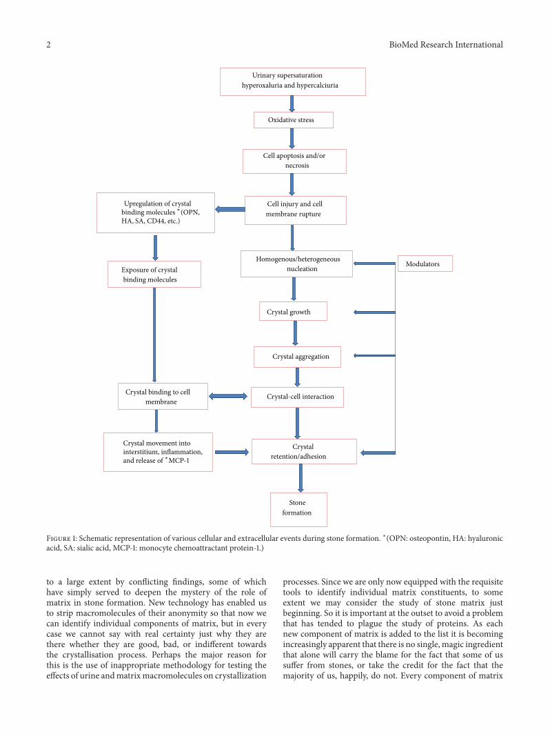
2 BioMed Research International
Urinary supersaturationhyperoxaluria and hypercalciuria
Oxidative stress
Cell apoptosis and/ornecrosis
Cell injury and cell membrane rupture
Homogenous/heterogeneous nucleation
Crystal growth
Crystal aggregation
Crystal-cell interaction
Crystal retention/adhesion
Stone formation
ModulatorsExposure of crystal binding molecules
Crystal binding to cell membrane
Upregulation of crystalbinding molecules ∗(OPN,HA, SA, CD44, etc.)
Crystal movement intointerstitium, inflammation,and release of ∗MCP-1
Figure 1: Schematic representation of various cellular and extracellular events during stone formation. ∗(OPN: osteopontin, HA: hyaluronicacid, SA: sialic acid, MCP-1: monocyte chemoattractant protein-1.)
to a large extent by conflicting findings, some of whichhave simply served to deepen the mystery of the role ofmatrix in stone formation. New technology has enabled usto strip macromolecules of their anonymity so that now wecan identify individual components of matrix, but in everycase we cannot say with real certainty just why they arethere whether they are good, bad, or indifferent towardsthe crystallisation process. Perhaps the major reason forthis is the use of inappropriate methodology for testing theeffects of urine andmatrixmacromolecules on crystallization
processes. Since we are only now equipped with the requisitetools to identify individual matrix constituents, to someextent we may consider the study of stone matrix justbeginning. So it is important at the outset to avoid a problemthat has tended to plague the study of proteins. As eachnew component of matrix is added to the list it is becomingincreasingly apparent that there is no single, magic ingredientthat alone will carry the blame for the fact that some of ussuffer from stones, or take the credit for the fact that themajority of us, happily, do not. Every component of matrix
BioMed Research International 3
is potentially an active protagonist in stone pathogenesisuntil proven otherwise. Doyle et al. reasoned that the studyof crystals enabled the study of urinary proteins directlyinvolved in the crucial crystal nucleation phase of stoneformation, free of any macromolecular contaminants thatmight be introduced into a stone by cellular injury [10].Adopting an approach,Morse and Resnick found that despitethe enormous array of proteins present in urine, the crystalsprecipitated from it contained relatively few proteins [11].However, although the presence of these proteins may implythat they fulfill some function in stone formation, it is equallypossible that their inclusion in the stone structuremay simplyhave been fortuitous or may have resulted from the injuriouseffects of the stone itself. Many proteins have been reportedto be from the renal stone matrix, but the study of functionof only few has been done. The common occurrence ofurine supersaturation and crystalluria has prompted studieson modulators of the processes beyond supersaturation. Toreduce the occurrence of urolithiasis, elucidation of themechanism of lithogenesis through fundamental research isessential. This review summarises what is currently knownor is hypothesised about the influences of urinary macro-molecules, especially proteins, on the formation of calciumoxalate crystals. Although a list of proteins is provided thathave either been detected in stones or have been implicated byvirtue of their effects on crystallization, only a select handful,which have been intensively studied, have been singled outfor individual discussion.
2. The Stone
Themain components of the stonematrix account for 2-3% oftheir total dry weight and consists of macromolecules gener-ally present in the urine [12, 13]. They are described by Boyceas 64% protein, 9.6% nonamino sugars, 5% hexosamineas glucosamine, 10% bound water, and the remainder asinorganic ash [14]. Although not detected by Boyce, lipidshave also been shown to be significant components of stonematrix [15]. Nonetheless, proteins comprise the major part ofmatrix, an observation confirmed by Sugimoto et al. [16]. Itwas inevitable therefore that protein would tend to dominatethe study of matrix, and consequently, considerably moreis known about them than the other two principal groups,namely, lipids and glycosaminoglycans (GAGs).
Boyce [14] defined and established the importance ofstone matrix in nephrolithiasis, proposing that the matrixactively participates in the assembly of kidney stones. In theirview, thematrix acts as a template and controls crystallizationwithin its bounds. An opposite hypothesis was advanced byVermeulen et al., who viewed the matrix and its ubiquitouspresence asmerely coincidental, because stones form by crys-tallization in urine in the presence of large macromolecules[17, 18]. Proteins formed a discontinuous coat around thecrystals ranging in thickness from 10 to 20 nm. It has beensuggested that newly formed crystals with a macromolecularcoat are less likely to dissolve during the routine urinary ionicand pH changes and thereinmay lay the importance ofmatrixin stone formation [19].
Urinarymolecule that affects themass of CaOx depositedfromurine or the size of the crystal particles produced has thepotential to influence the likelihood that crystalline particlesare retained in the renal collecting system, and thereby, thedevelopment of stone disease [20]. Some promote CaOxcrystal nucleation in inorganic solutions and in concentratedwhole urine [21]; others like albumin appear to exert nosignificant effect on crystallization in urine [22], but are,nonetheless, found in stones. Many studies, have shown thata variety of urinary proteins inhibit CaOx crystal growth [23]and aggregation [22]. More surprisingly, some can actuallydo more than one of these. Tamm-Horsfall mucoproteincan inhibit CaOx crystal aggregation, but can also act asa promoter of crystal deposition, depending upon experi-mental conditions [24]. It has also been demonstrated thatpolyelectrolytes and proteins that inhibit crystallization insolution can act as promoters when they are immobilized onto surfaces [25]. And the issue is further complicated by thefact that the potency of urinary macromolecules increasesinversely in relation to the prevailing ionic strength [26]. Theeffects of macromolecules are manifold, unpredictable, andparadoxical. Above all, they are certainly not amenable togeneralization; knowledge of their roles in stone formationwill therefore be obtained only by laboriously teasing out theinformation for each individual macromolecule. And in thefollowing sections, that is what we will attempt to do.
3. Mechanism of Calcium Oxalate RenalStone Formation: Urinary Supersaturationand Crystallization
The formation of renal stones is a consequence of increasedurinary supersaturation with subsequent formation of crys-talline particles. Supersaturation is the driving force forcrystallization in solutions like urine. When a salt is addedto a solvent it dissolves in the solvent until a particular con-centration is reached, beyond which no further dissolutionis possible. At this point, the solvent is said to be saturatedwith the salt. If more salt is added it crystallizes in solution,provided the temperature and pH are unchanged. The con-centration at which saturation is reached and crystallizationbegins is called the thermodynamic solubility product (Ksp).If inhibitors of crystallization were not able to act, the finalresult will be nephrolithiasis [27]. Inhibitors allow higherconcentration of calcium salts to be held in solution thanin pure solvents. Urine is thus metastable with respect tocalcium salts. Indeed, stone formers tend to excrete urine thatis more supersaturated than that of nonstone formers [13, 28].It has been suggested thatwith a transit time across the kidneyof 5 to 10min, residence time is too short for crystals tonucleate and grow large enough to be trapped in a normalperson [29].
3.1. Crystal Nucleation. The initial step in the transformationfrom a liquid to a solid phase in a supersaturated solution iscalled nucleation. This process begins with the combinationof stone salts in solution into loose clusters that may increasein size by addition of new components or clusters [30].
4 BioMed Research International
Nuclei form the first crystals that do not dissolve and have acharacteristic lattice pattern. In urine, nuclei usually form onexisting surfaces, a process called heterogeneous nucleation.Epithelial cells, urinary casts, RBCs, and other crystals can actas nucleating centers in urine. The saturation necessary forheterogeneous nucleation is much less than for homogenousnucleation [31]. Once a nucleus is created and principally if itis anchored, crystallization can occur at lower chemical pres-sures than required for the formation of the initial nucleus.Renal tubular cell injury can promote crystallization of CaOxcrystals by providing substances for their heterogeneousnucleation. In vitro cell degradation following renal tubularcell injury produces numerous membrane vesicles, whichhave been shown to be good nucleators of calcium crystals.In vivo crystals observed in the renal tubules of hyperoxaluricrats are always associated with cellular degradation products[32, 33].
3.2. Crystal Growth. Once a crystal nucleus has achieved acritical size and relative supersaturation remains above one,the overall free energy is decreased by adding new crystalcomponents to the nucleus. This process is called crystalgrowth. Crystal growth is one of the prerequisites for particleformation and thus for stone formation [34]. In each stepof stone formation, crystal growth and aggregation haveimportant functions. Honda et al. reported that the crystalsurface binding substance, which is found in CaOx crystalsgenerated from whole human urine, is a strong inhibitor ofCaOx crystal growth and contains proteins like human serumalbumin, retinol binding protein, transferrin, Tamm-Horsfallglycoprotein, and prothrombin [35]. However, it has beensuggested that the importance of crystal growth for CaOx,the most abundant stone component, is questionable. Sincethe rate of CaOx crystal growth is low and the transit timeof tubular fluid through the kidney amounts to only severalminutes, it has been calculated that the probability of a singleparticle achieving a pathophysiologically relevant size by theprocess of crystal growth alone is extremely low, even ifgrowth proceeds at an uninhibited rate of 2mm per minute[29]. The inhibitory effect of fibronectin (FN), distributedthroughout the extracellularmatrix and body fluids, onCaOxcrystal growth is small, considering the quantity normallyexcreted.
3.3. Crystal Aggregation. The process whereby crystals insolution stick together to form larger particles is calledaggregation. Some researchers have proposed that crystalaggregation is the most important step in stone formation.Although crystal growth is definitely a step in CaOx renalstone formation, the process of growth is so slow that crystalscannot become large enough to obstruct the renal tubules andbe retained there by thismechanism alone, as several minutesare required for the tubular fluid to pass through the kidney.For this reason, the more critical step is thought to be crystalaggregation. All models of CaOx urolithiasis concede thatcrystal aggregation is probably involved in crystal retentionwithin the kidneys, since aggregation of crystals can have aconsiderable effect on particle size and aggregated crystals
are commonly found in urine and renal stones [36]. Crystalaggregation is promoted by viscous binding, implying thatcrystal-foreign compounds with multiple binding sites, suchas abnormally self-aggregating Tamm-Horsfall glycoproteinor other macromolecules, attach to crystal surfaces and act asa kind of glue [37].
3.4. Crystal-Cell Interaction. The mechanisms of crystal-cellinteraction are thought to be very complex, and many ofthem remain unexplored. Crystallization is caused by thecondition of urinary supersaturation. Then, the crystals thathave formed attach to renal tubular epithelial cells and aretaken into them. The process of attachment or endocytosisof crystals to renal tubular cells is what is generally meantby crystal-cell interactions. These structural and functionalstudies of crystal-cell interactions in culture indicate thatCOM crystals rapidly adhere to microvilli on the cell surfaceand are subsequently internalized. Khan et al. concludedthat crystal-cell interaction is an essential element in thedevelopment of urinary stone disease [38]. Kohjimoto etal. reported that crystal-cell interactions may be amongthe earliest processes in the formation of kidney stones[39]. Finlayson and Reid hypothesized that it was unlikelythat CaOx crystals could grow large enough to be retainedwithin the renal tubules, and that attachment of crystals wasnecessary for initiation of stone formation [29]. There havebeen many reports on crystal attachment. Animal modeland tissue culture studies have provided evidence for crystalretentionwithin the kidneys via attachment to renal epithelialcells. Kok and Khan observed crystal attachment to the brushborder of proximal tubules in rats. Experimental inductionof CaOx urolithiasis starts with hyperoxaluria followed bycrystalluria and deposition in the kidney [36, 40]. Someurinary macromolecules have an inhibitory effect on CaOxcrystal attachment. Lieske et al. reported that diverse polyan-ionicmolecules in urine, such as specific glycosaminoglycans,glycoproteins, and citrate, block the binding of COM crystalsto the cell membrane. One common feature of molecules thatinhibit COM crystal adhesion to cells is their polyanioniccharacter. They mentioned that although polyanions presentin tubular fluid may coat crystals and thereby inhibit theiradhesion to tubular cells, a distinct and separate set of signalsacts on the cells to regulate their response to crystals that dobind [41, 42].
3.5. Endocytosis of CaOx Crystals by Renal Tubular EpithelialCells. Many studies of the endocytosis of crystals by cellshave been reported. Lieske et al. noted engulfment of crys-tals into tubular epithelial cells and cell proliferation in atransplanted kidney in a patient with primary hyperoxaluria[43] and confirmed this phenomenon experimentally usingcalcium-containing crystals and tubular cells in culture [44].Various substances have an inhibitory effect on CaOx crystalendocytosis. Tamm-Horsfall protein (THP) leads to [45],decreased COM crystal endocytosis by 34%, suggesting thatTHP in distal tubular fluid may block the uptake of COMcrystals by cells of this portion of the nephron and therebyprevent renal crystal retention and stone formation [46].
BioMed Research International 5
The inhibitory effect of FN on CaOx crystal endocytosis wasonly 18.4% at the physiological concentration of excreted FN(0.5mg/mL), though morphological examination revealedthat FN clearly inhibited the endocytosis of crystals byrenal tubular cells [47, 48]. Lieske and Toback reported thatthe internalization of CaOx crystals by BSC-1 and MDCKcells is a regulated event that can be modified by varioussignals [49]. In addition, they reported that the adsorptionof nephrocalcin, a urinary glycoprotein of renal cell origin,to COM crystals prevented attachment of the crystal tothe plasma membrane, engulfment, or both and therebyprevented mitogenic effects.
3.6. Relationship between Crystal-Cell Interaction and RenalTubular Cell Injury. Some investigators have obtained evi-dence that oxalate and CaOx crystals may be injurious torenal tubular cells. Cultured renal tubular cells exhibitedevidence of damage after exposure toCaOx crystals. Additionof CaOx crystals to monolayers of Madin-Darby caninekidney (MDCK) cells led to a marked increase in the releaseof lysosomal enzymes, prostaglandin E2, and, to a lesserextent, cytosolic enzymes [40, 50]. In animal models of renalstone disease produced by the administration of high oxalateloads, the presence of CaOx crystals within the renal tubulesis associated with renal tubular damage, as evidenced byenzymuria and the presence of membranous debris withinthe tubular lumina [51]. This membranous debris appearstogether with focal loss of the brush border from proximaltubular cells and the appearance of proximal tubular enzymesin the urine, suggesting that proximal tubular cells are thesource of the membranous debris [51–53]. Thus, in thisanimal model of stone disease, CaOx crystals or the highconcentrations of oxalate ion in proximal tubular fluid appearto be toxic for renal tubular epithelial cells.
Some reports have suggested the involvement of renaltubular epithelial cell injury in the crystal-cell interactionprocess. Wiessner et al. reported that individual cell injuryand generalized cell monolayer injury result in the pre-sentation of different cell surfaces, and that both types ofinjury result in increased affinity for crystal adhesion. Theyalso stated that both mechanisms could be important, eitherindependently or together, for the retention of microcrystalsadhering to renal collecting duct cells in nephrolithiasis. Fur-thermore, theymentioned that themechanism of adhesion ofcrystals to renal tubular cells is based on crystal interactionwith basolateral or basement membrane components. Thesecomponents may become exposed as a result of the loss ofcell polarity seen in several disease states in association withtissue injury, ischemia in kidney tubules, microvillus inclu-sion disease, and polycystic kidney disease [54]. Injury to therenal tubular epithelial cells results in cellular degradationand the production of membranous vesicles. The crystals areeither passed as crystalluria particles or are endocytosed bythe epithelial cells to be processed by their lysosomal systemor transported to the interstitium. CaOx crystal depositionin the kidneys upregulates the expression and/or synthesis ofmacromolecules that can promote inflammation and lead tofibrosis. Mild hyperoxaluriais is a known risk factor for CaOx
urolithiasis [55]. Mild hyperoxaluria promotes increasedproduction of crystallization modulators, such a Sosteopon-tin, bikunin [56], and FN [57]. These macromolecules areinvolved in controlling not only crystal nucleation, growth,and aggregation, but also crystal interaction with the renaltubular epithelial cells and crystal retentionwithin the kidney[9, 58].
4. Matrix Components
4.1. Lipids. The matrix of all stones examined to date,including struvite, uric acid, CaOx, and CaP, containslipids [59]. Phospholipids account for 8.6% of the totallipid, which in turn represents approximately 10.25% ofstone matrix [15]. Various identified phospholipids and gly-colipids include sphingomyelin (SM), phosphatidylcholine(PC), phosphatidylethanolamine (PE), cardiolipin (CL), andtrace amounts of phosphatidylserine (PS) in all stonematrices[60]. Occasionally, the stone matrix also contains phos-phatidylinositol (PI), lyso-PC, lysophosphatidic acid (PA)and lyso-PE. In all stones glycolipids include gangliosides,sphingosine and glucocerebrosides. Lipids play a more activerole since cell membranes [33, 61] and the lipids of CaOxstone matrix [59] can catalyse the nucleation of CaOxfrom a metastable solution. Cell membranes and their lipidsplay critical roles in the process of calcification. Particularmembrane phospholipids promote the formation of calciumoxalate and calcium phosphate and become a part of theorganic matrix of growing calcification [60].
4.2. Glycosaminoglycans (GAGs). The presence of GAGs instonematrix was inferred by Boyce and Garvey as long ago as1956 [62]. Nishio et al. reported that between 0.19 and 0.58%of a stone’s weight consists of GAGs, thereby providing uswith the evidence that GAGs may account for up to 20% ofthe weight of matrix [63]. This alone is sufficient to suggestthat they may fulfill some function in stone formation.
GAGs occurring in the urine of normal individualstypically consist of 55% chondroitin sulphate (CS), 20%heparan sulphate (HS), 11% low sulphated CS, and 4–10%hyaluronic acid (HA) [64]. It is therefore somewhat sur-prising that only HS and, to a lesser extent, HA have beenreported to be present in CaOx stones [63]. CS, the mostabundant GAG in urine, though present in small amountsin magnesium ammonium phosphate and apatite stones hasnot been detected in CaOx stones [63, 65]. These resultsstrongly suggest that the incorporation of GAGs into stonesis a selective process, a notion supported by the finding thatinclusion of GAGs into CaOx crystals freshly precipitatedfrom human urine is also highly selective. Like stones, suchcrystals contain only HS [65]; CS is incorporated into CaOxcrystals only in the absence of HA [66], indicating that theyprobably compete for the same binding sites on the crystalsurface. Similar results have been reported for uric acid stonesand crystals, with HS being the only GAG detected [67].
However, the simple fact of their presence in stones orcrystals tells us nothing about the mechanism by whichGAGs came to be there. However, recent evidence suggests
6 BioMed Research International
that urinary GAGs can fulfill both roles, promoting CaOxcrystal nucleation and reducing the final size of the crystalsproduced, thereby lessening their chance of retention withinthe urinary tract [68]. On the basis of present evidence, itwould appear that CS, though present in urine in largerquantities than HS, plays no significant role in CaOx crys-tallization or in stone formation. HS, on the other hand,may act as an inhibitor, although confirmation of such a rolemust await the results of further studies. What is quite clear,however, is that the routine measurement of total urinaryGAGs excretion is unlikely to be of any practical benefit inthe diagnosis and management of nephrolithiasis. With thebenefit of hindsight, it is not surprising that comparisons ofurinary GAG excretion in stone formers and normal subjectshave produced conflicting findings [68–71].
4.3. Proteins. Despite the fact that stone matrix has beenshown to contain an ever increasing list of proteins, in mostcases it is not possible to say with any certainty why theymight be there. One can speculate of course. For instance, thepresence of superoxide dismutase may perhaps be explainedby the fact that the enzyme acts as a protector of tissuedamage by scavenging the toxic superoxide anion [72]. Couldthe enzyme’s presence be explained by release of this radicalin response to cellular injury caused by the kidney stoneitself—an example of a macromolecule incorporated as asecondary matrix component? However, speculation alonewill not unravel the mystery of why proteins are in stonematrix, but detailed study of individual proteins may.We willnow discuss what is known about several urinary proteinsthat have been subjected to rigorous study because they havebeen found to occur in stones or because they have beenisolated fromurine and shown to influence the crystallizationofCaOx.Andwewill beginwithTamm-Horsfall glycoproteinbecause it has the longest historical association with stones,and, as a consequence, has been subjected to themost intenseexperimental scrutiny. A detailed table (Table 1) has beenprovided to show the effect of the modulators (protein) onstone formation.
4.3.1. Tamm-Horsfall Protein (THP). The most extensivelyinvestigated urinary macromolecule in nephrolithiasisresearch Tamm Horsfall glycoprotein (THP), enjoys adistinct position, perhaps because it is the most abundantprotein in human urine and was one of the first componentsof stone matrix to be identified by Boyce and Garvey [62].THP is a renal protein of all placental invertebrates [73, 74]. Itis known to have a monomeric molecular weight of 80 kDa.THP is present in urine in polymeric forms measuring upto several million Da. In humans, daily excretion rangesbetween 20 and 100mg/day [75] with a daily urinaryvolume of 1.5 litres, and in rats it ranges between 552 and2,865𝜇g/day with a daily urinary volume of 16.5mL [76]. Itis its extraordinary ability to self-associate due to presenceof carbohydrates in urine into large structures visible to thenaked eye that probably accounts for its known effects onCaOx crystallization [77], and it is these effects to whichwe will now primarily confine our discussion. THP may be
involved in the pathogenesis of nephropathy, nephrolithiasis,and tubule interstitial nephritis [78]. Mo et al. by ablatingthe murine THP gene established that THP is on the firstline of host defences against both renal stone formation andbacterial infection [79].
THP is often detected in stones, regardless of crystal com-ponents [80]. THP is absent from CaOx crystals precipitatedfrom whole urine [10], which would seem to indicate thatTHP binds only weakly, if at all, to CaOx crystals. Sinceit has been accepted for some time that inhibitors act bybinding to crystal surfaces, we might expect THP to be apoor inhibitor of CaOx crystallization—at least in urine,where stones form. The protein has been reported to act asan inhibitor [22, 24, 81–83] and a promoter [24, 84, 85] ofcrystallization. The picture is further complicated by the factthat conflicting findings were obtained in the only studies inwhich the effect of THP was tested in undiluted urine. Roseand Sulaiman [84] found that THP enhanced the depositionof CaOx crystals from urine concentrate by evaporation tohigh osmolalities, whereas Ryall et al. [22] and Grover etal. [83] found that the protein was a potent inhibitor ofCaOx crystal aggregation, although having no effect onCaOxdeposition. An explanation for these opposing findings is tobe found in a study by Grover et al. [24] who tested the effectof THP in the experimental systems used by the two researchgroups and found that while THP undoubtedly promotesCaOx precipitation under conditions of high osmolality,where it also links CaOx crystals together into large, looselyconnected agglomerates, it is a very effective inhibitor ofcrystal aggregation atmore usual urinary concentrations. It isalso apparent that THP inhibits crystal aggregation by sterichindrance rather than by binding to the crystal surfaces [22];binding might therefore not be a prerequisite for inhibitorypotency after all. Moreover, however potent an inhibitorit may be, it cannot account for the total inhibitory effectof urinary macromolecules on CaOx crystal aggregation incentrifuged and filtered urine [86], because it is removedfrom urine by centrifugation and filtration. Thus, there canbe no doubt that other urinary macromolecules contributeto this inhibition [22]. A reflection of the disagreementsurrounding the role of THP as a promoter or inhibitor, ofCaOx crystallization is to be found in similar conflict relatingto its urinary excretion. If indeed THP does play a directiverole in stone formation, we might expect that its excretionwould be different in stone formers and normal subjects. Butit is not [87]. Contrary to this, ratmodel studies have providedcontroversial results for THP. One study shows decreasedrenal expression of THP during CaOx crystal deposition[88], while results of another study show upregulation of theTHP gene [89]. However, it may be that stone formationis related more to the type of THP excreted than to thequantity. This fact led them to hypothesize that THP of stoneformers is structurally different from that of the healthysubjects [90]. THP isolated from the urine of stone formerscontained less carbohydrate (mainly sialic acid) than the THPobtained from control subjects [91]. Studies have also showndifferences in sialic acid contents and surface charge betweenTHP from stone formers and normal individuals. Isoelectricfocussing (IEF) studies have shown that THP from healthy
BioMed Research International 7
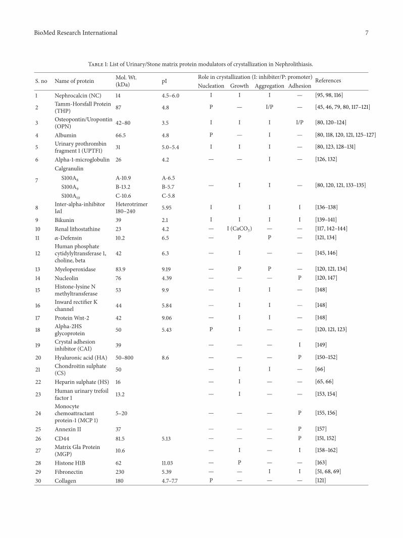
Table 1: List of Urinary/Stone matrix protein modulators of crystallization in Nephrolithiasis.
S. no Name of protein Mol. Wt.(kDa) pI Role in crystallization (I: inhibiter/P: promoter) References
Nucleation Growth Aggregation Adhesion1 Nephrocalcin (NC) 14 4.5–6.0 I I I — [95, 98, 116]
2 Tamm-Horsfall Protein(THP) 87 4.8 P — I/P — [45, 46, 79, 80, 117–121]
3 Osteopontin/Uropontin(OPN) 42–80 3.5 I I I I/P [80, 120–124]
4 Albumin 66.5 4.8 P — I — [80, 118, 120, 121, 125–127]
5 Urinary prothrombinfragment 1 (UPTF1) 31 5.0–5.4 I I I — [80, 123, 128–131]
6 Alpha-1-microglobulin 26 4.2 — — I — [126, 132]
7
CalgranulinS100A8 A-10.9 A-6.5
— I I — [80, 120, 121, 133–135]S100A9 B-13.2 B-5.7S100A10 C-10.6 C-5.8
8 Inter-alpha-inhibitorI𝛼I
Heterotrimer180–240 5.95 I I I I [136–138]
9 Bikunin 39 2.1 I I I I [139–141]10 Renal lithostathine 23 4.2 — I (CaCO3) — — [117, 142–144]11 𝛼-Defensin 10.2 6.5 — P P — [121, 134]
12Human phosphatecytidylyltransferase 1,choline, beta
42 6.3 — I — — [145, 146]
13 Myeloperoxidase 83.9 9.19 — P P — [120, 121, 134]14 Nucleolin 76 4.39 — — — P [120, 147]
15 Histone-lysine Nmethyltransferase 53 9.9 — I I — [148]
16 Inward rectifier Kchannel 44 5.84 — I I — [148]
17 Protein Wnt-2 42 9.06 — I I — [148]
18 Alpha-2HSglycoprotein 50 5.43 P I — — [120, 121, 123]
19 Crystal adhesioninhibitor (CAI) 39 — — — I [149]
20 Hyaluronic acid (HA) 50–800 8.6 — — — P [150–152]
21 Chondroitin sulphate(CS) 50 — I I — [66]
22 Heparin sulphate (HS) 16 — I — — [65, 66]
23 Human urinary trefoilfactor 1 13.2 — I — — [153, 154]
24Monocytechemoattractantprotein-1 (MCP 1)
5–20 — — — P [155, 156]
25 Annexin II 37 — — — P [157]26 CD44 81.5 5.13 — — — P [151, 152]
27 Matrix Gla Protein(MGP) 10.6 — I — I [158–162]
28 Histone H1B 62 11.03 — P — — [163]29 Fibronectin 230 5.39 — — I I [51, 68, 69]30 Collagen 180 4.7–7.7 P — — — [121]
8 BioMed Research International
individuals has a pI value of approximately 3.5, while THPfrom recurrent stone formers has pI values between 4.5 and6, and the two exhibit completely different IEF patterns [92].
Self-aggregation of THP may promote either heteroge-nous nucleation or formation of a protein and crystallinemass large enough to block the tubular lumen. Further studiesare still required to elucidate its real contribution, if any, tonephrolithiasis and its interaction, if any, with its urinarycompanions. And it is to those companions that we will nowdirect our attention.
4.3.2. Nephrocalcin (NC). Second to THP, nephrocalcin (NC)has been the most widely studied protein reported inthe stone literature. In 1978, Nakagawa and his colleaguesdescribed it as an unidentified acidic polypeptide [93] andthen for a number of years as a glycoprotein inhibitor of CaOxcrystal growth [94–97]. In 1987, after the first report of itsisolation from urine, the protein was named nephrocalcin[98]. NC holds a prominent position in urolithiasis research,having been claimed to be the principal inhibitor of CaOxcrystallization in urine [95], its activity reportedly accountingfor approximately 90% of urine’s total inhibitory effect onCaOx crystallization [94, 95, 97]. The molecular weight ofNC varies widely depending upon the state of aggregationof the protein, with the molecular weights of the monomer,dimer, trimer, and tetramer being reported as 14-15, 23–30, 45–48, and 60–68 kDa, respectively [94, 96]. There areat least four isoforms of nephrocalcin, that is, NC-A, NC-B, NC-C, and NC-D. Mustafi and Nakagawa explainedmechanism of inhibition of COM crystal growth by NC andcharacterised the Ca2+-binding sites present in nephrocalcin[99]. Nakagawa, in 1997-described that nonstone-formingpeople excrete more NC-A and NC-B isoforms in urine, butmoreNC-C andNC-D isoformswere found in stone formers’urine. The organic matrix of calcium oxalate kidney stonewas found to have greater quantities of NC-C and NC-Disoforms than those of NC-A and NC-B isoforms. IsoformsA and B changed their conformation upon Ca2+ binding, butthere was no change in the conformation of C and D. Allthese observations suggest that isoforms A and B are stronginhibitors of calcium oxalate monohydrate (COM) crystalgrowth and aggregation, whereas isoforms C and D act aspromotors for COM crystal growth [100].
The protein is located in the epithelium of the proximaltubules and thick ascending limb of the loops of Henle, inboth human and mouse kidneys [101]. NC was originallyisolated from human urine [93, 95–97] and has also beendetected in tissue culture medium of human kidney cell lines[94, 102] and kidney stones [98, 103]. A glycoprotein, NC hasbeen reported to occur in urine at concentrations rangingfrom 5mg/L 102 to 16mg/L 112 and to contain 2-3 residuesof 𝛾-carboxyglutamic acid (Gla) in its primary structure [94–96]. The Gla component confers the protein’s potent abilityto inhibit CaOx crystallization, previously reported to bedeficient in this amino acid. A lack of Gla in NC isolated fromkidney stones was suggested as the reason why the stones hadformed [98].
Despite its long history, there has been no report in theliterature of its primary amino acid sequence and the exact
nature of the protein remains unknown. Hochstrasser et al.suggested the identity of NC isolated from human urine withfragment HI-14 of the light chain (bikunin) of inter-𝛼-trypsininhibitor (ITI) [104]. They concluded that NC represents aportion of the bikunin chain of ITI.
The inhibitory activity of NC should now perhaps bereappraised, particularly since a recent paper byWorcester etal. reassessed its contribution to be no more than 16% [105].Moreover, all existing estimates of the protein’s inhibitorypotency have been obtained from crystallization systemsbased on inorganic metastable solutions. Although NC isundoubtedly a potent inhibitor of CaOx deposition in aninorganic metastable solution, observations [106, 107] haveconfirmed using material tentatively identified as NC. It isbecoming increasingly apparent that this potency is sharedwith a number of other urinary proteins, for example,uropontin, urinary prothrombin fragment 1, and UAP, and soforth.
4.3.3. Osteopontin (OPN). Osteopontin is a protein impor-tant in bone mineralization, where it is thought to anchorosteoblasts to bone [108]. Osteopontin is originally isolatedfrom rat bone matrix as a 44 kDa phosphorylated protein.It is rich in acidic amino acids like serine, aspartic acidand glutamic acid, which are commonly found in proteinsinvolved in biomineralization [109]. It has an amino acidsequence that serves as a recognition signal for interactingwith cell-surface receptor molecules known as integrins,which are involved in cell adhesion [110], and is a member ofa family of proteins rich in aspartic acid that have been shownin vitro to have stereospecific activity at the surface of crystals[111].
Osteopontin (OPN) is a negatively charged aspartic acidrich protein and is intimately involved in the regulation ofboth physiological and pathological mineralization. OPN isa phosphorylated protein of wide tissue distribution that isfound in association with dystrophic calcification includingin the organic matrix of kidney stones. OPN is synthesizedwithin the kidney and is present in the human urine.The bone-derived and kidney-derived forms of this proteinappear to be very similar in amino acid sequence. It isinvolved in various biological processes like inflammation,wound healing, cell survival, and leukocyte recruitment[112]. In 1992, Shiraga et al. reported the isolation fromhuman urine of a protein which they called uropontin (UP)[113]. It was isolated by immune affinity chromatographyusing a monoclonal rat antibody and had exhibited maximalinhibition of CaOx crystal growth in an inorganic metastablesolution. Total amino acid analysis of OPN revealed a highproportion of aspartic acid residues [113]. Molecular weightestimations of UP, which has an apparentmolecular weight of50 kDa in 16% SDS-PAGE gels and 72 kDa in 5–18% gradientgels, were also similar toUP [109].These similarities, togetherwith identical nucleotide sequences of cDNAs encoding UPfrom human kidney [113] and bone [114], indicate that UPis not a distinct protein, but rather a urinary form of OPN.OPN has been referred to by a variety of names includingsecreted phosphoprotein [115], 44 kDa bone phosphoprotein[109], and uropontin.
BioMed Research International 9
The distribution of OPN in humans was recently reportedby Brown et al. [164]. UP is present in normal adult urine ata mean concentration of approximately 6 × 10⁻⁸molar [165].The protein is widely distributed on the luminal surfaces ofspecific epithelial cells in the gall bladder, pancreas, urinarytract, reproductive tracts, gastrointestinal tract, lung, breast,salivary glands, and sweat glands. OPN was specificallyfound in the cytoplasm of many epithelial cells of the distaltubules and collecting ducts. Others have reported OPN tobe present in mice, but only in the thick ascending limbsof the loop of Henle and the distal convoluted tubules ina subset of nephrons [26]. It might perhaps be argued thatthe widespread distribution of OPN in humans militatesagainst it having a specific function in stone formation, butits potent effect on CaOx crystal growth would suggest thatit may influence the course of the disease. Certainly, it ispresent in kidney stones, with quantities in those composedprincipally of CaOx dehydrate being considerably less thanin calculi comprising mainly CaOx monohydrate [165], andrecently, OPNhas also been detected in uric acid stones alongwith COM and COD crystals [80], where its abundance issubstantially greater than that reported for NC [98]. In vitrostudies suggest that OPNmay inhibit the nucleation, growth,and aggregation of calcium oxalate crystals. In addition,it also inhibits the crystal adhesion to cultured epithelialcells [122]. Wesson et al. observed that it may direct CaOxcrystallisation to the CaOx dehydrate phase rather thanthe CaOx monohydrate (COM) phase, the dehydrate beingless adherent to renal tubular epithelial cells [166]. Clinicalstudies to date are inconclusive regarding the relationshipbetween OPN and renal stone disease. Some investigatorshave reported decreased concentrations of OPN in urinefrom stone formers compared to normal individuals [123],while others have not [167]. A single-base mutation in theOPN gene is seen at significantly higher incidence in patientswith recurrent stone formation or familial nephrolithiasis[168].The role ofOPN in nephrolithiasis is though somewhatunclear; due to very high content of aspartic and glutamicresidues, OPN is subjected to significant post translationalmodification, which may function as regulatory switchesin promotion or inhibition of mineralization [124]. Theproportional contribution of UP to the inhibitory activityof urine has not been assessed [165], but the fact that it ispresent in stones in greater quantities than NC, despite itslower concentration in urine,might suggest that it bindsmoreavidly to the CaOx crystal surface andmay consequently be amore potent inhibitor. However, like NC, its inhibitory effecton CaOx crystallization has not been tested in urine, so itis not presently possible to assess its likely effects on CaOxcrystallization in vivo.Thus, like all proteins currently underinvestigation for their possible roles in stone formation,significantlymore informationmust be obtained before it willbe possible to state with certainty that the presence of UPin urine is related specifically to its ability to inhibit CaOxcrystallization, and thereby, stone pathogenesis.
4.3.4. Urinary Prothrombin Fragment 1 (UPTF1). UrinaryProthrombin Fragment-1 was discovered from CaOx crystals
freshly precipitated from urine. With a molecular weightof 31 kDa and staining characteristics of a glycoprotein, itwas selectively incorporated into the crystals in quantitiesfar exceeding those of any other, and in amounts dispro-portionately greater than its concentration in the urine fromwhich the crystals had been derived. It was not possible toidentify the protein by western blotting using commercialantibodies such as crystal matrix protein (CMP). However,it has since been shown, both immunologically and by aminoacid sequence analysis, that the protein is related to humanprothrombin [128, 169].The link between nephrolithiasis andblood clotting was recently confirmed when its close identitywith the FI activation peptide of human prothrombin wasdemonstrated [170]. To avoid confusion, the name crystalmatrix protein has been abandoned and the protein is nowknown as urinary prothrombin fragment 1 (UPTFl). Theknown characteristics of UPTF1 and its presence in calciumoxalate stones indicate that it may fulfill some functionin stone pathogenesis. UPTF1 is one of the principal con-stituents of CaOx stones. Most notably, it was not foundin two struvite stones, indicating that its presence in CaOxstones is a consequence of direct inclusion into the crystallinearchitecture rather than a secondary product of tissue injury.Suzuki et al. studied the expression of prothrombin in humanand rat kidneys [171]. Analysis of the matrix of calciumphosphate crystal reveals that UPTF1 is a major component,while in urate crystals it is only a very minor constituent,which reflects the known relationship between this proteinand calcium ions [172]. Immunohistochemical studies havemapped its location in the human kidney, specifically to theepithelial cells of the thick ascending limb of the loops ofHenle and the distal convoluted tubules [173].Theproteinwasnot detected in any other human tissue, with the exception ofthe cytoplasmof hepatocytes. Limited data also demonstratedthat the amount of UPTF1 in the kidneys of stone formersis significantly greater than in those of healthy subjects [173,174].
UPTF1 is the most prominent protein in the organicmatrix of CaOx crystals precipitated from fresh human urine[10]. This combined with the observation that the organicmatrix is the most potent macromolecular inhibitor of CaOxcrystallization induced in human urine that has yet beendescribed led to the presumption that this inhibitory activitywas likely to be attributable to its component UPTF1 [20].This presumption was largely justified since UPTF1 purifiedby RPHPLC is now known to be a potent inhibitor of CaOxcrystal aggregation in undiluted urine [129], a feature thatcurrently distinguishes it from its peers, whose inhibitoryactivities, with the exception of THP, have only ever beentested in inorganic solutions.When crystallization inhibitorypotential of all the prothrombin-related peptides, the PT,thrombin (T), and fragments 1 and 2 was tested using asimple inorganic solution, both prothrombin and fragment1 were found to inhibit crystal aggregation [130]. However,when similar experiments were conducted using undiluted,centrifuged, and ultrafiltered human urine [131], crystalaggregationwas inhibited only by PT fragment 1.There seemsto be little doubt that the potent inhibitory effect of UPTF1on CaOx crystallization can be ascribed to the Gla domain

10 BioMed Research International
of the peptide, which is absent from thrombin and F2 andboth PT and F1 fragments [131]. Along with that of its peers,UP and UAP, the study of UPTF1 is still in its early stages.Certainly, preliminary data would indicate that it possessesall the features expected of a significant macromolecularurinary inhibitor, including potent activity in undilutedurine. Nonetheless, like all other proteins presumed to fulfillsome function in urolithiasis, the true role of UPTF1 mustremain speculative until such time as a cause and effectrelationship between the protein and stone pathogenesis canbe unequivocally demonstrated.
4.3.5. Inter-𝛼-Inhibitor (I𝛼I). Inter-𝛼-inhibitor (I𝛼I) belongsto the Kinitz-typeprotein superfamily, a group of proteinspossessing kunin as a common structural element and theability to inhibit serine proteases [175]. I𝛼I is a plasma glyco-protein normally synthesized in the liver and is composed of acombination of heavy chains, H1 (60 kDa), H2 (70 kDa), andand H3 (90 kDa) covalently linked via a chondroitin sulphatebridge to a light chain called bikunin (35–45 kDa) [106]. Theheavy and light chains also exist independently as singlemolecules. I𝛼I (180–240 kDa) is a heterotrimer consisting ofbikunin linked to heavy chains H1 and H2. Pre-𝛼-inhibitor(P𝛼I, 125 kDa) is composed of bikunin and heavy chain H3.The macromolecule consisting of bikunin linked to heavychain H2 is called I𝛼I-like inhibitor (I𝛼LI). I𝛼I and relatedproteins have been linked to various pathological conditionssuch as inflammatory diseases [176], cancer [177], renalfailure [178], and more recently the urinary stone disease.A possible role of I𝛼I in stone disease can be traced as farback as 1909, when Bauer and Reich demonstrated that theproteolytic activity of trypsin was inhibited by urine [179].Atmani et al., in 1993, isolated uronic acid-rich protein (UAP)from human urine by three gel filtration chromatographicsteps and, in the reduced form, had an estimated molecularweight of 35 kDa on an 8–20% SDS gradient gel [107]. UAPearned its name because of its high content of uronic acid;D-glucuronic and L-iduronic acids are major constituentsof GAGs, carbohydrate accounts for 8.5% of its weight, andamino acid analysis reveals a protein rich in glutamic andaspartic acids, glycine, and valine and it contains no Gla.N-terminal amino acid sequence analysis has demonstratedhomology with the light chain of inter-𝛼-trypsin inhibitor(ITI), that is, bikunin [136, 180]. This supposition is furthervindicated by the fact that the urinary derivative of ITI is alsoknown to be a GAG adduct [175] and has a relative molecularweight of 35 kDa on SDS-PAGE under reducing conditions[104]. Bikunin is a broad-spectrum protease inhibitor andan acute-phase reactant. It is excreted in urine where itdegrades further to fragments HI14 and HI8. Both heavyand light chains have also been identified in the urine [107,181, 182]. The average concentration of I𝛼I in the plasmaof healthy human subjects is approximately 450mg/L [137].Urinary excretion is 2 to 10mg/day, but can increase to 50–100-fold or more in certain pathological conditions such ascancer. Plasma concentration of I𝛼I is, on the other hand,reduced during various pathological conditions includingrenal failure. Bikunin is expressed mainly in the proximal
tubules and the thin descending segment near the loop ofHenle. Tissue-culture studies have shown that human renalproximal tubular epithelial cells constitutively express genesfor bikunin and H3 components [183]. Bikunin gene is alsoexpressed in MDCK cells and was upregulated when theywere exposed to oxalate [119]. In normal rat kidneys, stainingof the I𝛼I-related proteins is mostly limited to the proximaltubules and generally to their luminal contents [184]. Iidaet al. investigated renal and urinary expression of variousmembers of the I𝛼I family in male rats with or withoutexperimentally induced hyperoxaluria and CaOx crystaldeposition. The expression of bikunin mRNA increased inrenal epithelial cells exposed to oxalate and CaOx crystals[184]. As a result, an increase in CaOx inhibitory activity isanticipated. However, this was not the case, as there might besome structural abnormalities in stone formers’ bikunin. Itwas shown that bikunin isolated from the patients containedless sialic acid and exhibited less crystallization inhibitoryactivity than that purified from the urine of healthy subject[138]. Western analysis showed that a significantly higherproportion of stone patients had a 25 kDa bikunin in theirurine in addition to the normal 40 kDa species. 25 kDabikunin was similar to the deglycosylated bikunin and wasless inhibitory. In a separate study, mean urinary bikuninto creatinine ratio was found to be significantly higher instone formers than in nonstone-forming healthy male andfemale controls [185]. Contrary to this, another study founddecreased urinary excretion of bikunin by stone-formingpatients. Mean urinary excretion of bikunin in 18 healthyindividuals was 5.01 + 0.91 𝜇g/mL and 2.54 + 0.42 𝜇g/mL in31 stone patients [139].
The inhibitory activity of this protein from humanurine was determined in an inorganic CaOx crystallizationsystem, where it strongly retarded CaOx crystal growth. Itwas reported that this activity is reduced in stone formerscompared with normal controls [138]. I𝛼I proteins have beenshown to inhibit CaOx crystallization in vitro [107, 136,139, 180, 186, 187]. The inhibitory activity is confined to thecarboxy terminal of the bikunin fragment of I𝛼I [187]. Bothrat and human urinary bikunin inhibited nucleation andgrowth of CaOx crystals. Treatment with chondroitinase AChad no effect on this inhibitory activity which was destroyedby pronase treatment, indicating that the activity lies not withthe chondroitin chain but with the peptide. In 1999, Atmaniand Khan showed that urinary bikunin, at concentrations of2.5 to 20𝜇g/mL, retarded crystal nucleation by 67 to 58%and inhibited crystal aggregation by 59 to 80% [140]. Theseresults were confirmed later when inhibition of CaOx crystalgrowth and aggregation by I𝛼I, its heavy chains, light chain(bikunin) with or without chondroitinase treatment, andbikunin’s carboxy terminal domain (HI8) was tested in an invitro crystallization assay [187]. I𝛼I was aweak inhibitorwhileheavy chains showed no discernible activity. Bikunin andHI effectively inhibited the crystallization. Chondroitinasetreatment had no effect on the inhibitory activity of bikunin.I𝛼Imolecule itself is also an effective inhibitor ofCaOx crystalgrowth [136] and in another study was shown to be moreefficient than another crystal growth inhibitor, prothrombinfragment 1 [186]. Ebisuno et al. showed inhibitory effect of

BioMed Research International 11
bikunin on adhesion of calcium oxalate crystals to renaltubular cells in human urine [141]. MDCK cells were exposedin culture to CaOx monohydrate crystals in the presence orabsence of various protein fractions isolated from normalhuman urine. A single fraction with a molecular weight of35 kDa was found to be most inhibitory of crystal adhesion.This protein, on sequencing found, to be homologous tobikunin, inhibited crystal adhesion at the minimum concen-tration of 10 ng/mL, and completely blocked it at 200 ng/mL.It may contribute to the modulation of crystal adhesion andretentionwithin tubules during kidney stone formation [188].
Despite having been the subject of investigation formany years, the true physiological function of I𝛼I remainssomewhat a mystery, although it may play a role in cancer,adult respiratory distress syndrome, and septic shock andmay be a useful therapeutic agent in such conditions [183].Furthermore, the potent inhibition of CaOx crystal growth bythese proteins, coupled with the known presence of bikuninand its fragments in urine, suggested the possible existence ofa relationship between I𝛼I and CaOx stone formation [189].Then it is possible that its clinical usefulness may also extendto the treatment of human kidney stones.
4.3.6. Calgranulin. Calgranulin is a 28 kDa calcium-bindingprotein, member of S100 protein family, which are small,ubiquitous, and acidic proteins involved in normal devel-opmental and structural activities. This group of calcium-binding proteins are also implicated in a number of diseases[190]. There are 3 monomers of calgranulin (A, B, and C)all mapped to chromosome 1. It is present in the kidneyand human urine and can inhibit growth of CaOx crystals,which is the major component of kidney stones. The proteinwas isolated from human urine at a concentration of 3.5–10 nM. Purified urinary calgranulin inhibited both CaOxcrystal growth (44%) and aggregation (50%) in nanomolarrange. The inhibitory properties of calgranulin may be dueto its ability to bind to the crystal surface [133]. Westernanalysis of rat and human kidneys as well as renal epithelialcell lines including BSC-1 and MDCK confirmed its renalpresence. Calgranulin has also been identified in matricesof different type of stones, for example, calcium oxalatestones [120, 121, 134], uric acid stones [80], and infectiousor struvite stones [135], and in CaP deposits formed byMDCK cells [191]. Calgranulin is abundant in both ratand human kidney homogenates, and Pillay et al. isolatedthe cDNA of calgranulin by screening a human kidneyexpression library. Its renal presence was further evidencedby western and northern analysis of two renal epithelialcell lines. The inhibitory properties of calgranulin may beshared and expressed by other urinary S100 proteins; bovinebrain S100 protein inhibits COM growth and aggregation.The expression of calgranulin by renal cells indicates thaturine calgranulin could be produced by kidney cells andmay be regulated by them in ways that are important indefense against crystallization [133]. Calgranulin has alsobeen detected in the matrices of different types of stones,for example, calcium oxalate stones [120, 121, 134], uric acidstones [80], and infectious or struvite stones [135], and in CaPdeposits formed by MDCK cells [191].
4.3.7. Renal Lithostathine. Lithostathine is a glycoproteinsynthesized by acinar cells and secreted in pancreatic juice.Pancreatic juice is naturally supersaturated at calcium andbicarbonate ions, and lithostathine plays an important rolein inhibiting calcium carbonate crystal growth. A proteinimmunologically related to lithostathine and is actuallypresent in urine of healthy subjects and in renal stonesrenal lithostathine (RL) [192]. Western blot analysis of pro-teins extracted from concentrated normal urine or kid-ney stones demonstrated the presence of a protein withmolecular weight of 23 kDa [142]. Because of its structuraland functional similarities with pancreatic lithostathine, itwas called renal lithostathine. Renal lithostathine seems tocontrol growth of calcium carbonate crystals. Several reportsshowing the presence of calcium carbonate (CaCO
3) in renal
stones suggested that crystals of CaCO3might be present in
the early steps of stone formation. Such crystals, therefore,might promote CaOx crystallization from supersaturatedurine by providing an appropriate substrate for heteroge-neous nucleation [143, 144].
4.3.8. Albumin. Albumin is one of the most abundant pro-teins in the urine [62, 118] and has been detected in thematrix of both urinary stones [14, 62, 80, 118] and crystals[189, 193] made in the whole human urine. It is known tobind to CaOx as well as uric acid crystals [116, 126] butdoes not inhibit their growth [22, 116]. However, it has beenshown to inhibit CaOx crystal aggregation in concentrationdependent manner [194]. When immobilized to surfacesand exposed to metastable solutions, albumin promotescrystal nucleation [127, 195]. When dissolved in solutionalbumin exists either in monomeric and/or polymeric form[127]. In metastable CaOx solutions both monomeric andpolymeric forms promote nucleation of CaOx. In addition,nucleation by albumin leads exclusively to the formationof COD crystals. Urinary albumin purified from healthysubjects contained significantly more polymeric forms andwas a stronger promoter of CaOx nucleation than albuminfrom idiopathic calcium stone formers. Promotion of CaOxnucleation and formation of large number of COD crystalsmight be protective. Nucleation of large number of smallcrystals would allow their easy elimination and decreaseCaOx saturation preventing crystal growth and aggregationand subsequent stone formation. COD crystals are morecommon than COM crystals in nonstone formers’ urineand are generally found in less quantities in stones thanCOM crystals. In addition, crystals present in the urine fromnonstone formers are significantly smaller than those in stoneformers urine. Albumin also exhibits the capacity to bindsome of the urinary proteins. Interestingly, urinary proteinsthat show great affinity for albumin are also those that areincluded in the stone matrix. It is suggested that proteinsbecome a part of stone matrix by binding to the albumincoating of the CaOx crystals. It is also suggested that unlikeother calcium-binding urinary proteins, albumin promotesnucleation by interacting with calcium through the carboxylgroup. Strong nucleation activity was observed at pH 7 butwas totally eliminated at pH 4 when carboxyl groups are no

12 BioMed Research International
longer ionized. In addition, morphological studies showedCaOx crystals to nucleate through calcium-rich face [127].
4.3.9. Human Urinary Trefoil Factor 1 (THF1). Human uri-nary trefoil factor 1 (THF1) belongs to the trefoil factorfamily proteins. It is synthesised by mucosal epithelial cellsand is expressed in gastric mucosa. It has been describedas an antiapoptotic agent with mitogenic activities [196]. Itmay also act as a potent inhibitor of CaOx crystal growth.Functional studies of urinary THF1 demonstrated that itsinhibitory potency was similar to that of nephrocalcin. Theinhibitory activity of urinary THF1 was dose dependentand was inhibited by THF1 antisera. Concentrations andrelative amounts of THF1 in the urine of patients withidiopathic CaOx kidney stone were significantly less (2.5-fold for the concentrations and 5- to 22-fold for the relativeamounts) than those found in controls [153]. Urinary trefoilfactor 1 has the ability to inhibit growth and aggregation ofcalcium oxalate crystals and can transform calcium oxalatemonohydrate crystals to the dihydrate type [154].
4.3.10. Hyaluronic Acid (HA). HA is a linear glycosaminogly-can (GAG) that is composed of multiple units of glucuronicacid and N-acetylglucosamine (1, 4-GlcUA-1, 3-GlcNAc-)n.It is an extremely large and high molecular weight GAG,that is, >106Da, one disaccharide is approximately 400Da;high molecular weight HA is a chain of >2500 disacchariderepeats. HA chains occupy huge tissue domains and canentrap large amounts of solvent due to its expanded randomcoil structure [197]. HA is produced by HA synthase (HAS)proteins that are located at the inner face of the plasmamembrane, where it is extruded across the membrane intothe extracellular space. HA is a major component of theextracellular matrix (ECM) in the renal medullary intersti-tium and the pericellular matrix (PCM) of mitogen/stress-activated renal tubular cells. Hydrated PCM provides themicroenvironment that is conducive to adjustments in cellshape and epithelial architecture during dynamic morpho-genetic processes like wound healing, embryonic develop-ment, inflammation, and cancer [198].The size, negative ioniccharge, and ability to form hydrated gel-like matrices makeHA an excellent crystal-binding molecule. Crystal bindingto HA leads to crystal retention in the renal tubules and tothe formation of calcified plaques in the renal interstitium(Randall’s plaques). HA also directly influences cell behaviorthrough its ability to communicate with the cell interior viacell-surface receptors, such as CD44 and CD168 [197–199].
In a healthy kidney, HA is abundant in the renalmedullary interstitium, but in the cortex it is almost unde-tectable, and renal tubular cells normally do not expressHA. HA should be considered an enormous inhibitor ofcrystallization that efficiently prevents papillary calcification,for the reason that interstitial HA is low during antidiuresis;the highest risk for crystal formation most likely occursduring periods of water deprivation. On the other hand,high fluid intake leads to high interstitial HA which protectsagainst crystallization. Ca2+ also becomes associated withthe COO− groups in the HA matrix, thereby preventing
CaP precipitation. Precipitated calcium crystals (CaP) maybind to the HA matrix, which could play an important rolein interstitial plaque formation (Randall’s plaques) [150].Therefore, it was speculated that HA could be an inhibitorof crystallization as long as calcium salts are in solutionbecause the carboxyl groups of the HA chains race withanions such as phosphate and oxalate for binding to calcium,whereas HA may serve as binding substance for precipitatedcalcium salts because of the affinity of these crystals forhigh molecular weight HA. In case of renal cell injury, HAmay act as promoter for crystal adhesion to the cell surface,which will ultimately result into stone formation. Crystalretention in the human kidneymay depend on the expressionof damageddistal tubular epitheliumofCD44,OPN, andHA-rich cell coats [151]. HA becomes expressed in areas of thekidney where it is absent under normally conditions, suchas in the cortical interstitium and on the luminal surface ofrenal tubular cells [152]. HA is upregulated in the kidneyduring inflammatory renal disease states such as interstitialnephritis [200], acute ischemic injury [201], autoimmunerenal injury [202], acutely rejecting human kidney grafts[203], acute tubular necrosis [151], and obstructed kidneysand EG poisoning [152]. HA-expressing renal tubular cellsinvariably also express the HA receptor CD44 [151, 152, 200,204, 205]. HA is also one of the major constituents of theorganic matrix of renal stones. HA is present in kidneystones in fractions that are disproportionate to its urinaryconcentrations [63].
4.3.11. Annexin II. Annexin II protein is a 37 kDa member ofthe annexin family, that is, calcium-dependent phospholipid-binding protein family. These family members play a role inthe regulation of cellular growth and in signal transductionpathways. As a family, annexins are characterized by a con-served COOH-terminal protein “core” that mediates theirmembrane and calcium-binding properties. The conservedCOOH-terminal core is 70-amino acid-long chains and con-tains four repeats, each of which consist of a calcium-bindingmotif G-X-G-T [206]. Members of the annexin family differin binding specificity of the core for phospholipid headgroupslike PS, phosphatidic acid or phosphatidylinositol, as well asthe binding specificity of the NH2-terminal tail.
Annexin II is a calcium-dependent phospholipid-bindingprotein whose function is to help organize exocytosis ofintracellular proteins to the extracellular domain. AnnexinII is involved in diverse cellular processes like cell motility,linkage of membrane-associated protein complexes to theactin cytoskeleton, endocytosis, fibrinolysis, ion channelformation, and cell matrix interactions. Ax-II is a pleiotropicprotein, meaning that its function is dependent on the placeand time in the body. Ax-II has been demonstrated on thesurface of many cell types, including keratinocytes [207],endothelial cells, glioma cells, and smooth muscles cells[208], where it can serve receptor-like functions formoleculesincluding lipid A [209], cytomegalovirus [210], 1,25(OH)2D3[211], 2-glycoprotein I [212], andtissue plasminogen activator[213]. Annexin II can bind to the crystals while anchoredon the cell surface. Under certain circumstances, Ax-II is
BioMed Research International 13
known to interact with membranes via cholesterol moleculesin a calcium-independent mechanism, possibly leaving thecalcium-binding motifs available to bind calcium oxalatecrystals.TheNH2-terminus of Ax-II can bind a p11 dimer andbecome linked to another Ax-II molecule via its NH2 tail; aprocess that can mediate aggregation of membrane vesicles.Ax-II-p11 tetramermight link a crystal to amembrane insteadof linking two membranes [206].
Ax-II mediates the adhesion of COM crystals to renalcells and it may also play a role in their subsequent inter-nalization. Kumar et al., in 2003, used COM affinity toidentify apical membrane proteins that might mediate crystaladhesion to renal epithelial cells. They found annexin IIto be the major COM crystal-binding protein, and it wasidentified on the apical surface of intact MDCK-I cells. Theinteraction between crystals and renal tubular cells has beenproposed to be a crucial event that elicits subsequent cellularresponses, leading to kidney stone formation. Ax-II has a roleas a COM crystal-binding molecule on the surface of intactcells as COM crystal adhesion decreased significantly afterMDCKI cells were pretreated with a monoclonal antibodyagainst Ax-II. Studies suggest that Ax-IImay be one of severalcell surface crystal-binding molecules that might modulatecrystal retention [157]. Membrane-associated annexin II hasalso been reported to regulate extracellular matrix (ECM)metalloproteinase inducer (Emmprin) activity [150, 214].
4.3.12. CD44. CD44 is a multifunctional extracellular matrix(ECM) glycoprotein involved in adhesion, migration andcell-cell interactions. CD44 is the main cell surface receptorfor hyaluronan or hyaluronic acid (HA) as well as OPN[215] and can also interact with other ligands, collagens,and matrix metalloproteinases (MMPs). CD44 is expressedin a large number of mammalian cell types. It is alsofound to be associated with adhesion of crystals to renalepithelial cells. Retention of crystals in the kidney is anessential early step in renal stone formation. OPN, ratherthan HA, is the major ligand for CD44 on bone cells in theremodelling phase of healing of fractures [216]. CD44 playsan important role in calcium oxalate (CaOx) crystal bindingduring wound healing. Both CD44 and HA are upregulatedduring injury and inflammation and are involved in theformation of a cell coat or pericellular matrix on surfacesof proliferating and migrating cells. HA is restricted to theinner medullary interstitium of the normal kidneys. Distalcollecting duct cells express both CD44 and HA on apicalcell surfaces of the proliferating cells. At confluence, however,CD44 is expressed at the basolateral membrane while HA isundetectable. Proliferating cells are receptive to adhesion ofCaOx crystals, a property lost when cells become confluent.In addition, removal of pericellular matrix by hyaluronidasetreatment also results in loss of crystal adhesion property ofthe proliferating cells [151]. Asselman et al. studied crystalretention in vivo; intratubular crystals were found adheredto injured/regenerating tubular epithelial cells expressingHA,OPN, and CD44 at their luminal membrane [152]. Based onthese observations it has been proposed that intact epitheliumdoes not bind crystals because of the absence of a pericellular
matrix and crystal attachment depends upon the expressionof CD44, OPN, and HA by the damaged renal epithelial cells[217].
4.3.13. Matrix Gla Protein. Matrix Gla protein (MGP) is anatural inhibitor of vascular calcification. MGP, a vitamin K-dependent extracellular matrix protein, was initially isolatedfrom the bone and is also expressed in lung, heart, vascularsmooth muscle cells of the blood vessel wall, and kidney[218]. MGP is an 84-amino-acid protein that contains five𝛾-carboxyglutamic acid (Gla) residues which have a highaffinity for calcium and phosphate ions and hydroxyapatitecrystals [158]. The pathological mechanism of kidney stoneformation was partly similar to vascular calcification: form-ing calcific plaques, increasing expression of calcificationinhibitors, and regulating actively calcification process. MGPgenetic single nucleotide polymorphism was associated withthe individual susceptibility of nephrolithiasis [159]. MGPis involved in cell growth, differentiation, and regulation ofapoptosis and increase cell density in normal kidney cells[160].
Matrix Gla protein (MGP) is a molecular determinantregulating vascular calcification of the extracellular matrix.MGP is polarly distributed on the apical membrane of renaltubular epithelial cells and binds directly to crystals; it is alsofound in the ascending thick limbs of Henle’s loop and thedistal convoluted tubule in hyperoxaluric rats; its expressionwas present in themedullary collecting duct in stone-formingrats. Crystals with multilaminated structure were formed inthe injurious renal tubules with lack ofMGP expression [161].Lu et al. also reported that there was no crystal formation inrenal tubules withMGP expression, and crystals only depositin the damaged renal tubules with lack of MGP expression.MGPmRNA expression was found to be upregulated in renaltubular epithelial cells following exposure to calcium oxalatemonohydrate (COM) and oxalate [161, 162]. Luo et al. foundthat homozygous MGP-deficient mice died within 8 weeksas a result of arterial calcification that led to blood vesselrupture [219]. Vascular calcification in MGP-deficient micewas reversed by overexpressing MGP in vascular smoothmuscle cells [220].These findings imply thatMGPmay play acytoprotective role in maintaining cells’ survival and inhibit-ing crystal retention under oxalate and crystal exposure.
4.3.14. Monocyte Chemoattractant Protein-1 (MCP-1). Mono-cyte chemoattractant protein-1 (MCP-1/CCL2) is a memberof the CC chemokine family and belongs to the group ofinflammatory chemokines. MCP-1 is a potent chemotacticfactor formonocytes [221]. Chemokines are also grouped intotwo main functional subfamilies: inflammatory and homeo-static. Inflammatory chemokines control the recruitment ofleukocytes in inflammation and tissue injury. Homeostaticchemokines fulfill housekeeping functions, such as navigat-ing leukocytes to and within secondary lymphoid organs,in addition to in the bone marrow and the thymus duringhematopoiesis [222]. Chemokine activation of cell surface G-protein-coupled receptors results in directed cell migration,that is, chemotaxis [223].
14 BioMed Research International
The development of kidney disease involves a complexinterplay between neurohormonal, inflammatory, and bio-chemical changes which act on either intrinsic or extrinsicrenal cells or both. This can lead to the development ofan innate immune response predominantly characterized bythe accumulation and activation of leukocytes, particularlymonocytes/macrophages, in the kidney. Chemokine-inducedrecruitment of peripheral leukocytes into tissues is a criticalstep in the development of inflammatory responses [224].MCP-1 plays an important role in various pathophysiologicalconditions in many organ systems [225]. Even though MCP-1 is involved in various inflammatory diseases and may notbe specific for a particular disease, this should not necessarilypreclude its use as a biomarker. MCP-1 plays a critical role inthe development of kidney diseases. It has been investigatedas a potential urinary biomarker in several renal diseases,in a number of studies [155]. However, the exact role ofMCP-1 in stone formation is still not clear, but its expressionin renal epithelial cells on exposure to oxalate and CaOxcrystals indicates a close relationship between inflammationand nephrolithiasis [156].
Our research group has been conducting many studiesto explore the organic matrix of renal stone and revealedthe presence of a 66 kDa stimulator of calcium oxalatecrystal growth in the human renal calculi extract [6]. Laterthey investigated the activity of high and low molecularweight biomolecules present in the matrix of human CaOxstones not only on the initial mineral phase formation ofcalcium and phosphate (CaP) but also on its growth anddemineralization of the preformed mineral phase [226]. Wealso purified, identified, and characterized novel proteinsfrom human renal stone matrix. Some of them were potentinhibitors like urobilirubin (14.2–16.2 kDa) [227], a 36 kDainhibitor protein [228], anionic protein like human phos-phate cytidylyltransferase-1, 𝛽 (42 kDa), [145, 146] cationicproteins like histone-lysine N-methyltransferase (53 kDa),inward rectifier K channel (44 kDa), and protein Wnt-2(42 kDa) [148]. 2D map of >3 kDa fraction from matrix ofCaOx stone revealed the presence of disheveled-associatedactivator morphogenesis (123.9 kDa), glutamate receptordelta-1 subunit (113 kDa), caspase recruitment domain-containing protein (114.8 kDa), ALBU BOVIN (71.2 kDa),VP7 glycoprotein precursor (37.5 kDa), chymotrypsinogenA (26.2 kDa), and plasminogen (15.7 kDa). Also. our groupconducted various cell culture studies to explain the effect ofproteins in the stonematrix on oxalate-injured renal cell lines[229].
5. Conclusion
Various theories of pathogenesis of human kidney stonessuggest that their formation is too complex for simpleunderstanding. The pathogenesis of calcium oxalate stoneformation is a multistep process and essentially includesnucleation, crystal growth, crystal aggregation, and crystalretention. A detailed discussion has been made about severalurinary proteins found to occur in stones that modulatethe process of stone formation, thereby leading to better
understanding of the pathophysiology and pathogenesis ofkidney stone disease.Many aspects of themechanism of renalstone formation remain unclear at present; hence, a betterunderstanding of this intricate mechanism will lead to thedevelopment of a novel strategy for preventing this disease.
References
[1] G. Eknoyan, “History of urolithiasis,” Clinical Reviews in Boneand Mineral Metabolism, vol. 2, no. 3, pp. 177–185, 2004.
[2] M. Lopez and B. Hoppe, “History, epidemiology and regionaldiversities of urolithiasis,” Pediatric Nephrology, vol. 25, no. 1,pp. 49–59, 2010.
[3] H. Wilkinson, “Clinical investigation and management ofpatients with renal stones,” Annals of Clinical Biochemistry, vol.38, no. 3, pp. 180–187, 2001.
[4] G. Bihl and A. Meyers, “Recurrent renal stone disease—advances in pathogenesis and clinicalmanagement,”TheLancet,vol. 358, no. 9282, pp. 651–656, 2001.
[5] T. M. Reynolds, “Chemical pathology clinical investigation andmanagement of nephrolithiasis,” Journal of Clinical Pathology,vol. 58, no. 2, pp. 134–140, 2005.
[6] S. Aggarwal, C. D. Tandon, M. Forouzandeh, S. K. Singla, R.Kiran, and R. K. Jethi, “Role of biomolecules from human renalstone matrix on COM crystal growth,” Molecular and CellularBiochemistry, vol. 210, no. 1-2, pp. 109–119, 2000.
[7] W. B. Gill, K. W. Jones, and K. J. Ruggiero, “Protective effectsof heparin and other sulfated glycosaminoglycans on crystaladhesion to injured urothelium,”The Journal of Urology, vol. 127,no. 1, pp. 152–154, 1982.
[8] M.W. Bigelow, J. H.Wiessner, J. G. Kleinman, andN. S.Mandel,“Calcium oxalate-crystal membrane interactions: dependenceon membrane lipid composition,” The Journal of Urology, vol.155, no. 3, pp. 1094–1098, 1996.
[9] S. R. Khan and D. J. Kok, “Modulators of urinary stoneformation,” Frontiers in Bioscience, vol. 9, pp. 1450–1482, 2004.
[10] I. R. Doyle, R. L. Ryall, andV. R.Marshall, “Inclusion of proteinsinto calcium oxalate crystals precipitated from human urine: ahighly selective phenomenon,” Clinical Chemistry, vol. 37, no. 9,pp. 1589–1594, 1991.
[11] R. M. Morse and M. I. Resnick, “A new approach to the studyof urinary macromolecules as a participant in calcium oxalatecrystallization,”The Journal of Urology, vol. 139, no. 4, pp. 869–873, 1988.
[12] J. S. King Jr. andW.H. Boyce, “Immunological studies on serumand urinary proteins in urolith matrix in man,” Annals of theNew York Academy of Sciences, vol. 104, p. 5791, 1963.
[13] W. G. Robertson, M. Peacock, and B. E. C. Nordin, “Activityproducts in stone-forming and non-stone-forming urine,”Clin-ical Science, vol. 34, no. 3, pp. 579–594, 1968.
[14] W. H. Boyce, “Organic matrix of human urinary concretions,”The American Journal of Medicine, vol. 45, no. 5, pp. 673–683,1968.
[15] S. R. Khan, P. N. Shevock, and R. L. Hackett, “Presence of lipidsin urinary stones: results of preliminary studies,”CalcifiedTissueInternational, vol. 42, no. 2, pp. 91–96, 1988.
[16] T. Sugimoto, Y. Funae, H. Rubben, S. Nishio, R. Hautmann,and W. Lutzeyer, “Resolution of proteins in the kidney stonematrix using high-performance liquid chromatography,” Euro-pean Urology, vol. 11, no. 5, pp. 334–340, 1985.
BioMed Research International 15
[17] C. W. Vermeulen and E. S. Lyon, “Mechanisms of genesis andgrowth of calculi,” The American Journal of Medicine, vol. 45,no. 5, pp. 684–691, 1956.
[18] B. Finlayson, C. W. Vermeulen, and E. J. Stewart, “Stone matrixand mucoprotein from urine,” The Journal of Urology, vol. 86,pp. 355–363, 1961.
[19] S. R. Khan and R. L. Hackett, “Role of organic matrix inurinary stone formation: an ultrastructural study of crystalmatrix interface of calcium oxalate monohydrate stones,” TheJournal of Urology, vol. 15, no. 1, pp. 239–245, 1993.
[20] I. R. Doyle, V. R. Marshall, C. J. Dawson, and R. L. Ryall,“Calcium oxalate crystal matrix extract: themost potentmacro-molecular inhibitor of crystal growth and aggregation yet testedin undiluted human urine in vitro,” Urological Research, vol. 23,no. 1, pp. 53–62, 1995.
[21] G. W. Drach, S. Thorson, and A. Randolph, “Effects of urinaryorganic macromelecules on crystallization of calcium oxalate:enhancement of nucleation,”The Journal of Urology, vol. 123, no.4, pp. 519–523, 1980.
[22] R. L. Ryall, R. M. Harnett, C. M. Hibberd, K. A. Edyvane, andV. R. Marshall, “Effects of chondroitin sulphate, human serumalbumin and Tamm-Horsfall mucoprotein on calcium oxalatecrystallization in undiluted human urine,” Urological Research,vol. 19, no. 3, pp. 181–188, 1991.
[23] M. I. Resnick, M. E. Sorrell, J. A. Bailey, and W. H. Boyce,“Inhibitory effects of urinary calcium-binding substances oncalcium oxalate crystallization,”The Journal of Urology, vol. 127,no. 3, pp. 568–571, 1982.
[24] P. K. Grover, R. L. Ryall, and V. R. Marshall, “Does Tamm-Horsfall mucoprotein inhibit or promote calcium oxalate crys-tallization in human urine?” Clinica Chimica Acta, vol. 190, no.3, pp. 223–238, 1990.
[25] A. A. Campbell, A. Ebrahimpour, L. Perez, S. A. Smesko, andG. H. Nancollas, “The dual role of polyelectrolytes and proteinsas mineralization promoters and inhibitors of calcium oxalatemonohydrate,” Calcified Tissue International, vol. 45, no. 2, pp.122–128, 1989.
[26] M. Utsunomiya, T. Koide, T. Yoshioka, S. Yamaguchi, and A.Okyama, “Influence of ionic strength on crystal adsorptionand inhibitory activity of macromolecules,” British Journal ofUrology, vol. 71, no. 5, pp. 516–522, 1993.
[27] M. Carvahlo and Y. Nakagawa, “Urinary supersaturation andrecurrence in nephrolithiasis,” International Brazilian Journal ofUrology, vol. 25, pp. 475–479, 1999.
[28] D. J. Kok and S. E. Papapoulos, “Physicochemical considera-tions in the development and prevention of calcium oxalateurolithiasis,” Bone and Mineral, vol. 20, no. 1, pp. 1–15, 1993.
[29] B. Finlayson and S. Reid, “The expectation of free and fixedparticles in urinary stone disease,” Investigative Urology, vol. 15,no. 6, pp. 442–448, 1978.
[30] A. L. Boskey, “Current concepts of the physiology and bio-chemistry of calcification,” Clinical Orthopaedics and RelatedResearch, vol. 157, pp. 225–257, 1981.
[31] L. H. Smith, “Solutions and solute,” Endocrinology andMetabolism Clinics of North America, vol. 19, no. 4, pp. 767–772,1990.
[32] J. M. Fasano and S. R. Khan, “Intratubular crystallization ofcalcium oxalate in the presence of membrane vesicles,” KidneyInternational, vol. 59, no. 1, pp. 169–178, 2001.
[33] S. R. Khan, P. N. Shevock, and R. L. Hackett, “Membrane-associated crystallization of calcium oxalate,” Calcified TissueInternational, vol. 46, no. 2, pp. 116–120, 1990.
[34] B. Finlayson, S. R. Khan, and R. L. Hackett, “Mechanisms ofstone formation—an overview,” Scanning Electron Microscopy,vol. 3, pp. 1419–1425, 1984.
[35] M.Honda, T. Yoshioka, S. Yamaguchi et al., “Characterization ofprotein components of human urinary crystal surface bindingsubstance,”Urological Research, vol. 25, no. 5, pp. 355–360, 1997.
[36] D. J. Kok and S. R. Khan, “Calcium oxalate nephrolithiasis, afree or fixed particle disease,” Kidney International, vol. 46, no.3, pp. 847–854, 1994.
[37] B. Hess, L. Zipperle, and P. Jaeger, “Citrate and calcium effectsonTamm-Horsfall glycoprotein as amodifier of calciumoxalatecrystal aggregation,” American Journal of Physiology, vol. 265,no. 6, part 2, pp. F784–F791, 1993.
[38] S. R. Khan, K. J. Byer, S. Thamilselvan et al., “Crystal-cellinteraction and apoptosis in oxalate-associated injury of renalepithelial cells,” Journal of the American Society of Nephrology,vol. 10, no. 14, pp. S457–S463, 1999.
[39] Y. Kohjimoto, M. Ebisuno, M. Tamura, and T. Ohkawa,“Interactions between calcium oxalate monohydrate crystalsand Madin-Darby canine kidney cells: endocytosis and cellproliferation,” Urological Research, vol. 24, no. 4, pp. 193–199,1996.
[40] S. R. Khan, “Calcium oxalate crystal interaction with renaltubular epithelium, mechanism of crystal adhesion and itsimpact on stone development,” Urological Research, vol. 23, no.2, pp. 71–79, 1995.
[41] J. C. Lieske, S. Deganello, and F. G. Toback, “Cell-crystalinteractions and kidney stone formation,” Nephron, vol. 81, no.1, pp. 8–17, 1999.
[42] J. C. Lieske, R. Leonard, and F. G. Toback, “Adhesion of calciumoxalatemonohydrate crystals to renal epithelial cells is inhibitedby specific anions,”American Journal of Physiology, vol. 268, no.4, part 2, pp. F604–F612, 1995.
[43] J. C. Lieske, B. H. Spargo, and F. G. Toback, “Endocytosisof calcium oxalate crystals and proliferation of renal tubularepithelial cells in a patient with type 1 primary hyperoxaluria,”The Journal of Urology, vol. 148, no. 5, pp. 1517–1519, 1992.
[44] J. C. Lieske, H. Swift, T. Martin, B. Patterson, and F. G. Toback,“Renal epithelial cells rapidly bind and internalize calciumoxalate monohydrate crystals,” Proceedings of the NationalAcademy of Sciences of the United States of America, vol. 91, no.15, pp. 6987–6991, 1994.
[45] B. Hess, “Tamm-Horsfall glycoprotein-inhibitor or promoterof calcium oxalate monohydrate crystallization processes?”Urological Research, vol. 20, no. 1, pp. 83–86, 1992.
[46] S. Kumar and A. Muchmore, “Tamm-Horsfall protein-uromodulin (1950–1990),” Kidney International, vol. 37, no. 6,pp. 1395–1401, 1990.
[47] M. Tsujihata, O. Miyake, K. Yoshimura, K.-I. Kakimoto, S.Takahara, and A. Okuyama, “Fibronectin as a potent inhibitorof calcium oxalate urolithiasis,”The Journal of Urology, vol. 164,no. 5, pp. 1718–1723, 2000.
[48] M. Tsujihata, K. Yoshimura, K. Tsujikawa, N. Tei, and A.Okuyama, “Fibronectin inhibits endocytosis of calcium oxalatecrystals by renal tubular cells,” International Journal of Urology,vol. 13, no. 6, pp. 743–746, 2006.
[49] J. C. Lieske and F. G. Toback, “Regulation of renal epithelialcell endocytosis of calcium oxalate monohydrate crystals,”American Journal of Physiology, vol. 264, no. 5, pp. F800–F807,1993.
16 BioMed Research International
[50] J. H. Wiessner, A. T. Hasegawa, L. Y. Hung, and N. S. Mandel,“Oxalate-induced exposure of phosphatidylserine on the sur-face of renal epithelial cells in culture,” Journal of the AmericanSociety of Nephrology, vol. 10, supplement 14, pp. S441–S445,1999.
[51] S. R. Khan and R. L. Hackett, “Hyperoxaluria, enzymuria andnephrolithiasis,” Contributions to Nephrology, vol. 101, pp. 190–193, 1993.
[52] S. R. Khan, B. Finlayson, and R. L. Hackett, “Histologic study ofthe early events in oxalate induced intranephronic calculosis,”Investigative Urology, vol. 17, no. 3, pp. 199–203, 1979.
[53] S. R. Khan, B. Finlayson, and R. L. Hackett, “Experimentalcalciumoxalate nephrolithiasis in the rat,”TheAmerican Journalof Pathology, vol. 107, no. 1, pp. 59–69, 1982.
[54] J. H.Wiessner, A. T. Hasegawa, L. Y. Hung, G. S.Mandel, andN.S. Mandel, “Mechanisms of calcium oxalate crystal attachmentto injured renal collecting duct cells,” Kidney International, vol.59, no. 2, pp. 637–644, 2001.
[55] R. L. Hackett, P. N. Shevock, and S. R. Khan, “Madin-Darbycanine kidney cells are injured by exposure to oxalate and tocalcium oxalate crystals,” Urological Research, vol. 22, no. 4, pp.197–204, 1994.
[56] S. R. Khan, J. M. Johnson, A. B. Peck, J. G. Cornelius, and P. A.Glenton, “Expression of osteopontin in rat kidneys: inductionduring ethylene glycol induced calciumoxalate nephrolithiasis,”The Journal of Urology, vol. 168, no. 3, pp. 1173–1181, 2002.
[57] M. Tsujihata, O. Miyake, K. Yoshimura, K. I. Kakimoto, S.Takahara, and A. Okuyama, “Comparison of fibronectin con-tent in urinary macromolecules between normal subjects andrecurrent stone formers,” European Urology, vol. 40, no. 4, pp.458–462, 2001.
[58] S. R. Khan, “Role of renal epithelial cells in the initiation ofcalcium oxalate stones,”Nephron Experimental Nephrology, vol.98, no. 2, pp. e55–e60, 2004.
[59] S. R. Khan, F. Atmani, P. Glenton, Z. C. Hou, D. R. Talham,and M. Khurshid, “Lipids and membranes in the organicmatrix of urinary calcific crystals and stones,” Calcified TissueInternational, vol. 59, no. 5, pp. 357–365, 1996.
[60] S. R. Khan, P. A. Glenton, R. Backov, and D. R. Talham,“Presence of lipids in urine, crystals and stones: implicationsfor the formation of kidney stones,” Kidney International, vol.62, no. 6, pp. 2062–2072, 2002.
[61] S. R. Khan, P. O. Whalen, and P. A. Glenton, “Heterogeneousnucleation of calcium oxalate crystals in the presence of mem-brane vesicles,” Journal of Crystal Growth, vol. 134, no. 3-4, pp.211–218, 1993.
[62] W. H. Boyce and F. K. Garvey, “The amount and nature ofthe organic matrix in urinary calculi: a review,” The Journal ofUrology, vol. 76, no. 3, pp. 213–227, 1956.
[63] S. Nishio, Y. Abe, A. Wakatsuki et al., “Matrix glycosaminogly-can in urinary stones,”The Journal of Urology, vol. 134, no. 3, pp.503–505, 1985.
[64] E. Wessler, “The nature of the non-ultrafilterable glycosamino-glycans of normal human urine,” The Biochemical Journal, vol.122, no. 3, pp. 373–384, 1971.
[65] S. Yamaguchi, T. Yoshioka, M. Utsunomiya et al., “Heparansulfate in the stone matrix and its inhibitory effect on calciumoxalate crystallization,” Urological Research, vol. 21, no. 3, pp.187–192, 1993.
[66] K. Suzuki, K. Mayne, I. R. Doyle, and R. L. Ryall, “Urinary gly-cosaminoglycans are selectively included into calcium oxalatecrystals,” Scanning Microscopy, vol. 8, no. 3, pp. 523–530, 1994.
[67] H. Iwata, O. Kamei, Y. Abe et al., “The organic matrix of urinaryuric acid crystals,”The Journal of Urology, vol. 139, no. 3, pp. 607–610, 1988.
[68] D. K. Y. Shum and M. D. I. Gohel, “Separate effects ofurinary chondroitin sulphate and heparan sulphate on thecrystallization of urinary calcium oxalate: differences betweenstone formers andnormal control subjects,”Clinical Science, vol.85, no. 1, pp. 33–39, 1993.
[69] C. T. Samuell, “A study of glycosaminoglycan excretion innormal and stone-forming subjects using a modified cetylpyri-dinium chloride technique,” Clinica Chimica Acta, vol. 117, no.1, pp. 63–73, 1981.
[70] R. L. Ryall, “Glycosaminoglycans, proteins, and stone forma-tion: adult themes and child’s play,” Pediatric Nephrology, vol.10, no. 5, pp. 656–666, 1996.
[71] E. Erturk, M. Kiernan, and S. R. Schoen, “Clinical associationwith urinary glycosaminoglycans and urolithiasis,”Urology, vol.59, no. 4, pp. 495–499, 2002.
[72] M. C. Dalsing, G. L. Grosfeld, and M. A. Shiffler, “Superoxidedismutase: a cellular protective enzyme in bowel ischemia,”TheJournal of Surgical Research, vol. 34, no. 6, pp. 589–596, 1983.
[73] J. A. Gokhale, P. A. Glenton, and S. R. Khan, “Characterizationof Tamm-Horsfall protein in a rat nephrolithiasis model,” TheJournal of Urology, vol. 166, no. 4, pp. 1492–1497, 2001.
[74] K. L. Sikri, C. L. Foster, N. MacHugh, and R. D. Marshall,“Localization of Tamm-Horsfall glycoprotein in the human kid-ney using immune-fluorescence and immuno-electron micro-scopical techniques,” Journal of Anatomy, vol. 132, no. 4, pp. 597–605, 1981.
[75] J. S. Hunt, A. R. McGiven, A. Groufsky, K. L. Lynn, and M.C. Taylor, “Affinity-purified antibodies of defined specificity foruse in a solid-phase microplate radioimmunoassay of humanTamm-Horsfall glycoprotein in urine,” The The BiochemicalJournal, vol. 227, no. 3, pp. 957–963, 1994.
[76] J. A. Gokhale, P. A. Glenton, and S. R. Khan, “Biochemicaland quantitative analysis of Tamm Horsfall protein in rats,”Urological Research, vol. 25, no. 5, pp. 347–354, 1997.
[77] F. K. Stevenson, A. J. Cleave, and P. W. Kent, “The effect of ionson the viscometric and ultracentrifugal behaviour of Tamm-Horsfall glycoprotein,” Biochimica et Biophysica Acta, vol. 236,no. 1, pp. 59–66, 1971.
[78] F. Serafini-Cessi, N. Malagolini, and D. Cavallone, “Tamm-Horsfall glycoprotein: biology and clinical relevance,”AmericanJournal of Kidney Diseases, vol. 42, no. 4, pp. 658–676, 2003.
[79] L. Mo, H.-Y. Huang, X.-H. Zhu, E. Shapiro, D. L. Hasty, and X.-R. Wu, “Tamm-Horsfall protein is a critical renal defense factorprotecting against calcium oxalate crystal formation,” KidneyInternational, vol. 66, no. 3, pp. 1159–1166, 2004.
[80] K. Kaneko, R. Kobayashi, M. Yasuda, Y. Izumi, T. Yamanobe,and T. Shimizu, “Comparison of matrix proteins in differ-ent types of urinary stone by proteomic analysis using liq-uid chromatography-tandemmass spectrometry,” InternationalJournal of Urology, vol. 19, no. 8, pp. 765–772, 2012.
[81] W. G. Robertson, D. S. Scurr, and C. M. Bridge, “Factorsinfluencing the crystallisation of calcium oxalate in urine—critique,” Journal of Crystal Growth, vol. 53, no. 1, pp. 182–194,1981.
[82] B. Fellstrom, B. G. Danielson, S. Ljunghall, and B. Wikstrom,“Crystal inhibition: the effects of polyanions on calcium oxalatecrystal growth,” Clinica Chimica Acta, vol. 158, no. 3, pp. 229–235, 1986.
BioMed Research International 17
[83] P. K. Grover, V. R. Marshall, and R. L. Ryall, “Tamm-Horsfallmucoprotein reduces promotion of calcium oxalate crystalaggregation induced by urate in human urine in vitro,” ClinicalScience, vol. 87, no. 2, pp. 137–142, 1994.
[84] G. A. Rose and S. Sulaiman, “Tamm-Horsfall mucoproteinspromote calcium oxalate crystal formation in urine: quantita-tive studies,” The Journal of Urology, vol. 127, no. 1, pp. 177–179,1982.
[85] T. Yoshioka, T. Koide, M. Utsunomiya, H. Itatani, T. Oka, andT. Sonoda, “Possible role of Tamm-Horsefall glycoprotein incalcium oxalate crystallisation,” British Journal of Urology, vol.64, no. 5, pp. 463–467, 1989.
[86] K. A. Edyvane, C. M. Hibberd, R. M. Harnett, V. R. Marshall,and R. L. Ryall, “Macromolecules inhibit calcium oxalatecrystal growth and aggregation in whole human urine,” ClinicaChimica Acta, vol. 167, no. 3, pp. 329–338, 1987.
[87] C. Thornley, A. Dawnay, and W. R. Cattell, “Human Tamm-Horsfall glycoprotein: urinary and plasma levels in normalsubjects and patients with renal disease determined by a fullyvalidated radioimmunoassay,”Clinical Science, vol. 68, no. 5, pp.529–535, 1985.
[88] S. R.Marengo, D.H. C. Chen,H. L. C. Kaung,M. I. Resnick, andL. Yang, “Decreased renal expression of the putative calciumoxalate inhibitor Tamm-Horsfall protein in the ethylene glycolrat model of calcium oxalate urolithiasis,” The Journal ofUrology, vol. 167, no. 5, pp. 2192–2197, 2002.
[89] S. Katsuma, S. Shiojima, A. Hirasawa et al., “Global analysisof differentially expressed genes during progression of calciumoxalate nephrolithiasis,” Biochemical and Biophysical ResearchCommunications, vol. 296, no. 3, pp. 544–552, 2002.
[90] B. Hess, “The role of Tamm-Horsfall glycoprotein and nephro-calcin in calcium oxalate monohydrate crystallization pro-cesses,” Scanning Microscopy, vol. 5, no. 3, pp. 689–696, 1991.
[91] B. Hess, M. Jaggi, L. Zipperle, and P. Jaeger, “Reduced carbo-hydrate content of Tamm-Horsfall glycoprotein (THP) fromseverely recurrent Renal Calcium Stone Formers (RCSF),”Journal of American Society of Nephrology, vol. 6, pp. 949–952,1995.
[92] P. Schnierle, “A simple diagnostic method for the differentiationof Tamm-Horsfall glycoproteins from healthy probands andthose from recurrent calcium oxalate renal stone formers,”Experientia, vol. 51, no. 11, pp. 1068–1072, 1995.
[93] Y. Nakagawa, E. T. Kaiser, and F. L. Coe, “Isolation andcharacterization of calcium oxalate crystal growth inhibitorsfrom human urine,” Biochemical and Biophysical Research Com-munications, vol. 84, no. 4, pp. 1038–1044, 1978.
[94] Y. Nakagawa, H. C. Margolis, S. Yokoyama, F. J. Kezdy, E. T.Kaiser, and F. L. Coe, “Purification and characterization of acalcium oxalate monohydrate crystal growth inhibitor fromhuman kidney tissue culture medium,”The Journal of BiologicalChemistry, vol. 256, no. 8, pp. 3936–3944, 1981.
[95] Y. Nakagawa, V. Abram, and F. J. Kezdy, “Purification andcharacterization of the principal inhibitor of calcium oxalatemonohydrate crystal growth in human urine,” The Journal ofBiological Chemistry, vol. 258, no. 20, pp. 12594–12600, 1983.
[96] Y. Nakagawa, J. H. Parks, F. J. Kezdy, and F. L. Coe, “Molecularabnormality of urinary glycoprotein crystal growth inhibitorin calcium nephrolithiasis,” Transactions of the Association ofAmerican Physicians, vol. 98, pp. 281–289, 1985.
[97] F. L. Coe, H. C. Margolis, L. H. Deutsch, and A. L. Strauss,“Urinary macromolecular crystal growth inhibitors in calcium
nephrolithiasis,”Mineral and Electrolyte Metabolism, vol. 3, no.5, pp. 268–275, 1980.
[98] Y. Nakagawa, M. Ahmed, and S. L. Hall, “Isolation from humancalcium oxalate renal stones of nephrocalcin, a glycoproteininhibitor of calcium oxalate crystal growth. Evidence thatnephrocalcin frompatientswith calciumoxalate nephrolithiasisis deficient in 𝛾-carboxyglutamic acid,” The Journal of ClinicalInvestigation, vol. 79, no. 6, pp. 1782–1787, 1987.
[99] D.Mustafi and Y. Nakagawa, “Characterization of Ca2+-bindingsites in the kidney stone inhibitor glycoprotein nephrocalcinusing vanadyl ions: different metal binding properties in strongand weak inhibitor proteins revealed by EPR and ENDOR,”Biochemistry, vol. 35, no. 47, pp. 14703–14709, 1996.
[100] Y. Nakagawa, “Properties and function of nephrocalcin: mecha-nism of kidney stone inhibition or promotion,”TheKeio Journalof Medicine, vol. 46, no. 1, pp. 1–9, 1997.
[101] Y. Nakagawa, M. Netzer, and F. L. Coe, “Immunohistochemicallocalization of nephrocalcin (NC) to proximal tubule and thickascending limb of Henle’s loop (TALH) of human and mousekidney: resolution of a conflict,” Kidney International, vol. 37, p.474, 1990.
[102] Y. Nakagawa, D. Sirivongs, M. B. Novy et al., “Nephrocalcin:biosynthesis by human renal carcinoma cells in vitro and invivo,” Cancer Research, vol. 52, no. 6, pp. 1573–1579, 1992.
[103] B. Hess, Y. Nakagawa, and F. L. Coe, “Nephrocalcin isolatedfrom human kidney stones is a defective calcium-oxalate-monohydrate crystal-aggregation inhibitor,” in Urolithiasis, V.R. Walker, R. A. L. Sutton, E. C. Cameron, C. Y. C. Pak, and W.G. Robertson, Eds., p. 137, Plenum, New York, NY, USA, 1989.
[104] K. Hochstrasser, P. Reisinger, G. Albrecht, E. Wachter, andO. L. Schonberger, “Isolation of acid-resistant urinary trypsininhibitors by high performance liquid chromatography andtheir characterization by N-terminal amino-acid sequencedetermination,” Hoppe-Seyler’s Zeitschrift fur PhysiologischeChemie, vol. 365, no. 9, pp. 1123–1130, 1984.
[105] E.M.Worcester, J. L. Sebastian, J. G.Hiatt, A.M. Beshensky, andJ. A. Sadowski, “The effect of warfarin on urine calcium oxalatecrystal growth inhibition and urinary excretion of calcium andnephrocalcin,” Calcified Tissue International, vol. 53, no. 4, pp.242–248, 1993.
[106] Y. Tang, P. K. Grover, R. L. Moritz, J. Simpson, and R. L. Ryall,“Is nephrocalcin related to the urinary derivative (bikunin) ofinter-𝛼-trypsin inhibitor?”British Journal of Urology, vol. 76, no.4, pp. 425–430, 1995.
[107] F. Atmani, B. Lacour, T. Drueke, andM. Daudon, “Isolation andpurification of a new glycoprotein from human urine inhibitingcalcium oxalate crystallization,” Urological Research, vol. 21, no.1, pp. 61–66, 1993.
[108] F. P. Reinholt, K. Hultenby, A. Oldberg, and D. Heinegard,“Osteopontin—a possible anchor of osteoclasts to bone,” Pro-ceedings of the National Academy of Sciences of the United Statesof America, vol. 87, no. 12, pp. 4473–4475, 1990.
[109] C.W. Prince, T.Oosawa,W. T. Butler et al., “Isolation, character-ization, and biosynthesis of a phosphorylated glycoprotein fromrat bone,”The Journal of Biological Chemistry, vol. 262, no. 6, pp.2900–2907, 1987.
[110] A. Oldberg, A. Franzen, and D. Heinegard, “Cloning andsequence analysis of rat bone sialoprotein (osteopontin) cDNAreveals an Arg-Gly-Asp cell-binding sequence,” Proceedings ofthe National Academy of Sciences of the United States of America,vol. 83, no. 23, pp. 8819–8823, 1986.
18 BioMed Research International
[111] L. Addadi and S. Weiner, “Interactions between acidic proteinsand crystals: stereochemical requirements in biomineraliza-tion,” Proceedings of the National Academy of Sciences of theUnited States of America, vol. 82, no. 12, pp. 4110–4114, 1985.
[112] E. R. O’Brien, M. R. Garvin, D. K. Stewart et al., “Osteopontinis synthesized by macrophage, smooth muscle, and endothelialcells in primary and restenotic human coronary atheroscleroticplaques,” Arteriosclerosis and Thrombosis, vol. 14, no. 10, pp.1648–1656, 1994.
[113] H. Shiraga,W.Min,W. J. VanDusen et al., “Inhibition of calciumoxalate crystal growth in vitro by uropontin: another memberof the aspartic acid-rich protein superfamily,” Proceedings of theNational Academy of Sciences of the United States of America,vol. 89, no. 1, pp. 426–430, 1992.
[114] M. C. Kiefr, D. M. Bauer, and P. J. Barr, “The cDNA and derivedamino acid sequence for human osteopontin,” Nucleic AcidsResearch, vol. 17, no. 8, p. 3306, 1989.
[115] A. Franzen and D. Heinegard, “Isolation and characterizationof two sialoproteins present only in bone calcified matrix,”TheBiochemical Journal, vol. 232, no. 3, pp. 715–724, 1985.
[116] E. M. Worcester, “Urinary calcium oxalate crystal growthinhibitors,” Journal of the American Society of Nephrology, vol.5, no. 1, pp. S46–S53, 1994.
[117] D. R. Basavaraj, C. S. Biyani, A. J. Browning, and J. J. Cartledge,“The role of urinary kidney stone inhibitors and promoters inthe pathogenesis of calcium containing renal stones,” EAU-EBUUpdate Series, vol. 5, no. 3, pp. 126–136, 2007.
[118] B. M. Fraij, “Separation and identification of urinary proteinsand stone-matrix proteins bymini-slab sodiumdodecyl sulfate-polyacrylamide gel electrophoresis,” Clinical Chemistry, vol. 35,no. 4, pp. 658–662, 1989.
[119] S. Iida, A. B. Peck, K. J. Byer, and S. R. Khan, “Expression ofbikunin mRNA in renal epithelial cells after oxalate exposure,”The Journal of Urology, vol. 162, no. 4, pp. 1480–1486, 1999.
[120] M. L. Merchant, T. D. Cummins, D. W. Wilkey et al., “Pro-teomic analysis of renal calculi indicates an important role forinflammatory processes in calcium stone formation,” AmericanJournal of Physiology, vol. 295, no. 4, pp. F1254–F1258, 2008.
[121] B. K. Canales, L. Anderson, L. Higgins et al., “Proteome ofhuman calcium kidney stones,” Urology, vol. 76, no. 4, pp.1017.e13–1017.e20, 2010.
[122] E. M. Worcester and A. M. Beshensky, “Osteopontin inhibitsnucleation of calcium oxalate crystals,” Annals of the New YorkAcademy of Sciences, vol. 760, pp. 375–377, 1995.
[123] S. Nishio, M. Hatanaka, H. Takeda et al., “Calcium phosphatecrystal-associated proteins: 𝛼2-HS-glycoprotein, prothrombinF1, and osteopontin,” Molecular Urology, vol. 4, no. 4, pp. 383–390, 2000.
[124] B. Christensen, T. E. Petersen, and E. S. Sørensen, “Post-translationalmodification and proteolytic processing of urinaryosteopontin,”The Biochemical Journal, vol. 411, no. 1, pp. 53–61,2008.
[125] W. H. Boyce, J. King, and M. Fielden, “Total Non-DialyzableSolids (TNDS) in human urine XIII. Immunological detectionof a component peculiar to renal calculous matrix and to urineof calculous patients,” The Journal of Clinical Investigation, vol.41, no. 5, pp. 1180–1189, 1962.
[126] B. Dussol, S. Geider, A. Lilova et al., “Analysis of the solubleorganic matrix of five morphologically different kidney stones.Evidence for a specific role of albumin in the constitution of thestone protein matrix,”Urological Research, vol. 23, no. 1, pp. 45–51, 1995.
[127] C. Cerini, S. Geider, B. Dussol et al., “Nucleation of calciumoxalate crystals by albumin: involvement in the prevention ofstone formation,” Kidney International, vol. 55, no. 5, pp. 1776–1786, 1999.
[128] K. Suzuki, M. Moriyama, C. Nakajima et al., “Isolation andpartial characterization of crystal matrix protein as a potentinhibitor of calcium oxalate crystal aggregation: evidence ofactivation peptide of human prothrombin,”Urological Research,vol. 22, no. 1, pp. 45–50, 1994.
[129] R. L. Ryall, P. K. Grover, A.M. F. Stapleton et al., “The urinary FIactivation peptide of human prothrombin is a potent inhibitorof calcium oxalate crystallization in undiluted human urine invitro,” Clinical Science, vol. 89, no. 5, pp. 533–541, 1995.
[130] P. K. Grover and R. L. Ryall, “Inhibition of calcium oxalate crys-tal growth and aggregation by prothrombin and its fragmentsin vitro: relationship between protein structure and inhibitoryactivity,” European Journal of Biochemistry, vol. 263, no. 1, pp.50–56, 1999.
[131] P. K. Grover and R. L. Ryall, “Effect of prothrombin and itsactivation fragments on calcium oxalate crystal growth andaggregation in undiluted human urine in vitro: relationshipbetween protein structure and inhibitory activity,” ClinicalScience, vol. 102, no. 4, pp. 425–434, 2002.
[132] S. Tardivel, J. Medetognon, C. Randoux et al., “Alpha-1-microglobulin: inhibitory effect on calcium oxalate crystalliza-tion in vitro and decreased urinary concentration in calciumoxalate stone formers,” Urological Research, vol. 27, no. 4, pp.243–249, 1999.
[133] S.N. Pillay, J. R. Asplin, and F. L. Coe, “Evidence that calgranulinis produced by kidney cells and is an inhibitor of calciumoxalatecrystallization,” American Journal of Physiology, vol. 275, no. 2part 2, pp. F255–F261, 1998.
[134] S. Mushtaq, A. A. Siddiqui, Z. A. Naqvi et al., “Identification ofmyeloperoxidase,𝛼-defensin and calgranulin in calciumoxalaterenal stones,” Clinica Chimica Acta, vol. 384, no. 1-2, pp. 41–47,2007.
[135] J. Bennett, S. P. Dretler, J. Selengut, and W. H. Orme-Johnson,“Identification of the calcium-binding protein calgranulin inthe matrix of struvite stones,” Journal of Endourology, vol. 8, no.2, pp. 95–98, 1994.
[136] F. Atmani, J. Mizon, and S. R. Khan, “Identification of uronic-acid-rich protein as urinary bikunin, the light chain of inter-𝛼-inhibitor,” European Journal of Biochemistry, vol. 236, no. 3, pp.984–990, 1996.
[137] M. Steinbuch, “The inter-alpha-trypsin inhibitor,” Methods inEnzymology, vol. 45, pp. 760–772, 1976.
[138] F. Atmani, B. Lacour, P. Jungers, T. Drueke, and M. Daudon,“Reduced inhibitory activity of uronic-acid-rich protein inurine of stone formers,” Urological Research, vol. 22, no. 4, pp.257–260, 1994.
[139] J. Medetognon-Benissan, S. Tardivel, C. Hennequin, M.Daudon, T. Drueke, and B. Lacour, “Inhibitory effect of bikuninon calcium oxalate crystallization in vitro and urinary bikunindecrease in renal stone formers,”Urological Research, vol. 27, no.1, pp. 69–75, 1999.
[140] F. Atmani and S. R. Khan, “Role of urinary bikunin in theinhibition of calcium oxalate crystallization,” Journal of theAmerican Society of Nephrology, vol. 10, no. 14, pp. S385–S388,1999.
[141] S. Ebisuno, M. Nishihata, T. Inagaki, M. Umehara, and Y.Kohjimoto, “Bikunin prevents adhesion of calcium oxalate
BioMed Research International 19
crystal to renal tubular cells in human urine,” Journal of theAmerican Society of Nephrology, vol. 10, no. 14, pp. S436–S440,1999.
[142] J. M. Verdier, B. Dussol, P. Casanova et al., “Renal lithostathine,a new inhibitor of urinary stones formation,” Nephrologie, vol.14, no. 6, pp. 261–264, 1993.
[143] S. Geider, B. Dussol, S. Nitsche et al., “Calcium carbonate crys-tals promote calcium oxalate crystallization by heterogeneousor epitaxial nucleation: possible involvement in the control ofurinary lithogenesis,” Calcified Tissue International, vol. 59, no.1, pp. 33–37, 1996.
[144] P. K. Grover, D. S. Kim, and R. L. Ryall, “The effect of seedcrystals of hydroxyapatite and brushite on the crystallization ofcalcium oxalate in undiluted human urine in vitro: implicationsfor urinary stone pathogenesis,”Molecular Medicine, vol. 8, no.4, pp. 200–209, 2002.
[145] P. Priyadarshini, S. K. Singh, and C. Tandon, “Mass spectromet-ric identification of human phosphate cytidylyltransferase 1 asa novel calcium oxalate crystal growth inhibitor purified fromhuman renal stone matrix,” Clinica Chimica Acta, vol. 408, no.1-2, pp. 34–38, 2009.
[146] P. Priyadarshini, P. K. Naik, D. Sengupta, S. K. Singh, and C.D. Tandon, “Mode of interaction of calcium oxalate crystalwith human phosphate cytidylyltransferase 1: a novel inhibitorpurified from human renal stone matrix,” Journal of BiomedicalScience and Engineering, vol. 4, no. 9, pp. 591–598, 2011.
[147] E. A. Sorokina, J. A. Wesson, and J. G. Kleinman, “An acidicpeptide sequence of nucleolin-related protein can mediate theattachment of calcium oxalate to renal tubule cells,” Journal ofthe American Society of Nephrology, vol. 15, no. 8, pp. 2057–2065,2004.
[148] K. P. Aggarwal, S. Tandon, P. K. Naik, S. K. Singh, and C.D. Tandon, “Novel antilithiatic cationic proteins from humancalcium oxalate renal stone matrix identified by MALDI-TOF-MS endowed with cytoprotective potential: an insight intomolecularmechanismof urolithiasis,”ClinicaChimicaActa, vol.415, pp. 181–190, 2012.
[149] V. Kumar, S. Yu, G. Farell, F. G. Toback, and J. C. Lieske,“Renal epithelial cells constitutively produce a protein thatblocks adhesion of crystals to their surface,” American Journalof Physiology, vol. 287, no. 3, pp. F373–F383, 2004.
[150] C. F. Verkoelen, “Crystal retention in renal stone disease: acrucial role for the glycosaminoglycan hyaluronan?” Journal ofthe American Society of Nephrology, vol. 17, no. 6, pp. 1673–1687,2006.
[151] A. Verhulst, M. Asselman, V. P. Persy et al., “Crystal retentioncapacity of cells in the human nephron: involvement of CD44and its ligands hyaluronic acid and osteopontin in the transitionof a crystal binding—into a nonadherent epithelium,” Journalof the American Society of Nephrology, vol. 14, no. 1, pp. 107–115,2003.
[152] M. Asselman, A. Verhulst, M. E. De Broe, and C. F. Verkoe-len, “Calcium oxalate crystal adherence to Hyaluronan-,osteopontin-, and CD44-expressing injured/regenerating tubu-lar epithelial cells in rat kidneys,” Journal of the American Societyof Nephrology, vol. 14, no. 12, pp. 3155–3166, 2003.
[153] S. Chutipongtanate, Y. Nakagawa, S. Sritippayawan et al.,“Identification of human urinary trefoil factor 1 as a novelcalcium oxalate crystal growth inhibitor,” Journal of ClinicalInvestigation, vol. 115, no. 12, pp. 3613–3622, 2005.
[154] V. Thongboonkerd, S. Chutipongtanate, T. Semangoen, and P.Malasit, “Urinary trefoil factor 1 is a novel potent inhibitor of
calcium oxalate crystal growth and aggregation,”The Journal ofUrology, vol. 179, no. 4, pp. 1615–1619, 2008.
[155] M. J. Kim and F. W. K. Tam, “Urinary monocyte chemoattrac-tant protein-1 in renal disease,” Clinica Chimica Acta, vol. 412,no. 23-24, pp. 2022–2030, 2011.
[156] T. Umekawa, N. Chegini, and S. R. Khan, “Oxalate ions andcalcium oxalate crystals stimulate MCP-1 expression by renalepithelial cells,” Kidney International, vol. 61, no. 1, pp. 105–112,2002.
[157] V. Kumar, G. Farell, S. Deganello, and J. C. Lieske, “Annexin IIis present on renal epithelial cells and binds calcium oxalatemonohydrate crystals,” Journal of the American Society ofNephrology, vol. 14, no. 2, pp. 289–297, 2003.
[158] D. Proudfoot and C. M. Shanahan, “Molecular mechanismsmediating vascular calcification: role of matrix Gla protein,”Nephrology, vol. 11, no. 5, pp. 455–461, 2006.
[159] B. Gao, T. Yasui, Y. Itoh, K. Tozawa, Y. Hayashi, and K. Kohri,“A polymorphism of matrix Gla protein gene is associated withkidney stones,”The Journal of Urology, vol. 177, no. 6, pp. 2361–2365, 2007.
[160] M. L. Cancela, B. Hu, and P. A. Price, “Effect of cell density andgrowth factors onmatrix GLA protein expression by normal ratkidney cells,” Journal of Cellular Physiology, vol. 171, no. 2, pp.125–134, 1997.
[161] X. Lu, B. Gao, T. Yasui et al., “Matrix Gla protein is involved incrystal formation in kidney of hyperoxaluric rats,” Kidney andBlood Pressure Research, vol. 37, no. 1, pp. 15–23, 2013.
[162] B. Gao, T. Yasui, X. Lu et al., “Matrix Gla protein expressionin NRK-52E cells exposed to oxalate and calcium oxalatemonohydrate crystals,” Urologia Internationalis, vol. 85, no. 2,pp. 237–241, 2010.
[163] P. Latha, P. Kalaiselvi, P. Varalakshmi, and G. Rameshkumar,“Characterization of histone (H1B) oxalate binding protein inexperimental urolithiasis and bioinformatics approach to studyits oxalate interaction,” Biochemical and Biophysical ResearchCommunications, vol. 345, no. 1, pp. 345–354, 2006.
[164] L. P. Brown, B. Berse, L. Van De Water et al., “Expressionand distribution of osteopontin in human tissues: widespreadassociation with luminal epithelial surfaces,”Molecular Biologyof the Cell, vol. 3, no. 10, pp. 1169–1180, 1992.
[165] J. R. Hoyer, “Uropontin in urinary calcium stone formation,”Mineral and Electrolyte Metabolism, vol. 760, no. 6, pp. 375–377,1995.
[166] J. A.Wesson, E. M.Worcester, J. H.Wiessner, N. S. Mandel, andJ. G. Kleinman, “Control of calcium oxalate crystal structureand cell adherence by urinary macromolecules,” Kidney Inter-national, vol. 53, no. 4, pp. 952–957, 1998.
[167] R. C. Hedgepeth, L. Yang, M. I. Resnick, and S. R. Marengo,“Expression of proteins that inhibit calcium oxalate crystal-lization in vitro in the urine of normal and stone-formingindividuals,” American Journal of Kidney Diseases, vol. 37, no.1, pp. 104–112, 2001.
[168] T. Yamate, H. Tsuji, N. Amasaki, M. Iguchi, T. Kurita, andK. Kohri, “Analysis of osteopontin DNA in patients withurolithiasis,” Urological Research, vol. 28, no. 3, pp. 159–166,2000.
[169] A. M. F. Stapleton, R. J. Simpson, and R. L. Ryall, “Crystalmatrix protein is related to human prothrombin,” Biochemicaland Biophysical Research Communications, vol. 195, no. 3, pp.1199–1203, 1993.
20 BioMed Research International
[170] A. M. F. Stapleton and R. L. Ryall, “Blood coagulation proteinsand urolithiasis are linked: crystal matrix protein is the F1activation peptide of human prothrombin,” British Journal ofUrology, vol. 75, no. 6, pp. 712–719, 1995.
[171] K. Suzuki, T. Tanaka, K. Miyazawa et al., “Gene expressionof prothrombin in human and rat kidneys: basic and clinicalapproach,” Journal of the American Society of Nephrology, vol.10, no. 14, pp. S408–S411, 1999.
[172] A. M. F. Stapleton and R. L. Ryall, “Crystal matrix protein—getting blood out of a stone,” Mineral and ElectrolyteMetabolism, vol. 20, no. 6, pp. 399–409, 1995.
[173] A.M. F. Stapleton, A. E. Seymour, J. S. Brennan, I. R. Doyle, V. R.Marshall, and R. L. Ryall, “Immunohistochemical distributionand quantification of crystal matrix protein,” Kidney Interna-tional, vol. 44, no. 4, pp. 817–824, 1993.
[174] A. M. F. Stapleton, T. L. Timme, and R. L. Ryall, “Gene expres-sion of prothrombin in the human kidney and its potentialrelevance to kidney stone disease,” British Journal of Urology,vol. 81, no. 5, pp. 666–672, 1998.
[175] J.-P. Salier, “Inter-𝛼-trypsin inhibitor: emergence of a familywithin the Kunitz-type protease inhibitor superfamily,” Trendsin Biochemical Sciences, vol. 15, no. 11, pp. 435–439, 1990.
[176] C. Franck and J. Z. Pedersen, “Trypsin-inhibitory activities ofacid-stable fragments of the inter-alpha-trypsin inhibitor ininflammatory and uraemic conditions,” Scandinavian Journal ofClinical and Laboratory Investigation, vol. 43, no. 2, pp. 151–155,1983.
[177] R. K. Chawla, D. J. Rausch, F. W. Miller, W. R. Vogler, and D.H. Lawson, “Abnormal profile of serum proteinase inhibitors incancer patients,” Cancer Research, vol. 44, no. 6, pp. 2718–2723,1984.
[178] N. Toki and H. Sumi, “Urinary trypsin inhibitor and urokinaseactivities in renal diseases,” Acta Haematologica, vol. 67, no. 2,pp. 109–113, 1982.
[179] J. Bauer and Z. Reich, “Uber die antitryptische. Wirkung desHarns,” inMedizinische Klinik, vol. 46, pp. 1744–1747, 1909.
[180] F. Atmani, B. Lacour, G. Strecker, P. Parvy, T. Drueke, and M.Daudon, “Molecular characteristics of uronic-acid-rich protein,a strong inhibitor of calcium oxalate crystallization in vitro,”Biochemical and Biophysical Research Communications, vol. 191,no. 3, pp. 1158–1165, 1993.
[181] K. Hochstrasser, G. Bretzel, H. Feuth,W. Hilla, and K. Lempart,“The inter 𝛼 trypsin inhibitor as precursor of the acid stableproteinase inhibitors in human serum and urine,” Hoppe-Seyler’s Zeitschrift fur Physiologische Chemie, vol. 357, no. 2, pp.153–162, 1976.
[182] J. J. Enghild, I. B.Thogersen, S. V. Pizzo, and G. Salvesen, “Anal-ysis of inter-𝛼-trypsin inhibitor and a novel trypsin inhibitor,pre-𝛼-trypsin inhibitor, fromhuman plasma. Polypeptide chainstoichiometry and assembly by glycan,”The Journal of BiologicalChemistry, vol. 264, no. 27, pp. 15975–15981, 1989.
[183] U. Janssen, G.Thomas, T. Glant, and A. Phillips, “Expression ofinter-𝛼-trypsin inhibitor and tumor necrosis factor-stimulatedgene 6 in renal proximal tubular epithelial cells,” Kidney Inter-national, vol. 60, no. 1, pp. 126–136, 2001.
[184] S. Iida, A. B. Peck, J. Johnson-Tardieu et al., “Temporal changesin mRNA expression for bikunin in the kidneys of rats duringcalcium oxalate nephrolithiasis,” Journal of the American Societyof Nephrology, vol. 10, no. 5, pp. 986–996, 1999.
[185] S. Suzuki, H. Kobayashi, S. Kageyama, K. Shibata, M. Fujie, andT. Terao, “Excretion of bikunin and its fragments in the urine of
patients with renal stones,”The Journal of Urology, vol. 166, no.1, pp. 268–274, 2001.
[186] C. Dean, J. Kanellos, H. Pham et al., “Effects of inter-𝛼-inhibitorand several of its derivatives on calcium oxalate crystallizationin vitro,” Clinical Science, vol. 98, no. 4, pp. 471–480, 2000.
[187] H. Kobayashi, K. Shibata, M. Fujie, D. Sugino, and T.Terao, “Identification of structural domains in inter-𝛼-trypsininhibitor involved in calcium oxalate crystallization,” KidneyInternational, vol. 53, no. 6, pp. 1727–1735, 1998.
[188] M. Okuyama, S. Yamaguchi, and S. Yachiku, “Identification ofbikunin isolated from human urine inhibits calcium oxalatecrystal growth and its localization in the kidneys,” InternationalJournal of Urology, vol. 10, no. 10, pp. 530–535, 2003.
[189] F. Atmani, F. J. Opalko, and S. R. Khan, “Association of urinarymacromolecules with calcium oxalate crystals induced in vitroin normal human and rat urine,”Urological Research, vol. 24, no.1, pp. 45–50, 1996.
[190] D. Zimmer, E. Cornwall, A. Landar, and W. Song, “TheS100 protein family: history, function, and expression,” BrainResearch Bulletin, vol. 37, no. 4, pp. 417–429, 1995.
[191] Y. Naito, Y. Ohtawara, S. Kageyama et al., “Morphologicalanalysis of renal cell culture models of calcium phosphate stoneformation,” Urological Research, vol. 25, no. 1, pp. 59–65, 1997.
[192] J. M. Verdier, B. Dussol, P. Casanova et al., “Evidence thathuman kidney produces a protein similar to lithostathine,the pancreatic inhibitor of CaCO
3crystal growth,” European
Journal of Clinical Investigation, vol. 22, no. 7, pp. 469–474, 1992.[193] F. Atmani, P. A. Glenton, and S. R. Khan, “Identification of
proteins extracted from calcium oxalate and calcium phosphatecrystals induced in the urine of healthy and stone formingsubjects,” Urological Research, vol. 26, no. 3, pp. 201–207, 1998.
[194] B. Hess, U. Meinhardt, L. Zipperle, R. Giovanoli, and P. Jaeger,“Simultaneous measurements of calcium oxalate crystal nucle-ation and aggregation: impact of various modifiers,” UrologicalResearch, vol. 23, no. 4, pp. 231–238, 1995.
[195] A. Ebrahimpour, L. Perez, and G. H. Nancollas, “Inducedcrystal growth of calciumoxalatemonohydrate at hydroxapatitesurfaces. The influence of human serum albumin, citrate, andmagnesium,” Langmuir, vol. 7, no. 3, pp. 577–583, 1991.
[196] S. Ribieras, C. Tomasetto, and M. C. Rio, “The pS2/TFF1 trefoilfactor, from basic research to clinical applications,” Biochimicaet Biophysica Acta, vol. 1378, no. 1, pp. F61–F77, 1998.
[197] J. Y. Lee and A. P. Spicer, “Hyaluronan: a multifunctional,megaDalton, stealth molecule,” Current Opinion in Cell Biology,vol. 12, no. 5, pp. 581–586, 2000.
[198] B. P. Toole, “Hyaluronan in morphogenesis,” Journal of InternalMedicine, vol. 242, no. 1, pp. 35–40, 1997.
[199] B. P. Toole, “Developmental role of hyaluronate,” ConnectiveTissue Research, vol. 10, no. 1, pp. 93–100, 1982.
[200] V. Sibalic, X. Fan, J. Loffing, and R. P. Wuthrich, “Upregulatedrenal tubular CD44, hyaluronan, and osteopontin in kdkdmicewith interstitial nephritis,”Nephrology Dialysis Transplantation,vol. 12, no. 7, pp. 1344–1353, 1997.
[201] V. Goransson, C. Johnsson, A. Jacobson, P. Heldin, R. Hallgren,and P. Hansell, “Renal hyalurona accumulation and hyaluronansynthase expression after ischaemia-reperfusion injury in therat,”Nephrology Dialysis Transplantation, vol. 19, no. 4, pp. 823–830, 2004.
[202] E. Feusi, L. Sun, A. Sibalic, B. Beck-Schimmer, B. Oertli, andR. P. Wuthrich, “Enhanced hyaluronan synthesis in the MRL-Fas(lpr) kidney: role of cytokines,” Nephron, vol. 83, no. 1, pp.66–73, 1999.
BioMed Research International 21
[203] A. Wells, E. Larsson, E. Hanas, T. Laurent, R. Hallgren, andG. Tufveson, “Increased hyaluronan in acutely rejecting humankidney grafts,” Transplantation, vol. 55, no. 6, pp. 1346–1349,1993.
[204] R. P. Wuthrich, “The proinflammatory role of hyaluronan-CD44 interactions in renal injury,” Nephrology Dialysis Trans-plantation, vol. 14, no. 11, pp. 2554–2556, 1999.
[205] S. G. Jones, T. Ito, and A. O. Phillips, “Regulation of proximaltubular epithelial cell CD44-mediated binding and internalisa-tion of hyaluronan,” The International Journal of Biochemistryand Cell Biology, vol. 35, no. 9, pp. 1361–1377, 2003.
[206] V. Gerke and S. E.Moss, “Annexins: from structure to function,”Physiological Reviews, vol. 82, no. 2, pp. 331–371, 2002.
[207] A. S. P. Ma and L. J. Ozers, “Annexins I and II show differencesin subcellular localization and differentiation related changesin human epidermal keratinocytes,” Archives of DermatologicalResearch, vol. 288, no. 10, pp. 596–603, 1996.
[208] K. A. Hajjar, C. A. Guevara, E. Lev, K. Dowling, and J. Chacko,“Interaction of the fibrinolytic receptor, annexin II, with theendothelial cell surface. Essential role of endonexin repeat 2,”The Journal of Biological Chemistry, vol. 271, no. 35, pp. 21652–21659, 1996.
[209] D. A. Eberhard and S. R. Vandenberg, “Annexins I and II bindto lipid A: a possible role in the inhibition of endotoxins,” TheBiochemical Journal, vol. 330, no. 1 part 1, pp. 67–72, 1998.
[210] C. M. Raynor, J. F. Wright, D. M. Waisman, and E. L. Pryzdial,“Annexin II enhances cytomegalovirus binding and fusion tophospholipid membranes,” Biochemistry, vol. 38, no. 16, pp.5089–5095, 1999.
[211] D. T. Baran, J. M. Quail, R. Ray, J. Leszyk, and T. Honeyman,“Annexin II is the membrane receptor that mediates the rapidactions of 1𝛼,25-dihydroxyvitamin D3,” Journal of CellularBiochemistry, vol. 78, no. 1, pp. 34–46, 2000.
[212] K. Ma, R. Simantov, J. C. Zhang, R. Silverstein, K. A. Hajjar, andK. R. McCrae, “High affinity binding of 𝛽2-glycoprotein I tohuman endothelial cells is mediated by annexin II,”The Journalof Biological Chemistry, vol. 275, no. 20, pp. 15541–15548, 2000.
[213] K. S. Hajjar, L. Mauri, A. T. Jacovina et al., “Tissue plasminogenactivator binding to the annexin II tail domain: direct modula-tion by homocysteine,”The Journal of Biological Chemistry, vol.273, no. 16, pp. 9987–9993, 1998.
[214] L. Yan, S. Zucker, and B. P. Toole, “Roles of the multifunctionalglycoprotein, emmprin (basigin; CD147), in tumour progres-sion,” Thrombosis and Haemostasis, vol. 93, no. 2, pp. 199–204,2005.
[215] G. F. Weber, S. Ashkar, M. J. Glimcher, and H. Cantor,“Receptor-ligand interaction between CD44 and osteopontin(Eta-1),” Science, vol. 271, no. 5248, pp. 509–512, 1996.
[216] M. Yamazaki, F. Nakajima, A. Ogasawara, H. Moriya, R. J.Majeska, and T. A. Einhorn, “Spatial and temporal distributionof CD44 and osteopontin in fracture callus,” Journal of Bone andJoint Surgery B, vol. 81, no. 3, pp. 508–515, 1999.
[217] C. F. Verkoelen, B. G. Van der Boom, and J. C. Romijn,“Identification of hyaluronan as a crystal-binding molecule atthe surface of migrating and proliferating MDCK cells,” KidneyInternational, vol. 58, no. 3, pp. 1045–1054, 2000.
[218] J. D. Fraser and P. A. Price, “Lung, heart, and kidney expresshigh levels of mRNA for the vitamin K-dependent matrix Glaprotein. Implications for the possible functions of matrix Glaprotein and for the tissue distribution of the 𝛾-carboxylase,”TheJournal of Biological Chemistry, vol. 263, no. 23, pp. 11033–11036,1988.
[219] G. Luo, P. Ducy, M. D. McKee et al., “Spontaneous calcificationof arteries and cartilage in mice lacking matrix GLA protien,”Nature, vol. 386, no. 6620, pp. 78–81, 1997.
[220] M.Murshed, T. Schinke,M. D.McKee, andG. Karsenty, “Extra-cellular matrix mineralization is regulated locally; differentroles of two gla-containing proteins,” Journal of Cell Biology, vol.165, no. 5, pp. 625–630, 2004.
[221] L. Gu, S. C. Tseng, and B. J. Rollins, “Monocyte chemoattractantprotein-1,” Chemical Immunology, vol. 72, pp. 7–29, 1999.
[222] W. Wagner, C. Roderburg, F. Wein et al., “Molecular andsecretory profiles of human mesenchymal stromal cells andtheir abilities to maintain primitive hematopoietic progenitors,”Stem Cells, vol. 25, no. 10, pp. 2638–2647, 2007.
[223] S. J. Hyduk, J. R. Chan, S. T. Duffy et al., “Phospholipase C, cal-cium, and calmodulin are critical for 𝛼4𝛽1 integrin affinity up-regulation andmonocyte arrest triggered by chemoattractants,”Blood, vol. 109, no. 1, pp. 176–184, 2007.
[224] J. S. Duffield, “Macrophages and immunologic inflammation ofthe kidney,” Seminars in Nephrology, vol. 30, no. 3, pp. 234–254,2010.
[225] A. Yadav, V. Saini, and S. Arora, “MCP-1: chemoattractant witha role beyond immunity: a review,” Clinica Chimica Acta, vol.411, no. 21-22, pp. 1570–1579, 2010.
[226] P. Pathak, S. K. Singh, and C. Tandon, “Effect of biomoleculesfrom human renal matrix of calcium oxalate monohydrate(CaOx) stones on in vitro calcium phosphate crystallization,”International Brazilian Journal of Urology, vol. 36, no. 5, pp. 621–628, 2010.
[227] M. F. Moghadam, C. Tandon, S. Aggarwal et al., “Concentra-tion of a potent calcium oxalate monohydrate crystal growthinhibitor in the urine of normal persons and kidney stonepatients by ELISA-based assay system employing monoclonalantibodies,” Journal of Cellular Biochemistry, vol. 90, no. 6, pp.1261–1275, 2003.
[228] S. Aggarwal, C. Tandon,M. Forouzandeh, S. K. Singla, R. Kiran,and R. K. Jethi, “Role of a protein inhibitor isolated fromhuman renal stone matrix in urolithiasis,” Indian Journal ofBiochemistry and Biophysics, vol. 42, no. 2, pp. 113–117, 2005.
[229] K. P. Aggarwal, S. Tandon, S. K. Singh, and C. D. Tandon, “2Dmap of proteins from human renal stone matrix and evaluationof their effect on oxalate induced renal tubular epithelial cellinjury,” International Brazilian Journal of Urology, vol. 39, no.1, pp. 128–136, 2013.

Submit your manuscripts athttp://www.hindawi.com
Stem CellsInternational
Hindawi Publishing Corporationhttp://www.hindawi.com Volume 2014
Hindawi Publishing Corporationhttp://www.hindawi.com Volume 2014
MEDIATORSINFLAMMATION
of
Hindawi Publishing Corporationhttp://www.hindawi.com Volume 2014
Behavioural Neurology
EndocrinologyInternational Journal of
Hindawi Publishing Corporationhttp://www.hindawi.com Volume 2014
Hindawi Publishing Corporationhttp://www.hindawi.com Volume 2014
Disease Markers
Hindawi Publishing Corporationhttp://www.hindawi.com Volume 2014
BioMed Research International
OncologyJournal of
Hindawi Publishing Corporationhttp://www.hindawi.com Volume 2014
Hindawi Publishing Corporationhttp://www.hindawi.com Volume 2014
Oxidative Medicine and Cellular Longevity
Hindawi Publishing Corporationhttp://www.hindawi.com Volume 2014
PPAR Research
The Scientific World JournalHindawi Publishing Corporation http://www.hindawi.com Volume 2014
Immunology ResearchHindawi Publishing Corporationhttp://www.hindawi.com Volume 2014
Journal of
ObesityJournal of
Hindawi Publishing Corporationhttp://www.hindawi.com Volume 2014
Hindawi Publishing Corporationhttp://www.hindawi.com Volume 2014
Computational and Mathematical Methods in Medicine
OphthalmologyJournal of
Hindawi Publishing Corporationhttp://www.hindawi.com Volume 2014
Diabetes ResearchJournal of
Hindawi Publishing Corporationhttp://www.hindawi.com Volume 2014
Hindawi Publishing Corporationhttp://www.hindawi.com Volume 2014
Research and TreatmentAIDS
Hindawi Publishing Corporationhttp://www.hindawi.com Volume 2014
Gastroenterology Research and Practice
Hindawi Publishing Corporationhttp://www.hindawi.com Volume 2014
Parkinson’s Disease
Evidence-Based Complementary and Alternative Medicine
Volume 2014Hindawi Publishing Corporationhttp://www.hindawi.com