ReversalofapoE4-DrivenBrainPathologyandBehavioral ... · PDF fileSigma), and phosphatase...
9
Neurobiology of Disease Reversal of apoE4-Driven Brain Pathology and Behavioral Deficits by Bexarotene Anat Boehm-Cagan and Daniel M. Michaelson Department of the Neurobiology, The George S. Wise Faculty of Life Sciences, The Sagol School of Neuroscience, Tel Aviv University, Tel Aviv 69978, Israel Apolipoprotein E4 (apoE4), the most prevalent genetic risk factor for Alzheimer’s disease (AD), is less lipidated than its corresponding AD-benign form, apoE3, and it has been suggested that the pathological effects of apoE4 are mediated by lipid-related mechanisms. ATP-binding cassette transporters A1 and G1 (ABCA1 and ABCG1, respectively) are the most important apoE-lipidating proteins. The expression of these proteins, as well as that of apoE, is controlled by the transcription regulation retinoid X receptor (RXR)–liver X receptor (LXR) system. In the present study, we investigated the effects of the RXR agonist bexarotene on mRNA and protein levels of apoE, ABCA1, and ABCG1 in young, naive apoE3- and apoE4-targeted replacement mice and assessed the extent to which this reverses the apoE4-driven pathological phenotype. This investigation reveled that bexarotene increases the mRNA and protein levels of ABCA1 and ABCG1 in hippocampal neurons, but has no effect on the corresponding levels of apoE. These findings were associated with reversal of the lipidation deficiency of apoE4 and of the cognitive impairments of apoE4 mice in several tests. Furthermore, bexarotene reversed the apoE4-driven accumulation of A42 and hyperphosphorylated tau in hippocampal neurons, as well as the apoE4-induced reduction in the levels of the presynaptic marker vesicular glutamatergic transporter 1 (VGluT1). In conclusion, the results show that treatment of apoE4 mice with the RXR agonist bexarotene reverses the apoE4-induced cognitive and neuronal impairments in vivo and suggest that this is due to reversal of the lipidation deficiency of apoE4. This puts forward the possibility that RXR activation and increased levels of ABCA1 and ABCG1 could be useful in the treatment of human apoE4 carriers. Key words: ABCA1; ABCG1; Alzheimer’s disease; apoE-targeted mice; ApoE4; bexarotene Introduction Genetic studies of Alzheimer’s disease (AD) revealed allelic seg- regation of the apoE gene to sporadic AD and to families with a higher risk of late-onset AD (Corder et al., 1993; Saunders et al., 1993; Roses, 1996). There are three major alleles of apoE, termed E2 (apoE2), apoE3, and apoE4, of which apoE4 is the AD risk factor. It had been suggested that the pathological effects of apoE4 are mediated by lipid-related mechanisms (Puglielli et al., 2003; Vance and Hayashi, 2010). Accordingly, studies performed in humans suggest that apoE4 is significantly less lipidated than apoE3 (Hanson et al., 2013). Corresponding studies with trans- genic mice expressing human apoE show that less apoE4 is asso- ciated with lipoproteins compared with apoE3 (Youmans et al., 2012; Tai et al., 2013). Furthermore, apoE4 is markedly less po- tent than apoE3 in promoting the efflux of cholesterol and phos- pholipids from both astrocytes and neurons in culture (Michikawa et al., 2000; Riddell et al., 2008; Minagawa et al., 2009). The ATP-binding cassette transporters A1 and G1 (ABCA1 and ABCG1, respectively) are important apoE-lipidating pro- teins (Hirsch-Reinshagen et al., 2004; Wahrle et al., 2004). ABCA1 preferentially stimulates the efflux of cholesterol and its binding to lipid-free acceptors, such as lipid-free apoE, whereas ABCG1 is more selective for partially lipidated lipoprotein com- plexes (Karten et al., 2006; Kim et al., 2008). The retinoid X receptor (RXR) system regulates the expression of ABCA1 and ABCG1, as well as that of apoE, by forming heterodimers with the liver X receptor (LXR), which then binds to distinct promoter sequences, thereby activating gene expression (Chawla et al., 2001; Laffitte et al., 2001; Liang et al., 2004). It has been shown recently that treatment of APP/PS1-transgenic mice with the RXR agonist bexarotene stimulates the synthesis of mouse ABCA1, ABCG1, and apoE and that this leads to the clearance of brain A and reversal of cognitive deficits (Cramer et al., 2012). Application of such an approach to mice that express human apoE4 could potentially be double edged. Bexarotene induces an increase in ABCA1 and ABCG1 levels, which is expected to in- crease the lipidation of apoE4 and thus to have a beneficial effect, compensating for the decreased lipidation of native apoE4. How- ever, because some of the pathological effects of apoE4 are asso- ciated with a gain of toxic function (Levi and Michaelson, 2007; Zepa et al., 2011), it is also possible that activation of the RXR Received Dec. 12, 2013; revised April 2, 2014; accepted April 3, 2014. Author contributions: A.B.-C. and D.M.M. designed research; A.B.-C. performed research; A.B.-C. analyzed data; A.B.-C. and D.M.M. wrote the paper. The research leading to these results received funding from the EU FP7 (Project Grant Agreement No. 211696), the Joseph K. and Inez Eichenbaum Foundation, the Diane Pregerson Glazer and Guilford Glazer Foundation, and from Harold and Eleanore Foonberg. We thank Alex Smolar for technical assistance and for maintaining the mouse colonies. The authors declare no competing financial interests. Correspondence should be addressed to Daniel M. Michaelson, Department of Neurobiology, The George S. Wise Faculty of Life Sciences, Tel Aviv University, Ramat Aviv 69978, Tel Aviv, Israel. E-mail: [email protected]. DOI:10.1523/JNEUROSCI.5198-13.2014 Copyright © 2014 the authors 0270-6474/14/347293-09$15.00/0 The Journal of Neuroscience, May 21, 2014 • 34(21):7293–7301 • 7293
Transcript of ReversalofapoE4-DrivenBrainPathologyandBehavioral ... · PDF fileSigma), and phosphatase...
Neurobiology of Disease
Reversal of apoE4-Driven Brain Pathology and BehavioralDeficits by Bexarotene
Anat Boehm-Cagan and Daniel M. MichaelsonDepartment of the Neurobiology, The George S. Wise Faculty of Life Sciences, The Sagol School of Neuroscience, Tel Aviv University, Tel Aviv 69978, Israel
Apolipoprotein E4 (apoE4), the most prevalent genetic risk factor for Alzheimer’s disease (AD), is less lipidated than its correspondingAD-benign form, apoE3, and it has been suggested that the pathological effects of apoE4 are mediated by lipid-related mechanisms.ATP-binding cassette transporters A1 and G1 (ABCA1 and ABCG1, respectively) are the most important apoE-lipidating proteins. Theexpression of these proteins, as well as that of apoE, is controlled by the transcription regulation retinoid X receptor (RXR)–liver Xreceptor (LXR) system. In the present study, we investigated the effects of the RXR agonist bexarotene on mRNA and protein levels ofapoE, ABCA1, and ABCG1 in young, naive apoE3- and apoE4-targeted replacement mice and assessed the extent to which this reverses theapoE4-driven pathological phenotype. This investigation reveled that bexarotene increases the mRNA and protein levels of ABCA1 andABCG1 in hippocampal neurons, but has no effect on the corresponding levels of apoE. These findings were associated with reversal of thelipidation deficiency of apoE4 and of the cognitive impairments of apoE4 mice in several tests. Furthermore, bexarotene reversed theapoE4-driven accumulation of A�42 and hyperphosphorylated tau in hippocampal neurons, as well as the apoE4-induced reduction inthe levels of the presynaptic marker vesicular glutamatergic transporter 1 (VGluT1). In conclusion, the results show that treatment ofapoE4 mice with the RXR agonist bexarotene reverses the apoE4-induced cognitive and neuronal impairments in vivo and suggest thatthis is due to reversal of the lipidation deficiency of apoE4. This puts forward the possibility that RXR activation and increased levels ofABCA1 and ABCG1 could be useful in the treatment of human apoE4 carriers.
Key words: ABCA1; ABCG1; Alzheimer’s disease; apoE-targeted mice; ApoE4; bexarotene
IntroductionGenetic studies of Alzheimer’s disease (AD) revealed allelic seg-regation of the apoE gene to sporadic AD and to families with ahigher risk of late-onset AD (Corder et al., 1993; Saunders et al.,1993; Roses, 1996). There are three major alleles of apoE, termedE2 (apoE2), apoE3, and apoE4, of which apoE4 is the AD riskfactor.
It had been suggested that the pathological effects of apoE4 aremediated by lipid-related mechanisms (Puglielli et al., 2003;Vance and Hayashi, 2010). Accordingly, studies performed inhumans suggest that apoE4 is significantly less lipidated thanapoE3 (Hanson et al., 2013). Corresponding studies with trans-genic mice expressing human apoE show that less apoE4 is asso-ciated with lipoproteins compared with apoE3 (Youmans et al.,2012; Tai et al., 2013). Furthermore, apoE4 is markedly less po-tent than apoE3 in promoting the efflux of cholesterol and phos-
pholipids from both astrocytes and neurons in culture(Michikawa et al., 2000; Riddell et al., 2008; Minagawa et al.,2009).
The ATP-binding cassette transporters A1 and G1 (ABCA1and ABCG1, respectively) are important apoE-lipidating pro-teins (Hirsch-Reinshagen et al., 2004; Wahrle et al., 2004).ABCA1 preferentially stimulates the efflux of cholesterol and itsbinding to lipid-free acceptors, such as lipid-free apoE, whereasABCG1 is more selective for partially lipidated lipoprotein com-plexes (Karten et al., 2006; Kim et al., 2008). The retinoid Xreceptor (RXR) system regulates the expression of ABCA1 andABCG1, as well as that of apoE, by forming heterodimers with theliver X receptor (LXR), which then binds to distinct promotersequences, thereby activating gene expression (Chawla et al.,2001; Laffitte et al., 2001; Liang et al., 2004). It has been shownrecently that treatment of APP/PS1-transgenic mice with theRXR agonist bexarotene stimulates the synthesis of mouseABCA1, ABCG1, and apoE and that this leads to the clearance ofbrain A� and reversal of cognitive deficits (Cramer et al., 2012).
Application of such an approach to mice that express humanapoE4 could potentially be double edged. Bexarotene induces anincrease in ABCA1 and ABCG1 levels, which is expected to in-crease the lipidation of apoE4 and thus to have a beneficial effect,compensating for the decreased lipidation of native apoE4. How-ever, because some of the pathological effects of apoE4 are asso-ciated with a gain of toxic function (Levi and Michaelson, 2007;Zepa et al., 2011), it is also possible that activation of the RXR
Received Dec. 12, 2013; revised April 2, 2014; accepted April 3, 2014.Author contributions: A.B.-C. and D.M.M. designed research; A.B.-C. performed research; A.B.-C. analyzed data;
A.B.-C. and D.M.M. wrote the paper.The research leading to these results received funding from the EU FP7 (Project Grant Agreement No. 211696),
the Joseph K. and Inez Eichenbaum Foundation, the Diane Pregerson Glazer and Guilford Glazer Foundation, andfrom Harold and Eleanore Foonberg. We thank Alex Smolar for technical assistance and for maintaining the mousecolonies.
The authors declare no competing financial interests.Correspondence should be addressed to Daniel M. Michaelson, Department of Neurobiology, The George S. Wise
Faculty of Life Sciences, Tel Aviv University, Ramat Aviv 69978, Tel Aviv, Israel. E-mail: [email protected]:10.1523/JNEUROSCI.5198-13.2014
Copyright © 2014 the authors 0270-6474/14/347293-09$15.00/0
The Journal of Neuroscience, May 21, 2014 • 34(21):7293–7301 • 7293

system will have detrimental effects due to an increase in thelevels of the apoE4 protein.
In the present study, we investigated the effects of bexarotenetreatment on the levels of apoE, ABCA1, and ABCG1 in young,naive apoE3- and apoE4-targeted replacement (TR) mice andassessed the extent to which this is associated with reversal orexacerbation of the apoE4-driven pathological phenotype. Thisincludes lipidation and synaptic impairments, as well as accumu-lation of A�42 and hyperphosphorylated tau in hippocampalneurons of young apoE4 mice and their behavioral impairments(Liraz et al., 2013; Salomon-Zimri et al., 2014).
Materials and MethodsMiceApoE-TR mice, in which the endogenous mouse apoE was replaced byeither human apoE3 or apoE4, were created by gene targeting as de-scribed previously (Sullivan et al., 1997). The mice used were purchasedfrom Taconic. Mice were back-crossed to wild-type C57BL/6J mice (2BL/610; Harlan Laboratories) for 10 generations and were homozygous forthe apoE3 (3/3) or apoE4 (4/4) alleles. These mice are referred to hereinas apoE3 and apoE4 mice, respectively. The apoE genotype of the micewas confirmed by PCR analysis, as described previously (Levi et al., 2003;Belinson and Michaelson, 2009). All experiments were performed onage-matched male animals (4 months of age) and were approved by theTel Aviv University Animal Care Committee. Every effort was made toreduce animal stress and to minimize animal usage. The source of bex-arotene used was the commercial formulation Targretin from Eisai (100mg/kg, which translates to 2.5 mg dissolved in 200 �l of double distilledwater (DDW) per mouse weighing 25 g), which has better solubility andtissue accessibility than bexarotene powder. Bexarotene or DDW (con-trol) were administered to 4-month-old apoE3 and apoE4 mice by oralgavage daily for 10 d. After treatment, the mice were anesthetized withketamine and xylazine and perfused transcardially with PBS. Their brainswere then removed and halved and each hemisphere was further pro-cessed for either biochemical or histological analysis, as outlined in thesucceeding paragraph. Each of the four groups (apoE3 or apoE4 � con-trol or bexarotene) consisted of five to seven mice and the experimentwas performed on two different cohorts of mice.
Immunohistochemistry and immunofluorescenceconfocal microscopyOne brain hemisphere was fixed overnight with 4% paraformaldehyde in0.1 M phosphate buffer, pH 7.4, and then placed in 30% sucrose for 48 h.Frozen coronal sections (30 �m) were then cut on a sliding microtome,collected serially, placed in 200 �l of cryoprotectant (containing glycerin,ethylene glycol, and 0.1 M sodium-phosphate buffer, pH 7.4), and storedat �20°C until use. The free-floating sections were immunostained withthe following primary antibodies (Abs): rabbit anti-A�42 (1:500; Millipore);mouse anti-202/205 phosphorylated tau (AT8, 1:200; Innogenetics); mouseanti-212/214 phosphorylated tau (AT100, 1:500; Innogenetics); mouse anti-Thr181 phosphorylated tau (AT270, 1:500; Innogenetics); guinea pig anti-vesicular glutamatergic transporter 1 (VGluT1, 1:2000; Millipore).
Immunohistochemistry was performed as described previously (Be-linson et al., 2008). Sections were washed with 10 mM PBS, pH 7.4, andblocked for 1 h in 20% serum diluted in PBS with 0.1% Triton X-100(PBST), after which the primary Ab, diluted in PBST containing 2% ofthe appropriate serum, was applied overnight at 4°C. The sections werethen rinsed in PBST and incubated for 1 h at room temperature with thecorresponding secondary Ab (Vector Laboratories) diluted 1:1000 inPBST containing 2% of the appropriate serum. After several additionalrinses in PBST, the sections were incubated for 0.5 h in avidin-biotin-horseradish peroxidase complex (ABC Elite; Vector Laboratories) inPBST. After rinses in PBST, sections were placed for up to 10 min indiaminobenzidine chromagen solution (Vector Laboratories). To mini-mize variability, sections from all animals of the same cohort werestained simultaneously. The reaction was monitored visually andstopped by rinses in PBS. The sections were mounted on a dry-gelatin-coated slide and then dehydrated and sealed with coverslips. A� staining
was performed similarly except that the sections were preincubated with70% formic acid for 7 min to increase antigen retrieval before staining.The immunostained sections were viewed using a Zeiss Axioskop lightmicroscope interfaced with a Kodak Megaplus CCD video camera. Pho-tographs of stained brains were obtained at 10� magnification. Analysisand quantification of the staining (2 hippocampal sections per animal atbregma �1.7 to �2.06) were performed using the Image-Pro plus systemfor image analysis (version 5.1; Media Cybernetics). The images wereanalyzed by marking the area of interest (e.g., a hippocampal CA3 sub-field) and setting a threshold for all sections subjected to the same stain-ing. The stained area above the threshold relative to the total area wasthen determined for each section. All of the groups were stained togetherand the results presented correspond to the mean � SEM of the percentarea stained normalized relative to the young control apoE3 mice.
Immunofluorescence staining was performed using fluorescent chro-mogens. Accordingly, sections were first blocked (incubation with 20%normal goat serum in PBST for 1 h at room temperature) and thenreacted for 48 h at 4°C with the primary Abs (dissolved in 2% normal goatserum in PBST). Next, the bound primary Abs were visualized by incu-bating the sections for 1 h at room temperature with Alexa Fluor 488-conjugated goat anti-guinea pig (1:1000; Invitrogen). The sections werethen mounted on dry-gelatin-coated slides. Sections stained for immu-nofluorescence were visualized using a confocal scanning laser micro-scope (LSM 510; Zeiss). Images (1024 � 1024 pixels, 12 bit) wereacquired by averaging eight scans. Control experiments revealed nostaining in sections lacking the first Ab. The intensities of immunofluo-rescence staining were calculated using the Image-Pro Plus system (ver-sion 5.1; Media Cybernetics) as described previously (Belinson et al.,2008). All images for each immunostaining were obtained under identi-cal conditions and their quantitative analyses were performed with nofurther handling. Moderate adjustments for contrast and brightness wereperformed similarly on all the presented images of the different mousegroups. The images were analyzed by setting a threshold for all sections ofa specific labeling. The area of staining over the threshold relative to thetotal area of interest was determined and averaged for each mouse andeach group.
A�42 ELISA measurementsThe levels of mouse A� X-42 (A�42) were determined using the BetaAmyloid X-42 ELISA kit from Covance (catalog #SIG-38952). Wholehippocampi were homogenized in 180 �l of Tris-buffered saline (TBS; 20mM Tris, pH 7.4, containing 150 mM NaCl) with protease inhibitor(Roche). Triton X-100 was then added to a final concentration of 1%(TBSX) and the samples were agitated by pipetting up and down. Thehomogenates were than ultracentrifuged (350,000 � g for 20 min) andthe supernatant subjected to ELISA according to the manufacturer’sspecifications.
Immunoblot analysisImmunoblot analysis was performed as described previously (Haas et al.,2012; Kariv-Inbal et al., 2012). In brief, the hippocampus was rapidlyremoved from one freshly excised hemisphere and stored frozen at�70°C until use. The dissected hippocampus was then homogenized in200 �l of the following buffer: 10 mM HEPES, pH 7, containing 2 mM
EDTA, 2 mM EGTA, 0.5 mM DTT, protease inhibitor mixture (P8340;Sigma), and phosphatase inhibitor mixture (P5726; Sigma). The ho-mogenates were then aliquoted and stored at �70°C. For SDS-electrophoresis, the samples were boiled for 10 min with 0.5% SDS andimmunoblotted as described previously (Belinson et al., 2008; Haas et al.,2012). The following Abs were used: mouse anti-VGluT1 (1:1000; Milli-pore); goat anti-apoE (1:10,000, Millipore); rabbit anti-ABCA1 (1:500;Novus); rabbit anti-ABCG1 (1:2000; Novus); and mouse anti-GAPDH(1:1000; Abcam). Protein concentration was determined using the BCAprotein assay kit (23225; Pierce). The immunoblot bands were visualizedusing the ECL chemiluminescent substrate (Pierce), after which theirintensity was quantified using EZQuantGel software (EZQuant).GAPDH levels were used as gel-loading controls and the results are pre-sented relative to the control apoE3 mice.
7294 • J. Neurosci., May 21, 2014 • 34(21):7293–7301 Boehm-Cagan and Michaelson • Bexarotene Counteracts apoE4-Driven Pathology

Nondenaturing (native) immunoblot analysisFreshly excised hippocampai from control and bexarotene-treatedapoE4 mice and their corresponding apoE3 mice were gently pressedthrough a 40 �m cell strainer using a plunger from a 1 ml syringe (Evanset al., 2014). The extract was briefly centrifuged to dispose of crude debrisand then run on a nondenaturing 3–16% gradient gel. The gels were thentransferred to a nitrocellulose membrane and stained with goat anti-apoE Ab (1:10000; Millipore). The immunoblot bands were visualizedusing the ECL chemiluminescent substrate (Pierce). Analysis of the apoEcontent in the tissue extract and the remaining tissue by SDS gel revealedthat �80% of the total apoE was extracted by this procedure.
qRT-PCR analysisqRT-PCR analysis was performed as described previously (Gilat-Frenkelet al., 2013). In brief, the hippocampus was rapidly excised from onefreshly removed hemisphere and stored frozen at �70°C until use. RNAwas extracted from the tissue using the MasterPure RNA purification kit(Epicenter). RNA was transformed into cDNA using the High CapacitycDNA reverse transcription kit (Applied Biosystems). TaqMan qRT-PCR assays were conducted according to the manufacturer’s specifica-tions (Applied Biosystems). Oligonucleotides ( probes) for TaqMan qRTPCR were attached to FAM (6-carboxyfluorescin) at the 5� end and aquencher dye at the 3� end. ApoE, ABCA1, and ABCG1 gene expressionlevels were determined using TaqMan qRT-PCR specific primers (Ap-plied Biosystems). Analysis and quantification were conducted using the7300 system software and compared with the expression of the house-keeping HPRT-1 gene.
Behavioral testingThe behavioral tests were initiated 10 d after the beginning of the bex-arotene treatment. The mice were first subjected to the novel objectrecognition test for 3 d and then, after a 2 d interval, to the Morris watermaze for 4 d. The mice were administered either DDW or bexarotenedaily throughout this testing period.
Novel object recognition test. This was performed as described previ-ously (Salomon-Zimri et al., 2014). In brief, the mice were first placed inan arena (60 � 60 cm with 50 cm walls) in the absence of objects, afterwhich two identical objects were added. Either 2 h (short-term memorytest) or 24 h (long-term memory test) later, the mice were reintroducedto the arena in which one of the objects was replaced by a novel one. Thebehavior of the mice was then monitored using the EthoVision XT 9program for 5 min, and the time and number of visits that the mice paidto each of the objects were measured. The results are presented as theratio in percentage of the time spent near the novel object relative to thetotal time spent near both new and old objects.
Morris water maze. The Morris water maze test was performed asdescribed previously (Salomon-Zimri et al., 2014). Mice were placed in a140 cm circular pool with the water rendered opaque with milk powder.A 10 cm circular platform submerged 1 cm below the surface of the waterwas placed at a fixed position. The mice were subjected to 4 trials per dayfor 4 d such that, for each trial, the mice were placed in 1 of equally spacedlocations along the perimeter of the pool. The intertrial interval was 30min and the location of the platform was unchanged between days. Themice were introduced to the arena from four random locations, the orderof which was unchanged between days. The performance of the mice wasmonitored by measuring the time they took to reach the platform. Aprobe test was performed after the last trial of the fourth day, in which thehidden platform was removed from the arena and the amount of timethe mice spent in the quadrant in which the platform was located andin the other quadrants was measured. Measurements were performedusing the computerized video-assisted HVS water maze system (HVSImage).
Statistical analysisThe experimental design consisted of two genotypes (apoE3 and apoE4)and two treatments (control and bexarotene) and the results were ana-lyzed using two-way ANOVA testing with STATISTICA software (ver-sion 8.0; StatSoft). Further post hoc Tukey analysis was performed to testfor individual effects. Each of the four groups contained five to sevenmice and the experiments were performed on two different cohorts of
mice. The histological, biochemical, and behavioral results obtained withthe two cohorts were similar and they are presented jointly after normal-ization of each of the experiments relative to the apoE3 control group.Similar results were obtained when the two cohorts were analyzedseparately.
ResultsEffect of bexarotene on the levels of ABCA1, ABCG1,and apoEThe effects of the RXR-agonist bexarotene on the expression lev-els of ABCA1, ABCG1, and apoE are depicted in Figure 1. As canbe seen, the mRNA levels of ABCA1 and ABCG1 were both sim-ilar in control apoE3 and apoE4 mice and were similarly in-creased in these mice after bexarotene treatment (p � 0.0001 andp � 0.001 by 2-way ANOVA for the effect of treatment on themRNA levels of ABCA1 and ABCG1, respectively). In contrast,the apoE mRNA levels, which were similar in the control apoE3and apoE4 mice, were not affected by bexarotene treatment (Fig.1A). Measurements of the corresponding protein levels revealedthat the levels of ABCA1 were similar in the apoE3 and apoE4control mice and were similarly increased by bexarotene (p �0.0001 by 2-way ANOVA for the effect of treatment). Measure-ments of the ABCG1 protein levels also revealed a significanteffect of treatment (p � 0.0001 by 2-way ANOVA), which wasaccompanied by a small yet significant effect of apoE genotype(p � 0.002 by 2-way ANOVA). Consistent with previous results(Liraz et al., 2013), measurements of apoE protein levels revealedthat the apoE4 control mice had lower levels of apoE than thecorresponding apoE3 mice ( p � 0.0001 by 2-way ANOVA forthe effect of genotype). Bexarotene had no significant effect onthe apoE levels of either mouse group (Fig. 1B). The finding thatapoE4 mice have lower levels of apoE compared with the apoE3mice, but that they have similar mRNA levels, suggests that thelower apoE levels in apoE4 mice are due to a posttranslationaleffect such as increased degradation.
The extent to which the increase in ABCA1 and ABCG1 levelsby bexarotene is associated with increased lipidation of apoE wasinvestigated using native gel electrophoresis. As can be seen inFigure 2, apoE positive reactivity in the native gel is significantlylower in the control apoE4 mice than in the control apoE3 mice.Importantly, the decreased levels of native apoE4 mice comparedwith native apoE3 mice is markedly more pronounced than thecorresponding decrease in total apoE levels between these mousegroups (cf. Figs. 2, 1B), suggesting that, in the control mice,apoE4 is indeed less lipidated than apoE3. Interestingly, controlapoE3 homogenates contain a broader spectrum of apoE speciesthan the apoE4 homogenates. Bexarotene treatment had amarked effect on the lipidation of apoE. Accordingly, in theapoE4 mice, bexarotene increased the level of high-molecular-weight apoE species, whereas in the apoE3 mice, this was associ-ated with an increase in the high-molecular-weight species and aconcurrent decrease in the low-molecular-weight species. To-gether, these results suggest that the bexarotene-induced increasein the levels of ABCA1 and ABCG1 results in increased lipidationof apoE.
Reversal of apoE4-driven brain pathology by bexaroteneThe extent to which bexarotene can reverse the pathological phe-notype of apoE4 in TR mice was first assessed by focusing on theapoE4-driven accumulation of A�42 and hyperphosphorylatedtau in CA3 neurons, which is the hippocampal subfield in whichthese pathologies were most pronounced (Liraz et al., 2013). Ascan be seen in Figure 3A, and consistent with previous results
Boehm-Cagan and Michaelson • Bexarotene Counteracts apoE4-Driven Pathology J. Neurosci., May 21, 2014 • 34(21):7293–7301 • 7295

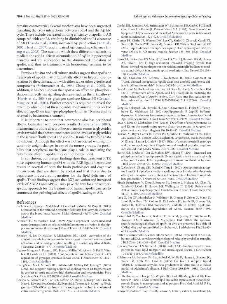
(Liraz et al., 2013), the levels of immunohistochemically deter-mined A�42 were higher in the control apoE4 mice than in thecontrol apoE3 mice. Bexarotene treatment markedly reduced thelevels of A�42 in both the apoE3- and the apoE4-treated mice(p � 0.0001 by 2-way ANOVA for the effect of treatment). Thiseffect was more substantial in the apoE4 mice and virtually abol-ished the difference in the A�42 levels between the apoE4 andapoE3 mice. Similar results were obtained using an ELISA kit forA�42 and whole hippocampal homogenates. As shown in Figure3B, this revealed that the levels of A�42 were higher in the controlapoE4 mice compared with the control apoE3 mice and that thiseffect was counteracted by treatment with bexarotene (p � 0.012by 2-way ANOVA for the effect of treatment � genotype). Fur-ther post hoc analysis revealed that the levels of A�42 were signif-icantly higher in the apoE4 control mice compared with thecorresponding apoE3 mice (p � 0.04) and that the bexarotene-induced decrease in A�42 levels was also significant (p � 0.024).
The ELISA results, which were obtained using TBSX-extractedA�42, correspond to measurements of intracellular and extracel-lular A�42, whereas the immunohistochemical staining mainlydetects intracellular A�42. Accordingly, because both techniquesyielded a similar apoE4-driven effect, this suggests that theapoE4-driven effects on A�42 are primarily intracellular. To-gether, these results suggest that bexarotene counteracts theapoE4-induced accumulation of A�42. This effect seems to bespecific to A�42, because the histochemically measured levelsA�40 are not affected by either genotype or treatment (data notshown).
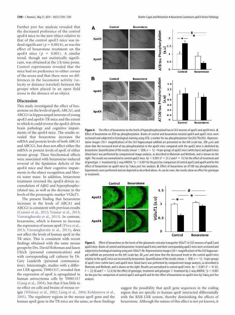
The effects of apoE genotype and bexarotene treatment on tauhyperphosphorylation in CA3 hippocampal neurons are shownin Figure 4. As can be seen and consistent with previous findings(Liraz et al., 2013), the levels of tau phosphorylation using theAT8 mAb were higher in the control apoE4 mice than in thecorresponding apoE3 mice. Bexarotene treatment induced amarked decrease in the levels of AT8 tau phosphorylation inapoE4-treated mice, but not in the apoE3-treated mice (p �0.0002 for the effect of treatment and p � 0.00035 for the effect ofgenotype � treatment by 2-way ANOVA). Post hoc analysis re-vealed that the levels of tau phosphorylation were indeed higherin the apoE4 control mice compared with the correspondingapoE3 mice (p � 0.005) and that the bexarotene-induced de-crease in tau phosphorylation in the apoE4 mice was also signif-icant (p � 0.0002). In contrast to the results obtained with theAT8 mAb, the extent of phosphorylation of tau at the sites recog-nized by AT100 mAb was not affected by either apoE genotype ortreatment (Fig. 4B). Similar results were obtained using the an-tiphosphorylated tau mAb AT270 (data not shown).
The effects of bexarotene on the levels of the synaptic markerVGluT1 are depicted in Figure 5. As can be seen, and consistentwith previous findings (Liraz et al., 2013), the levels of VGluT1 in
Figure 1. The effect of bexarotene on the mRNA and protein levels of apoE, ABCA1, and ABCG1 in hippocampal neurons of apoE3 and apoE4 mice. A, qRT-PCR measurements of the mRNA levelsof ABCA1, ABCG1, and apoE mice. The experiments were performed as described in Materials and Methods and the results are normalized relative to control apoE3 mice (mean � SEM; n � 5 pergroup). White bars correspond to apoE3 mice, whereas black bars correspond to apoE4 mice. B, Immunoblot assays of the corresponding protein levels of ABCA1, ABCG1, and apoE mice. Wholehippocampi from control and bexarotene apoE3 and apoE4 mice were subjected to immunoblot assays using anti-apoE, anti-ABCG1, and anti-ABCA1 Abs as described in Materials and Methods. Theresults are normalized relative to control apoE3 mice (mean � SEM; n � 10 per group). #p � 0.0001 by 2-way ANOVA for the effect of treatment on ABCA1 and ABCG1 protein levels and on ABCA1mRNA levels and #p � 0.001 for the effect of treatment on the ABCG1 mRNA levels (F � 148.4, F � 48.06 for ABCA1 and ABCG1 protein levels and F � 17.82 and F � 17.02 for ABCA1 and ABCG1mRNA levels, respectively). #p � 0.0001 (F � 45.79) for the effect of genotype on apoE protein levels by 2-way ANOVA.
Figure 2. The effect of bexarotene on apoE lipidation in the hippocampus of apoE3 andapoE4 mice. Freshly excised hippocampi from control and bexarotene-treated apoE4 and apoE3mice were subjected to a nondenaturing 3–16% gradient gel and blotted using anti-apoE Ab, asdescribed in Materials and Methods. As can be seen, the levels of apoE reactivity are lower in thecontrol apoE4 mice compared with the control apoE3 mice and bexarotene treatment causes asignificant increase in the appearance of high-molecular-weight species of apoE in both apoE4and apoE3 mice.
7296 • J. Neurosci., May 21, 2014 • 34(21):7293–7301 Boehm-Cagan and Michaelson • Bexarotene Counteracts apoE4-Driven Pathology

the apoE4 control mice were lower than those observed in thecorresponding apoE3 mice. Treatment with bexarotene in-creased the levels of VGluT1 in the apoE4 mice. Bexarotene hadno effect on the apoE3-treated mice, thus rendering the levels ofVGluT1 similar in the treated apoE3- and apoE4 mice (p � 0.001for the effect of genotype � treatment by 2-way ANOVA). Fur-ther post hoc analysis revealed that the reduced levels of VGluT1in the apoE4 mice was significant relative to control apoE3 mice(p � 0.0002), as was the effect of bexarotene treatment on theapoE4 mice (p � 0.0003). Similar results were obtained by West-ern blot analysis of VGluT1 levels of the corresponding mousegroups (data not shown).
Bexarotene also counteracted the effect of apoE4 on the levelsof A�42, hyperphosphorylated tau, and VGluT1 in the CA1 andDG hippocampal subfields. These effects, however, were less pro-nounced than those observed in the CA3 (Liraz et al., 2013).Specifically, the levels of A�42, which were higher in controlapoE4 compared with the control apoE3 in the CA1 (135 � 2% ofcontrol apoE3 mice) and similar in the DG subfields (101 � 2%of control apoE3 mice), were both decreased in the apoE4 miceafter bexarotene treatment (92 � 13% of control apoE3 and 79 �7% of control apoE3 in the CA1 and DG, respectively). Similarresults were obtained with AT8 tau-phosphorylation levels(115 � 7% and 116 � 17% for control apoE4 mice relative tocontrol apoE3 in the CA1 and DG, respectively, and 82 � 9% and
55 � 9% for bexarotene-treated apoE4mice in the CA1 the DG). For the VGluT1,the levels of staining were lower for thecontrol apoE4 mice compared with thecorresponding apoE3 mice in both theCA1 and DG (87 � 12% and 79 � 3%,respectively) and were elevated by bexaro-tene in both subfields (95 � 12% in theCA1 and 106 � 2% in the DG).
Reversal of apoE4-driven behavioraldeficits by bexaroteneThe extent to which the reversal of theapoE4-induced brain pathological effectsby bexarotene is associated with improve-ment of the cognitive performance of theapoE4 mice was examined next. Resultsobtained in the Morris water maze are de-picted in Figure 6. As can be seen, allmouse groups performed similarly on thefirst day of the experiment. However,whereas the control apoE3 mice improvedtheir performance in the swim test andreached a plateau by the second day of thetest, the control apoE4 mice were impairedin their performance and took longer toreach the plateau. This apoE4-induced def-icit was reversed by bexarotene treatment,which had no effect on the apoE3 mice’sperformance (p � 0.047 for the effect ofgenotype � treatment by repeated-measures ANOVA analysis and p �0.0006 for the corresponding Tukey posthoc comparison of the control andbexarotene-treated apoE4 mice). After thelast trial on the fourth day of the Morriswater maze, the mice were subjected to aprobe test in which the hidden platform
was removed from the arena and the mice’s preferences for thedifferent quadrants of the arena were assessed. This test revealedthat the control apoE3 mice spent more time in the quadrantwhere the platform was originally located than in the other quad-rants, whereas the apoE4 mice showed no preference to any of thequadrants (Fig. 6B). Treatment with bexarotene resulted in anincrease in the time that the apoE4 mice spent in the relevantquadrant and rendered their performance equal to that of theapoE3 mice, the performance of which was not affected by bex-arotene. These effects were associated with a significant effect oftreatment (p � 0.028 by 2-way ANOVA).
The mice were next subjected to the novel object recogni-tion test in which their tendency to approach a novel objectwas measured. Mice were exposed to new and old objects 2 h(short-time interval) and 24 h (long-time interval) after theinitial exposure to 2 similar objects. The results are depicted inFigure 7. As can be seen, at both time points, the control apoE3mice spent more time near the new object, whereas the controlapoE4 mice showed no such preference and visited the newand old object similarly. This difference between the controlapoE3 and apoE4 mice was abolished by bexarotene at bothtime points, such that, under these conditions,both mousegroups approached the new object preferentially. Statisticalanalysis revealed a significant effect of genotype � treatmentat the 24 h time point ( p � 0.034 by 2-way ANOVA analysis).
Figure 3. Effect of bexarotene on the levels of A�42 in the hippocampus of apoE3 and apoE4 mice. A, A�42 immunohisto-chemistry. Brains of control and bexarotene-treated apoE4 and apoE3 mice were sectioned and subjected to histological stainingwith anti-A�42 Ab. Representative images (10� magnification) of the CA3 hippocampal field stained with anti-A�42 arepresented on the left (scale bar, 200 �m) and show that the higher levels of A�42 in control apoE4 mice relative to the apoE3 miceare reduced in both apoE3 and apoE4 mice. Quantification of the results (mean � SEM; n � 12–14 per group) of apoE3 mice(white bars) and apoE4 mice (black bars) was performed by computerized image analysis and is shown on the right. The experi-ment and data analysis were performed as described in Materials and Methods. The results are normalized relative to control apoE3mice. #p � 0.001 (F � 41.61) for the effect of treatment by 2-way ANOVA. B, A�42 ELISA. Whole hippocampi from control andbexarotene-treated apoE4 and their corresponding apoE3 mice were subjected to A� X-42 ELISA kit, as described in Materials andMethods. The results (mean�SEM; n�5 per group) are normalized relative to control apoE3 mice and show a higher level of totalA�42 in the control apoE4 mice, which is counteracted by the bexarotene treatment. #p � 0.05 (F � 7.86) for the effect ofgenotype � treatment by 2-way ANOVA; *p � 0.05 for the post hoc comparison of control apoE3 and apoE4 and for the effect ofbexarotene on apoE4 mice by Tukey post hoc analysis.
Boehm-Cagan and Michaelson • Bexarotene Counteracts apoE4-Driven Pathology J. Neurosci., May 21, 2014 • 34(21):7293–7301 • 7297
Further post hoc analysis revealed thatthe decreased preference of the controlapoE4 mice to the new object relative tothat of the control apoE3 mice was in-deed significant ( p � 0.0014), as was theeffect of bexarotene treatment on theapoE4 mice ( p � 0.001). A similartrend, though not statistically signifi-cant, was obtained at the 2 h time point.Control experiments revealed that themice had no preference to either cornerof the arena and that there were no dif-ferences in the locomotor activity (ve-locity or distance traveled) between thegroups when placed in an open fieldarena in the absence of an object.
DiscussionThis study investigated the effect of bex-arotene on the levels of apoE, ABCA1, andABCG1 in hippocampal neurons of youngapoE3 and apoE4-TR mice and the extentto which it could reverse the apoE4-drivenbrain pathology and cognitive impair-ments of the apoE4 mice. The results re-vealed that bexarotene increases themRNA and protein levels of both ABCA1and ABCG1, but does not affect either themRNA or protein levels of apoE of eithermouse group. These biochemical effectswere associated with bexarotene-inducedreversal of the lipidation deficits of theapoE4 mice and their cognitive impair-ments in the object recognition and Mor-ris water maze. In addition, bexarotenetreatment reversed the apoE4-driven ac-cumulation of A�42 and hyperphospho-rylated tau, as well as the decrease in thelevels of the presynaptic marker VGluT1.
The present finding that bexaroteneincreases in the levels of ABCA1 andABCG1 is consistent with previous results(Cramer et al., 2012; Tesseur et al., 2013;Veeraraghavalu et al., 2013). In contrast,bexarotene, which is known to increasethe expression of mouse apoE (Price et al.,2013; Veeraraghavalu et al., 2013), doesnot affect the levels of human apoE in theTR mice. This is consistent with recentfindings obtained with the same mousegroups by Drs. David Holtzman and JasonUlrich (personal communication) andwith corresponding cell cultures by Dr.Gary Landerth (personal communica-tion). Interestingly, studies with a differ-ent LXR agonist, T0901317, revealed thatthe expression of apoE is upregulated inhuman astrocytoma cells by T0901317(Liang et al., 2004), but that it has little tono effect on cells and brains of mouse or-igin (Whitney et al., 2002; Liang et al., 2004; Koldamova et al.,2005). The regulatory regions in the mouse apoE gene and thehuman apoE gene in the TR mice are the same, so these findings
suggest the possibility that apoE gene sequences in the codingregion that are specific to human apoE interacted differentiallywith the RXR-LXR system, thereby diminishing the effects ofbexarotene. Although the nature of this effect is not yet known, it
Figure 4. The effect of bexarotene on the levels of hyperphosphorylated tau in CA3 neurons of apoE3 and apoE4 mice. A,Effect of bexarotene on AT8 tau phosphorylation. Brains of control and bexarotene-treated apoE4 and apoE3 mice weresectioned and subjected to histological staining using AT8, a marker for tau phosphorylation (Ser202/Thr205). Represen-tative images (10� magnification) of the CA3 hippocampal subfield are presented on the left (scale bar, 200 �m) andshow that the increased level of tau phosphorylation in the apoE4 mice compared with the apoE3 mice is abolished bybexarotene. Quantification of the results (mean � SEM; n � 12–14 per group) of apoE3 mice (white bars) and apoE4 mice(black bars) was performed by computerized image analysis, as described in Materials and Methods, and is shown on theright. The results are normalized to control apoE3 mice. #p � 0.001 (F � 23.2 and F � 15) for the effect of treatment andof genotype � treatment by 2-way ANOVA; *p � 0.001 for the post hoc comparison of control apoE3 and apoE4 and for theeffect of bexarotene on apoE4 mice by Tukey post hoc analysis. B, Effect of bexarotene on AT100 tau phosphorylation.Experiments were performed and are depicted as described above. As can be seen, the results show no effect for genotypeor treatment.
Figure 5. Effect of bexarotene on the levels of the glutamate vesicular transporter VGluT1 in CA3 neurons of apoE3 andapoE4 mice. Brains of control and bexarotene-treated apoE4 mice and their corresponding apoE3 mice were sectioned andsubjected to histological staining using anti-VGluT1 Ab. Representative images (20� magnification) of the CA3 hippocam-pal subfield are presented on the left (scale bar, 80 �m) and show that the decreased levels in the control apoE4 micerelative to the apoE3 mice are increased by bexarotene. Quantification of the results (mean � SEM: n � 12–14 per group)of apoE3 mice (white bars) and apoE4 mice (black bars) was performed by computerized image analysis, as described inMaterials and Methods, and is shown on the right. Results are normalized to control apoE3 mice. #p � 0.001 (F � 41.65,F � 22.58 and F � 12.4) for the effect of genotype, treatment and genotype � treatment by 2-way ANOVA; *p � 0.001for the post hoc comparison of control apoE3 and apoE4 and for the effect of bexarotene on apoE4 mice by Tukey post hocanalysis.
7298 • J. Neurosci., May 21, 2014 • 34(21):7293–7301 Boehm-Cagan and Michaelson • Bexarotene Counteracts apoE4-Driven Pathology

has beneficial significance in terms of the ability to differentiatebetween effects of bexarotene related to lipidation and those re-lated to modulation of the levels of apoE.
Our finding that bexarotene both increases the levels ofABCA1 and ABCG1 and the lipidation of apoE in both apoE3 andapoE4 hippocampal homogenates, as seen by native gels, is con-sistent with previous findings that bexarotene increases the levelsof these proteins and the lipidation of mouse apoE in the brains ofAPP/PS1 mice (Cramer et al., 2012). Careful examination of thepatterns of native apoE reveals that the apoE4 mice have lowerlevels of both the high- and the low-molecular-weight species ofapoE compared with apoE3 (Fig. 2). Assuming that the low-molecular-weight particles are precursors of the high-molecular-weight species, this finding is consistent with apoE4-drivenimpairment in the initiation of the formation of the larger spe-cies. Nevertheless, the possibility that apoE4 enhances the degra-dation of the large and small apoE species cannot be ruled out.The mechanism by which the initial basal lipidation of apoE4 islower than that of apoE3 may be related to differences in theinteraction between apoE3 and apoE4 with the lipidation pro-teins and/or to the effect of apoE genotype on the compartmen-
talization and accessibility of apoE3 andapoE4 to the lipidation proteins. The ef-fect of bexarotene on apoE lipidation,which is most likely driven by the in-creased levels of ABCA1 and ABCG1,results in increased conversion of the low-molecular-weight species to the high-molecular-weight species in the apoE3mice. In contrast, in the apoE4 mice, themain effect is an increase in the high-molecular-weight species with no signifi-cant effect on the low-molecular-weightapoE particles. Further studies of the lipidand protein composition of the apoE spe-cies and the time course of their formationand degradation are required to unravelthe effect of apoE genotype on apoElipidation.
The present findings suggest that thesequence of events generated by the intro-duction of bexarotene is as follows: in na-ive mice, apoE4 is hypolipidated relativeto apoE3. Bexarotene increases the mRNAlevels of ABCA1 and ABCG1, thus leadingto an increase in their protein level.ABCA1 and ABCG1 then mediate an in-crease in the lipidated species of apoE, inparticular, a compensatory increase in thelipidation of apoE4. In the following para-graphs, we discuss the functional implica-tions of these effects.
It was shown that, in astrocytic cul-tures from TR mice, the expression ofapoE4 induces a deficiency in the trans-port of cholesterol relative to that ob-served in corresponding apoE3-secretingcells (Gong et al., 2002). Similar resultswere obtained in neuronal culture studiesin which apoE was added exogenously,showing that apoE3 has a greater ability toinduce cholesterol efflux than apoE4 (Mi-chikawa et al., 2000; Minagawa et al.,
2009). In view of the importance of cholesterol to membranehomeostasis and to the well-being of synapses, it is tempting tosuggest that apoE4-indcued decrease in VGluT1 levels is also re-lated to apoE4-driven specific impairments in lipidation of gluta-matergic neurons, which, after bexarotene treatment, is reversed andreturns to normal.
The effect of bexarotene on the levels of A�40 and A�42 wasstudied recently using different animal models and the same bex-arotene preparation used in the present study. The majority ofstudies were performed on APP/PS1 transgenic mouse modelsand show a bexarotene-driven decrease in the levels of solubleA�40 or A�42 in the interstitial fluid (Cramer et al., 2012; Ulrichet al., 2013). A similar bexarotene-induced decrease in A� levelswas observed in other mouse models, such as APP/human apoE(Fitz et al., 2013) and 5XFAD mice (Veeraraghavalu et al., 2013),and in in vitro models (Bachmeier et al., 2013). Our present find-ing that bexarotene decreases the level of intracellular A�42 in thehippocampus of TR mice is consistent with these studies. In con-trast to the consistency in the literature regarding the effect ofbexarotene on the levels of A�40 and A�42, the observed effect ofbexarotene on the size and number of A� plaques in these studies
Figure 6. Effect of bexarotene on the performance of apoE3 and apoE4 mice in the Morris water maze. Control and bexaroteneapoE4 mice and their corresponding apoE3 mice were subjected to a Morris water maze test. A, Latency to reach the platform.Latency was tested across 4 daily trials for 4 d, as described in Materials and Methods. The results shown are the average latenciesof the 4 daily trials of each group in seconds (n � 12–14 per group). � and f correspond to control apoE3 and apoE4 mice,respectively. andŒ correspond to bexarotene-treated apoE3 and apoE4 mice, respectively. # p � 0.05 (F � 2.86) for the effectof genotype � treatment by 2-way ANOVA; *p � 0.05 for the effect of bexarotene on apoE4 mice by Tukey post hoc analysis. B,Probe test. The hidden platform was removed from the arena and the time the mice spent in the quadrant in which the platformwas located was measured, as described in Materials and Methods. The results are depicted as the percentage of time spent in theplatform’s quadrant out of the total trial time (n � 6 –7 per group). White bars correspond to apoE3 mice, whereas black barscorrespond to apoE4 mice. #p � 0.05 (F � 5.47) for the effect of treatment by 2-way ANOVA.
Figure 7. Effect of bexarotene on the performance of the apoE3 and apoE4 mice in the novel object recognition test. Control andbexarotene-treated apoE4 mice and corresponding apoE3 mice were first exposed to two identical objects (training session). Thiswas followed by a delay of either 2 or 24 h, after which the mice were exposed to an old and a new object. The preference of the miceto the different objects was monitored, as described in Materials and Methods. A, Short-term memory test performed 2 h after thetraining session. B, Long-term memory test performed 24 h after the training session. The results obtained are presented as thepercentage of time the mice spent near the novel object out of the total time spent near both old and novel objects. White barscorrespond to apoE3 mice, whereas black bars correspond to apoE4 mice (n � 6 – 8 per group). #p � 0.05 (F � 5.13) for the effectof genotype � treatment by 2-way ANOVA; *p � 0.05 for the post hoc comparison of control apoE3 and apoE4 and for the effectof bexarotene on apoE4 mice by Tukey post hoc analysis.
Boehm-Cagan and Michaelson • Bexarotene Counteracts apoE4-Driven Pathology J. Neurosci., May 21, 2014 • 34(21):7293–7301 • 7299
remains controversial. Several mechanisms have been suggestedregarding the cross interactions between apoE4 and the A� lifecycle. These include decreased binding efficiency of apoE4 to A�compared with apoE3, resulting in diminished apoE4 clearanceof A� (Tokuda et al., 2000), increased A� production (Ye et al.,2005; He et al., 2007), and impaired A�-degrading efficiency (Ji-ang et al., 2008). The extent to which these different mechanismsmediate the apoE4-driven accumulation of A� in hippocampalneurons and are susceptible to the diminished lipidation ofapoE4, and thus to treatment with bexarotene, remains to bedetermined.
Previous in vitro and cell culture studies suggest that apoE4 orfragments of apoE4 may differentially affect tau hyperphospho-rylation by direct interaction with either tau or other cytoskeletalcomponents (Strittmatter et al., 1994; Chang et al., 2005). Inaddition, it has been shown that apoE4 can affect tau phosphor-ylation indirectly via signaling elements such as the Erk pathway(Harris et al., 2004) or glycogen synthase kinase-3� (Cedazo-Mínguez et al., 2003). Further research is required to reveal theextent to which one of these possible mechanisms underlies theeffects of apoE4 on tau hyperphosphorylation in TR mice and itsreversal by bexarotene treatment.
It is important to note that bexarotene also has peripheraleffects. Consistent with previous results (Lalloyer et al., 2009),measurements of the effects of bexarotene on serum triglycerideslevels revealed that bexarotene increases the levels of triglyceridesin the serum of both apoE4 and apoE3-TR mice by twofold (datanot shown). Although this effect was not associated with signifi-cant body weight changes in any of the mouse groups, the possi-bility that peripheral mechanisms play a role in mediating thebexarotene effect in apoE4 mice cannot be excluded.
In conclusion, our present findings show that treatment of TRmice expressing human apoE4 with the RXR ligand bexaroteneresults in reversal of both the brain pathologies and cognitiveimpairments that are driven by apoE4 and that this is due tobexarotene induced compensation for the lipid deficiency ofapoE4. These findings suggest that RXR activation and increasedlevels of ABCA1 and ABCG1 may pave the way for a novel ther-apeutic approach for the treatment of human apoE4 carriers tocounteract the pathological effects of apoE4 leading to AD.
ReferencesBachmeier C, Beaulieu-Abdelahad D, Crawford F, Mullan M, Paris D (2013)
Stimulation of the retinoid X receptor facilitates beta-amyloid clearanceacross the blood-brain barrier. J Mol Neurosci 49:270 –276. CrossRefMedline
Belinson H, Michaelson DM (2009) ApoE4-dependent Abeta-mediatedneurodegeneration is associated with inflammatory activation in the hip-pocampus but not the septum. J Neural Transm 116:1427–1434. CrossRefMedline
Belinson H, Lev D, Masliah E, Michaelson DM (2008) Activation of theamyloid cascade in apolipoprotein E4 transgenic mice induces lysosomalactivation and neurodegeneration resulting in marked cognitive deficits.J Neurosci 28:4690 – 4701. CrossRef Medline
Cedazo-Mínguez A, Popescu BO, Blanco-Millan JM, Akterin S, Pei JJ, Win-blad B, Cowburn RF (2003) Apolipoprotein E and beta-amyloid (1– 42)regulation of glycogen synthase kinase-3beta. J Neurochem 87:1152–1164. CrossRef Medline
Chang S, ran Ma T, Miranda RD, Balestra ME, Mahley RW, Huang Y (2005)Lipid- and receptor-binding regions of apolipoprotein E4 fragments actin concert to cause mitochondrial dysfunction and neurotoxicity. ProcNatl Acad Sci U S A 102:18694 –18699. CrossRef Medline
Chawla A, Boisvert WA, Lee CH, Laffitte BA, Barak Y, Joseph SB, Liao D,Nagy L, Edwards PA, Curtiss LK, Evans RM, Tontonoz P (2001) A PPARgamma-LXR-ABCA1 pathway in macrophages is involved in cholesterolefflux and atherogenesis. Mol Cell 7:161–171. CrossRef Medline
Corder EH, Saunders AM, Strittmatter WJ, Schmechel DE, Gaskell PC, SmallGW, Roses AD, Haines JL, Pericak-Vance MA (1993) Gene dose of apo-lipoprotein E type 4 allele and the risk of Alzheimer’s disease in late onsetfamilies. Science 261:921–923. CrossRef Medline
Cramer PE, Cirrito JR, Wesson DW, Lee CY, Karlo JC, Zinn AE, Casali BT,Restivo JL, Goebel WD, James MJ, Brunden KR, Wilson DA, Landreth GE(2012) ApoE-directed therapeutics rapidly clear beta-amyloid and re-verse deficits in AD mouse models. Science 335:1503–1506. CrossRefMedline
Evans TA, Barkauskas DS, Myers JT, Hare EG, You JQ, Ransohoff RM, HuangAY, Silver J (2014) High-resolution intravital imaging reveals thatblood-derived macrophages but not resident microglia facilitate second-ary axonal dieback in traumatic spinal cord injury. Exp Neurol 254:109 –120. CrossRef Medline
Fitz NF, Cronican AA, Lefterov I, Koldamova R (2013) Comment on“ApoE-directed therapeutics rapidly clear beta-amyloid and reverse def-icits in AD mouse models”. Science 340:924-c. CrossRef Medline
Gilat-Frenkel M, Boehm-Cagan A, Liraz O, Xian X, Herz J, Michaelson DM(2013) Involvement of the Apoer2 and Lrp1 receptors in mediating thepathological effects of ApoE4 in vivo. Curr Alzheimer Res. Advance on-line publication. doi:10.2174/1567205010666131119232444. CrossRefMedline
Gong JS, Kobayashi M, Hayashi H, Zou K, Sawamura N, Fujita SC, Yanag-isawa K, Michikawa M (2002) Apolipoprotein E (ApoE) isoform-dependent lipid release from astrocytes prepared from human ApoE3 andApoE4 knock-in mice. J Biol Chem 277:29919 –29926. CrossRef Medline
Haas A, Liraz O, Michaelson DM (2012) The effects of apolipoproteins E3and E4 on the transforming growth factor-beta system in targeted re-placement mice. Neurodegener Dis 10:41– 45. CrossRef Medline
Hanson AJ, Bayer-Carter JL, Green PS, Montine TJ, Wilkinson CW, BakerLD, Watson GS, Bonner LM, Callaghan M, Leverenz JB, Tsai E, PostupnaN, Zhang J, Lampe J, Craft S (2013) Effect of apolipoprotein E genotypeand diet on apolipoprotein E lipidation and amyloid peptides: random-ized clinical trial. JAMA Neurol 70:972–980. CrossRef Medline
Harris FM, Brecht WJ, Xu Q, Mahley RW, Huang Y (2004) Increased tauphosphorylation in apolipoprotein E4 transgenic mice is associated withactivation of extracellular signal-regulated kinase: modulation by zinc.J Biol Chem 279:44795– 44801. CrossRef Medline
He X, Cooley K, Chung CH, Dashti N, Tang J (2007) Apolipoprotein recep-tor 2 and X11 alpha/beta mediate apolipoprotein E-induced endocytosisof amyloid-beta precursor protein and beta-secretase, leading to amyloid-beta production. J Neurosci 27:4052– 4060. CrossRef Medline
Hirsch-Reinshagen V, Zhou S, Burgess BL, Bernier L, McIsaac SA, Chan JY,Tansley GH, Cohn JS, Hayden MR, Wellington CL (2004) Deficiency ofABCA1 impairs apolipoprotein E metabolism in brain. J Biol Chem 279:41197– 41207. CrossRef Medline
Jiang Q, Lee CY, Mandrekar S, Wilkinson B, Cramer P, Zelcer N, Mann K,Lamb B, Willson TM, Collins JL, Richardson JC, Smith JD, Comery TA,Riddell D, Holtzman DM, Tontonoz P, Landreth GE (2008) ApoE pro-motes the proteolytic degradation of Abeta. Neuron 58:681– 693.CrossRef Medline
Kariv-Inbal Z, Yacobson S, Berkecz R, Peter M, Janaky T, Lutjohann D,Broersen LM, Hartmann T, Michaelson DM (2012) The isoform-specific pathological effects of apoE4 in vivo are prevented by a fish oil(DHA) diet and are modified by cholesterol. J Alzheimers Dis 28:667–683. CrossRef Medline
Karten B, Campenot RB, Vance DE, Vance JE (2006) Expression of ABCG1,but not ABCA1, correlates with cholesterol release by cerebellar astroglia.J Biol Chem 281:4049 – 4057. CrossRef Medline
Kim WS, Weickert CS, Garner B (2008) Role of ATP-binding cassette trans-porters in brain lipid transport and neurological disease. J Neurochem104:1145–1166. CrossRef Medline
Koldamova RP, Lefterov IM, Staufenbiel M, Wolfe D, Huang S, Glorioso JC,Walter M, Roth MG, Lazo JS (2005) The liver X receptor ligandT0901317 decreases amyloid beta production in vitro and in a mousemodel of Alzheimer’s disease. J Biol Chem 280:4079 – 4088. CrossRefMedline
Laffitte BA, Repa JJ, Joseph SB, Wilpitz DC, Kast HR, Mangelsdorf DJ, Ton-tonoz P (2001) LXRs control lipid-inducible expression of the apolipo-protein E gene in macrophages and adipocytes. Proc Natl Acad Sci U S A98:507–512. CrossRef Medline
Lalloyer F, Pedersen TA, Gross B, Lestavel S, Yous S, Vallez E, Gustafsson JA,
7300 • J. Neurosci., May 21, 2014 • 34(21):7293–7301 Boehm-Cagan and Michaelson • Bexarotene Counteracts apoE4-Driven Pathology
Mandrup S, Fievet C, Staels B, Tailleux A (2009) Rexinoid bexarotenemodulates triglyceride but not cholesterol metabolism via gene-specificpermissivity of the RXR/LXR heterodimer in the liver. ArteriosclerThromb Vasc Biol 29:1488 –1495. CrossRef Medline
Levi O, Michaelson DM (2007) Environmental enrichment stimulates neu-rogenesis in apolipoprotein E3 and neuronal apoptosis in apolipoproteinE4 transgenic mice. J Neurochem 100:202–210. CrossRef Medline
Levi O, Jongen-Relo AL, Feldon J, Roses AD, Michaelson DM (2003) ApoE4impairs hippocampal plasticity isoform-specifically and blocks the envi-ronmental stimulation of synaptogenesis and memory. Neurobiol Dis13:273–282. CrossRef Medline
Liang Y, Lin S, Beyer TP, Zhang Y, Wu X, Bales KR, DeMattos RB, May PC, LiSD, Jiang XC, Eacho PI, Cao G, Paul SM (2004) A liver X receptor andretinoid X receptor heterodimer mediates apolipoprotein E expression,secretion and cholesterol homeostasis in astrocytes. J Neurochem 88:623–634. CrossRef Medline
Liraz O, Boehm-Cagan A, Michaelson DM (2013) ApoE4 induces Abeta42,tau, and neuronal pathology in the hippocampus of young targeted re-placement apoE4 mice. Mol Neurodegener 8:16. CrossRef Medline
Michikawa M, Fan QW, Isobe I, Yanagisawa K (2000) Apolipoprotein Eexhibits isoform-specific promotion of lipid efflux from astrocytes andneurons in culture. J Neurochem 74:1008 –1016. CrossRef Medline
Minagawa H, Gong JS, Jung CG, Watanabe A, Lund-Katz S, Phillips MC,Saito H, Michikawa M (2009) Mechanism underlying apolipoprotein E(ApoE) isoform-dependent lipid efflux from neural cells in culture.J Neurosci Res 87:2498 –2508. CrossRef Medline
Price AR, Xu G, Siemienski ZB, Smithson LA, Borchelt DR, Golde TE, Felsen-stein KM (2013) Comment on “ApoE-directed therapeutics rapidlyclear beta-amyloid and reverse deficits in AD mouse models.” Science340:924-d. CrossRef Medline
Puglielli L, Tanzi RE, Kovacs DM (2003) Alzheimer’s disease: the choles-terol connection. Nat Neurosci 6:345–351. CrossRef Medline
Riddell DR, Zhou H, Atchison K, Warwick HK, Atkinson PJ, Jefferson J, Xu L,Aschmies S, Kirksey Y, Hu Y, Wagner E, Parratt A, Xu J, Li Z, Zaleska MM,Jacobsen JS, Pangalos MN, Reinhart PH (2008) Impact of apolipopro-tein E (ApoE) polymorphism on brain ApoE levels. J Neurosci 28:11445–11453. CrossRef Medline
Roses AD (1996) Apolipoprotein E alleles as risk factors in Alzheimer’s dis-ease. Annu Rev Med 47:387– 400. CrossRef Medline
Salomon-Zimri S, Boehm-Cagan A, Liraz O, Michaelson DM (2014)Hippocampus-related cognitive impairments in young apoE4 targetedreplacement mice. Neurodegener Dis 13:86 –92. CrossRef Medline
Saunders AM, Strittmatter WJ, Schmechel D, George-Hyslop PH, Pericak-Vance MA, Joo SH, Rosi BL, Gusella JF, Crapper-MacLachlan DR, AlbertsMJ, et al. (1993) Association of apolipoprotein E allele epsilon 4 withlate-onset familial and sporadic Alzheimer’s disease. Neurology 43:1467–1472. CrossRef Medline
Strittmatter WJ, Saunders AM, Goedert M, Weisgraber KH, Dong LM, JakesR, Huang DY, Pericak-Vance M, Schmechel D, Roses AD (1994)Isoform-specific interactions of apolipoprotein E with microtubule-associated protein tau: implications for Alzheimer disease. Proc Natl AcadSci U S A 91:11183–11186. CrossRef Medline
Sullivan PM, Mezdour H, Aratani Y, Knouff C, Najib J, Reddick RL,Quarfordt SH, Maeda N (1997) Targeted replacement of the mouse
apolipoprotein E gene with the common human APOE3 allele en-hances diet-induced hypercholesterolemia and atherosclerosis. J BiolChem 272:17972–17980. CrossRef Medline
Tai LM, Bilousova T, Jungbauer L, Roeske SK, Youmans KL, Yu C, Poon WW,Cornwell LB, Miller CA, Vinters HV, Van Eldik LJ, Fardo DW, Estus S, BuG, Gylys KH, Ladu MJ (2013) Levels of soluble apolipoproteinE/amyloid-beta (Abeta) complex are reduced and oligomeric Abeta in-creased with APOE4 and Alzheimer disease in a transgenic mouse modeland human samples. J Biol Chem 288:5914 –5926. CrossRef Medline
Tesseur I, Lo AC, Roberfroid A, Dietvorst S, Van Broeck B, Borgers M, GijsenH, Moechars D, Mercken M, Kemp J, D’Hooge R, De Strooper B (2013)Comment on “ApoE-directed therapeutics rapidly clear beta-amyloidand reverse deficits in AD mouse models.” Science 340:924-e. CrossRefMedline
Tokuda T, Calero M, Matsubara E, Vidal R, Kumar A, Permanne B, ZlokovicB, Smith JD, Ladu MJ, Rostagno A, Frangione B, Ghiso J (2000) Lipida-tion of apolipoprotein E influences its isoform-specific interaction withAlzheimer’s amyloid beta peptides. Biochem J 348:359 –365. CrossRefMedline
Ulrich JD, Burchett JM, Restivo JL, Schuler DR, Verghese PB, Mahan TE,Landreth GE, Castellano JM, Jiang H, Cirrito JR, Holtzman DM (2013)In vivo measurement of apolipoprotein E from the brain interstitial fluidusing microdialysis. Mol Neurodegener 8:13. CrossRef Medline
Vance JE, Hayashi H (2010) Formation and function of apolipoproteinE-containing lipoproteins in the nervous system. Biochim Biophys Acta1801:806 – 818. CrossRef Medline
Veeraraghavalu K, Zhang C, Miller S, Hefendehl JK, Rajapaksha TW, Ulrich J,Jucker M, Holtzman DM, Tanzi RE, Vassar R, Sisodia SS (2013) Com-ment on “ApoE-directed therapeutics rapidly clear beta-amyloid and re-verse deficits in AD mouse models.” Science 340:924-f. CrossRef Medline
Wahrle SE, Jiang H, Parsadanian M, Legleiter J, Han X, Fryer JD, KowalewskiT, Holtzman DM (2004) ABCA1 is required for normal central nervoussystem ApoE levels and for lipidation of astrocyte-secreted apoE. J BiolChem 279:40987– 40993. CrossRef Medline
Whitney KD, Watson MA, Collins JL, Benson WG, Stone TM, Numerick MJ,Tippin TK, Wilson JG, Winegar DA, Kliewer SA (2002) Regulation ofcholesterol homeostasis by the liver X receptors in the central nervoussystem. Mol Endocrinol 16:1378 –1385. Medline
Ye S, Huang Y, Mullendorff K, Dong L, Giedt G, Meng EC, Cohen FE, KuntzID, Weisgraber KH, Mahley RW (2005) Apolipoprotein (apo) E4 en-hances amyloid beta peptide production in cultured neuronal cells: apoEstructure as a potential therapeutic target. Proc Natl Acad Sci U S A 102:18700 –18705. CrossRef Medline
Youmans KL, Tai LM, Nwabuisi-Heath E, Jungbauer L, Kanekiyo T, Gan M,Kim J, Eimer WA, Estus S, Rebeck GW, Weeber EJ, Bu G, Yu C, Ladu MJ(2012) APOE4-specific changes in Abeta accumulation in a new trans-genic mouse model of Alzheimer disease. J Biol Chem 287:41774 – 41786.CrossRef Medline
Zepa L, Frenkel M, Belinson H, Kariv-Inbal Z, Kayed R, Masliah E, Michael-son DM (2011) ApoE4-driven accumulation of intraneuronal oli-gomerized Abeta42 following activation of the amyloid cascade in vivo ismediated by a gain of function. Int J Alzheimers Dis 2011:792070.CrossRef Medline
Boehm-Cagan and Michaelson • Bexarotene Counteracts apoE4-Driven Pathology J. Neurosci., May 21, 2014 • 34(21):7293–7301 • 7301


![Sigma 3-18KS Sigma 3-18KHS · PDF fileSigma 3-18KS Sigma 3-18KHS. ... (with 13190 and 13194) 11133 ... RFI suppression EN 61326 EN 61326 Weight without rotor [kg]](https://static.fdocuments.us/doc/165x107/5a790eff7f8b9a9a188b7ade/sigma-3-18ks-sigma-3-18khs-3-18ks-sigma-3-18khs-with-13190-and-13194-11133.jpg)