Rev. Vol. XXXIV, Nr.1-2, 2015srh.org.ro/wp-content/uploads/2016/10/Revista Vol. XXXIV, Nr. 1-2,...
54
Nr. XXXIV, Vol. 1-2, 2015
Transcript of Rev. Vol. XXXIV, Nr.1-2, 2015srh.org.ro/wp-content/uploads/2016/10/Revista Vol. XXXIV, Nr. 1-2,...
Nr. XXXIV, Vol. 1-2, 2015
Editor-in-ChiefDaniel CoriuUniversity of Pharmacy “Carol Davila” Bucharest, Fundeni Institute, Bucharest, Rom niaa
Deputy/ Managing EditorRadu Niculescu, Fundeni Clinical Institute, Bucharest, Rom nia a
Editorial Advisory BoardIulia Ursuleac, University of Medicine and Pharmacy “Carol Davila”, Bucharest, Fundeni Clinical Institute, Bucharest, Rom niaaLumini a Rusen, țNational Centre of Haematology and Transfusion, Bucharest, Rom niaaAmelia G man, ăUniversity of Medicine and Pharmacy Craiova, Rom nia â
Editors Anca Bojan, University of Medicine and Pharmacy “Iuliu Hatieganu”, Cluj Napoca, Romania
Domnița Crișan, University of Michigan Medical School, USAC t lin Danaila, ă ă University of Medicine and Pharmacy “Grigore Popa", Ia , Rom nia și aGabriel Ghiaur, The Johns Hopkins University School of MedicineErnst Holler, University of Regensburg, Germany Hortensia Ioni , ță University of Medicine and Pharmacy “Victor Babe ”, Timi oara, Romania ș șAnca Lupu, University of Medicine and Pharmacy “Carol Davila”, Bucharest, Col ea Hospital, Bucharest, Romaniaț Galafteon Oltean, University of Medicine and Pharmacy T rgu Mure , Romania â șAna Maria Vl d reanu, ă ă University of Medicine and Pharmacy “Carol Davila”, Bucharest, Emergency University Hospital, Bucharest, Romania Florentina Vl d reanu, ă ă National Centre of Haematology and Transfusion, Bucharest, Romania
Language Editors Bardas Alexandru, Fundeni Clinical Institute
Technical Editors Cretu Teodor, Medmun, Bucharest, Romania
PublisherDE GRUYTER OPENBogumiła Zuga 32A Str.01-811 Warsaw, PolandT: +48 22 701 50 15
I
II
Scientific Reviewers
Mihail Badea (UMF Craiova)Marius Balea (Spitalul Clinic Colentina, Bucureşti)Sorina Bădeliţă (Institutul Clinic Fundeni, Bucureşti)Erzsebet Benedek Lazar (Spitalul Clinic Județean de Urgență Tg.Mureş)Istvan Benedek (UMF Târgu Mureş)Nicoleta Berbec (UMF “Carol Davila”, Bucureşti)Anca Bojan (UMF “Iuliu Hațieganu” Cluj)Horia Bumbea (UMF “Carol Davila” Bucureşti) Cristina Burcoveanu (Institutul Regional de Oncologie Iaşi)Leny Caban (Institutul Clinic Fundeni, Bucureşti)Despina Calamar Popovici (UMF “Victor Babeş” Timişoara)Carmen Călugăroiu (Institutul Clinic Fundeni, Bucureşti)Alina Cătană (Spitalul Clinic Judetean de Urgenta Sibiu)Adriana Coliță (UMF”Carol Davila” Bucureşti)Anca Coliță (UMF”Carol Davila” Bucureşti)Andrei Coliță (UMF”Carol Davila” Bucureşti)Dan Coliță (UMF “Carol Davila”, Bucureşti)Daniel Coriu (UMF “Carol Davila”, Bucureşti)Coralia Cotoraci (Universitatea de Vest “Vasile Goldiş” Arad)Domnița Crişan (Michigan, USA)Cătălin Dănăilă (UMF “Gr.T,Popa” Iaşi)Smaranda Demian (UMF Târgu Mureş)Camelia Dobrea (UMF “Carol Davila”, Bucureşti)Aurora Dragomirişteanu (Registrul Național al Donatorilor Voluntari de C.S.H.)Amelia Găman (UMF Craiova)Emanuil Gheorghiță (Spitalul Clinic Judetean de Urgență Braşov)Mihaela Ghinea (Universitatea Ovidius Constanța)Marian Giovani (Spitalul Județean de Urgenta Brăila)Ana Marcela Grigoriu (Spitalul Județean de Urgență Ploieşti)Ecaterina Hanganu (Institutul Regional de Oncologie Iaşi)Ernst Holler (KLinikum der Universität Regensburg, Germany)
Anca Ion (Institutul Clinic Fundeni, Bucureşti)Bogdan Ionescu (Institutul Clinic Fundeni, Bucureşti)Hortensia Ioniță (UMF “Victor Babeş” Timişoara)Ioana Ioniță (UMF “Victor Babeş” Timişoara)Cerasela Jardan (Institutul Clinic Fundeni, Bucureşti)Anca Roxana Lupu (UMF “Carol Davila” Bucureşti)Ioan Macarie (UMF Târgu Mureş)Romeo Mihăilă (Universitatea ”Lucian Blaga” Sibiu)Ana Maria Moldovianu (Institutul Clinic Fundeni, Bucureşti)Radu Niculescu (Institutul Clinic Fundeni, Bucureşti)Galafteon Oltean (UMF Târgu Mureş)Ljubomir Petrov (UMF “Iuliu Hațieganu” Cluj)Alina Roşca (Spitalul Clinic Judetean de Urgență Braşov)Luminița Rusen (Institutul Național de Hematologie Transfuzională)Silvia Sirian (Institutul Național de Hematologie Transfuzională)Răzvan Stoia (Institutul Clinic Fundeni, Bucureşti)Aurelia Tatic (UMF “Carol Davila” Bucureşti)Alina Tănase (Institutul Clinic Fundeni, Bucureşti)Rodica Tălmaci (UMF “Carol Davila” Bucureşti)Mihaela Tevet (Spitalul Clinic Colentina, Bucureşti)Iulia Ursuleac (UMF “Carol Davila” Bucureşti)Valentina Uscătescu (Institutul Clinic Fundeni, Bucureşti)Zsofia Varady (Institutul Clinic Fundeni, Bucureşti)Anca Vasilache (“Prof.Dr.Ion Chiricuță” Oncological Institute, Cluj)Didona Vasilache (Institutul Clinic Fundeni, Bucureşti)Mariana Vasilică (Institutul Clinic Fundeni, Bucureşti)Ana Maria Vlădăreanu (UMF “Carol Davila” Bucureşti)Florentina Vlădăreanu (Institutul Național de Hematologie Transfuzională)
III
Updates in acquired aplastic anemia: Can we do more for our patients?Ana-Maria Moldovianu, Anca Popp, Zsofia Varady, Alina Tanase, Alexandra Marculescu, Camelia Dobrea,Didona Vasilache, Cerasela Jardan, Radu Niculescu, Daniel Coriu ........................................................................................ .....5
Haplo-identical one arrow ransplant protocol using reduced B M Tintensity conditioning for undeni linical nstituteF C IVarady Zsofia, Coriu Daniel, Ghiaur Gabriel,Richard J Jones ................................................................... ............2....... 5
Clinical Case Of Aplastic Crisis Associated With Extramedullary Hematopoiesis In An Adult With Hereditary Spherocytosis And Parvovirus B19 InfectionAndreea Jercan, Rusu Munteanu Gina, Camelia Dobrea, Daniel Coriu, Aurelia Tatic .....................................................................31
Clonal evolution in a patient with aplastic anemia – case reportM , , ,elen Brinza Cerasela Jardan Didona Vasilache Camelia Dobrea, Daniel Coriu ...........................................................................................37
Quercetin, Menadione, Doxorubicin combination as a potential alternative to Doxorubicin monotherapy of acute lymphoblastic leukemiaRuxandra Irimia, Ioana Teodora Tofolean, Roxana Gabriela Sandu, Oana Elena Băran, Maria Cătălina Ceauşescu, Vlad Coşoreanu, Maria Teodora Ilie, Ramona Babeş, Constanţa Ganea, Irina Băran .............................................................................................45
CONTENTS

6
7
1. Definition Aplastic anemia is defined as pancytopenia with a hypocellular bone marrow. Although bone marrow function is a lways diminished affecting all hematopoietic lineages, levels of blood cells may not be
depressed uniformly and different degrees of cytopenia can be observed. There are standard definition criteria of AA with different severity degrees. Severe AA is defined as BM cellularity < 25% and at least two of three criteria consisting in neutrophil count < 0.5 x 10⁹/L,
Updates in acquired aplastic anemia: Can we do more for our patients?
Ana-Maria Moldovianu, Anca Popp, Zsofia Varady, Alina Tanase, Alexandra Marculescu, Camelia Dobrea, Didona Vasilache, Cerasela Jardan, Radu Niculescu, Daniel CoriuCentre of Hematology and Bone Marrow Transplant, Fundeni Clinical Institute, Bucharest, Romania
Abstract: The purpose of this work is to present the results of allogeneic stem cell transplantation as therapy for patients diagnosed with acquired aplastic anemia in the Department of Bone Marrow Transplantation of Fundeni Clinical Institute and to elaborate an algorithm of treatment in aplastic anemia starting with the observations obtained from our clinical practice and following the European treatment guidelines in this group of patients.Aplastic Anemia (AA) is a rare hematological disease characterized by pancytopenia and a hypocellular bone marrow. The paradigm of bone marrow failure syndromes, aplastic anemia is a diagnosis of exclusion despite the precision of its diagnosis criteria. Although AA is not a malignant disease, but an autoimmune disorder, the grave consequences of pancytopenia and clonal transformation into acute leukemia make it a potentially fatal condition. The management of AA patients is challenging and necessitates a very well established treatment plan from the diagnosis. We present the treatment algorithm for AA patients with recommendations based on both recent guidelines in the field and on our experience treating AA patients with allogeneic stem cell transplant. Therapeutic procedure algorithm comprises different approaches for different patient populations, age categories and availability of immunosuppression therapy or different types of donors. According to the recent EBMT recommendations the treatment of choice for young patients (younger than 40 years) who have a matched sibling donor is hematopoietic stem cell transplantation (HSCT). For those patients who don't have a matched sibling donor or are not candidates for HSCT due to older age, the immunosuppression with ATG and cyclosporine is an efficient treatment. The supportive care has an important role and the patients with aplastic anemia should be managed by a multidisciplinary team. For patients older than 40 years, the choice between immunosuppressive therapy (IST) and upfront transplant with HLA identical sibling donor remains a key question. However, the standard approaches for this category of patients is front line immunosuppression with ATG and cyclosporine and if they become refractory to at least one course of IST the allogeneic stem cell transplant using fludarabine-based conditioning is the second-line treatment option. In our institution there were eleven AA patients treated with allogeneic stem cell transplantation from 2009 till 2015. They were all young patients with age between 19 and 42 years old and all had severe acquired aplastic anemia with transfusion dependence. Six cases were transplanted from a matched sibling donor and five patients had undergone an unrelated matched donor transplant. The allogeneic HSCT procedure was done both as front line therapy in the case of three patients and as second treatment choice in the rest of eight patients. Four patients died, three of them due to transplant related toxicity and one patient experienced severe autoimmune reaction with transfusion inefficacy complicated with intracerebral haemorrhage at four months from transplant. In our opinion the most challenging aspect in treating AA patients is choosing the best treatment option taking into account the patient age and performance status, the severity of the disease and the availability of a donor for allogeneic HSCT.Although the treatment strategy must be individualized in every patient case, it is necessary to make a standardization of treatment procedures in AA and to follow the evidence based recommendations available in the management of this rare disease.
Keywords: aplastic anemia, bone marrow transplant, bone marrow failure, allogeneic stem cell transplantation, anti-thymocyte globulin, cycloporine A.
Corresponding authorAna-Maria Moldovianu, Department of Hematology, Fundeni Clinical Institute, Sos. Fundeni nr. 258, sector 2, Bucharest, Romania, phone+40745538575, e-mail: [email protected]
DOI: 10.1515/dch-2015-0001
D DE GRUYTEREOPEN
5
8
DOCUMENTA HAEMATOLOGICAVol. XXXI , Nr. - , 20V 1 2 51 Updates in acquired aplastic anemia: Can we do more for our patients?
Ana-Maria Moldovianu
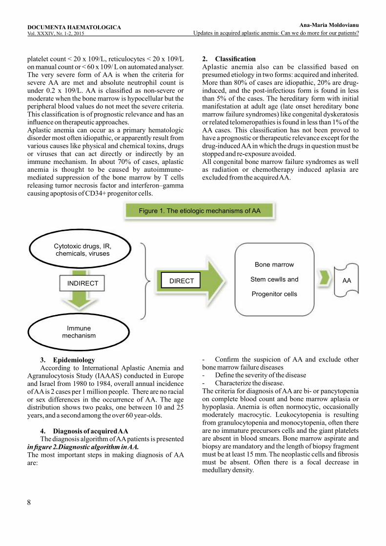
platelet count < 20 x 109/L, reticulocytes < 20 x 109/L on manual count or < 60 x 109/ L on automated analyser. The very severe form of AA is when the criteria for severe AA are met and absolute neutrophil count is under 0.2 x 10⁹/L. AA is classified as non-severe or moderate when the bone marrow is hypocellular but the peripheral blood values do not meet the severe criteria. This classification is of prognostic relevance and has an influence on therapeutic approaches.Aplastic anemia can occur as a primary hematologic disorder most often idiopathic, or apparently result from various causes like physical and chemical toxins, drugs or viruses that can act directly or indirectly by an immune mechanism. In about 70% of cases, aplastic anemia is thought to be caused by autoimmune-mediated suppression of the bone marrow by T cells releasing tumor necrosis factor and interferon–gamma causing apoptosis of CD34+ progenitor cells.
Figure 1. The etiologic mechanisms of AA
Cytotoxic drugs, IR,chemicals, viruses
Immunemechanism
AA
Bone marrow
Stem cewlls and
Progenitor cells
DIRECTINDIRECT
2. ClassificationAplastic anemia also can be classified based on presumed etiology in two forms: acquired and inherited. More than 80% of cases are idiopathic, 20% are drug-induced, and the post-infectious form is found in less than 5% of the cases. The hereditary form with initial manifestation at adult age (late onset hereditary bone marrow failure syndromes) like congenital dyskeratosis or related telomeropathies is found in less than 1% of the AA cases. This classification has not been proved to have a prognostic or therapeutic relevance except for the drug-induced AA in which the drugs in question must be stopped and re-exposure avoided. All congenital bone marrow failure syndromes as well as radiation or chemotherapy induced aplasia are excluded from the acquired AA.
3. Epidemiology According to International Aplastic Anemia and Agranulocytosis Study (IAAAS) conducted in Europe and Israel from 1980 to 1984, overall annual incidence of AA is 2 cases per 1 million people. There are no racial or sex differences in the occurrence of AA. The age distribution shows two peaks, one between 10 and 25 years, and a second among the over 60 year-olds.
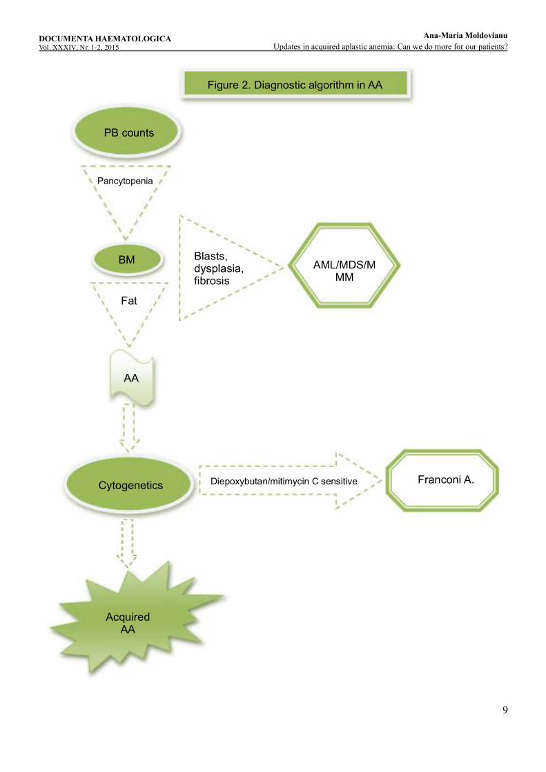
4. Diagnosis of acquired AA The diagnosis algorithm of AA patients is presented in figure 2.Diagnostic algorithm in AA.The most important steps in making diagnosis of AA are:
- Confirm the suspicion of AA and exclude other bone marrow failure diseases- Define the severity of the disease- Characterize the disease.The criteria for diagnosis of AA are bi- or pancytopenia on complete blood count and bone marrow aplasia or hypoplasia. Anemia is often normocytic, occasionally moderately macrocytic. Leukocytopenia is resulting from granulocytopenia and monocytopenia, often there are no immature precursors cells and the giant platelets are absent in blood smears. Bone marrow aspirate and biopsy are mandatory and the length of biopsy fragment must be at least 15 mm. The neoplastic cells and fibrosis must be absent. Often there is a focal decrease in medullary density.
9
Figure 2. Diagnostic algorithm in AA
PB counts
BM AML/MDS/MMM
Fat
AA
Franconi A.Cytogenetics Diepoxybutan/mitimycin C sensitive
AcquiredAA
Blasts,dysplasia,fibrosis
Pancytopenia
DOCUMENTA HAEMATOLOGICAVol. XXXI , Nr. - , 20V 1 2 51 Updates in acquired aplastic anemia: Can we do more for our patients?
Ana-Maria Moldovianu

10
In children and young adults, acquired AA should be distinguished from the inherited forms of bone marrow failure such as Fanconi's anemia as the differentiation has therapeutic implications. Patients with Fanconi's anemia often have physical anomalies, but the distinction depends on the laboratory finding of abnormal chromosome fragility seen readily in metaphase preparations of peripheral blood lymphocytes cultured with phytohemagglutinin. Chromosomal breakage is strikingly enhanced compared with controls if clastogenic agents, such as diepoxybutane, are added to the culture. However, in older patients, the major differential diagnosis is between AA and hypoplastic myelo-displastic syndrome which sometimes may be one of the most difficult diagnostic issues. The main differences between these two entities are the absence of dysplasia, splenomegaly, blast cells, marrow fibrosis and cytogenetic anomalies in AA which may be present in MDS; CD 34 positive cells are absent in AA, but in MDS may be normal or increased, and myelopoiesis and megakariopoiesis usually are absent in AA, but possible in hypoplastic MDS. The severity of AA is exclusively based on peripheral blood parameters and not on bone marrow cellularity. The criteria defining the severity of the disease were presented in the definition paragraph.Regarding the characterization of AA, there are some important interrelations between AA and other conditions that may have impact on disease outcome and prognostic and therapeutic relevance. Acquired AA and Paroxysmal Nocturnal Hemoglobinuria (PNH) have a close interrelationship. Patients diagnosed with AA often present a PNH clone. Also, patients with PNH can develop AA in the course of their disease. In the AA patients most clones are small and they do not have symptoms related to PNH. However, in some patients the PNH clone can increase in the evolution of AA and the PNH characteristic symptoms and complications become the problem to deal with. There are some data predicting a better response to immunosuppressive therapy in AA patients with significant clones of PNH. Other association observed in 5-10% of aplastic anemia cases is with seronegative hepatitis. It is characterized by self-limited liver inflammation, elevated liver enzymes often severe, negative viral serology for hepatitis A, B or C. The post-hepatitis AA typically occurs in young, healthy males and it seems to have a poorer prognosis than idiopathic AA, with early estimates of mortality of 90% at one year, and a history of hepatitis in AA has been considered an indication for early BMT. Patients with posthepatitis AA can successfully undergo BMT without an increased risk of veno-occlusive disease. Patients with hepatitis-
associated AA have markers of immune system activation and respond well to intensive immuno-suppressive therapy. Pregnancy is common in the age groups most susceptible to BM failure, and in many cases, its association is probably only coincidental. The true frequency of AA in pregnancy is unknown, but from the number of cases reported, it appears rare, although bone marrow hypoplasia may be relatively common during pregnancy. Survival rates for AA in pregnancy have been relatively high for the mother and baby, with the most pregnancies being successful. The published data are insufficient to guide the management of pregnant women with AA. A woman who desires a child can be maintained with transfusions, as hemorrhage is the most common cause of death from AA during pregnancy. In a large multicenter study of the Severe AA Working Party of the EBMT the association between aplastic anemia and an autoimmune disease was reported in 50 of 1251 AA patients, especially in older patients. Aplastic anemia is a component of the collagen vascular syndrome called eosinophilic fasciitis. This severe, scleroderma-like disease is characterized by fibrosis of subcutaneous and fascial tissue, localized sk in induration, eos inophilia , hypergamma-globul inemia, and an e levated ery throcyte sedimentation rate. The rheumatologic symptoms of fasciitis respond to corticosteroids, but the associated AA has a very poor prognosis. More rarely, AA has complicated systemic lupus erythematosus (SLE) and rheumatoid arthritis, but in many cases, the role of concomitant drug therapy is intricately. Rarely, AA can accompany Sjogren syndrome, multiple sclerosis, and immune thyroid disease. AA occasionally occurs in individuals with hypogammaglobulinemia or congenital immuno deficiency syndrome, thymoma or -thymic hyperplasia. 5. Differential diagnosis and diagnosis by exclusion The differential diagnosis for acquired aplastic anemia must include: hypoplastic acute leukemia, myelodisplastic syndrome (hypoplastic form), hairy cell leukemia and other lymphomas, bone marrow infiltration by solid tumors, osteomyelofibrosis, hypersplenism, severe megaloblasticanemia, anorexia nervosa, systemic lupus erythematosus, paroxysmal nocturnal hemoglobinuria, Fanconi anemia, congenital dyskeratosis, Shwachman-Diamond syndrome, isolated aplastic anemia (“pure red cell aplasia”), aplasia after chemo- or radiation therapy. 6. Diagnostic workup for AA - Detailed medical drug therapy history - Clinical examination paying attention to clinical signs relevant for cytopenic complications or d i ffe rent ial d iagnos is : infec tion , b leeding ,
DOCUMENTA HAEMATOLOGICAVol. XXXI , Nr. - , 20V 1 2 51 Updates in acquired aplastic anemia: Can we do more for our patients?
Ana-Maria Moldovianu
11
lymphadenopathy, hepatomegaly, splenomegaly, jaundice, nail dystrophies, leukoplakias, pigment or skeletal or dental anomalies- Complete blood counts including differential and reticulocytes- Bone marrow biopsy and aspirate, cytogenetic studies
- LDH, haptoglobin, hemosiderin in the urine looking for hemolysis- Coagulation tests- Reactive C Protein test- Blood total protein, serum protein electrophoresis and immunoglobulins
Legend:
Modulate according to disease severity, favour transplant if very severe diseasea
In the very elderly treatment may be limited to Cyclosporine Ab
BMT – bone marrow transplant, Cy200/ATG-cyclophosphamide 200 mg/kgc and ATG; FluCy/ATG-1
Fludarabine 120mg/m2, low-dose Cyclophosphamide 120mg/kgc and ATG; TBI-total body irradiationATG – antithymocyte globulin, CSA-cyclosporine A, IST-immunosuppressive therapy2
NR – non - responder3
MUD – Matched Unrelated Donor4
BSC – Best supportive care including transfusional support, trial of androgens5
Exp. IST – experimental immunosuppressive therapy (Alemtuzumab, high dose cyclophosphamide)6
Figure 3. The therapeuric algorithm for adult patients with aquired Aplastic Anemia
DOCUMENTA HAEMATOLOGICAVol. XXXI , Nr. - , 20V 1 2 51 Updates in acquired aplastic anemia: Can we do more for our patients?
Ana-Maria Moldovianu
12
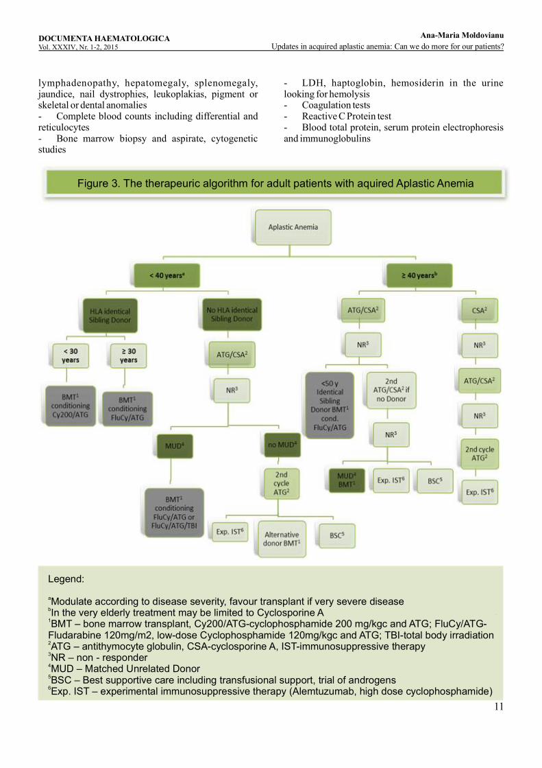
- GOT, GPT, creatinine, urea, uric acid, glucose- Ferritin- Vitamin B12, folic acid- Immunological tests like ANA, anti-DNA antibodies- Blood group, Coombs tests- Chest X-ray, abdominal sonography- EBV, CMV, hepatitis A+B+C, HIV, Parvovirus B19- Flow cytometric analysis of GPI-anchored proteins on granulocytes and erythrocytes- Telomeric length measurement by means of the Flow-Fish method (if shortened telomeres then mutation analysis for TERT, TERC, TIN2, and further components of telomerase complex if possible) in the case of adolescents and young adults if congenital bone marrow failure syndromes are suspected- For differential diagnosis with Fanconianemia: chromosome fragmentation test, mutation analysis of the Fanconianemia genes- HLA class I and II typing for allogeneic HSCT transplant candidates 7. Therapya. Indication The management for AA patients should be based on a therapy plan promptly made once the diagnosis has been confirmed. Treatment options must be identified and chosen with alacrity. The choice of therapy depends on the age of the patient, the severity of the disease, and the degree of the HLA-identity in a potential related or unrelated bone marrow donor (Figure 3.The therapeutic algorithm for adult patients with Aplastic Anemia). The interval between diagnosis and therapy, as studies have shown in cases of bone marrow transplantations, has a significant impact on the outcome. A therapy that is intended to cure will be indicated in three situations: symptomatic disease, severe disease or high risk disease. The patients with severe and very severe form of AA according to definition and the cases with non-severe AA but with severe cytopenia of at least one cell line which requires regular transfusions or with an increased risk for infections or bleeding invariably need definitive treatment. Also, progression of non-severe AA to severe AA is an indication for definitive therapy. Other situations are to be individually evaluated taking into consideration the course of the disease.The current indications for allogeneic HSCT in patients diagnosed with acquired AA according to the last report of the European Society for Blood and Marrow Transplantation published in 2015 regarding the current practice and indications for HSCT for hematological diseases, solid tumors and immune disorders in Europe are the following:· For newly diagnosed AA patients allogeneic HSCT
from sibling donor is a standard procedure, allogeneic HSCT from well matched unrelated donor is also standard of care in children and may be a clinical option in adult patients. Alternative donor allogeneic HSCT is generally not recommended as front-line treatment in AA. · For relapsed/refractory AA patients standard of care includes sibling allogeneic HSCT or well matched unrelated donor allogeneic HSCT in the absence of a suitable family donor. Alternative allogeneic HSCT remains a clinical option which can be carried after careful assessment of risks and benefits.· Autologous stem cell transplantation has no role in the management of AA. Well-matched unrelated donor means a 10/10, 8/8 or 9/10 (if mismatch in DQB1) allele matched donor. Alternative donors denote multiple mismatched unrelated donor, cord blood, and haploidentical transplants.a. Allogeneic Stem Cell TransplantationAllogeneic Stem Cell Transplantation from an HLA-matched Sibling DonorSince 2009, in our Hematology and Bone Marrow Transplantation Center, six patients diagnosed with severe acquired AA were treated with bone marrow transplant (BMT) from identical sibling donor. Three patients received this therapy as frontline treatment, the other three as a secondary therapy after failing combination immunosuppressive therapy (IST) with ATG and cyclosporine A (CsA). The characteristics of the patients are presented in Table 1. Characteristics of patients treated with allogeneic HSCT from HLA identical sibling donor.The median age at the time of BMT in this group of patients was 29 years (19-37 years). The conditioning regimen used was Cy200/ATG and the source of stem cells was the peripheral blood (PBSC) in all the six cases. Graft versus host disease (GvHD) prophylaxis was done with CsA and short course methotrexate (MTX). After 4-year median follow up, five of six patients are surviving.Unfortunately, one patient died day + 16 after BMT due to severe infection. In the group of second–line therapy transplanted patients there was one case of graft failure at one year after BMT. The patient successfully received a second stem cell transplant. The second BMT conditioning regimen used in this case included fludarabine and total body irradiation (TBI). He had increasingly mixed chimerism at the time of graft rejection. The posttransplant immunosuppressive therapy with cyclosporine A was continued for about one year with slow dose tapering in all the cases. Remarkably, in both groups of patients there was no acute or chronic grade III/ IV GvHD.
DOCUMENTA HAEMATOLOGICAVol. XXXI , Nr. - , 20V 1 2 51 Updates in acquired aplastic anemia: Can we do more for our patients?
Ana-Maria Moldovianu

13
Characteristics
Patients with sibling matched donor BMT
First-line therapy Second-line therapy (Relapsed/refractory pts)
Age
Diagnosis
Conditioning
Blood group
matching
GvHD
Chimerism
Graft failure
Stem cell S.
Time DTx
Status
Complications
29 y
SAA
Cy200/ATG
Yes
No
100%
No
PB
3 months
Alive
No
37 y
SAA
Cy200/ATG
Yes
NA
NA
NA
PB
2 months
Deceased Day
+16 after BMT
MSOF
37 y
SAA
Cy200/ATG
Yes
No
100%
No
PB
5 months
Alive
Pretransplant
Siphilis
infection-AB
prophylaxis
19 y
SAA
Cy200/ATG
Yes
No
100%
No
PB
1,5 years
Alive
No
26 y
SAA
Cy200/ATG
Yes
No
Mixt donor
Yes, 1 y
after BMT
PB
3,5 years
Alive
2nd SCT
Flu+TBI,
100%
chimerism
22 y
SAA
Cy200/ATG
Yes
No
100%
No
PB
1,5 years
Alive
No
Infection, sepsis,
Table 1. Characteristics of patients treated with allogeneic HSCT from HLA identical sibling donor
Legend:BMT – bone marrow transplant, Cy200/ATG-cyclophosphamide 200 mg/kgc and ATG; FluCy/ATG-Fludarabine 120mg/m2, low-dose Cyclophosphamide 120mg/kgc and ATG; TBI-total body irradiationATG – antithymocyte globulin, CSA-cyclosporine A, IST-immunosuppressive therapySCT – stem cell transplantAB – antibiotic therapyPB – peripheral blood cellsDTx – time between diagnosis and transplantationMSOF – multiple system organ failure GvHD – graft versus host disease
DOCUMENTA HAEMATOLOGICAVol. XXXI , Nr. - , 20V 1 2 51 Updates in acquired aplastic anemia: Can we do more for our patients?
Ana-Maria Moldovianu
14
Discussion: Allogeneic bone marrow transplantation from an HLA-identical sibling donor is the standard of care in the following situations: as front-line therapy in cases of severe or very severe AA and age < 40 years and as second line therapy in cases of severe AA and age ˂ 50 years after the failure of at least one cycle of immunosuppressive combination therapy with ATG and cyclosporine A (Figure 3. The therapeutic algorithm for adult patients with Aplastic Anemia). Transplantation for AA from an HLA-identical sibling donor has improved considerably over the years, with a 75% to 80% chance of long-term cure. A recent report from the EBMT of over 1500 patients confirmed that predictors of survival following BMT included matched sibling donor, age of less than 16 years, early transplant (time from diagnosis to transplant of less than 83 days) and a non-radiation conditioning regimen. In our opinion, the most important aspects regarding best frontline therapy in AA patients are the age of the patient, the severity of the disease and the availability of an HLA-identical sibling donor. The cutoff age of 40 years for frontline BMT is controversial. The latest studies showed that the outcome of patients in the ranges of 20 to 30 years, 30 to 40 years and 40 to 50 years tend to be similar. However, even if the actual tendency is to shift the upper age limit to 50, we consider that upper limit of “young age” should be probably situated around age 30-35 and the disease severity and performance status should be taken into discussion. Obviously, response to allogeneic HSCT may be more rapid as compared to IST and may be preferable in a situation of an infected or transfusion refractory patient. Also, one study demonstrated that the outcome in patients undergoing transplantation after failing IST is worse than undergoing transplantation upfront. However, retrospective analysis of our AA patients grafted from sibling matched donor showed that at 4 years median follow-up the mortality in both group of patients- first line BMT and second line BMT, was related only to transplant procedure (infection precociously occurred after BMT ). The meaning is that transplant related toxicity is a real concern. In younger patients with AA, the standard conditioning proposed by the Working Party on SAA is cyclophosphamide 50 mg/kg x 4 + ATG (Cy200+ ATG). This regimen is nonmyeloablative and highly immunosuppressive to prevent graft rejection and GvHD. Because of the unsatisfactory results and in order to reduce transplant-related toxicity and improve survival of patients older than 30 years with sibling transplantation, the use of less cytotoxic but more immunosuppressive regimens was explored in several studies with encouraging results. Modified regimens
included low dose cyclophosphamide (300 mg/m2 x 4) in combination with fludarabine (30 mg/m2 x 4) and ATG. Also, according to the EBMT SAA working party retrospective analysis of 30 patients older than 30 years receiving reduced-intensity conditioning BMT, there was a higher probability of age-adjusted overall survival than the control group. Based on these results, we consider that treating AA patients older than 30 years with up front allogeneic stem cell transplant from sibling HLA-matched donor using fludarabine – based conditioning may be a better option than with standard high dose cyclophosphamide conditioning HSCT. This new conditioning protocol is being evaluated in a study of the EBMT Aplastic Anemia Working Party. Also, in this combination of fludarabine with low dose cyclophosphamide, alemtuzumab may be an alternative to ATG. GvHD prophylaxis consists of cyclosporine combined with short course of methotrexate (5 mg/m2 on day +1, +3, +6) and is the standard regimen associated with significant advantage in survival compared to CsA alone. It is recommended to administer therapeutic doses of cyclosporine over o period of at least nine months and then gradually reduce and stop therapy at least over a period of three months on the surveillance of the chimerism status. The risk of graft failure is increased in the case of progressive increase of host cells > 5%. The lowest risk of graft failure is achieved when there is full donor chimerism or stable mixed chimerismby one year after BMT, but no more than 5 % host. Stem cell source should be the bone marrow as retrospective studies (observational study by Center for International Bone and Marrow Transplant Research and EBMT) have showed that peripheral blood stem cells grafts are associated with higher rates of both chronic GvHD development and mortality than the transplantation with bone marrow stem cells. In a retrospective analysis of EBMT the authors concluded that bone marrow grafts are preferable especially in young patients undergoing HLA-matched sibling donor transplantation for SAA, as there was an approximately 10% survival advantage between the two types of transplantation favouring the bone marrow stem cells ( the 5-year overall survival was 85% after transplantation with bone marrow cells and only 73% after peripheral stem cells grafting). In our group of patients the source of stem cells used in all the six grafts was only PBSC and as we can see there is no significant GvHD, acute or chronic. Allogeneic Stem Cell Transplantation from an Unrelated Donor or Alternative Donor In our medical center, in the same period of time as mentioned above, five patients diagnosed with severe AA and without a sibling donor received allogeneic
DOCUMENTA HAEMATOLOGICAVol. XXXI , Nr. - , 20V 1 2 51 Updates in acquired aplastic anemia: Can we do more for our patients?
Ana-Maria Moldovianu
15
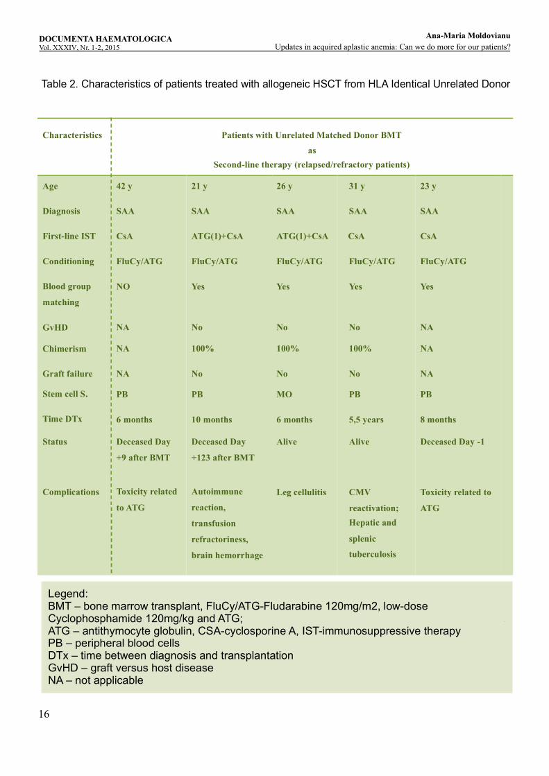
stem cell transplant from unrelated HLA compatible donor. The characteristics of the patients are presented in Table 2. Characteristics of patients treated with allogeneic HSCT from HLA Identical Unrelated Donor. All unrelated transplant cases were with 10/10 allele matched donor. In all the cases unrelated transplantation was done as secondary therapy after failing immunosuppressive therapy (IST) consisting of combination with ATG and CsA in the case of two patients and respectively monotherapy with CsA in three cases. The median age at the time of BMT was 26 years (21-42 years). The median time between diagnosis and BMT was 10 months (5 months-66 months). Conditioning regimen used included fludarabine 30 mg/m2 x 4, low dose cyclophosphamide 30 mg/kg x 4 and ATG 10 mg/kg x 4. The source of stem cells was bone marrow in one case and the peripheral blood stem cells were used in the rest of the cases. Three patients died, one on day -1, one on day +9 and one at four months after BMT. The probable causes of death were toxicity related to conditioning regimen, especially to ATG, in two of the cases and an autoimmune reaction occurred after approximately four months postt-ranplantation causing transfusion refractoriness and severe bleeding in the case of one patient. There was no significant acute or chronic GvHD and no graft rejection, all the patients having stable complete donor chimerism. The GvHD prophylaxis was cyclosporine and mini dose methotrexate. The complications after BMT developed by our patients included opportunistic infections like CMV reactivation in two cases and hepato-splenic tuberculosis in one patient. With adequate treatment these complications were manageable and patients were cured.
DiscussionStandard indication for unrelated s tem cell transplantation is as secondary therapy in the case of severe AA or very severe AA and age ≤ 40 years after the failure of at least one cycle of immunosuppressive combination therapy with ATG and CsA and absence of suitable sibling donor (Figure 3. The therapeutic algorithm for adult patients with Aplastic Anemia). Unrelated matched donor stem cell transplant may be taken into discussion in patients ˃ 40 years of age and with a good performance status if other treatments failed. As primary therapy, at present, there is no consensus. In young patients with very severe AA, unrelated BMT may be an option if a donor with a 10/10 match is available. As in the case of sibling donor BMT, the patient age and performance status and the severity degree of the disease come into question for an unrelated transplantation. The search for an unrelated donor
shouldbe initiated at an early stage. Suitable donor in AA is considered only when there is a high resolution HLA match 10/10. There are no recommendations about when to accept mismatched unrelated donors. More than 70% of patients will do not have an HLA-matched sibling donor. Alternative potential donors include relatives who are phenotipically matched or partially matched and HLA phenotipically matched but unrelated volunteers. Although phenotipically identical family donors are occasionally available, mismatched family members and matched but unrelated donors represent a much larger pool. Unfortunately, unrelated donors and mismatched transplants have almost twice the transplant-related mortality and risk of GvHD as matched sibling donor transplants. Also, graft rejection is a big obstacle for unrelated transplants. Historically, in the large European experience, for phenotipically identical family matches, the actuarial survival rate was 45%, for patients with a single-locus mismatch, it was 25%, and for those with two to three loci mismatched, the survival rate was 11%. According to an early EBMT report, the best results with unrelated or mismatched transplantation are seen in patients under the age of 21 years with disease duration of less than one year. Also, the International Bone Marrow Transplant Registry reported on a group of 318 alternative donor transplants in patients with SAA between 1988 and 1998, a 20% probability of graft failure and a survival probability at 5 years less than 40%. Most patients were young, heavily transfused and of poor performance status. The Fred Hutchinson Cancer Research Center (Seattle, WA, USA) reported on the results of unrelated allogeneic BMT in SAA after conditioning with low-dose TBI, high-dose cyclophosphamide and ATG. The median age was 19 years, and with a median follow-up of 7 years, 61% of HLA-identical and 40% of HLA mismatched transplant recipients survived the procedure; however, more than 70% of patients acquired acute GvHD and over 50% developed chronic GvHD. A recent meta-analysis of 18 heterogeneous trials evaluating the outcomes for patients who received unrelated donor transplants after failure to respond to IST, suggests that a good performance status and detailed HLA-matching contribute to improved survival. A review of data from the EBMT analyzed 498 patients transplanted during 1990–2005. Only the year of BMT was associated with increased survival. Survival at 5 years increased from 38% before 1998 to 57% after 1998. Also after 1998, there was less graft failure, less acute and chronic GvHD. The authors
DOCUMENTA HAEMATOLOGICAVol. XXXI , Nr. - , 20V 1 2 51 Updates in acquired aplastic anemia: Can we do more for our patients?
Ana-Maria Moldovianu
16
Characteristics
Patients with Unrelated Matched Donor BMT
as Second-line therapy (relapsed/refractory patients)
Age
Diagnosis
First-line IST
Conditioning
Blood group
matching
GvHD
Chimerism
Graft failure
Stem cell S.
Time DTx
Status
Complications
42 y
SAA
CsA
FluCy/ATG
NO
NA
NA
NA
PB
6 months
Deceased Day
+9 after BMT
Toxicity related
to ATG
21 y
SAA
ATG(1)+CsA
FluCy/ATG
Yes
No
100%
No
PB
10 months
Deceased Day
+123 after BMT
Autoimmune
reaction,
transfusion
refractoriness,
brain hemorrhage
26 y
SAA
ATG(1)+CsA
FluCy/ATG
Yes
No
100%
No
MO
6 months
Alive
Leg cellulitis
31 y
SAA
CsA
FluCy/ATG
Yes
No
100%
No
PB
5,5 years
Alive
CMV
reactivation;Hepatic and
splenic
tuberculosis
23 y
SAA
CsA
FluCy/ATG
Yes
NA
NA
NA
PB
8 months
Deceased Day -1
Toxicity related to
ATG
Legend:BMT – bone marrow transplant, FluCy/ATG-Fludarabine 120mg/m2, low-dose Cyclophosphamide 120mg/kg and ATG;ATG – antithymocyte globulin, CSA-cyclosporine A, IST-immunosuppressive therapyPB – peripheral blood cellsDTx – time between diagnosis and transplantationGvHD – graft versus host diseaseNA – not applicable
Table 2. Characteristics of patients treated with allogeneic HSCT from HLA Identical Unrelated Donor
DOCUMENTA HAEMATOLOGICAVol. XXXI , Nr. - , 20V 1 2 51 Updates in acquired aplastic anemia: Can we do more for our patients?
Ana-Maria Moldovianu
17
suggest that these improvements in outcomes are due to better donor matching. The outcome of unrelated donor transplants for SAA patients has improved not only due to better selection of HLA matched donors but also to significant changes in the conditioning regimen. A recent analysis from the EBMT-SAA working party retrospectively reviewed the outcome of 100 patients treated with alternative donor transplant as secondary therapy after the failure of IST. All patients received conditioning with a combination of fludarabine 30 mg/m2x4, cyclophosphamide 300 mg/m2x4 and ATG (FCA) with or without low dose (2 Gy) TBI. The actuarial 5-year survival was 73% for the group that received FCA and 79% for the group given the conditioning regimen including TBI. The most significant predictor of survival was the interval between diagnosis and transplantation, with 5-year survival rates of 87 and 55% for patients grafted within 2 years of diagnosis and more than 2 years after diagnosis, respectively. The overall cumulative incidence of acute GvHD grades II–IV and III–IV was 18% and 7%, respectively, with no difference between the two regimens. Chronic GvHD was recorded in 27% of the FCA group and 50% of the FCA-TBI group. This study confirmed that survival of patients with SAA treated with unrelated donor transplant has almost doubled in the past decade. Even more than in standard sibling transplants, age is a crucial risk factor in unrelated transplants and probably more important than the level of match, conditioning regimen, or use of T-cell depletion. EBMT recommends that treatment with alternative donor transplant should be adapted. In children and young adults (between 20 and 30 years) without a matched sibling donor, an unrelated donor should be started at diagnosis and transplantation should be considered after one course of IST had failed, in the presence of a suitable donor (10/10 matched donor). At the best centers, survival rates now are almost as good as with sibling donors. Adults do less well, primarily because of transplant-related mortality from the intensive conditioning regimen and so for adults over the age of 30 there are no clear indications and enrollment in a prospective trial is advised. Alternative donor transplant may be on option for second line treatment after failure of one or two courses of IST for this patient category depending on patient performance status and comorbidities and disease severity. Current protocols within EBMT include two dose-reduced conditioning regimens which are modified relative to sibling donor transplantations: a radiation free regimen with a combination of low-dose cyclophosphamide 300 mg/m2x4, fludarabine 30 mg/m2x4 and ATG x4 days (FCA) or alemtuzumab for patients under the age of 14 years and an adjusted
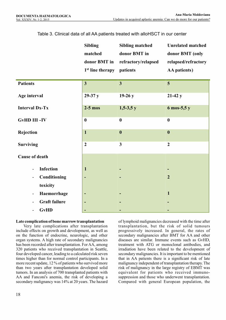
regimen with addition of 2 Gy total body irradiation (TBI) to Flu-Cy and administration of half of the dose of ATG (2 days instead of 4 days) in patients above the age of 14 years up to the age of 55 years. The reason to add TBI in adults was based on a high rejection rate with FCA in patients over the age of 14. An alternative approach consists in conventional cyclophosphamide conditioning Cy200 mg/kg with ATG and low-dosed TBI (2Gy) or the combination of reduced dose of cyclophosphamide Cy120 mg/kg and 8 Gy TBI. Donor selection includes suitable donors in the following order: 1. 10/10 allele matched unrelated donor (MUD), 2. One antigen mismatched UD, 3. matched or minimally mismatched cord blood or haploidentical donor. Graft rejection and GvHD are the major complications of allogeneic transplantation in AA. Graft rejection is a major predictor of posttrans-plantation survival. The rate of graft rejection decreased with intensification of the immunosuppressive conditioning regimen from 15% to 4% in Europe and from 35% to between 10% in Seattle and has remained stable in the last decade. Graft rejection can be caused by the pathophysiology of AA, a finding supported by the unexpectedly high proportion of failure in unprepared patients receiving syngeneic transplants and even in adequately preconditioned patients receiving syngeneic transplants. In a group of untransfused patients who received allogeneic stem cells, the incidence of graft rejection was 10%, indicating that AA patients may be particularly sensitive to alloimmunization. -Nevertheless, the influence of the number of transfusions on graft rejection is relative, and modest number of blood donations (40 units in the International Bone Marrow Transplant Registry experience and less than 10 units of erythrocytes or 40 units of platelets in Seattle) did not greatly increase the risk of graft rejection. Despite progress, matched but unrelated transplantation in AA patients is associated with a high mortality rate as it was resulted also from our experience in our center. More than half of our patients grafted from unrelated matched donor (three of five patients) died (Table 3. Clinical data of all AA patients treated with allo HSCT in our center). Refractory and high-risk patients are selected for this procedure and it is likely the poor results may be a consequence of this. However, alternative donor transplant is feasible. Alternative donor transplantation represents an option, especially for the young patient with very severe pancytopenia in whom immunosuppressive therapy has failed.
DOCUMENTA HAEMATOLOGICAVol. XXXI , Nr. - , 20V 1 2 51 Updates in acquired aplastic anemia: Can we do more for our patients?
Ana-Maria Moldovianu
18
Table 3. Clinical data of all AA patients treated with alloHSCT in our center
Sibling
matched
donor BMT in
1st line therapy
Sibling matched
donor BMT in
refractory/relapsed
patients
Unrelated matched
donor BMT (only
relapsed/refractory
AA patients)
Patients 3 3 5
Age interval 29-37 y 19-26 y 21-42 y
Interval Dx-Tx 2-5 mos 1,5-3,5 y 6 mos-5,5 y
GvHD III -IV
0
0
0
Rejection
1
0
0
Surviving
2
3
2
Cause of death
-
Infection
-
Conditioning
toxicity
-
Haemorrhage
-
Graft failure
-
GvHD
1
-
-
-
-
-
-
-
-
-
-
2
1
-
-
Late complication of bone marrow transplantation Very late complications after transplantation include effects on growth and development, as well as on the function of endocrine, neurologic, and other organ systems. A high rate of secondary malignancies has been recorded after transplantation. For AA, among 320 patients who received transplantation in Seattle, four developed cancer, leading to a calculated risk seven times higher than for normal control participants. In a more recent update, 12 % of patients who survived more than two years after transplantation developed solid tumors. In an analysis of 700 transplanted patients with AA and Fanconi's anemia, the risk of developing a secondary malignancy was 14% at 20 years. The hazard
of lymphoid malignancies decreased with the time after transplantation, but the risk of solid tumours progressively increased. In general, the rates of secondary malignancies after BMT for AA and other diseases are similar. Immune events such as GvHD, treatment with ATG or monoclonal antibodies, and irradiation have been related to the development of secondary malignancies. It is important to be mentioned that in AA patients there is a significant risk of late malignancy independent of transplantation therapy. The risk of malignancy in the large registry of EBMT was equivalent for patients who received immuno-suppression and those who underwent transplantation. Compared with general European population, the
DOCUMENTA HAEMATOLOGICAVol. XXXI , Nr. - , 20V 1 2 51 Updates in acquired aplastic anemia: Can we do more for our patients?
Ana-Maria Moldovianu
19
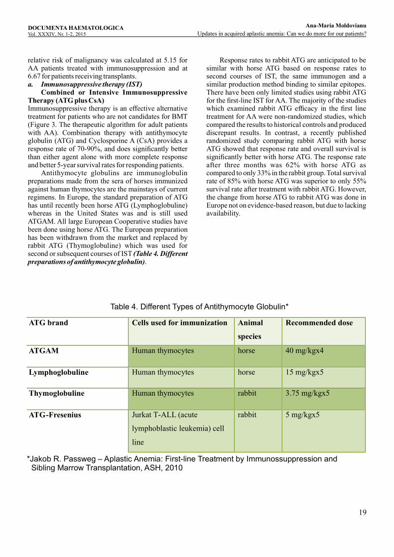
relative risk of malignancy was calculated at 5.15 for AA patients treated with immunosuppression and at 6.67 for patients receiving transplants. a. Immunosuppressive therapy (IST) Combined or Intensive Immunosuppressive Therapy (ATG plus CsA)Immunosuppressive therapy is an effective alternative treatment for patients who are not candidates for BMT (Figure 3. The therapeutic algorithm for adult patients with AA). Combination therapy with antithymocyte globulin (ATG) and Cyclosporine A (CsA) provides a response rate of 70-90%, and does significantly better than either agent alone with more complete response and better 5-year survival rates for responding patients. Antithymocyte globulins are immunoglobulin preparations made from the sera of horses immunized against human thymocytes are the mainstays of current regimens. In Europe, the standard preparation of ATG has until recently been horse ATG (Lymphoglobuline) whereas in the United States was and is still used ATGAM. All large European Cooperative studies have been done using horse ATG. The European preparation has been withdrawn from the market and replaced by rabbit ATG (Thymoglobuline) which was used for second or subsequent courses of IST (Table 4. Different preparations of antithymocyte globulin).
Response rates to rabbit ATG are anticipated to be similar with horse ATG based on response rates to second courses of IST, the same immunogen and a similar production method binding to similar epitopes. There have been only limited studies using rabbit ATG for the first-line IST for AA. The majority of the studies which examined rabbit ATG efficacy in the first line treatment for AA were non-randomized studies, which compared the results to historical controls and produced discrepant results. In contrast, a recently published randomized study comparing rabbit ATG with horse ATG showed that response rate and overall survival is significantly better with horse ATG. The response rate after three months was 62% with horse ATG as compared to only 33% in the rabbit group. Total survival rate of 85% with horse ATG was superior to only 55% survival rate after treatment with rabbit ATG. However, the change from horse ATG to rabbit ATG was done in Europe not on evidence-based reason, but due to lacking availability.
ATG brand Cells used for immunization Animal
species
Recommended dose
ATGAM Human thymocytes horse 40 mg/kgx4
Lymphoglobuline Human thymocytes horse 15 mg/kgx5
Thymoglobuline Human thymocytes rabbit 3.75 mg/kgx5
ATG-Fresenius Jurkat T-ALL (acute
lymphoblastic leukemia) cell
line
rabbit 5 mg/kgx5
Table 4. Different Types of Antithymocyte Globulin*
*Jakob R. Passweg – Aplastic Anemia: First-line Treatment by Immunossuppression and Sibling Marrow Transplantation, ASH, 2010
DOCUMENTA HAEMATOLOGICAVol. XXXI , Nr. - , 20V 1 2 51 Updates in acquired aplastic anemia: Can we do more for our patients?
Ana-Maria Moldovianu
20
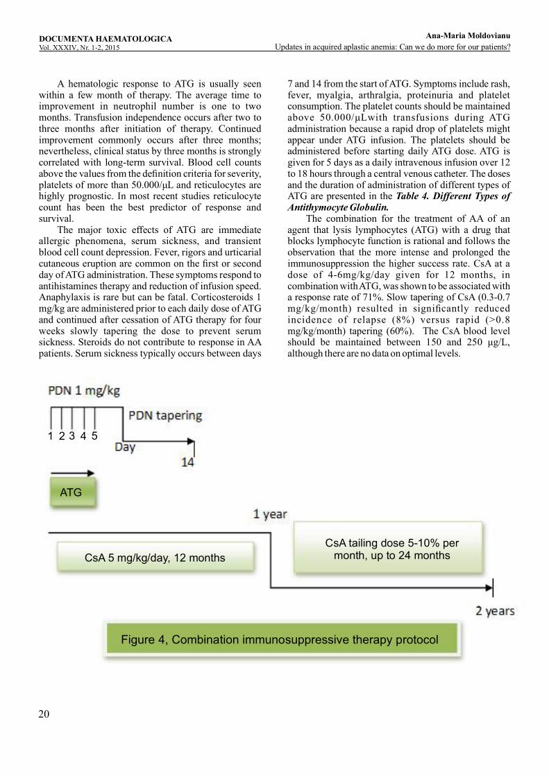
A hematologic response to ATG is usually seen within a few month of therapy. The average time to improvement in neutrophil number is one to two months. Transfusion independence occurs after two to three months after initiation of therapy. Continued improvement commonly occurs after three months; nevertheless, clinical status by three months is strongly correlated with long-term survival. Blood cell counts above the values from the definition criteria for severity, platelets of more than 50.000/ L and reticulocytes are μhighly prognostic. In most recent studies reticulocyte count has been the best predictor of response and survival. The major toxic effects of ATG are immediate allergic phenomena, serum sickness, and transient blood cell count depression. Fever, rigors and urticarial cutaneous eruption are common on the first or second day of ATG administration. These symptoms respond to antihistamines therapy and reduction of infusion speed. Anaphylaxis is rare but can be fatal. Corticosteroids 1 mg/kg are administered prior to each daily dose of ATG and continued after cessation of ATG therapy for four weeks slowly tapering the dose to prevent serum sickness. Steroids do not contribute to response in AA patients. Serum sickness typically occurs between days
7 and 14 from the start of ATG. Symptoms include rash, fever, myalgia, arthralgia, proteinuria and platelet consumption. The platelet counts should be maintained above 50.000/μLwith transfusions during ATG administration because a rapid drop of platelets might appear under ATG infusion. The platelets should be administered before starting daily ATG dose. ATG is given for 5 days as a daily intravenous infusion over 12 to 18 hours through a central venous catheter. The doses and the duration of administration of different types of ATG are presented in the Table 4. Different Types of Antithymocyte Globulin. The combination for the treatment of AA of an agent that lysis lymphocytes (ATG) with a drug that blocks lymphocyte function is rational and follows the observation that the more intense and prolonged the immunosuppression the higher success rate. CsA at a dose of 4-6mg/kg/day given for 12 months, in combination with ATG, was shown to be associated with a response rate of 71%. Slow tapering of CsA (0.3-0.7 mg/kg/month) resulted in significantly reduced incidence of relapse (8%) versus rapid (>0.8 mg/kg/month) tapering (60%). The CsA blood level should be maintained between 150 and 250 μg/L, although there are no data on optimal levels.
1 2 3 4 5
ATG
CsA tailing dose 5-10% permonth, up to 24 monthsCsA 5 mg/kg/day, 12 months
Figure 4, Combination immunosuppressive therapy protocol
DOCUMENTA HAEMATOLOGICAVol. XXXI , Nr. - , 20V 1 2 51 Updates in acquired aplastic anemia: Can we do more for our patients?
Ana-Maria Moldovianu
21
-Cyclosporine has considerable toxicity. Hypertension and renal toxicity are the most common serious side effects. Hirsutism and gingival hypertrophy are also frequent side effects. Increasing serum creatinine levels are an indication for dose reduction. The risk of nephropathy is increased by high doses and longer duration of therapy and occurs more often in older patients. Convulsions, possibly related to hypo-magnesemia, are another serious complication of CsA therapy. Infections are the principal cause of death during combination IST and having adverse effect on survival. In order to reduce infection related mortality, granulocytes stimulating growth factor (G-CSF) has been added to ATG and CsA. The current EBMT protocol recommends to use G-CSF only “on demand” during infectious episodes in neutropenic patients since there are some data reporting increased risk for clonal diseases after IST containing G-CSF. In disease refractory to initial therapy with ATG and CsA, a repeat course as salvage regimen can produce a response rate of 30%. Other immunosuppressantsBecause half of patients with AA experience failure to IST, there have been many efforts to improve its efficacy by adding other agents. However, prospective studies conducted by the NIH group failed to show that the addition of mycophenolatemofetil or sirolimus to horse ATG/CsA results in improved overall response rate and reduced relapse rate. Other immunosuppressants are used by some centers. High dose cyclophosphamide 45-50 mg/kg/day for 4 days without stem cell rescue is used by Johns Hopkins Group as treatment for patients with newly diagnosed AA with overall response rate claimed similar to that achieved with ATG + CsA. A prospective randomized study comparing this with ATG and CsA was terminated early because of excess deaths and fungal infections in the cyclophosphamide arm. Therefore, high dose cyclophosphamide without stem cell support is reserved for second or third line therapy. Mycophenolatemofetil has been used in organ transplantation and autoimmune disorders. There are data showing that addition of this drug to ATG and CsA did not improve response nor reduce the relapse rate. Alemtuzumab treatment has been retrospectively investigated in a limited cohort of 35 heterogenous patients with a response rate of 60%. Immunosuppressive therapy in older patients For older patients, consideration for treatment should be preceded by medical assessment to exclude significant co-morbidities and requires discussion of the risk with the patient. Although there is no upper age limit for ATG administration, CsA alone may be considered for patients older than 60 years. The current EBMT
treatment algorithm proposes to treat hospitalized patients who are severely ill by ATG and CsA if considered to be tolerable considering the co-morbidity profile, whereas to start newly diagnosed patients who are well and in an outpatient setting with CsA alone. The outcomes for tolerability and toxicity, response and relapse rates were examined in 24 older patients (over 60 years of age) receiving IST. Seven patients received standard IST consisting of standard-dose ATG with or without CsA, and 17 patients received attenuated IST consisting of at least a 50% dose reduction of ATG with CsA or CsA alone. Six patients (25%) had early deaths, mostly due to infection. Early mortality appeared higher in the standard IST group, although this was not statistically significant (43 vs 18%; p = 0.4). The 2-year cumulative incidence of response was 42% (95% CI: 26–69%). Responders had significantly better survival than non-responders (p = 0.0002). The 3-year probability of OS was 49% (95% CI: 27–68%). Nine out of 14 evaluable patients in the attenuated IST group had durable responses to treatment. These data from this small cohort suggest that attenuated dose IST could be a reasonable treatment option for patients deemed unfit for standard-dose IST. Late Complication of Immunosuppressive TherapyRelapse after IST is common. About 30% or more of the patients that responded require reinstitution of IST. Most relapse respond to retreatment, and there is no clear relationship with worse survival. Late complication of IST therapy with great impact on survival is the development of clonal hematologic disorders, especially myelodysplasia and acute myeloid leukemia. In the National Institutes of Health trials, the overall rate of clonal evolution is 12% to 15% at about one decade. Over time, a minority of patients may develop a large clone of PNH and the haemolytic and thrombotic manifestations of PNH. Immunosuppressive therapy for non-severe AA There is only one prospective randomized trial of CsA alone or the combination of ATG/CsA treatments in patients with non-severe or moderate AA which showed that the outcomes of two groups of patients were comparable, with a 93% survival probability in the CsA group and 91% in the combination therapy group of patients. Immunosuppressive therapy versus sibling matched donor BMT as frontline treatment A recent analysis of outcomes from the EBMT comprising 2479 patients with SAA compared the survival after frontline treatment with IST versus BMT. Theresults showed a survival rate at 10 years of 73% in BMT recipients and 68% in those treated with IST (p = 0.002). It is important to mention that the frequency of
DOCUMENTA HAEMATOLOGICAVol. XXXI , Nr. - , 20V 1 2 51 Updates in acquired aplastic anemia: Can we do more for our patients?
Ana-Maria Moldovianu
22
secondary malignancy was much higher in the patients who received IST alone (1.2%) compared with those who received BMT (0.1%). d. Supportive care in Aplastic Anemia Supportive management for AA patients include all the measures necessary to prevent the occurrence of the redoubtable complications of pancytopenia like bleeding, infections and iron overload. The overall survival has greatly improved for AA patients in the last 30 years due to prophylaxis and treatment of infections, bleeding prophylaxis, restrictive transfusion strategy, and iron chelation. The management of AA patients should be made by a multidisciplinary team and in a specialized center, especially the patients with severe neutropenia who are at risk for severe infections. Bleeding Platelet transfusions have greatly improved survival in patients with aplastic anemia and severe thrombocytopenia. Modern transfusion practice has made platelets safe to administer. The major problem related to platelet support is the development of alloimmunization in the recipient. Host antibodies directed against transfused platelet in the circulation shorten the transfused platelet life span and are almost always directed to HLA-A and HLA-B antigens. Alloimmunization is suggested by poor recovery of the 1-hour posttransfusion platelet count and confirmed by detection of specific HLA antibodies in serum. If HLA-antibodies are detected, HLA-matched platelets should be used for further transfusions. Alloimmunization can be prevented or delayed by the use of single-donor platelets rather than pooled platelets and by physical leukocyte depletion by filtration or ultraviolet treatment of blood products. Avoidance of platelet transfusions except when there is active bleeding is another alternative to prevent alloimmunization. The dose relationship between exposure to different donors' platelets and the probabil i ty of developing refractoriness is not clearly established. The risk for alloimmunization is thought to increase after more than 40 units of platelets have been administered.Prophylactic transfusion of platelets is recommended in case of platelets < 10 x 109/L without fever, bleeding signs or history of major bleeding events. AA patients with fever, or bleeding signs or history of relevant bleeding like cerebral hemorrhage should receive prophylactic platelet transfusions in case of platelet counts < 20 x 109/L. Bleeding prophylaxis must be individualized for each patient, but in general, a goal of maintaining platelet counts higher than 10 x 109/L is reasonable. For invasive procedures platelet transfusions must be given to achieve the recommended levels. In case of patients receiving treatment with ATG the platelet counts should be increased to 50 x 109/L prior to the onset of ATG infusions, as rapid drop of
platelets might ensue under ATG infusion. Prophylactic transfusions of platelets have not been shown to alter survival and their use should not be withheld if indicated just for fear of alloimmunization or increased risk of graft rejection after allogeneic stem cell transplantation. The beneficial effects of platelet transfusions meaning avoidance of bleeding complications and improvement of quality of life justify their use. Prevention of bleeding complications may include some general measures like hormone therapy for women to control menorrhagia, avoidance of aspirin or other platelet aggregation inhibitor, and the use of tranexamic acid. Anemia The indication for erythrocyte concentrates (RBC) transfusions is hypoxic anemia. Symptomatic anemia develops at different haemoglobin levels depending on physical state and co-morbidities of the patient. Therefore the decision to transfuse RBCs depends on clinical symptoms, haemoglobin level and quality of life. Frequent RBCs transfusions might result in the alloimmunization against erythrocyte antigens and iron overload. Iron chelation should be used in patients who are unresponsive to immunosuppressive therapy, necessitate transfusions over a long period of life and have a reasonable expectation of survival. Alloimmunization secondary to transfusions increases the risk for graft rejection and mortality after BMT. Blood products from a potential bone marrow donor (sibling or parent) should be avoided. The transfusion of leukocyte-depleted blood products is mandatory in AA patients. The 5% risk of graft rejection after transplantation in untransfused patients was increased to 15 % with receipt of 1 to 40 units and to more than 25% in more heavily transfused patients. These data are from a period before leukocyte depleted blood products were routinely used and it is likely that graft rejection would be lower with the reduced patient alloimmunization by using leukocyte-depleted blood products. Further studies are needed to confirm this. The irradiation of blood products for aplastic anemia patients is recommended during and after treatment with ATG or alemtuzumab as long as patients are immunosuppressed with a reduced CD4/CD8 ratio or a minimum of 6 months after IST and in case of patients receiving allogeneic BMT. The main reasons for using irradiate blood products are to prevent allosensitization and transfusion-associated GvHD. Also, granulocyte and HLA-matched platelets concentrates must be irradiated. CMV-negative blood products are given in case of patients undergoing allogeneic SCT where both the patient and donor are CMV negative. In general, there is no need for CMV-negative blood products when
DOCUMENTA HAEMATOLOGICAVol. XXXI , Nr. - , 20V 1 2 51 Updates in acquired aplastic anemia: Can we do more for our patients?
Ana-Maria Moldovianu
23
leukodepletion is applied.Granulocyte concentrates are used as a temporary measure and are reserved for the cases of life-threatening infections and severe neutropenia. Infections There is very little data regarding infections prophylaxis and treatment in AA. The recommendations are based on reports of infections in neutropenic patients with malignant diseases and chemotherapy. The risk of infection is determined by the degree and duration of neutropenia. Susceptibility to infection is extremely high with an absolute neutrophil count (ANC) of less than 200 cells/µL. With longer periods of neutropenia, the probability of serious bacterial or fungal infections increases. Therefore, almost all severe AA patients unresponsive to IST are at high risk. Recommendations for initiation of empiric antibiotic therapy are similar in AA patients and other patients with neutropenia. A basic rule is, if ANC is less than 500 cells/µL and infection is suspected, immediate hospitalisation and broad-spectrum intravenous antibiotics should be institute. After the isolation of infectious agents in cultures or if new signs or symptoms of localized infection are identified the antibiotic regimen can be modified. Local hospital guidelines for treatment of febrile neutropenia should be followed. Usually, a combination of broad-spectrum beta lactam antibiotic and an aminoglycoside is the first choice. The patient's infection history and recent medication should also be taken into consideration. If the fever persists despite adequate antibiotic therapy or reappears, antifungal therapy should be given. Earlier addition of antifungals in AA patients is advisable if there are findings on chest tomography and a positive test result for galactomannan antigen. Candida and Aspergillus spp. are the most common agents incriminated for fungal infections in AA patients. The newer antifungal agents such as voriconazol or caspofungin, as studies have demonstrated, have the same or superior efficacy and less toxicity compared to amphotericin for the treatment of persistent fever in neutropenic patients and established Candida and Aspergillus infection. If a viral infection is suspected, an antiviral agent should be included in the treatment. The use of G-CSF is recommended to be given for the treatment of infectious complications in AA patients. Because there are no guidelines and some studies have shown that prophylactic growth factors do not improve overall outcome, their use as infections prophylaxis is not advised. Hospitalised patients with AA and severe neutropenia should ideally be cared for in isolation, in rooms with air filtration and should receive prophylactic antibiotics and antifungals. Prevention of bacterial infections in severe
neutropenic AA patients should be realised with a quinolone antibiotic or a combination of non-absorbable antibiotics such as neomycin and colistin depending on the local microbiological flora and rates of resistance. Antifungal prophylaxis should be done in all patients with severe AA and prolonged neutropenia. Itraconazole, voriconazole or posaconazole appear to be more effective than fluconazole, as their activity against Aspergillus whereas fluconazole does not. Antiviral prophylaxis and Pneumocystis jiroveci pneumonia (PJP) prophylaxis are not standardized. In case of allogeneic stem cell transplantation acyclovir and an anti pneumocystis drug are routinely given. For –patients treated with ATG the usage of acyclovir and PJP-prophylaxis is also a rule in many centers. Iron chelation It is now known that iron overload has a negative influence on the outcome of patients undergoing stem cell transplantation as well as a potential worsened effect on bone marrow function. Therefore serum ferritin level above 1000 µg/L requires starting iron chelation therapy. On the other hand due to potential side effects of chelate therapy a risk-benefit analysis should be assessed for individual patients. When remission is achieved an iron overload can be treated with phlebotomy. Psychological support The diagnosis of Aplastic Anemia has an important psychological impact on the patients, most of them being at a young age with life expectancies. It is a serious condition with potential fatal complications, a chronic disease and a life changing experience. The patient and its family must be informed about the nature of the disease, treatment, prognostic and social impact. Some patients may need professional psychological support. Androgens Testosterone and synthetic anabolic steroids were introduced in the 1960s and at that moment appeared to have major benefits in the treatment of AA. The high rates of response in early reports may be retrospectively attributed to the inclusion of patients with moderate acquired AA and constitutional AA. For severe AA, controlled trials have not demonstrated any benefits regarding survival rates or hematologic improvement. When added to immunosuppressive therapy androgens have failed to show an increase in response rates. Androgens were preponderantly used and still are for second-line treatment of AA patients, but there had been a limitation of their use due to side effects such as hirsutism or hepatic toxicity and also due to restrictions of their availability and manufacture. Occasionally, there may be observed a hematological improvement with a course of androgen therapy, may be more
DOCUMENTA HAEMATOLOGICAVol. XXXI , Nr. - , 20V 1 2 51 Updates in acquired aplastic anemia: Can we do more for our patients?
Ana-Maria Moldovianu

24
effective if combined with CsA. 8. Prognosis In the modern era, indicators for prognosis, besides the classic initial blood counts of a patient with AA, are considered to be absolute reticulocyte count and absolute lymphocytes predicting a good response to immunosuppression and survival. The sturdiness of the platelet and reticulocyte response after immuno-suppressive therapy also correlates with long-term survival. Clonal evolution is a poor prognostic factor, especially the acquisition of monosomy 7. The rate of spontaneous recovery is low, untreated severe AA being invariably fatal. Moderate AA, in contrast with the severe form, has a good prognosis. Some patients may experience a hematologic recovery with no treatment. 9. Conclusions The outcome of patients diagnosed with acquired Aplastic anemia has greatly improved over time. Age remains a major predictor of outcome for both BMT and immunosuppressive treatment. Early intervention is associated with a significantly superior outcome. Improved survival is seen both in patients grafted from a sibling matched donor, and those with a unrelated matched and alternative donor, suggesting that better supportive care and improved management of infections have an major impact on outcome.
Conflict of interest: Authors state no conflict of interest
References1. Passweg J, et al. The complete treatment algorithm for SAA, EBMT, 2012, 1-41.2. Sureda A, et al. Indications for allo- and auto-SCT for haematological diseases, solid tumours and immune disorders: current practice in Europe, 2015, Bone Marrow Transplantation (2015), 1-20.3. Schrezenmeier et al. Aplastic Anemia- Diagnostics and Therapy of Acquired Aplastic Anemia, 2012, 1-154. Passweg JR, et al. Aplastic Anemia: First-line Treatment by Immunosuppression and Sibling Marrow Transplantation, ASH, 2010.5. Guinan E. Diagnosis and Management of Aplastic Anemia, ASH, 2011.6. DeZern AE, Brodsky R. Clinical management of aplastic anemia, Expert Rev Hematol. 2011 Apr; 4(2): 221–230.7. Locasciulli A, Oneto R, Bacigalupo A, et al. Outcome of patients with acquired aplastic anemia given first line bone marrow transplantation or immunosuppressive treatment in the last decade: a report from the European Group for Blood and Marrow Tr a n s p l a n t a t i o n ( E B M T ) H a e m a t o l o g i c a . 2007;92(1):11–18. Guidance for immunosuppressive therapy or bone marrow transplantation as first-line
therapy.8. Lawler M, McCann SR, Marsh JC, et al. Serial chimerism analyses indicate that mixed haemopoietic chimerism influences the probability of graft rejection and disease recurrence following allogeneic stem cell transplantation (SCT) for severe aplastic anaemia (SAA): indication for routine assessment of chimerism post SCT for SAA. Br. J. Haematol. 2009; 144(6):933–945.9. Schrezenmeier H, Passweg JR, Marsh JC, et al. Worse outcome and more chronic GVHD with peripheral blood progenitor cells than bone marrow in HLA-matched sibling donor transplants for young patients with severe acquired aplastic anemia. Blood. 2007;110:1397–1400.10. Kahl C , Leisenr ing W, Deeg HJ, e t a l . Cyclophosphamide and anti-thymocyte globulin as a conditioning regimen for allogeneic marrow transplantation in patients with aplastic anaemia: a long-term follow-up. Br. J. Haematol. 2005;130(5):747–751. 11. Bacigalupo A. Bone marrow transplantation for severe aplastic anemia from HLA identical siblings. Haematologica. 1999;84(1):2–4. 12. Champlin RE, Perez WS, Passweg JR, et al. Bone marrow transplantation for severe aplastic anemia: a randomized controlled study of conditioning regimens. Blood. 2007;109(10):4582–4585. 13. Al-Zahrani H, Nassar A, Al-Mohareb F, et al. Fludarabine based conditioning chemotherapy for allogeneic hematopoietic stem cell transplantation in acquired severe aplastic anemia. Biol. Blood Marrow Transplant. 2010 DOI: 10.1016/j. bbmt.2010.08.013.14. Maury S, et al. Improving outcome of patients older than 30 years receiving HLAidentical sibling HSCT for severe acquired aplastic anemia using fludarabine-based conditioning: a comparison with conventional conditioning regimen. Haematologica. 2009.15. Srinivasan R, Takahashi Y, McCoy JP, et al. Overcoming graft rejection in heavily transfused and allo-immunised patients with bone marrow failure syndromes using fludarabine-based haematopoietic cell transplantation. Br. J. Haematol. 2006;133(3):305–314.16. Deeg HJ, O'Donnell M, Tolar J, Agarwal R, Harris RE, Feig SA, et al. Optimization of conditioning for marrow transplantation from unrelated donors for patients with aplastic anemia after failure of immunosuppressive therapy. Blood. 2006 Sep 1;108(5):1485-91.17. Maury S, Balere-Appert ML, Chir Z, Boiron JM, Galambrun C, Yakouben K, et al. Unrelated stem cell transplantation for severe acquired aplastic anemia: improved outcome in the era of high-resolution HLA matching between donor and recipient. Haematologica. 2007 May;92(5):589-96.18. Young N, Bacigalupo A, Marsh J. Aplastic Anemia:
DOCUMENTA HAEMATOLOGICAVol. XXXI , Nr. - , 20V 1 2 51 Updates in acquired aplastic anemia: Can we do more for our patients?
Ana-Maria Moldovianu
25
Pathophysiology and Treatment. Biol Blood Marrow Transplant. 2009 Sep 23.19. Viollier R, Passweg J, Gregor M, et al. Quality-adjusted survival analysis shows differences in outcome a f te r im mu no su pp res s i on or b on e mar ro w transplantation in aplastic anemia. Ann. Hematol. 2005;84(1):47–55.20. Siegal D, Xu W, Sutherland R J Kuruvilla, J H Lipton, M Minden, H Messner, V Gupta. Graft-versus-host disease following marrow transplantation for aplastic anemia: different impact of two GVHD prevention strategies. Bone Marrow Transplantation 2008; 42, 51 – 56.21. Locasciulli A, Oneto R, Bacigalupo A, et al. Outcome of patients with acquired aplastic anemia given first line bone marrow transplantation or immunosuppressive treatment in the last decade: a report from the European Group for Blood and Marrow Tr a n s p l a n t a t i o n ( E B M T ) H a e m a t o l o g i c a . 2007;92(1):11–18.22. Peinemann F, Grouven U, Kroger N, Pittler M, Zschorlich B, Lange S. Unrelated donor stem cell transplantation in acquired severe aplastic anemia: a s y s t e m a t i c r e v i e w . H a e m a t o l o g i c a . 2009;94(12):1732–1742. 23. Viollier R, Socie G, Tichelli A, et al. Recent improvement in outcome of unrelated donor transplantation for aplastic anemia. Bone Marrow Transplant. 2008;41(1):45–50. 24. Bacigalupo A, Socie G, Lanino E, et al. Fludarabine, cyclophosphamide, anti-thymocyte globulin, with or without low dose total body irradiation, for alternative donor transplants, in acquired severe aplastic anemia: a retrospective study from the EBMT-SAA working par ty. Haematologica. 2010;95(6):976–982. 25. Gafter-Gvili A, Ram R, Gurion R, et al. ATG plus cyclosporine reduces all-cause mortality in patients with severe aplastic anemia–systematic review and meta-analysis. ActaHaematol. 2008;120(4):237–243. 26. Marsh J, Schrezenmeier H, Marin P, et al. Prospective randomized multicenter study comparing cyclosporin alone versus the combination of anti-thymocyte globulin and cyclosporin for treatment of patients with nonsevere aplastic anemia: a report from the European Blood and Marrow Transplant (EBMT) Severe Aplastic Anaemia Working Party. Blood. 1999;93(7):2191–2195.27. Scheinberg P, Nunez O, Wu C, Young NS. Treatment of severe aplastic anaemia with combined immunosuppression: anti-thymocyte globulin, ciclosporin and mycophenolatemofetil. Br. J. Haematol. 2006;133(6):606–61128. Kao SY, Xu W, Brandwein JM, et al. Outcomes of older patients (> or = 60 years) with acquired aplastic
anaemia treated with immunosuppressive therapy. Br. J. Haematol. 2008;143(5):738–743.29. Rosenfeld S, Follmann D, Nunez O, Young NS. Anti-thymocyte globulin and cyclosporine for severe aplastic anemia: association between hematologic r e s p o n s e a n d l o n g - t e rm o u t co m e . J AM A. 2003;289(9):1130–1135. Randomized trial showing anti-thymocyte globulin and cyclosporine as combination immunosuppressive therapy.30. Scheinberg P, Nunez O, Wu C, Young NS. Treatment of severe aplastic anaemia with combined immunosuppression: anti-thymocyte globulin, ciclosporin and mycophenolatemofetil. Br. J. Haematol. 2006;133(6):606–611.31. Gluckman E, Rokicka-Milewka R, Hann I, Nikiforakis E, Tavakoli F, Cohen-Scali S, Bacigalupo A. Results and follow-up of a phase III randomized study of recombinant humangranulocyte stimulating factor as support for immunosuppressive therapy in patients with s e v e r e a p l a s t i c a n a e m i a . B r J H ae m a t o l . 2002;119(4):1075-82.32. Ohara A, Kojima S, Hamajima N, et al . M y e l o d y s p l a s t i c s y n d r o m e a n d a c u t e myelogenousleukemia as a late clonal complication in children with acquired aplastic anemia. Blood. 1997;90(3):1009–1013.33. Brodsky RA, Chen AR, Dorr D, et al. High-dose cyclophosphamide for severe aplastic anemia: long-term follow-up. Blood. 2010;115(11):2136–2141.34. Gómez-Almaguer D, Jaime-Pérez JC, Garza-Rodríguez V, Chapa-Rodríguez A, Tarín-Arzaga L, Herrera-Garza JL, Ruiz-Argüelles GJ, López-Otero A, González-Llano O, Rodríguez-Romo L. (2009) Subcutaneous alemtuzumab plus cyclosporine for the treatment of aplastic anemia. Annals of Haematology, DOI 10.1007/s00277-009-0816-5.35. Risitano AM, et al. on behalf of the Working Party Severe Aplastic Anemia (WPSAA) of the European Group for Blood and Marrow Transplantation (EBMT). A l e m t u z u m a b I s S a f e A n d E f f e c t i v e A s Immunosuppressive Treatment For Aplastic Anemia And Single-Lineage Marrow Failure: A Pilot Study And A Survey From The EBMT WPSAA. British Journal of Haematology, 2010.
DOCUMENTA HAEMATOLOGICAVol. XXXI , Nr. - , 20V 1 2 51 Updates in acquired aplastic anemia: Can we do more for our patients?
Ana-Maria Moldovianu

26
27
Background: Even if the romanian population is ethnically compact caucasian-type population, many of the patients referred for bone marrow transplantation lack a suitable donor. In order to expand the donor pool and the accesibility to transplant procedure for those who have indications it is necessary to introduce the haplo-identical bone marrow transplant procedure with myeloablative and reduced-intensity conditioning in Fundeni Clinical Institute.
Introduction: Bone marrow transplantation is the only curative option for many adult hematological malignancies. Donor availability in a timely manner for those patients is one of the major challenge in the treatment. The likelihood of finding an HLA-matched sibling donor (MSD) for a patient is 25% as the mendelian rule shows, and the fact that in Romania we have more and more families with only one child, making the sibling-donor search more and more difficult. Since 2009 Romania
Haplo- dentical one arrow ransplant rotocol using reduced intensity I B M T Pconditioning for undeni linical nstituteF C I
Varady Zsofia , Coriu Daniel , Ghiaur Gabrie Richard J Jones .1 ,1,2 l 3 3l , .1.Bone Marrow Transplant Center, Fundeni Clinical Institute, Bucharest, Romania2. Carol Davila University, Bucharest, Romania3. Sidney Kimmel Comprehensive Cancer Center, Johns Hopkins University School of Medicine, Baltimore, Maryland, USA
Abstract: Purpose: Even if the romanian population is ethnically compact caucasian-type population, many of the patients referred for bone marrow transplantation lack a suitable donor. In order to expand the donor pool and the accesibility to transplant for those who have indications it is necessary to perform haplo-identical bone marrow transplant procedure in Fundeni Clinical Institute. Since 2009 Romania established a National Volunteer Stem Cell Donor Registry (RNDVCSH), the goal was to enlarge the possibility to find HLA-matched unrelated donors (MUD) for patients. This approach offered transplant for only up to 60%patients referred for transplantation in 2014, even we chose one HLA-mismatch donors. The haploidentical transplant protocol proposed for our institution is based on Sidney Kimmel Comprehensive Cancer Center protocol from Johns Hopkins University School of Medicine. The major milestones of this protocol include: patient eligibility, donor selection criterias, evaluation of the haplo donor, the conditioning regimen plan and additional supportive care, the bone marrow harvest, prophylaxis of graft versus host disease, assessment during and after the transplant. Donor must be HLA-haploidentical first-degree relatives of the patient with signed consent. The patient, parents and children are typed at the allelic level for HLA-A, -B,-C,-DRB1 and -DQB1. They will perform also de HLA-antibody search using cross-match test in complement-dependent cytotoxicity. The conditioning regimen is composed by Fludarabine 30 mg/m2/day (from day -6 to day -2) combined with Cyclophosphamide 14,5 mg/kgIBW/day (from day -6 to day-5) and TBI at 2 Gyîn day -1. In case of lacking TBI procedure at 2 Gy dose in day -1, it could be replaced by two dose of Busulfan iv in day -3 and day-2 (dose=3,2 mg/kgIBW/day) for those with acute and chronic leukemias. The donor will have general anesthesia,the target yield of marrow is 4 x 10^8 total nucleated cells/kg recipient using his IBW. The GVHD prophylaxis consisted of post-transplant Cyclophosphamide (PTCy) of 50mg/kgIBW/day in IV administration in day +3 and +4, followed by tacrolimus and mycophenolatemofetil (MMF) beginning day +5. The MMF will be stopped at day+35, the tacrolimus will continue till 6 months after the transplantation. Conclusion: One of the most important factors affecting transplantation outcome is proper timing.Therefore, donor availability is an crucial issue. Haploidentical related donors are available for almost all patients, so the use of those donors is a viable alternative.
*Coresponding author:VaradyZsofia, Bone Marrow Transplant Center, Fundeni Clinical Institute, Sos. Fundeni nr.258, sector 2, Bucharest, Romania, phone:+40723293853, e-mail: zsofi2005@yahoo.
D DE GRUYTEREOPEN
DOI: 10.1515/dch-2015-0002

28
established a National Volunteer Stem Cell Donor Registry (RNDVCSH) and the goal was to enlarge the possibility to find HLA-matched unrelated donors (MUD) for patients. This approach offered transplant for only up to 60%patients referred for transplantation in 2014, even we chose one HLA-mismatch donors. For these reasons it is important to introduce HLA-haploidentical transplantation. Potential HLA-haploidentical donors include biological parents or children of a patient, and each sibling has a 50% chance of sharing one HLA-haplotype. So it is a rapid possibility to find a donor and it will be available in very short time for the transplant. The major drawback is the intense bidirectional alloreactivity with graft failure and graft versus host disease (GVHD), who leads to increased non-relapse mortality (NRM). Initially in haplo-setting, knowing that T-cells are responsible in graft versus host disease (GVHD), studies were made removing T cells from the donor graft ex-vivo. But the graft failure increased. The post-transplant high-dose Cyclophosphamide (PTCy) it was the next step in order to deplete in-vivo alloreactive T cells from the donor and the host at the same time, and reduce both GVHD and rejection. This DNA-damaging agent take action over the proliferative, alloreactive T cells, but not over resting T cells and allow a better immune reconstitution and lower non-relapse mortality (NRM).Patient eligibility Patients over 18 years of age and under 65, with a diagnosis of hematological malignancy, having an indication for allogeneic transplant within the Fundeni Clinical Institute local practice. The eligibility should be assessed by the Fundeni Clinical Institute transplant committee after the hematology specialist fulfilled the transplant-specific medical chart of the patient. The patient must have one or more potential related mismatched donor (biological parents, siblings or children) typed in high-resolution for HLA-A, -B, -C, -DRB1 and -DPB1. The exclusion criterias are: a) hematological malignancy in evolution b) suitable matched sibling or unrelated donor, defined by local committee c) prior autologous stem cell transplantation, if the disease relapse under 6 months after autologous transplant. d) prior allogeneic stem cell transplant e) poor performance status: Karnofsky under 70% f) poor cardiac function: left ventricular ejection fraction under 45% g) poor pulmonary function: DLCO, FEV1 and FVC under 50% h) poor liver function: transaminase more than 3 time of normal level and bilirubin over 2,5 mg/dl
DOCUMENTA HAEMATOLOGICAVol. XXXI , Nr. - , 20V 1 2 51
Haplo- dentical one arrow ransplant I B M TP F C Irotocol using reduced intensity conditioning for undeni linical nstitute
Varady Zsofia
(except the Gilbert syndrome) i) poor renal function: elevated creatinine above 1,5 mg/dl. j) curent uncontrolled bacterial, viral or fungal infection k) anti-donor HLA-antibodies defined by positive crossmatch test of any titer rely on complement-dependent cytotoxicity l) evidence of HIV positive serology or infection l) pregnancy and breast-feeding. m) lack of a signed informed consent for the patient and for his donor. After the patient passed this exclusion criterias he will be screened for HbsAg; anti Hbcore; anti HIV 1,2; HIV1,2 NAT; HCV NAT; HBV NAT; anti-HTLV; anti-HCV; anti-CMV, anti-EBV; anti-toxoplasma and serology for syphilis.Donor selection criterias, in decreasing order of priority: 1. donor must be medically, socially and psychologically fit to donate 2. donor must be HLA-haploidentical first-degree relatives of the patient with signed consent. For donors under 18 years of age a local judge will take the consent, obeying Romanian rules and laws. 3. the maximum recipient actual body weight should not exceed 1,25 times the donor actual body weight. 4. the patient must lack antibodies against donor HLA molecules (specifically complement dependent cytotoxicity) 5. no major ABO incompatibility between donor and recipient ( include: recipient “O” with donor “A”, “B” or “AB”; recipient “A” with donor “B” or “AB”; recipient “B” with donor “A” or “AB”) 6. matched CMV IgG serologic status between donor and recipient (for a recipient who is CMV negative, use a CMV negative donor; for a positive recipient use a positive donor) . use an ABO compatible donor over a minor ABO incompatible donor. 8. other donor characteristics (in no order of priority): preferring donors above 18 years of age over donor under 18; among donors above 18, prefer younger and lighter donor; for male recipients male donors are preferred.Evaluation of the haplo-identical donor Haplo-identical bone marrow donor has to be extensively evaluated before donation in order to assess the potential risks to the donor and recipient. Donor eligibility must be documented prior to the start of the preparative regimen for the recipient in the recipient chart. This task will be completed by the transplant physician who will take care of the recipient, under the supervision of Fundeni Clinical Institute transplant
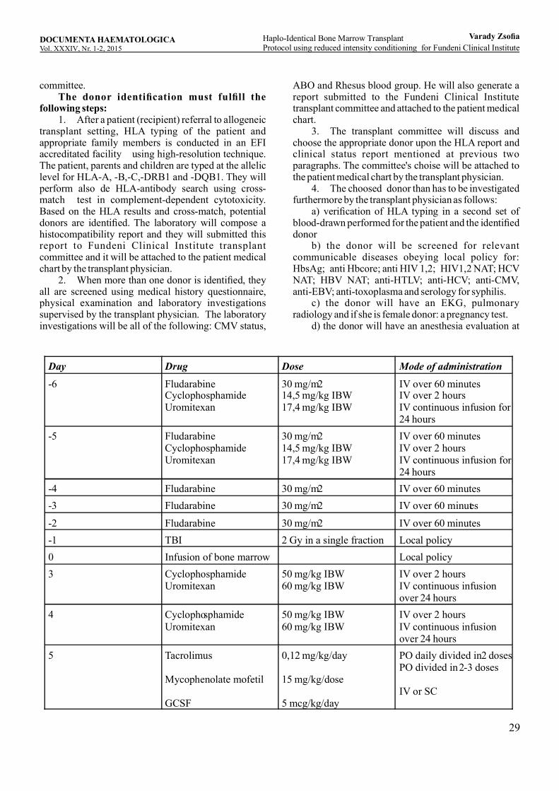
29
committee. The donor identification must fulfill the following steps: 1. After a patient (recipient) referral to allogeneic transplant setting, HLA typing of the patient and appropriate family members is conducted in an EFI accreditated facility using high-resolution technique. The patient, parents and children are typed at the allelic level for HLA-A, -B,-C,-DRB1 and -DQB1. They will perform also de HLA-antibody search using cross-match test in complement-dependent cytotoxicity. Based on the HLA results and cross-match, potential donors are identified. The laboratory will compose a histocompatibility report and they will submitted this report to Fundeni Clinical Institute transplant committee and it will be attached to the patient medical chart by the transplant physician. 2. When more than one donor is identified, they all are screened using medical history questionnaire, physical examination and laboratory investigations supervised by the transplant physician. The laboratory investigations will be all of the following: CMV status,
Day Drug Dose Mode of administration
-6 Fludarabine Cyclophosphamide Uromitexan
30 mg/m2 14,5 mg/kg IBW 17,4 mg/kg IBW
IV over 60 minutes IV over 2 hours IV continuous infusion for 24 hours
-5 Fludarabine Cyclophosphamide Uromitexan
30 mg/m2 14,5 mg/kg IBW 17,4 mg/kg IBW
IV over 60 minutes IV over 2 hours IV continuous infusion for 24 hours
-4 Fludarabine 30 mg/m2 IV over 60 minutes -3 Fludarabine 30 mg/m2 IV over 60 minutes
-2 Fludarabine 30 mg/m2 IV over 60 minutes -1 TBI 2 Gy in a single fraction Local policy 0 Infusion of bone marrow Local policy 3 Cyclophosphamide
Uromitexan 50 mg/kg IBW 60 mg/kg IBW
IV over 2 hours IV continuous infusion over 24 hours
4 Cyclophosphamide Uromitexan
50 mg/kg IBW 60 mg/kg IBW
IV over 2 hours IV continuous infusion over 24 hours
5 Tacrolimus Mycophenolate mofetil GCSF
0,12 mg/kg/day 15 mg/kg/dose 5 mcg/kg/day
PO daily divided in 2 doses PO divided in 2-3 doses IV or SC
ABO and Rhesus blood group. He will also generate a report submitted to the Fundeni Clinical Institute transplant committee and attached to the patient medical chart. 3. The transplant committee will discuss and choose the appropriate donor upon the HLA report and clinical status report mentioned at previous two paragraphs. The committee's choise will be attached to the patient medical chart by the transplant physician. 4. The choosed donor than has to be investigated furthermore by the transplant physician as follows: a) verification of HLA typing in a second set of blood-drawn performed for the patient and the identified donor b) the donor will be screened for relevant communicable diseases obeying local policy for: HbsAg; anti Hbcore; anti HIV 1,2; HIV1,2 NAT; HCV NAT; HBV NAT; anti-HTLV; anti-HCV; anti-CMV, anti-EBV; anti-toxoplasma and serology for syphilis. c) the donor will have an EKG, pulmonary radiology and if she is female donor: a pregnancy test. d) the donor will have an anesthesia evaluation at
DOCUMENTA HAEMATOLOGICAVol. XXXI , Nr. - , 20V 1 2 51
Haplo- dentical one arrow ransplant I B M TP F C Irotocol using reduced intensity conditioning for undeni linical nstitute
Varady Zsofia
30
the Anesthesia Department of Fundeni Clinical Institute.All the documentation mentioned above at point 4 will be done by the transplant physician and kept in the patient medical chart.
The conditioning regimen plan and the additional supportive care:
The treatment plan for the haplo-identical transplant setting it is a non-myeloablative conditioning regimen, using the following:In case of lacking TBI procedure at 2 Gy dose in day -1, it could be replaced by two dose of Busulfan iv in day -3 and day-2 (dose=3,2 mg/kgIBW/day) for those with acute and chronic leukemias. The IBW (ideal body weight) will be calculated using Devine formula and the clearance of creatinine will be calculated using the Cockcroft formula. The fludarabine will be reduced with 20% if de creatinine clearance is below 70 ml/min. The following additional supportive measures will be kept within this protocol: 1. hydration, anti-nausea treatment, fluid-balance, transfusional and nutritional support will respect Fundeni Clinical Institute local standards 2. the central venous access and his care will respect institutional practice. 3. it will be minimum 4-6 hours between the TBI and infusion of bone marrow cells 4. if there is a major ABO incompatibility the marrow will be red-cell depleted using institutional practice 5. prophylaxis for antibacterial, antifungal and antiviral agents will be start in day 0 using institutional practice. The stop of this drugs will respect also the local policy. 6. post-transplant cyclophosphamide will start at minimum 60-72 hours after the start of marrow infusion 7. it is prohibited to use corticosteroids until at least 24 hours after the finish of cyclophosphamide infusion. The only exception is using corticosteriods if anaphylaxis. 8. residual serum level of tacrolimus will be first measured at day +7 and it will be kept between 5-15 ng/ml using institutional practice till 6 months after transplant. 9. every tacrolimus dose change will be controlled by residual serum level at 48 hours 10. mycophenolatemofetil prophylaxis will be ended at day +35. 11. GCSF will be given until absolute neutrophil count is above 500/mm3 for three consecutive days. 12. thetacrolimus levels and PCR-CMV will determined every week within the first 60 days and
wherever is clinically indicated.Bone marrow harvest The bone marrow harvest will respect the Fundeni Institution local practice. It will be performed in operating theater by the one hematologist or transplant physician and one 3-5 year hematology resident with the help of two nurses from the Fundeni Stem Cell Bank. The donor will have general anesthesia done and supervised by the ICU physician.The target yield of marrow is 4 x 10^8 total nucleated cells/kg recipient using his IBW. Minimum recommended yield will be 2,5 x 10^8 total nucleated cells/kg recipient using his IBW. A sample of the product to be infused will be sent to flow-cytometry to determine the CD34+ cell count. If the marrow has major ABO incompatibility it will be depleted in red cells using institutional standards. The marrow will be infused in the same day to the patient.Graft versus host disease (GVHD) prophylaxis The GVHD prophylaxis consisted of post-t r a n s p l a n t C y c l o p h o s p h a m i d e ( P T C y ) o f 50mg/kgIBW/day in IV administration in day +3 and +4, followed by tacrolimus and mycophenolatemofetil (MMF) beginning day +5. The tacrolimus and MMF will be used in oral administration as the transplant protocol show above.The MMF will be stopped at day+35, the tacrolimus will continue till 6 months after the transplantation. If GVHD occur it will be addressed using Fundeni institutional policies.Risks and toxicities 1. Cyclophophamide side effects could be : nausea/vomiting, cardiomyopathy, skin rash, mucositis, sterility, hemorrhagic cystitis, fluid gain and edema with weight gain, hemolysis and alopecia. 2. F ludarabine s ide e ff ect s could be : neurotoxicity (agitation, confusion, blurred vision, peripheral neurophaty, loss of hearing, weakness, blidness, coma), autoimmune hemolytic anemia, deep venous thrombosis, transient ischemic attack, phlebitis, fever, chills, skin rash, nausea/vomiting, diarrhea, stomatitis, anorexia, abnormal liver function tests, abnormal renal function tests, peripheral edema, myalgia, pulmonary toxicity. 3. Tota l Bod y I r rad ia t io n can cau se : nausea/vomiting, parotiditis, diarrhea, erythema, fever, mucositis, alopecia. Late effects include hiper-pigmantation, cataract, risk for secondary malignancies, sterility, pneumonitis, nephropathy. 4. Tacrolimus side effects could be: renal insufficiency, hypertension, hypomagnesemia, hypokalemia, hyperglycemia and neurologic toxicity (tremor). 5. Mycophenolatemofetil side effects could be: nausea/vomiting, diarrhea, headache, hypertension, dizziness, pancytopenia, insomnia, rash, bone pain,
DOCUMENTA HAEMATOLOGICAVol. XXXI , Nr. - , 20V 1 2 51
Haplo- dentical one arrow ransplant I B M TP F C Irotocol using reduced intensity conditioning for undeni linical nstitute
Varady Zsofia
31
electrolyte imbalances, hyperglycemia.Assessment during the transplant During the transplant (and in neutrophil-recovery period) patient will be hospitalized in Bone Marrow Transplant Unit at Fundeni Clinical Institute respecting the local institutional practice for hospitalized patients. He will be in care of a transplant physician. Before admittance all patients will have a bacterial portage evaluation (nasal and pharyngeal swabs, cultures of urine and stool. The transplant phase will be assessed as follows: 1. daily physical examination for the assessment of general health and well-being, infectious complications, medication related problems and GVHD. 2. Daily CBC (complete blood count). 3. At least three times weekly (or wherever needed): creatinine, uric acid, bilirubin, LDH, AST, FAS, Na, K, Cl, Ca, Mg, Ph, blood sugar. 4. Weekly (or wherever needed): coagulation panel, residual tacrolimus level, PCR-CMV. 5. Cultures wherever the clinical situation of the patient indicates, using institutional practice. 6. Any other evaluations or imaging could be performed at any time with medical indication from the transplant physician who has the patient in charge.Assessment in the post-transplant period In the post-transplant period the transplant physician will assess the patient regularly in outpatient manner and the patient will be admitted as needed. This phase will be assessed as follows: 1. At day +30, +60,+120, +180 and 1 year: the patient will have a complete physical examination, CBC, creatinine, uric acid, bilirubin, LDH, AST, FAS, Na, K, Cl, Ca, Mg, Ph, blood sugar, coagulation panel, residual tacrolimus level (if it needed), PCR-CMV, peripheral blood chimerism (unsorted and T cell). 2. Between 6 months and 1 year the patient will come monthly to be evaluated with: complete physical examination, CBC, creatinine, uric acid, bilirubin, LDH, AST, FAS, Na, K, Cl, Ca, Mg, Ph, blood sugar, coagulation panel, residual tacrolimus level (if it needed). 3. At day +30, +180 and at 1 year the patient will have bone marrow aspirate/biopsy, endocrinological testing and consult..Disease status evaluation with specific leukemia and lymphoma panel. 4. In the second year the patient will be evaluated at every third months with: complete physical examination, CBC, creatinine, uric acid, bilirubin, LDH, AST, FAS, Na, K, Cl, Ca, Mg, Ph, blood sugar, coagulation panel, residual tacrolimus level (if it needed). 5. In the third year the patient will be evaluated at every six months with: complete physical examination,
CBC, creatinine, uric acid, bilirubin, LDH, AST, FAS, Na, K, Cl, Ca, Mg, Ph, blood sugar, coagulation panel.6. Starting from the second year the patient will have yearly peripheral blood chimerism, disease status evaluations, endocrinological evaluations with hormone-dosage and thyroid ultrasonography, mammography and genital evaluation with Papanicolau smear for women. Cardiological (EKG and ultrasound) and pulmonary evaluation ( function tests). 7. Any other evaluations or imaging could be performed at any time with medical indication. 8. The patient will be re-immunize starting from 1 year (if he is not having extended chronic GVHD) using institutional standard. One of the most important factors affecting transplantation outcome is proper timing.Therefore, donor availability is an crucial issue. Haploidentical related donors are available for almost all patients, so the use of those donors is a viable alternative. Acknowledgements to my host mentors Gabriel Ghiaur and Richard J Jones from the Bone Marrow Transplant P rogram at theS idney Kimmel Comprehensive Cancer Center, Johns Hopkins University School of Medicine, Baltimore, Maryland, USA andtheVisitorTranining Program within American Society of Hematology.
DOCUMENTA HAEMATOLOGICAVol. XXXI , Nr. - , 20V 1 2 51
Haplo- dentical one arrow ransplant I B M TP F C Irotocol using reduced intensity conditioning for undeni linical nstitute
Varady Zsofia

32
33
DOI: 10.1515/dch-2015-0003
D DE GRUYTEREOPEN
Clinical ase f Aplastic Crisis ssociated ith Extramedullary Hematopoiesis n n c o a w i aa w adult ith Hereditary Spherocytosis nd Parvovirus B19 Infection
Andreea Jercan , Rusu Munteanu Gina, Camelia Dobrea, Daniel Coriu, Aurelia Tatic*Center of Hematology and Bone Marrow Transplant, Fundeni Clinical Institute, Bucharest, Romania
Abstract: Hereditary spherocytosis is an inherited hemolytic anemia due to red cell membrane defects, characterised by chronic hemolysis with different severity degrees, splenomegaly and microspherocytosis on the peripheral blood film. Among the possible complications in these patients are aplastic crisis and extramedullary hematopoiesis. In this article we present the case of a 42 years old man with hereditary spherocytosis diagnosed during childhood (average haemoglobin (Hb) value of 11-12 g/dl), which presented with worsening anemia, fever, chills, bone and muscle pain. The evolution was with accentuation of anemia (Hb 4.2 g/dl), decease of reticulocyte number (Ret 0,8%) and bilirubin (indirect bilirubin 2.7 g/dl). ParvovirusB19 DNA was 100.000.000 copies/ml. A computer tomography (CT)scan was performed and showed extramedullary hematopoiesis areas situated paravertebraly in the inferior thorax and hepatosplenomegaly. The infectious episode was self-limited and improved with substitution treatment.
Key ordsW :Spherocytosis, aplastic crisis, extramedullary hematopoiesis, Parvovirus B19 infection.
* : Jercan Andreea, Department of Hematology, Fundeni Clinical Institute, Sos. Fundeni nr. 258, sector 2, Corresponding authorBucharest, Romania, phone:+40761612450, e-mail:[email protected]
Introduction Hereditary spherocitosis is an inherited hemolytic anemia characterized by a red cell membrane defect, increased number of reticulocytes, increased MCHC (mean corpuscular hemoglobin concentration) index, presence of microspherocites on the peripheral blood smear, intermittent jaundice, splenomegaly and abnormal osmotic fragility test.1
Among it's complications there are worsening anemia, wich can be secondary to the accentuation of hemolysis due to unspecific viral infections (low Hb, high Ret,highbilirubin (Br)), aplastic crysis in Parvovirus B19 infection (low Hb(suddenly), low Ret, low Br) and folatedepletion secondary to accelerated hematopoiesi s. Other complicat ions can be cholelithiasis and extramedullary hematopoiesis. Parvovirus B19 infection is global, it is more common in childhood and is transmitted respiratory. In temperate areas, the infection usually occurs in spring and in small outbreaks every few years.2
In general , Parvovirus B19 infection is asymptomatic. The most common presentation of the infection is infectious erythema (“fifth disease”), a childhood exanthema characterized by “slapped cheeks”.2
It has two phases, initially with flu-like symptoms
(viraemia phase), followed by, when the immune complexes are formed, the rash (rarely seen in adults) and the arthropathy (arthralgia and even arthritis, mimicking rheumatoid arthritis, that resolves in a few weeks).2
Case resentationP A 42 years old male, with mild hereditary spherocytosis, diagnosed during childhood, with a family history of hereditary spherocytosis (daughter), presents to the territorial hospital in March 2013 for fever, chills, vomiting, myalgia. Lab tests showed anemia (Hb 7.1 g/dl), indirect bilirubin 6,7 mg/dl, heterogenous splenomegaly (ultrasonography) and hepatocytolisis. He was sent to our Clinic for diagnosis and treatment, with an average general status, pallor, jaundice, splenomegaly and flu-like symptoms. The lab tests show worsening anemia (Hb 6.1 g/dl, Ht 16.8%, Ret 3.8%, MCV(mean corpuscular volume) 83 fl, MCH (mean corpuscular hemoglobin) 30,3pg, MCHC (mean corpuscular hemoglobin concentration) 36.6 g/dl), L ( l euko co cyte s) 86 00/m mc, PLT (p la te le t s ) 164000/mmc, with microspherocytes on the peripheral blood smear. The indirect bilirubin was lower (from 6.7 to 5.2 mg/dl) and the hepatocytolisis persisted.
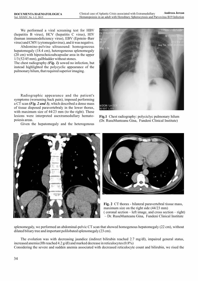
34
We performed a viral screening test for HBV (hepatitis B virus), HCV (hepatitis C virus), HIV (human immunodeficiency virus), EBV (Epstein–Barr virus) and CMV (cytomegalovirus), and it was negative. Abdomino-pelvine ultrasound: homogeneous hepatomegaly (18.4 cm), heterogeneous splenomegaly (20 cm) with hiperechoicsubcapsular area in the upper 1/3 (52/45 mm), gallbladder without stones.The chest radiography sowed no infection, but (Fig. 1)instead highlighted the polycyclic appearance of the pulmonary hilum, that required superior imaging.
Radiographic appearance and the patient's symptoms (worsening back pain), imposed performing a CT scan , which described a dense mass (Fig. 2 and 3)of tissue disposed paravertebraly in the lower thorax, with maximum size of 44/23 mm (to the right). These lesions were interpreted asextramedullary hemato-poiesis areas. Given the hepatomegaly and the heterogenous
Fig.1 Chest radiography: polyciclyc pulmonary hilum (Dr. RusuMunteanu Gina, Fundeni Clinical Institute)
Fig. 2 CT thorax - bilateral paravertebral tissue mass, maximum size on the right side (44/23 mm) ( coronal section – left image, and cross section – right) – Dr. RusuMunteanu Gina, Fundeni Clinical Institute
splenomegaly, we performed an abdominal-pelvic CT scan that showed homogenous hepatomegaly (22 cm), without dilated biliary tree and important polilobated splenomegaly (23 cm).
The evolution was with decreasing jaundice (indirect bilirubin reached 2.7 mg/dl), impaired general status, increased anemia (Hb reached 4.2 g/dl) and marked decrease in reticulocytes (0.8%)Considering the severe and sudden anemia associated with decreased reticulocyte count and bilirubin, we rised the
DOCUMENTA HAEMATOLOGICAVol. XXXI , Nr. - , 20V 1 2 51
Andreea JercanClinical ase f Aplastic Crisis ssociated ith Extramedullary c o a wi a a w aHematopoiesis n n dult ith Hereditary Spherocytosis nd Parvovirus B19 Infection
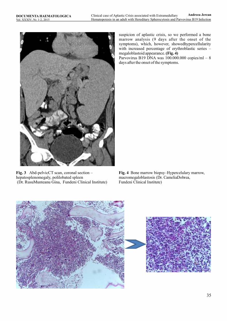
35
Fig. 3 Abd-pelvicCT scan, coronal section – hepatosplenomegaly, polilobated spleen (Dr. RusuMunteanu Gina, Fundeni Clinical Institute)
suspicion of aplastic crisis, so we performed a bone marrow analysis (9 days after the onset of the symptoms), which, however, showedhypercellularity with increased percentage of erythroblastic series – megaloblastoid appearance. (Fig. 4)Parvovirus B19 DNA was 100.000.000 copies/ml – 8 days after the onset of the symptoms.
Fig. 4 Bone marrow biopsy–Hypercelulary marrow, macromegaloblastosis (Dr. CameliaDobrea,
Fundeni Clinical Institute)
DOCUMENTA HAEMATOLOGICAVol. XXXI , Nr. - , 20V 1 2 51
Andreea JercanClinical ase f Aplastic Crisis ssociated ith Extramedullary c o a wi a a w aHematopoiesis n n dult ith Hereditary Spherocytosis nd Parvovirus B19 Infection
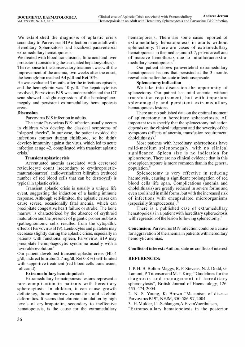
36
We established the diagnosis of aplastic crisis secondary to Parvovirus B19 infection in an adult with Hereditary Spherocitosis and localized paravertebral extramedullary hematopoiesis.We treated with blood transfusions, folic acid and liver protectors (considering the associated hepatocytolisis).The response to the conservatory treatment was with the improvement of the anemia, two weeks after the onset, the hemoglobin reached 9.4 g/dl and Ret 10%.He was evaluated 3 months after the infectious episode, and the hemoglobin was 10 g/dl. The hepatocytolisis resolved, Parvovirus B19 was undetectable and the CT scan showed a slight regression of the hepatospleno-megaly and persistent extramedullary hematopoiesis areas.Discussion Parvovirus B19 infection in adults. The acute Parvovirus B19 infection usually occurs in children who develop the classical symptoms of “slapped cheeks”. In our case, the patient avoided the infectious contact during childhood, so he didn't develop immunity against the virus, which led to acute infection at age 42, complicated with transient aplastic crisis. 2
Transient aplastic crisis Accentuated anemia associated with decreased reticulocyte count (secondary to erythropoietic-maturationarrest) andlowerindirect bilirubin (reduced number of red blood cells that can be destroyed) is typical in aplastic crisis. Transient aplastic crisis is usually a unique life event, suggesting the induction of a lasting immune response. Although self-limited, the aplastic crises can cause severe, occasionally fatal anemia, which can precipitate congestive heart failure or stroke. The bone marrow is characterized by the absence of erythroid maturation and the presence of gigantic pronormoblasts (pathognomonic cells resulted from the cytopathic effect of Parvovirus B19). Leukocytes and platelets may decrease slightly during the aplastic crisis, especially in patients with functional spleen. Parvovirus B19 may precipitate hemophagocytic syndrome usually with a favorable evolution.2
Our patient developed transient aplastic crisis (Hb 4 g/dl, indirect bilirubin 2.7 mg/dl, Ret 0.8 %) self-limited with supportive treatment (red blood cells transfusion, folic acid). Extramedullary hematopoiesis Extramedullary hematopoiesis lesions represent a rare complication in patients with hereditary spherocytosis. In children, it can cause growth deficiency, bone marrow expansion and skeletal deformities. It seems that chronic stimulation by high levels of erythropoietin, secondary to ineffective hematopoiesis, is the cause for the extramedullary
hematopoiesis. There are some cases reported of extramedullary hematopoiesis in adults without splenectomy. There are cases of extramedullary hematopoiesis in the mediastinum3-7, pelvic area8 and of massive hemothorax due to intrathoracicextra-medullary hematopoiesis .9
Our patient shows paravertebral extramedullary hematopoiesis lesions that persisted at the 3 months reevaluation after the acute infectious episode. Splenectomy indication We take into discussion the opportunity of splenectomy. Our patient has mild anemia, without transfusion requirement, but with important splenomegaly and persistent ex tramedullary hematopoiesis lesions. There are no published data on the optimal moment of splenectomy in hereditary spherocitosis. All important texts specify that the splenectomy indication depends on the clinical judgment and the severity of the symptoms (effects of anemia, transfusion requirement, cholelithiasis).1
Most patients with hereditary spherocitosis have mild-medium splenomegaly, with no clinical significance. Spleen size is no indication for splenectomy. There are no clinical evidence that in this case spleen rupture is more common than in the general population.10
Splenectomy is very effective in reducing haemolysis, causing a significant prolongation of red blood cells life span. Complications (anemia and cholelithiasis) are greatly reduced in severe forms and even abolished in mild forms, but with the increased risk of infections with encapsulated microorganisms (especially Streptoccocus).10
There is a published case of extramedullary hematopoiesis in a patient with hereditary spherocitosis with regression of the lesion following splenectomy.8
Conclusion: Parvovirus B19 infection could be a cause for aggravation of the anemia in patients with hereditary hemolytic anemias.
Conflict of interest: Authors state no conflict of interest
REFERENCES:
1. P. H. B. Bolton-Maggs, R. F. Stevens, N. J. Dodd, G. Lamont, P. Tittensor and M. J. King, “Guidelines for the d i a g n o s i s an d m a n a g e m e n t o f h e r e d i t a r y spherocytosis”, British Journal of Haematology, 126: 455–474, 2004.2. N. S. Young, K. Brown “Mecanism of disease Parvovirus B19”, NEJM, 350:586-97, 2004.3. H. Mulder, J.T.Schlangen,A.E.vanVoorthuisen,“Extramedullary hematopoiesis in the posterior
DOCUMENTA HAEMATOLOGICAVol. XXXI , Nr. - , 20V 1 2 51
Andreea JercanClinical ase f Aplastic Crisis ssociated ith Extramedullary c o a wi a a w aHematopoiesis n n dult ith Hereditary Spherocytosis nd Parvovirus B19 Infection
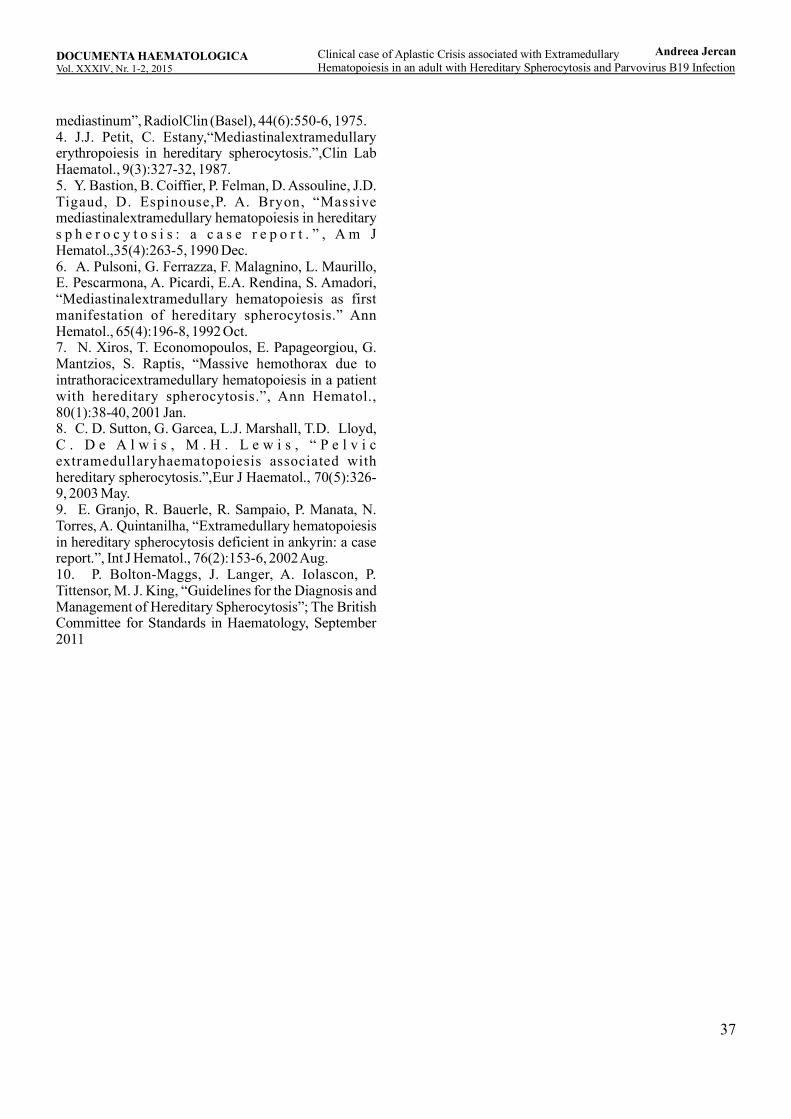
37
mediastinum”, RadiolClin (Basel), 44(6):550-6, 1975.4. J.J. Petit, C. Estany,“Mediastinalextramedullary erythropoiesis in hereditary spherocytosis.”,Clin Lab Haematol., 9(3):327-32, 1987.5. Y. Bastion, B. Coiffier, P. Felman, D. Assouline, J.D. Tigaud, D. Espinouse,P. A. Bryon, “Massive mediastinalextramedullary hematopoiesis in hereditary s p h e r o c y t o s i s : a c a s e r e p o r t . ” , A m J Hematol.,35(4):263-5, 1990 Dec.6. A. Pulsoni, G. Ferrazza, F. Malagnino, L. Maurillo, E. Pescarmona, A. Picardi, E.A. Rendina, S. Amadori, “Mediastinalextramedullary hematopoiesis as first manifestation of hereditary spherocytosis.” Ann Hematol., 65(4):196-8, 1992 Oct.7. N. Xiros, T. Economopoulos, E. Papageorgiou, G. Mantzios, S. Raptis, “Massive hemothorax due to intrathoracicextramedullary hematopoiesis in a patient with hereditary spherocytosis.”, Ann Hematol., 80(1):38-40, 2001 Jan.8. C. D. Sutton, G. Garcea, L.J. Marshall, T.D. Lloyd, C . D e A l w i s , M . H . L e w i s , “ P e l v i c extramedullaryhaematopoiesis associated with hereditary spherocytosis.”,Eur J Haematol., 70(5):326-9, 2003 May.9. E. Granjo, R. Bauerle, R. Sampaio, P. Manata, N. Torres, A. Quintanilha, “Extramedullary hematopoiesis in hereditary spherocytosis deficient in ankyrin: a case report.”, Int J Hematol., 76(2):153-6, 2002 Aug.10. P. Bolton-Maggs, J. Langer, A. Iolascon, P. Tittensor, M. J. King, “Guidelines for the Diagnosis and Management of Hereditary Spherocytosis”; The British Committee for Standards in Haematology, September 2011
DOCUMENTA HAEMATOLOGICAVol. XXXI , Nr. - , 20V 1 2 51
Andreea JercanClinical ase f Aplastic Crisis ssociated ith Extramedullary c o a wi a a w aHematopoiesis n n dult ith Hereditary Spherocytosis nd Parvovirus B19 Infection

38
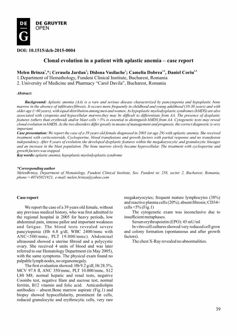
39
DOI: 10.1515/dch-2015-0004
D DE GRUYTEREOPEN
Clonal evolution in a patient with aplastic anemia – case report
Melen Brinza ,*; Cerasela Jardan ; Didona Vasilache ; Camelia Dobrea , Daniel Coriu1 1 1 1,2 1,2
1.Department of Hemathology, Fundeni Clinical Institute, Bucharest, Romania2. University of Medicine and Pharmacy “Carol Davila”, Bucharest, Romania
Abstract:
Aplastic anemia (AA) is a rare and serious disease characterized by pancytopenia and hypoplastic bone Background:marrow in the absence of infiltrates/fibrosis. It occurs more frequently in childhood and young adulthood (10-30 years) and with older age (>60 years), with equal distribution among men and women. As hypoplastic myelodysplastic syndromes (hMDS) are also associated with cytopenia and hypocellular marrow,they may be difficult to differentiate from AA. The presence of dysplastic features (others than erythroid) and/or blast cells >5% is essential to distinguish hMDS from AA. Cytogenetic tests may reveal clonal evolution in hMDS. As the two disorders differ greatly in means of management and prognosis, the correct diagnostic is very important.Case presentation: We report the case of a 39 years old female diagnosed in 2005 (at age 29) with aplastic anemia. She received treatment with corticosteroids, Cyclosporine, blood transfusions and growth factors with partial response and no transfusion independency. After 8 years of evolution she developed dysplastic features within the megakaryocytic and granulocytic lineages and an increase in the blast population. The bone marrow slowly became hypercellular. The treatment with cyclosporine and growth factors was stopped.Key words: aplastic anemia, hypoplastic myelodysplastic syndrome
*Corresponding authorMelenBrinza, Department of Hematology, Fundeni Clinical Institute, Sos. Fundeni nr. 258, sector 2, Bucharest, Romania, phone:+40745021923, e-mail: [email protected]
Case report
We report the case of a 39 years old female, without any previous medical history, who was first admitted to the regional hospital in 2005 for heavy periods, low abdominal pain, intense pallor and important weakness and fatigue. The blood tests revealed severe pancytopenia (Hb 6.8 g/dl, WBC 2400/mmc with ANC<500/mmc, PLT 19.000/mmc). Abdominal ultrasound showed a uterine fibroid and a polycystic ovary. She received 4 units of blood and was later referred to our Hematology Department (in May 2005), with the same symptoms. The physical exam found no palpable lymph nodes, no organomegaly. The first evaluation showed: Hb 9.2 g/dl, Ht 28.5%, MCV 97.8 fl, ANC 350/mmc, PLT 10.000/mmc, S12 L80 M8; normal hepatic and renal tests, negative Coombs test, negative Ham and sucrose test, normal ferritin, B12 vitamin and folic acid. Anticardiolipin antibodies – absent.Bone marrow aspirate (Fig.1) and biopsy showed hypocellularity, prominent fat cells, reduced granulocytic and erythrocytic cells, very rare
megakaryocytes; frequent mature lymphocytes (38%) and reactive plasma cells (20%); absent fibrosis; CD34+ cells <5% (Fig.1) The cytogenetic exam was inconclusive due to insufficient metaphases. Serum erythropoietin (EPO): 45 mU/ml In vitro cell cultures showed very reduced cell grow and colony formation (spontaneous and after growth factors). The chest X-Ray revealed no abnormalities.
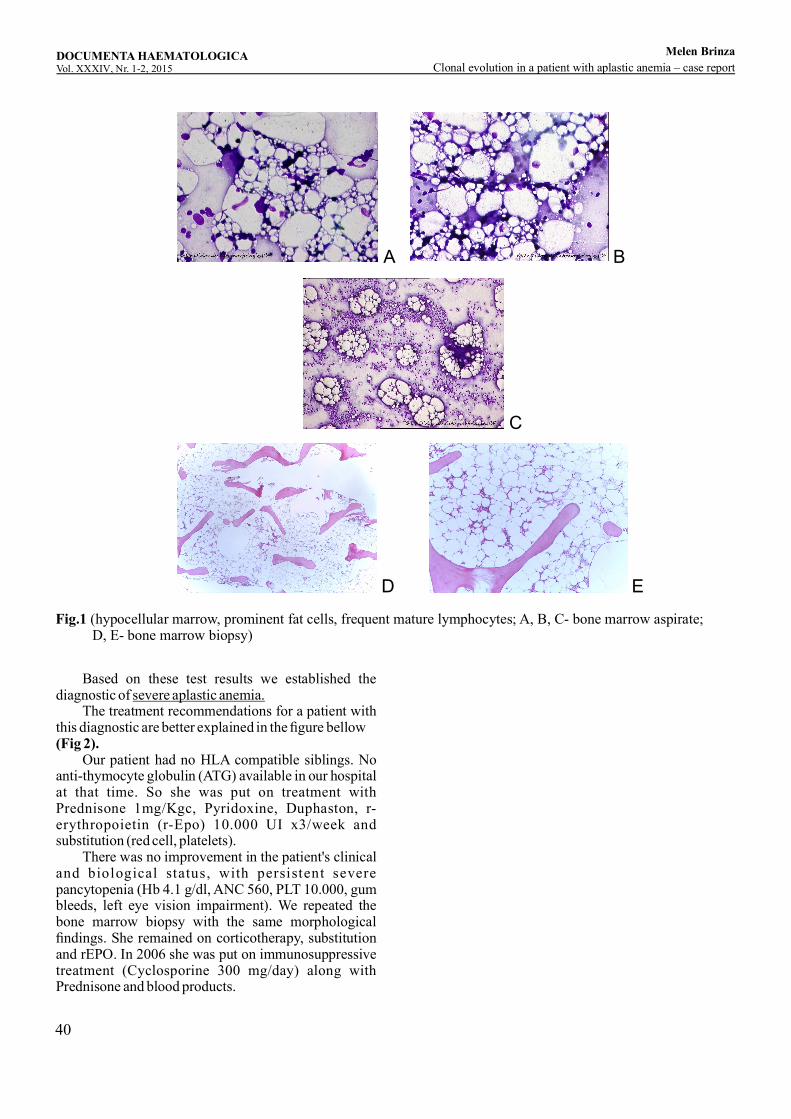
40
A
ED
B
C
Fig.1 (hypocellular marrow, prominent fat cells, frequent mature lymphocytes; A, B, C- bone marrow aspirate; D, E- bone marrow biopsy)
Based on these test results we established the diagnostic of severe aplastic anemia. The treatment recommendations for a patient with this diagnostic are better explained in the figure bellow (Fig 2). Our patient had no HLA compatible siblings. No anti-thymocyte globulin (ATG) available in our hospital at that time. So she was put on treatment with Prednisone 1mg/Kgc, Pyridoxine, Duphaston, r-erythropoietin (r-Epo) 10.000 UI x3/week and substitution (red cell, platelets). There was no improvement in the patient's clinical and biological status, with persistent severe pancytopenia (Hb 4.1 g/dl, ANC 560, PLT 10.000, gum bleeds, left eye vision impairment). We repeated the bone marrow biopsy with the same morphological findings. She remained on corticotherapy, substitution and rEPO. In 2006 she was put on immunosuppressive treatment (Cyclosporine 300 mg/day) along with Prednisone and blood products.
DOCUMENTA HAEMATOLOGICAVol. XXXI , Nr. - , 20V 1 2 51 Clonal evolution in a patient with aplastic anemia – case report
Melen Brinza
41
Fig. 2 [9]
DOCUMENTA HAEMATOLOGICAVol. XXXI , Nr. - , 20V 1 2 51 Clonal evolution in a patient with aplastic anemia – case report
Melen Brinza
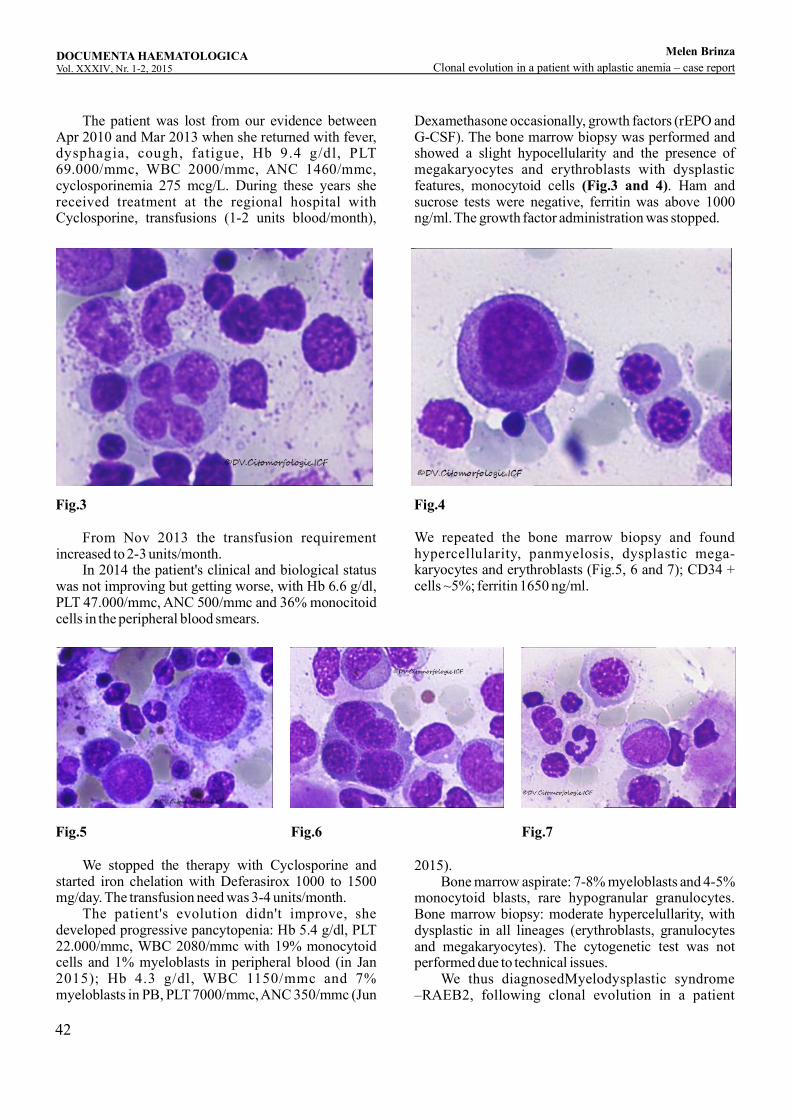
42
The patient was lost from our evidence between Apr 2010 and Mar 2013 when she returned with fever, dysphagia, cough, fatigue, Hb 9.4 g/dl, PLT 69.000/mmc, WBC 2000/mmc, ANC 1460/mmc, cyclosporinemia 275 mcg/L. During these years she received treatment at the regional hospital with Cyclosporine, transfusions (1-2 units blood/month),
Dexamethasone occasionally, growth factors (rEPO and G-CSF). The bone marrow biopsy was performed and showed a slight hypocellularity and the presence of megakaryocytes and erythroblasts with dysplastic features, monocytoid cells . Ham and (Fig.3 and 4)sucrose tests were negative, ferritin was above 1000 ng/ml. The growth factor administration was stopped.
Fig.3
Fig.5
Fig.4
Fig.6 Fig.7
From Nov 2013 the transfusion requirement increased to 2-3 units/month. In 2014 the patient's clinical and biological status was not improving but getting worse, with Hb 6.6 g/dl, PLT 47.000/mmc, ANC 500/mmc and 36% monocitoid cells in the peripheral blood smears.
We repeated the bone marrow biopsy and found hypercellularity, panmyelosis, dysplastic mega-karyocytes and erythroblasts (Fig.5, 6 and 7); CD34 + cells ~5%; ferritin 1650 ng/ml.
We stopped the therapy with Cyclosporine and started iron chelation with Deferasirox 1000 to 1500 mg/day. The transfusion need was 3-4 units/month. The patient's evolution didn't improve, she developed progressive pancytopenia: Hb 5.4 g/dl, PLT 22.000/mmc, WBC 2080/mmc with 19% monocytoid cells and 1% myeloblasts in peripheral blood (in Jan 2015); Hb 4.3 g/dl, WBC 1150/mmc and 7% myeloblasts in PB, PLT 7000/mmc, ANC 350/mmc (Jun
2015). Bone marrow aspirate: 7-8% myeloblasts and 4-5% monocytoid blasts, rare hypogranular granulocytes. Bone marrow biopsy: moderate hypercelullarity, with dysplastic in all lineages (erythroblasts, granulocytes and megakaryocytes). The cytogenetic test was not performed due to technical issues. We thus diagnosedMyelodysplastic syndrome –RAEB2, following clonal evolution in a patient
DOCUMENTA HAEMATOLOGICAVol. XXXI , Nr. - , 20V 1 2 51 Clonal evolution in a patient with aplastic anemia – case report
Melen Brinza
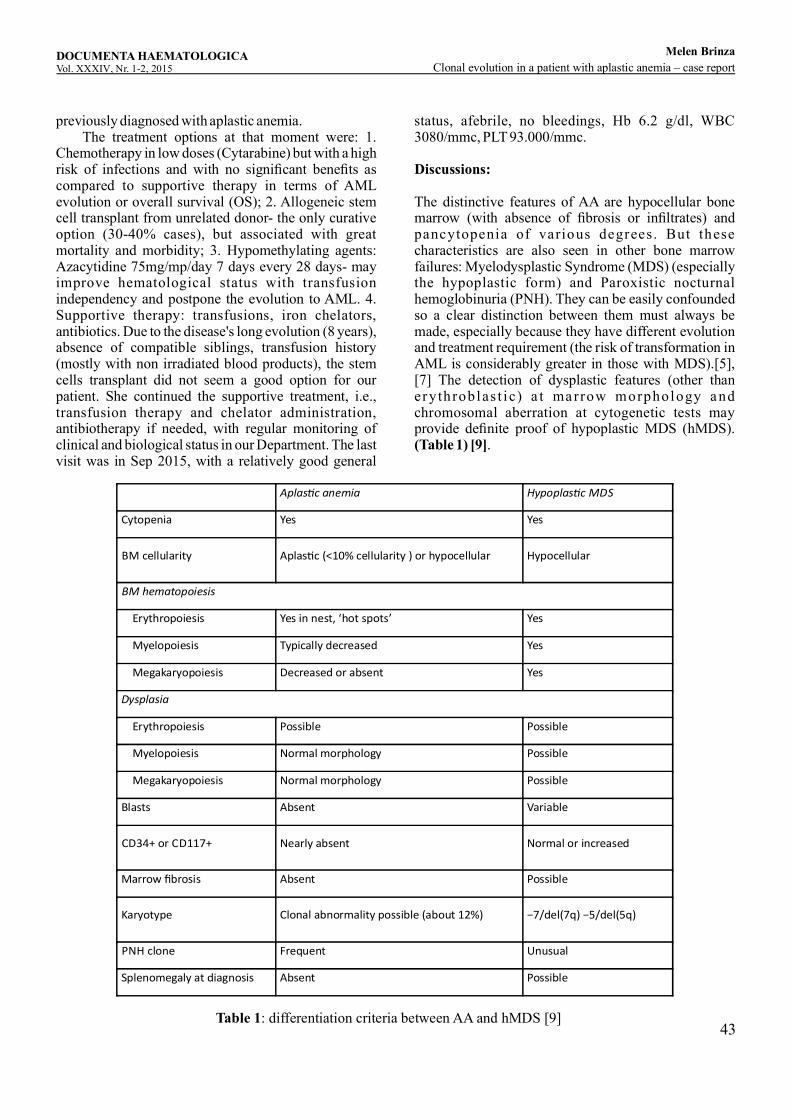
43
previously diagnosed with aplastic anemia. The treatment options at that moment were: 1. Chemotherapy in low doses (Cytarabine) but with a high risk of infections and with no significant benefits as compared to supportive therapy in terms of AML evolution or overall survival (OS); 2. Allogeneic stem cell transplant from unrelated donor- the only curative option (30-40% cases), but associated with great mortality and morbidity; 3. Hypomethylating agents: Azacytidine 75mg/mp/day 7 days every 28 days- may improve hematological status with transfusion independency and postpone the evolution to AML. 4. Supportive therapy: transfusions, iron chelators, antibiotics. Due to the disease's long evolution (8 years), absence of compatible siblings, transfusion history (mostly with non irradiated blood products), the stem cells transplant did not seem a good option for our patient. She continued the supportive treatment, i.e., transfusion therapy and chelator administration, antibiotherapy if needed, with regular monitoring of clinical and biological status in our Department. The last visit was in Sep 2015, with a relatively good general
status, afebrile, no bleedings, Hb 6.2 g/dl, WBC 3080/mmc, PLT 93.000/mmc.
Discussions:
The distinctive features of AA are hypocellular bone marrow (with absence of fibrosis or infiltrates) and pancytopenia of various degrees. But these characteristics are also seen in other bone marrow failures: Myelodysplastic Syndrome (MDS) (especially the hypoplastic form) and Paroxistic nocturnal hemoglobinuria (PNH). They can be easily confounded so a clear distinction between them must always be made, especially because they have different evolution and treatment requirement (the risk of transformation in AML is considerably greater in those with MDS).[5], [7] The detection of dysplastic features (other than ery throblas t i c) a t marrow morphology and chromosomal aberration at cytogenetic tests may provide definite proof of hypoplastic MDS (hMDS). (Table 1) [9].
Aplas c anemia Hypoplas c MDS
Cytopenia Yes Yes
BM cellularity
Aplas c (<10% cellularity ) or hypocellular Hypocellular
BM hematopoiesis
Erythropoiesis
Yes in nest, ‘hot spots’
Yes
Myelopoiesis
Typically decreased
Yes
Megakaryopoiesis
Decreased or absent
Yes
Dysplasia
Erythropoiesis
Possible
Possible
Myelopoiesis
Normal morphology
Possible
Megakaryopoiesis
Normal morphology
Possible
Blasts
Absent
Variable
CD34+ or CD117+
Nearly absent
Normal or increased
Marrow fibrosis
Absent
Possible
Karyotype
Clonal abnormality possible (about 12%)
−7/del(7q) −5/del(5q)
PNH clone
Frequent
Unusual
Splenomegaly at diagnosis Absent Possible
Table 1: differentiation criteria between AA and hMDS [9]
DOCUMENTA HAEMATOLOGICAVol. XXXI , Nr. - , 20V 1 2 51 Clonal evolution in a patient with aplastic anemia – case report
Melen Brinza

44
However, some consider AA as a clonal hematopoietic disorder and the immunosuppressive therapy may reveal pre-existing abnormalities in those patients who were misclassified initially with AA instead of hMDS. This may have been possible due to the fact that in AA patients were too few cells to be analyzed morphologically and by conventional cytogenetic tests. Nowadays it is well known that AA is mostly an immune-mediated disorder caused by an abnormal activation of the cytotoxic T cells (that destroy the normal hematopoietic elements); this explains why the main therapy consists of immunosuppressive agents like cyclosporine, antithymocyte globulin (ATG) and steroids, with or without G-CSF (Fig.2). But, at the same time, this treatment may increase the risk of late clonal complications (PNH, MDS, AML transformation) by allowing the abnormal pre-existing clone to escape immunosurveillance and to overgrow and become detectable, allowing AA to be considered as a preleukemic disorder with clonal hematopoiesis and defective stem cells. [6][8] M D S i s c h a r a c t e r i z e d b y i n e f f e c t i v e hematopoiesis, dysplastic features and chromosomal abnormalities. The treatment is more aggressive and includes chemotherapy, hypomethylating agents and allotransplant. Hypocellular MDS is a subgroup that accounts for 7-15% of MDS cases. The most common chromosomal aberration is monosomy 7. [3] But there are cases of hMDS with normal cytogenetic tests or with good response to immunosuppressive treatment and patients with AA with some cytogenetic abnormalities or poor response to immunotherapy. This makes the distinction between the two (AA and hMDS) even more challenging. Furthermore, the absence of dysplasia does not exclude the possibility of hMDS, especially in patients refractory to the therapy designated for AA or with relatively short interval from the initial diagnosis of AA to the development of MDS/AML. For the case of our patient, the lack of dysplastic elements in the initial bone marrow aspirate and biopsy was insufficient to exclude the diagnostic of hMDS because.No cytogenetic analysis had beenperformed, so we did not know whether there were any chromosomal abnormalities. The fact that she was refractory to the i mm un o su p p res s i v e t he r apy ( s t e ro id s an d cyclosporine) is a sign that the patient is more likely to have had hMDS rather than AA at the time of the initial diagnostic. Alternatively, the evolution to MDS was secondary: the therapy with growth factors may have stimulated the defective clone to expand and become detectable after a long period of evolution (8 years). The indication of allogeneic stem cell transplant from unrelated donor should have been evaluated earlier in the patient's evolution, but at that time this procedure
was not available in our country.Conclusion: There are several reports of patients who first had AA that later transformed into MDS. It is still unclear why this happens, some suggesting the abnormal s t i m u l a t i o n b y g r o w t h f a c t o r s o r t h e immunosuppression as possible explanations. [1]. Others argue that these patients had hypoplastic MDS at the initial onset of the pancytopenia, but there were not sufficient cells for the dysplasia to be detected on marrow morphology or clonal detection by cytogenetic tests. Our case underlines the importance of cytogenetic analysis at the time of first presentation of a patient with pancytopenia and hypocellular bone marrow. [2] If the number of metaphases is insufficient, a FISH test for the detection of chromosome 7 abnormalities (the most commonly affected in hMDS) may be useful. Although p a t i e n t s w i t h h M D S m ay r e s p o n d t o t h e immunosuppressive therapy, the evolution to AML is faster and more frequent.
Conflict of interest: Authors state no conflict of interest.
References1. Kaito K, Kobayashi M, Katayama T, et al. Long-term administration of G-CSF for aplastic anaemia is closely related to the early evolution of monosomy 7 MDS in adults. Br J Haematol. 1998;103:297–303.2. Maciejewski JP, Risitano A, Sloand EM, Nunez O, Young NS. Distinct clinical outcomes for cytogenetic abnormalities evolving from aplastic anemia. Blood 2002;99:3129–3135.3. Maciejewski JP, Selleri C. Evolution of clonal cytogenetic abnormalities in aplastic anemia. Leuk Lymphoma. 2004; 45:433-440.4. Appelbaum FR, Barrall J, Storb R, et al. Clonal cytogenetic abnormalities in patients with otherwise typical aplastic anemia. Exp Hematol. 1987; 15:1134-11395. Tichelli A, Gratwohl A, Wursch A, Nissen C, Speck B. Late haematological complications in severe aplastic anaemia. Br J Haematol. 1988; 69:413-418.6. Sergej K, L. Jeffrey M, Patrick A, et al. Therapy may unmask hypoplastic myelodysplastic syndrome that mimics aplastic anemia. Cancer. 2007; 110(7):1520-1526.7. John M. Bennett, Attilio o, et al. Diagnostic criteria to distinguish hypocellular acute myeloid leukemia from hypocellular myelodysplastic syndromes and aplastic anemia: recommendations for a standardized approach. Haematologica 2009; 94(2):264-2688. Seiji K, Akira O, et al. Risk factors for evolution of acquired aplastic anemia into myelodysplastic
DOCUMENTA HAEMATOLOGICAVol. XXXI , Nr. - , 20V 1 2 51 Clonal evolution in a patient with aplastic anemia – case report
Melen Brinza
45
syndrome and acute myeloid leukemia after immunosuppressive therapy in children. Blood. 2002; 100(3):786-790.9. Phillip Scheinberg, Neal S. Young, How I treat acquired aplastic anemia. Blood. 2012 Aug 9; 120(6): 1185–1196
DOCUMENTA HAEMATOLOGICAVol. XXXI , Nr. - , 20V 1 2 51 Clonal evolution in a patient with aplastic anemia – case report
Melen Brinza

46
47
DOI: 10.1515/dch-2015-0005
D DE GRUYTEREOPEN
Quercetin, Menadione, Doxorubicin combination as a potential alternative to Doxorubicin monotherapy of acute lymphoblastic leukemia
Ruxandra Irimia ¹, *,**, Ioana Teodora Tofolean ¹, **, Roxana Gabriela Sandu ¹, Oana Elena Băran ¹, Maria Cătălina Ceauşescu ¹, Vlad Coşoreanu ¹, Maria Teodora Ilie ¹, Ramona Babeş ¹, Constanţa Ganea ¹, Irina Băran ¹¹ “Carol Davila” University of Medicine and Pharmacy, Dept. of Biophysics, 8 Eroii Sanitari, 050474 Bucharest, Romania** These authors contributed equally to the work
Abstract: Doxorubicin is a widely used chemotherapeutic drug, effective on patients with acute lymphoblastic leukemia, but associated with significant long term cardiotoxicity. Menadione (vitamin K3) and the flavonoid quercetin are known as strong apoptogens in human leukemia Jurkat T cells. We explored the potential synergic cytotoxic effects of doxorubicin in association with quercetin and menadione in this cellular model for acute lymphoblastic leukemia. Apoptosis, necrosis and cell cycle distributions were determined by flow cytometry on Jurkat lymphoblasts labeled with Annexin V-FITC/7-Aminoactinomycin D and propidium iodide, respectively. Oxidative stress was assessed by flow cytometry using CM-H2DCFDA/7-Aminoactinomycin D labeling. Results indicate a dose-dependent oxidative-stress generation, cell cycle arrest and apoptosis induction by doxorubicin alone, correlated with a decrease of the required doses when the anticancer drug was associated with quercetin and menadione. Data also support the theory of an additive cytotoxic effect of the three agents on leukemia cells. Introducing quercetin-menadione combinations in leukemia doxorubicin-based treatment could significantly increase the treatment's efficacy. The main mechanism responsible for this effect appears to be the increase in the affinity of doxorubicin for DNA, which enables lowering of the therapeutic dose.
Keywords: doxorubicin, acute lymphoblastic leukemia, oxidative stress, apoptosis, necrosis, cell cycle
*Corresponding author: Ruxandra Irimia, “Carol Davila” University of Medicine and Pharmacy, Dept. of Biophysics, 8 Eroii Sanitari, 050474 Bucharest, Romania, phone +40748152880, e-mail: [email protected]
Introduction In the past years, a great interest has been focused on the health benefits of natural flavonoids. They are chemical compounds widely found in fruits, leaves and vegetables, responsible for their bright colors, and also well-known potent antioxidants. The possible antioxidant effects of flavonoids were suspected after it was shown that a red wine rich Mediterranean diet was associated with a lower cardiovascular risk [1]. Numerous studies have shown that they possess antiatherogenic - cardioprotective effects and exert antitumoral, anti-mutagenic and anti-inflammatory activities [2-4]. Quercetin (QC) is one of the best characterized flavonoids, found prevalently in fresh parsley, onions, olives and green salad [1]. This flavonoid is known to have pro-oxidative properties under various conditions,
and a number of both in vitro and in vivo studies indicate a greater susceptibility of malignant cells to the cytotoxic effect of the flavonoid with respect to their normal counterparts [5,6]. Flavonoids appear to bind to a sugar molecule and originally it was considered that only free flavonoids could be absorbed in the human intestine. Quercetin in its native state is usually bound to a glucose or rutinose molecule. Studies have indicated a superior absorption of the quercetin-glucose complex, compared to the quercetin-rutinose one, with a 20 times higher peak plasma level reached in 10 times less time from the ingestion, suggesting a small intestine absorption for the first compound and a large intestine post-deglycosylation for the latter [7]. The main limitation of the clinical use of quercetin is linked to its poor bioavailability and its rapid metabolic
48
conversion in the liver. Pharmacokinetic studies had shown that tolerable QC plasma levels efficient for cancer therapy (>10 μM) are maintained for about 30 minutes after intravenous administration, the average half-life being little above 40 minutes [8,9]. Liposomal carriers used to deliver quercetin can increase the half-life and the bioavailability of the flavonoid [10] and thus could lower the threshold level required for efficient cancer treatment protocols. Flavonoids possess at least 3 antioxidant mechanisms, as well as the capacity of stimulating the natural protective capacity. One mechanism (probably responsible for the anti-atherogenic effect) consists of direct reactive oxygen species scavenging through the high hydroxyl functional group reactivity, resulting in the inactivation of radical species [7]. A second cellular pathway stimulates the nitric oxide (NO) production. Studies conducted on acute lymphoblastic leukemia cells after quercetin exposure show increased nitric oxide levels and NO synthase inhibition by 1-(2-Mercaptoethyl)guanidine increases the apoptotic fraction of leukemic post-antioxidant treatment cells. The increase in NO production appears to be a pro tect ive mechanism at physiological low concentrations, but at high levels it reacts with reactive oxygen species, forming peroxynitrite, with deleterious effects on cell membranes [11]. The third antioxidant mechanism can be explained by quercetin's iron chelation properties, demonstrated in in vitro studies on mice erythrocytes after glutathione depletion. In the absence of flavonoids, significant membrane lipid peroxidation and hemolysis may occur due to iron release, while association with quercetin can exert protective effects via iron chelation [12]. In the human leukemia Jurkat T cell line, quercetin activates the ryanodine receptor (a calcium channel), hence inducing calcium release from the endoplasmic reticulum, acting as a potent apoptogen and enhancing apoptosis induced by menadione (MD) [11,13,14].Menadione, also known as vitamin K3, is a well-known pro-oxidant agent, with antitumoral activity in various cellular lines, including Jurkat T cells. MD is a liposoluble synthetic form of vitamin K, which is metabolized in the human body to vitamin K2. Recent studies have shown a chemosensitizing effect of menadione in different cancer models [5,15], as well as a powerful corrector of doxorubicin resistance in leukemia cells. More than half of the intracellular metabolism of menadione is developed through redox cycling, which can generate high quantities of free radicals [16]. Both menadione and quercetin are known as pro-apoptotic agents acting via the calcium-dependent mitochondrial pathway, promoting calcium release from the endoplasmic reticulum and opening of the
mitochondrial permeability transition pore. The opened pore induces the collapse of the mitochondrial transmembrane potential and the release of the cytochrome c from the mitochondria. Cytochrome c is associated with the internal mitochondrial membrane and is an essential component of the electron transport chain. When released into the cytoplasm, it activates the apoptotic protease activation factor-1, thus triggering apoptosis [17,18]. Doxorubicin (DOX), also known as adriamycin, is a broad spectrum chemotherapeutic drug compared to other antineoplastic drugs, first extracted from Streptomyces peucetius in the 1970s. Its cytotoxic properties are either a consequence of DNA intercalation and topoisomerase II-mediated DNA repair disruption, or a result of free oxygen radicals generation, that determine lipid peroxidation, DNA and protein injury, thus triggering apoptosis [19]. Doxorubicin enters the cells through passive diffusion [20] and once it has entered the cell, it can bind to the proteasome. The doxorubicin-proteasome complex is translocated into the nucleus, where it dissociates and DOX inserts itself between the strands of DNA, thus inhibiting the cellular metabolism [21]. Doxorubicin molecules that did not intercalate into the DNA stabilize the complex between topoisomerase II and DNA strings, leading to an increase in the damaged genomic material, associated with G2/M arrest and necrosis/apoptosis [22,23]. During the intracellular metabolic conversion of doxorubicin, one electron is added to the quinone, leading to the formation of a semiquinone, which is converted back to its quinone form when entering a redox reaction. Reactive oxygen species are then produced leading to cellular oxidative stress [24]. A major limitation in the clinical use of doxorubicin is its cardiotoxicity. The myocardial involvement is dependent on the cumulative dose and may occur even decades after finishing the treatment [24]. It appears that this severe side effect is determined by iron release and free radicals generation. Doxorubicin is reduced to doxorubicinol, an active metabolite which interferes with intracellular iron deposits. This theory is supported by the fact that dexrazoxane, an iron chelator, is protective against doxorubicin induced toxicity. In this case, association of quercetin could not only improve the therapeutic index of doxorubicin, but also decrease its cardiac toxicity, through its iron chelating properties [25,26].
Methods Jurkat cells (clone E6-1, ATCC TIB-152) were cultured in GLUTAMAX-I and HEPES-containing RPMI 1640 medium (Invitrogen), supplemented with 10% fetal bovine serum, 100 units/ml penicillin and 100
DOCUMENTA HAEMATOLOGICAVol. XXXI , Nr. - , 20V 1 2 51
Ruxandra IrimiaQuercetin, Menadione, Doxorubicin combination as a potential alternative to Doxorubicin monotherapy of acute lymphoblastic leukemia
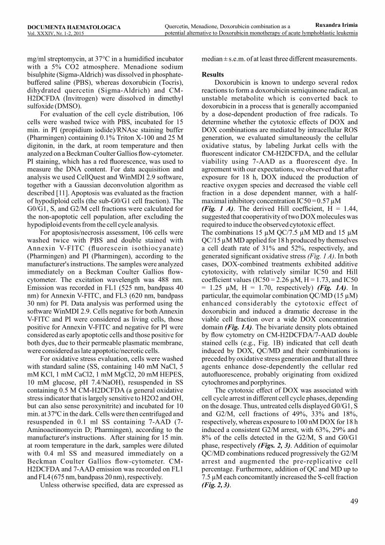
49
mg/ml streptomycin, at 37°C in a humidified incubator with a 5% CO2 atmosphere. Menadione sodium bisulphite (Sigma-Aldrich) was dissolved in phosphate-buffered saline (PBS), whereas doxorubicin (Tocris), dihydrated quercetin (Sigma-Aldrich) and CM-H2DCFDA (Invitrogen) were dissolved in dimethyl sulfoxide (DMSO). For evaluation of the cell cycle distribution, 106 cells were washed twice with PBS, incubated for 15 min. in PI (propidium iodide)/RNAse staining buffer (Pharmingen) containing 0.1% Triton X-100 and 25 M digitonin, in the dark, at room temperature and then analyzed on a Beckman Coulter Gallios flow-cytometer. PI staining, which has a red fluorescence, was used to measure the DNA content. For data acquisition and analysis we used CellQuest and WinMDI 2.9 software, together with a Gaussian deconvolution algorithm as described [11]. Apoptosis was evaluated as the fraction of hypodiploid cells (the sub-G0/G1 cell fraction). The G0/G1, S, and G2/M cell fractions were calculated for the non-apoptotic cell population, after excluding the hypodiploid events from the cell cycle analysis. For apoptosis/necrosis assessment, 106 cells were washed twice with PBS and double stained with Annexin V-FITC (fluorescein isothiocyanate) (Pharmingen) and PI (Pharmingen), according to the manufacturer's instructions. The samples were analyzed immediately on a Beckman Coulter Gallios flow-cytometer. The excitation wavelength was 488 nm. Emission was recorded in FL1 (525 nm, bandpass 40 nm) for Annexin V-FITC, and FL3 (620 nm, bandpass 30 nm) for PI. Data analysis was performed using the software WinMDI 2.9. Cells negative for both Annexin V-FITC and PI were considered as living cells, those positive for Annexin V-FITC and negative for PI were considered as early apoptotic cells and those positive for both dyes, due to their permeable plasmatic membrane, were considered as late apoptotic/necrotic cells. For oxidative stress evaluation, cells were washed with standard saline (SS, containing 140 mM NaCl, 5 mM KCl, 1 mM CaCl2, 1 mM MgCl2, 20 mM HEPES, 10 mM glucose, pH 7.4/NaOH), resuspended in SS containing 0.5 M CM-H2DCFDA (a general oxidative stress indicator that is largely sensitive to H2O2 and OH, but can also sense peroxynitrite) and incubated for 10 min. at 37ºC in the dark. Cells were then centrifuged and resuspended in 0.1 ml SS containing 7-AAD (7-Aminoactinomycin D; Pharmingen), according to the manufacturer's instructions. After staining for 15 min. at room temperature in the dark, samples were diluted with 0.4 ml SS and measured immediately on a Beckman Coulter Gallios flow-cytometer. CM-H2DCFDA and 7-AAD emission was recorded on FL1 and FL4 (675 nm, bandpass 20 nm), respectively. Unless otherwise specified, data are expressed as
median ± s.e.m. of at least three different measurements.
Results Doxorubicin is known to undergo several redox reactions to form a doxorubicin semiquinone radical, an unstable metabolite which is converted back to doxorubicin in a process that is generally accompanied by a dose-dependent production of free radicals. To determine whether the cytotoxic effects of DOX and DOX combinations are mediated by intracellular ROS generation, we evaluated simultaneously the cellular oxidative status, by labeling Jurkat cells with the fluorescent indicator CM-H2DCFDA, and the cellular viability using 7-AAD as a fluorescent dye. In agreement with our expectations, we observed that after exposure for 18 h, DOX induced the production of reactive oxygen species and decreased the viable cell fraction in a dose dependent manner, with a half-maximal inhibitory concentration IC50 = 0.57 µM (Fig. 1 A). The derived Hill coefficient, H = 1.44, suggested that cooperativity of two DOX molecules was required to induce the observed cytotoxic effect. The combinations 15 µM QC/7.5 µM MD and 15 µM QC/15 µM MD applied for 18 h produced by themselves a cell death rate of 31% and 52%, respectively, and generated significant oxidative stress . In both (Fig. 1 A)cases, DOX-combined treatments exhibited additive cytotoxicity, with relatively similar IC50 and Hill coefficient values (IC50 = 2.26 µM, H = 1.73, and IC50 = 1.25 µM, H = 1.70, respectively) . In (Fig. 1A)particular, the equimolar combination QC/MD (15 µM) enhanced considerably the cytotoxic effect of doxorubicin and induced a dramatic decrease in the viable cell fraction over a wide DOX concentration domain . The bivariate density plots obtained (Fig. 1A)by flow cytometry on CM-H2DCFDA/7-AAD double stained cells (e.g., Fig. 1B) indicated that cell death induced by DOX, QC/MD and their combinations is preceded by oxidative stress generation and that all three agents enhance dose-dependently the cellular red autofluorescence, probably originating from oxidized cytochromes and porphyrines. The cytotoxic effect of DOX was associated with cell cycle arrest in different cell cycle phases, depending on the dosage. Thus, untreated cells displayed G0/G1, S and G2/M, cell fractions of 49%, 33% and 18%, respectively, whereas exposure to 100 nM DOX for 18 h induced a consistent G2/M arrest, with 63%, 29% and 8% of the cells detected in the G2/M, S and G0/G1 phase, respectively . Addition of equimolar (Figs. 2, 3)QC/MD combinations reduced progressively the G2/M arrest and augmented the pre-replicative cell percentage. Furthermore, addition of QC and MD up to 7.5 µM each concomitantly increased the S-cell fraction (Fig. 2, 3).
DOCUMENTA HAEMATOLOGICAVol. XXXI , Nr. - , 20V 1 2 51
Ruxandra IrimiaQuercetin, Menadione, Doxorubicin combination as a potential alternative to Doxorubicin monotherapy of acute lymphoblastic leukemia
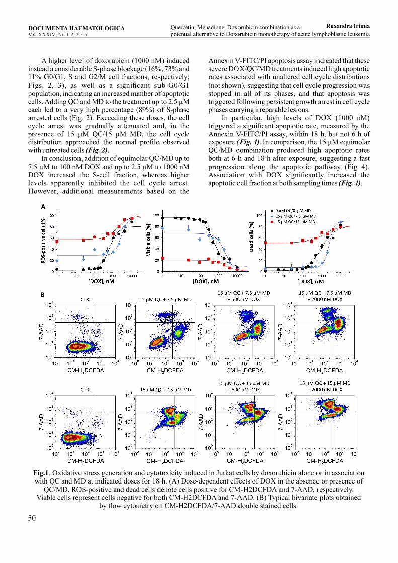
50
A higher level of doxorubicin (1000 nM) induced instead a considerable S-phase blockage (16%, 73% and 11% G0/G1, S and G2/M cell fractions, respectively; Figs. 2, 3), as well as a significant sub-G0/G1 population, indicating an increased number of apoptotic cells. Adding QC and MD to the treatment up to 2.5 µM each led to a very high percentage (89%) of S-phase arrested cells (Fig. 2). Exceeding these doses, the cell cycle arrest was gradually attenuated and, in the presence of 15 µM QC/15 µM MD, the cell cycle distribution approached the normal profile observed with untreated cells . (Fig. 2) In conclusion, addition of equimolar QC/MD up to 7.5 µM to 100 nM DOX and up to 2.5 µM to 1000 nM DOX increased the S-cell fraction, whereas higher levels apparently inhibited the cell cycle arrest. However, additional measurements based on the
Annexin V-FITC/PI apoptosis assay indicated that these severe DOX/QC/MD treatments induced high apoptotic rates associated with unaltered cell cycle distributions (not shown), suggesting that cell cycle progression was stopped in all of its phases, and that apoptosis was triggered following persistent growth arrest in cell cycle phases carrying irreparable lesions. In particular, high levels of DOX (1000 nM) triggered a significant apoptotic rate, measured by the Annexin V-FITC/PI assay, within 18 h, but not 6 h of exposure . In comparison, the 15 µM equimolar (Fig. 4)QC/MD combination produced high apoptotic rates both at 6 h and 18 h after exposure, suggesting a fast progression along the apoptotic pathway (Fig 4). Association with DOX significantly increased the apoptotic cell fraction at both sampling times .(Fig. 4)
Fig.1. Oxidative stress generation and cytotoxicity induced in Jurkat cells by doxorubicin alone or in association with QC and MD at indicated doses for 18 h. (A) Dose-dependent effects of DOX in the absence or presence of
QC/MD. ROS-positive and dead cells denote cells positive for CM-H2DCFDA and 7-AAD, respectively. Viable cells represent cells negative for both CM-H2DCFDA and 7-AAD. (B) Typical bivariate plots obtained
by flow cytometry on CM-H2DCFDA/7-AAD double stained cells.
DOCUMENTA HAEMATOLOGICAVol. XXXI , Nr. - , 20V 1 2 51
Ruxandra IrimiaQuercetin, Menadione, Doxorubicin combination as a potential alternative to Doxorubicin monotherapy of acute lymphoblastic leukemia
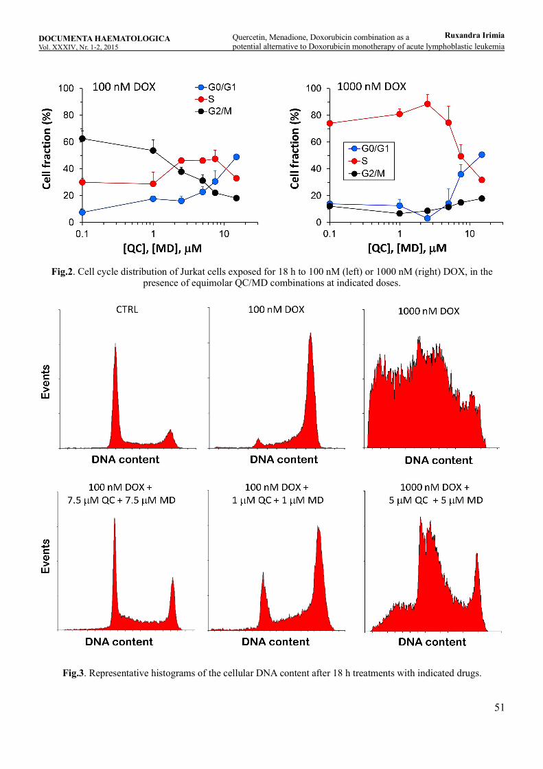
51
Fig.2. Cell cycle distribution of Jurkat cells exposed for 18 h to 100 nM (left) or 1000 nM (right) DOX, in the presence of equimolar QC/MD combinations at indicated doses.
Fig.3. Representative histograms of the cellular DNA content after 18 h treatments with indicated drugs.
DOCUMENTA HAEMATOLOGICAVol. XXXI , Nr. - , 20V 1 2 51
Ruxandra IrimiaQuercetin, Menadione, Doxorubicin combination as a potential alternative to Doxorubicin monotherapy of acute lymphoblastic leukemia
52
Fig.4. Apoptosis induction determined by the Annexin V/PI assay after exposure for 6 h (A) or 18 h (B) to indicated drugs.
Discussion Current data suggest that the inclusion of the QC/MD combination in doxorubicin-based treatment schemes for leukemia could improve the growth-suppressive effect of the therapeutic drug by promoting cell cycle arrest, oxidative stress generation and apoptosis induction, in a dose dependent manner. The primary mechanisms responsible for the cytotoxic effect of doxorubicin are generally recognized to be the formation of DNA adducts leading to topoisomerase II inhibition, and oxidative stress generation with subsequent release of intracellular reactive oxygen species. Doxorubicin's cardiotoxicity is most likely due to the latter mechanism, which may be enhanced considerably by the high mitochondrial density in cardiomyocytes and the high affinity for cardiolipin exhibited by doxorubicin. Quercetin generally exhibits both a pro-oxidant and an antioxidant character, with a primarily antioxidant effect exerted on healthy cells and a major pro-oxidant effect in neoplastic cells. This dual behavior could not only prevent healthy cell injury promoted by doxorubicin treatment, but also play a significant role as a protective agent against doxorubicin-induced cardiac toxicity. A relevant effect of doxorubicin observed in our current investigations at low doses of DOX (100 nM) consisted in G2/M phase cell cycle arrest, likely
determined by topoisomerase II inhibition and oxidative DNA injury. When using higher doxorubicin doses (1000 nM), S-phase arrest was detected, probably due to DNA breakage and ATM (ataxia telangiectasia mutated) activation. High levels of doxorubicin also induced consistent apoptotic rates in Jurkat cells. QC/MD equimolar combinations at physiological levels maintained the DOX-induced cell cycle arrest and enhanced DOX-induced apoptosis. In conclusion, this study shows that doxorubicin induces apoptosis, cell cycle arrest and oxidative stress in a dose-dependent manner in human leukemia Jurkat T cells. The cytotoxic effects of DOX and QC/MD at clinically relevant levels appear to be additive, which could allow for a reduction of the therapeutic doses of doxorubicin.
Acknowledgments This work was supported by a grant of the Romanian National Authority for Scientific Research, CNCS - UEFISCDI, project number PN-II-ID-PCE-2011-3-0800. Work for I.T.T. was supported by the Sectoral Operational Programme Human Resources Development (SOP HRD), financed from the European Social Fund and by the Romanian Government under the contract number POSDRU/187/1.5/S/156069. Work for R.G.S., O.E.B., V.C. and M.T.I. were supported by four Young Researchers Fellowships of the Romanian
DOCUMENTA HAEMATOLOGICAVol. XXXI , Nr. - , 20V 1 2 51
Ruxandra IrimiaQuercetin, Menadione, Doxorubicin combination as a potential alternative to Doxorubicin monotherapy of acute lymphoblastic leukemia
53
Ministry of Education, UEFISCDI, project numbers 6/2014, 8/2015, 6/2015 and 7/2015, respectively. The funders had no role in the study design, data collection and analysis, decision to publish, or preparation of the manuscript.
Abbreviations C M - H 2 D C F DA : 5 - ( a n d - 6 ) - c h l o r o m e t h y l -2',7'dichlorodihydrofluorescein diacetate, acetyl esterDMSO: dimethyl sulfoxide DOX: doxorubicinFITC: fluorescein isothiocyanate MD: menadione (2-methyl-1,4-naphthoquinone)NO: nitric oxidePBS: phosphate buffer salinePI: propidium iodideQC: quercetinROS: reactive oxygen speciesSS: standard salineTriX: Triton X-100 7-AAD: 7-Aminoactinomycin D
References[1] Nijveldt R.J., van Nood E., van Hoorn D.E.C., Boelens P.G., van Norren K., van Leeuwen P.A.M., Flavonoids: a review of probable mechanisms of action and potent ial applicat ions, Am J Clin Nutr 2001;74(4):418-425.[2] Baran I., Cell proliferation versus apoptosis. Mechanisms and particularities under genotoxic or oxidative stress conditions, “Carol Davila” University Publishing House, Bucharest, 2014.[3] Murakami A., Ashida H., Terao J., Multitargeted cancer prevention by quercetin, Cancer Lett 2008;269:315–325.[4] Yamamoto Y., Gaynor R.B., Therapeutic potential of inhibition of the NF-κB pathway in the treatment of inflammation and cancer, Clin Oral Investig 2001; 107(2):135–142.[5] Chen G., Wang F., Trachootham D., Huang P., Preferential killing of cancer cells with mitochondrial dysfunction by natural compounds, Mitochondrion 2010;10:614–625.[6] Jeong J.H., An J., Kwon Y.T., Rhee J.G., Lee Y.J., Effects of low dose quercetin: cancer specific inhibition of cell cycle progression, J Cell Biochem 2009;106:73-82.[7] Gibellini L., Pinti M., Nasi M., Montagna J.P., De Biasi S., Roat E., et al., Quercetin and cancer chemoprevention, Evid Based Complement Alternat Med 2011; 2011:591356.[8] Kelly G.S., Quercetin. Monograph, Altern Med Rev 2011;16(2):172-194.[9] Ferry D.R., Smith A., Malkhandi J., Fyfe D.W., deTakats P.G., Anderson D., et al., Phase I clinical trial
of the flavonoid quercetin: pharmacokinetics and evidence for in vivo tyrosine kinase inhibition, Clin Cancer Res 1996;2(4):659-668.[10] Yuan Z.P., Chen L.J., Fan L.Y., Tang M.H., Yang G.L., Yang H.S., et al, Liposomal quercetin efficiently suppresses growth of solid tumors in murine models, Clin Cancer Res 2006;12(10):3193-3199.[11] Baran I., Ganea C., Scordino A., Musumeci F., Barresi F., Tudisco S., et al., Effects of menadione, hydrogen peroxide and quercetin on apoptosis and delayed luminescence of human leukemia Jurkat T-cells, Cell Biochem Biophys 2010;58(3):169-179.[12] Ferrali M., Signorini C., Caciotti B., Sugherini L., Ciccoli L., Giachetti D., et al., Protection against oxidative damage of erythrocyte membrane by the flavonoid quercetin and its relation to iron chelating activity, FEBS Lett. 1997;416(2):123-129.[13] Baran I., Katona E., Ganea C., Quercetin as a fluorescent probe for the ryanodine receptor activity in Jurkat cells, Pflugers Arch Eur J Phy 2013;465(8):1101-1119.[14] Baran I., Ganea C., RyR3 in situ regulation by Ca2+ and quercetin and the RyR3-mediated Ca2+ release flux in intact Jurkat cells, Arch Biochem Biophys 2013;540(1-2):145-159.[15] Laux I., Nei A., Evidence that oxidative stress-induced apoptosis by menadione involves Fas-dependent and Fas-independent pathways, Clin Immunol 2001;101(3):335-344.[16] Floreani M., Carpenedo F., One- and two-electron reduction of menadione in guinea-pig and rat cardiac tissue, Gen Pharmacol 1992;23(4):757-762. [17] Baran I., Ionescu D., Filippi A., Mocanu M.M., Iftime A., Babes R., et.al., Novel insights into the antiproliferative effects and synergism of quercetin and menadione in human leukemia Jurkat T cells, Leuk Res 2014;38(7):836-849.[18] De Marchi U., Biasutto L., Garbisa S., Toninello A., Zoratti M., Quercetin can act either as an inhibitor or an inducer of the mitochondrial permeability transition pore: a demonstration of the ambivalent redox character o f p o l y p h e n o l s , B i o c h i m B i o p h y s A c t a 2009;1787(12):1425-1432.[19] Gewirtz D.A., A critical evaluation of the mechanisms of action proposed for the antitumor effects of the anthracycline antibiotics adriamycin and daunorubicin, Biochem Pharmacol 1999;57(7):727-741.[20] Skovsgaard T., Nissen N.I., Membrane transport of anthracyclines, Pharmacol Ther 1982;18(3):293-311.[21] Chaires J.B., Dattagupta N., Crothers D.M., Studies on interaction of anthracycline antibiotics and deoxyribonucleic acid: equilibrium binding studies on interaction of daunomycin with deoxyribonucleic acid, Biochemistry 1982;21(17):3933-3940.
DOCUMENTA HAEMATOLOGICAVol. XXXI , Nr. - , 20V 1 2 51
Ruxandra IrimiaQuercetin, Menadione, Doxorubicin combination as a potential alternative to Doxorubicin monotherapy of acute lymphoblastic leukemia
54
[22] Tewey K.M., Rowe T.C., Yang L., Halligan B., Liu L.F., Adriamycin-induced DNA damage mediated by mammalian DNA topoisomerase I I , Science 1984;226(4673):466-468.[23] Perego P., Corna E., De Cesare M., Gatti L., Polizzi D., Pratesi G., et al., Role of apoptosis and apoptosis-related genes in cellular response and antitumor efficacy of anthracyclines, Curr Med Chem 2001;8(1): 31-37.[24] Thorn C.F., Oshiro C., Marsh S., Hernandez-Boussard T., McLeod H., Klein T.E., et al., Doxorubicin pathways: pharmacodynamics and adverse effects, Pharmacogenet Genomics 2011;21(7):440-446.[25] Du G., Lin H., Wang M., Zhang S., Wu X., Lu L., et al., Quercetin greatly improved therapeutic index of doxorubicin against 4T1 breast cancer by its opposite effect on HIF-1α tumor and normal cells, Cancer Chemother Pharmacol 2010;65:277-287.[26] Wang G., Zhang J., Liu L., Sharma S., Dong Q., Quercetin potentiates doxorubicin mediated antitumor effects against liver cancer through p53/Bcl-xl, PLos One 2012;7(12):e51764.
DOCUMENTA HAEMATOLOGICAVol. XXXI , Nr. - , 20V 1 2 51
Ruxandra IrimiaQuercetin, Menadione, Doxorubicin combination as a potential alternative to Doxorubicin monotherapy of acute lymphoblastic leukemia