Rev Invest Desarr Pesq 17 43-53.pdf - OceanDocs
13
Morphological development of the mouth and improvement in feeding ability in the early larval stages of red porgy, Pagrus pagrus (L.) Item Type Journal Contribution Authors Aristizabal, E.O. Citation Revista de Investigación y Desarrollo Pesquero, 17. p. 43-53 Publisher Mar del Plata: Instituto Nacional de Investigación y Desarrollo Pesquero (INIDEP) Rights Publisher permission Download date 16/12/2021 17:08:18 Link to Item http://hdl.handle.net/1834/1560
Transcript of Rev Invest Desarr Pesq 17 43-53.pdf - OceanDocs

Morphological development of the mouth andimprovement in feeding ability in the early
larval stages of red porgy, Pagrus pagrus (L.)
Item Type Journal Contribution
Authors Aristizabal, E.O.
Citation Revista de Investigación y Desarrollo Pesquero, 17. p. 43-53
Publisher Mar del Plata: Instituto Nacional de Investigación y DesarrolloPesquero (INIDEP)
Rights Publisher permission
Download date 16/12/2021 17:08:18
Link to Item http://hdl.handle.net/1834/1560

43
MORPHOLOGICAL DEVELOPMENT OF THE MOUTH AND IMPROVEMENTIN FEEDING ABILITY IN THE EARLY LARVAL STAGES OF RED PORGY,
Pagrus pagrus (L.)*†
by
EDDIE O. ARISTAZABAL
Instituto Nacional de Investigación y Desarrollo Pesquero (INIDEP),Paseo Victoria Ocampo Nº l, Escollera Norte, B7602HSA - Mar del Plata, Argentina
e-mail: [email protected]
SUMMARY
The appearance, ossification and growth of cartilaginous structures of the mouth in early larval stages of laboratoryreared red porgy, Pagrus pagrus, were examined. At the time of mouth opening, red porgy larvae are equipped with thefundamental components of the oral cavity to allow the sucking motion for acquiring food. During the initial feedingstage, from day 5 after hatching (AH) to day 7 AH, the hyoid and lower branchial archs were completed, and the headlength and other structural elements, like the ceratohyal-epihyal cartilage, increased rapidly. These events intensifiedsucking mechanisms by increasing the amount of ingested rotifers. After day 13 AH, the presence of the bony pre-maxilla and angular, the ossification of the Meckel´s cartilage and the appearance of pharyngeal teeth (ceratobranchialteeth), allowed grasping of food items. Finally, at the end of the study period, the appearance of new elements, the ossi-fication of previous ones, and the improvement of swimming capacity, increased the amount of ingested rotifers by thecombination of sucking and grasping mechanisms. As a result of the cartilaginous-osteological development of themouth, a modification of the feeding schedule of red porgy larvae reared in captivity is proposed.
RESUMEN
Desarrollo morfológico de la boca y mejoras de la habilidad alimentaria en los estadios larvales tempranos delbesugo Pagrus pagrus (L.). Se examinó la aparición de las estructuras cartilaginosas de la boca y su osificación y creci-miento en larvas del besugo Pagrus pagrus obtenidas en laboratorio. Al momento de la apertura de la boca, las larvasestán equipadas con los elementos estructurales fundamentales de la cavidad oral para adquirir alimento por succión.Durante la fase inicial de alimentación, entre el quinto y séptimo día después de la eclosión (DDE), se completó la for-mación de los arcos hioide y branquial inferior, a la vez que se incrementó rápidamente el largo de la cabeza y de otroselementos, como el cartílago ceratohial-epihial. Estas estructuras mejoran la capacidad de succión que se refleja en unaumento del número de rotíferos ingeridos. Transcurridos 13 DDE, la presencia del premaxilar y angular óseos, elcomienzo de la osificación del cartílago de Meckel y la aparición de los dientes faríngeos (ceratobranquial), permitierondesarrollar también el mecanismo de alimentación por captura. Finalmente, durante las últimas etapas del estudio, las lar-
*Contribución INIDEP Nº 1353†This work is part of the Doctorate Thesis degree of the author.
REV. INVEST. DESARR. PESQ. Nº 17: 43-53 (2005)
44 REV. INVEST. DESARR. PESQ. Nº 17: 43-53 (2005)
vas de besugo utilizaron para alimentarse una combinación de succión y captura, posibilitadas por la aparición de nuevos elementos estruc-turales, por la osificación de otros pre-existentes y por la mejora de la capacidad de natación. A partir de los resultados del estudio del des-arrollo cartilaginoso-osteológico, se propone modificar el esquema alimenticio de las larvas de besugo criadas en cautiverio.
Key words: Pagrus pagrus, larval development, feeding, osteology, mouth parts, marine fish culture.Palabras clave: Pagrus pagrus, desarrollo larval, alimentación, osteología, partes de la boca, cultivo de peces marinos.
INTRODUCTION
The red porgy, Pagrus pagrus, is an importantcommercial species from the coasts of BuenosAires Province, Argentina. Natural spawning of abroodstock has been achieved through photoperi-od and temperature manipulation at the NationalFisheries Research and Development Institute(INIDEP) (Aristizabal Abud et al., 1997;Aristizabal Abud, 2003). For this reason, thestudy of larval development of red porgy is a keyfeature not only to understand those factorsaffecting early stages but also to improve aqua-culture techniques applied to this species.
The transition from endogenous to exoge-nous feeding is one of the most critical periodsin the development of marine fish larvae(Conides and Glamuzina, 2001; Moteki et al.,2001) and is the cause of mass mortalities(Kamler, 1992), which have been clearlyobserved in captivity (Kanazawa et al., 1989;Nakagawa et al., 1991; Ottera, 1993; Kjorsvikand Holmefjord, 1995; Hernández-Cruz et al.,1999; Dou et al., 2003). Kohno et al. (1997)examined the osteological development of theoral cavity in early stages of Epinepheluscoioides and concluded that the delayed devel-opment of the feeding apparatus caused difficul-ties in larval rearing. Machinandiarena et al.(2000) studied external morphologic develop-ment of larvae and juvenile red porgy reared incaptivity, while Aristizabal Abud (2003), pro-vided detailed information on the energeticevents of the transition from endogenous toexogenous nutritional sources in the samespecies.
This paper reports the cartilaginous-osteologi-cal development of the mouth in early stages of P.pagrus larvae, in order to have a better under-standing of their feeding mechanisms and theeffect of the appearance of those structures ontheir feeding ability.
MATERIALS AND METHODS
Fertilized eggs of P. pagrus were collectedfrom natural spawnings of the broodstock at theLaboratory of Mariculture at INIDEP, with aplanktonic net 300 µm mesh, and transferred to acircular 0.5 m3 fiberglass tank. During incuba-tion and larval rearing, water temperature waskept at 18.0 ± 0.5 °C, salinity ranged from 33‰to 34‰, and the photoperiod was set to 12 h L:12 h D. Two experimental tanks connected to arecirculation system were stocked with 26,000larvae each. Larvae were fed with rotifers(Brachionus plicatilis) at densities greater than10 individuals ml-1.
From time of hatching to day 21 after hatching(AH), 20 larvae were daily removed from thetanks, anesthetized and preserved in 10% neutral-ized formalin. Before preservation (Klaoudatos etal., 1990), total length was measured on livespecimens from the tip of the snout to the end ofthe caudal fin. Head length was measured fromthe tip of the snout to the posterior margin of thecleithrum. Larvae were stained for bones and car-tilage following a modification of the methoddescribed by Potthoff (1984) and Taylor and vanDyke (1985). Mouth opening was measuredusing the length of the Meckel´s cartilage accord-ing to the formulae proposed by Shirota (1970):
Mo = 2 x Mc
Heartbeat rate was measured under the micro-scope only at time of measurement and for 1minute in order to avoid changes of heartbeat rateowing to extreme conditions of light and temper-ature under the microscope.
RESULTS
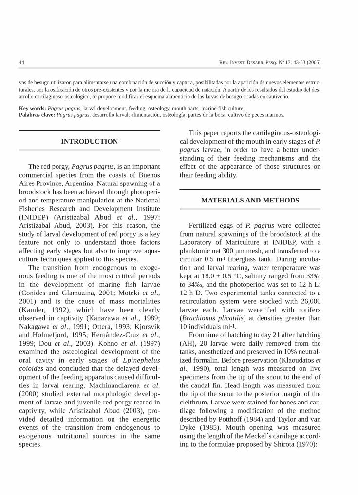
The eggs of the red porgy are spherical with amean diameter of 903 ± 12 m. They have a single oilglobule with a diameter of 180-220 µm. Hatchingoccurred after 48-50 hours at 18 °C. Newly hatchedlarvae of P. pagrus have a mean total length of 2.32± 0.12 mm. Eyes are unpigmented, the mouth isundifferentiated and the anus is closed. The diges-
tive tract is visible as a straight thin tube over theyolk sac. With the exception of sporadic pulses ofactivity, larvae do not move during the first 24hours. The body of the larva is surrounded by theprimordial fin. There are 2-3 chromatophore cellson the yolk sac and the oil globule, and several oth-ers randomly located on the sides of the body.
At day 1 AH, the mean total length of the larvais 2.86 ± 0.12 mm, otoliths become visible at thehead region, and mean heartbeats are 81 min-1.The pigmentation pattern has not changed.Xanthophores were distinguished all over thebody, mainly in the head and around the tail, andthe body is almost straight. At day 2 AH, themean total length of the larva is 3.11 ± 0.10 mm,blood circulation is not yet visible, and heartbeatsincrease to 148 min-1. Auditory capsules are visi-ble and the convolution process of the digestivetract has started (Figure 1).
45ARISTIZABAL: DEVELOPMENT OF THE MOUTH IN EARLY STAGE LARVAE OF RED PORGY
Figure 1. Image of a yolk sac larvae of red porgy 2 days after hatching.Figura 1. Larva de besugo con vitelo dos días después de la eclosión.

46 REV. INVEST. DESARR. PESQ. Nº 17: 43-53 (2005)
Figure 2. Development of the oral cavity and related structures in the larvae of red porgy. A) 3 days after hatching (DAH). B) 5DAH. C) 21 DAH. D) Branchial arch 5 DAH. ACp = anterior-directed coracoid process; An = angular; Bb = basibranchial;Br = brachiostegal ray; Cb1 = ceratobranchial 1; CEc = ceratohyal-epihyal cartilage; Ch = ceratohyal; Cl = cleithrum;CSc = coraco-scapular cartilage; De = dentary; Ec = ethmoid cartilage; EEb = ectethmoid bar; Et = epiphysial tectum; Fr= frontal; Hb1 = hypobranchial 1; Hh = hypohyal; Hm = hyomandibular; Ih = Interhyal; Mc = Meckel´s cartilage; Mx =maxilla; PCc = palatoquadrate cartilage; PCp = posterior-directed coracoid process; Pmx = premaxilla; Pop = pre-oper-cle; Qu = quadrate; RAc = retroarticular cartilage; Sc = sclerotic cartilage; SCl = supracleithrum; SHc = symplectic-hyomandibular cartilage; Sy = symplectic; Tr = trabecula. Stripped area, cartilage; open area, ossification.
Figura 2. Desarrollo de la cavidad oral y estructuras relacionadas de larvas de besugo. A) 3 días después de la eclosión (DDE).B) 5 días DDE. C) 21 días DDE. D) Arcos branquiales, 5 días DDE. ACp = proceso coracoide anterior; An = angular;Bb = basibranquial; Br = radio braquiostegal; Cb1 = ceratobranquial 1; CSc = cartílago coraco-escapular; Ch = cera-tohial; Cl = cleitro; De = dentario; Ec = cartílago etmoides; EEb = barra ectetmoides; Et = téctum epifisial; CEc = car-tílago ceratohial-epihial; Fr = frontal; Hb1 = hipobranquial 1; Hh = hipohial; Hm = hiomandibular; Ih = Interhial; Mc= cartílago de Meckel; Mx = maxilar; PCc = cartílago palatocuadrado; PCp = proceso coracoide posterior; Pmx = pre-maxilar; Pop = preopérculo; Qu = cuadrado; RAc = cartílago retroarticular; Sc = cartílago esclerótico; SCl = supra-cleitro; SHc = cartílago simpléctico-hiomandibular; Sy = simpléctico; Tr = trabécula. La zona sombreada representa elcartílago y la blanca la osificación.

At day 3 AH, the mean total length of the larvais 3.24 ± 0.70 mm. Eyes become pigmented andmove. The mouth is opened and the digestivetract convulses and differentiates. The heartbeatsreach 160 min-1. Xanthophore cells decreased innumber and belt to the posterior spine and oper-cular area. The bony cleithrum and the coraco-scapular cartilage appear, bearing the recentlyformed pectoral fins (Figure 2 A). The first jawelement to appear was the Meckel´s cartilage.The bony maxilla was the first element to appearin the upper jaw. The symplectic-hyomandibularcartilage first appeared as an element of the sus-pensorium. The ceratohyal-epihyal cartilage fromthe hyoid arch was detected. The cartilaginousquadrate was detected in front of the symplectic-hyomandibular cartilage. The cartilaginous basi-branchial and the two anterior-most cerato-branchials appeared as lower arch elements.
At day 4 AH, the mean total length of thelarva is 3.32 ± 0.15 mm. Blood circulation isvisible. The mouth and the digestive tract arefunctional, and food particles can be found. Theedge of the primordial fin is serrated. Number ofchromophores has decreased while xanthophorecells have disappeared. Only a small part of theoil globule and yolk sac still remains. Heartbeatsincrease to 260 min-1. At day 5 AH, the meantotal length of the larva is 3.54 ± 0.13 mm. Theyolk sac and oil globule have disappeared. Adense blood circulation around organs can beobserved. The primordial fin is not divided yetexcept a small notch appearing just behind thehead region. The symplectic-hyomandibularcartilage presents a small foramen in the upperpart, while the coraco-scapular cartilage devel-ops a small anterior-directed coracoid processon the anteroventral part (Figure 2 B). Thehypohyal and interhyal cartilages wereobserved. The hypobranchial 1 and cerato-branchial 3 and 4 were detected at the base ofthe arch (Figure 2 D).
At day 6 AH, the mean total length of thelarva is 3.75 ± 0.12 mm. Traces of the caudal fin
rays appear, and the olfactory organ appears onthe snout of the larva. The chromophore cellsdisappear except for some, randomly locatedalong the digestive tract. Some individuals bear-ing swim bladder were detected. Hearbeatsdecrease to 206 min-1. At day 8 AH, the meantotal length of the larva is 4.30 ± 0.15 mm. Thedigestive tract is complete, functional and fullof rotifers. The lateral arteries of the body arevisible. The opercle and pre-opercle areobserved. The bony dentary is formed in theanterior part of the Meckel´s cartilage.Ossification processes began at the lower andupper part of the symplectic-hyomandibularcartilage and on the anterior part of the cerato-hyal-epihyal cartilage.
Larvae at day 10 AH, present a cartilaginousoccipital process and a single bony brachiostegalray. The cartilaginous palatine and the retroarticu-lar cartilages were first detected. Between days 13-15 AH, an anterodorsally directed projection fromthe posterodorsal part of the Meckel´s cartilagestarted to form, while the bony angular ossificatesfrom the posterioventral part of the Meckel´s carti-lage. The bony premaxilla and the ossification ofthe outer part of the ceratohyal-epihyal cartilagewere observed (Figure 2 C). The quadrate becamecontiguous to the palatine to form the palato-quadrate cartilage A single tooth located at the cer-atobranchial-1 was observed in some individuals.
The bony supracleithrum was observed at day16 AH, with a posterior-directed spine evident atthe end of the study period. The parasphenoid andthe starting of notochord flexion were detected.By day 21 AH, a single tooth was observed on thedentary. The partial ossification of the symplectic-hyomandibular and quadrate cartilage was alsodetected at this time. Four branchiostegal rayswere evident in the hyoid arch (Figure 2 C).
Head length and mouth opening increased lin-early with time (Figure 3), while the ceratohyal-epihyal cartilage showed an asymptotic growthcurve. Number of rotifers in the gut increasedwith time (Figure 4).
47ARISTIZABAL: DEVELOPMENT OF THE MOUTH IN EARLY STAGE LARVAE OF RED PORGY

DISCUSSION
Most teleost ingest their food by suckingmechanisms created by the negative pressure ofthe oral cavity (Gerking, 1994). At the time ofmouth opening, red porgy larvae are equippedwith the following fundamental components ofthe oral cavity (Figure 5): the trabecula of theneurocranium comprising the roof of the oralcavity; the ceratohyal and epihyal from thehyoid arch, and some other elements of thelower branchial arch forming the cavity floor;the quadrate and symplectic-hyomandibularcartilages comprising the sides; and finally, thebony maxilla and the Meckel´s cartilage as jawelements. The bony cleithrum and coraco-scapular cartilage were also present at the time
48 REV. INVEST. DESARR. PESQ. Nº 17: 43-53 (2005)
Figure 3. Changes in head (A) and ceratohyal-epihyal carti-lage length (B), and mouth opening (C) in larvae ofred porgy.
Figura 3. Cambios en la longitud de la cabeza (A), en la lon-gitud del cartílago ceratohial-epihial (B) y en eltamaño de la boca (C) de larvas de besugo.
Figure 4. Number of rotifers found in the gut of red porgylarvae. The arrow indicates the time of mouth open-ing. Vertical bars show standard error. Samplingwas performed one hour after adding rotifers to thelarvae tank.
Figura 4. Cantidad de rotíferos encontrados en el intestino delas larvas de besugo. La flecha indica el momento dela apertura de la boca. Las barras verticales indicanel error estándar. El muestreo se realizó una horadespués del agregado de rotíferos al tanque de larvas.

of mouth opening. Matsuoka (1987) described alarge levator arcus palatini (peripheral headmuscles) 11 hs after mouth opening in larvae ofPagrus major, which expands oral andbranchial cavities allowing sucking motion toacquire food. Based on the development of themouth in early stages of the red porgy, previ-ously mentioned muscles must be present inthis species too, when initial feeding starts, 24h after mouth opening. It can be stated thatfeeding during this time was performed exclu-sively by sucking mechanisms, because ele-ments that facilitate the grasping of food, suchas the premaxilla and jaw teeth, were not pres-ent (Kohno et al., 1996 a). During the first daysof exogenous feeding, from day 5 to 7 afterhatching, hyoid and lower branchial archs werecompletely formed (Figure 5). At the same timethe length of the head and the ceratohyal-epi-hyal cartilage were rapidly increased. Redporgy larvae feed more as they grow, so theimprovement in their sucking ability during thisstage would depend mainly on size develop-ment (Moteki et al., 2001).
The respiratory gas exchange is basicallyperformed by the skin until the time of mouthopening. Thus, the functional development ofthe branchial system must be completed whenlarvae reach a certain critical size, which isaround day 6 AH in this species (AristizabalAbud, 2003). Heartbeats decreased after mouthopening, showing the participation of thebranchial system in the respiration.
After day 8 AH, several developmentsoccurred. These included the appearance of newstructures in the oral cavity, like the dentary,opercle and preopercle. The general growth of thelarvae, the ossification of existing elements and arapid increase in bone length of the oral cavitysuggest an enhancement of feeding by suckingmechanisms (Figure 5). Kohno et al. (1996 b)described the shift in feeding mechanisms fromsucking to grasping, in larval sea bass Lates cal-carifer. From day 13 AH, the appearance of the
bony premaxilla and angular, the ossification ofMeckel´s cartilage and the appearance of lowerpharyngeal teeth (ceratobranchial), would allowfeeding by grasping mechanisms (Moteki, 2002).At day 16 AH, swimming became more activeand caudal propulsion increased supported by theappearance of new fin elements and the startingof the notochord flexion. According toMachinandiarena et al. (2000), postflexion stagewas detected from day 29 AH. As larvae grew,they started to swim against the current, anddeployed attacking movements over rotifers.
Larvae of P. pagrus share common osteolog-ical characters of sparids, like spines on the pre-opercle and supracleithrum (Houde andPotthoff, 1976; Hussain et al., 1981; Griswoldand McKenney, 1984; Matsuoka, 1987; Zieske,1989; Fukuhara, 1991; Tucher and Alshuth,1997). The supraoccipital crest is not diagnosticof sparids, but have been reported in a fewspecies (Leis et al., 2002).
Leis et al. (2002), stated that the Atlanticspecies Pagrus pagrus have virtually identicalhead spination to that of Pagellus species(Figure 6), while the Mediterranean Pagruspagrus differ in having about one less spine inall series. The same authors hypothesize thatPagellus affinis, Pagellus bellotti and Pagelusnatalensis and Pagrus pagrus are closely relat-ed, and that the other described larvae of Pagrus(the indo-Pacific P.auratus and P. major), do notbelong to the spiny Pagellus-Pagrus pagrusclade. Although solving this problem is beyondthe scope of this paper, Leis et al. (2002) pro-posed an interesting taxonomic maze of rela-tionships between sparid fishes, which will beimportant for the development of specific rear-ing techniques for Pagellus-Pagrus larvae.
The present schedule of red porgy seed pro-duction at INIDEP follows the general develop-ment of the larvae. Rotifers are the first live fooditem offered to larvae in the rearing tank fromthe time of mouth opening to day 30 AH. Theshift to a second live food item (Artemia sp.),
49ARISTIZABAL: DEVELOPMENT OF THE MOUTH IN EARLY STAGE LARVAE OF RED PORGY

50 REV. INVEST. DESARR. PESQ. Nº 17: 43-53 (2005)
Figure 5. Schematic representation of the development of elements comprising the mouth and fins of red porgy. (•) Appearanceof cartilaginous element; (•) appearance of ossified bone or the beginning of ossification of cartilaginous element.Development refers to the enhancement of feeding ability and changes of feeding mode. For feeding mechanism, thick-ness of the arrows indicates the degree of ability in each mode.
Figura 5. Representación esquemática del desarrollo de los elementos de la cavidad oral y aletas de besugo. (•) Simboliza la apa-rición de un elemento cartilaginoso y (•) la aparición de un elemento óseo o el inicio de la osificación del cartílago. Eldesarrollo hace referencia a los principales eventos que contribuyen a la mejora de los mecanismos alimenticios. El gro-sor de la flecha indica la capacidad desarrollada para cada tipo de mecanismo alimenticio.

occurs from day 20 AH. Artemia nauplii is acrustacean that actively move along the watercolumn in the rearing tank. At this time, feedingby sucking alone would not be an efficientmechanism to grasp Artemia nauplii. Based onthe feeding abilities developed by the red porgylarvae and the mouth width, it would be possibleto schedule the time of Artemia nauplii supply tostart by day 13 AH, since larvae are ready tofeed by grasping mechanism. The possibility ofreducing the period of rotifer supply is veryimportant in terms of production workload, witha consequent reduction of costs.
ACKNOWLEDGEMENTS
The author wishes to thank Lic. LauraMachinandiarena for helping in the dyeing tech-nique of larvae and the review of this paper. AlsoLic. Andrea López and Lic. Mariana Cadaveirafor the supply of microalgae and rotifers.
REFERENCES
ARISTIZABAL ABUD, E.O. 2003. Bioenergética delbesugo, Pagrus pagrus (L.). Tesis de
Doctorado, Facultad de Ciencias Exactas yNaturales, Universidad Nacional de Mar delPlata, 145 pp.
ARISTIZABAL ABUD, E.O., MULLER, M., BAMBIL,G., LOPEZ, A., SABATINI, M., COSTAGLIOLA, M.,INCORVAIA, S., VEGA, A., CARRIZO, J.C. &MANCA, E. 1997. Producción de alimento vivoy cría de besugo. Período 1995-1996. Inf. Téc.Int. DNI-INIDEP Nº 83/97, 92 pp.
CONIDES, A.J. & GLAMUZINA, B. 2001. Study onthe early larval development and growth of thered porgy, Pagrus pagrus with emphasis onthe mass mortalities observed during thisphase. Sci. Mar., 65 (3): 193-200.
DOU, S., MASUDA, R., TANAKA, M. &TSUKAMOTO, K. 2003. Identification of factorsaffecting the growth and survival of the set-tling japanese flounder larvae, Paralichthysolivaceus. Aquaculture, 218: 309-327.
FUKUHARA, O. 1991. Size and age at transforma-tion in red sea bream, Pagrus major, reared inthe laboratory. Aquaculture, 95: 117-124.
GERKING, S.D. 1994. Feeding ecology of fishes.Academic Press, London, 420 pp.
GRISWOLD, C.A. & MCKENNEY, T.W. 1984.Larval development of the scup, Stenotomuschrysops (Pisces: Sparidae). Fish. Bull., U. S.,82: 77-84.
HERNÁNDEZ-CRUZ, C.M., SALHI, M., BESSONART,M., IZQUIERDO, M.S., GONZÁLEZ, M.M. &
51ARISTIZABAL: DEVELOPMENT OF THE MOUTH IN EARLY STAGE LARVAE OF RED PORGY
Figure 6. 7.75 mm larvae of Pagrus pagrus reared at the laboratory (from Machinandiarena et al., 2000). Note supraoccipitalspine and strong preopercular spination.
Figura 6. Larva de Pagrus pagrus de 7,75 mm nacida en laboratorio (tomada de Machinandiarena et al., 2000). Se observan laespina supraoccipital y las espinas preoperculares.
FERNÁNDEZ-PALACIOS, H. 1999. Rearing tech-niques for red porgy (Pagrus pagrus) duringlarval development. Aquaculture, 179: 489-497.
HOUDE, E.D. & POTTHOFF, T. 1976. Egg and lar-val development of the sea breamArchosargus rhomboidalis (Linnaeus): Pisces,Sparidae. Bull. Mar. Sci., 26: 506-529.
HUSSAIN, N., AKATSU, S. & EL-ZAHAR, C. 1981.Spawning, egg and early larval development,and growth of Acanthopagrus cuvieri(Sparidae). Aquaculture, 22: 125-136.
KAMLER, E. 1992. Early Life History of Fish. AnEnergetic approach. Chapman & Hall,London, 183 pp.
KANAZAWA, A., KOSHIO, S. & TESHIMA, S. 1989.Growth and survival of larval red sea breamPagrus major and japanese flounderParalichthys olivaceus fed microbond diets. J.World Aquacult. Soc., 20 (2): 31-37
KJORSVIK, E. & HOLMEFJORD, I. 1995. Atlantichalibut (Hippoglossus hippoglossus) and cod(Gadus morhua). In: BROMAGE, N.R. &ROBERTS, R.J. (Eds.). Broodstock manage-ment and egg and larval quality, BlackwellScience, Oxford, 169-196.
KLAOUDATOS, S., TSEVIS, N. & CONIDES, A. 1990.Energy sources during early larval develop-ment of the European Sea Bass, Dicentrarchuslabrax (L.). Aquaculture, 87: 361-372.
KOHNO, H., ORDONIO-AGUILAR, R., OHNO, A. &TAKI, Y. 1996 a. Morphological aspects of fee-ding mode and improvement of feeding abilityin early stage larvae of the milkfish, Chanoschanos. Ichthyol. Res., 43: 133-140.
KOHNO, H., ORDONIO-AGUILAR, R., OHNO, A. &TAKI, Y. 1996 b. Osteological development ofthe feeding apparatus in early stage of larvaeof the sea bass, Lates calcarifer. Ichthyol.Res., 43: 1-9.
KOHNO, H., ORDONIO-AGUILAR, R., OHNO, A. &TAKI, Y. 1997. Why is grouper larval rearingdifficult? An approach from the developmentof the feeding apparatus in early stage larvae
of the grouper, Epinephelus coioides.Ichthyol. Res., 44: 267-274.
LEIS, J.M., TRNSKI, T. & BECKLEY, L.E. 2002.Larval development of Pagellus natalensisand what larval morphology indicates aboutrelationships in the perciform fish familySparidae (Teleostei). Mar. Freshwater Res.,53: 367-376
MACHINANDIARENA, L., MÜLLER, M., LÓPEZ, A.,ARISTIZABAL ABUD, E. & BAMBILL, G. 2000.Desarrollo de los estadios iniciales del besugo(Pagrus pagrus) en cautiverio, en la Provinciade Buenos Aires, Argentina. Inf. Téc. Int.DNI-INIDEP Nº 30/00, 16 pp.
MATSUOKA, M. 1987. Development of the skele-tal tissues and skeletal muscles in red seabream. Bull. Seikai Reg. Fish. Res. Lab., 65:1-114.
MOTEKI, M. 2002. Morphological aspects of fee-ding and improvement in feeding ability in theearly larval stages of red sea bream Pagrusmajor. Fish. Sci., 68: 996-1003.
MOTEKI, M., ISHIKAWA, T., TERAOKA, N. &FUSHIMI, H. 2001. Transition from endoge-nous to exogenous nutritional sources in larvalsea bream, Pagrus major. Suisanzoshoku, 49:323-328.
NAKAGAWA, H., IMABAYASHI, H., KUROKURA, H.& KASAHARA, S. 1991. Changes in body cons-tituents of young red sea bream, Pagrusmajor, in reference to survival during experi-mental stocking. Biochem. Syst. Ecol., 19 (2):105-110.
OTTERA, H. 1993. Feeding, growth, and survivalof Atlantic cod (Gadus morhua) larvae rearedin replicate plastic enclosures. Can. J. Fish.Aquat. Sci., 50 (5): 913-924
POTTHOFF, T., 1984, Clearing and staining techni-ques. In: MOSER, H.G., RICHARDS, W.J.,COHEN, D.M., FAHAY-JR., M.P. KENDALL A.W.& RICHARDSON, S.L. (Eds.). Ontogeny andSystematics of Fishes. American Society ofIchthyologists and Herpetologists, (Spec.Publ. 1), Lawrence: 35-37.
52 REV. INVEST. DESARR. PESQ. Nº 17: 43-53 (2005)

SHIROTA, A. 1970. Studies on the mouth size offish larvae. Bull. Jap. Soc. Sci. Fish., 36 (4):353-368.
TAYLOR, W.R. & VAN DIKE, G.C. 1985. Revisedprocedures for staining and clearing small fis-hes and other vertebrates for bone and cartila-ge study. Cybium, 9 (2): 107-119.
TUCKER, J.W. & ALSHUTH, S.R. 1997.Development of laboratory-reared sheepshe-
ad, Archosargus probatocephalus (Pisces:Sparidae). Fish. Bull., U. S., 95: 394-401.
ZIESKE, G.G. 1989. Redescription of larvae of thepinfish, Lagodon rhomboides (Linnaeus)(Pisces, Sparidae). Contr. Mar. Sci., 31: 51-91.
Recibido: 15-04-2004Aceptado: 24-10-2004
53ARISTIZABAL: DEVELOPMENT OF THE MOUTH IN EARLY STAGE LARVAE OF RED PORGY