RESTORING FUNCTION AFTER SPINAL CORD...
15
RESTORING FUNCTION AFTER SPINAL CORD INJURY Daniel Becker, MD, Cristina L. Sadowsky, MD and John W. McDonald, MD, PhD BACKGROUND– By affecting young people during the most productive period of their lives, spinal cord injury (SCI) is a devastating problem for modern society. A decade ago, treating SCI seemed frustrating and hopeless because of the tremendous morbidity and mortality, life-shattering impact, and limited therapeutic options associated with the condition. Today, however, an understanding of the underlying pathophysiological mechanisms, the development of neuroprotective interventions, and progress toward regenerative interventions are increasing hope for functional restoration. REVIEW SUMMARY– This study addresses the present understanding of SCI, including the etiology, pathophysiology, treatment, and scientific advances. The discussion of treatment options includes a critical review of high-dose methyl- prednisolone and GM-1 ganglioside therapy. The concept that limited rebuilding can provide a disproportionate improve- ment in quality of life is emphasized throughout. CONCLUSIONS– New surgical procedures, pharmacologic treatments, and functional neuromuscular stimulation meth- ods have evolved over the last decades that can improve functional outcomes after spinal cord injury, but limiting secondary injury remains the primary goal. Tissue replacement strategies, including the use of embryonic stem cells, become an important tool and can restore function in animal models. Controlled clinical trials are now required to confirm these observations. The ultimate goal is to harness the body’s own potential to replace lost central nervous system cells by activation of endogenous progenitor cell repair mechanisms. KEY WORDS excitotoxicity, regeneration, rehabilitation, spinal cord injury, stem cell (THE NEUROLOGIST 9:1–15, 2003) The annual incidence of traumatic spinal cord injury (SCI) in the United States is approximately 40 cases per million. The estimated prevalence is between 183,000 and 230,000 cases (1–3), of which more than 80% are male (4). Moreover, 55% of SCIs occur between ages 16 and 30 years, the average age at injury being 32.1 years. The incidence, prevalence, and causes of SCI vary greatly in other countries; rates of occupational SCI tend to be higher outside the United States. The most frequent neurologic category of traumatic SCI is incomplete tetraplegia (30.2%), followed by complete para- plegia (26.1%), complete tetraplegia (23.3%), and incomplete paraplegia (19.7%) (5). Since 1994, motor vehicle crashes have accounted for 40.7% of injuries in this country, followed by acts of violence (primarily gunshot wounds) (21.8%), falls (21.3%), and rec- reational activities (7.9%) (5). More than 88% of people who leave the hospital after treatment for SCI go to private, noninstitutional resi- dences. However, average yearly health care expenses and estimated lifetime costs directly attributable to SCI vary greatly according to age and severity of injury. Lifetime costs range from $300,000 for a person who suffers an incomplete motor lesion at any level at age 50 years to $1.7 From the Department of Neurology, Spinal Cord Injury Neuro-Reha- bilitation Section, Restorative Treatment and Research Program (D.B., C.L.S., J.W.M.); Center for the Study of Nervous System Injury (D.B., C.L.S., J.W.M.); and Department of Neurological Surgery (J.W.M.), Washington University School of Medicine, St Louis, Missouri. Send reprint requests to Dr John W. McDonald, Department of Neu- rology, Restorative Treatment and Research Program, Campus Box 8518, Washington University School of Medicine, 4444 Forest Park Blvd, St Louis, MO, 63108. E-mail: [email protected] 1
Transcript of RESTORING FUNCTION AFTER SPINAL CORD...
RESTORING FUNCTION AFTERSPINAL CORD INJURYDaniel Becker, MD, Cristina L. Sadowsky, MD and John W. McDonald, MD, PhD
BACKGROUND– By affecting young people during the most productive period of their lives, spinal cord injury (SCI) is adevastating problem for modern society. A decade ago, treating SCI seemed frustrating and hopeless because of thetremendous morbidity and mortality, life-shattering impact, and limited therapeutic options associated with the condition.Today, however, an understanding of the underlying pathophysiological mechanisms, the development of neuroprotectiveinterventions, and progress toward regenerative interventions are increasing hope for functional restoration.
REVIEW SUMMARY– This study addresses the present understanding of SCI, including the etiology, pathophysiology,treatment, and scientific advances. The discussion of treatment options includes a critical review of high-dose methyl-prednisolone and GM-1 ganglioside therapy. The concept that limited rebuilding can provide a disproportionate improve-ment in quality of life is emphasized throughout.
CONCLUSIONS– New surgical procedures, pharmacologic treatments, and functional neuromuscular stimulation meth-ods have evolved over the last decades that can improve functional outcomes after spinal cord injury, but limitingsecondary injury remains the primary goal. Tissue replacement strategies, including the use of embryonic stem cells,become an important tool and can restore function in animal models. Controlled clinical trials are now required to confirmthese observations. The ultimate goal is to harness the body’s own potential to replace lost central nervous system cellsby activation of endogenous progenitor cell repair mechanisms.
KEY WORDS excitotoxicity, regeneration, rehabilitation, spinal cord injury, stem cell
(THE NEUROLOGIST 9:1–15, 2003)
The annual incidence of traumatic spinal cord injury(SCI) in the United States is approximately 40 cases permillion. The estimated prevalence is between 183,000 and230,000 cases (1–3), of which more than 80% are male (4).Moreover, 55% of SCIs occur between ages 16 and 30 years,the average age at injury being 32.1 years. The incidence,prevalence, and causes of SCI vary greatly in other countries;
rates of occupational SCI tend to be higher outside theUnited States.
The most frequent neurologic category of traumatic SCIis incomplete tetraplegia (30.2%), followed by complete para-plegia (26.1%), complete tetraplegia (23.3%), and incompleteparaplegia (19.7%) (5).
Since 1994, motor vehicle crashes have accounted for40.7% of injuries in this country, followed by acts of violence(primarily gunshot wounds) (21.8%), falls (21.3%), and rec-reational activities (7.9%) (5).
More than 88% of people who leave the hospital aftertreatment for SCI go to private, noninstitutional resi-dences. However, average yearly health care expenses andestimated lifetime costs directly attributable to SCI varygreatly according to age and severity of injury. Lifetimecosts range from $300,000 for a person who suffers anincomplete motor lesion at any level at age 50 years to $1.7
From the Department of Neurology, Spinal Cord Injury Neuro-Reha-bilitation Section, Restorative Treatment and Research Program (D.B.,C.L.S., J.W.M.); Center for the Study of Nervous System Injury (D.B.,C.L.S., J.W.M.); and Department of Neurological Surgery (J.W.M.),Washington University School of Medicine, St Louis, Missouri.
Send reprint requests to Dr John W. McDonald, Department of Neu-rology, Restorative Treatment and Research Program, Campus Box8518, Washington University School of Medicine, 4444 Forest ParkBlvd, St Louis, MO, 63108. E-mail: [email protected]
1
million for someone who develops high tetraplegia at age25 years (6).
Over the last decade, the life expectancy of individualswith SCI has continued to improve, and it now approachesnormal for young individuals suffering lower level injuries.Mortality rates are higher during the first year after injury,particularly for individuals with high cervical lesions andassociated multiorgan injuries. In the past, the leading causeof death was renal failure, but advances in urological man-agement have dramatically lowered rates of genitourinarycomplications. Today, pneumonia, pulmonary emboli, andsepticemia are the leading causes of death in individuals withSCI (4).
The epidemiology of SCI is largely restricted to trau-matic causes, because state and federal registries are availablefor traumatic etiologies but not for nontraumatic causes.Estimates of the incidence and prevalence of nontraumaticcauses are conservatively 4 to 5 times those corresponding totraumatic etiologies.
MECHANISMS OF SCI
Primary Injury
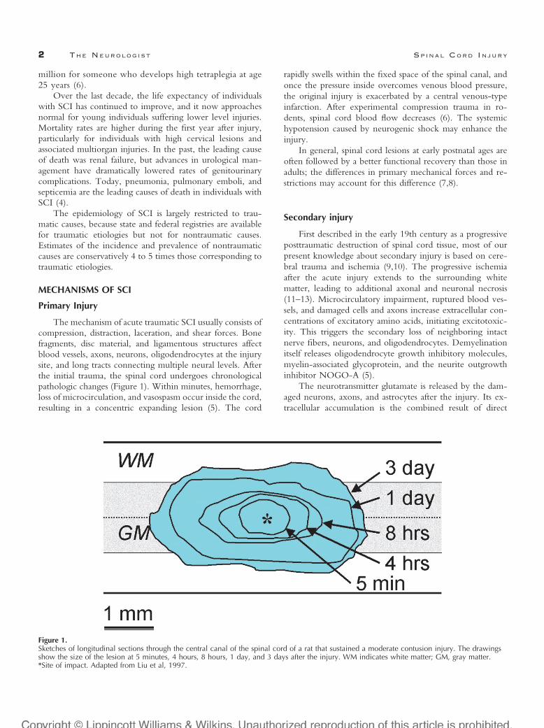
The mechanism of acute traumatic SCI usually consists ofcompression, distraction, laceration, and shear forces. Bonefragments, disc material, and ligamentous structures affectblood vessels, axons, neurons, oligodendrocytes at the injurysite, and long tracts connecting multiple neural levels. Afterthe initial trauma, the spinal cord undergoes chronologicalpathologic changes (Figure 1). Within minutes, hemorrhage,loss of microcirculation, and vasospasm occur inside the cord,resulting in a concentric expanding lesion (5). The cord
rapidly swells within the fixed space of the spinal canal, andonce the pressure inside overcomes venous blood pressure,the original injury is exacerbated by a central venous-typeinfarction. After experimental compression trauma in ro-dents, spinal cord blood flow decreases (6). The systemichypotension caused by neurogenic shock may enhance theinjury.
In general, spinal cord lesions at early postnatal ages areoften followed by a better functional recovery than those inadults; the differences in primary mechanical forces and re-strictions may account for this difference (7,8).
Secondary injury
First described in the early 19th century as a progressiveposttraumatic destruction of spinal cord tissue, most of ourpresent knowledge about secondary injury is based on cere-bral trauma and ischemia (9,10). The progressive ischemiaafter the acute injury extends to the surrounding whitematter, leading to additional axonal and neuronal necrosis(11–13). Microcirculatory impairment, ruptured blood ves-sels, and damaged cells and axons increase extracellular con-centrations of excitatory amino acids, initiating excitotoxic-ity. This triggers the secondary loss of neighboring intactnerve fibers, neurons, and oligodendrocytes. Demyelinationitself releases oligodendrocyte growth inhibitory molecules,myelin-associated glycoprotein, and the neurite outgrowthinhibitor NOGO-A (5).
The neurotransmitter glutamate is released by the dam-aged neurons, axons, and astrocytes after the injury. Its ex-tracellular accumulation is the combined result of direct
Figure 1.Sketches of longitudinal sections through the central canal of the spinal cord of a rat that sustained a moderate contusion injury. The drawingsshow the size of the lesion at 5 minutes, 4 hours, 8 hours, 1 day, and 3 days after the injury. WM indicates white matter; GM, gray matter.*Site of impact. Adapted from Liu et al, 1997.
2 THE NEUROLOGIST SPINAL CORD INJURY
release from disrupted membranes and axons and failure oreven reversal of normal energy-dependent glutamate uptake.Such abnormally high levels of glutamate overexcite neigh-boring neurons, causing them to admit waves of calciumions. The sudden influx of calcium triggers a series of de-structive events, including the production of highly reactivefree radicals that attack membranes and other cellular com-ponents, killing previously healthy neurons.
Excitotoxic shock can harm myelinating oligodendro-cytes as well as neurons (14,15). Because one oligodendro-cyte myelinates 10 to 40 different axons, loss of even a singleoligodendrocyte can contribute to the demyelination of sev-eral axons that remain intact after the primary injury. Oligo-dendrocytes are highly vulnerable to excitotoxic signalsmediated by glutamate receptors of the alpha-amino-3-hydroxy-5-methyl-4-isoxazolepropionate (AMPA) and kainateclasses (16). Cellular Ca2� overload is a key trigger of thisprocess (17).
Oligodendrocytes may also kill themselves after SCIthrough apoptosis (programmed cell death). Days or weeksafter the initial trauma, apoptotic death of oligodendrocytes,located as far as 4 segments away from the trauma site,magnifies the original injury (18). In a rat model of contusioninjury, cell death peaks 1 week after the injury and continuesthroughout the first month (19). Treatment with drugs thatalter protein synthesis limit white-matter injury and improve
behavioral recovery in a rodent spinal cord contusion injurymodel (20).
Many factors can promote this delayed death of oligoden-drocytes, including tumor necrosis factor-�, the apoptosis anti-gen ligand FAS/p75 cytokine pathway, and lipid byproducts.Cytokine-mediated activation of FAS and p75 death-receptorpathways might be essential molecular events contributing tooligodendrocyte apoptosis (21). 4-Hydroxynonenal, a lipid per-oxidation byproduct that accumulates after SCI, is directly cy-totoxic to oligodendrocyte precursors (22). Mechanisms that killoligodendrocyte precursors or inhibit their proliferation or mi-gration could limit remyelination and recovery of function.Targeting the delayed, protracted wave of oligodendrocytedeath, which might proceed for months in humans, is openingdoors to novel protective therapies.
Many secondary mechanisms of injury to oligodendro-cytes are common to traumatic and nontraumatic SCI. Thus,chronic demyelination after CNS injury shares commonfeatures with chronic degenerative disorders such as multiplesclerosis (23). Treatment with antagonists of AMPA-typeglutamate receptors reduces injury severity and oligodendro-cyte loss in experimental allergic encephalitis, a murinemodel of multiple sclerosis (24–27). Nontraumatic SCI hasmultiple etiologies that result in the same symptoms as trau-matic SCI (Figure 2). However, these disease-specific causesare beyond the focus of this review.
Investigations of the immune system’s role in secondaryinjury are challenging the traditional view that autoimmunityafter CNS trauma is purely destructive (28,29). In animalmodels, proinflammatory cytokines help prevent injury,macrophages are needed for CNS repair, and SCI activates Tcells that recognize CNS myelin basic protein (30–32). Thus,growing evidence suggests that the T cell–dependent immu-nity is a physiologic response to CNS trauma that, in part,limits secondary injury. Indeed, T cell–based active vaccina-tion against myelin-associated antigens has proved neuropro-tective in animal models and is being studied as a potentialhuman therapy (33).
ACUTE TREATMENT
The management of acute SCI should seek primarily toprotect the individual from additional injury. It is mainlydirected at the prevention of secondary injury and control ofthe systemic physiologic derangements resulting from theoriginal injury. Most traumatic SCI occurs as a result of rapidcord compression because of a fracture-dislocation or burstfracture (34). One necessary step is to decompress the swollencord by removing damaging bone, disk, and ligament frag-ments. Early surgery is typically limited to individuals withcontinued neurologic decline and evidence from magneticresonance imaging of acute compression (35). However,there is no standard of care regarding the role and timing ofearly surgical intervention because of insufficient data tosupport overall treatment standards. However, the clinicalAmerican Spinal Injury Association (ASIA) injury grade (Ta-ble 1) should not be used in the equation of who shouldreceive early surgery, as has been the case in the past. Becauseof spinal shock, many individuals will present as ASIA A or acomplete injury but will improve to a higher grade withresolution of spinal shock.
Some animal data strongly suggest that early decompres-sion (�24 hours) can improve neurologic recovery andlower the rate of complications (36,37). There is a lot ofclinical uncertainty about surgical intervention and its timewindow in the setting of human SCI. Most studies cannotidentify a difference in clinical outcomes between operatedand nonoperated patients (37–40). However, most studiesare small and define early intervention as less than 72 hours,a period long after most secondary injury is complete (41,42).
The progressive ischemia after theacute injury extends to thesurrounding white matter, leading toadditional axonal and neuronalnecrosis
VOL. 9 / NO. 1 / JANUARY 2003 THE NEUROLOGIST 3
To answer these questions, randomized, controlled prospec-tive human trials are needed.
Nevertheless, new surgical procedures and better hard-ware for internal spine stabilization have evolved in the past
decade (43). Moreover, early surgery might be justified evenif immediate recovery is not expected, because internal sta-bilization of the spine permits earlier mobilization, an essen-tial approach to limiting secondary complications such as skin
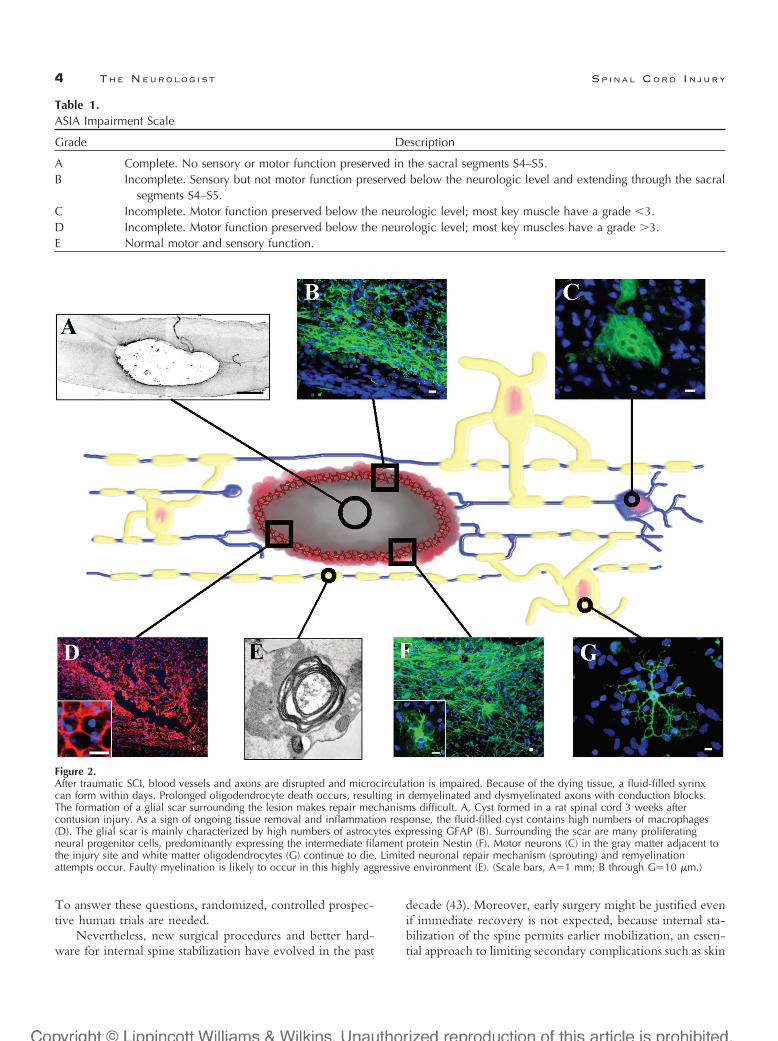
Table 1.ASIA Impairment Scale
Grade Description
A Complete. No sensory or motor function preserved in the sacral segments S4–S5.B Incomplete. Sensory but not motor function preserved below the neurologic level and extending through the sacral
segments S4–S5.C Incomplete. Motor function preserved below the neurologic level; most key muscle have a grade �3.D Incomplete. Motor function preserved below the neurologic level; most key muscles have a grade �3.E Normal motor and sensory function.
Figure 2.After traumatic SCI, blood vessels and axons are disrupted and microcirculation is impaired. Because of the dying tissue, a fluid-filled syrinxcan form within days. Prolonged oligodendrocyte death occurs, resulting in demyelinated and dysmyelinated axons with conduction blocks.The formation of a glial scar surrounding the lesion makes repair mechanisms difficult. A, Cyst formed in a rat spinal cord 3 weeks aftercontusion injury. As a sign of ongoing tissue removal and inflammation response, the fluid-filled cyst contains high numbers of macrophages(D). The glial scar is mainly characterized by high numbers of astrocytes expressing GFAP (B). Surrounding the scar are many proliferatingneural progenitor cells, predominantly expressing the intermediate filament protein Nestin (F). Motor neurons (C) in the gray matter adjacent tothe injury site and white matter oligodendrocytes (G) continue to die. Limited neuronal repair mechanism (sprouting) and remyelinationattempts occur. Faulty myelination is likely to occur in this highly aggressive environment (E). (Scale bars, A�1 mm; B through G�10 �m.)
4 THE NEUROLOGIST SPINAL CORD INJURY
breakdown, pulmonary and urological infections, autonomicdysfunction, and loss of muscle mass and bone density. In-dividuals with internal spine stabilization can participate inrehabilitation programs more fully than those with externalstabilization devices (eg, halo). Thus, the entire process ofrecovery needs to be considered when deciding whether toperform surgery early or late.
Where do we stand with pharmacologic treatment ofacute SCI in humans? To date, 8 prospective clinical trialshave been completed in acute SCI, including study of meth-ylprednisolone, naloxone, tirilazad, GM-1 ganglioside, thy-rotropin-releasing hormone, and nimodipine. Excellent re-views on this subject are available, and additional discussionwill focus on methylprednisolone and GM-1 gangliosides(44). The first neuroprotective agent for SCI was introducedin the 1990s after a multicenter randomized clinical study(National Acute Spinal Cord Injury Study [NASCIS-2])found that a high dose of the steroid methylprednisolonesodium succinate (MPSS) resulted in a modest, althoughstatistically significant, recovery effect when administeredwithin 8 hours of trauma (45,46). The subsequent multi-center NASCIS-3 trial in 1997 examined the potential ben-efit of extending the NASCIS-2 regimen of 24-hour therapyto 48 hours of MPSS treatment and treatment with tirilazadmesylate, a synthetic steroid. Most benefit from the 48-hourtreatment was reported in individuals whose MPSS therapy
was initiated between 3 hours and 8 hours after injury andresulted in higher motor improvement scores (47,48).
However, there is considerable controversy about the useof MPSS in the acute SCI setting because of scientific limi-tations of the original studies. The design and outcomelimitations and lack of public availability of the data havebeen extensively reviewed, and the interested reader is re-ferred to excellent reviews in this area for additional infor-mation (49–52). Suffice to say, treatment with MPSS re-mains largely a clinical decision, incompletely supported byscientific data, requiring clinical expertise and cautious appli-cation. Although a consensus is not clear, most centers treatwith MPSS using the following guideline based on onset totreatment relative to injury: MPSS bolus (30 mg/kg) deliv-ered over the first hour for individuals within the first 8 hours
of injury; treatment is continued (5.4 mg/kg per hour) forthe next 23 hours for individuals treated within 0 to 3 hoursand for the next 47 hours for individuals treated within 3 to8 hours of injury. MPSS is not indicated to be administeredmore than 8 hours after trauma or for acute penetrating SCI(53).
The high steroid dose used in the treatment of SCI isassociated with side effects such as increased incidence ofgastric bleeding, sepsis, pneumonia, acute corticosteroid my-opathy, and wound infection. The elderly are particularlyprone to these complications and therefore deserve specialconsideration (54). Rapid mobilization to rehabilitation isperhaps the most important variable in limiting infectious andskin breakdown complications.
The mechanism by which MPSS acts in acute SCI isunclear but is largely held to reduce the inflammatory cas-cade. Studies have shown reductions in inflammation,edema, lipid peroxidation, and improvements in blood flow.MPSS treatment also reduces “dieback” of vestibulospinalfibers, enhances axonal sprouting, inhibits tumor necrosisfactor-� and nuclear factor-�B binding activity, inhibits cal-pain, and reduces the release of excitatory amino acids (47).However, it is difficult to assign mechanisms based on in vivostudies, because any neuroprotective intervention will likelyproduce many of the above-mentioned observations.
Following primary outcome measures in a preliminarystudy of monosialotetrahexosylganglioside (GM-1 ganglio-side) (55), a prospective, randomized, placebo-controlled,double-blind trial of 2 doses of GM-1 ganglioside versusplacebo was evaluated in acute traumatic SCI (56). Treat-ment with GM-1 had to begin within 72 hours of injuryonset; an initial bolus (300 mg IV over 30 minutes in normalsaline) was given after MPSS, followed by 56 daily doses (100mg each). The high-dose group received a 600-mg bolus and200-mg daily dose. Although not proven in primary out-comes analysis, GM-1 seems to be beneficial in individualswith incomplete SCI, and faster neurologic recovery wasachieved in all individuals. There were significant effects inall patients in the primary outcome variable (percentage ofmarked recovery) at week 8, the end of the dosing period.Consistent positive trends favoring GM-1 were observed inASIA motor and sensory scores, bowel and bladder function,sacral sensation, and anal contraction.
Unfortunately, GM-1 is not readily available for acutetreatment in the United States. GM-1 was available for use inthe United States under an open-label trial from Fidia Phar-maceuticals Corporation, but presently all studies have beencompleted. The Food and Drug Administration has notapproved it for general distribution. Presently, it can only beused under an Emergency IND from the Food and DrugAdministration on a case-by-case basis and pragmatically canonly be obtained for more subacute injuries, where clinicaldata are not available.
The proposed mechanism of action of GM-1 is also notclear, with studies suggesting multiple effects, including an-
Treatment with methyl-prednisoloneremains largely a clinical decision,incompletely supported by scientificdata, requiring clinical expertise andcautious application.
VOL. 9 / NO. 1 / JANUARY 2003 THE NEUROLOGIST 5
tiexcitotoxic activity, apoptosis prevention, neurite sproutingpromotion, and nerve growth factor effects.
Nonetheless, the MPSS and GM-1 ganglioside trials offerencouraging progress in a very difficult clinical researcharena, each trial improving on its predecessor. A criticalprocess to enable faster delivery of acute therapies beforeentry to the emergency room is needed to maximize theeffectiveness of any future therapy to prevent secondaryinjury. The present multihour interval to pharmacologictreatment is too long.
Future acute therapies for SCI are likely to be moremechanistically focused and based on current advances in thepathophysiology of secondary injury. Promising agents in-clude those that protect cells from excess glutamate andexcitotoxicity. Recently, white matter oligodendrocyteswere discovered to be highly vulnerable to AMPA-typeglutamate receptor overactivation (excitotoxicity) (15). Thiswork supports previous data demonstrating a prominent neu-roprotective (particularly white matter preservation) and re-covery effect of AMPA receptor antagonists in experimentalacute SCI (57,58). Only a few early clinical drug trials ofAMPA/kainate receptor antagonists have been reported, andmost have been completed in stroke. Derivatives ofGYKI53655 have apparently been abandoned, but YM90Kdemonstrated safety and pharmacokinetic profiles compatiblewith clinical use (59,60). Such compounds offer selectiveadvantages for additional use in the orphan disease SCI,because they are being clinically pursued for treatment ofdisorders with much larger population bases, including mi-graine and nausea.
Other neuroprotective agents of the future might includelipid peroxidase inhibitors, reactive oxygen species derivedfrom nitric oxide, peroxynitrite inhibitors, inhibitors of cal-pain (which degrades the spinal cord cytoskeleton), and in-hibitors of posttraumatic apoptosis of neurons and oligoden-drocytes. All of these approaches have been supported bypromising animal studies, but translation into clinical trials isa major hurdle.
LONG-TERM TREATMENT
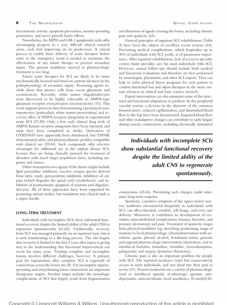
Individuals with incomplete SCIs show substantial func-tional recovery despite the limited ability of the adult CNS toregenerate spontaneously (61,62). Traditionally, recoveryfrom SCI was managed primarily on an inpatient basis, but itis now transitioning to a life-long process. The old dogmathat recovery is limited to the first 2 years after injury is givingway to the understanding that functional improvement canoccur for many years. Treating complete and incompletelesions involves different challenges, however. A primarygoal for regeneration after complete SCI is regrowth ofconnections across the lesion site (8). For incomplete injuries,sprouting and remyelinating intact connections are importanttherapeutic targets. Another target includes the neurologiccomplications of SCI that largely result from fragmentation
and distortion of signals crossing the lesion, including chronicpain and spasticity (63).
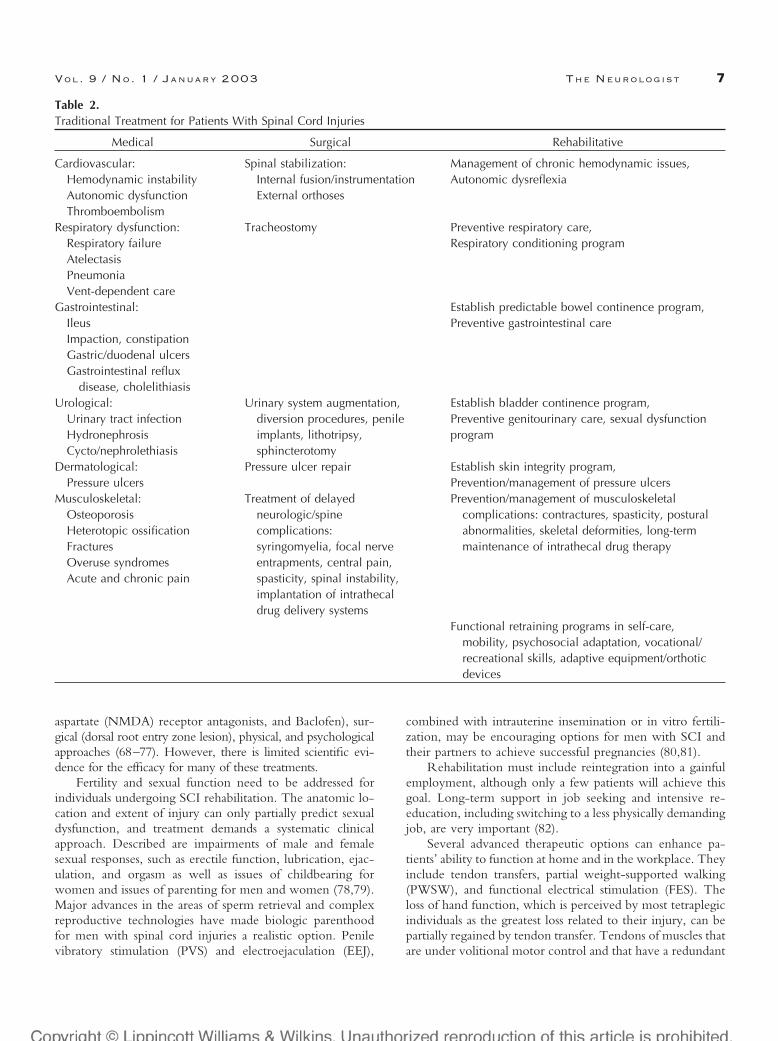
General principles of inpatient SCI rehabilitation (Table2) have been the subject of excellent recent reviews (64).Preventing medical complications, which hospitalize up to30% of individuals with SCI yearly, is of paramount impor-tance. After inpatient rehabilitation, lack of access to specialtycenters limits specialist care for most individuals with SCI.However, annual follow-ups should include both medicaland functional evaluations and therefore are best performedby neurologists, physiatrists, and other SCI experts. They canhelp to tailor physical fitness programs for each patient tocombat functional loss and adjust therapies to the most cur-rent advances in clinical and basic science research.
Expert intervention can also minimize some of the struc-tural and functional adaptations to paralysis. In the peripheralvascular system, a decrease in the diameter of the commonfemoral artery, reduced capillarization, and diminished bloodflow to the legs have been documented. Impaired blood flowand other maladaptive changes can contribute to early fatigueduring muscle contractions, including electrically stimulated
contractions (65,66). Preventing such changes could mini-mize long-term sequelae.
Spasticity, a positive symptom of the upper motor neu-ron syndrome encountered frequently in individuals withSCI, can affect function, comfort, self-image, and even caredelivery. Moreover, it contributes to development of sec-ondary musculoskeletal complications (trauma, fractures, andpressure ulcerations) and pain. Treatment of spasticity rangesfrom physical modalities (eg, stretching, positioning, range ofmotion) to local pharmacologic (chemodenervation with an-esthetic agents, phenol, alcohol, botulinum toxin), general,and regional pharmacologic interventions (dantrolene, oral orintrathecal baclofen, tizanidine, clonidine, benzodiazepines,gabapentin) and surgery (posterior rhizotomy).
Chronic pain is also an important problem for peoplewith SCI. The reported incidence varies but conservativelyoccurs in most individuals, and one third rate their pain assevere (67). Present treatments use a variety of pharmacologic(oral or intrathecal opioids, �-adrenergic agonists, anti-depressants, anticonvulsants, local anesthetics, N-methyl-D-
Individuals with incomplete SCIsshow substantial functional recovery
despite the limited ability of theadult CNS to regenerate
spontaneously.
6 THE NEUROLOGIST SPINAL CORD INJURY
aspartate (NMDA) receptor antagonists, and Baclofen), sur-gical (dorsal root entry zone lesion), physical, and psychologicalapproaches (68–77). However, there is limited scientific evi-dence for the efficacy for many of these treatments.
Fertility and sexual function need to be addressed forindividuals undergoing SCI rehabilitation. The anatomic lo-cation and extent of injury can only partially predict sexualdysfunction, and treatment demands a systematic clinicalapproach. Described are impairments of male and femalesexual responses, such as erectile function, lubrication, ejac-ulation, and orgasm as well as issues of childbearing forwomen and issues of parenting for men and women (78,79).Major advances in the areas of sperm retrieval and complexreproductive technologies have made biologic parenthoodfor men with spinal cord injuries a realistic option. Penilevibratory stimulation (PVS) and electroejaculation (EEJ),
combined with intrauterine insemination or in vitro fertili-zation, may be encouraging options for men with SCI andtheir partners to achieve successful pregnancies (80,81).
Rehabilitation must include reintegration into a gainfulemployment, although only a few patients will achieve thisgoal. Long-term support in job seeking and intensive re-education, including switching to a less physically demandingjob, are very important (82).
Several advanced therapeutic options can enhance pa-tients’ ability to function at home and in the workplace. Theyinclude tendon transfers, partial weight-supported walking(PWSW), and functional electrical stimulation (FES). Theloss of hand function, which is perceived by most tetraplegicindividuals as the greatest loss related to their injury, can bepartially regained by tendon transfer. Tendons of muscles thatare under volitional motor control and that have a redundant
Table 2.Traditional Treatment for Patients With Spinal Cord Injuries
Medical Surgical Rehabilitative
Cardiovascular:Hemodynamic instabilityAutonomic dysfunctionThromboembolism
Spinal stabilization:Internal fusion/instrumentationExternal orthoses
Management of chronic hemodynamic issues,Autonomic dysreflexia
Respiratory dysfunction:Respiratory failureAtelectasisPneumoniaVent-dependent care
Tracheostomy Preventive respiratory care,Respiratory conditioning program
Gastrointestinal:IleusImpaction, constipationGastric/duodenal ulcersGastrointestinal reflux
disease, cholelithiasis
Establish predictable bowel continence program,Preventive gastrointestinal care
Urological:Urinary tract infectionHydronephrosisCycto/nephrolethiasis
Urinary system augmentation,diversion procedures, penileimplants, lithotripsy,sphincterotomy
Establish bladder continence program,Preventive genitourinary care, sexual dysfunctionprogram
Dermatological:Pressure ulcers
Pressure ulcer repair Establish skin integrity program,Prevention/management of pressure ulcers
Musculoskeletal:OsteoporosisHeterotopic ossificationFracturesOveruse syndromesAcute and chronic pain
Treatment of delayedneurologic/spinecomplications:syringomyelia, focal nerveentrapments, central pain,spasticity, spinal instability,implantation of intrathecaldrug delivery systems
Prevention/management of musculoskeletalcomplications: contractures, spasticity, posturalabnormalities, skeletal deformities, long-termmaintenance of intrathecal drug therapy
Functional retraining programs in self-care,mobility, psychosocial adaptation, vocational/recreational skills, adaptive equipment/orthoticdevices
VOL. 9 / NO. 1 / JANUARY 2003 THE NEUROLOGIST 7
role (eg, both the biceps brachii and brachioradialis can flexthe elbow) are surgically rerouted to allow the muscles totake over lost motor functions. It requires sufficiently strong(4–5/5) muscles, which must be trained to assume their newroles. The most frequent tendon transfers are performed torestore voluntary thumb pinch, improve grip strength, andregain active elbow and wrist extension (83,84).
PWSW may improve gait in individuals with incompleteSCI (85,86). PWSW consists of walking on a treadmill whilesupported by a harness and a pneumatic suspension device.Some pilot studies have provided an adequate basis for alarger, controlled clinical trial comparing PWSW with con-ventional gait training. However, this potential treatment isonly applicable to a small subset of individuals with SCI,primarily ASIA C/D classification (Table 1).
FES and evolving neuroprostheses are dramaticallychanging therapeutic strategies for patients with SCI (87).However, muscles can be safely electrostimulated directly ortranscutaneously only by activating a functional nerve end-ing, which requires the lower motor neuron and peripheralnerve to be intact. FES cannot be applied to denervatedmuscle because injurious currents would be required.
FES can strengthen muscles, condition the heart, pacethe diaphragm, enhance bowel and bladder function, facili-tate erection and ejaculation, and control pain. Some FESsystems reproduce walking, but engineering complexities andhigh human energy requirements preclude their routine use.However, using FES to facilitate standing can greatly im-prove quality of life and increase the cross-sectional area oflarge conduit arteries, improving blood flow into the para-lyzed leg (88).
Involuntary exercise by electrically stimulated contrac-tions of the paralyzed limbs is used for strength training.Muscle strengthening can partially reverse neurologic muscleweakness, slow or reverse osteopenia, and improve musclemass and blood flow. It is possible that FES systems can beused to effectively decrease spasticity, provide cardiovascularconditioning, and reduce medical complications such as ve-nous thrombosis and skin breakdown (89–92).
Individuals who sustain an injury at the cervical levelhave a high incidence of respiratory compromise; approxi-mately 20% will require mechanical ventilatory support and5% will require chronic mechanical ventilation (93). Me-chanical ventilation has a high number of complications, suchas increased risk of infection, interference with speech, in-creased need for assistance, and high costs. In the last twodecades, the alternative therapy of choice was bilateralphrenic nerve stimulation, which requires intact bilateralphrenic nerve function (94). Combined intercostals and uni-lateral phrenic nerve pacing can also provide long-term ven-tilatory support in patients with only a single functionalphrenic nerve. The potential morbidity associated with tra-ditional phrenic nerve pacing may soon be replaced by asimple outpatient procedure that allows direct pacing of thediaphragm using intramuscular electrodes attached to themotor end points (95).
Loss of neurocontrol after SCI has many different aspects:dysphagia, difficulty breathing, loss of bowel and bladdercontrol, and control of limb movement. Perhaps the functionthat individuals with SCI would most like to regain is boweland bladder control. FES technology is designed to achievethis goal. Benefits of a FES bladder system include urinationon demand, elimination of catheters, improved continence,fewer urinary tract infections, improved quality of life andsocial ease, and long-term cost savings (96,97).
Neuroprosthesis that uses FES technology for tetraplegicindividuals can help to restore hand grasp and release. Mul-tichannel, implanted FES systems are being used or evaluatedfor their ability to improve motor function of the upperextremities (98,99). Several systems that use FES technologyare presently undergoing clinical trials (100–102). Throughthe latest developments in cortical neuroprosthetics, for ex-ample, it may be feasible to reconstruct voluntary motoractivity in paralyzed patients. These implantable devices thatinterface parts of the brain with a computer can bypassdamaged motor pathways and could one day be used torestore sensory and motor functions lost through injury ordisease (103–106). Present efforts to develop smaller elec-trodes, recording techniques, control systems, and hardwareshould eventually make such systems smaller, less expensive,and easier to implant (107). However, these are early days.
RESTORING FUNCTION STEP BY STEP
Targets
Pragmatic approaches to the treatment of SCI are neededto improve quality of life. A clear understanding of thehierarchy of needs for individuals with SCI is thereforenecessary. Although needs vary according to the level andseverity of injury, most affected individuals prioritize boweland bladder function, sexual function, hand function, andbreathing; walking is a distant competitor.
The management to restore these functions should bedesigned in a stepwise manner. It is important to explain tothe individual that a discovery of a cure will not occur in thenear-term and that a cure is not the goal, but only partialrestoration is required. Restorative therapies should focus onmultiple targets and be applied at different intervals afterinjury. Important strategies should include limiting secondaryinjury and promoting regeneration.
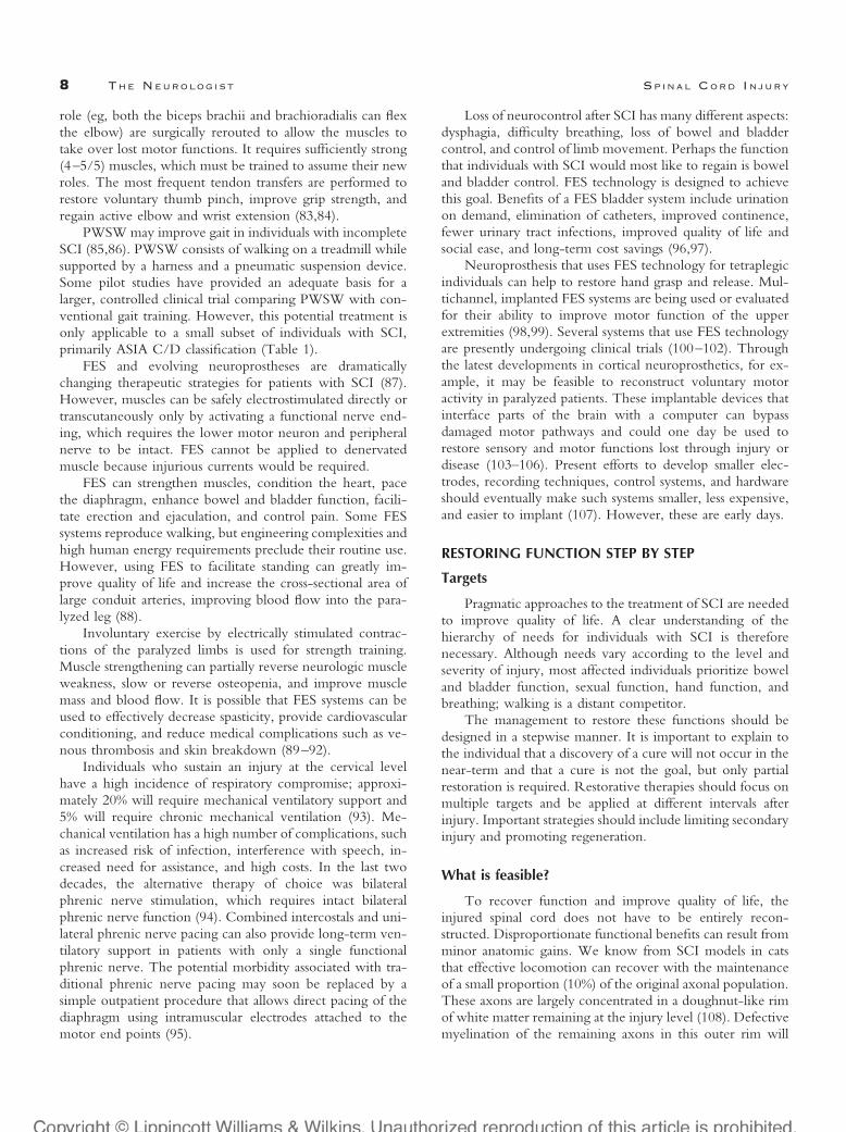
What is feasible?
To recover function and improve quality of life, theinjured spinal cord does not have to be entirely recon-structed. Disproportionate functional benefits can result fromminor anatomic gains. We know from SCI models in catsthat effective locomotion can recover with the maintenanceof a small proportion (10%) of the original axonal population.These axons are largely concentrated in a doughnut-like rimof white matter remaining at the injury level (108). Defectivemyelination of the remaining axons in this outer rim will
8 THE NEUROLOGIST SPINAL CORD INJURY
contribute to functional deficits (34). Consequently, to im-prove function, remyelination of the exposed axons is onepractical approach. It probably will not allow patients withsevere SCI to walk, but they might regain some neurocontrolover such domains as bowel and bladder function, breathing,and hand grasp. Thus, it is useful to consider that secondaryinjury prevention is more feasible than rebuilding damagedspinal tissue.
Present regenerative research efforts are aimed at geneticexpression of nerve growth factors and molecules that sup-press inhibitors of axonal growth to promote the regrowth ofinterrupted nerve fibers (109). In rodent models, oligoden-drocytes have been enticed to remyelinate axons and im-prove axonal conduction. Thus, it seems that restoring theintegrity of existing neuronal circuits is the most feasiblemethod of enhancing recovery. Neither in rodents nor inhumans has long-tract rebuilding of functional neuronal cir-cuits with establishment of appropriate neuronal reconnec-tions been demonstrated. However, there is evidence forpartial construction of local nonspecific neuronal connec-tions. For example, human fetal striatal tissue has been trans-planted into humans and animals with Huntington’s disease,showing establishment of afferent and efferent connectionsand functional benefits (110,111). Transplantation of humanembryonic dopamine neurons into the striatum of patientswith Parkinson’s disease has proved beneficial in open clinicaltrials, and transplanted cells have been monitored to store and
release dopamine for more than a decade (112–115). Focalepilepsy, stroke, and SCI are other applications for neuraltransplantation in humans (116–118). Greater discussion ofthe different regenerative strategies is warranted.
Enhancing regeneration – Regenerative sprouting occursspontaneously but is short lived and diminishes within weeksafter SCI (119). A potential reason for this phenomenoncould be local production of specific inhibitory proteins thatblock neurite outgrowth (120). Early successful attempts toneutralize these factors include specific antibodies (eg, inhib-itor neutralizing antibody) that counteract the myelin proteinNOGO-A (121). Other strategies use neurotrophic factors,such as neurotrophin-3, which can actively promote axonalgrowth (122). Today’s research is focused on NOGO-Ablocking agents and other inhibitory proteins, including the
development of antibodies against these molecules, their re-ceptors, or signal pathways (123). The role of guidanceproteins (netrins and semaphorins) as molecular cues in re-generation of the spinal cord is under close observation.Their signaling pathway may play a key role in limiting orchanneling the regeneration of certain neurons (124,125).
Gene therapy – Transferring therapeutic genes directly intocells surrounding a lesion could enhance the ability of resi-dent cells to produce large quantities of neurotrophic factorssuch as nerve growth factor, brain derived neurotrophicfactor, glial cell line derived neurotrophic factor, plateletderived growth factor, and neurotrophin 3 (126,127). Ge-netically modifying cells to express neurotrophins or growthfactors within the damaged spinal cord increases neuronalsurvival, sprouting, and regeneration (128). Overexpressionof these factors most likely increases the intrinsic ability ofaxons to grow and potentially compensates for elevated levelsof inhibitory molecules surrounding a lesion (129). Long-term expression of these genes can be achieved primarily byviral vector systems (129). Animal studies indicate that in-traspinal administration of the antiapoptotic gene Bcl-2 canprevent retrograde cell loss and reduce atrophy of axoto-mized red nucleus and Clarke’s nucleus neurons after SCI(130). For now, though, efficient gene transfer and appro-priate gene expression are still major challenges.
Bridging scars – In the adult CNS, both glial substrates andthe extracellular matrix influence axonal regeneration afterinjury. Numerous groups claiming functional recovery haveused therapeutic spinal cord grafting procedures. It has beenknown for many years that Schwann cell grafts can promoteaxonal regeneration in the central nervous system (131–134).They are capable of ensheathing and myelinating regenerat-ing axons (135). Olfactory ensheathing cell transplantationpoints to the neuroprotective mechanism of reducing astro-cytic gliosis and cystic cavitations (136). Solid human embry-onic spinal cord xenografts transplanted into a cavity in theadult injured spinal cord produce beneficial morphologiceffects (137). Delayed cotransplantation of fetal cerebral tissueand nerve tissue can achieve anatomic remodeling and long-term functional recovery in rats (138). Polymer scaffolds thatcontain nerve growth factors and other small molecules orneural stem cells were transplanted into the lesion site to forma growth-permissive environment with limited success. Aprimary concern of these grafting approaches is induction ofadditional scar formation that might inhibit axon growth(109,139).
Replacing cells – Cellular transplantation has been used as aprimary strategy for replacing cells lost after injury. Endoge-nous stem cells isolated from spinal cord, fetal spinal cord, andembryonic stem cells have been used successfully for trans-plantation in animal SCI models, some showing promisingresults (125,140,141). Neural precursor or stem cells derived
To recover function and improvequality of life, the injured spinalcord does not have to be entirelyreconstructed.
VOL. 9 / NO. 1 / JANUARY 2003 THE NEUROLOGIST 9
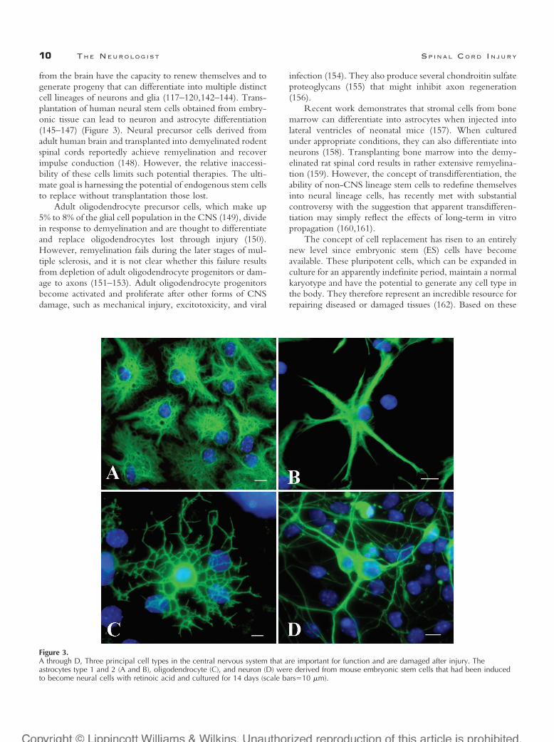
from the brain have the capacity to renew themselves and togenerate progeny that can differentiate into multiple distinctcell lineages of neurons and glia (117–120,142–144). Trans-plantation of human neural stem cells obtained from embry-onic tissue can lead to neuron and astrocyte differentiation(145–147) (Figure 3). Neural precursor cells derived fromadult human brain and transplanted into demyelinated rodentspinal cords reportedly achieve remyelination and recoverimpulse conduction (148). However, the relative inaccessi-bility of these cells limits such potential therapies. The ulti-mate goal is harnessing the potential of endogenous stem cellsto replace without transplantation those lost.
Adult oligodendrocyte precursor cells, which make up5% to 8% of the glial cell population in the CNS (149), dividein response to demyelination and are thought to differentiateand replace oligodendrocytes lost through injury (150).However, remyelination fails during the later stages of mul-tiple sclerosis, and it is not clear whether this failure resultsfrom depletion of adult oligodendrocyte progenitors or dam-age to axons (151–153). Adult oligodendrocyte progenitorsbecome activated and proliferate after other forms of CNSdamage, such as mechanical injury, excitotoxicity, and viral
infection (154). They also produce several chondroitin sulfateproteoglycans (155) that might inhibit axon regeneration(156).
Recent work demonstrates that stromal cells from bonemarrow can differentiate into astrocytes when injected intolateral ventricles of neonatal mice (157). When culturedunder appropriate conditions, they can also differentiate intoneurons (158). Transplanting bone marrow into the demy-elinated rat spinal cord results in rather extensive remyelina-tion (159). However, the concept of transdifferentiation, theability of non-CNS lineage stem cells to redefine themselvesinto neural lineage cells, has recently met with substantialcontroversy with the suggestion that apparent transdifferen-tiation may simply reflect the effects of long-term in vitropropagation (160,161).
The concept of cell replacement has risen to an entirelynew level since embryonic stem (ES) cells have becomeavailable. These pluripotent cells, which can be expanded inculture for an apparently indefinite period, maintain a normalkaryotype and have the potential to generate any cell type inthe body. They therefore represent an incredible resource forrepairing diseased or damaged tissues (162). Based on these
Figure 3.A through D, Three principal cell types in the central nervous system that are important for function and are damaged after injury. Theastrocytes type 1 and 2 (A and B), oligodendrocyte (C), and neuron (D) were derived from mouse embryonic stem cells that had been inducedto become neural cells with retinoic acid and cultured for 14 days (scale bars�10 �m).
10 THE NEUROLOGIST SPINAL CORD INJURY
findings and the fact that they are very amenable to geneticmanipulation, ES cells are becoming an invaluable researchtool. Harnessing their potential is a major goal now that theBush administration has made federal funding available forhuman ES cell research (163). Mouse ES cells have beenstudied for more than 20 years, but research on human EScells is still in its earliest stages.
TOWARD THE FUTURE
It is impossible to predict the success of any one ap-proach, but it is important to understand the relative feasi-bility of each strategy and for science to put efforts intoshort-, intermediate-, and long-range therapeutic goals.
The discovery that the CNS of adult vertebrates, includ-ing humans, contains endogenous progenitor cells capable ofmaking new neurons and glia raises the distinct possibility ofsomeday harnessing this potential for replacing cells lost dur-ing nervous system injury (164). It was not long ago that webelieved the adult human CNS was hardwired and unable tomend. Now, with each passing year, there is a growingunderstanding of the increasing regenerative potential of theadult CNS. It is possible that the adult CNS has a muchgreater capacity of repair than we realize and that perhaps wehave not optimized the setting for regeneration to be maxi-mized. It is just a matter of time, resources, and scientificeffort before we learn how to program and optimize func-tional recovery of the human CNS. The old statements toooften stated by the bedside that most recovery will occurwithin 6 months and it will be complete by two years willcertainly be giving way to allow hope for some recovery longafter an injury (5 to 10 years).
Meanwhile, we can focus on other important ways ofimproving the lives of individuals with SCI. One is todevelop new pharmacologic strategies to minimize CNSinjury and optimize recovery. Animal studies have iden-tified numerous neuroprotective agents, such as AMPAreceptor antagonists, anandamide (endogenous cannabi-noid receptor ligand), dehydroascorbic acid (potent bloodbrain barrier penetrating antioxidant), and hydroxy-fasudil(Rho-kinase inhibitor), that may protect the spinal cordfrom injury and ischemia (165–167). Also, reevaluation ofclinically available drugs that have shown early promise atthe basic science level is a pragmatic approach. For exam-ple, topiramate, an antiepileptic drug, can promote neuriteoutgrowth in vitro and recovery of nerve function in vivo(168). However, there is only limited evidence for theefficacy of these agents (169). Moreover, the clinical ef-fects of such drugs might vary according to the severityand location of a lesion.
The neuroregenerative field has seen impressive advancesduring the last 20 years. It is now time to unlock the door torepairing the nervous system and minimizing the catastrophicconsequences of SCI. Additional investigations into the mo-lecular and cellular mechanisms of spinal cord damage, per-severance in moving potential interventions from bench to
bedside, and advances in rehabilitation techniques shouldhelp us achieve these goals. The concept of replacing lost cellsvia transplantation or activation of endogenous stem cells isexciting, and progress is speeding forward faster than ever.Understanding the factors important for optimizing sponta-neous recovery and regeneration will be important(170,171). It is nearly impossible to know where we will bein 10 years, but it promises to surprise us all.
In 2500 BC, Edwin Smith’s papyrus stated that SCI is“an ailment not to be treated.” Four thousand five hundredyears later, we unlocked mechanisms dealing with cellularprotection and neuroregeneration, but clinically we are stillproviding mostly supportive care to individuals with SCIs. Itis time to aggressively evaluate and transition the gains ob-tained in the laboratory at cellular and animal levels to patientcare so that the dream of getting out of a chair and walkingagain can become more of a rule than an exception.
ACKNOWLEDGMENTS
This work was supported by NIH NINDS grantsNS37927, NS39577, NS40520, and NS01931 (J.W.M.) andthe Sam Schmidt Foundation, the ALS Hope Foundation,and the Christopher Reeve Paralysis Foundation. Yun Qu,MD, kindly provided electron microscopy data. We thankSudhakar Vadivelu, Linda Schultz, PhD, Oksana Volshteyn,MD, and Jenny Edrington for their important input.
REFERENCES
1. Go BK, DeVivo MJ, Richards JS. The epidemiology of spinal cordinjury. In: Stover SL, DeLisa JA, Whiteneck GG, eds. Spinal CordInjury: Clinical Outcomes From the Model Systems. Gaithersburg, Md:Aspen; 1995:21–55.
2. Richards JS, Go BK, Rutt RD, et al. The national spinal cord injurydatabase. In: Stover SL, DeLisa JA, Whiteneck GG, eds. Spinal CordInjury: Clinical Outcomes From the Model Systems. Gaithersburg, Md:Aspen; 1995:10–20.
3. Surkin J, Smith M, Penman A, et al. Spinal cord injury incidence inMississippi: a capture-recapture approach. J Trauma. 1998;45:502–504.
4. National Spinal Cord Injury Database. Spinal Cord Injury, Facts andFigures. 2001. Birmingham, AL: National Spinal Cord InjuryStatistical Center (NSCISC).
5. Tator CH, Fehlings MG. Review of the secondary injury theory ofacute spinal cord trauma with emphasis on vascular mechanisms.J Neurosurg. 1991;75:15–26.
6. Westergren H, Farooque M, Olsson Y, et al. Spinal cord blood flowchanges following systemic hypothermia and spinal cord compressioninjury: an experimental study in the rat using Laser-Dopplerflowmetry. Spinal Cord. 2001;39:74–84.
7. Bregman BS, Goldberger ME. Infant lesion effect, II: sparing andrecovery of function after spinal cord damage in newborn and adultcats. Brain Res. 1983;285:119–135.
8. Raineteau O, Schwab ME. Plasticity of motor systems afterincomplete spinal cord injury. Nat Rev Neurosci. 2001;2:263–273.
9. McIntosh TK, Juhler M, Wieloch T. Novel pharmacologic strategiesin the treatment of experimental traumatic brain injury: J Neurotrauma.1998;15:731–769.
10. Lee JM, Zipfel GJ, Choi DW. The changing landscape of ischaemicbrain injury mechanisms. Nature. 1999;399(suppl):A7–A14.
11. Tator CH, Koyanagi I. Vascular mechanisms in the pathophysiology ofhuman spinal cord injury. J Neurosurg. 1997;86:483–492.
VOL. 9 / NO. 1 / JANUARY 2003 THE NEUROLOGIST 11
12. Fehlings MG, Tator CH, Linden RD. The effect of nimodipine anddextran on axonal function and blood flow following experimentalspinal cord injury. J Neurosurg. 1989;71:403–416.
13. Kobrine AI, Doyle TF, Martins AN. Local spinal cord blood flow inexperimental traumatic myelopathy. J Neurosurg. 1975;42:144–149.
14. Matute C, Sanchez-Gomez MV, Martinez-Millan L, et al. Glutamatereceptor-mediated toxicity in optic nerve oligodendrocytes. Proc NatlAcad Sci U S A. 1997;94:8830–8835.
15. McDonald JW, Althomsons SP, Hyrc KL, et al. Oligodendrocytesfrom forebrain are highly vulnerable to AMPA/kainate receptor-mediated excitotoxicity. Nat Med. 1998;4:291–297.
16. Matute C, Alberdi E, Domercq M, et al. The link between excitotoxicoligodendroglial death and demyelinating diseases. Trends Neurosci.2001;24:224–230.
17. Choi DW. Calcium: still center-stage in hypoxic-ischemic neuronaldeath. Trends Neurosci. 1995;18:58–60.
18. Beattie MS, Farooqui AA, Bresnahan JC. Review of current evidencefor apoptosis after spinal cord injury. J Neurotrauma. 2000;17:915–925.
19. Li GL, Farooque M, Holtz A, et al. Apoptosis of oligodendrocytesoccurs for long distances away from the primary injury aftercompression trauma to rat spinal cord. Acta Neuropathol (Berl). 1999;98:473–480.
20. Kamencic H, Griebel RW, Lyon AW, et al. Promoting glutathionesynthesis after spinal cord trauma decreases secondary damage andpromotes retention of function. FASEB J. 2001;15:243–250.
21. Casha S, Yu WR, Fehlings MG. Oligodendroglial apoptosis occursalong degenerating axons and is associated with FAS and p75expression following spinal cord injury in the rat. Neuroscience. 2001;103:203–218.
22. Gard AL, Solodushko VG, Waeg G, et al. 4-Hydroxynonenal, a lipidperoxidation byproduct of spinal cord injury, is cytotoxic foroligodendrocyte progenitors and inhibits their responsiveness toPDGF. Microsc Res Tech. 2001;52:709–718.
23. Crowe MJ, Bresnahan JC, Shuman SL, et al. Apoptosis and delayeddegeneration after spinal cord injury in rats and monkeys. Nat Med.1997;3:73–76.
24. Werner P, Pitt D, Raine CS. Multiple sclerosis: altered glutamatehomeostasis in lesions correlates with oligodendrocyte and axonaldamage. Ann Neurol 2001;50:169–180.
25. Werner P, Pitt D, Raine CS. Glutamate excitotoxicity–a mechanismfor axonal damage and oligodendrocyte death in Multiple Sclerosis?J Neural Transm Suppl. 2000;60:375–385.
26. Pitt D, Werner P, Raine CS. Glutamate excitotoxicity in a model ofmultiple sclerosis. Nat Med 2000;6:67–70.
27. Smith T, Groom A, Zhu B, et al. Autoimmune encephalomyelitisameliorated by AMPA antagonists. Nat Med. 2000;6:62–66.
28. Popovich PG, Wei P, Stokes BT. Cellular inflammatory response afterspinal cord injury in Sprague- Dawley and Lewis rats. J Comp Neurol.1997;377:443–464.
29. Schwartz M, Cohen I, Lazarov-Spiegler O, et al. The remedy may liein ourselves: prospects for immune cell therapy in central nervoussystem protection and repair. J Mol Med. 1999;77:713–717.
30. Klusman I, Schwab ME. Effects of pro-inflammatory cytokines inexperimental spinal cord injury. Brain Res. 1997:762;173–184.
31. Prewitt CM, Niesman IR, Kane CJ, et al. Activated macrophage/microglial cells can promote the regeneration of sensory axons into theinjured spinal cord. Exp Neurol. 1997;148:433–443.
32. Hauben E, Nevo U, Yoles E, et al. Autoimmune T cells as potentialneuroprotective therapy for spinal cord injury. Lancet. 2000;355:286–287.
33. Hauben E, Agranov E, Gothilf A, et al. Posttraumatic therapeuticvaccination with modified myelin self-antigen prevents completeparalysis while avoiding autoimmune disease. J Clin Invest. 2001;108:591–599.
34. Bunge RP, Puckett WR, Becerra JL, et al. Observations on thepathology of human spinal cord injury: a review and classification of 22new cases with details from a case of chronic cord compression withextensive focal demyelination. Adv Neurol. 1993;59:75–89.
35. Esce PG, Haines SJ. Acute treatment of spinal cord injury. Curr TreatOptions Neurol. 2000;2:517–524.
36. Duh MS, Shepard MJ, Wilberger JE, et al. The effectiveness of surgeryon the treatment of acute spinal cord injury and its relation topharmacological treatment. Neurosurgery. 1994;35:240–248.
37. Dimar JR, Glassman SD, Raque GH, et al. The influence of spinalcanal narrowing and timing of decompression on neurologic recoveryafter spinal cord contusion in a rat model. Spine. 1999;24:1623–1633.
38. Fehlings MG, Sekhon LH, Tator C. The role and timing ofdecompression in acute spinal cord injury: what do we know? Whatshould we do? Spine. 2001;26(24 suppl):S101–S110.
39. Marshall LF, Knowlton S, Garfin SR, et al. Deterioration followingspinal cord injury: a multicenter study. J Neurosurg. 1987;66:400–404.
40. Vaccaro AR, Daugherty RJ, Sheehan TP, et al. Neurologic outcomeof early versus late surgery for cervical spinal cord injury. Spine.1997;22:2609–2613.
41. Chen TY, Dickman CA, Eleraky M, et al. The role of decompressionfor acute incomplete cervical spinal cord injury in cervical spondylosis.Spine. 1998;23:2398–2403.
42. Mirza SK, Krengel WF III, Chapman JR, et al. Early versus delayedsurgery for acute cervical spinal cord injury. Clin Orthop. 1999;359:104–114.
43. Ulrich C, Arand M, Nothwang J. Internal fixation on the lowercervical spine: biomechanics and clinical practice of procedures andimplants. Eur Spine J. 2001;10:88–100.
44. Geisler FH. Clinical trials of pharmacotherapy for spinal cord injury.Ann N Y Acad Sci. 1998;845:374–381.
45. Bracken MB, Shepard MJ, Collins WF, et al. Methylprednisolone ornaloxone treatment after acute spinal cord injury: 1-year follow-updata. Results of the second National Acute Spinal Cord Injury Study.J Neurosurg. 1992;76:23–31.
46. Bracken MB, Shepard MJ, Collins WF, et al. A randomized,controlled trial of methylprednisolone or naloxone in the treatment ofacute spinal-cord injury: results of the Second National Acute SpinalCord Injury Study. N Engl J Med. 1990;322:1405–1411.
47. Bracken MB. Methylprednisolone and acute spinal cord injury: anupdate of the randomized evidence. Spine. 2001;26(24 suppl):S47–S54.
48. Bracken MB, Shepard MJ, Holford TR, et al. Administration ofmethylprednisolone for 24 or 48 hours or tirilazad mesylate for 48hours in the treatment of acute spinal cord injury: results of the ThirdNational Acute Spinal Cord Injury Randomized Controlled Trial.National Acute Spinal Cord Injury Study. JAMA. 1997;277:1597–1604.
49. Ducker TB, Zeidman SM. Spinal cord injury: role of steroid therapy.Spine. 1994;19:2281–2287.
50. Nesathurai S. Steroids and spinal cord injury: revisiting the NASCIS 2and NASCIS 3 trials. J Trauma. 1998;45:1088–1093.
51. Coleman WP, Benzel D, Cahill DW, et al. A critical appraisal of thereporting of the National Acute Spinal Cord Injury Studies (II and III)of methylprednisolone in acute spinal cord injury. J Spinal Disord.2000;13:185–199.
52. Hurlbert RJ. Methylprednisolone for acute spinal cord injury: aninappropriate standard of care. J Neurosurg. 2000;93(1 suppl):1–7.
53. Fehlings MG. Editorial: recommendations regarding the use ofmethylprednisolone in acute spinal cord injury: making sense out ofthe controversy. Spine. 2001;26(24 suppl):S56–S57.
54. Matsumoto T, Tamaki T, Kawakami M, et al. Early complications ofhigh-dose methylprednisolone sodium succinate treatment in thefollow-up of acute cervical spinal cord injury. Spine. 2001;26:426–430.
55. Geisler FH, Dorsey FC, Coleman WP. Recovery of motor functionafter spinal-cord injury: a randomized, placebo-controlled trial withGM-1 ganglioside. N Engl J Med. 1991;324:1829–1838.
56. Geisler FH, Coleman WP, Grieco G, et al. The Sygen multicenteracute spinal cord injury study. Spine. 2001;26(24 suppl):S87–S98.
57. Wrathall JR, Teng YD, Choiniere D. Amelioration of functionaldeficits from spinal cord trauma with systemically administered
12 THE NEUROLOGIST SPINAL CORD INJURY
NBQX, an antagonist of non-N-methyl-D-aspartate receptors. ExpNeurol. 1996;137:119–126.
58. Stys PK. Anoxic and ischemic injury of myelinated axons in CNSwhite matter: from mechanistic concepts to therapeutics. J Cereb BloodFlow Metab. 1998;18:22–25.
59. Umemura K, Kondo K, Ikeda Y, et al. Pharmacokinetics and safety ofthe novel amino-3-hydroxy-5-methylisoxazole-4-propionate receptorantagonist YM90K in healthy men. J Clin Pharmacol. 1997;37:719–727.
60. Lees GJ. Pharmacology of AMPA/kainate receptor ligands and theirtherapeutic potential in neurological and psychiatric disorders. Drugs.2000;59:33–78.
61. Burns SP, Golding DG, Rolle WA Jr, et al. Recovery of ambulationin motor-incomplete tetraplegia. Arch Phys Med Rehabil. 1997;78:1169–1172.
62. Waters RL, Sie I, Adkins RH, et al. Injury pattern effect on motorrecovery after traumatic spinal cord injury. Arch Phys Med Rehabil.1995;76:440–443.
63. Wong ST, Atkinson BA, Weaver LC. Confocal microscopic analysisreveals sprouting of primary afferent fibres in rat dorsal horn after spinalcord injury. Neurosci Lett. 2000;296:65–68.
64. Stover SL. Review of forty years of rehabilitation issues in spinal cordinjury. J Spinal Cord Med. 1995;18:175–182.
65. Burnham R, Martin T, Stein R, et al. Skeletal muscle fibre typetransformation following spinal cord injury. Spinal Cord. 1997;35:86–91.
66. Gerrits HL, de Haan A, Hopman MT, et al. Contractile properties ofthe quadriceps muscle in individuals with spinal cord injury. MuscleNerve. 1999;22:1249–1256.
67. Siddall PJ, Loeser JD. Pain following spinal cord injury. Spinal Cord.2001;39:63–73.
68. Fenollosa P, Pallares J, Cervera J, et al. Chronic pain in the spinal cordinjured: statistical approach and pharmacological treatment. Paraplegia.1993;31:722–729.
69. Glynn CJ, Jamous MA, Teddy PJ, et al. Role of spinal noradrenergicsystem in transmission of pain in patients with spinal cord injury.Lancet. 1986;2:1249–1250.
70. Sandford PR, Lindblom LB, Haddox JD. Amitriptyline andcarbamazepine in the treatment of dysesthetic pain in spinal cordinjury. Arch Phys Med Rehabil. 1992;73:300–301.
71. Ness TJ, San Pedro EC, Richards JS, et al. A case of spinal cordinjury-related pain with baseline rCBF brain SPECT imaging andbeneficial response to gabapentin. Pain. 1998;78:139–143.
72. Loubser PG, Donovan WH. Diagnostic spinal anaesthesia in chronicspinal cord injury pain. Paraplegia. 1991;29:25–36.
73. Eide PK, Stubhaug A, Stenehjem AE. Central dysesthesia pain aftertraumatic spinal cord injury is dependent on N-methyl-D-aspartatereceptor activation. Neurosurgery. 1995;37:1080–1087.
74. Loubser PG, Akman NM. Effects of intrathecal baclofen on chronicspinal cord injury pain. J Pain Symptom Manage. 1996;12:241–247.
75. Loubser PG, Clearman RR. Evaluation of central spinal cord injurypain with diagnostic spinal anesthesia. Anesthesiology. 1993;79:376–378.
76. Friedman AH, Nashold BS Jr. DREZ lesions for relief of pain relatedto spinal cord injury. J Neurosurg. 1986;65:465–469.
77. Umlauf RL. Psychological interventions for chronic pain followingspinal cord injury. Clin J Pain. 1992;8:111–118.
78. Linsenmeyer TA. Sexual function and infertility following spinal cordinjury. Phys Med Rehabil Clin North Am. 2000; 11:141–156.
79. Sipski ML. Sexual functioning in the spinal cord injured. Int J ImpotRes. 1998;10(suppl 2):S128–S130.
80. Biering-Sorensen F, Sonksen J. Sexual function in spinal cord lesionedmen. Spinal Cord. 2001;39:455–470.
81. Ekland M, Griffin S, Copeland J, et al. Exploring male fertility optionsafter spinal cord injury: the role of the nurse clinician. SCI Nurs.1998;15:95–98.
82. Tomassen PC, Post MW, van Asbeck FW. Return to work after spinalcord injury. Spinal Cord. 2000;38:51–55.
83. Freehafer AA. Tendon transfers in tetraplegic patients: the Clevelandexperience. Spinal Cord. 1998;36:315–319.
84. Johnstone BR, Jordan CJ, Buntine JA. A review of surgicalrehabilitation of the upper limb in quadriplegia. Paraplegia. 1988;26:317–339.
85. Protas EJ, Holmes SA, Qureshy H, et al. Supported treadmillambulation training after spinal cord injury: a pilot study. Arch PhysMed Rehabil. 2001;82:825–831.
86. Field-Fote EC. Combined use of body weight support, functionalelectric stimulation, and treadmill training to improve walking abilityin individuals with chronic incomplete spinal cord injury. Arch PhysMed Rehabil. 2001;82:818–824.
87. Grill WM, McDonald JW, Peckham PH, et al. At the interface:convergence of neural regeneration and neural prostheses forrestoration of function. J Rehabil Res Dev. 2001;38:633–639.
88. Gerrits HL, de Haan A, Sargeant AJ, et al. Peripheral vascular changesafter electrically stimulated cycle training in people with spinal cordinjury. Arch Phys Med Rehabil. 2001;82:832–839.
89. Belanger M, Stein RB, Wheeler GD, et al. Electrical stimulation: canit increase muscle strength and reverse osteopenia in spinal cord injuredindividuals? Arch Phys Med Rehabil. 2000;81:1090–1098.
90. Scremin AM, Kurta L, Gentili A, et al. Increasing muscle mass in spinalcord injured persons with a functional electrical stimulation exerciseprogram. Arch Phys Med Rehabil. 1999;80:1531–1536.
91. Nash MS, Montalvo BM, Applegate B. Lower extremity blood flowand responses to occlusion ischemia differ in exercise-trained andsedentary tetraplegic persons. Arch Phys Med Rehabil. 1996;77:1260–1265.
92. Stein RB. Functional electrical stimulation after spinal cord injury.J Neurotrauma. 1999;16:713–717.
93. Carter RE, Donovan WH, Halstead L, et al. Comparative study ofelectrophrenic nerve stimulation and mechanical ventilatory support intraumatic spinal cord injury. Paraplegia. 1987;25:86–91.
94. Glenn WW, Phelps ML, Elefteriades JA, et al. Twenty years ofexperience in phrenic nerve stimulation to pace the diaphragm. PacingClin Electrophysiol. 1986;9(6 pt 1):780–784.
95. DiMarco AF. Neural prostheses in the respiratory system. J Rehabil ResDev. 2001;38:601–607.
96. Creasey GH, Dahlberg JE. Economic consequences of an implantedneuroprosthesis for bladder and bowel management. Arch Phys MedRehabil. 2001;82:1520–1525.
97. Creasey GH, Grill JH, Korsten M, et al. An implantableneuroprosthesis for restoring bladder and bowel control to patientswith spinal cord injuries: a multicenter trial. Arch Phys Med Rehabil.2001;82:1512–1519.
98. Peckham PH, Keith MW, Kilgore KL, et al. Efficacy of an implantedneuroprosthesis for restoring hand grasp in tetraplegia: a multicenterstudy. Arch Phys Med Rehabil. 2001;82:1380–1388.
99. Keith MW, Kilgore KL, Peckham PH, et al. Tendon transfers andfunctional electrical stimulation for restoration of hand function inspinal cord injury. J Hand Surg [Am ]. 1996;21:89–99.
100. Fromm B, Rupp R, Gerner HJ. The Freehand System: an implantableneuroprosthesis for functional electrostimulation of the upperextremity [in German]. Handchir Mikrochir Plast Chir. 2001;33:149–152.
101. Keith MW. Neuroprostheses for the upper extremity. Microsurgery.2001;21:256–263.
102. Triolo RJ, Liu MQ, Kobetic R, et al. Selectivity of intramuscularstimulating electrodes in the lower limbs. J Rehabil Res Dev. 2001;38:533–544.
103. Shoham S, Halgren E, Maynard EM, et al. Motor-cortical activity intetraplegics. Nature. 2001;413(6858):793.
104. Nicolelis MA. Actions from thoughts. Nature. 2001;409:403–407.105. Wessberg J, Stambaugh CR, Kralik JD, et al. Real-time prediction of
hand trajectory by ensembles of cortical neurons in primates. Nature.2000;408:361–365.
VOL. 9 / NO. 1 / JANUARY 2003 THE NEUROLOGIST 13
106. Kennedy PR, Bakay RA, Moore MM, et al. Direct control of acomputer from the human central nervous system. IEEE Trans RehabilEng. 2000;8:198–202.
107. Bhadra N, Kilgore KL, Peckham PH. Implanted stimulators forrestoration of function in spinal cord injury. Med Eng Phys. 2001;23:19–28.
108. Blight AR. Cellular morphology of chronic spinal cord injury in thecat: analysis of myelinated axons by line-sampling. Neuroscience. 1983;10:521–543.
109. Schwab ME. Repairing the injured spinal cord. Science. 2002;295:1029–1031.
110. Hauser RA, Furtado S, Cimino CR, et al. Bilateral human fetal striataltransplantation in Huntington’s disease. Neurology. 2002;58:687–695.
111. Bachoud-Levi AC, Remy P, Nguyen JP, et al. Motor and cognitiveimprovements in patients with Huntington’s disease after neuraltransplantation. Lancet. 2000;356:1975–1979.
112. Freed CR, Greene PE, Breeze RE, et al. Transplantation of embryonicdopamine neurons for severe Parkinson’s disease. N Engl J Med. 2001;344:710–719.
113. Piccini P, Lindvall O, Bjorklund A, et al. Delayed recovery ofmovement-related cortical function in Parkinson’s disease after striataldopaminergic grafts. Ann Neurol. 2000;48:689–695.
114. Piccini P, Brooks DJ, Bjorklund A, et al. Dopamine release from nigraltransplants visualized in vivo in a Parkinson’s patient. Nat Neurosci.1999;2:1137–1140.
115. Hauser RA, Freeman TB, Snow BJ, et al. Long-term evaluation ofbilateral fetal nigral transplantation in Parkinson disease. Arch Neurol.1999;56:179–187.
116. Falci S, Holtz A, Akesson E, et al. Obliteration of a posttraumatic spinalcord cyst with solid human embryonic spinal cord grafts: first clinicalattempt. J Neurotrauma. 1997;14:875–884.
117. Brevig T, Holgersson J, Widner H. Xenotransplantation for CNSrepair: immunological barriers and strategies to overcome them. TrendsNeurosci. 2000;23:337–344.
118. Melton L. Neural transplantation: new cells for old brains. Lancet.2000;355(9221):2142.
119. von Meyenburg J, Brosamle C, Metz GA, et al. Regeneration andsprouting of chronically injured corticospinal tract fibers in adult ratspromoted by NT-3 and the mAb IN-1, which neutralizes myelin-associated neurite growth inhibitors. Exp Neurol. 1998;154:583–594.
120. Schwab ME, Caroni P. Oligodendrocytes and CNS myelin arenonpermissive substrates for neurite growth and fibroblast spreading invitro. J Neurosci. 1988;8:2381–2393.
121. Chen MS, Huber AB, van der Haar ME, et al. Nogo-A is a myelin-associated neurite outgrowth inhibitor and an antigen for monoclonalantibody IN-1. Nature. 2000;403:434–439.
122. Schwab ME, Bartholdi D. Degeneration and regeneration of axons inthe lesioned spinal cord. Physiol Rev. 1996;76:319–370.
123. Brittis PA, Flanagan JG. Nogo domains and a Nogo receptor:implications for axon regeneration. Neuron. 2001;30:11–14.
124. Shifman MI, Selzer ME. Expression of the netrin receptor UNC-5 inlamprey brain: modulation by spinal cord transection. NeurorehabilNeural Repair. 2000;14:49–58.
125. McDonald JW. Repairing the damaged spinal cord. Sci Am. 1999;281:64–73.
126. Jones LL, Oudega M, Bunge MB, et al. Neurotrophic factors, cellularbridges and gene therapy for spinal cord injury. J Physiol. 2001;533(pt1):83–89.
127. Li H, Zou X, Bunger C. Gene therapy and spinal disorders. Int Orthop.2001;25:11–4.
128. Smith GM, Romero MI. Adenoviral-mediated gene transfer toenhance neuronal survival, growth, and regeneration. J Neurosci Res.1999;55:147–157.
129. Rabchevsky AG, Smith GM. Therapeutic interventions followingmammalian spinal cord injury. Arch Neurol. 2001;58:721–726.
130. Shibata M, Murray M, Tessler A, et al. Single injections of a DNAplasmid that contains the human Bcl-2 gene prevent loss and atrophyof distinct neuronal populations after spinal cord injury in adult rats.Neurorehabil Neural Repair. 2000;14:319–330.
131. Blakemore WF. Remyelination of CNS axons by Schwann cellstransplanted from the sciatic nerve. Nature. 1977;266:68–69.
132. Kromer LF, Cornbrooks CJ. Transplants of Schwann cell culturespromote axonal regeneration in the adult mammalian brain. Proc NatlAcad Sci U S A. 1985;82:6330–6334.
133. Morrissey TK, Bunge RP, Kleitman N. Human Schwann cells invitro, I: Failure to differentiate and support neuronal health underco-culture conditions that promote full function of rodent cells.J Neurobiol. 1995;28:171–189.
134. Oudega M, Xu XM, Guenard V, et al. A combination of insulin-likegrowth factor-I and platelet-derived growth factor enhancesmyelination but diminishes axonal regeneration into Schwann cellgrafts in the adult rat spinal cord. Glia. 1997;19:247–258.
135. Xu XM, Guenard V, Kleitman N, et al. Axonal regeneration intoSchwann cell-seeded guidance channels grafted into transected adultrat spinal cord. J Comp Neurol. 1995;351:145–160.
136. Verdu E, Garcia-Alias G, Fores J, et al. Effects of ensheathing cellstransplanted into photochemically damaged spinal cord. Neuroreport.2001;12:2303–2309.
137. Akesson E, Holmberg L, Jonhagen ME, et al. Solid human embryonicspinal cord xenografts in acute and chronic spinal cord cavities: amorphological and functional study. Exp Neurol. 2001;170:305–316.
138. Zurita M, Vaquero J, Oya S, et al. Functional recovery in chronicparaplegic rats after co-grafts of fetal brain and adult peripheral nervetissue. Surg Neurol. 2001;55:249–254.
139. Teng YD, Lavik EB, Qu X, et al. Functional recovery followingtraumatic spinal cord injury mediated by a unique polymer scaffoldseeded with neural stem cells. Proc Natl Acad Sci U S A. 2002;99:3024–3029.
140. Liu S, Qu Y, Stewart TJ, et al. Embryonic stem cells differentiate intooligodendrocytes and myelinate in culture and after spinal cordtransplantation. Proc Natl Acad Sci U S A. 2000;97:6126–6131.
141. McDonald JW, Liu XZ, Qu Y, et al. Transplanted embryonic stemcells survive, differentiate and promote recovery in injured rat spinalcord. Nat Med. 1999;5:1410–1412.
142. Lois C, Alvarez-Buylla A. Proliferating subventricular zone cells in theadult mammalian forebrain can differentiate into neurons and glia. ProcNatl Acad Sci U S A. 1993;90:2074–2077.
143. Morshead CM, Reynolds BA, Craig CG, et al. Neural stem cells in theadult mammalian forebrain: a relatively quiescent subpopulation ofsubependymal cells. Neuron. 1994;13:1071–1082.
144. Reynolds BA, Weiss S. Generation of neurons and astrocytes fromisolated cells of the adult mammalian central nervous system. Science.1992;255:1707–1710.
145. Chalmers-Redman RM, Priestley T, Kemp JA, et al. In vitropropagation and inducible differentiation of multipotential progenitorcells from human fetal brain. Neuroscience. 1997;76:1121–1128.
146. Moyer MP, Johnson RA, Zompa EA, et al. Culture, expansion, andtransplantation of human fetal neural progenitor cells. Transplant Proc.1997;29:2040–2041.
147. Svendsen CN, Caldwell MA, Shen J, et al. Long-term survival ofhuman central nervous system progenitor cells transplanted into a ratmodel of Parkinson’s disease. Exp Neurol. 1997;148:135–146.
148. Akiyama Y, Honmou O, Kato T, et al. Transplantation of clonalneural precursor cells derived from adult human brain establishesfunctional peripheral myelin in the rat spinal cord. Exp Neurol. 2001;167:27–39.
149. Vaughn JE, Peters A. A third neuroglial cell type: an electronmicroscopic study. J Comp Neurol. 1968;133:269–288.
150. Raff MC, Miller RH, Noble M. A glial progenitor cell that developsin vitro into an astrocyte or an oligodendrocyte depending on culturemedium. Nature. 1983;303:390–396.
14 THE NEUROLOGIST SPINAL CORD INJURY
151. Keirstead HS, Blakemore WF. Identification of post-mitoticoligodendrocytes incapable of remyelination within the demyelinatedadult spinal cord. J Neuropathol Exp Neurol. 1997;56:1191–1201.
152. Carroll WM, Jennings AR, Ironside LJ. Identification of the adult restingprogenitor cell by autoradiographic tracking of oligodendrocyte precursorsin experimental CNS demyelination. Brain. 1998;121(pt 2):293–302.
153. Redwine JM, Armstrong RC. In vivo proliferation of oligodendrocyteprogenitors expressing PDGF�R during early remyelination.J Neurobiol. 1998;37:413–428.
154. Gensert JM, Goldman JE. Endogenous progenitors remyelinatedemyelinated axons in the adult CNS. Neuron. 1997;19:197–203.
155. Fawcett JW, Asher RA. The glial scar and central nervous systemrepair. Brain Res Bull. 1999;49:377–391.
156. McKeon RJ, Hoke A, Silver J. Injury-induced proteoglycans inhibitthe potential for laminin-mediated axon growth on astrocytic scars.Exp Neurol. 1995;136:32–43.
157. Kopen GC, Prockop DJ, Phinney DG. Marrow stromal cells migratethroughout forebrain and cerebellum, and they differentiate intoastrocytes after injection into neonatal mouse brains. Proc Natl Acad SciU S A. 1999;96:10711–10716.
158. Woodbury D, Schwarz EJ, Prockop DJ, et al. Adult rat and humanbone marrow stromal cells differentiate into neurons. J Neurosci Res.2000;61:364–370.
159. Sasaki M, Honmou O, Akiyama Y, et al. Transplantation of an acutelyisolated bone marrow fraction repairs demyelinated adult rat spinalcord axons. Glia. 2001;35:26–34.
160. Morshead CM, Benveniste P, Iscove NN, et al. Hematopoieticcompetence is a rare property of neural stem cells that may depend ongenetic and epigenetic alterations. Nat Med. 2002;8:268–273.
161. D’Amour KA, Gage FH. Are somatic stem cells pluripotent or lineage-restricted? Nat Med. 2002;8:213–214.
162. Donovan PJ, Gearhart J. The end of the beginning for pluripotent stemcells. Nature. 2001;414:92–97.
163. Stephenson J. Green light for federally funded research on embryonicstem cells. JAMA. 2000;284:1773–1774.
164. Kempermann G, Gage FH. New nerve cells for the adult brain. SciAm. 1999;280:48–53.
165. van der SM, Veldhuis WB, van Haaften GW, et al. Exogenousanandamide protects rat brain against acute neuronal injury in vivo.J Neurosci. 2001;21:8765–8771.
166. Huang J, Agus DB, Winfree CJ, et al. Dehydroascorbic acid, a blood-brain barrier transportable form of vitamin C, mediates potentcerebroprotection in experimental stroke. Proc Natl Acad Sci U S A.2001;98:11720–11724.
167. Satoh S, Utsunomiya T, Tsurui K, et al. Pharmacological profile ofhydroxy fasudil as a selective rho kinase inhibitor on ischemic braindamage. Life Sci. 2001;69:1441–1453.
168. Smith-Swintosky VL, Zhao B, Shank RP, et al. Topiramate promotesneurite outgrowth and recovery of function after nerve injury.Neuroreport. 2001;12:1031–1034.
169. de Haan P, Kalkman CJ, Jacobs MJ. Pharmacologic neuroprotection inexperimental spinal cord ischemia: a systematic review. J NeurosurgAnesthesiol. 2001;13:33–12.
170. McDonald JW, Sadowsky C. Spinal-cord injury. Lancet. 2002;359:417–425.
171. McDonald JW, Becker D, Sadowsky CL, Jane JA Sr, Conturo TE,Schultz LM. Late recovery following spinal cord injury. Case reportand review of the literature. J Neurosurg 2002;97(2 Suppl):252–265.
VOL. 9 / NO. 1 / JANUARY 2003 THE NEUROLOGIST 15