Spinal cord
30
BY, DAWN V TOMY M.PHARM., ASST. PROFESSOR, DEPT. OF PHARMACOLOGY, ST.JOSEPH’S COLLEGE OF PHARMACY, CHERTHALA. Wednesday, December 17, 2014 ST.JOSEPH’S COLLEGE OF PHARMACY, CHERTHALA. 1
-
Upload
dawn-v-tomy -
Category
Health & Medicine
-
view
139 -
download
1
Transcript of Spinal cord
BY,
DAWN V TOMY M.PHARM.,ASST. PROFESSOR,
DEPT. OF PHARMACOLOGY,
ST.JOSEPH’S COLLEGE OF PHARMACY, CHERTHALA.
Wednesday, December 17,
2014ST.JOSEPH’S COLLEGE OF PHARMACY, CHERTHALA.
1
Wednesday, December 17,
2014ST.JOSEPH’S COLLEGE OF PHARMACY, CHERTHALA.
2
INTRODUCTION• The central nervous system (CNS) consists of the brain and spinal
cord.
• The brainstem connects the brain to the spinal cord. Brain
communicate with the peripheral nervous system (PNS) through the
spinal cord.
• The spinal cord is continuous with the brain and lies caudally to the
brain, protected by the vertebra and spinal meninges .
• The spinal cord starts from the base of the skull, starting below
the foramen magnum and terminates at the first or second lumbar
vertebra.
STRUCTURE AND FUNCTION OF
SPINAL CORD.
Wednesday, December 17,
2014ST.JOSEPH’S COLLEGE OF PHARMACY, CHERTHALA.
3
Spinal Cord
Wednesday, December 17,
2014ST.JOSEPH’S COLLEGE OF PHARMACY, CHERTHALA.
4
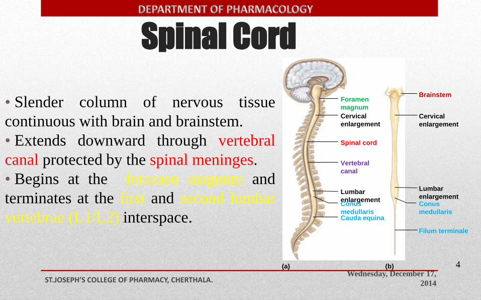
• Slender column of nervous tissue
continuous with brain and brainstem.
• Extends downward through vertebral
canal protected by the spinal meninges.
• Begins at the foramen magnum and
terminates at the first and second lumbar
vertebrae (L1/L2) interspace.
Brainstem
Spinal cord
(a) (b)
Foramen
magnum
Cervical
enlargement
Vertebral
canal
Lumbar
enlargementConus
medullarisCauda equina
Filum terminale
Conus
medullaris
Lumbar
enlargement
Cervical
enlargement
Wednesday, December 17,
2014ST.JOSEPH’S COLLEGE OF PHARMACY, CHERTHALA.
5
Brainstem
Spinal cord
(a) (b)
Foramen
magnum
Cervical
enlargement
Vertebral
canal
Lumbar
enlargementConus
medullarisCauda equina
Filum terminale
Conus
medullaris
Lumbar
enlargement
Cervical
enlargement
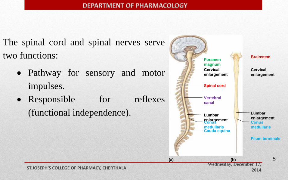
The spinal cord and spinal nerves serve
two functions:
Pathway for sensory and motor
impulses.
Responsible for reflexes
(functional independence).
Structure of the Spinal Cord:
Wednesday, December 17,
2014ST.JOSEPH’S COLLEGE OF PHARMACY, CHERTHALA.
6
Brainstem
Spinal cord
(a) (b)
Foramen
magnum
Cervical
enlargement
Vertebral
canal
Lumbar
enlargementConus
medullarisCauda equina
Filum terminale
Conus
medullaris
Lumbar
enlargement
Cervical
enlargement
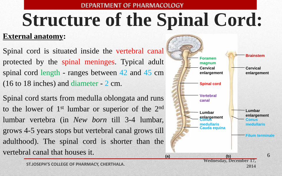
External anatomy:
Spinal cord is situated inside the vertebral canal
protected by the spinal meninges. Typical adult
spinal cord length - ranges between 42 and 45 cm
(16 to 18 inches) and diameter - 2 cm.
Spinal cord starts from medulla oblongata and runs
to the lower of 1st lumbar or superior of the 2nd
lumbar vertebra (in New born till 3-4 lumbar,
grows 4-5 years stops but vertebral canal grows till
adulthood). The spinal cord is shorter than the
vertebral canal that houses it.
Wednesday, December 17,
2014ST.JOSEPH’S COLLEGE OF PHARMACY, CHERTHALA.
7
Brainstem
Spinal cord
(a) (b)
Foramen
magnum
Cervical
enlargement
Vertebral
canal
Lumbar
enlargementConus
medullarisCauda equina
Filum terminale
Conus
medullaris
Lumbar
enlargement
Cervical
enlargement
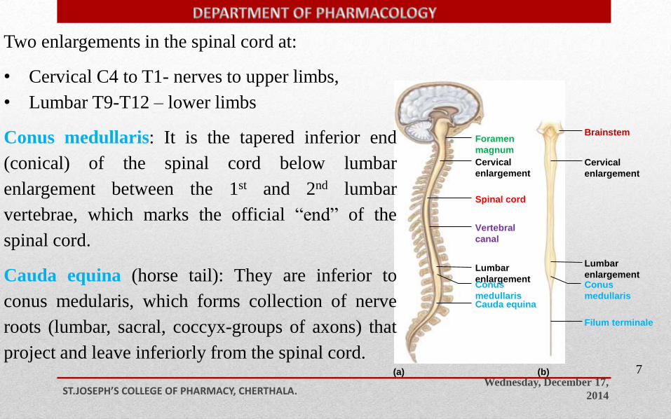
Two enlargements in the spinal cord at:
• Cervical C4 to T1- nerves to upper limbs,
• Lumbar T9-T12 – lower limbs
Conus medullaris: It is the tapered inferior end
(conical) of the spinal cord below lumbar
enlargement between the 1st and 2nd lumbar
vertebrae, which marks the official “end” of the
spinal cord.
Cauda equina (horse tail): They are inferior to
conus medularis, which forms collection of nerve
roots (lumbar, sacral, coccyx-groups of axons) that
project and leave inferiorly from the spinal cord.
Wednesday, December 17,
2014ST.JOSEPH’S COLLEGE OF PHARMACY, CHERTHALA.
8
Brainstem
Spinal cord
(a) (b)
Foramen
magnum
Cervical
enlargement
Vertebral
canal
Lumbar
enlargementConus
medullarisCauda equina
Filum terminale
Conus
medullaris
Lumbar
enlargement
Cervical
enlargement
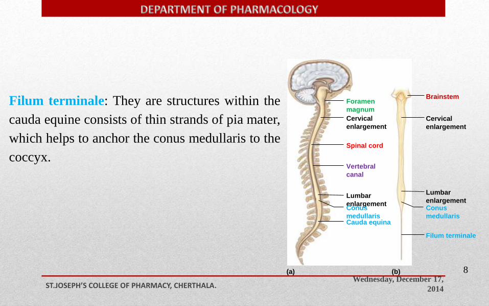
Filum terminale: They are structures within the
cauda equine consists of thin strands of pia mater,
which helps to anchor the conus medullaris to the
coccyx.
Wednesday, December 17,
2014ST.JOSEPH’S COLLEGE OF PHARMACY, CHERTHALA.
9
Brainstem
Spinal cord
(a) (b)
Foramen
magnum
Cervical
enlargement
Vertebral
canal
Lumbar
enlargementConus
medullarisCauda equina
Filum terminale
Conus
medullaris
Lumbar
enlargement
Cervical
enlargement
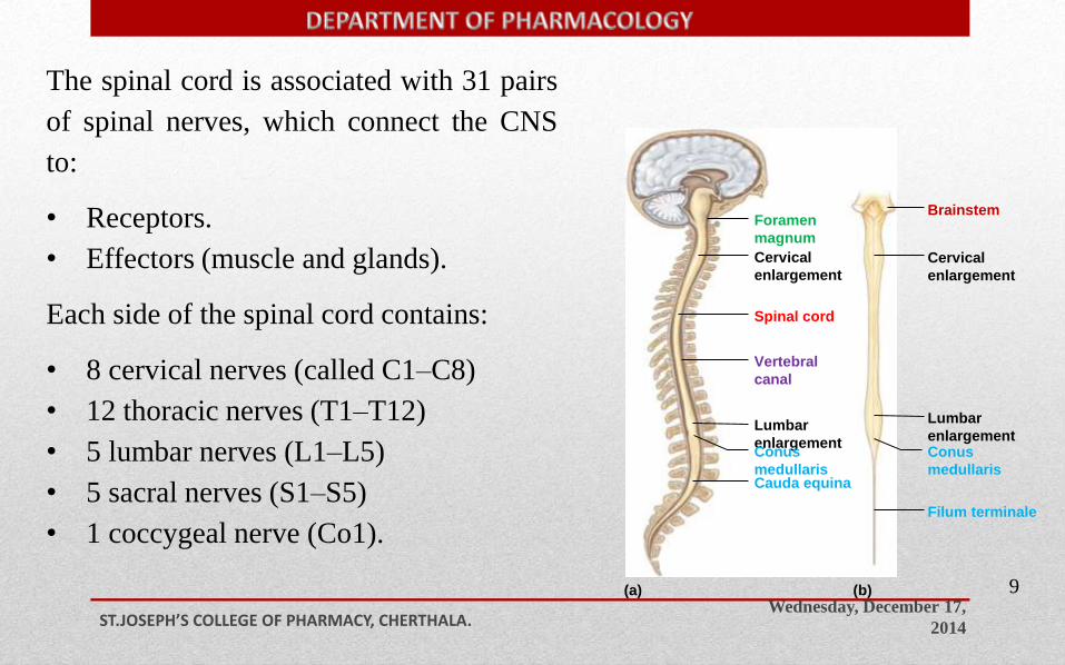
The spinal cord is associated with 31 pairs
of spinal nerves, which connect the CNS
to:
• Receptors.
• Effectors (muscle and glands).
Each side of the spinal cord contains:
• 8 cervical nerves (called C1–C8)
• 12 thoracic nerves (T1–T12)
• 5 lumbar nerves (L1–L5)
• 5 sacral nerves (S1–S5)
• 1 coccygeal nerve (Co1).
Wednesday, December 17,
2014ST.JOSEPH’S COLLEGE OF PHARMACY, CHERTHALA.
1
0
Brainstem
Spinal cord
(a) (b)
Foramen
magnum
Cervical
enlargement
Vertebral
canal
Lumbar
enlargementConus
medullarisCauda equina
Filum terminale
Conus
medullaris
Lumbar
enlargement
Cervical
enlargement
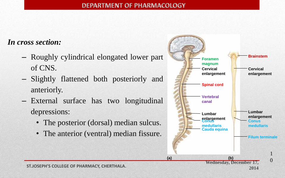
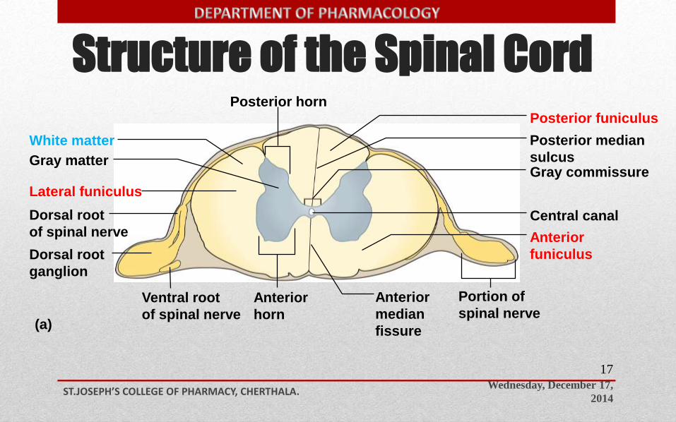
In cross section:
– Roughly cylindrical elongated lower part
of CNS.
– Slightly flattened both posteriorly and
anteriorly.
– External surface has two longitudinal
depressions:
• The posterior (dorsal) median sulcus.
• The anterior (ventral) median fissure.
Structure of the Spinal Cord
Wednesday, December 17,
2014ST.JOSEPH’S COLLEGE OF PHARMACY, CHERTHALA.
11
White matter
Gray matter
Lateral funiculus
Posterior funiculus
Gray commissure
Central canal
(a)
Posterior horn
Dorsal root
of spinal nerve
Dorsal root
ganglion
Ventral root
of spinal nerve
Anterior
horn
Anterior
median
fissure
Portion of
spinal nerve
Anterior
funiculus
Posterior median
sulcus
Wednesday, December 17,
2014ST.JOSEPH’S COLLEGE OF PHARMACY, CHERTHALA.
12
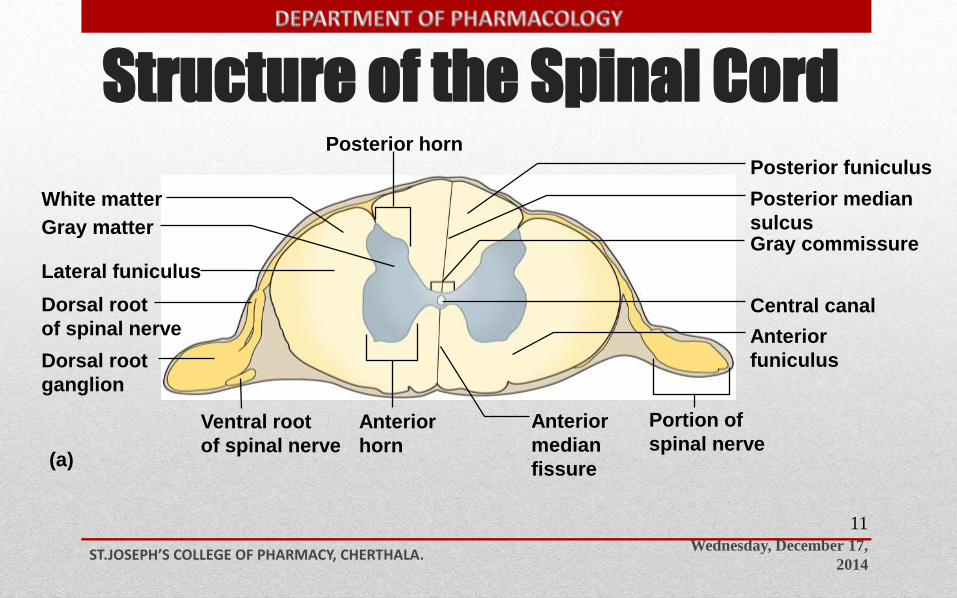
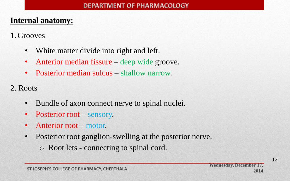
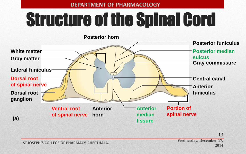
Internal anatomy:
1. Grooves
• White matter divide into right and left.
• Anterior median fissure – deep wide groove.
• Posterior median sulcus – shallow narrow.
2. Roots
• Bundle of axon connect nerve to spinal nuclei.
• Posterior root – sensory.
• Anterior root – motor.
• Posterior root ganglion-swelling at the posterior nerve.
o Root lets - connecting to spinal cord.
Structure of the Spinal Cord
Wednesday, December 17,
2014ST.JOSEPH’S COLLEGE OF PHARMACY, CHERTHALA.
13
White matter
Gray matter
Lateral funiculus
Posterior funiculus
Gray commissure
Central canal
(a)
Posterior horn
Dorsal root
of spinal nerve
Dorsal root
ganglion
Ventral root
of spinal nerve
Anterior
horn
Anterior
median
fissure
Portion of
spinal nerve
Anterior
funiculus
Posterior median
sulcus
Wednesday, December 17,
2014ST.JOSEPH’S COLLEGE OF PHARMACY, CHERTHALA.
14
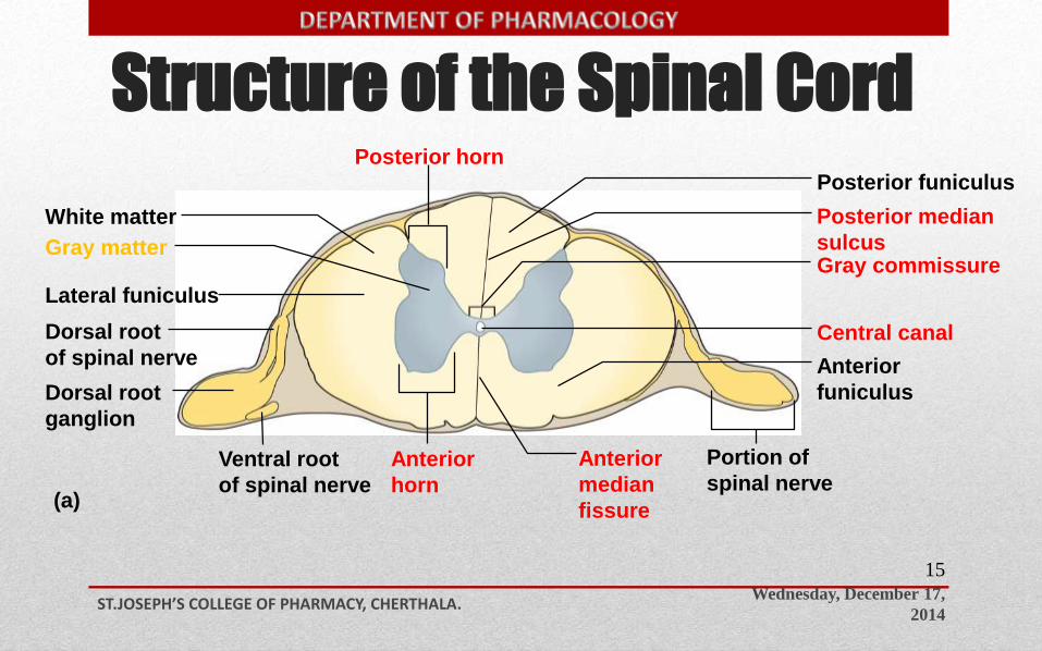
Location and Distribution of Gray Matter:
In the spinal cord, it is centrally located
• Its shape resembles a letter H or a butterfly.
• Surrounded by white matter, dendrites, cell bodies, unmyelinated axons and neuroglia.
• Central canal filled with CSF.
• The gray matter may be subdivided into the following components:
o Anterior horns.
o Lateral horns.
o Posterior horns.
o The gray commissure - cross bar which connects 2 halves horns.
o Nuclei - clusters of cell bodies form functional groups.
Sensory and motor nuclei.
Lateral only in thoracic area autonomic motor.
Structure of the Spinal Cord
Wednesday, December 17,
2014ST.JOSEPH’S COLLEGE OF PHARMACY, CHERTHALA.
15
White matter
Gray matter
Lateral funiculus
Posterior funiculus
Gray commissure
Central canal
(a)
Posterior horn
Dorsal root
of spinal nerve
Dorsal root
ganglion
Ventral root
of spinal nerve
Anterior
horn
Anterior
median
fissure
Portion of
spinal nerve
Anterior
funiculus
Posterior median
sulcus
Wednesday, December 17,
2014ST.JOSEPH’S COLLEGE OF PHARMACY, CHERTHALA.
16
Location and Distribution of White Matter:
• The white matter of the spinal cord is external to the gray matter.
• Three regions. Composed of tracts – (bundles of axon).
Ascending sensory tract.
Descending motor tract.
o Posterior (white column) funiculus:
lies between the posterior gray horns and the posterior median sulcus.
o Lateral (white column) funiculus.
o Anterior (white column) funiculus.
Between the anterior gray horns and the anterior median fissure.
• The anterior funiculi are interconnected by the white commissure.
Structure of the Spinal Cord
Wednesday, December 17,
2014ST.JOSEPH’S COLLEGE OF PHARMACY, CHERTHALA.
17
White matter
Gray matter
Lateral funiculus
Posterior funiculus
Gray commissure
Central canal
(a)
Posterior horn
Dorsal root
of spinal nerve
Dorsal root
ganglion
Ventral root
of spinal nerve
Anterior
horn
Anterior
median
fissure
Portion of
spinal nerve
Anterior
funiculus
Posterior median
sulcus
Tracts of the Spinal Cord
Wednesday, December 17,
2014ST.JOSEPH’S COLLEGE OF PHARMACY, CHERTHALA.
18
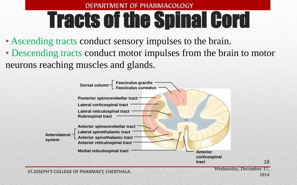
• Ascending tracts conduct sensory impulses to the brain.
• Descending tracts conduct motor impulses from the brain to motor
neurons reaching muscles and glands.
Posterior spinocerebellar tract
Lateral corticospinal tract
Lateral reticulospinal tract
Rubrospinal tract
Anterior spinocerebellar tract
Lateral spinothalamic tract
Anterior reticulospinal tract
Medial reticulospinal tract
Fasciculus cuneatus
Fasciculus gracilisDorsal column
Anterior spinothalamic tractAnterolateral
system
Anterior
corticospinal
tract
Ascending Tracts
Wednesday, December 17,
2014ST.JOSEPH’S COLLEGE OF PHARMACY, CHERTHALA.
19
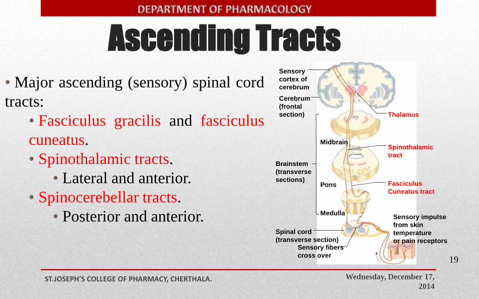
• Major ascending (sensory) spinal cord
tracts:
• Fasciculus gracilis and fasciculus
cuneatus.
• Spinothalamic tracts.
• Lateral and anterior.
• Spinocerebellar tracts.
• Posterior and anterior.
Midbrain
Pons
Medulla
Thalamus
Sensory
cortex of
cerebrum
Cerebrum
(frontal
section)
Brainstem
(transverse
sections)
Spinal cord
(transverse section)Sensory fibers
cross over
Spinothalamic
tract
Fasciculus
Cuneatus tract
Sensory impulse
from skin
temperature
or pain receptors
Descending Tracts
Wednesday, December 17,
2014ST.JOSEPH’S COLLEGE OF PHARMACY, CHERTHALA.
2
0
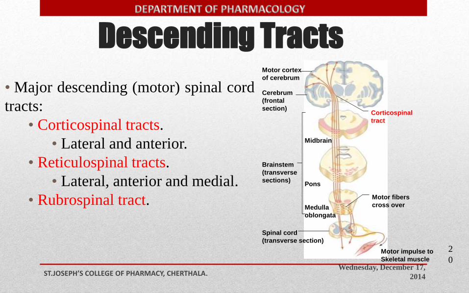
• Major descending (motor) spinal cord
tracts:
• Corticospinal tracts.
• Lateral and anterior.
• Reticulospinal tracts.
• Lateral, anterior and medial.
• Rubrospinal tract.
Midbrain
Pons
Brainstem
(transverse
sections)
Spinal cord
(transverse section)
Motor cortex
of cerebrum
Cerebrum
(frontal
section)Corticospinal
tract
Medulla
oblongata
Motor fibers
cross over
Motor impulse to
Skeletal muscle
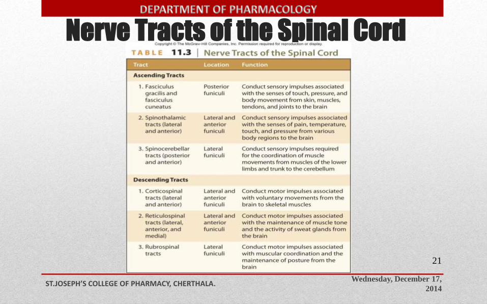
Nerve Tracts of the Spinal Cord
Wednesday, December 17,
2014ST.JOSEPH’S COLLEGE OF PHARMACY, CHERTHALA.
21
Functions of Spinal Cord
Wednesday, December 17,
2014ST.JOSEPH’S COLLEGE OF PHARMACY, CHERTHALA.
22
• Center for spinal reflexes.
• Conduct pathway for nerve impulses to and from the
brain and brainstem.
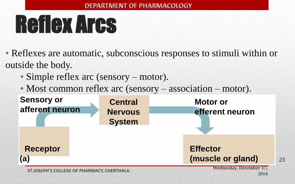
Reflex Arcs
Wednesday, December 17,
2014ST.JOSEPH’S COLLEGE OF PHARMACY, CHERTHALA.
23
• Reflexes are automatic, subconscious responses to stimuli within or
outside the body.
• Simple reflex arc (sensory – motor).
• Most common reflex arc (sensory – association – motor).
Receptor
(a)
Sensory or
afferent neuronMotor or
efferent neuronCentral
Nervous
System
Effector
(muscle or gland)
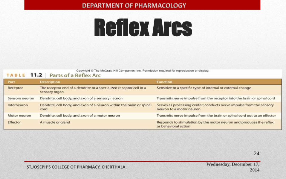
Reflex Arcs
Wednesday, December 17,
2014ST.JOSEPH’S COLLEGE OF PHARMACY, CHERTHALA.
24
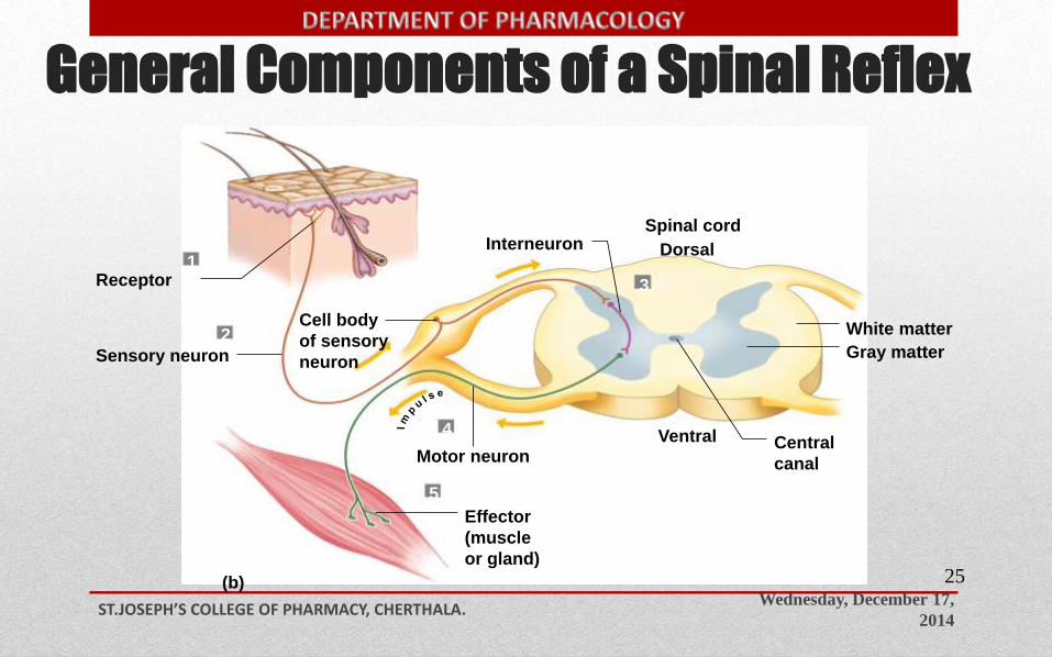
General Components of a Spinal Reflex
Wednesday, December 17,
2014ST.JOSEPH’S COLLEGE OF PHARMACY, CHERTHALA.
25
Receptor
Sensory neuron
Motor neuron
White matter
Gray matter
Spinal cord
DorsalInterneuron
4
5
3
2
1
(b)
Cell body
of sensory
neuron
Effector
(muscle
or gland)
Central
canal
Ventral
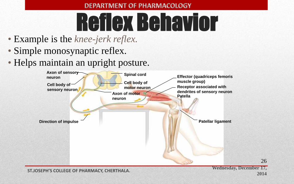
Reflex Behavior
Wednesday, December 17,
2014ST.JOSEPH’S COLLEGE OF PHARMACY, CHERTHALA.
26
• Example is the knee-jerk reflex.
• Simple monosynaptic reflex.
• Helps maintain an upright posture.
Copyright © The McGraw-Hill Companies, Inc. Permission required for reproduction or display.
Spinal cord
Patella
Patellar ligamentDirection of impulse
Axon of sensory
neuron
Cell body of
sensory neuron
Cell body of
motor neuron
Axon of motor
neuron
Effector (quadriceps femoris
muscle group)
Receptor associated with
dendrites of sensory neuron
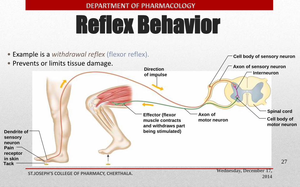
Reflex Behavior
Wednesday, December 17,
2014ST.JOSEPH’S COLLEGE OF PHARMACY, CHERTHALA.
27
• Example is a withdrawal reflex (flexor reflex).• Prevents or limits tissue damage.
Interneuron
Spinal cord
Axon of sensory neuron
Cell body of sensory neuron
Dendrite of
sensory
neuronPain
receptor
in skin
Direction
of impulse
Cell body of
motor neuron
Axon of
motor neuronEffector (flexor
muscle contracts
and withdraws part
being stimulated)
Tack
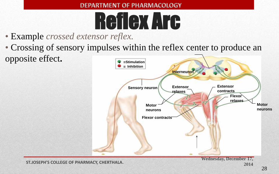
Reflex Arc
Wednesday, December 17,
2014ST.JOSEPH’S COLLEGE OF PHARMACY, CHERTHALA.
28
• Example crossed extensor reflex.
• Crossing of sensory impulses within the reflex center to produce an
opposite effect.=Stimulation
= Inhibition
Interneuron
Flexor contracts
Sensory neuron
+
++
–
––
Motor
neurons
Extensor
contracts
Flexor
relaxesMotor
neurons
Extensor
relaxes
Wednesday, December 17,
2014ST.JOSEPH’S COLLEGE OF PHARMACY, CHERTHALA.
29

Wednesday, December 17,
2014ST.JOSEPH’S COLLEGE OF PHARMACY, CHERTHALA.
30