
Respiratory - uploads-ssl.webflow.com
52
Respiratory 1 Lecture 7 [email protected] www.bitemedicine.com www.facebook.com/biteemedicine @bitemedicine Dr Azeem Alam, MBBS BSc (Hons) Surgical AFP Guy’s and St. Thomas’ Hospital Content reviewed on 21/04/2020.
Transcript of Respiratory - uploads-ssl.webflow.com

Respiratory
1
Lecture 7
www.bitemedicine.comwww.facebook.com/biteemedicine
@bitemedicine
Dr Azeem Alam, MBBS BSc (Hons)Surgical AFPGuy’s and St. Thomas’ Hospital
Content reviewed on 21/04/2020.

Learning objectives• 2 respiratory topics: Pneumothorax and Pulmonary Embolism
• Case-based discussion(s) to identify the top differentials and why
• Theory to cover pathophysiology, diagnostic criteria, investigations and
management
• Quiz (Mentimeter and multi-step SBAs)
2www.bitemedicine.com Instagram: @bitemedicine Facebook: /biteemedicine
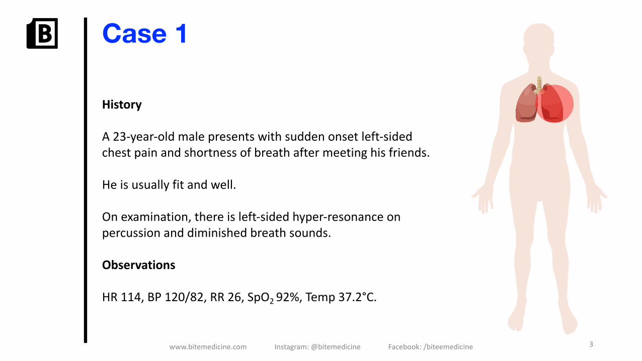
Case 1
History
A 23-year-old male presents with sudden onset left-sided chest pain and shortness of breath after meeting his friends.
He is usually fit and well.
On examination, there is left-sided hyper-resonance on percussion and diminished breath sounds.
Observations
HR 114, BP 120/82, RR 26, SpO2 92%, Temp 37.2°C.
3www.bitemedicine.com Instagram: @bitemedicine Facebook: /biteemedicine


5www.bitemedicine.com Instagram: @bitemedicine Facebook: /biteemedicine
PathophysiologyDefinition: accumulation of air within the pleural space
Spontaneous occurs without trauma• Primary pneumothorax: without underlying pulmonary disease• Secondary pneumothorax: complication secondary to underlying pulmonary
disease
Traumatic pneumothorax• Penetrating or blunt injury to the chest, including iatrogenic causes
Tension pneumothorax (EMERGENCY)• Intrapleural pressure exceeds atmospheric

6www.bitemedicine.com Instagram: @bitemedicine Facebook: /biteemedicine
(1)

7www.bitemedicine.com Instagram: @bitemedicine Facebook: /biteemedicine
Pathophysiology
Primary spontaneous
Pathogenesis Spontaneous rupture of a subpleural bleb
Typical presentation Young, tall, healthy, male presenting with sudden onset breathlessness and chest pain
Underlying lung disease?
No
Risk factors • Tall, slender, young (20-30)
• Smoking• Marfan syndrome• Family history• Diving or flying
(2)

8www.bitemedicine.com Instagram: @bitemedicine Facebook: /biteemedicine
PathophysiologySecondary spontaneous
Pathogenesis Rupture of damaged pulmonary tissue
Typical presentation Middle-aged patient with COPD presenting with sudden onset breathlessness and chest pain
Underlying lung disease? Yes: occurs due to ruptured bleb or bullae secondary to lung disease
Risk factors • Underlying lung disease: COPD, asthma, lung cancer
• Tuberculosis• Pneumocystis
jirovecii
(3)

9www.bitemedicine.com Instagram: @bitemedicine Facebook: /biteemedicine
PathophysiologyTension (emergency)
Pathogenesis • Air is forced to enter the thoracic cavity without any means of escape
• Results in a ‘one-way-valve’
Typical presentation Ventilated patient suddenly becomes breathless and acutely unwell
Underlying lung disease? Yes/no: usually occurs in ventilated or trauma patients
Risk factors • Mechanical ventilation
• Trauma• Iatrogenic: central
line insertion, biopsy
(4)

10www.bitemedicine.com Instagram: @bitemedicine Facebook: /biteemedicine
Clinical featuresSymptoms Signs
Sudden onset pleuritic chest pain Tachycardia and tachypnoea
Sudden onset dyspnoea Cyanosis
Hyper-resonance ipsilaterally
Reduced breath sounds ipsilaterally
Hyperexpanded chest ipsilaterally: associated with tension pneumothorax
Contralateral tracheal deviation and circulatory shock in tension pneumothorax

11www.bitemedicine.com Instagram: @bitemedicine Facebook: /biteemedicine
DifferentialsPneumothorax Pulmonary embolism Pneumonia
• SOB• Pleuritic chest pain
• SOB• Pleuritic chest pain• Haemoptysis• Pain / swelling in one leg
• SOB• Pleuritic chest pain• Productive cough• Fever
• Any age• Primary spontaneous• Secondary spontaneous• Tension
• Risk factors for thromboembolism• Obesity• Prolonged bed rest • Pregnancy • Malignancy
• Usually middle-aged or elderly
• More common with underlying lung disease
Confirmed on CXR ECG usually non-specific, but sinus tachycardia and S1Q3T3
Usually confirmed on CXR

12www.bitemedicine.com Instagram: @bitemedicine Facebook: /biteemedicine
InvestigationsImaging• Chest x-ray: visible visceral pleural edge with no lung margins peripheral to this• CT chest: gold-standard imaging method but not routinely performed
Bedside• ECG: exclude a cardiac cause
Bloods• Arterial blood gas: may demonstrate respiratory failure
Additional points• Other investigations will depend on the aetiology• ALL patients require a repeat CXR after intervention
• Tension pneumothorax: decompress prior to imaging if high clinical suspicion

13(5)

14(6)

15


(5)

(5)


20www.bitemedicine.com Instagram: @bitemedicine Facebook: /biteemedicine
Management: spontaneous • Needle aspiration: 2nd intercostal space midclavicular line
• Chest drain: 5th intercostal space mid-axillary line; triangle of safety
• Remember to always insert above the upper border of the rib
• High-flow oxygen

21www.bitemedicine.com Instagram: @bitemedicine Facebook: /biteemedicine
(7)
22www.bitemedicine.com Instagram: @bitemedicine Facebook: /biteemedicine
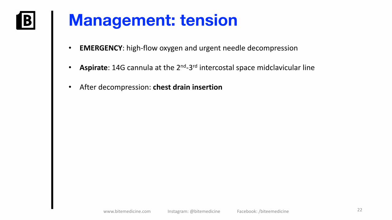
Management: tension• EMERGENCY: high-flow oxygen and urgent needle decompression
• Aspirate: 14G cannula at the 2nd-3rd intercostal space midclavicular line
• After decompression: chest drain insertion

23www.bitemedicine.com Instagram: @bitemedicine Facebook: /biteemedicine
Chest drain insertion
Base of axilla
Lateral edge of latissimus dorsi
Lateral edge of pectoris major
Nipple or 5th intercostal space

24www.bitemedicine.com Instagram: @bitemedicine Facebook: /biteemedicine
Chest drain insertion


26www.bitemedicine.com Instagram: @bitemedicine Facebook: /biteemedicine
Management: recurrentpneumothoracesOptions• Open thoracotomy and pleurectomy: lowest recurrence rate (1%)• VATS pleurectomy: lower morbidity than open• Surgical chemical pleurodesis: less popular now
Indications for referral to a thoracic surgeon
First contralateral pneumothorax Second ipsilateral pneumothorax
Bilateral spontaneous pneumothorax Persistent air-leak despite chest drain
High risk professions: e.g. pilots Pregnancy

27www.bitemedicine.com Instagram: @bitemedicine Facebook: /biteemedicine
Top decile question


29www.bitemedicine.com Instagram: @bitemedicine Facebook: /biteemedicine
Management: follow-upFlying• Patients can fly 1 week post check CXR as long as the pneumothorax has resolved
Diving• Avoid indefinitely until the patient has had a definitive bilateral surgical
pleurectomy, post-operative CT chest and normal lung function tests
30www.bitemedicine.com Instagram: @bitemedicine Facebook: /biteemedicine
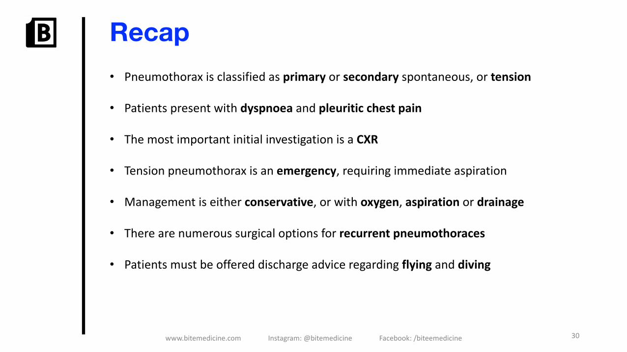
Recap• Pneumothorax is classified as primary or secondary spontaneous, or tension
• Patients present with dyspnoea and pleuritic chest pain
• The most important initial investigation is a CXR
• Tension pneumothorax is an emergency, requiring immediate aspiration
• Management is either conservative, or with oxygen, aspiration or drainage
• There are numerous surgical options for recurrent pneumothoraces
• Patients must be offered discharge advice regarding flying and diving

Case 2
History A 65-year-old female presents with sudden onset shortness of breath and pleuritic chest pain.
She has a history of a right-sided mastectomy for breast cancer, 1 year ago.
She has a BMI of 27.
Observations
HR 125, BP 85/60, RR 28, SpO2 89%, Temp 37.7°C
31www.bitemedicine.com Instagram: @bitemedicine Facebook: /biteemedicine


PathophysiologyDefinition: obstruction of the pulmonary vasculature secondary to an embolus
• Virchow’s triad• Often secondary to deep vein thrombosis• Embolus dislodges and migrate to the lung circulation• Obstructed pulmonary vasculature ⟶ increased pulmonary vascular resistance
• Can result in arrhythmias, pulmonary infarction, cor pulmonale and cardiac arrest

Pathophysiology

35www.bitemedicine.com Instagram: @bitemedicine Facebook: /biteemedicine
Clinical featuresSymptoms SignsPleuritic chest pain Tachypnoea and tachycardiaDyspnoea HypoxiaCough or haemoptysis Deep vein thrombosis: swollen,
tender calfFever PyrexiaSyncope: a red flag symptom Hypotension: SBP < 90mmHg
suggests massive PEElevated JVP: suggests corpulmonaleRight parasternal heave: suggests right ventricular strain

36www.bitemedicine.com Instagram: @bitemedicine Facebook: /biteemedicine
DifferentialsPneumothorax Pulmonary embolism Pneumonia
• SOB• Pleuritic chest pain
• SOB• Pleuritic chest pain• Haemoptysis• Pain / swelling in one leg
• SOB• Pleuritic chest pain• Productive cough• Fever
• Any age• Primary spontaneous• Secondary spontaneous• Tension
• Risk factors for thromboembolism• Obesity• Prolonged bed rest • Pregnancy • Malignancy
• Usually middle-aged or elderly
• More common with underlying lung disease
Confirmed on CXR ECG usually non-specific, but sinus tachycardia and S1Q3T3
Usually confirmed on CXR

Case 2
History A 65-year-old female presents with sudden onset shortness of breath and pleuritic chest pain.
She has a history of a right-sided mastectomy for breast cancer, 1 year ago.
She has a BMI of 27.
Observations
HR 125, BP 85/60, RR 28, SpO2 89%, Temp 37.7°C
37www.bitemedicine.com Instagram: @bitemedicine Facebook: /biteemedicine


39www.bitemedicine.com Instagram: @bitemedicine Facebook: /biteemedicine
Wells scoreWells Two-Level PE Score
Clinical feature Points
Clinical signs and symptoms of a DVT 3.0
PE is number 1 diagnosis or equally likely 3.0
Tachycardia (>100 BPM) 1.5
Immobilisation for more than three days or surgery in the previous four weeks
1.5
Previous, objectively diagnosed PE or DVT 1.5
Malignancy with treatment within the last 6 months, or palliative 1.0
Haemoptysis 1.0

40www.bitemedicine.com Instagram: @bitemedicine Facebook: /biteemedicine
InvestigationsBedside• ECG: sinus tachycardia (most common); RBBB and right axis deviation; S1Q3T3
Bloods• ABG: may demonstrate respiratory failure
Imaging• CXR: typically normal, although a wedge-shaped opacification can be seen• ECHO: assess for right ventricular strain in massive PE
Specialist tests: depends on Wells score• CTPA is performed if high probability (Wells score > 4) or• D-dimer performed if low probability (Wells score ≤ 4)

(8)

(9)

43www.bitemedicine.com Instagram: @bitemedicine Facebook: /biteemedicine
Investigations


45www.bitemedicine.com Instagram: @bitemedicine Facebook: /biteemedicine
Further investigations: unprovoked PEInvestigations for cancer• All patients: full set of blood tests, CXR, and urinalysis• Patients > 40 years old: CT abdomen and pelvis should be considered
Investigations for thrombophilia• Antiphospholipid antibodies: considered in people who have an unprovoked PE• Hereditary thrombophilia: considered in people who have an unprovoked PE and a
first-degree relative who has had a DVT

46www.bitemedicine.com Instagram: @bitemedicine Facebook: /biteemedicine
ManagementMassive PE• Thrombolysis: e.g. alteplase
Non-massive PE• Anticoagulation:
• Oral anticoagulation: warfarin or DOAC for 3 months if provoked, or 6 months if unprovoked
• LMWH used for 6 months in cases of active cancer
Alternative treatments• Inferior vena cava filter: consider in patients with recurrent PEs, despite
anticoagulation• Surgical embolectomy: when thrombolysis has failed or is contraindicated

47www.bitemedicine.com Instagram: @bitemedicine Facebook: /biteemedicine
Recap• A pulmonary embolism presents with dyspnoea and pleuritic chest pain
• Risk factors can be remembered using Virchow’s triad
• A massive PE can cause cor pulmonale and rapid deterioration
• Initial investigations include ABG, ECG, CXR, D-dimer, CTPA and ECHO for a massive PE
• Patients with an unprovoked PE require further investigations
• Management options include thrombolysis, DOAC, LMWH or specialist interventions

48www.bitemedicine.com Instagram: @bitemedicine Facebook: /biteemedicine
Top decile question


Further information• We need your feedback!
• Lecture series / schedule
• New, interactive website coming very soon
• Stay up-to-date!• Website: www.bitemedicine.com• Facebook: https://www.facebook.com/biteemedicine• Instagram: @bitemedicine• Email: [email protected]
• Want to get involved? Contact us at [email protected] to get your information pack
50Buy our textbook at www.bitemedicine.com – SOLD OUT


References1. OpenStax College / CC BY (https://creativecommons.org/licenses/by/3.0).
https://upload.wikimedia.org/wikipedia/commons/0/0d/2313_The_Lung_Pleurea.jpg2. Mileny ES Colovati, Luciana RJ da Silva, Sylvia S Takeno, Tatiane I Mancini, Ana R N Dutra, Roberta S Guilherme,
Cláudia B de Mello, Maria I Melaragno and Ana B A Perez / CC BY (https://creativecommons.org/licenses/by/2.0)
3. National Heart Lung and Blood Institute / Public domain4. Royalty—free stock illustration from Shutterstock.5. James Heilman, MD / CC BY (https://creativecommons.org/licenses/by/3.0)6. Photographed by User Clinical Cases 00:42, 7 November 2006 [<a
href="https://creativecommons.org/licenses/by-sa/2.5">CC BY-SA 7. Egmason / CC BY-SA (https://creativecommons.org/licenses/by-sa/4.0).
https://upload.wikimedia.org/wikipedia/commons/e/e2/Endothoracic_fascia.svg8. James Heilman, MD / CC BY-SA (https://creativecommons.org/licenses/by-sa/3.0).
https://upload.wikimedia.org/wikipedia/commons/b/bd/Sinustachy.JPG9. James Heilman, MD / CC BY-SA (https://creativecommons.org/licenses/by-sa/3.0).
https://upload.wikimedia.org/wikipedia/commons/4/4e/Cardiogram_indicating_right_bundle_branch_block_with_tachycardia.jpg
52Buy our textbook at www.bitemedicine.com


