Respiratory system - General Histologyrespiratory system . • Describe the embryologic steps in the...
12
10/2/19 1 Respiratory System Kristine Krafts, M.D. 1 Respiratory System Objectives • Describe the components and functions of the conducting and respiratory portions of the respiratory system. • Describe the embryologic steps in the development of the respiratory system. • Describe and compare the characteristic microscopic features and functions of the different regions and airways of the respiratory system, including lining epithelium, glands, cartilage, smooth muscle and elastic fibers. 2 Respiratory System Lecture Outline • Introduction • Conducting portion • Respiratory portion • Pleura 3 Respiratory System Lecture Outline • Introduction • Anatomy • Embryology 4 Respiratory System Lecture Outline • Introduction • Anatomy 5 Anatomy of the respiratory system 6
Transcript of Respiratory system - General Histologyrespiratory system . • Describe the embryologic steps in the...

10/2/19
1
Respiratory SystemKristine Krafts, M.D.
1
Respiratory System Objectives
• Describe the components and functions of the conducting and respiratory portions of the respiratory system.
• Describe the embryologic steps in the development of the respiratory system.
• Describe and compare the characteristic microscopic features and functions of the different regions and airways of the respiratory system, including lining epithelium, glands, cartilage, smooth muscle and elastic fibers.
2
Respiratory System Lecture Outline
• Introduction
• Conducting portion
• Respiratory portion
• Pleura
3
Respiratory System Lecture Outline
• Introduction• Anatomy• Embryology
4
Respiratory System Lecture Outline
• Introduction• Anatomy
5
Anatomy of the respiratory system
6
10/2/19
2
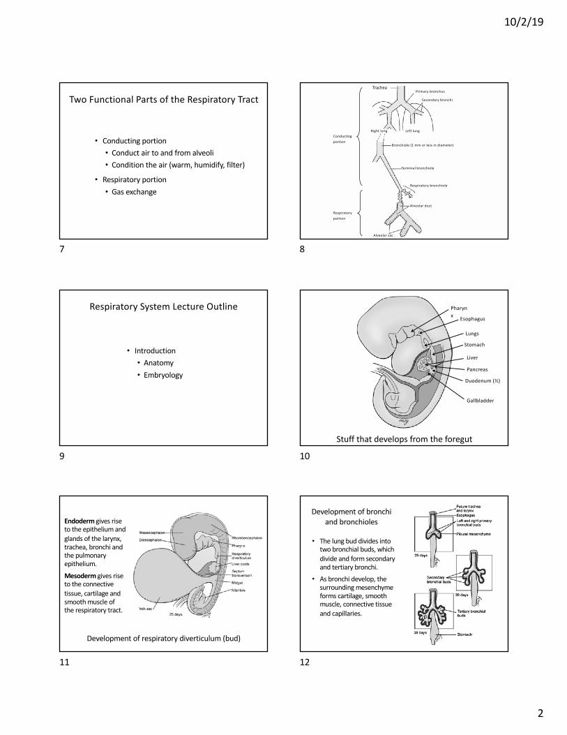
Two Functional Parts of the Respiratory Tract
• Conducting portion• Conduct air to and from alveoli• Condition the air (warm, humidify, filter)
• Respiratory portion• Gas exchange
7
Trachea
Right lung Left lung
Primary bronchus
Secondary bronchi
Bronchiole (1 mm or less in diameter)
Terminal bronchiole
Respiratory bronchiole
Alveolar duct
Alveolar sac
Conductingportion
Respiratoryportion
8
Respiratory System Lecture Outline
• Introduction• Anatomy• Embryology
9
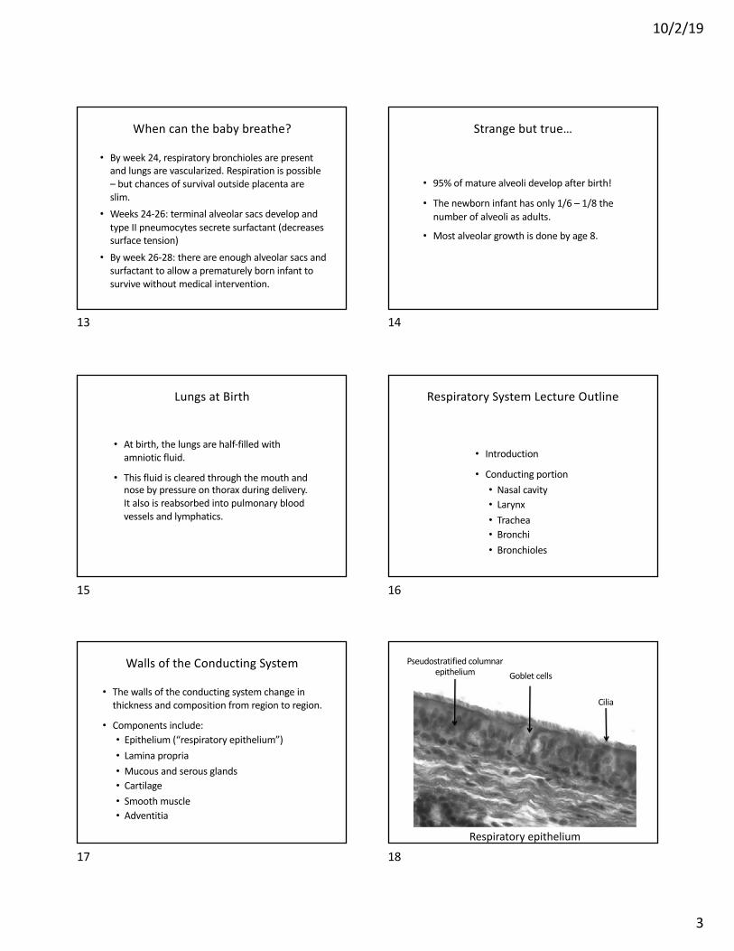
Pharynx
Lungs
Esophagus
Gallbladder
Stomach
Liver
Stuff that develops from the foregut
Duodenum (½)
Pancreas
10
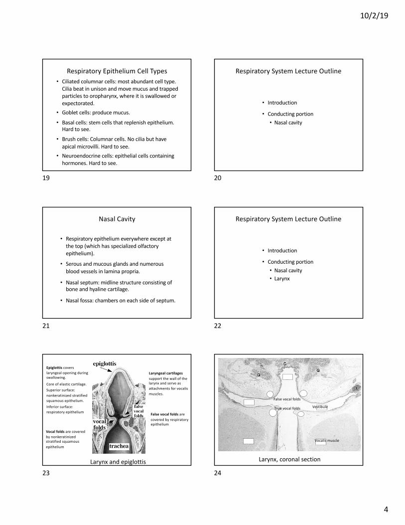
Development of respiratory diverticulum (bud)
Endoderm gives rise to the epithelium and glands of the larynx, trachea, bronchi and the pulmonary epithelium.
Mesoderm gives rise to the connective tissue, cartilage and smooth muscle of the respiratory tract.
11
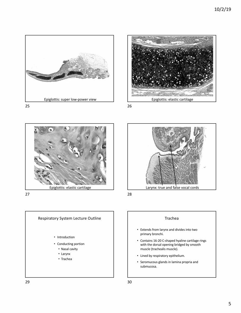
• The lung bud divides into two bronchial buds, which divide and form secondary and tertiary bronchi.
• As bronchi develop, the surrounding mesenchyme forms cartilage, smooth muscle, connective tissue and capillaries.
Development of bronchi and bronchioles
12
10/2/19
3
• By week 24, respiratory bronchioles are present and lungs are vascularized. Respiration is possible – but chances of survival outside placenta are slim.
• Weeks 24-26: terminal alveolar sacs develop and type II pneumocytes secrete surfactant (decreases surface tension)
• By week 26-28: there are enough alveolar sacs and surfactant to allow a prematurely born infant to survive without medical intervention.
When can the baby breathe?
13
• 95% of mature alveoli develop after birth!
• The newborn infant has only 1/6 – 1/8 the number of alveoli as adults.
• Most alveolar growth is done by age 8.
Strange but true…
14
• At birth, the lungs are half-filled with amniotic fluid.
• This fluid is cleared through the mouth and nose by pressure on thorax during delivery. It also is reabsorbed into pulmonary blood vessels and lymphatics.
Lungs at Birth
15
Respiratory System Lecture Outline
• Introduction
• Conducting portion• Nasal cavity• Larynx• Trachea• Bronchi• Bronchioles
16
• The walls of the conducting system change in thickness and composition from region to region.
• Components include:• Epithelium (“respiratory epithelium”)• Lamina propria• Mucous and serous glands• Cartilage• Smooth muscle• Adventitia
Walls of the Conducting System
17Respiratory epithelium
Pseudostratified columnar epithelium
Cilia
Goblet cells
18
10/2/19
4
• Ciliated columnar cells: most abundant cell type. Cilia beat in unison and move mucus and trapped particles to oropharynx, where it is swallowed or expectorated.
• Goblet cells: produce mucus.
• Basal cells: stem cells that replenish epithelium. Hard to see.
• Brush cells: Columnar cells. No cilia but have apical microvilli. Hard to see.
• Neuroendocrine cells: epithelial cells containing hormones. Hard to see.
Respiratory Epithelium Cell Types
19
Respiratory System Lecture Outline
• Introduction
• Conducting portion• Nasal cavity
20
• Respiratory epithelium everywhere except at the top (which has specialized olfactory epithelium).
• Serous and mucous glands and numerous blood vessels in lamina propria.
• Nasal septum: midline structure consisting of bone and hyaline cartilage.
• Nasal fossa: chambers on each side of septum.
Nasal Cavity
21
Respiratory System Lecture Outline
• Introduction
• Conducting portion• Nasal cavity• Larynx
22
Epiglottis covers laryngeal opening during swallowing.
Core of elastic cartilage.Superior surface: nonkeratinized stratified squamous epithelium.Inferior surface: respiratory epithelium
Laryngeal cartilages support the wall of the larynx and serve as attachments for vocalis muscles.
Vocal folds are covered by nonkeratinized stratified squamous epithelium
False vocal folds are covered by respiratory epithelium
Larynx and epiglottis
23
False vocal folds
True vocal folds
Vocalis muscle
Vestibule
Larynx, coronal section
24
10/2/19
5
Epiglottis: super low-power view
25Epiglottis: elastic cartilage
26
Epiglottis: elastic cartilage
27Larynx: true and false vocal cords
28
Respiratory System Lecture Outline
• Introduction
• Conducting portion• Nasal cavity• Larynx• Trachea
29
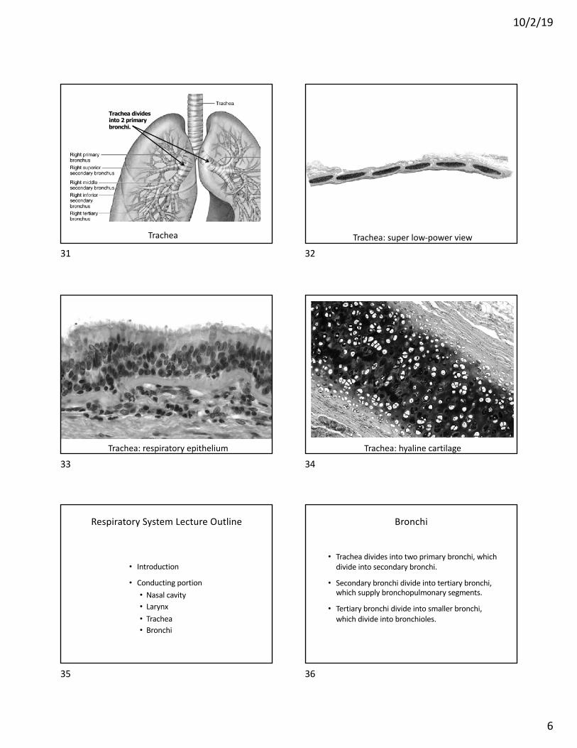
• Extends from larynx and divides into two primary bronchi.
• Contains 16-20 C-shaped hyaline cartilage rings with the dorsal opening bridged by smooth muscle (trachealis muscle).
• Lined by respiratory epithelium.
• Seromucous glands in lamina propria and submucosa.
Trachea
30
10/2/19
6
Trachea
Trachea divides into 2 primary bronchi.
31Trachea: super low-power view
32
Trachea: respiratory epithelium
33Trachea: hyaline cartilage
34
Respiratory System Lecture Outline
• Introduction
• Conducting portion• Nasal cavity• Larynx• Trachea• Bronchi
35
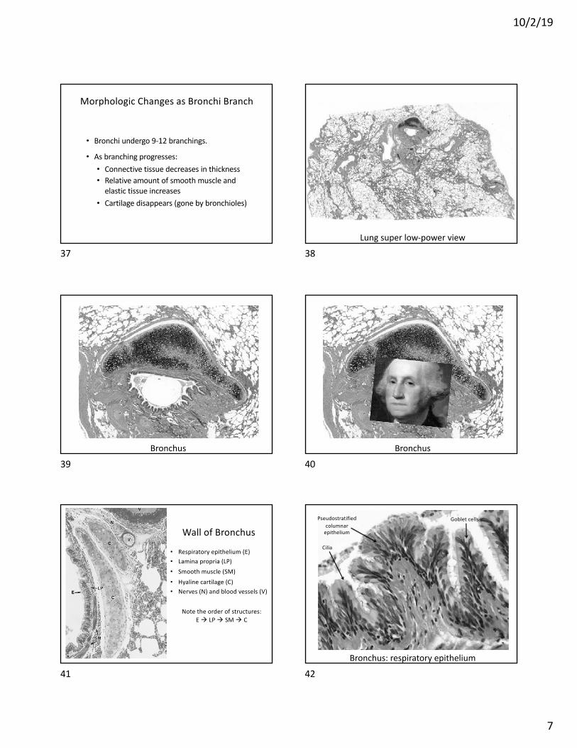
• Trachea divides into two primary bronchi, which divide into secondary bronchi.
• Secondary bronchi divide into tertiary bronchi, which supply bronchopulmonary segments.
• Tertiary bronchi divide into smaller bronchi, which divide into bronchioles.
Bronchi
36
10/2/19
7
• Bronchi undergo 9-12 branchings.
• As branching progresses:• Connective tissue decreases in thickness• Relative amount of smooth muscle and
elastic tissue increases• Cartilage disappears (gone by bronchioles)
Morphologic Changes as Bronchi Branch
37Lung super low-power view
38
Bronchus
39Bronchus
40
Wall of Bronchus
• Respiratory epithelium (E)• Lamina propria (LP) • Smooth muscle (SM) • Hyaline cartilage (C)• Nerves (N) and blood vessels (V)
Note the order of structures: E à LP à SM à C
41Bronchus: respiratory epithelium
Goblet cellsPseudostratifiedcolumnar
epithelium
Cilia
42
10/2/19
8
Respiratory System Lecture Outline
• Introduction
• Conducting portion• Nasal cavity• Larynx• Trachea• Bronchi• Bronchioles
43
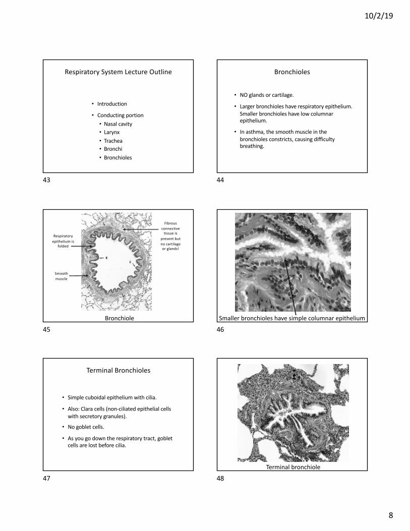
• NO glands or cartilage.
• Larger bronchioles have respiratory epithelium. Smaller bronchioles have low columnar epithelium.
• In asthma, the smooth muscle in the bronchioles constricts, causing difficulty breathing.
Bronchioles
44
Bronchiole
Smooth muscle
Respiratoryepithelium is
folded
Fibrous connective
tissue is present but no cartilage or glands!
45Smaller bronchioles have simple columnar epithelium
46
• Simple cuboidal epithelium with cilia.
• Also: Clara cells (non-ciliated epithelial cells with secretory granules).
• No goblet cells.
• As you go down the respiratory tract, goblet cells are lost before cilia.
Terminal Bronchioles
47Terminal bronchiole
48
10/2/19
9
Clara cells
Is there anything they don’t do?Make surfactant components, break down mucus,
detoxify harmful substances, transfer IgA, fight bacteria…
49
Respiratory System Lecture Outline
• Introduction
• Conducting portion
• Respiratory portion• Respiratory bronchioles• Alveoli
50
Respiratory System Lecture Outline
• Introduction
• Conducting portion
• Respiratory portion• Respiratory bronchioles
51
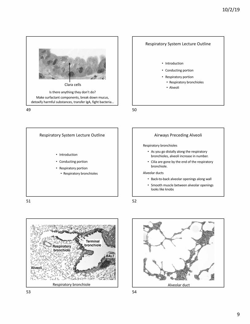
Respiratory bronchioles
• As you go distally along the respiratory bronchioles, alveoli increase in number.
• Cilia are gone by the end of the respiratory bronchiole.
Alveolar ducts
• Back-to-back alveolar openings along wall
• Smooth muscle between alveolar openings looks like knobs
Airways Preceding Alveoli
52
Respiratory bronchiole
53Alveolar duct
54

10/2/19
10
Alveolar duct (AD), alveolar sac (AS) and alveolus (A)
55
Respiratory System Lecture Outline
• Introduction
• Conducting portion
• Respiratory portion• Respiratory bronchioles• Alveoli
56
• Sac-like structures with super-thin walls so O2 and CO2 can diffuse between air and blood.
• Separated by interalveolar septae, which contain capillaries.
• Cells lining interalveolar septae:• Type I cells (thin, flat squamous cells)• Type II cells (pneumocytes): produce surfactant• Alveolar macrophages (dust cells)
Alveoli
57Alveolus
58
• Cover 95% of alveolar surface
• Simple squamous cells with thin cytoplasm
• Blood-air barrier includes (from air to blood):• Type I cells• Fused basal laminae of type I cells and
capillary endothelial cells• Capillary endothelial cells
Type I Cells
59
• Cover 5% of alveolar surface.
• Large cuboidal cells with round nuclei.
• Typical secretory cell structure. Lamellar bodies in cytoplasm make and store surfactant.
• Surfactant decreases surface tension in alveoli and prevents collapse of alveoli during expiration.
Type II Cells (Pneumocytes)
60

10/2/19
11
Alveoli lined by type I and II cells
61
• Found on surface of alveoli, within alveoli and in interstitial connective tissue.
• Remove debris and particles that escape mucus and cilia in conducting portion of respiratory tract
Alveolar Macrophages (Dust Cells)
62
Macrophages (dust cells)
63
Respiratory System Lecture Outline
• Introduction
• Conducting portion
• Respiratory portion
• Pleura
64
• The outer surface of the lung and the inner surface of the thoracic cavity are covered by the pleura, which is a serous membrane (serosa).
• Parietal pleura lines the thoracic cavity; visceral pleura covers the lungs.
• Serous membranes consist of simple squamous epithelial cells called mesothelium plus a thin layer of connective tissue.
• The pleural cavity contains serous fluid made by the pleura.
Parietal and Visceral Pleura
65 66

10/2/19
12
Respiratory System Lecture Outline
• Introduction
• Conducting portion
• Respiratory portion
• Pleura
67