RESEARCH ARTICLE Open Access Effects of steroid …
16
RESEARCH ARTICLE Open Access Effects of steroid hormones on differentiated glandular epithelial and stromal cells in a three dimensional cell culture model of the canine endometrium Cordula Bartel 1* , Alexander Tichy 2 , Susanne Schoenkypl 3 , Christine Aurich 4 and Ingrid Walter 1,5 Abstract Background: Oestrogens and progesterone have a significant impact on the endometrium during the canine oestrous cycle. Their receptors mediate plasma steroid hormone levels and are expressed in several endometrial cell types. Altered steroid receptor expression patterns are involved in serious uterine diseases; however the mechanisms of hormone action during pathogenesis in these tissues remain unclear. The development of 3D culture systems of canine endometrial cells provides an opportunity for the effects of steroid hormones to be quantitatively assessed in a more in vivo-like setting. The present study aimed to determine the effects of the steroid hormones 17β-estradiol (E) and progesterone (P) on the expression of the oestrogen and progesterone receptors (ER and PR), and on proliferative activity, in a 3D co-culture system of canine uterine origin, comprising differentiated endometrial glands, and stromal cells (SCs). Results: Morphology, differentiation, and apical-basolateral polarity of cultured glandular epithelial cells (GECs) were comparable to those in native uterine tissue as assessed by immunohistochemistry using differentiation markers (β-catenin, laminin), lectin histochemistry, and transmission electron microscopy. Supplementation of our 3D-culture system with E (at 15, 30 and 100 pg/mL) resulted in constant levels of ER expression in GECs, but reduced expression levels in SCs. PR expression was reduced in both GECs and SCs following treatment with E. 3 ng/mL P resulted in increased ER expression in GECs, but a decrease in SCs. PR expression in GECs increased in all P-treated groups, whereas PRs in SCs decreased with the lowest and highest doses, but increased with the middle dose of treatment. Proliferative activity, assessed by Ki67 staining, remained below 1% in all assays and cell types. Conclusions: The present study demonstrates the applicability of our 3D organotypic canine endometrium-derived culture system for cellular-level studies. 3D cultures represent near-physiological systems allowing reproducible quantitative experimentation, thus reducing the need to experiment on living animals. The results of the present investigation emphasize the importance of co-culture of the uterine glands with SCs, as it was shown that the responsiveness of the different cell types to steroid hormones were divergent in the 3D cell culture model. Keywords: Dog, Endometrium, 3D cell culture, Glandular epithelium, Stroma, Steroid hormones, Oestrogen receptor, Progesterone receptor, Proliferative activity, Immunohistochemistry, Apical polarity * Correspondence: [email protected] 1 Department of Pathobiology, Institute of Anatomy, Histology and Embryology, University of Veterinary Medicine, Veterinaerplatz 1, Vienna A – 1210, Austria Full list of author information is available at the end of the article © 2013 Bartel et al.; licensee BioMed Central Ltd. This is an Open Access article distributed under the terms of the Creative Commons Attribution License (http://creativecommons.org/licenses/by/2.0), which permits unrestricted use, distribution, and reproduction in any medium, provided the original work is properly cited. Bartel et al. BMC Veterinary Research 2013, 9:86 http://www.biomedcentral.com/1746-6148/9/86
Transcript of RESEARCH ARTICLE Open Access Effects of steroid …

Bartel et al. BMC Veterinary Research 2013, 9:86http://www.biomedcentral.com/1746-6148/9/86
RESEARCH ARTICLE Open Access
Effects of steroid hormones on differentiatedglandular epithelial and stromal cells in a threedimensional cell culture model of the canineendometriumCordula Bartel1*, Alexander Tichy2, Susanne Schoenkypl3, Christine Aurich4 and Ingrid Walter1,5
Abstract
Background: Oestrogens and progesterone have a significant impact on the endometrium during the canineoestrous cycle. Their receptors mediate plasma steroid hormone levels and are expressed in several endometrial celltypes. Altered steroid receptor expression patterns are involved in serious uterine diseases; however themechanisms of hormone action during pathogenesis in these tissues remain unclear. The development of 3Dculture systems of canine endometrial cells provides an opportunity for the effects of steroid hormones to bequantitatively assessed in a more in vivo-like setting. The present study aimed to determine the effects of thesteroid hormones 17β-estradiol (E) and progesterone (P) on the expression of the oestrogen and progesteronereceptors (ER and PR), and on proliferative activity, in a 3D co-culture system of canine uterine origin, comprisingdifferentiated endometrial glands, and stromal cells (SCs).
Results: Morphology, differentiation, and apical-basolateral polarity of cultured glandular epithelial cells (GECs) werecomparable to those in native uterine tissue as assessed by immunohistochemistry using differentiation markers(β-catenin, laminin), lectin histochemistry, and transmission electron microscopy. Supplementation of our 3D-culturesystem with E (at 15, 30 and 100 pg/mL) resulted in constant levels of ER expression in GECs, but reducedexpression levels in SCs. PR expression was reduced in both GECs and SCs following treatment with E. 3 ng/mL Presulted in increased ER expression in GECs, but a decrease in SCs. PR expression in GECs increased in all P-treatedgroups, whereas PRs in SCs decreased with the lowest and highest doses, but increased with the middle dose oftreatment. Proliferative activity, assessed by Ki67 staining, remained below 1% in all assays and cell types.
Conclusions: The present study demonstrates the applicability of our 3D organotypic canine endometrium-derivedculture system for cellular-level studies. 3D cultures represent near-physiological systems allowing reproduciblequantitative experimentation, thus reducing the need to experiment on living animals. The results of the presentinvestigation emphasize the importance of co-culture of the uterine glands with SCs, as it was shown that theresponsiveness of the different cell types to steroid hormones were divergent in the 3D cell culture model.
Keywords: Dog, Endometrium, 3D cell culture, Glandular epithelium, Stroma, Steroid hormones, Oestrogenreceptor, Progesterone receptor, Proliferative activity, Immunohistochemistry, Apical polarity
* Correspondence: [email protected] of Pathobiology, Institute of Anatomy, Histology andEmbryology, University of Veterinary Medicine, Veterinaerplatz 1,Vienna A – 1210, AustriaFull list of author information is available at the end of the article
© 2013 Bartel et al.; licensee BioMed Central Ltd. This is an Open Access article distributed under the terms of the CreativeCommons Attribution License (http://creativecommons.org/licenses/by/2.0), which permits unrestricted use, distribution, andreproduction in any medium, provided the original work is properly cited.

Bartel et al. BMC Veterinary Research 2013, 9:86 Page 2 of 16http://www.biomedcentral.com/1746-6148/9/86
BackgroundAltered patterns of oestrogen receptor (ER) and proges-terone receptor (PR) expression have been suggested toplay roles in the etiology of serious, occasionally life-threatening pathological alterations of the canine endo-metrium, concerning mainly the uterine surface, glandsand the stroma, including cystic endometrial hyperplasia(CEH), and pyometra [1]. Alterations in the plasmalevels of progesterone are involved [2,3], but detailedknowledge of processes controlling these serious endo-metrial alterations at the cellular and molecular levels islacking. As a prerequisite tool to study the effects ofsteroid hormones and other processes on the cellularlevel, a three dimensional (3D) co-culture system of thecanine endometrium was established in our laboratory [4],and further developed for the present study. Previousin vitro studies demonstrated the responsiveness of canineendometrial epithelial and stromal cells to oestrogen andprogesterone in a monolayer cell culture system [5]. How-ever a 3D co-culture system can much better mimicconditions present in the endometrium, due to the main-tenance of epithelial cell differentiation, cell migration, cellsignaling and drug responses [6-10]. The 3D co-culturesystem is designed to provide an appropriate microenvi-ronment for the correct structure and function of epithe-lial cells, including cell-cell interactions, media, andcomposition of extracellular matrix (ECM), which definescellular and tissue stiffness [10]. The structure and func-tion of cells are closely intertwined, and therefore we usedprimary isolated uterine glands with their natural tissuestructure featuring polarized glandular epithelial cells(GECs), surrounded by their original basement mem-brane, and stromal cells (SCs). The different cell types, inparticular endometrial GECs, surface epithelial cells, andSCs, show strong interactions with diverse expressionpatterns of ERs and PRs during the canine oestrous cycleand among the different regions of the canine endome-trium [11,12]. It is well known that the different cell typesof the canine endometrium show different ER and PRexpression patterns during the oestrous cycle in relationto fluctuations of plasma steroid concentrations [11-13].Increased plasma oestrogen concentrations in general leadto an increased expression of ERs and PRs, whereas a risein plasma progesterone levels is accompanied by decreasedexpression of ERs and PRs [11,12]. Increasing plasmaoestrogen levels have been reported to lead to an increasedER expression in endometrial luminal epithelial andmyometrial cells, but to a decreased ER expression in SCsand GECs [5,11,12]. It has been shown that proliferationpatterns of the canine endometrium are influenced byplasma steroid hormone levels as well [14,15]. Oestrogensstimulate growth, vascularity and edema of the endome-trium as well as proliferation of the glandular epithelia,whereas progesterone promotes proliferation of SCs and
secretory activity of the endometrial glands [3,11,12,16].These results underline the distinct responsiveness of thedifferent endometrial cell populations to the respectivesteroid hormones. The advantages of 3D co-culture werestudied in human systems with a main focus on mammaryglandular epithelial cells to mimic and study the humanbreast in culture [17-20], as well as endometrial andovarian cells [21,22], mainly for cancer research. In veteri-nary medicine only a few 3D cell cultures have beenestablished for experimental approaches [23-26], and a cellculture system of complete endometrial glands with theirspecific environment has not existed until now.The aim of our study was to apply our established 3D
co-culture system, which mimics the in vivo canineendometrium with intact primary uterine glands in theiroriginal structural environment (basement membrane,ECM, SCs), to study the influence of steroid hormoneson the uterine glands and the surrounding SCs. Wehypothesized that different physiological concentrationsof progesterone or oestrogens influence the expressionpatterns of steroid hormone receptors in these cellsin vitro. Furthermore, the effects of these hormones onproliferative activity of the in vitro endometrial model wereevaluated. Besides a morphological evaluation (histologyand transmission electron microscopy) several markers(immunohistochemistry for β-catenin, laminin, cytokeratin,vimentin, Ki67, ER, PR) were used to verify differentiationas demonstrated by cell-cell-contacts, cytoskeleton, polarityof the cultured glandular epithelial cells, and lectin bindingpatterns, also in comparison with the in vivo situation inthe canine endometrium. This 3D cell culture systemallows the study of physiological and pathological mecha-nisms acting in the canine endometrium at the cellularlevel, which is almost impossible in the living animal. Onthe basis of the demonstrated responsiveness of the 3Dcultured endometrial GECs and SCs to supplementedsteroid hormones we expect this system to make a signifi-cant contribution to the knowledge about the endocrineregulation of endometrial cell populations. In addition, thedevelopment of similar 3D cultures will be applicable forthe experimental investigation of other biological systems.
MethodsAnimals and tissue samplingUterine tissue for the present study was collected fromroutine ovariohysterectomy of ten bitches of differentbreeds (Deer Pinscher, Beagle, Collie, Chihuahua, YorkshireTerrier, Pekinese, Great Dane and two mongrel) and ages(mean age: two years, range: 1–5 years). Surgery wasperformed under general anesthesia at the Department ofCompanion Animals and Horses, Section of Obstetrics,Gynecology and Andrology of the University of VeterinaryMedicine at Vienna, Austria and at a veterinary practice inVienna, Austria.

Bartel et al. BMC Veterinary Research 2013, 9:86 Page 3 of 16http://www.biomedcentral.com/1746-6148/9/86
Tissue sampling and evaluation as well as anonymizedpublication of the received data were in accordance withthe pet owners and the project was approved by thelocal ethical commission at the Vetmeduni Vienna(ETK) to be based on the respective regulations of goodscientific practice.The dissected uterine tracts were transported in sterile
Dulbecco’s Phosphate Buffered Saline (DPBS-AB, withoutCa & Mg; PAA Laboratories, Pasching, Austria) con-taining 0.5% Gentamicin (PAA Laboratories) and 1.5%Nystatin suspension (10,000 units/mL in DPBS; SigmaAldrich, Vienna, Austria), at 4-8°C.
Native tissue histologyVisceral fat was removed from the uterine tracts understerile conditions. For the comparison between in vivoendometrial tissue and cultured uterine glands, samples(1 cm3) of the uterine horns (cranial and caudal regions)as well as of the uterine body (bifurcation) wereseparated and immersion fixed in 4% buffered formalde-hyde for 24 to 48 hr at 4°C and then embedded inParaplast® (Vogel, Giessen, Germany). Samples from thesame regions were reduced to 1–2 mm3 and separatedfor transmission electron microscopy.For the determination of the oestrous cycle stage,
histological sections of 2 μm thickness were cut andstained with haematoxylin and eosin (H&E) according toRomeis [27]. Histological evaluation of the stage of theoestrous cycle was performed according to Barrau et al.[16] and Galabova et al. [14]. Furthermore, immunohisto-chemical oestrogen and progesterone receptor expressionpatterns of the endometrial tissue were evaluated for cyclestage determination according to Vermeirsch et al. [12,28],and allocation of proliferative activity was assessed by anti-Ki67 staining, according to van Cruchten et al. [15].
3D cell cultureIsolation of uterine glands and stromal cellsUterine tissue preparation for 3D cell culture wasperformed according to Stadler et al. [4] with modifi-cations. In brief, the uterine tissue was rinsed withDPBS-AB and cut into pieces of about 2 cm2 with sterilescalpel blades. These pieces were opened longitudinallyand then cut into further pieces (2–3 mm2). Subse-quently, the minced tissue was placed into a Petri dish(80 cm diameter) and gently rinsed three times withDPBS-AB to remove the majority of erythrocytes. Subse-quently, pieces were placed into a glass beaker containingstandard medium [88% medium M199 with L-glutamine,10% fetal calf serum FCS, 1% antibiotic-antimycoticsolution (PAA Laboratories, Pasching, Austria) and 1%Fungizone®, liquid (GIBCO / Life Technologies, Austria)]with 1 mg collagenase I / mL (from Clostridium histo-lyticum, prepared from Type XI, Sigma-Aldrich, Vienna,
Austria) and a sterile magnetic stirrer for tissue disintegra-tion. The mixture was mildly stirred for 4–6 hr (depen-ding on the tissue structure of the obtained uterus) in atissue culture incubator at 37°C and 5% CO2. Subse-quently, the solution was filtered through a stainless steelfilter (pore size 280 μm; Bellco Glass, Dunn Labortechnik,Asbach, Germany) to remove tissue parts that had notdisintegrated. The filtered solution containing uterineglands and stromal cells was further filtered through a cellfilter (pore size 40 μm, BD Falcon™, Becton DickinsonGmbH, Heidelberg, Germany) into a sterile Petri dish.Isolated stromal cells passed through the filter whereasglandular structures remained on the filter mesh and werewashed into a separate Petri dish with standard medium.Stromal and glandular fractions were centrifuged in asterile 15 mL tube for one minute at 0.6 × g. Each pelletwas resuspended in 1 mL sterile distilled water for 30 s toeliminate remaining erythrocytes. Afterwards, pellets werewashed in 10 mL standard medium and the fractions werecentrifuged again. The supernatant was removed and thepellets were resuspended in hormone-free medium (stro-mal fraction with 1300 μl, glandular fractions with 5200 μl).The hormone-free medium was prepared by filtration
of standard medium through a Sep-Pak C18 column(Sep-Pak C18 Classic Type, Waters, Vienna, Austria)prepared as specified by the manufacturer, to extractpresent steroids from the standard cell culture mediumcontaining fetal calf serum, to enable the application ofspecified hormone concentrations in the cell culturesystem. The concentrations of E and P in the standardmedium (M199 + 10%FCS) were among 11 pg/mL E and25 pg/mL P, determined with a competitive enzymeimmunoassays using biotin linked steroids [29]. Afterfiltration through the Sep-Pak C18 column neither es-trogens nor 20-oxo-gestagens (including progesterone)were present in the hormone free medium.At this preparation stage, glandular (200 μl) and stro-
mal (50 μl) fractions were collected for histological andelectron microscopical evaluation of the morphologicalstate of the cells after the isolation procedure.
Experimental designExperiments were performed on Matrigel™ 24-well plates(BD BioCoat 24-well Multiwell Plates, Becton DickinsonGmbH, Heidelberg, Germany), thawed from −20°C to 4°C for 5–7 hr. After the thawing process, six wells wererehydrated with 1 mL standard medium, 18 wells with 1mL hormone free medium for 30 min at 37°C. Mediawere removed and 750 μl fresh medium (6× standardmedium, 18× hormone-free medium) were added toeach well, and plates were ready to use. Plates were pro-vided with 200 μl glandular fraction and 50 μl stromalfractions in each well.

Bartel et al. BMC Veterinary Research 2013, 9:86 Page 4 of 16http://www.biomedcentral.com/1746-6148/9/86
Three different physiological concentrations of thesteroid hormones 17β-estradiol (Sigma Aldrich, Germany;E; 15, 30, and 100 pg/mL, i.e. 5.5×10⁻², 11×10-2, and36.7×10-2 nM, respectively) and progesterone (SigmaAldrich, Germany; P; 3, 15, and 30 ng/mL, i.e. 9.54, 47.7,and 95.4 nM respectively) were tested. On each 24-wellplate, 6 wells prepared with hormone-free mediumwere supplemented with the respective hormone solu-tion (10 μL/mL medium) of one concentration. Six wellswith standard medium and 6 wells with hormone-freemedium served as controls. 3D co-cultures with uterineglands and stromal cells were incubated for either 24 hror 48 hr in each medium group. Medium was changedafter 24 hr, including two washing steps with pre-warmedDPBS. Each hormone concentration was tested in threeindependent experiments (tissue originating from threedifferent dogs).Additional experiments (12 wells) with standard
medium containing 10 μl sterile distilled water / mLrepresented the control for the solvent of the steroidhormones.
Preparation of 3D co-culture samplesTo harvest the cultured uterine glands and stromal cells,the Matrigel™ gel was gently mixed with the medium inthe well and then aspirated using a plastic pipette(tip diameter 2–3 mm) to ensure the integrity of theglandular structures. The gel containing the glandularstructures and stromal cells was transferred to a 2 mltube, centrifuged (3 min, 0.84 × g) and the gelatinouspellet was treated with the respective fixative. At eachtime point, one well was used for electron microscopyand two wells were pooled for histological preparation.
HistologyFor histological preparation samples were fixed for 24 hrat 4°C, then centrifuged again (3 min, 0.84 × g) andpellets were embedded in Histogel® (Richard-Allan Sci-entific, Microm International, Walldorf, Germany; asspecified by the manufacturer) and subsequently inParaplast® (Vogel) by means of an automatic embedding
Table 1 Sources, pre-treatments and dilutions of the antibod
Antibody Source
Anti-oestrogen receptor Zymed / Life Technologies, USA
Anti-progesterone receptor Immunotech SAS, Marseille, France
Anti-Ki-67 Thermo Fisher Scientific, Fremont, CA, USA
Anti-cytokeratin Sigma-Aldrich, Germany
Anti-vimentin Dako, Glostrup, Denmark
Anti-laminin Dako, Glostrup, Denmark
Anti-β-catenin Acris Antibodies, Herford, Germany
device. Serial sections of 3 μm thickness were cut andstained with H&E according to Romeis [27].
ImmunohistochemistrySerial sections (3 μm) of native tissue and 3D cultureswere mounted on APES/glutaraldehyde -coated slides.Endogenous peroxidase activity was blocked by incuba-tion in 0.6% H2O2 in methanol for 15 min at roomtemperature. A protein block (1.5% normal goat serum)was used to minimize unspecific binding of the primaryantibody. The unlabeled primary antibodies (anti-oestrogen receptor, anti-progesterone receptor, anti-Ki-67;for sources, pre-treatments and dilutions see Table 1) weredetected with the ImmunoVision secondary system(ImmunoVision Technologies, Brisbane, CA, USA) usingDAB (3,3’diaminobenzidine-tetrahydrochloride substratein Tris buffer pH 7.4 and 0.03% H2O2) as chromogen.Finally, slides were washed with distilled water, coun-terstained with haemalum, dehydrated and mountedby use of xylene-soluble medium (DPX, Fluka, Buchs,Switzerland). For the fluorescent detection (anti-cytokeratin, anti-vimentin, anti-laminin, anti-β-catenin)Alexa Fluor™ 488 goat anti-mouse (Molecular Probes,Eugene, OR, USA; dilution 1:100) secondary antibodywas used and nuclear counterstaining was performedwith 4’,6-diamidino-2-phenylindole (DAPI, MolecularProbes / Life Technologies, Vienna, Austria).Negative controls were performed by substitution of
the primary antibodies with PBS. Evaluation of thesections was performed using light microscopy (Polyvar,Reichert-Jung, Vienna, Austria) and confocal laser scan-ning microscopy (Zeiss, LSM 510 Meta, Vienna, Austria).
Lectin histochemistryParaffin sections were pre-treated as described by Bartel etal. [30] and incubated with the respective biotinylatedlectin (Ulex Europaeus Agglutinin – UEA I, Wheat GermAgglutinin -WGA; Helix Pomatia Agglutinin - HPA,Vector Laboratories, Burlingame, CA, USA) at a concentra-tion of 10 μg/mL. After incubation, sections were washedin PBS solution and incubated with avidin-biotin
ies used in this study
Clone Pre-treatment Dilution
Poly rabbit Nuclear Decloaker. Biocare Medical. Concord.CA. USA. pH 9.5. 3× 5 min.
1:200
10A9 rabbit Boil in citrate buffer, pH 6.0, 4× 5 min. 1:200
SP6 rabbit Boil in citrate buffer, pH 6.0, 3× 5 min. 1:400
8.13 mouse Boil in citrate buffer, pH 6.0, 2× 5 min. 1:100
V9 mouse No pre-treatment 1:400
Poly rabbit Protease (Sigma) 1mg/mL PBS, 20 min,room temp.
1:500
9G2 mouse Boil in citrate buffer, pH 6.0, 3× 5 min. 1:100

Bartel et al. BMC Veterinary Research 2013, 9:86 Page 5 of 16http://www.biomedcentral.com/1746-6148/9/86
-peroxidase complex (Vectastain ABC Kit, Vector Labora-tories) according to the manufacturer’s instructions, thenwashed and developed with DAB.In case of double labelling, fluorescent Alexa Fluor™
488 goat anti-mouse secondary antibody was used forantigen (β−catenin) detection, and streptavidin (MolecularProbes, Eugene, OR, USA, 568 nm, red) for lectin (UEAI,HPA, WGA) demonstration.
Transmission electron microscopySamples of native tissue and 3D cultures were fixed in3% buffered glutaraldehyde (pH 7.4) at 4°C for at least12 hr, flushed three times with phosphate buffer, andpost-fixed in 1% OsO4 for 2 hr at room temperature,followed by washing with phosphate buffer. Dehydrationwas performed in a series of graded ethanol solutions.Infiltration with propylene oxide was followed byincreasing ratios of epoxy resin: propylene oxide (1:1, 3:1)and finally pure resin. After two additional changes, theresin was polymerized at 60°C. Semithin sections (0.7 μm)were stained with toluidine blue and embedded with DPX(Fluka, Buchs, Switzerland). Ultrathin sections were cut at70 nm, contrasted with alkaline-lead citrate and metha-nolic-uranyl acetate and viewed with a Zeiss EM 902electron microscope and a SiS-software assisted CCDcamera (Nikon).
Scoring and statistical analysisThe scoring of proliferative activity (anti-Ki-67 staining)as well as ER and PR expression was performed on twoslides for each sample, counting in three differentrandomly-selected areas (40× magnification) using a lightmicroscope with a Nikon DS-Fi1 digital camera systemand Nikon NIS-Elements imaging and counting software(Nikon). The number of nuclei positive for the respectiveimmunohistochemical staining was correlated to the totalnumber of cells counted in that area. Glandular epithelialcells and stromal cells were counted separately. A mini-mum of 50 cells for cell culture samples, and 100 cells fornative tissue samples were used per area.The statistical analyses were performed using IBM®
SPSS® Statistics 20 software. The assumption of normaldistribution was tested using the Kolmogorov-Smirnovtest. All data were normally distributed. Values areexpressed as means ± standard deviations (SD). To ana-lyze the effects of time, medium and concentration onthe expression of ER and PR as well as proliferativeactivity, a linear mixed model was performed appointingtime and media as repeated measures. The significanceof the differences between factor levels was determinedusing Bonferroni alpha correction as a post hoc proce-dure. For all analyses, a p-value < 0.05 was consideredsignificant.
ResultsHistological and immunohistochemical evaluation of thenative endometrial tissueThe canine uterine tissues used in this study for the 3D cellculture systems were classified as late metestrous (n = 3)and anestrous (n = 6) by means of histological and immu-nohistochemical assessment, including oestrogen and pro-gesterone receptor expression (Figure 1) and proliferativeactivity (Figure 2).
Histological and immunohistochemical evaluation of the3D cultured endometrial glands and stromal cellsH&E-stained histological sections of the 3D-culturedglandular structures surrounded by stromal cells did notshow any significant structural changes during theculture period of 48 hr. Luminal secretions appeared inall glandular structures indicating grossly correctphysiological function of the glandular epithelial cells(Figure 3A).To distinguish the phenotypes of the different isolated
primary endometrial cell populations (glandular epithe-lial cells and stromal cells) as well as to examine thestructural and functional characteristics of the cells, theexpression of a series of key markers was assessed bymeans of immunohistochemistry.Vimentin was used as a mesenchymal marker to iden-
tify stromal cells in the native tissue and the culturedendometrial structures, and to distinguish them fromepithelial cells, which were used as an internal negativecontrol (Figure 3B). Cytokeratin, used to characterizeglandular epithelial cells, identified clear lumen forma-tion and the cytoskeletal composition of the GECs with-out staining the stromal cells (Figure 3C). Laminin wasused to assess the presence of the basement membranesurrounding the endometrial glands that is important forthe tissue-specific structure and function correlated tothe basolateral and apical polarization of the glandularepithelial cells (Figure 3D). Apical polarization of the3D-cultured glandular epithelial cells was demonstratedby transmission electron microscopy (TEM), showingmicrovilli at the apical luminal region of the glandularepithelial cells and cell-cell contacts characteristic forglandular epithelial cells (tight junctions, zonulaadhaerens, desmosomes) (Figure 3E).Immunohistochemical anti β-catenin staining demon-
strated the basolateral domains of the cell membranes ofGECs, and was applied to detect β-catenin as a proteinbound to the cytoplasmic domain of E-cadherin (Fig-ure 3F). This protein complex is important for cell-to-cell adhesions, and this correlates with correct lumenformation of the GECs in vivo and in vitro. To demon-strate functional apical polarity of the glandular epithe-lial cells, different lectins (UEA I, HPA and WGA) wereapplied to analyse surface glycoconjugates. In contrast to

Figure 1 Estrogen and progesterone receptor expression in 3D cultured and native canine endometrial cells. Percentage of oestrogenreceptor ER (A) and progesterone receptor PR (B) positive glandular epithelial cells (GECs) and stromal cells (SCs) in the canine uterine tissue oforigin (native) as well as after 24 and 48 hr of cell culture in standard medium and hormone free medium. Statistical significances (p < 0.05) areindicated as letters (A-E for GECs, a-e for SCs) above the related columns.
Bartel et al. BMC Veterinary Research 2013, 9:86 Page 6 of 16http://www.biomedcentral.com/1746-6148/9/86
that of the other lectins, WGA-binding, with strongaffinity to N-acetylglucosamine sugar residues of theglycocalyx, remained stable during the whole cultureperiod in the standard and hormone-free medium, andwas therefore chosen to test the apical polarization of theglandular epithelial cells. Based on these results, a newdouble-staining method using β-catenin immunohisto-chemistry in combination with WGA lectin histochemis-try was established in our laboratory to demonstrateapical and basolateral polarization of the 3D-culturedcanine endometrial glandular structures (Figure 3G); thismethod showed the 3D-cultured structures to be compar-able with those found in the native tissue (Figure 3H).The above-mentioned immunohistochemical markers
were used as quality assurance for the cell-specific
characteristics. The protein expression patterns of thesemarkers did not show any alterations, either in the standardmedium, or in any treatment group, compared to theoriginal endometrial tissue during the whole culture period.Staining patterns of nuclear oestrogen- and proges-
terone-receptors in the canine endometrial glandularepithelial and stromal cells cultured in standardmedium (Figure 4A and 4B) were comparable tothose in native uterine tissue samples (Figure 4C and4D). Low proliferative activity, as demonstratedby immunohistochemical anti-Ki-67 staining, wasdetected in both 3D-cultured endometrial glands andstromal cells (Figure 4E) and glandular epitheliumand stromal cells in native uterine tissue samples(Figure 4F).

Figure 2 Proliferative activity in 3D cultured and native canineendometrial cells. Percentage of anti-Ki67 positive glandularepithelial cells (GECs) and stromal cells (SCs) indicating proliferativeactivity in the canine uterine tissue of origin (native) as well as after24 and 48 hr of cell culture in standard medium and hormone freemedium. Statistical significances (p < 0.05) are indicated as letters (Afor GECs, a-d for SCs) above the related columns.
Bartel et al. BMC Veterinary Research 2013, 9:86 Page 7 of 16http://www.biomedcentral.com/1746-6148/9/86
Scoring and statistical analysis of steroid hormonereceptor expression and proliferative activity ofendometrial glands and stromal cells in response tohormone supplementation in the 3D cultureGlandular structures and surrounding stromal cellscultured in standard medium for 24 hr maintained theircharacteristic expression patterns concerning ER and PRcomparable to the in vivo situation of the tissue of origin(Figure 1). After 48-hr culture in the standard medium,ERs of both cell types decreased, whereas PR expressionwas mainly reduced in SCs (Figure 1). Proliferative acti-vity was below 1% in both standard medium culturedcells and in the native uterine tissue (Figure 2).To exclude potential effects of the supplemented FCS,
with its undefined components, the standard mediumwas filtered through a Sep-Pak C18 column to removesteroid-based molecules (hormone-free medium). ER ex-pression patterns in GECs and SCs declined after 48 hrculture in hormone-free conditions, whereas PRsdeclined only in SCs significantly (Figure 1). Proliferativeactivity was below 0.4 % in both cell types after a 48-hrhormone-free culture period (Figure 2). These hormone-free medium conditions were then used as baselines(control group) for testing the effects of steroidhormones on ER and PR expression, and on proliferativeactivity.
Effects of different concentrations of 17β-estradiol on ERand PR expression and proliferative activity ofendometrial glands and stromal cells in 3D cultureDifferent concentrations of 17β-estradiol (E) inducedsignificant changes in the expression patterns of ER andPR in the 3D-cultured endometrial glandular epithelialcells and stromal cells during the 24- and 48-hr culture
period compared to the control group (hormone freemedium) (Figures 5A and 6A). E supplementation had asignificant stimulatory effect on ERs in GECs in allconcentrations. In SCs ER expression was reduced aftertreatment with 30 pg/mL E for 48 hr. PR expression inGECs increased after 24-hr culture time with 15 pg/mLand 100 pg/mL E but decreased due to supplementationof 30 pg/mL E (Figure 6A). In SCs an increase of PRswas observed after 24 hr in all of the treatment groupsbut after supplementation of 30 pg/mL E for 48 hr PRsof the SCs declined compared to the control group(Figure 6A).Beside the comparison with the control group signifi-
cant results were also obtained between the differentconcentration groups of E supplementation. The effectsof the different concentrations of E on ER expressionwere mainly present in SCs with a significant reductionbetween 15 and 30 pg/mL E (p < 0.01) and a significantincrease of ER positive SCs between 30 to 100 pg/mL E(p < 0.01) after 48-hr culturing period. Furthermore, PRexpression in SCs was reduced after 48hr - treatmentwith 30 pg/mL E compared to the expression levels in SCstreated with 15 (p < 0.01) and 100 pg/mL E (p < 0.01),respectively, for the same culturing period.The proliferative activities of GECs and SCs were
assessed using an anti-Ki67 based assay. Although forboth cell types an increase in Ki67-positive cells was mea-sured following treatment with 30 pg/mL and 15 pg/mL Efor 24 hr compared to hormone-free cultured cells, asignificant decrease in SCs was demonstrated with100 pg/mL E (Figure 7A).
Effects of different concentrations of progesterone on ERand PR expression and proliferative activity ofendometrial glands and stromal cells in 3D cultureThe application of progesterone to 3D-cultured GECsand SCs induced significant changes in the expressionpatterns of the ER and PR during a 24-hr and 48-hrculture period compared to the control group, closelydepending on the concentration of the supplemented P(Figures 5B and 6B).In all concentrations P had a stimulatory effect on ER
expression in GECs compared to the control group(Figure 5B). In SCs an increase of PRs was observed after3 ng/mL and 30 ng/mL P treatment but due to supple-mentation of 15 ng/mL P PRs in SCs decreased. P had areducing effect on PR expression in GECs after 24-hrculturing period compared to the control group, but after48-hr culturing period PRs increased in GECs. In the SCsa reducing effect of only 15 ng/mL P after 24 hr treatmentwas observed followed by an increase of PRs in all treat-ment groups compared to the control group after 48 hrculturing time (Figure 6B).

Figure 3 A-H: 3D cell culture of canine endometrial glands with surrounding stromal cells cultured for 48 hr in standard medium(M199 + 10% FCS). (A) H&E-stained histological section of 3D-cultured canine endometrial glands (asterisk) and surrounding stromal cells(arrows) featuring physiological morphology with luminal formation and secretory activity(s). (B) Immunohistochemical characterization of stromalcells by anti-vimentin staining (green); glandular epithelial cells lack any staining reaction,counterstained with DAPI (blue). (C) Glandular epithelialcells were identified by immunohistochemical anti-cytokeratin staining (green), counterstained with DAPI (blue). (D) Demonstration of the intactbasement membrane (green) surrounding the glandular structures with anti-laminin immunohistochemical staining, counterstained with DAPI(blue). (E) Transmission electron micrograph of 3D-cultured canine endometrial epithelial cells to demonstrate apical polarity (mv, microvilli)andintact junctional complex (arrows) featuring tight junctions, zonulaadherens and desmosomes, characteristic for differentiated glandular epithelialcells. (F) Basolateral polarization of the 3D-cultured glandular epithelial cells, ensuring correct luminal formation and function,demonstrated withanti-b-catenin immunohistochemical staining (green),counterstained with DAPI (blue nuclei). (G) Combined demonstration of apical polarization,with histochemical WGA lectin binding to the glycocalyx (red), and basolateral polarisation demonstrated with immunohistochemical anti-b-catenin staining (green) on 3D-cultured canine endometrial glands(nuclei demonstrated by DAPI counterstaining, blue). (H)The same combineddouble-staining method (compare with panel G)of the canine endometrium in vivo, featuring apical and basolateral polarization of theendometrial glands demonstrated histochemically byapical WGA lectin binding to the glycocalyx (red), and immunohistochemicallybybasolateralanti-b-catenin staining (green),comparable to the 3D in vitro cultured glandular structures (panel G); DAPI counterstaining (blue).Scale bars A-D, F-H25 µm; E 1 µm.
Bartel et al. BMC Veterinary Research 2013, 9:86 Page 8 of 16http://www.biomedcentral.com/1746-6148/9/86
The clear dose dependent effects of P on ER expres-sion in GECs were demonstrated by the reduction of ERpositive GECs after 15 ng/mL P treatment compared tothe increased amounts of ER positive GECs after 3 ng/mLP (p = 0.016) and 30 ng/mL P (p = 0.013), respectively,24 hr-treatment. In the SCs ER expression was reduceddue to supplementation of 15 (p < 0.01) and 30 ng/mL P(p = 0.035), respectively, compared to the high expressionlevels in SCs with 3 ng/mL P treatment after 24 hr-culturing period. After 48hr-culturing period with15 ng/mL P ERs in GECs were reduced compared to the 3
(p < 0.01) and 30 ng/mL P (p = 0.036) treatment. In SCscomparable results were obtained with a significant reduc-tion of ERs after 48hr - culturing period with 15ng/mL Pcompared to 3 (p < 0.01) and 30 ng/mL P (p < 0.01),respectively. The high amount of ER positive SCs after48hr - treatment with 30 ng/mL compared to 3 ng/mL P(p < 0.01) reflects the dose- and time-dependent reactionsof SCs to P.For all concentrations of P, proliferative activity of
GECs and SCs was below 1%, but showed indication of asmall enhancement, following treatment with 3 ng/mL

Figure 4 Immunohistochemical demonstration of oestrogen and progesterone receptorexpression and proliferative activityin 3D cellcultured canine endometrial cells compared to the native tissue. Oestrogen (A) and progesterone (B) receptor expression of glandularepithelial and stromal cells cultured for 48 hr in standard mediumcompared to the native tissue (Coestrogen receptor, Dprogesterone receptor)indicated by immunohistochemical nuclear staining. Proliferative activity in 3D cell-cultured canine glandular epithelial cells (E) (arrows) comparedto the in vivo situation (F) of uterine glands and surrounding stromal cells by means of anti-Ki67 immunohistochemical staining. Scale bars A-F, 25µm.
Bartel et al. BMC Veterinary Research 2013, 9:86 Page 9 of 16http://www.biomedcentral.com/1746-6148/9/86
and 30 ng/mL P for 24 hr, relative to the hormone-freemedium controls (Figure 7B). SCs treated with 15 ng/mLP for 48 hr showed a reduction in PR expression com-pared to the control group.Detailed results, including all percentage values with
their ± SDs, are listed in Additional file 1: Table S1.
DiscussionThe canine organotypic endometrial model featuresdifferentiated glandular structures with proper cellcharacteristicsThe application of a 3D organotypic endometrial culturesystem using differentiated glandular structures and
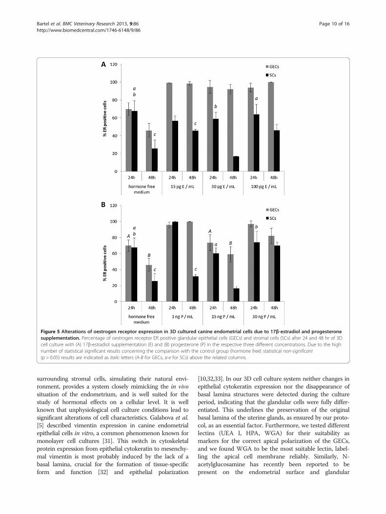
Figure 5 Alterations of oestrogen receptor expression in 3D cultured canine endometrial cells due to 17β-estradiol and progesteronesupplementation. Percentage of oestrogen receptor ER positive glandular epithelial cells (GECs) and stromal cells (SCs) after 24 and 48 hr of 3Dcell culture with (A) 17β-estradiol supplementation (E) and (B) progesterone (P) in the respective three different concentrations. Due to the highnumber of statistical significant results concerning the comparison with the control group (hormone free) statistical non-significant(p > 0.05) results are indicated as italic letters (A-B for GECs, a-e for SCs) above the related columns.
Bartel et al. BMC Veterinary Research 2013, 9:86 Page 10 of 16http://www.biomedcentral.com/1746-6148/9/86
surrounding stromal cells, simulating their natural envi-ronment, provides a system closely mimicking the in vivosituation of the endometrium, and is well suited for thestudy of hormonal effects on a cellular level. It is wellknown that unphysiological cell culture conditions lead tosignificant alterations of cell characteristics. Galabova et al.[5] described vimentin expression in canine endometrialepithelial cells in vitro, a common phenomenon known formonolayer cell cultures [31]. This switch in cytoskeletalprotein expression from epithelial cytokeratin to mesenchy-mal vimentin is most probably induced by the lack of abasal lamina, crucial for the formation of tissue-specificform and function [32] and epithelial polarization
[10,32,33]. In our 3D cell culture system neither changes inepithelial cytokeratin expression nor the disappearance ofbasal lamina structures were detected during the cultureperiod, indicating that the glandular cells were fully differ-entiated. This underlines the preservation of the originalbasal lamina of the uterine glands, as ensured by our proto-col, as an essential factor. Furthermore, we tested differentlectins (UEA I, HPA, WGA) for their suitability asmarkers for the correct apical polarization of the GECs,and we found WGA to be the most suitable lectin, label-ling the apical cell membrane reliably. Similarly, N-acetylglucosamine has recently been reported to bepresent on the endometrial surface and glandular

Figure 6 Alterations of progesterone receptor expression in 3D cultured canine endometrial cells due to 17β-estradiol andprogesterone supplementation. Percentage of progesterone receptor PR positive glandular epithelial cells (GECs) and stromal cells (SCs) after24 and 48 hr of 3D cell culture with (A) 17β-estradiol (E) and (B) progesterone (P) supplementation in the respective three differentconcentrations. Statistical significances (p < 0.05) are indicated as letters (A-E for GECs, a-e for SCs) above the related columns for comparison withthe control group (hormone free medium).
Bartel et al. BMC Veterinary Research 2013, 9:86 Page 11 of 16http://www.biomedcentral.com/1746-6148/9/86
epithelial cells during the physiological canine oestrouscycle as well as in pathological alterations of the uterinetissue [34]. Based on this knowledge and our results, wedecided to use WGA as a marker for apical polarity of theglandular epithelial cells in our 3D cell culture system. To-gether with β-catenin we created a new double-labellingcombination to assay apical and basolateral polarisationand using this were able to demonstrate the differentiationof cultured GECs in the 3D system. Loss of polarity is gen-erally associated with increased proliferation and migra-tion, two factors related to malignant alterations in tissues
and dedifferentiation processes in monolayer or co-culture systems of primary cells [10,35].
In the 3D endometrial cell culture system ER and PRexpression patterns in endometrial glands and stromalcells follow exogenous hormonal stimulationIn the dog, several studies have been performed to eluci-date the specific effects of steroid hormones on thedifferent cell types of the endometrium in vivo [36-38].Even with progesterone concentrations at physiologicallevels, altered steroid hormone receptor expression

Figure 7 Alterations of proliferative activity in 3D cultured canine endometrial cells due to 17β-estradiol and progesteronesupplementation. Percentage of anti-Ki67 positive glandular epithelial cells (GECs) and stromal cells (SCs) indicating proliferative activity after 24and 48 hr of 3D cell culture with (A) 17β-estradiol (E) and (B) progesterone (P) supplementation in the respective three different concentrations.Statistical significances (p < 0.05) are indicated as letters (A-E for GECs, a-e for SCs) above the related columns.
Bartel et al. BMC Veterinary Research 2013, 9:86 Page 12 of 16http://www.biomedcentral.com/1746-6148/9/86
patterns in the uterus are suggested to be involved inthe pathogenesis of serious uterine diseases, such ascystic endometrial hyperplasia and pyometra [1,3,39,40].Therefore, knowledge about the direct effects of differ-ent progesterone and oestrogen concentrations on endo-metrial epithelial and stromal cells could help theunderstanding of physiological and pathological pro-cesses in the endometrium.Compared to the initial situation of ER and PR status
in the uterus, ER expression of GECs and SCs declinedafter a 48 hr hormone-free culture period, whereas PRexpression was nearly stable in GECs and declined onlyin SCs. These effects were also observed in extenuatedpatterns in standard medium. We interpret this
observation as a response to the lack of a stimulus underthese culture conditions. Consistent with our observa-tions, Pierro et al. [41] described altered ER expressionpatterns and responsiveness of human endometrialepithelial and stromal cells to E in vitro as a result ofmissing paracrine factors (e.g. insulin).The application of different physiological concen-
trations of steroid hormones according to the canineoestrous cycle [42,43] demonstrated the capability of theendometrial GECs and SCs to respond to these supple-ments in an organotypic endometrial cell culture model.In our 3D cell culture system addition of E resulted inincreased ER expression in GECs (100 pg/mL and15 pg/mL E), but this was decreased in SCs with a

Bartel et al. BMC Veterinary Research 2013, 9:86 Page 13 of 16http://www.biomedcentral.com/1746-6148/9/86
minimum level following 30 pg/mL E for 48 hr. These re-sults are at least in part comparable with the physiologicalsituation. In the native canine endometrial tissue ERexpression in GECs is high in proestrus (E 5 – 10 pg/mL;P 0.2 - 0.4 ng/mL), and oestrus (E 20 – 80 pg/mL,P 1 – 5 ng/mL) but decreasing during early metestrus(E 10 – 25 pg/mL, P 15 – 80 ng/mL) [43]. SCs follow thesame scheme but with more pronounced fluctuations asreported before [28]. Compared to the anestrous ERexpression in SCs a decline during oestrus and earlymetestrus but an increase during early proestrus wasreported by Dhaliwal et al. [44] and Veremirsch et al. [28]in the native canine endometrium. Therefore, the reduc-tion of ERs in SCs due to 30pg/mL E (comparable toestrous/early metestrous plasma E levels) appears to beanalogous to the in vivo situation.In the monolayer culture system of canine endometrial
cells ERs in both cell types increased due to 100 pg/mLE [5]. This reaction of SCs may be a result of separatedsingle cell-type culturing in a 2D cell culture system dueto a lack of interactions between stromal and epithelialcells. Haslam et al. [20] demonstrated that in the mam-mary gland oestrogenic effects on glandular epithelialcells are controlled by mammary stromal cells in vivoand in vitro [45]. Direct supplementation of E tomammary epithelial cells cultured alone did not showany effects, neither morphological nor pro-proliferative,although ERs were expressed in these epithelial cells[46]. Pierro et al. [41] were able to induce an E dose-dependent increase in proliferative activity in humanendometrial epithelial cells only when these were co-cultured with endometrial stromal cells, underlining theimportance of epithelial-stromal signaling in hormonaltransduction.PR expression in the GECs was reduced in our 3D cell
culture system following E supplementation (100 pg/mLand 30 pg/mL E) but increased with 15 pg/mL E for 48hr. In the native endometrium PR expression in theGECs is high during early proestrus and oestrus butdeclines during early and late metestrus compared toanestrous levels [12,44]. PR expression in SCs in vitroincreased following supplementation of 15 pg/mL and100 pg/mL E, but decreased after treatment with 30 pg/mLE for 48 hr. PR expression in SCs in vivo declines fromearly to late metestrus compared to anestrous levels, butincreases during early proestrus to reach anestrous levelsduring oestrus [12,44]. The achieved results demonstratethat the two different cell types of the 3D cell culturesystem partially react comparable to the normal canineendometrial tissue. The increase of PR-positive GECs inthe 3D cell culture system upon treatment with 15 pg/mLE is comparable to the increase of PR expression duringproestus/early oestrus, and the decline of PR expression inSCs with 30 pg/mL E in vitro mirrors the effects during
early metestrus in vivo. In a human endometrial co-culture system, addition of E induced increased PR-positive immunoreactivity of endometrial epithelial cells[47]. Comparable results were observed in the murineendometrium model of Chung et al. [48], demonstratingan increase of PR expression in co-cultured endometrialepithelial cells due to E supplementation. In both studies,endometrial epithelial cells were separated by membranes(inserts) from the stromal cells. In contrast to our 3Dorganotypic endometrial model neither physiologicalglandular architecture nor contact between stromal andepithelial cells was provided. Bläuer et al. [47] usedepithelial organoids embedded in Matrigel™, which formedglandular structures within 24 hr, to show the positiveresponse of epithelial PRs to E supplementation. Thedecrease of PRs in our studies after treatment with100 pg/mL and 30 pg/mL E in GECs and 30 pg/mL E inSCs, in contrast to the positive regulation of PRs in GECsand SCs following 15 pg/mL E after 48h culturing period,may reflect the importance of tissue composition (cell-cellcontacts) on the one hand, and may display canine-specific reactions of GECs and SCs to the supplementedhormones on the other. Interpretation of species-specificreactions to supplemented steroid hormones in co-cultureor 3D-culture systems has to be carried out carefullybecause of the different culture techniques and ECMcompositions.Another important fact concerning endometrial ER
and PR expression patterns in GECs and SCs in thenative canine endometrium is that, especially for PRs,different regions of the glands have to be considered dueto the different expression patterns of PRs. Vermeirschet al. [12] described distinct PR expression of the basalportion of endometrial glands and their glandular ductsreaching to the surface epithelium. Due to our extractiontechnique that obtains intact endometrial glands, theseregions are no longer distinguishable in the histologicalsections, and furthermore we do not know if the loss ofcontact to the surface epithelium induces additionalchanges of PR (and ER) expression patterns. Never-theless, we are convinced that working with isolatedprimary endometrial glands more closely resembles thephysiological situation than working with secondaryconstructed spheroids [10].The effects of P in the 3D cell-culture system were
demonstrated in a decline of ERs in GECs and SCs dueto supplementation of 15 ng/mL P for 48 hr. Supple-mentation of 3 ng/mL P for 48 hr led to a reduction ofER-positive SCs, whereas GECs showed a minimalincrease of ER expression after 48 hr in culture. Differentauthors [13,28] reported that ER expression patterns inthe native canine endometrium showed declining valuesdue to increasing serum P levels during oestrus. Increas-ing ER expression accompanied in vivo decreasing serum

Bartel et al. BMC Veterinary Research 2013, 9:86 Page 14 of 16http://www.biomedcentral.com/1746-6148/9/86
P values (below 1 ng/mL) during late metestrus wereobserved in GECs after treatment with 3 ng/mL P. Incontrast, in the 2D cell culture system of Galabova et al.[5] ER expression in co-cultured GECs was reducedfollowing 3 ng/mL P. In the human endometrial cellculture model of Classen Linke et al. [49] endometrialepithelial and stromal cells showed significant down-regulation of ER and PR expression following gestagensupplementation. Similarly Bläuer et al. [47] demonstratedreduced PR expression in epithelial organoids after me-droxyprogesterone acetate supplementation, to an unde-tectable level. PR expression in our 3D-cultured SCsincreased after 24 hr supplementation with 30 ng/mL P(compared to the hormone free cultivated control group),comparable to the increase of PR expression in SCs at theend of oestrus or early metestrus. These discrepancieswith other endometrial epithelial cell cultures may beexplained by species-specific reactions of endometrialGECs and SCs, corroborated by the fact that theprolonged serum P levels in the bitch during metestrousare unique to canids [43,50].
Proliferative activity of endometrial glands and stromalcells in the 3D cell culture system show minor reactionsto the added steroid hormonesAs the proliferative activity of GECs and SCs in thestandard medium was below 1% and of GECs and SCsin the hormone-free medium below 0.2% after a 48-hrculture period, high SD values were inevitably producedfrom the statistical analysis. In the basal glands of thenative canine endometrium, proliferative activity is atlow levels in proestrus, with values rapidly increasing inoestrus and early metestrus, followed by declining valuesin late metestrus and anestrus [15]. Neither in the caninemonolayer endometrial culture system [5] nor in our 3Dcell-culture system were clear effects of E on prolifera-tive activity observed.However, supplementation of 30 ng/mL P for 24 hr
induced a significant rise in proliferative activity in GECsand SCs in the 3D cell-culture system, compared to thehormone-free cultured cells. Comparable effects of lowdoses of P were observed in the 2D cell-culture systemof Galabova et al. [5]. In the native canine endometrialtissue, proliferative activity of the stromal cells is highestin proestrus under oestrogenic influence, and decreasesduring oestrus with increasing plasma P levels [15].These divergent reactions of the cells to the steroidhormones in vitro concerning proliferative activitycompared to the in vivo situation may be a result of thesingle use of either E or P. For example, in the murineendometrium model of Chang et al. [48] P supplementa-tion alone did not show any effects on the proliferativeactivity of SCs, but in combination with E it had inhibi-tory effects on E-induced proliferative activity. It has to
be considered that in the (canine) endometrium, differ-ent cell populations (surface and glandular epithelialcells, epithelial cells of the crypts as well as stromal cellsand endothelial cells) in different regions (surface, basalregion) show different patterns of proliferative activityduring the individual cyclic phases. Van Cruchten et al.[15] described increased mitotic activity of the surfaceepithelium, the stroma, the blood vessels and the cryptsduring proestrus, whereas for the basal glands, prolifera-tive activity increased during oestrus compared to latemetestrus and anestrus. In the basal endometrial glandsa positive correlation of serum P levels and proliferativeactivity were observed, whereas in the other cell groupsthis activity positively correlated with levels of E. VanCruchten et al. [15] concluded that regulation of theproliferation in the canine endometrial surface epithelium,the stroma, the blood vessels and the crypts is differentfrom that in the basal glands. These results highlight theproblems faced by researchers isolating, and subsequentlyculturing and analyzing, isolated endometrial glands andstromal cells originating from the different endometrialregions.
ConclusionsWe have been able to demonstrate the advantages of a3D cell-culture model of the canine endometrium overmonolayer cultures, for experimental approaches due tothe differentiated and polarized cell conditions. Wefound pronounced effects due to single steroid hormonesupplementation on ER and PR expression in epithelialand stromal cells. However, in the bitch the prolonged Pphase during metestrous as well as the importance ofthe E:P ratio for the sensitive balance of steroid hormonereceptor expression are unique scenarios, and thereforethe mimicry of this special hormonal situation in thecycling canine endometrium deserves particular consi-deration in further studies.
Additional file
Additional file 1: Table S1. Detailed scoring results for expression ofestrogen and progesterone receptors and proliferative activity inglandular epithelial cells (GECs) and stromal cells (SCs) during differentculturing media (standard and hormone free medium as well assupplemented estrogen E and progesterone P in different dosages for 24and 48 hours, respectively); all values are listed as percentage values ±SD.
Abbreviations3D: Three dimensional; DAPI: 4’,6-Diamidin-2-phenylindol; DPBS-AB: Dulbecco’s Phosphate Buffered Saline containing antibiotics; E: oestrogen(17β−estradiol); ECM: Extracellular matrix; ER: Oestrogen receptor; FCS: Fetalcalf serum; GEC: Glandular epithelial cell; HPA: Helix Pomatia Agglutinin;P: Progesterone; PR: Progesterone receptor; SC: Stromal cell; SD: Standarddeviations; TEM: Transmission electron microscopy; UEA: Ulex EuropaeusAgglutinin; WGA: Wheat Germ Agglutinin.

Bartel et al. BMC Veterinary Research 2013, 9:86 Page 15 of 16http://www.biomedcentral.com/1746-6148/9/86
Competing interestsThe authors declare that they have no competing interests.
Authors’ contributionsCB conceived the study, performed cell culture experiments, histological,histochemical and immunohistochemical as well as transmission electronmicroscopy evaluation, and drafted the manuscript. TA participated in thedesign of the study regarding statistical analyses, and helped to draft thestatistical part of the manuscript. SS provided the tissue samples, preparedclinical anamneses and cycle determination of the animals, and revised themanuscript. CA participated in the study design and revised the manuscript.IW conceived the study, participated in the study design and coordination,and helped to draft the manuscript. All authors read and approved the finalmanuscript.
AcknowledgementsPart of this study was performed within the framework of the InitiativeDoctoral College BIOREC (Biological Responses to Environmental Challenges)programme of the University of Veterinary Medicine of Vienna. The experttechnical assistance of Magdalena Helmreich, Stefanie Burger, WaltraudTschulenk, Floriane Schaub, Brigitte Machac and Anne Flemming as well asthe biochemical input concerning steroid hormones of Prof. Dr. Erich Möstlis greatly acknowledged. We also thank James Hutchins for editing thescientific English of the manuscript.
Author details1Department of Pathobiology, Institute of Anatomy, Histology andEmbryology, University of Veterinary Medicine, Veterinaerplatz 1,Vienna A – 1210, Austria. 2Department of Biomedical Science, Institute ofPopulation Genetics, Platform Biostatistics, University of Veterinary Medicine,Veterinaerplatz 1, Vienna A – 1210, Austria. 3Small Animal Practice, Vienna1220, Austria. 4Centre for Artificial Insemination and Embryo Transfer,University of Veterinary Medicine, Veterinaerplatz 1, Vienna A – 1210, Austria.5VetCore Facility for Research, University of Veterinary Medicine,Veterinaerplatz 1, Vienna A – 1210, Austria.
Received: 27 August 2012 Accepted: 22 April 2013Published: 24 April 2013
References1. Noakes DE, Dhaliwal GK, England GC: Cystic endometrial hyperplasia/
pyometra in dogs: a review of the causes and pathogenesis. J ReprodFertil Suppl 2001, 57:395–406.
2. De Bosschere H, Ducatelle R, Vermeirsch H, Van Den Broeck W, Coryn M:Cystic endometrial hyperplasia-pyometra complex in the bitch: shouldthe two entities be disconnected? Theriogenology 2001, 55(7):1509–1519.
3. De Cock H, Vermeirsch H, Ducatelle R, De Schepper J:Immunohistochemical analysis of estrogen receptors in cystic-endometritis-pyometra complex in the bitch. Theriogenology 1997,48(6):1035–1047.
4. Stadler K, Handler J, Schoenkypl S, Walter I: A three-dimensional culturemodel of canine uterine glands. In Vitro Cell Dev Biol Anim 2009,45(1–2):35–43.
5. Galabova-Kovacs G, Walter I, Aurich C, Aurich JE: Steroid receptors incanine endometrial cells can be regulated by estrogen andprogesterone under in vitro conditions. Theriogenology 2004,61(5):963–976.
6. Blow N: Cell culture: building a better matrix. Nat Meth 2009, 6(8):619–622.7. Cukierman E, Pankov R, Yamada KM: Cell interactions with three-
dimensional matrices. Curr Opin Cell Biol 2002, 14(5):633–639.8. Abbott A: Cell culture: biology’s new dimension. Nature 2003,
424(6951):870–872.9. Birgersdotter A, Sandberg R, Ernberg I: Gene expression perturbation
in vitro–a growing case for three-dimensional (3D) culture systems.Semin Cancer Biol 2005, 15(5):405–412.
10. Inman JL, Bissell MJ: Apical polarity in three-dimensional culture systems:where to now? J Biol 2010, 9(1):2.
11. Vermeirsch H, Van den Broeck W, Coryn M, Simoens P:Immunohistochemical detection of androgen receptors in the canineuterus throughout the estrus cycle. Theriogenology 2002, 57(9):2203–2216.
12. Vermeirsch H, Simoens P, Hellemans A, Coryn M, Lauwers H:Immunohistochemical detection of progesterone receptors in the canineuterus and their relation to sex steroid hormone levels. Theriogenology2000, 53(3):773–788.
13. Johnston SD, Kiang DT, Seguin BE, Hegstad RL: Cytoplasmic estrogen andprogesterone receptors in canine endometrium during the estrous cycle.Am J Vet Res 1985, 46(8):1653–1658.
14. Galabova G, Egerbacher M, Aurich JE, Leitner M, Walter I: Morphologicalchanges of the endometrial epithelium in the bitch during metoestrusand anoestrus. Reprod Domest Anim 2003, 38(5):415–420.
15. Van Cruchten S, Van den Broeck W, D’Haeseleer M, Simoens P: Proliferationpatterns in the canine endometrium during the estrous cycle.Theriogenology 2004, 62(3–4):631–641.
16. Barrau MD, Abel JH Jr, Verhage HG, Tietz WJ Jr: Development of theendometrium during the estrous cycle in the bitch. Am J Anat 1975,142(1):47–65.
17. Hebner C, Weaver VM, Debnath J: Modeling morphogenesis andoncogenesis in three-dimensional breast epithelial cultures. Annu RevPathol 2008, 3:313–339.
18. Lee GY, Kenny PA, Lee EH, Bissell MJ: Three-dimensional culture models ofnormal and malignant breast epithelial cells. Nat Methods 2007,4(4):359–365.
19. Debnath J, Brugge JS: Modelling glandular epithelial cancers in three-dimensional cultures. Nat Rev Cancer 2005, 5(9):675–688.
20. Haslam SZ, Woodward TL: Host microenvironment in breast cancerdevelopment: epithelial-cell-stromal-cell interactions and steroidhormone action in normal and cancerous mammary gland. Breast CancerRes 2003, 5(4):208–215.
21. Grun B, Benjamin E, Sinclair J, Timms JF, Jacobs IJ, Gayther SA, Dafou D:Three-dimensional in vitro cell biology models of ovarian andendometrial cancer. Cell Prolif 2009, 42(2):219–228.
22. Kim JB: Three-dimensional tissue culture models in cancer biology.Semin Cancer Biol 2005, 15(5):365–377.
23. Kunz-Schughart LA, Groebe K, Mueller-Klieser W: Three-dimensional cellculture induces novel proliferative and metabolic alterations associatedwith oncogenic transformation. Int J Cancer Journal international du cancer1996, 66(4):578–586.
24. Allen RL, Wright RW Jr: In vitro development of porcine embryos incoculture with endometrial cell monolayers or culture supernatants.J Anim Sci 1984, 59(6):1657–1661.
25. Yamauchi N, Yamada O, Takahashi T, Imai K, Sato T, Ito A, Hashizume K: Athree-dimensional cell culture model for bovine endometrium:regeneration of a multicellular spheroid using ascorbate. Placenta 2003,24(2–3):258–269.
26. Korff T, Augustin HG: Integration of endothelial cells in multicellularspheroids prevents apoptosis and induces differentiation. J Cell Biol 1998,143(5):1341–1352.
27. Mulisch Maria WU: Romeis Mikroskopische Technik. 18th edition. SpektrumAkademischer Verlag Heidelberg: Imprint Springer; 2010.
28. Vermeirsch H, Simoens P, Lauwers H, Coryn M: Immunohistochemicaldetection of estrogen receptors in the canine uterus and their relationto sex steroid hormone levels. Theriogenology 1999, 51(4):729–743.
29. Mostl E, Brunner I: Comparison of different progestagen assays formeasuring progesterone metabolites in faeces of the bitch. ZentralblVeterinarmed A 1997, 44(9–10):573–578.
30. Bartel C, Schonkypl S, Walter I: Pseudo-placentational endometrial cysts ina bitch. Anat Histol Embryol 2010, 39(1):74–80.
31. Classen-Linke I, Kusche M, Knauthe R, Beier HM: Establishment of a humanendometrial cell culture system and characterization of its polarizedhormone responsive epithelial cells. Cell Tissue Res 1997, 287(1):171–185.
32. Petersen OW, Ronnov-Jessen L, Howlett AR, Bissell MJ: Interaction withbasement membrane serves to rapidly distinguish growth anddifferentiation pattern of normal and malignant human breast epithelialcells. Proc Natl Acad Sci USA 1992, 89(19):9064–9068.
33. Streuli CH, Bailey N, Bissell MJ: Control of mammary epithelialdifferentiation: basement membrane induces tissue-specific geneexpression in the absence of cell-cell interaction and morphologicalpolarity. J Cell Biol 1991, 115(5):1383–1395.
34. Leitner M, Aurich JE, Galabova G, Aurich C, Walter I: Lectin binding patternsin normal canine endometrium and in bitches with pyometra and cysticendometrial hyperplasia. Histol Histopathol 2003, 18(3):787–795.

Bartel et al. BMC Veterinary Research 2013, 9:86 Page 16 of 16http://www.biomedcentral.com/1746-6148/9/86
35. Eritja N, Llobet D, Domingo M, Santacana M, Yeramian A, Matias-Guiu X,Dolcet X: A novel three-dimensional culture system of polarizedepithelial cells to study endometrial carcinogenesis. Am J Pathol 2010,176(6):2722–2731.
36. Buhi WC, Thatcher MJ, Shille VM, Alvarez IM, Lannon AP, Johnson J:Synthesis of uterine endometrial proteins during early diestrus in thecyclic and pregnant dog, and after estrogen and progesteronetreatment. Biol Reprod 1992, 47(3):326–336.
37. Chu PY, Lee CS, Moore PF, Wright PJ: Oestrogen and progestagen treatedovariectomized bitches: a model for the study of uterine function.J Reprod Fertil Suppl 2001, 57:45–54.
38. De Bosscher H, Ducatelle R, Tshamala M, Coryn M: Changes in sexhormone receptors during administration of progesterone to preventestrus in the bitch. Theriogenology 2002, 58(6):1209–1217.
39. Dhaliwal GK, England GC, Noakes DE: Oestrogen and progesteronereceptors in the uterine wall of bitches with cystic endometrialhyperplasia/pyometra. Vet Rec 1999, 145(16):455–457.
40. Smith FO: Canine pyometra. Theriogenology 2006, 66(3):610–612.41. Pierro E, Minici F, Alesiani O, Miceli F, Proto C, Screpanti I, Mancuso S,
Lanzone A: Stromal-epithelial interactions modulate estrogenresponsiveness in normal human endometrium. Biol Reprod 2001,64(3):831–838.
42. Jeffcoate IA, Lindsay FE: Ovulation detection and timing of inseminationbased on hormone concentrations, vaginal cytology and the endoscopicappearance of the vagina in domestic bitches. J Reprod Fertil Suppl 1989,39:277–287.
43. Concannon PW: Reproductive cycles of the domestic bitch. Anim ReprodSci 2011, 124(3–4):200–210.
44. Dhaliwal GK, England GC, Noakes DE: Immunocytochemical localization ofoestrogen and progesterone receptors in the uterus of the normal bitchduring oestrus and metoestrus. J Reprod Fertil Suppl 1997, 51:167–176.
45. Woodward TL, Xie JW, Haslam SZ: The role of mammary stroma inmodulating the proliferative response to ovarian hormones in thenormal mammary gland. J Mammary Gland Biol Neoplasia 1998,3(2):117–131.
46. Zhang HZ, Bennett JM, Smith KT, Sunil N, Haslam SZ: Estrogen mediatesmammary epithelial cell proliferation in serum-free culture indirectly viamammary stroma-derived hepatocyte growth factor. Endocrinology 2002,143(9):3427–3434.
47. Blauer M, Heinonen PK, Martikainen PM, Tomas E, Ylikomi T: A novelorganotypic culture model for normal human endometrium: regulationof epithelial cell proliferation by estradiol and medroxyprogesteroneacetate. Hum Reprod 2005, 20(4):864–871.
48. Chung D, Das SK: Mouse primary uterine cell coculture system revisited:ovarian hormones mimic the aspects of in vivo uterine cell proliferation.Endocrinology 2011, 152(8):3246–3258.
49. Classen-Linke I, Alfer J, Hey S, Krusche CA, Kusche M, Beier HM: Markermolecules of human endometrial differentiation can be hormonallyregulated under in-vitro conditions as in-vivo. Hum Reprod Update 1998,4(5):539–549.
50. Concannon PW, Castracane VD, Temple M, Montanez A: Endocrine controlof ovarian function in dogs and other carnivores. Anim Reprod 2009,6(1):172–193.
doi:10.1186/1746-6148-9-86Cite this article as: Bartel et al.: Effects of steroid hormones ondifferentiated glandular epithelial and stromal cells in a threedimensional cell culture model of the canine endometrium. BMCVeterinary Research 2013 9:86.
Submit your next manuscript to BioMed Centraland take full advantage of:
• Convenient online submission
• Thorough peer review
• No space constraints or color figure charges
• Immediate publication on acceptance
• Inclusion in PubMed, CAS, Scopus and Google Scholar
• Research which is freely available for redistribution
Submit your manuscript at www.biomedcentral.com/submit