Estimation of Stapedius-Muscle Activation using Ear Canal ...
Research ArticleComparison of Muscle Onset Activation Sequencesbetween a Golf or Tennis Swing and Common Training ExercisesUsing Surface Electromyography: A Pilot Study
John M. Vasudevan,1 Andrew Logan,2 Rebecca Shultz,2,3 Jeffrey J. Koval,2
Eugene Y. Roh,3 and Michael Fredericson3
1Department of Physical Medicine & Rehabilitation, University of Pennsylvania, 1800 Lombard Street, Philadelphia, PA 19146, USA2Human Performance Lab, Sports Medicine Center, Department of Orthopaedic Surgery, Stanford University, 341 Galvez Street,Lower Level, Stanford, CA 94305, USA3Division of Sports Medicine, Department of Orthopaedic Surgery, Stanford University, 450 Broadway Street,Redwood City, CA 94063, USA
Correspondence should be addressed to John M. Vasudevan; [email protected]
Received 16 November 2015; Revised 21 February 2016; Accepted 10 May 2016
Academic Editor: S. John Sullivan
Copyright © 2016 John M. Vasudevan et al. This is an open access article distributed under the Creative Commons AttributionLicense, which permits unrestricted use, distribution, and reproduction in any medium, provided the original work is properlycited.
Aim. The purpose of this pilot study is to use surface electromyography to determine an individual athlete’s typical muscle onsetactivation sequence when performing a golf or tennis forward swing and to use the method to assess to what degree the sequenceis reproduced with common conditioning exercises and a machine designed for this purpose. Methods. Data for 18 healthy malesubjects were collected for 15 muscles of the trunk and lower extremities. Data were filtered and processed to determine the averageonset ofmuscle activation for eachmotion.A Spearman correlation estimated congruence of activation order between the swing andeach exercise. Correlations of each group were pooled with 95% confidence intervals using a random effects meta-analytic strategy.Results. The averaged sequences differed among each athlete tested, but pooled correlations demonstrated a positive associationbetween each exercise and the participants’ natural muscle onset activation sequence. Conclusion. The selected training exercisesand Turning Point� device all partially reproduced our athletes’ averaged muscle onset activation sequences for both sports. Theresults support consideration of a larger, adequately powered study using thismethod to quantify to what degree each of the selectedexercises is appropriate for use in both golf and tennis.
1. Introduction
Golf and tennis are popular sports for athletes of all ages andability levels. There are over 30 million golfers and 28 millionactive tennis players in the United States and over 50 milliongolfers worldwide [1–4]. Both sports are associated withoveruse injuries, as they rely on transferring the momentumfrom a club or a racquet to a ball for maximum velocity andaccuracy [5].
The modern golf swing is characterized by increasedshoulder rotation with restricted hip turning to maxi-mize hip-shoulder separation. This technique may produceincreased ball velocity and distance but comes at a cost of
increased risk of musculoskeletal injury [5–10]. The moderngolf swing has been shown to place nearly as much stressupon the spine as forces measured in college football linemenhitting a blocking sled [11]. Several biomechanical studieshave assessed not only the golf swing, but also the role ofaxial rotation on loading and injury of the thoracolumbarspine, in which a supramaximal angle (termed the “dynamicX-factor”) takes the player beyond a safe limit of flexibility [8–14]. Control of the dynamic X-factor requires conditioningnot only spinal stabilizing muscles, but all limb and trunkmuscles required of the swing.
The golf swing motion is separated into several phases:backswing, forward swing, acceleration, early follow through,
Hindawi Publishing CorporationJournal of Sports MedicineVolume 2016, Article ID 3987486, 9 pageshttp://dx.doi.org/10.1155/2016/3987486

2 Journal of Sports Medicine
and late follow through. The primary lower limb and trunkmuscles involved in these phases have been described [2, 5].Although EMG studies have identified muscles active in eachphase, few studies have detailed the precise order of activationin an individual athlete or have onlymeasured a limited set ofmuscles (i.e., lower limb or trunk, but rarely are both regionsstudied at once) [8, 9, 15].
As with golf, tennis has evolved over the years to becomea more aggressive sport, requiring increasing loads to maxi-mize power. This, in turn, increases the risk of injury acrossall skill levels [16]. There are no longitudinal cohort studiesto identify risk factors and no randomized controlled trialsdesigned to determine methods of injury prevention.
The tennis forehand stroke is divided into the backswingand forward swing. Power in the tennis forward swing isderived from sequential contractions in the lower limbs,axial torque through the pelvis and trunk in the transverseplane, and sequential rotation through the kinetic chain tothe shoulder girdle and upper limb [17, 18]. As with thegolf swing, this also requires hip-shoulder separation duringbackswing to increase potential energy and power from thestroke. Studies sequencingmuscle activation in tennis are fewin number, and most focus on the serve rather than forwardswing [17]. Studies comparing athletes of varying skill levelshave found that appropriate timing and velocity of pelvisand trunk rotation are critical to performance in athletes ofall levels [19]. However, there is little information regardingtypical muscle activation patterns of the trunk and pelvis inthe tennis forward swing.
Conditioning exercises specific to the golf and ten-nis swing have been described to maximize efficiency ofmovement through the kinetic chain [2, 4, 17]. Althoughprecise reproduction of sport-specific movement may not benecessary to condition or rehabilitate athletes, exercises areoften tailored to athletes to accommodate for the complexmovement patterns required of their sport. The purposeof this pilot study is to use surface electromyography todetermine an individual athlete’s typical muscle onset acti-vation sequence from the limbs through the trunk, with agoal of determining how well the sequence is reproducedwith common conditioning exercises (wood chop cablepull, medicine ball wall throw, or the novel Turning Point4.0 Rotational Training Platform). We sought to determinewhether this electromyographic measurement method usedis appropriate to analyzemotions used in both golf and tennis.Understanding how well an exercise simulates an athlete’sswingmayprovide insight into the development of an optimaland individualized exercise regimen.
2. Materials and Methods
2.1. Participants. Informed consent for participation in studywas obtained from 18 healthy male subjects (9 golfers and 9tennis players). The number of participants was kept low forthe pragmatic purpose of generating and analyzing pilot data.The participants ranged from 20 to 33 (mean = 22.2) years ofage. The 9 golfers ranged from 20 to 33 (22.3) years of age.All golfers had been playing regularly for 1–14 (7) years and 2
of the participants played on an intercollegiate club golf team.The 9 tennis players were between the ages of 19 and 26 (22.4).Participants for tennis had been playing tennis ranging from6 to 15 (9.9) years. Of the 9 tennis players, 2 had played withhigh school varsity, 2 are currently at the division III collegiatelevel and 3 at the club level, and the remaining 2 participantsplay recreationally. In an effort to minimize variability in theswing, onlymale university students without acute or chronicinjuries of the spine or limbs (that could interfere withclub or racquet swing capability) were recruited. All subjectsprimarily play one of the two sports. The study protocolwas approved by the Stanford University Institutional ReviewBoard.
2.2. Procedures. For this study, we chose the wood chopcable pull (WC) and medicine ball wall throw (MB) exercisesbecause strength and conditioning coaches commonly usethese exercises to train and rehabilitate athletes in thesesports.These two exercises in isolation are by nomeans com-prehensive in the training of the coremusculature and propermotion through the kinetic chain. Rather, they are represen-tative of many exercises used to train rotational movementand chosen based upon the advice of the strength andconditioning coaches at our university.The Turning Point 4.0Rotational Training Platform (Turning Point Biotechnology,Toledo, Ohio; http://www.turningpointbiotech.com/indexdraft.php) (TP) was included in this study because it was notonly designed specifically for training of rotational strength,but also intended to provide more consistent and measurablemovement due to its adjustable settings and computerizedmotion analysis [20, 21].
Prior to testing, participants were given five minutes towarm up with low-intensity cardiovascular exercise (e.g.,treadmill walking) or stretching to their preference. Onceready, participants had the muscles of interest prepared forEMG electrode placement by gentle abrasion of the skinand treatment with alcohol to clean the area and provide anaccurate reading. Ultrasound gel was also used to ensure thata clean EMG signal was recorded for all 15 channels used inthis study. Location of electrode placement on each musclewas determined according to Cram’s Introduction to SurfaceElectromyography [22].
Once all EMG electrodes were properly placed, partici-pants completed four motions, performing 5 repetitions ofeach—a golf or tennis forward swing with ball contact, awood chop cable pull (Figure 1), an 8-pound medicine ballwall throw in a perpendicular stance to a wall (Figure 2),and the Turning Point 4.0 Rotational Platform (Figure 3).We chose five repetitions for each motion to sample enoughdata while reducing any adverse effects of fatigue as eachathlete progressed through the motions. The wood chopcable pull exercise is a dynamic, functional exercise thatemploys the lower limbs, trunk, and upper limbs in a naturalmotion. The person performing the exercise moves a bar orball in diagonal fashion (e.g., from lower right to upper leftin a right-handed golfer) in a controlled, spiral movementwhile keeping the torso as still as possible by coactivationof abdominals and paraspinals. A medicine ball wall throw

Journal of Sports Medicine 3
Figure 1: Surface EMG setup for wood chop cable pull exercise.
Figure 2: Surface EMG setup for medicine ball wall throw exercise.
exercise can be used in similar fashion to the wood chop butallows for a more natural take away (simulating backswing)and release of the ball toward a wall (simulating followthrough), since the athlete is not tethered to a machine.The Turning Point 4.0 Rotational Training Platform placesthe user in a standing position and allows for shoulder-hipseparation required of rotational movements as used in golfand tennis.
These exercises were performed in a randomized orderfor each participant. Randomization was done by use of arandomized number sheet. Participantswere told to completeall motions at a self-selected comfortable pace. Resistance fortheWC exercise was standardized to equal the torque createdby an 8-pound medicine ball. Because settings on the TPplatform were not directly comparable to the other exercises,resistance on the platform for each subject was set accordingto an equal level of self-perceived exertion when comparedto the WC and MB exercises. All exercises were performedin the individual’s self-selected natural swing stance. Eachparticipant was given up to 5 minutes to practice the motionsfor each exercise and the Turning Point machine, followed by5 minutes of rest prior to recorded trials. Five minutes of restwere given between each of the four motions to control foreffects of fatigue.
2.3. Measuring Methods. Electromyographic (EMG) datawere recorded (1200Hz) from lower limb and trunk muscles(Bagnoli Desktop, Delsys Inc., Boston. MA, USA) using
Figure 3: Surface EMG setup for Turning Point 4.0 RotationalPlatform.
preamplified single differential, rectangular Ag electrodeswith 10mm interelectrode distance (DE-2.1, Delsys, Inc.,Boston, MA, USA). The 15 specific muscles were bilateralerector spinae, pectoralis major, external oblique, rectusabdominis, internal oblique, vastus lateralis, gluteus max-imus, and unilateral vastus medialis of each participant’strailing leg.
EMG data were bandpass filtered at 30–500Hz, rectified,and low-pass filtered at 30Hz. The evaluation of EMG onsetwas determined with computer analysis (MATLAB version7.10.0. Natick,Massachusetts:TheMathWorks Inc., 2010).Thealgorithm involved identification of the time point when themean of a specified number of surrounding time samplesexceeded the mean level by a predetermined number ofstandard deviations. This time point is considered the onsetof activation for eachmuscle studied.The baselinemean levelwas averaged for 50ms prior to signal to perform the activity.Following this, EMG activity was averaged over a slidingtime window of 25ms. If the average of the time window didnot exceed the baseline by 3 standard deviations, the slidingtime window was advanced one sample at a time until theonset was found. The algorithm and parameters used havepreviously been demonstrated to reduce observer bias andincrease reliability [23].
The muscles studied in each subject were ranked in orderof activation as described above from 1 to 15. For example, ifthe external oblique was the first to activate, it was ranked1, and the last muscle to activate was ranked 15. In orderto account for variability among swings in each individual,this activation order was averaged across the natural swingfor each subject to determine his own “gold standard.” Weanticipated this variability based on a study by Meister et al.that demonstrated decreased consistency of the golf swingas handicap increases [6]. The muscle activation sequencesfor the wood chop cable pull (WC), medicine ball wallthrow (MB), andTurning Point (TP) exercisesweremeasuredin similar fashion for comparison to each subject’s naturalswing.
2.4. Statistical Analyses. For each athlete, a Spearman corre-lation was calculated to estimate the congruence of muscleonset activation order between golf swing and the otherexercises (i.e., WC, MB, and TP). For each exercise pair(e.g., golf swing versus WC) the correlations of each group

4 Journal of Sports Medicine
Muscle activation sequence Muscle activation sequence
Muscle activation sequence Muscle activation sequence
R_ESL_VLR_PecR_IOL_Pec
R_GMR_EOR_RAVMOL_ES
R_VLL_GM
L_IOL_RAL_EO
Golf
Medicine ball throw Turning point35003000250020001500100050002200200018001600140012001000800600400200
2800260024002200200018001600140012001000800 500 1000 2500 30000
Cable wood chop
R_ES
L_VL
R_PecR_IO
L_Pec
R_GM
R_EOR_RA
VMO
L_ES
R_VL
L_GM
L_IO
L_RA
L_EO
R_ES
L_VL
R_PecR_IO
L_Pec
R_GM
R_EO
R_RA
VMO
L_ES
R_VL
L_GML_IO
L_RAL_EO
R_ES
L_VL
R_Pec
R_IO
L_Pec
R_GM
R_EO
R_RA
VMO
L_ES
R_VL
L_GM
L_IO
L_RA
L_EO
20001500
Figure 4: Example of one golf athlete’s averagedmuscle onset activation sequence for the forward golf swing, wood chop cable pull, medicineball wall throw, and Turning Point 4.0 Rotational Platform. Each bar represents the average onset and duration of each subject’s individualmuscle activation. (𝑥-axis: time (msec)). 𝑦-axis: muscles (e.g., L EO = left external oblique).
of 9 athletes were pooled into overall estimates with 95%confidence intervals using a random effects meta-analyticstrategy, implemented in the Metafor Package of the Rstatistical program. Meta-analysis is a statistical strategyto average or pool independent statistical results (in thiscase, player-specific Spearman’s correlations) in a mannerthat retains information regarding within-player variabilityand number of observations [24]. Differences between thepooled correlations (e.g., golf swing versus WC comparedto golf swing versus TP) were evaluated by inspecting theoverlap between the 95% confidence intervals for the pooledcorrelations. The same method was used to estimate thecongruence of muscle activation order between tennis swingand WC, MB, and TP exercises.
3. Results
3.1. Golf. Averaging of the 5 motions for each athletedetermined the natural forehand swing for that athlete. Anexample of an individual athlete’s muscle activation sequencefor the golf swing and the three exercises studied is shownin Figure 4. There were differences in correlation (ranging
from negative to positive) between the forward swing andeach exercise among the 9 golf athletes, but these differenceswere within the range of the 95% confidence intervals.This isdisplayed along the 𝑥-axis in Figures 5(a)–5(c).
The majority of all athletes demonstrated a positivecorrelation between all exercises and the natural swing. Forthe minority of athletes in which there was a negative corre-lation, all confidence intervals crossed into the positive range(Figure 5, top 9 lines). When averaged among all athletestested, the coefficients of correlation (with 95% confidenceinterval) between theWC,MB, andTPmovements to the golfforward swingwere 0.16 (95%CI:−0.01 to 0.32), 0.26 (95%CI:−0.01 to 0.52), and 0.23 (95% CI: 0.02 to 0.45), respectively(Figure 5, bottom line). The confidence intervals of thesecorrelations overlap, indicating a nonsignificant differenceamong the exercises.
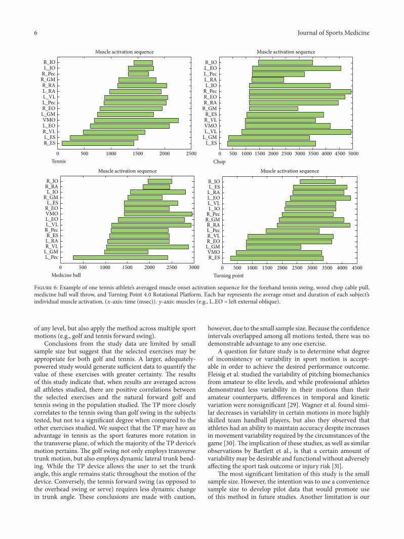
3.2. Tennis. An example of an individual athlete’s muscleactivation sequence for the tennis swing and the threeexercises studied is shown in Figure 6, using the samemethodology as with the golf athletes. Again, there were

Journal of Sports Medicine 5
Correlation between golf and chop
1
2
3
4
5
6
7
8
9
RE model
0.31 [−0.16, 0.78]
−0.28 [−0.76, 0.20]
−0.07 [−0.59, 0.45]
0.33 [−0.13, 0.80]
0.29 [−0.19, 0.77]
−0.12 [−0.64, 0.39]
0.17 [−0.34, 0.68]
0.25 [−0.25, 0.74]
0.40 [−0.03, 0.84]
0.16 [−0.01, 0.32]
1.000.500.00−0.50−1.00
Muscle activation order in 9 players
(a)
Correlation between golf and medicine ball
0.16 [−0.36, 0.67]
0.45 [0.03, 0.87]
0.66 [0.37, 0.96]
0.81 [0.62, 0.99]
−0.07 [−0.59, 0.45]
0.23 [−0.27, 0.73]
−0.28 [−0.76, 0.20]
−0.02 [−0.54, 0.51]
−0.05 [−0.58, 0.47]
0.26 [−0.01, 0.52]
−1.00
1
2
3
4
5
6
7
8
9
RE model
1.000.500.00−0.50
Muscle activation order in 9 players
(b)
Correlation between golf and TP
−0.03 [−0.55, 0.49]
0.50 [0.11, 0.89]
0.48 [0.07, 0.88]
0.63 [0.31, 0.95]
0.30 [−0.18, 0.77]
0.30 [−0.17, 0.78]
−0.01 [−0.54, 0.51]
−0.25 [−0.74, 0.24]
−0.16 [−0.67, 0.35]
0.23 [0.02, 0.45]
−1.00
1
2
3
4
5
6
7
8
9
RE model
1.000.500.00−0.50
Muscle activation order in 9 players
(c)
Figure 5: Correlation of muscle onset activation sequences between golf swing and the wood chop cable pull, medicine ball wall throw, andTurning Point 4.0 Rotational Platform. The top nine bars indicate each subject’s individual correlation (with 95% confidence interval), andthe bottom bar indicates the correlation averaged across all subjects tested (with 95% confidence interval).
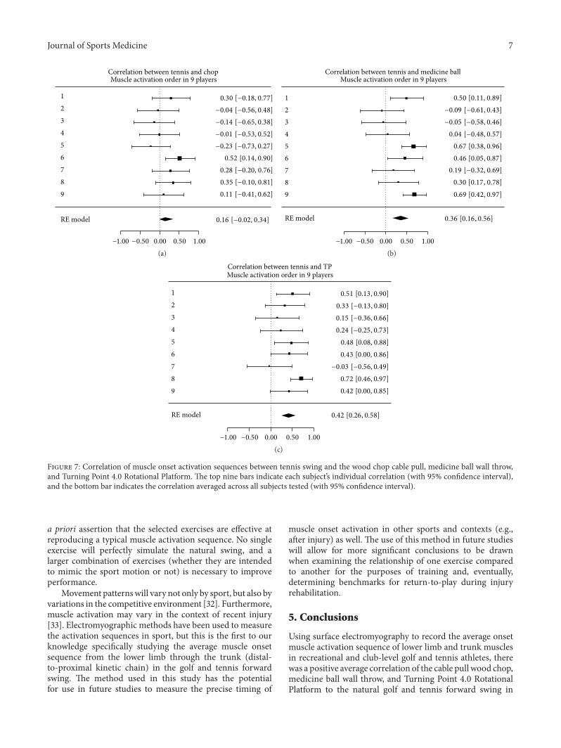
differences in correlation (ranging from negative to positive)between the forward swing and each exercise among the 9tennis athletes, with these differences within the range of the95% confidence intervals. This is displayed along the 𝑥-axisin Figures 7(a)–7(c). This particular group studied exhibitedless inconsistency than the golf athletes.
As with the golf cohort, the majority of all athletesdemonstrated a positive correlation between all exercises andthe natural swing. For theminority of athletes, in which therewas a negative correlation, confidence intervals crossed intothe positive range (Figure 7, top 9 lines). When averagedacross all participants, the coefficients forWC,MB, and TP tothe tennis forward swing were 0.16 (−0.02–0.34), 0.36 (0.16–0.56), and 0.42 (0.26–0.58), respectively (Figure 7, bottomline). The confidence intervals of these correlations overlap,indicating a nonsignificant difference among the exercises.
4. Discussion
This study utilized surface electromyography to observe themuscle activation sequences among recreational and club-level golf and tennis athletes. Seminal studies by Perry andcolleagues, such as the ones by Pink et al. in 1990 and1993 and Watkins et al. in 1996, helped establish the useof electromyography to evaluate muscle activation in thegolf swing [25–27]. Because most of the available data isfocused on trunk and shoulder musculature, recent reviewsof electromyography variables related to the golf swing haveidentified a gap in the knowledge of lower limb muscleactivation [28]. Based on what was already known (and yetunknown), the purpose of this pilot study was to not onlydefine amethod to readily identify the precise onset sequenceof the lower limb through the trunk muscles in an athlete
6 Journal of Sports Medicine
Muscle activation sequence Muscle activation sequence
Muscle activation sequence Muscle activation sequence
3500300025002000150010005000 45004000 5000
Chop15001000 2000 25005000
Tennis
500 1000 1500 2000 300025000
Medicine ball40001000 1500 2000 2500 30005000 45003500
Turning point
L_ESL_GML_VLVMOR_VLR_ES
R_GMR_RAR_EOR_PecL_IO
L_RAL_PecL_EOR_IO
R_ESVMO
L_GMR_EOR_VLL_PecR_RA
R_GMR_PecL_IOL_VLL_EOL_RAL_ESR_IO
L_PecL_GMR_VLL_RAR_ES
R_PecL_VLL_EOVMOR_EOL_ES
R_GML_IO
R_RAR_IO
R_ESL_ES
R_VLL_EOVMO
L_GMR_EOL_PecL_VLL_RAR_RA
R_GMR_PecL_IOR_IO
Figure 6: Example of one tennis athlete’s averaged muscle onset activation sequence for the forehand tennis swing, wood chop cable pull,medicine ball wall throw, and Turning Point 4.0 Rotational Platform. Each bar represents the average onset and duration of each subject’sindividual muscle activation. (𝑥-axis: time (msec)). 𝑦-axis: muscles (e.g., L EO = left external oblique).
of any level, but also apply the method across multiple sportmotions (e.g., golf and tennis forward swing).
Conclusions from the study data are limited by smallsample size but suggest that the selected exercises may beappropriate for both golf and tennis. A larger, adequately-powered study would generate sufficient data to quantify thevalue of these exercises with greater certainty. The resultsof this study indicate that, when results are averaged acrossall athletes studied, there are positive correlations betweenthe selected exercises and the natural forward golf andtennis swing in the population studied. The TP more closelycorrelates to the tennis swing than golf swing in the subjectstested, but not to a significant degree when compared to theother exercises studied. We suspect that the TP may have anadvantage in tennis as the sport features more rotation inthe transverse plane, of which the majority of the TP device’smotion pertains. The golf swing not only employs transversetrunk motion, but also employs dynamic lateral trunk bend-ing. While the TP device allows the user to set the trunkangle, this angle remains static throughout the motion of thedevice. Conversely, the tennis forward swing (as opposed tothe overhead swing or serve) requires less dynamic changein trunk angle. These conclusions are made with caution,
however, due to the small sample size. Because the confidenceintervals overlapped among all motions tested, there was nodemonstrable advantage to any one exercise.
A question for future study is to determine what degreeof inconsistency or variability in sport motion is accept-able in order to achieve the desired performance outcome.Fleisig et al. studied the variability of pitching biomechanicsfrom amateur to elite levels, and while professional athletesdemonstrated less variability in their motions than theiramateur counterparts, differences in temporal and kineticvariation were nonsignificant [29]. Wagner et al. found simi-lar decreases in variability in certain motions in more highlyskilled team handball players, but also they observed thatathletes had an ability to maintain accuracy despite increasesin movement variability required by the circumstances of thegame [30]. The implication of these studies, as well as similarobservations by Bartlett et al., is that a certain amount ofvariability may be desirable and functional without adverselyaffecting the sport task outcome or injury risk [31].
The most significant limitation of this study is the smallsample size. However, the intention was to use a conveniencesample size to develop pilot data that would promote useof this method in future studies. Another limitation is our
Journal of Sports Medicine 7
Correlation between tennis and chop
1
2
3
4
5
6
7
8
9
RE model
1.000.500.00−0.50−1.00
0.30 [−0.18, 0.77]
−0.04 [−0.56, 0.48]
−0.14 [−0.65, 0.38]
−0.01 [−0.53, 0.52]
−0.23 [−0.73, 0.27]
0.52 [0.14, 0.90]
0.28 [−0.20, 0.76]0.35 [−0.10, 0.81]
0.11 [−0.41, 0.62]
0.16 [−0.02, 0.34]
Muscle activation order in 9 players
(a)
0.50 [0.11, 0.89]
−0.09 [−0.61, 0.43]
−0.05 [−0.58, 0.46]
0.04 [−0.48, 0.57]
0.67 [0.38, 0.96]
0.46 [0.05, 0.87]
0.19 [−0.32, 0.69]0.30 [0.17, 0.78]0.69 [0.42, 0.97]
0.36 [0.16, 0.56]
Correlation between tennis and medicine ballMuscle activation order in 9 players
1
2
3
4
5
6
7
8
9
RE model
1.000.500.00−0.50−1.00
(b)
0.51 [0.13, 0.90]
0.33 [−0.13, 0.80]
0.15 [−0.36, 0.66]
0.24 [−0.25, 0.73]
0.48 [0.08, 0.88]
0.43 [0.00, 0.86]
−0.03 [−0.56, 0.49]
0.72 [0.46, 0.97]
0.42 [0.00, 0.85]
0.42 [0.26, 0.58]
Correlation between tennis and TP
1
2
3
4
5
6
7
8
9
RE model
1.000.500.00−0.50−1.00
Muscle activation order in 9 players
(c)
Figure 7: Correlation of muscle onset activation sequences between tennis swing and the wood chop cable pull, medicine ball wall throw,and Turning Point 4.0 Rotational Platform. The top nine bars indicate each subject’s individual correlation (with 95% confidence interval),and the bottom bar indicates the correlation averaged across all subjects tested (with 95% confidence interval).
a priori assertion that the selected exercises are effective atreproducing a typical muscle activation sequence. No singleexercise will perfectly simulate the natural swing, and alarger combination of exercises (whether they are intendedto mimic the sport motion or not) is necessary to improveperformance.
Movement patternswill vary not only by sport, but also byvariations in the competitive environment [32]. Furthermore,muscle activation may vary in the context of recent injury[33]. Electromyographic methods have been used to measurethe activation sequences in sport, but this is the first to ourknowledge specifically studying the average muscle onsetsequence from the lower limb through the trunk (distal-to-proximal kinetic chain) in the golf and tennis forwardswing. The method used in this study has the potentialfor use in future studies to measure the precise timing of
muscle onset activation in other sports and contexts (e.g.,after injury) as well. The use of this method in future studieswill allow for more significant conclusions to be drawnwhen examining the relationship of one exercise comparedto another for the purposes of training and, eventually,determining benchmarks for return-to-play during injuryrehabilitation.
5. Conclusions
Using surface electromyography to record the average onsetmuscle activation sequence of lower limb and trunk musclesin recreational and club-level golf and tennis athletes, therewas a positive average correlation of the cable pull wood chop,medicine ball wall throw, and Turning Point 4.0 RotationalPlatform to the natural golf and tennis forward swing in

8 Journal of Sports Medicine
the amateur athlete population studied. Our pilot data sug-gest that a larger study using this method may effectivelyquantify to what degree these and other exercises are appro-priate for golf and tennis athletes.
Disclosure
The Turning Point 4.0 Rotational Training Platform wasdonated as a gift from an alumnus of the University to theHuman Performance Lab.There was no direct funding of thisresearch study from Turning Point Biotechnology.
Competing Interests
Michael Fredericson was briefly a member of the ScientificAdvisory Board for Turning Point Biotechnology, but not atthe time of the study.
References
[1] J. F. Beditz and J. R. Kass, Golf Participation in America, 2010–2020, National Golf Foundation, Jupiter, Fla, USA, 2010, http://www.ngf.org/pages/future-of-golf-participation-us/.
[2] B. Brandon and P. Z. Pearce, “Training to prevent golf injury,”Current Sports Medicine Reports, vol. 8, no. 3, pp. 142–146, 2009.
[3] United States Tennis Association, 2012 Year in Review: By TheNumbers, http://www.usta.com/2012 year in review by thenumbers/.
[4] M. Reid andK. Schneiker, “Strength and conditioning in tennis:current research and practice,” Journal of Science and Medicinein Sport, vol. 11, no. 3, pp. 248–256, 2008.
[5] E. Y. Roh, M. C. Geraci, and M. Smuck, “Preseason training forgolfers with low back pain,” SpineLine, vol. 1, pp. 17–21, 2012.
[6] D. W. Meister, A. L. Ladd, E. E. Butler et al., “Rotationalbiomechanics of the elite golf swing: benchmarks for amateurs,”Journal of Applied Biomechanics, vol. 27, no. 3, pp. 242–251, 2011.
[7] J.Myers, S. Lephart, Y. S. Tsai, T. Sell, J. Smoliga, and J. Jolly, “Therole of upper torso and pelvis rotation in driving performanceduring the golf swing,” Journal of Sports Sciences, vol. 26, no. 2,pp. 181–188, 2008.
[8] A. McHardy and H. Pollard, “Muscle activity during the golfswing,”British Journal of SportsMedicine, vol. 39, no. 11, pp. 799–804, 2005.
[9] M.H.Cole andP.N.Grimshaw, “Electromyography of the trunkand abdominal muscles in golfers with and without low backpain,” Journal of Science and Medicine in Sport, vol. 11, no. 2, pp.174–181, 2008.
[10] M. H. Cole and P. N. Grimshaw, “Trunk muscle onset andcessation in golfers with and without low back pain,” Journal ofBiomechanics, vol. 41, no. 13, pp. 2829–2833, 2008.
[11] G. S. Gluck, J. A. Bendo, and J.M. Spivak, “The lumbar spine andlow back pain in golf: a literature review of swing biomechanicsand injury prevention,”The Spine Journal, vol. 8, no. 5, pp. 778–788, 2008.
[12] W. S. Marras and K. P. Granata, “A biomechanical assessmentand model of axial twisting in the thoracolumbar spine,” Spine,vol. 20, no. 13, pp. 1440–1451, 1995.
[13] S. H. M. Brown and S. M. McGill, “Co-activation altersthe linear versus non-linear impression of the EMG–torque
relationship of trunk muscles,” Journal of Biomechanics, vol. 41,no. 3, pp. 491–497, 2008.
[14] S. Kumar, Y. Narayan, and D. Garand, “An electromyographicstudy of isokinetic axial rotation in young adults,” Spine Journal,vol. 3, no. 1, pp. 46–54, 2003.
[15] J. F. Horton, D. M. Lindsay, and B. R. MacIntosh, “Abdominalmuscle activation of elite male golfers with chronic low backpain,” Medicine and Science in Sports and Exercise, vol. 33, no.10, pp. 1647–1654, 2001.
[16] B. M. Pluim, J. B. Staal, G. E. Windler, and N. Jayanthi, “fTennisinjuries: occurrence, aetiology, and prevention,” British Journalof Sports Medicine, vol. 40, no. 5, pp. 415–423, 2006.
[17] E. P. Roetert, M. Kovacs, D. Knudson, and J. L. Groppel,“Biomechanics of the tennis groundstrokes: implications forstrength training,” Strength & Conditioning Journal, vol. 31, no.4, pp. 41–49, 2009.
[18] B. Elliott, “Biomechanics and tennis,” British Journal of SportsMedicine, vol. 40, no. 5, pp. 392–396, 2006.
[19] J. Landlinger, S. Lindinger, T. Stoggl, H. Wagner, and E. Muller,“Key factors and timing patterns in the tennis forehand ofdifferent skill levels,” Journal of Sports Science andMedicine, vol.9, no. 4, pp. 643–651, 2010.
[20] K. Marbaugh, V. K. Goel, D. Dick, and D. M. Pincivero, “Trunkand leg muscle EMG and perceived exertion during resistedtrunk rotation exercise,” in Proceedings of the Annual Meeting ofthe American Society of Biomechanics, State College, Pa, USA,August 2009, http://www.asbweb.org/conferences/2009/1065.pdf.
[21] B. J. AlSarraf, J. Brown, M. A. Waller, P. Esenman, and C.Hicks-Little, “Reliability of the turning point core traineras a measure of peak upper torso rotational velocity, peakpelvic rotational velocity, and peak pelvic torso separationangle,” in Proceedings of the National Strength and ConditioningAssociated National Conference, Las Vegas, Nev, USA, July 2013,http://www.nsca.com/uploadedFiles/NSCA/Inactive Content/Program Books/Abstracts%20Inclusive.pdf.
[22] E. Criswell, Cram’s Introduction to Surface Electromyography,Jones & Bartlett Publishers, Sudbury, Mass, USA, 2nd edition,2011.
[23] P. W. Hodges and B. H. Bui, “A comparison of computer-basedmethods for the determination of onset of muscle contractionusing electromyography,” Electroencephalography and ClinicalNeurophysiology—Electromyography and Motor Control, vol.101, no. 6, pp. 511–519, 1996.
[24] L. V. Hedges and I. Olkin, Statistical Methods for Meta-Analysis,Academic Press, Cambridge, Mass, USA, 2014.
[25] M. Pink, F.W. Jobe, and J. Perry, “Electromyographic analysis ofthe shoulder during the golf swing,” American Journal of SportsMedicine, vol. 18, no. 2, pp. 137–140, 1990.
[26] M. Pink, J. Perry, and F.W. Jobe, “Electromyographic analysis ofthe trunk in golfers,” American Journal of Sports Medicine, vol.21, no. 3, pp. 385–388, 1993.
[27] R. G. Watkins, G. S. Uppal, J. Perry, M. Pink, and J. M. Dinsay,“Dynamic electromyographic analysis of trunk musculature inprofessional golfers,” The American Journal of Sports Medicine,vol. 24, no. 4, pp. 535–538, 1996.
[28] S. Marta, L. Silva, M. A. Castro, P. Pezarat-Correia, and J. Cabri,“Electromyography variables during the golf swing: a literaturereview,” Journal of Electromyography andKinesiology, vol. 22, no.6, pp. 803–813, 2012.
Journal of Sports Medicine 9
[29] G. Fleisig, Y. Chu, A. Weber, and J. Andrews, “Variabilityin baseball pitching biomechanics among various levels ofcompetition,” Sports Biomechanics, vol. 8, no. 1, pp. 10–21, 2009.
[30] H. Wagner, J. Pfusterschmied, M. Klous, S. P. von Duvillard,and E. Muller, “Movement variability and skill level of variousthrowing techniques,” Human Movement Science, vol. 31, no. 1,pp. 78–90, 2012.
[31] R. Bartlett, J. Wheat, and M. Robins, “Is movement variabilityimportant for sports biomechanists?” Sports Biomechanics, vol.6, no. 2, pp. 224–243, 2007.
[32] H. Wagner, J. Pfusterschmied, M. Tilp, J. Landlinger, S. P. VonDuvillard, and E. Muller, “Upper-body kinematics in team-handball throw, tennis serve, and volleyball spike,” Scandina-vian Journal of Medicine & Science in Sports, vol. 24, no. 2, pp.345–354, 2014.
[33] G. Sole, S. Milosavljevic, H. Nicholson, and S. J. Sullivan,“Alteredmuscle activation following hamstring injuries,” BritishJournal of Sports Medicine, vol. 46, no. 2, pp. 118–123, 2012.

Submit your manuscripts athttp://www.hindawi.com
Stem CellsInternational
Hindawi Publishing Corporationhttp://www.hindawi.com Volume 2014
Hindawi Publishing Corporationhttp://www.hindawi.com Volume 2014
MEDIATORSINFLAMMATION
of
Hindawi Publishing Corporationhttp://www.hindawi.com Volume 2014
Behavioural Neurology
EndocrinologyInternational Journal of
Hindawi Publishing Corporationhttp://www.hindawi.com Volume 2014
Hindawi Publishing Corporationhttp://www.hindawi.com Volume 2014
Disease Markers
Hindawi Publishing Corporationhttp://www.hindawi.com Volume 2014
BioMed Research International
OncologyJournal of
Hindawi Publishing Corporationhttp://www.hindawi.com Volume 2014
Hindawi Publishing Corporationhttp://www.hindawi.com Volume 2014
Oxidative Medicine and Cellular Longevity
Hindawi Publishing Corporationhttp://www.hindawi.com Volume 2014
PPAR Research
The Scientific World JournalHindawi Publishing Corporation http://www.hindawi.com Volume 2014
Immunology ResearchHindawi Publishing Corporationhttp://www.hindawi.com Volume 2014
Journal of
ObesityJournal of
Hindawi Publishing Corporationhttp://www.hindawi.com Volume 2014
Hindawi Publishing Corporationhttp://www.hindawi.com Volume 2014
Computational and Mathematical Methods in Medicine
OphthalmologyJournal of
Hindawi Publishing Corporationhttp://www.hindawi.com Volume 2014
Diabetes ResearchJournal of
Hindawi Publishing Corporationhttp://www.hindawi.com Volume 2014
Hindawi Publishing Corporationhttp://www.hindawi.com Volume 2014
Research and TreatmentAIDS
Hindawi Publishing Corporationhttp://www.hindawi.com Volume 2014
Gastroenterology Research and Practice
Hindawi Publishing Corporationhttp://www.hindawi.com Volume 2014
Parkinson’s Disease
Evidence-Based Complementary and Alternative Medicine
Volume 2014Hindawi Publishing Corporationhttp://www.hindawi.com