Related carbapenemase-producing Klebsiella isolates detected … · clinical species, K. pneumoniae...
11
ORIGINAL ARTICLE Related carbapenemase-producing Klebsiella isolates detected in both a hospital and associated aquatic environment in Sweden Faisal Ahmad Khan 1 & Bengt Hellmark 2 & Ralf Ehricht 3,4,5 & Bo Söderquist 2 & Jana Jass 1 Received: 19 May 2018 /Accepted: 20 August 2018 /Published online: 31 August 2018 # The Author(s) 2018 Abstract Carbapenem antibiotics are one of the last-resort agents against multidrug-resistant (MDR) bacteria. The occurrence of carbapenemase-producing Enterobacteriaceae (CPE) in wastewater and aquatic environments is an indication of MDR bacteria in the community. This study evaluated CPE in aquatic environments and compared them to the local hospital isolates in Sweden. Phenotypic and genotypic analyses of antibiotic resistance of environmental and clinical CPE were performed. The relatedness of the isolates and possible clonal dissemination was evaluated using phylogenetic and phyloproteomic analysis. Klebsiella oxytoca carrying carbapenemase genes (bla VIM-1 , bla IMP-29 ) were isolated from wastewater and the recipient river, while K. oxytoca (bla VIM-1 ) and Klebsiella pneumoniae (bla VIM-1 , bla OXA-48 , bla NDM-1 , bla KPC-3 ) were isolated from patients at the local clinics or hospital. The K. oxytoca classified as sequence type 172 (ST172) isolated from the river was genotypically related to two clinical isolates recovered from patients. The similarity between environmental and clinical isolates suggests the dispersion of bla VIM-1 producing K. oxytoca ST172 from hospital to aquatic environment and the likelihood of its presence in the community. This is the first report of CPE in aquatic environments in Sweden; therefore, surveillance of aquatic and hospital environments for CPE in other urban areas is important to determine the major transfer routes in order to formulate strategies to prevent the spread of MDR bacteria. Keywords Antimicrobial resistance . Klebsiella pneumoniae . Klebsiella oxytoca . Multidrug resistance . Extended spectrum beta-lactamase . Carbapenemase-producing Enterobacteriaceae Introduction Infections caused by antibiotic-resistant Enterobacteriaceae are difficult to treat because of their resistance to a broad-spectrum of antibiotics, such as 3rd and 4th generation cephalosporins [1]. Resistance to cephalosporins is primarily due to the pres- ence of extended spectrum β-lactamase (ESBL) enzymes. Carbapenems, including imipenem, meropenem, and ertapenem, are the drugs of choice for treating infections by ESBL producing Enterobacteriaceae; however, resistance to carbapenems has greatly increased worldwide during the last decade [2]. Carbapenemase-producing Enterobacteriaceae (CPE) are an increasing threat to human health due to the limited treatment options for CPE infections such as febrile urinary tract infections (UTI), ventilator-associated pneumo- nia, abdominal sepsis, and bacteremia [3]. Resistance to carba- penems is primarily attributed to acquired metallo-β-lactamase and serine carbapenemase enzymes (ESBL carba ); however, they may also be combined with other resistance mechanisms such as the loss of outer membrane porins that influence anti- biotic uptake [4]. An increase in the number of CPE cases has been reported in hospitals in Sweden and has therefore become notifiable according to the Swedish Communicable Diseases Act since 2012. CPE infections (both infections and carriers of Electronic supplementary material The online version of this article (https://doi.org/10.1007/s10096-018-3365-9) contains supplementary material, which is available to authorized users. * Jana Jass [email protected] 1 The Life Science Centre - Biology, School of Science and Technology, Örebro University, 701 82 Örebro, Sweden 2 School of Medical Sciences, Faculty of Medicine and Health, Örebro University, 701 82 Örebro, Sweden 3 Abbott (Alere Technologies GmbH), 07749 Jena, Germany 4 InfectoGnostics Research Campus, 07743 Jena, Germany 5 Research Alliance - Leibniz Health Technologies, Leibniz Institute of Photonic Technology, Albert-Einstein-Straße 9, 07745 Jena, Germany European Journal of Clinical Microbiology & Infectious Diseases (2018) 37:2241–2251 https://doi.org/10.1007/s10096-018-3365-9
-
Upload
nguyenquynh -
Category
Documents
-
view
217 -
download
0
Transcript of Related carbapenemase-producing Klebsiella isolates detected … · clinical species, K. pneumoniae...

ORIGINAL ARTICLE
Related carbapenemase-producing Klebsiella isolates detectedin both a hospital and associated aquatic environment in Sweden
Faisal Ahmad Khan1& Bengt Hellmark2 & Ralf Ehricht3,4,5 & Bo Söderquist2 & Jana Jass1
Received: 19 May 2018 /Accepted: 20 August 2018 /Published online: 31 August 2018# The Author(s) 2018
AbstractCarbapenem antibiotics are one of the last-resort agents against multidrug-resistant (MDR) bacteria. The occurrence ofcarbapenemase-producing Enterobacteriaceae (CPE) in wastewater and aquatic environments is an indication of MDR bacteriain the community. This study evaluated CPE in aquatic environments and compared them to the local hospital isolates in Sweden.Phenotypic and genotypic analyses of antibiotic resistance of environmental and clinical CPEwere performed. The relatedness ofthe isolates and possible clonal dissemination was evaluated using phylogenetic and phyloproteomic analysis. Klebsiella oxytocacarrying carbapenemase genes (blaVIM-1, blaIMP-29) were isolated from wastewater and the recipient river, while K. oxytoca(blaVIM-1) and Klebsiella pneumoniae (blaVIM-1, blaOXA-48, blaNDM-1, blaKPC-3) were isolated from patients at the local clinics orhospital. The K. oxytoca classified as sequence type 172 (ST172) isolated from the river was genotypically related to two clinicalisolates recovered from patients. The similarity between environmental and clinical isolates suggests the dispersion ofblaVIM-1 producing K. oxytoca ST172 from hospital to aquatic environment and the likelihood of its presence in thecommunity. This is the first report of CPE in aquatic environments in Sweden; therefore, surveillance of aquatic andhospital environments for CPE in other urban areas is important to determine the major transfer routes in order to formulatestrategies to prevent the spread of MDR bacteria.
Keywords Antimicrobial resistance . Klebsiella pneumoniae . Klebsiella oxytoca . Multidrug resistance . Extended spectrumbeta-lactamase . Carbapenemase-producing Enterobacteriaceae
Introduction
Infections caused by antibiotic-resistant Enterobacteriaceae aredifficult to treat because of their resistance to a broad-spectrum
of antibiotics, such as 3rd and 4th generation cephalosporins[1]. Resistance to cephalosporins is primarily due to the pres-ence of extended spectrum β-lactamase (ESBL) enzymes.Carbapenems, including imipenem, meropenem, andertapenem, are the drugs of choice for treating infections byESBL producing Enterobacteriaceae; however, resistance tocarbapenems has greatly increased worldwide during the lastdecade [2]. Carbapenemase-producing Enterobacteriaceae(CPE) are an increasing threat to human health due to thelimited treatment options for CPE infections such as febrileurinary tract infections (UTI), ventilator-associated pneumo-nia, abdominal sepsis, and bacteremia [3]. Resistance to carba-penems is primarily attributed to acquired metallo-β-lactamaseand serine carbapenemase enzymes (ESBLcarba); however,they may also be combined with other resistance mechanismssuch as the loss of outer membrane porins that influence anti-biotic uptake [4]. An increase in the number of CPE cases hasbeen reported in hospitals in Sweden and has therefore becomenotifiable according to the Swedish Communicable DiseasesAct since 2012. CPE infections (both infections and carriers of
Electronic supplementary material The online version of this article(https://doi.org/10.1007/s10096-018-3365-9) contains supplementarymaterial, which is available to authorized users.
* Jana [email protected]
1 The Life Science Centre - Biology, School of Science andTechnology, Örebro University, 701 82 Örebro, Sweden
2 School of Medical Sciences, Faculty ofMedicine and Health, ÖrebroUniversity, 701 82 Örebro, Sweden
3 Abbott (Alere Technologies GmbH), 07749 Jena, Germany4 InfectoGnostics Research Campus, 07743 Jena, Germany5 Research Alliance - Leibniz Health Technologies, Leibniz Institute of
Photonic Technology, Albert-Einstein-Straße 9,07745 Jena, Germany
European Journal of Clinical Microbiology & Infectious Diseases (2018) 37:2241–2251https://doi.org/10.1007/s10096-018-3365-9

CPE) have increased twofold in Sweden, from 47 cases (0.48/100,000 inhabitants) in 2014 to 115 cases (1.16/100,000 in-habitants) in 2015 [5]. These numbers have further increased to126 (1.26/100,000 inhabitants) in 2016. The most commonlyidentified CPE during 2014 was Klebsiella pneumoniae(61%), followed by Escherichia coli (33%) [6].
Klebsiella spp. are ubiquitous in the environment, found insoil, surface water, and on plants [7]. The most importantclinical species, K. pneumoniae and Klebsiella oxytoca, arefrequently the causative agents of nosocomial infections andhave been associated with community-acquired infectionssuch as bacteremia, pneumonia, meningitis, and UTI [8, 9].The carbapenemase genes, blaOXA-48 (oxacillinase type-48),blaKPC (K. pneumoniae carbapenemase), blaVIM (Veronain tegron encoded meta l lo -β - l ac tamase) , bla IMP
(imipenemase), and blaNDM (New Delhi metallo-β-lactamase), have been identified in Klebsiella species [10].Moreover, the NDM-1 gene was first identified in aK. pneumoniae isolated in Örebro, Sweden, from a patientpreviously hospitalized in New Delhi, India [11]. Klebsiellaspp. harboring carbapenemase genes often carry a variety ofother antibiotic resistance genes conferring resistance to ami-noglycosides and quinolones [12].Multidrug-resistant (MDR)Klebsiella spp. that include resistance to carbapenems signif-icantly limit the therapeutic options for CPE infections.Colistin and tigecycline are among the remaining drugs effec-tive to treat infections caused by CPE. However, colistin re-sistance mediated by plasmid-bornemcr-1 andmcr-2 genes aswell as tigecycline resistance due to efflux pump overexpres-sion have been recently reported in Enterobacteriaceae[13–16].
The majority of patients with CPE reported (both carriersor with infections) in Sweden have contracted the CPE abroad[5]. In 71% of CPE cases in 2016, patients were reported to becolonized during international travels and only 27% have ac-quired CPE from domestic sources [5]. With the increasingoccurrence of infections with CPE, there is growing con-cern over the dissemination of CPE in the natural environ-ment since that may have a direct or indirect effect onhuman health. Wastewater effluents from hospitals oftenrelease clinically relevant bacteria [17], which increasethe risk of dissemination of CPE into the environment.Moreover, carbapenemase-harboring bacteria have beenreported from non-human sources including animal sew-age and river water in both developing and developedcountries [17]. Currently, the situation of CPE in aquaticenvironments and wastewater effluent in Sweden is un-known; therefore, appropriate monitoring of wastewatereffluents and environmental waters is ongoing.
The aim of this study was to evaluate the presence of CPEin wastewater and the recipient-river and lake water in Örebro,located in central Sweden, and to investigate the relationshipof these bacteria to the clinical isolates obtained from patients
in the same city by comparing the genotypic, phenotypic, andphyloproteomic profiles of the isolates.
Methods
Sampling locations
Environmental water samples were collected during October2015 from Svartån River and Hjälmaren Lake near Örebro,Sweden, a city that reflects urban areas in Nordic countries.The Svartån River flows through Örebro city and is the recip-ient of the effluent water from Örebro wastewater treatmentplant (WWTP) before it flows into Hjälmaren Lake. TheWWTP serves a population of 140,000 people and processesan average of 45,000 m3 of wastewater per day (www.orebro.se). The untreated wastewater from Örebro hospital,veterinary clinics, household, and industries is transportedvia the sewage system to the WWTP. There are nopharmaceutical industries in Örebro city, and most of theagricultural activities are outside of the urban area. Surfacewater samples from the river were collected from upstream(59° 16′ 03.5″ N, 15° 08′ 49.9″ E) and downstream (59° 16′42.5″ N, 15° 15′ 41.5″ E) of Örebro city and WWTP. Surfacewater samples from Hjälmaren Lake (59° 16′ 40.2″ N, 15° 17′31.1″ E) were collected approximately 1 nautical miledownstream of WWTP effluent point. The influent andeffluent water from Örebro WWTP was collected to analyzefor the presence of CPE. Three separate water samples werecollected in sterile 1-litre glass bottles from each location atdifferent times of the day, transported and stored at 4 °C, andanalyzed within 24 h.
CPE from a pre-existing collection at Örebro UniversityHospital were included in the study. The isolates were collect-ed from patients during the years 2008 to 2015.
Bacterial isolation and identification
Water samples were filtered through 0.45 μm polyethylenesulfonate membrane filters (Sartorius Stedim Biotech,Sweden) and placed on selective chromogenic agar mediumfor carbapenem-resistant bacteria, chromID™ CARBA(bioMérieux, Marcy-l’Etoile, France) and chromID™ OXA-48 (bioMérieux,Marcy-l’Etoile, France). The agar plates wereincubated for 18 h at 37 °C, and suspected CPE colonies werepicked and streaked onto Chromocult® Coliform Agar(Merck, Darmstadt, Germany) to further identify the isolatesas Enterobacteriaceae and to obtain pure cultures.
Isolates were identified using matrix-assisted laser de-sorption ionization-time of flight mass spectrometry(MALDI-TOF MS) with Microflex LT system (BrukerDaltonik GmbH, Bremen, Germany) according to manu-facturer’s instructions.
2242 Eur J Clin Microbiol Infect Dis (2018) 37:2241–2251
Analysis of carbapenemase production
Putative carbapenemase producers were screened for theirsusceptibility to meropenem and imipenem using the diskdiffusion method according to the EUCAST specifications.The isolates with inhibition zone diameters of ˂ 25 mm and˂ 23 mm for meropenem (10 μg) and imipenem (10 μg),respectively, were considered presumed carbapenemase pro-ducers [18].
Presumed carbapenemase producers were further tested fortheir carbapenemase production using RAPIDEC® CARBANP test (BioMérieux, Marcy-l’Etoile, France) according tomanufacturer’s instructions [19].
Antibiotic susceptibility testing
The antibiotic susceptibility of isolates was tested for six dif-ferent classes of antibiotics: beta-lactams (imipenem,meropenem, cefotaxime, ceftazidime, piperacillin-tazobac-tam), aminoglycosides (gentamicin, amikacin, tobramycin),fluoroquinolone (ciprofloxacin), trimethoprim-sulfamethoxa-zole, tetracycline (tigecycline), and polymyxin B (colistin).The disk diffusion method was used according to the specifi-cations of the European Committee on AntimicrobialSusceptibility Testing (EUCAST) [20]. Minimum inhibitoryconcentrations were determined using ETEST® (bioMérieux,Marcy-l’Etoile, France) following manufacturer instructions.The plates were incubated at 37 °C for 18 h. EUCASTclinicalbreakpoint values for zone diameters (mm), and the MICvalues were used to categorize the isolates as susceptible (S),intermediate (I), and resistant (R) [21].
DNA isolation and whole genome sequencing
Genomic DNA of Klebsiella spp. was isolated from an over-night culture in nutrient broth (Merck, Germany) usingguanidinium thiocyanate-phenol-chloroform extraction meth-od [22]. DNA samples were sent to GATCBiotech (Konstanz,Germany) for whole genome sequencing (WGS). Shortpaired-end reads of 150 bp were generated using IlluminaHiSeq. Quality trimming of the paired-end reads was per-formed with Trimmomatic version 0.32.3 [23]. De novo ge-nome assembly was performed using SPAdes GenomeAssembler version 3.10.1 [24]. The sequences have been sub-mitted to GenBank ID: 451179 (https://www.ncbi.nlm.nih.gov/bioproject/PRJNA451179).
Multilocus sequence typing and phylogenetic analysis
MLST was performed using MLST-1.8 Server provided byCenter for Genomic Epidemiology [25], and sequence types(STs) were confirmed using SRST2 [26]. For genomic com-parisons, we performed core genome SNP analysis and core
genome gene-by-gene analysis. To increase the shared se-quences for analysis, only K. oxytoca were included in theanalysis. Phylogenetic maximum likelihood tree based oncore genome SNPs analysis was generated in Parsnp [27]using the default parameters. K. oxytoca JKo3 was selectedas the reference genome. Neighbor-joining tree based on coregenome MLST (allelic comparison) was performed inSeqSphere+ (Ridom Muenster, Germany) using default pa-rameters, and K. oxytoca JKo3 was selected as the referencegenome. Phylogenetic trees were reconstructed in FigTreeversion 1.4.3 (Institute of Evolutionary Biology, Universityof Edinburgh). To provide a wider phylogenetic context, weincluded nine epidemiologically unrelated K. oxytoca ge-nomes in the analysis. The assembled genomes were random-ly selected from Ensembl bacterial genome database (bacteria.ensembl.org). The strain names and archive numbers are inSupplementary Table S1.
Antibiotic resistance gene profiling
The antibiotic resistance gene profiling of isolates was per-formedwith oligonucleotide microarray-based assay developedby Alere Technologies [28, 29]. This was complemented withallelic identification of the antibiotic resistance genes (ARGs)from the assembled genomes using Resfinder, server 2.1 [30].For microarray-based identification of ARGs, fresh culturesfrom Mueller Hinton agar (BD, LePont de Claix,France) were inoculated to AMIES agar gel tubes (Copan,Brescia, Italy) and sent to Alere Technologies (Germany) forgenotyping. The antibiotic resistance gene profiles of both clin-ical and environmental isolates were analyzed for clinicallyimportant carbapenemase genes along with narrow spectrumβ-lactamase, ESBL, and other antibiotic resistance genes. Theassay included the following carbapenemase genes, blaKPC,blaVIM, blaNDM, blaBIC, blaDIM, blaGES, blaGOB, blaPAM,blaSFH, blaSMB, blaSME, blaSPM, blaTMB, blaGIM, blaIND,blaKHM, as well as blaOXA-23, blaOXA-40, blaOXA-48, blaOXA-50, blaOXA-51, blaOXA-54, and blaOXA-58, and the followingESBL genes, blaCTX-M-1/M-15, blaSHV and blaTEM. In addition,genes conferring resistance to aminoglycosides (e.g., aac, aad,ant2, aphA, strA, strB), quinolones (e.g., qepA, qnrA, qnrB,qnrC, qnrD, qnrS), macrolides (mph, mrx), sulfonamides(sul1, sul2, sul3), and trimethoprim (e.g., dfrA genes) wereevaluated (Alere Technologies, http://www.alere-technologies.com). Four genes related to two bacterial toxin-antitoxin sys-tems higB-higA and splT-splAwere also included in the assay.
Mass spectra analysis
A loop (approx. 1 μl) of freshly grown bacteria was suspendedin ethanol (900 μl, 99.5%), centrifuged at 11,000×g for 2 min,and the pellet was air-dried at room temperature for 3 min. Thepellet was incubated in 70% formic acid (20 μl) for 3 min at
Eur J Clin Microbiol Infect Dis (2018) 37:2241–2251 2243

room temperature to lyse the cells and the released proteinswere isolated with acetonitrile (20 μl). The mixture wasvortexed and centrifuged at 11,000×g for 2 min. The superna-tant was spotted onto a MALDI-TOF MS target plate withBruker Matrix solution (1 μl). MALDI-TOF MS plates wereair-dried at room temperature, and mass spectrometry wasperformed. Two replicate analyses were performed for eachbacterial isolate.
The mass spectra of the bacterial proteins of 2000–20,000mass-to-charge ratio (m/z) were analyzed using BioNumericsversion 7.5 created by Applied Maths NV (http://www.applied-maths.com). Default parameters for strict pre-processing of spectrum data using baseline subtraction, noiseelimination, and curve smoothing were selected. Similaritycomparison was performed with peak-based Pearson coeffi-cient using default parameters, and phyloproteomic dendro-gram was created.
Data availability The datasets generated during the currentstudy have been deposited in GenBank ID: 451179 (https://www.ncbi.nlm.nih.gov/bioproject/PRJNA451179).
Results
Characterization of carbapenemase-producingKlebsiella isolates
A total of 31 putative carbapenem-resistant Gram-negativebacilli were recovered from the WWTP influent (n = 23,1.87 × 103 CFU/100 ml) and effluent (n = 8, 40 CFU/100 ml) water. From the Svartån River downstream of theWWTP and Hjälmaren Lake, 22 (12 CFU/100 ml) and 16(8 CFU/100 ml) putative carbapenem-resistant Gram-nega-tive bacilli were recovered, respectively. A total of twoisolates were identified as K. oxytoca, one each fromSvartån River downstream of the WWTP (4.5% of totalcarbapenem-resistant Gram-negative bacilli) and influentsewage water (2.5% of total carbapenem-resistant Gram-negative bacilli). Klebsiella spp. were not found in theeffluent water from WWTP, Svartån river upstream of thecity and Hjälmaren Lake.
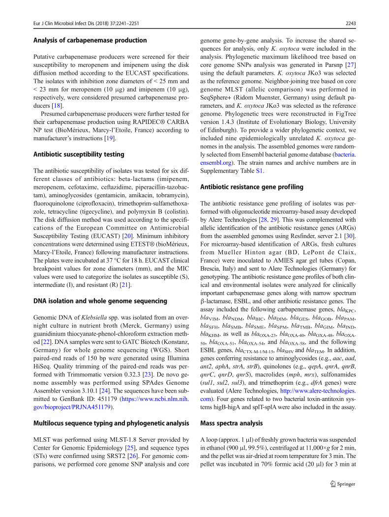
In Örebro County, 1132 clinical isolates of ESBL-producing Enterobacteriaceae were reported between 2008and 2015 [5]. Among those, six (0.53%) isolates were identi-fied as carbapenem-resistant Enterobacteriaceae and were pre-served at the Department of Laboratory Medicine, ClinicalMicrobiology, Örebro University Hospital (Table 1). All theisolates wereKlebsiella spp., eitherK. pneumoniae (n = 4) andK. oxytoca (n = 2), and all were positive for carbapenemaseproduction. It is likely that majority of the patients contractedthe CPE abroad since domestic CPE is rare in Sweden.
Genetic analysis of Klebsiella isolates
The antibiotic resistance genes conferring resistance to thedifferent classes of antibiotics were initially determined byan oligonucleotide hybridization assay, and subsequently, thewhole genome analysis was used for allelic confirmation ofARGs. The results of these analyses are consolidated inTable 1. A remarkable similarity was observed between oneof the K. oxytoca isolates from the river, E1, and two from thehospital (H4 and H6), where H4 showed identical antibioticresistance gene profiles, while H6 only lacked the strA gene(Table 1). All isolates from the environment and hospital har-bored one of five carbapenemase genes. The blaVIM-1 was themost commonly detected carbapenemase gene with 50% iso-lates carrying this locus. Other carbapenemase genes, blaNDM-
1, blaIMP-29, blaKPC-3, and blaOXA-48, were uniquely detectedin environmental and clinical isolates (Table 1). Six isolateshad one of the metallo-β-lactamase (blaVIM-1, blaNDM-1,bla IMP-29) genes, including the two environmentalK. oxytoca isolates from the river (E1) and sewage water(E2). Two clinical K. pneumoniae isolates, H2 and H5, har-bored serine-type carbapenemase genes, blaKPC-3 and blaOXA-48, respectively. The genes for two bacterial toxin-antitoxinsystems, higB-higA and splT-splA, were not detected in anyisolate in this study.
Interestingly, ESBL genes were only detected inK. pneumoniae isolates, while no ESBL genes were detectedin any of the K. oxytoca strains (Table 1). K. pneumoniae H1carried blaCTX-M-15, blaSHV-11 and blaTEM-1, whileK. pneumoniae H2 carried two ESBL genes, blaSHV andblaTEM-1. The K. pneumoniae H3 harbored blaCTX-M-9 genetogether with blaSHV-28 while H5 harbored blaCTX-M-15 andblaTEM-1 while isolate H3 only harbored blaSHV. All isolatescarried antibiotic resistance genes for aminoglycosides andsulfonamides. The common aminoglycoside resistance geneswere aac(6′)-Ib, aadA1, strA, and strB. For sulfonamide resis-tance, sul and dfrA genes were detected. With the exception ofthe K. oxytoca E2 isolate from the river, quinolone resistancegenes were detected in all isolates. Macrolide resistance geneswere detected only in K. pneumoniaeH1 (mph) and H2 (mph,mrx).
Since K. oxytoca from the river (E1) and hospital (H4)showed identical antibiotic resistance gene profiles accordingto oligonucleotide hybridization assay, we performed amultilocus sequence typing (MLST) and whole genome phy-logenetic analysis to further characterize the isolates. Both ofthe hospital (H4 and H6) and the environmental (E1) isolatesof K. oxytoca belonged to the same sequence type, ST172(Table 1). Isolate E2 showed a novel allelic profile, whichwas identified as new sequence type, ST192. AllK. pneumoniae isolates showed unique sequence types(Table 1). For phylogenetic analysis, only K. oxytoca wereselected while K. pneumoniae were excluded to increase the
2244 Eur J Clin Microbiol Infect Dis (2018) 37:2241–2251
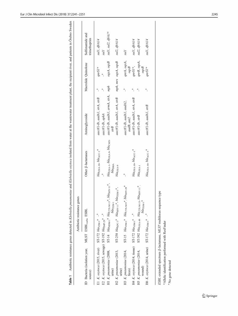
Table1
Antibiotic
resistance
genesdetected
inKlebsiella
pneumoniaeandKlebsiella
oxytocaisolated
from
water
atthewastewatertreatm
entp
lant,the
recipientriver,and
patientsin
ÖrebroSweden
Antibiotic
resistance
genes
IDBacteria(isolatio
nyear,
source)
MLST
ESBLcarbaESBL
Other
β-lactamases
Aminoglycoside
Macrolid
eQuinolone
Sulfonamideand
trim
ethoprim
E1
K.oxytoca
(2015,river)
ST-172bla V
IM-1*
–abla O
XA-10,blaACC-1*
aac(6′)-Ib,aadA1,strA,strB
–aqnrS1*
sul1,dfrA14
E2
K.oxytoca
(2015,sewage)
ST-192bla IMP-29*–a
–aaac(6′)-Ib,aphA
–a–a
sul1
H1K.pneum
oniae(2008,
urine)
ST-14
bla N
DM-1*bla C
TX-M
-15*,bla S
HV-11*,
bla T
EM-1
bla O
XA-1,blaOXA-9,blaCMY,
bla D
HA
aac(6′)-Ib,aadA1,armA,strA,
strB
mph
oqxA
,oqxB
sul1,sul2,dfrA1*
H2K.pneum
oniae(2013,
urine)
ST-258bla K
PC-3*
bla S
HV-11*,bla T
EM-1*,
bla O
XA-9
aac(6′)-Ib,aadA1,strA,strB
mph,m
rxoqxA
,oqxB
sul2,dfrA14
H3K.pneum
oniae(2014,
feces)
ST-15
bla V
IM-1*
bla C
TX-M
-9* ,bla S
HV-28*
–aaac(6′)-Ib,aadA1,aadA
2,aadB
,ant2
–aqnrA,oqxA,
oqxB
sul1
H4K.oxytoca
(2014,tissue)
ST-172bla V
IM-1*
–abla O
XA-10,blaACC-1*
aac(6′)-Ib,aadA1,strA,strB
–aqnrS1*,
sul1,dfrA14
H5K.pneum
oniae(2014,
wound)
ST-392bla O
XA-48
bla C
TX-M
-15,blaSHV-11* ,
bla T
EM-1*
bla O
XA-1
aac(6′)-Ib,strB
–aqnrB,oqxA,
oqxB
sul2,dfrA14
H6K.oxytoca
(2014,urine)
ST-172bla V
IM-1*
–abla O
XA-10,blaACC-1*
aac(6′)-Ib,aadA1,strB
–aqnrS1*
sul1,dfrA14
ESB
Lextended
spectrum
β-lactamase,MLS
Tmultilocus
sequence
type
*Allelic
identificationperformed
with
ResFinder
aNogene
detected
Eur J Clin Microbiol Infect Dis (2018) 37:2241–2251 2245

resolution of core genome analysis. A total of 13 K. oxytocagenomes including isolates from this study (n = 4) and epide-miologically unrelated isolates (n = 9) were subjected to singlenucleotide polymorphisms (SNPs) analysis in Parsnp, andphylogenetic tree was constructed based on SNPs comparison(see methods). The Parsnp phylogenetic tree revealed threedistinct clusters (Fig. 1a). The K. oxytoca isolates E1, H4,and H6 are together in cluster 3 and are highly related as theyshare a common genetic ancestor base. The K. oxytoca isolateE2 is placed separately in cluster 2, showing its genetic dis-tance from otherK. oxytoca in this study.We further comparedhomologous coding sequences of isolates using core genomeMLST (cgMLST) in SeqSphere+ software. K. oxytoca JKo3was used as a reference genome, and a set of 3089 commongenes in 13 K. oxytoca isolates was included in the analysis.The cgMLSTevaluation produced highly similar results to theSNPs analysis (Fig. 1b). Similar to the SNP-based analysis,cgMLST shows that the clinical isolates H4 and H6 and envi-ronmental isolate E1 are highly similar and cluster together(cluster 3), while E2 is in a separate cluster (cluster 2).
Multidrug resistance in all the Klebsiella isolates
The eight Klebsiella spp. were tested for susceptibility to sixclasses of antibiotics (Table 2). K. oxytoca E1 isolate from theriver showed intermediate resistance to imipenem whileK. oxytoca E2 from incoming wastewater was resistant toimipenem and meropenem but susceptible to ertapenem.Both environmental isolates (E1 and E2) were susceptible togentamicin, amikacin, colistin, and tigecycline. Among theclinical isolates, both K. oxytoca isolates H4 and H6 were
clinically not resistant to all tested carbapenems, aminoglyco-sides, tetracycline, and polymyxin but showed resistance tocephalosporins, piperacillin-tazobactam, and ciprofloxacin.TheK. pneumoniaeH1 isolated from a UTI patient was highlyresistant to all tested antibiotics except colistin and tigecycline(Table 2), while K. pneumoniae H2 was only susceptible tocolistin. All Klebsiella isolates (n = 8) were resistant to ≥ 3classes of antibiotics and were classified as MDR.
Phyloproteomic comparison of mass spectraof isolates
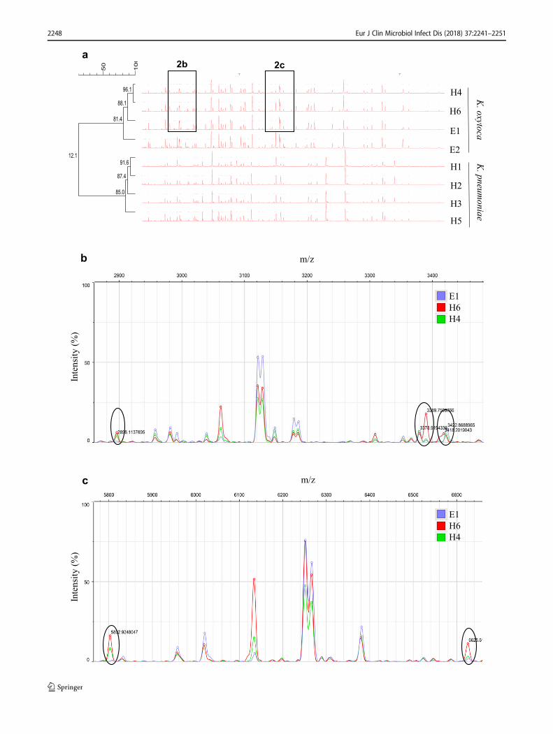
Strain level comparison between isolates was performed usingMALDI-TOF MS and Pearson similarity coefficient analysis.The similarity of mass spectra between each replicate pair wasgreater than 99% (supplementary Fig. S1), which was used asa threshold value to define related types. K. oxytoca andK. pneumoniae clustered separately with only 12.1% spectralsimilarity between them (Fig. 2a). The highest similarity of96% was observed between two clinical K. oxytoca isolates,H4 and H6. A detailed analysis of the spectra was done for theK. oxytoca isolates E1, H4, and H6 to determine their related-ness since they had identical antibiotic resistance genes andH6 only differed by one gene. However, the spectra showedthat peaks at mass to charge ratio (m/z) of 2890, 3389, 3418(Fig. 2b), 5802, and 6625 (Fig. 2c) were absent for E1 butwere present in either H4 or H6. The peak corresponding tom/z of 3422 was unique to E1 and absent in H4 and H6. Bothenvironmental isolates of K. oxytoca (E1 and E2) appeareddifferent from the clinical strains with less than 90% similarity(Fig. 2a). Furthermore, a comparison of spectral profiles of
Fig. 1 Phylogenetic relationship of theK. oxytoca isolates from this studycompared to other strains. a Phylogenetic maximum-likelihood treeconstructed in FigTree on the basis of core genome SNP analysis of usingParsnp. The isolates from the present study are highlighted in red.Scale bar represents genetic distances based on nucleotide substitutions
per SNP site. b Allele-based neighbor-joining tree based on 3089cgMLST target genes showing the clustering of environmental andclinical K. oxytoca strains. cgMLST was performed using K. oxytocaJKo3 as a reference in SeqSphere+. Scale bar represents allelic distance(color figure online)
2246 Eur J Clin Microbiol Infect Dis (2018) 37:2241–2251

environmental K. oxytoca isolates, E1 and E2, revealed 78%similarity and showed a large difference in terms of presenceor absence and displacement of peaks (data not shown).
Discussion
In the present study, carbapenemase-producing K. oxytocawere isolated from sewage and river waters running throughÖrebro city in central Sweden. To the best of our knowledge,this was the first CPE isolated from the environment inSweden, thus prior to 2015, no isolates were reported. Thisis in contrast to other European countries that have reportedhealth-care associated CPE since 2011 [31]. Only twocarbapenemase-producing Klebsiella isolates were recoveredfrom the environment, indicating that the levels of CPE inenvironmental waters remain very low. However, the presenceof these resistant bacteria in the river, even at low levels,remains a concern. Although carbapenemase-producingK. oxytoca were found in Svartån River, they were not detect-ed in the downstream lake water, which suggests that CPEmay be present at even lower levels or completely absent inthe lake due to the increased dilution or die-off under environ-mental conditions. Environmental K. oxytoca isolates, E1 andE2, harbored blaVIM-1 and blaIMP-29, respectively, and alsoshowed phenotypic resistance to either one or two carbapen-em antibiotics (Table 2). These carbapenemase genes are themost commonly identified worldwide and blaVIM-1 is predom-inant in Europe, while blaIMP alleles are prevalent in Asia[32]. As none of the isolates from patients in ÖrebroUniversity Hospital were identified with blaIMP-29, this im-plies that the source of CPE in the wastewater most likelyoriginate from the community.
Only six cases of CPE infections were reported in ÖrebroCounty between 2008 and 2015, and the majority of thesepatients had likely contracted their resistant bacteria fromabroad rather than domestic acquisition in the Swedish hospi-tal environment. The clinical K. pneumoniae isolates H1 andH2 harbored blaNDM-1 and blaKPC-3, respectively, and theblaNDM type metallo-β-lactamases are highly prevalent inthe Indian sub-cont inent [33, 34] , while blaKPC
carbapenemases have worldwide prevalence [35–38]. Theseisolates harbored antibiotic resistance genes for a broad rangeof antibiotics as well as showed functional resistance to mostof the antibiotics tested in this study. The K. pneumoniae H1co-harbored other ESBL genes such as blaCTX-M-15, which isfrequently detected in blaNDM-1-positive Enterobacteriaceae[39] and also the predominant ESBL type in Örebro County[40]. Strains H4 and H6 only carried blaVIM-1, and these hadhigh genome similarity to the strain from river (E1) indicatingthat this isolate might be widespread in the community.
Resistance to carbapenems was due to the presence of func-tional carbapenemase genes; however, other mechanisms thatwere not analyzed in this study could also contribute the finalphenotype. These mechanisms include the overexpression ofefflux-pumps and downregulation or modification of outer-membrane proteins such as ompK and phoE [41]. Mostcarbapenemase genes are found on mobile plasmids that canbe exchanged between the members of Enterobacteriaceaethrough horizontal gene transfer [42]. Some plasmids carrycarbapenemase genes along with toxin-antitoxin systems thatincrease plasmid prevalence by post-segregational killing ofdaughter cells that lack the plasmid [43]. Thus, antibiotic se-lection pressure is not always necessary to maintain antibioticresistance plasmids in a bacterial population. The recipientWWTP of the hospital and municipal wastewater is a major
Table 2 Antibiotic susceptibility patterns of Klebsiella pneumoniae and Klebsiella oxytoca isolated from the hospital, river water, and wastewaterincoming to the WWTP
Antibiotic susceptibility
IMP MEM ETP CTX CAZ TZP GN AK TOB CIP SXT CT TGC
E1 I (4) S (2) S (0.125) R (> 32) R (48) R (nd) S (1.5) S (2) S (4) R (3) R (> 32) S (0.5) S (0.25)
E2 I (8) R (16) I (0.75) R (> 32) R (> 256) R (nd) S (1) S (8) R (6) R (> 32) S (0.25) S (0.25) S (0.38)
H1 R (> 32) R (> 32) R (> 32) R (> 32) R (> 256) R (nd) R (> 256) R (> 256) R (> 256) R (> 32) R (> 32) S (2) S (1)
H2 R (24) R (> 32) R (> 32) R (> 32) R (> 256) R (nd) I (4) R (32) R (24) R (> 32) R (> 32) S (0.2) R (6)
H3 S (1.5) S (0.75) S (0.5) R (> 32) R (> 256) R (nd) R (6) S (8) R (12) R (> 32) R (> 32) S (2) S (2)
H4 S (1.5) S (0.25) S (0.125) R (32) R (48) R (nd) S (1) S (1) I (3) R (2) R (> 32) S (0.125) S (0.38)
H5 S (0.25) S (0.25) S (0.38) R (> 32) R (48) R (nd) S (1) S (4) R (6) R (> 32) R (> 32) S (0.125) R (12)
H6 S (0.5) S (0.19) S (0.125) R (> 32) R (32) R (nd) S (1) S (1) S (2) R (2) S (2) S (0.125) S (0.5)
Based on antibiotic susceptibility test using Etest or disk diffusion method and EUCAST breakpoints v 8.0. Values in parentheses indicate MIC inmicrograms per milliliter. nd = MIC not determined, and clinical resistance was determined using disk diffusion method
Antibiotics: IMP imipenem, MEM meropenem, CTX cefotaxime, CAZ ceftazidime, TZP piperacillin-tazobactam (30 μg–6 μg), GN gentamicin, AKamikacin, TOB tobramycin, CIP ciprofloxacin, SXT trimethoprim-sulfamethoxazole, CT colistin, TGC tigecycline, R resistant, S susceptible, Iintermediate
Eur J Clin Microbiol Infect Dis (2018) 37:2241–2251 2247
b
E1
H6
H4
Intensity
(%)
m/z
c
Intensity
(%)
m/z
E1
H6
H4
96.1
88.1
81.4
91.6
87.4
85.0
100
50
0 5000 10000
H4
H6
E1
E2
H1
H2
H3
H5
K.oxytoca
K.pneum
oniae
a2c2b
12.1
2248 Eur J Clin Microbiol Infect Dis (2018) 37:2241–2251

source of antibiotic-resistant Enterobacteriaceae, and the treat-ment process often reduces but does not eliminate these or-ganisms [44]. Complex and nutrient-rich microbial environ-ment in the WWTP may provide ideal conditions for the ex-pansion of antibiotic resistance into natural environments [45]and may help to disseminate plasmids with toxin-antitoxinsystems in a bacterial population without any selection pres-sure. It is interesting to note that our strains did not have thehigB-higA and splT-splA toxin-antitoxin genes. However,there is a possibility of the presence of other toxin-antitoxinsystems that were not analyzed in this study.
In countries with low to middle-income economies, un-treated wastewaters from households and pharmaceutical in-dustries are discharged into natural environments without pri-or treatment, and this has been shown to increase antibiotic-resistant bacteria in the environment [46]. In Sweden, howev-er, the regulations for wastewater disposal have been strictsince the approval of the Environmental Protection Act in1969. Our study revealed the presence of CPE in theSwedish aquatic environment, albeit at low levels, regardlessof the stringent regulations for antibiotic use. Although, thestudy is based on the comparison of a small set of strains, thepresence of related CPE in both hospital and associated riverand lake environments is an important observation. Thisshows the possibility of transmission of CPE from hospitalto aquatic environment since the effluent wastewater fromhospital is transported through the drainage system into theWWTP without prior treatment. In addition to the hospital,the community and veterinary clinics are other potentialsources of CPE that may reach the aquatic environment,with the WWTP being the distributor. The clinical isolatescarrying carbapenemase genes were most probably ac-quired from abroad and not generated at the hospital, sug-gesting that alternate strategies must be employed to avoidthe further environmental contamination and transmissionof MDR in the community. Although WWTP plays animportant role to minimize the organic pollution from thewastewater, they can be ultimate points of disseminationfor MDR bacteria into natural environments [47].Therefore, WWTP must apply processes for eradicatingbacteria, including MDR bacteria from wastewater beforedischarging into the environment. Thus, attempts to reducethe spread of CPE to the aquatic environment in Swedenand surrounding countries are of vital importance since thepresence of CPE in these niches is still rare.
Acknowledgments The authors thank Ellinor Rapp and Ines Engelmannfor technical assistance.
Author contributions J.J. and B.S. supervised the project and togetherwith F.A.H. designed the study, analyzed, and interpreted the data. F.A.H.collected the data and wrote the initial draft of the manuscript. B.H.contributed to analysis of the clinical strains and MALDI-TOF MS data,and R.E. provided the gene array analysis of all the strains. J.J., B.S., andR.E. critically revised the manuscript, and all authors contributed to thewriting and approved the manuscript.
Funding Svenska Forskningsrådet Formas (219-2014-837) (JJ) andNyckelfonden at Örebro University Hospital (BS).
Compliance with ethical standards
Conflict of interest R.E. is employed at Abbott (ALERE TechnologiesGmbH). This has not influenced the study.
Ethical approval For this type of study, formal consent is not required.
Open Access This article is distributed under the terms of the CreativeCommons At t r ibut ion 4 .0 In te rna t ional License (h t tp : / /creativecommons.org/licenses/by/4.0/), which permits unrestricted use,distribution, and reproduction in any medium, provided you giveappropriate credit to the original author(s) and the source, provide a linkto the Creative Commons license, and indicate if changes were made.
References
1. Cao X, Cavaco LM, Lv Y, Li Y, Zheng B, Wang P, Hasman H, LiuY, Aarestrup FM (2011) Molecular characterization and antimicro-bial susceptibility testing of Escherichia coli isolates from patientswith urinary tract infections in 20 Chinese hospitals. J ClinMicrobiol 49(7):2496–2501. https://doi.org/10.1128/JCM.02503-10
2. Braykov NP, Eber MR, Klein EY, Morgan DJ, Laxminarayan R(2013) Trends in resistance to carbapenems and third-generationcephalosporins among clinical isolates of Klebsiella pneumoniaein the United States, 1999–2010. Infect Control Hosp Epidemiol34(3):259–268. https://doi.org/10.1086/669523
3. Tzouvelekis LS, Markogiannakis A, Piperaki E, Souli M, DaikosGL (2014) Treating infections caused by carbapenemase-producingEnterobacteriaceae. Clin Microbiol Infect 20(9):862–872. https://doi.org/10.1111/1469-0691.12697
4. Yang D, Guo Y, Zhang Z (2009) Combined porin loss and extendedspectrum β-lactamase production is associated with an increasingimipenem minimal inhibitory concentration in clinical Klebsiellapneumoniae strains. Curr Microbiol 58(4):366–370. https://doi.org/10.1007/s00284-009-9364-4
5. Folkhälsomyndigheten (2016) Yearly statistics of ESBLcarba pro-ducing Enterobacteriaceae in Sweden by Public Health Agency
6. SWEDRES-SVARM (2014) Consumption of antibiotics and occur-rence of antibiotic resistance in Sweden. Solna/Uppsala ISSN1650–6332
7. Bagley ST (1985) Habitat association of Klebsiella species. InfectControl 6(2):52–58
8. Lin YT, Jeng YY, Chen TL, Fung CP (2010) Bacteremiccommunity-acquired pneumonia due to Klebsiella pneumoniae:clinical and microbiological characteristics in Taiwan, 2001–
�Fig. 2 MALDI-TOF mass spectra of the Klebsiella isolates. aPhyloproteomic dendrogram of MALDI-TOF mass spectra ofK. pneumoniae and K. oxytoca isolated from Svartån River, incomingsewage to the WWTP and patients from Örebro University Hospital. b,c Zoom into spectral regions with differences between K. oxytoca isolatesfrom the river (E1) and hospital (H4 and H6), based on the presence andabsence of ions of specific mass to charge ratio. Five major differencesbetween the strains are shown in ellipses
Eur J Clin Microbiol Infect Dis (2018) 37:2241–2251 2249
2008. BMC Infect Dis 10:307. https://doi.org/10.1186/1471-2334-10-307
9. Podschun R, UllmannU (1998)Klebsiella spp. as nosocomial path-ogens: epidemiology, taxonomy, typing methods, and pathogenici-ty factors. Clin Microbiol Rev 11(4):589–603
10. Patel G, Bonomo RA (2011) Status report on carbapenemases:challenges and prospects. Expert Rev Anti-Infect Ther 9(5):555–570. https://doi.org/10.1586/eri.11.28
11. Yong D, Toleman MA, Giske CG, Cho HS, Sundman K, Lee K,Walsh TR (2009) Characterization of a new metallo-beta-lactamasegene, bla(NDM-1), and a novel erythromycin esterase gene carriedon a unique genetic structure in Klebsiella pneumoniae sequencetype 14 from India. Antimicrob Agents Chemother 53(12):5046–5054. https://doi.org/10.1128/aac.00774-09
12. Uz Zaman T, Aldrees M, Al Johani SM, Alrodayyan M,Aldughashem FA, Balkhy HH (2014) Multi-drug carbapenem-re-sistant Klebsiella pneumoniae infection carrying the OXA-48 geneand showing variations in outer membrane protein 36 causing anoutbreak in a tertiary care hospital in Riyadh, Saudi Arabia. Int JInfect Dis 28:186–192. https://doi.org/10.1016/j.ijid.2014.05.021
13. Yao X, Doi Y, Zeng L, Lv L, Liu J-H (2016) Carbapenem-resistantand colistin-resistant Escherichia coli co-producing NDM-9 andMCR-1. Lancet Infect Dis 16(3):288–289. https://doi.org/10.1016/S1473-3099(16)00057-8
14. Marchaim D, Chopra T, Pogue JM, Perez F, Hujer AM, Rudin S,Endimiani A, Navon-Venezia S, Hothi J, Slim J, Blunden C,Shango M, Lephart PR, Salimnia H, Reid D, Moshos J, HafeezW, Bheemreddy S, Chen T-Y, Dhar S, Bonomo RA, Kaye KS(2011) Outbreak of colistin-resistant, carbapenem-resistantKlebsiella pneumoniae in Metropolitan Detroit, Michigan.Antimicrob Agents Chemother 55(2):593–599. https://doi.org/10.1128/aac.01020-10
15. Xavier BB, Lammens C, Ruhal R, Kumar-Singh S, Butaye P,Goossens H, Malhotra-Kumar S (2016) Identification of a novelplasmid-mediated colistin-resistance gene, mcr-2, in Escherichiacoli, Belgium, June 2016. Euro Surveill 21(27). https://doi.org/10.2807/1560-7917.es.2016.21.27.30280
16. Pournaras S, Koumaki V, Spanakis N, Gennimata V, Tsakris A(2016) Current perspectives on tigecycline resistance inEnterobacteriaceae: susceptibility testing issues and mechanismsof resistance. Int J Antimicrob Agents 48(1):11–18. https://doi.org/10.1016/j.ijantimicag.2016.04.017
17. Woodford N, Wareham DW, Guerra B, Teale C (2014)Carbapenemase-producing Enterobacteriaceae and non-Enterobacteriaceae from animals and the environment: an emerg-ing public health risk of our ownmaking? J Antimicrob Chemother69(2):287–291. https://doi.org/10.1093/jac/dkt392
18. EUCAST (2013)Guidelines for detection of resistancemechanismsand specific resistances of clinical and/or epidemiologicalimportance
19. Poirel L, Nordmann P (2015) Rapidec Carba NP test for rapiddetection of carbapenemase producers. J Clin Microbiol 53(9):3003–3008. https://doi.org/10.1128/jcm.00977-15
20. EUCAST (2013) Disk diffusion test methodology21. EUCAST (2016) Breakpoint tables for interpretation of MICs and
zone diameters22. Lemarchand K, Berthiaume F, Maynard C, Harel J, Payment P,
Bayardelle P, Masson L, Brousseau R (2005) Optimization of mi-crobial DNA extraction and purification from raw wastewater sam-ples for downstream pathogen detection by microarrays. JMicrobiol Methods 63(2):115–126. https://doi.org/10.1016/j.mimet.2005.02.021
23. Bolger AM, Lohse M, Usadel B (2014) Trimmomatic: a flexibletrimmer for Illumina sequence data. Bioinformatics 30(15):2114–2120. https://doi.org/10.1093/bioinformatics/btu170
24. Bankevich A, Nurk S, Antipov D, Gurevich AA, Dvorkin M,Kulikov AS, Lesin VM, Nikolenko SI, Pham S, Prjibelski AD,Pyshkin AV, Sirotkin AV, Vyahhi N, Tesler G, Alekseyev MA,Pevzner PA (2012) SPAdes: a new genome assembly algorithmand its applications to single-cell sequencing. J Comput Biol19(5):455–477. https://doi.org/10.1089/cmb.2012.0021
25. LarsenMV, Cosentino S, Rasmussen S, Friis C, Hasman H, MarvigRL, Jelsbak L, Sicheritz-Ponten T, Ussery DW, Aarestrup FM,Lund O (2012) Multilocus sequence typing of total-genome-sequenced bacteria. J Clin Microbiol 50(4):1355–1361. https://doi.org/10.1128/jcm.06094-11
26. Inouye M, Dashnow H, Raven L-A, Schultz MB, Pope BJ, TomitaT, Zobel J, Holt KE (2014) SRST2: rapid genomic surveillance forpublic health and hospital microbiology labs. Genome Med 6(11):90. https://doi.org/10.1186/s13073-014-0090-6
27. Treangen TJ, Ondov BD, Koren S, Phillippy AM (2014) TheHarvest suite for rapid core-genome alignment and visualizationof thousands of intraspecific microbial genomes. Genome Biol15(11):524. https://doi.org/10.1186/s13059-014-0524-x
28. Braun SD, Monecke S, Thürmer A, Ruppelt A, Makarewicz O,Pletz M, Reißig A, Slickers P, Ehricht R (2014) Rapid identificationof carbapenemase genes in gram-negative bacteria with an oligo-nucleotide microarray-based assay. PLoS One 9(7):e102232.https://doi.org/10.1371/journal.pone.0102232
29. Braun SD, Dorneanu OS, Vremera T, Reissig A, Monecke S,Ehricht R (2016) Carbapenemase-producing Enterobacteriaceae: a2-year surveillance in a hospital in Iasi, Romania. Future Microbiol11:391–401. https://doi.org/10.2217/fmb.15.148
30. Kleinheinz KA, Joensen KG, Larsen MV (2014) Applying theResFinder and VirulenceFinder web-services for easy identificationof acquired antibiotic resistance and E. coli virulence genes in bac-teriophage and prophage nucleotide sequences. Bacteriophage 4:e27943. https://doi.org/10.4161/bact.27943
31. Albiger B, Glasner C, Struelens MJ, Grundmann H, Monnet DL(2015) Carbapenemase-producing Enterobacteriaceae in Europe:assessment by national experts from 38 countries, May 2015.Euro Surveill 20(45). https://doi.org/10.2807/1560-7917.es.2015.20.45.30062
32. Tato M, Coque TM, Baquero F, Cantón R (2010) Dispersal ofcarbapenemase blaVIM-1 gene associated with different Tn402variants, mercury transposons, and conjugative plasmids inEnterobacteriaceae and Pseudomonas aeruginosa. AntimicrobAgents Chemother 54(1):320–327. https://doi.org/10.1128/aac.00783-09
33. Perry JD, Naqvi SH, Mirza IA, Alizai SA, Hussain A, Ghirardi S,Orenga S, Wilkinson K, Woodford N, Zhang J, Livermore DM,Abbasi SA, Raza MW (2011) Prevalence of faecal carriage ofEnterobacteriaceae with NDM-1 carbapenemase at military hospi-tals in Pakistan, and evaluation of two chromogenic media. JAntimicrob Chemother 66(10):2288–2294. https://doi.org/10.1093/jac/dkr299
34. Bharadwaj R, Joshi S, Dohe V, Gaikwad V, Kulkarni G, Shouche Y(2012) Prevalence of New Delhi metallo-beta-lactamase (NDM-1)-positive bacteria in a tertiary care centre in Pune, India. Int JAntimicrob Agents 39(3):265–266. https://doi.org/10.1016/j.ijantimicag.2011.09.027
35. Kitchel B, Rasheed JK, Patel JB, Srinivasan A, Navon-Venezia S,Carmeli Y, Brolund A, Giske CG (2009) Molecular epidemiologyof KPC-producing Klebsiella pneumoniae isolates in the UnitedStates: clonal expansion of multilocus sequence type 258.Antimicrob Agents Chemother 53(8):3365–3370. https://doi.org/10.1128/aac.00126-09
36. Sheppard AE, Stoesser N, Wilson DJ, Sebra R, Kasarskis A,Anson LW, Giess A, Pankhurst LJ, Vaughan A, Grim CJ, CoxHL, Yeh AJ, Sifri CD, Walker AS, Peto TE, Crook DW,Mathers AJ (2016) Nested Russian doll-like genetic mobility
2250 Eur J Clin Microbiol Infect Dis (2018) 37:2241–2251

drives rapid dissemination of the carbapenem resistance geneblaKPC. Antimicrob Agents Chemother 60(6):3767–3778.https://doi.org/10.1128/aac.00464-16
37. Stoesser N, Sheppard AE, Peirano G, Anson LW, Pankhurst L,Sebra R, Phan HTT, Kasarskis A, Mathers AJ, Peto TEA,Bradford P, Motyl MR, Walker AS, Crook DW, Pitout JD (2017)Genomic epidemiology of global Klebsiella pneumoniaecarbapenemase (KPC)-producing Escherichia coli. Sci Rep 7(1):5917. https://doi.org/10.1038/s41598-017-06256-2
38. Nordmann P, Cuzon G, Naas T (2009) The real threat of Klebsiellapneumoniae carbapenemase-producing bacteria. Lancet Infect Dis9(4):228–236. https://doi.org/10.1016/S1473-3099(09)70054-4
39. Poirel L, Dortet L, Bernabeu S, Nordmann P (2011) Genetic fea-tures of bla(NDM-1)-positive Enterobacteriaceae. AntimicrobAgents Chemother 55(11):5403–5407. https://doi.org/10.1128/AAC.00585-11
40. Önnberg A, Söderquist B, Persson K, Mölling P (2014)Characterization of CTX-M-producing Escherichia coli by repeti-tive sequence-based PCR and real-time PCR-based replicon typingof CTX-M-15 plasmids. APMIS 122(11):1136–1143. https://doi.org/10.1111/apm.12270
41. Yigit H, Marie Queenan A, Kamile Rasheed J, Biddle JW,Doménech-Sánchez A, Alberti S, Bush K, Tenover FC (2004)Carbapenem-resistant strain of Klebsiella oxytoca harboringcarbapenem-hydrolyzing -lactamase KPC-2. 47. https://doi.org/10.1128/AAC.47.12.3881-3889.2003
42. Goren MG, Carmeli Y, Schwaber MJ, Chmelnitsky I, Schechner V,Navon-Venezia S (2010) Transfer of Carbapenem-resistant plasmid
from Klebsiella pneumoniae ST258 to Escherichia coli in patient.Emerg Infect Dis 16(6):1014–1017. https://doi.org/10.3201/eid1606.091671
43. Mosqueda N, Gato E, Roca I, Lopez M, de Alegria CR, FernandezCuenca F, Martinez-Martinez L, Pachon J, Cisneros JM,Rodriguez-Bano J, Pascual A, Vila J, Bou G, Tomas M (2014)Characterization of plasmids carrying the blaOXA-24/40carbapenemase gene and the genes encoding the AbkA/AbkB pro-teins of a toxin/antitoxin system. J Antimicrob Chemother 69(10):2629–2633. https://doi.org/10.1093/jac/dku179
44. Aali R, Nikaeen M, Khanahmad H, Hassanzadeh A (2014)Monitoring and comparison of antibiotic resistant bacteria and theirresistance genes in municipal and hospital wastewaters. Int J PrevMed 5(7):887–894
45. Kummerer K (2009) Antibiotics in the aquatic environment—areview—part II. Chemosphere 75(4):435–441. https://doi.org/10.1016/j.chemosphere.2008.12.006
46. Marathe N, Pal C, Gaikwad S, Jonsson V, Kristiansson E, Larsson J(2017) Untreated urban waste contaminates Indian river sedimentswith resistance genes to last resort antibiotics. 124. https://doi.org/10.1016/j.watres.2017.07.060
47. Rutgersson C, Fick J, Marathe N, Kristiansson E, Janzon A,Angelin M, Johansson A, Shouche Y, Flach CF, Larsson DG(2014) Fluoroquinolones and qnr genes in sediment, water, soil,and human fecal flora in an environment polluted bymanufacturingdischarges. Environ Sci Technol 48(14):7825–7832. https://doi.org/10.1021/es501452a
Eur J Clin Microbiol Infect Dis (2018) 37:2241–2251 2251