Recombination Proteins Mediate Meiotic Spatial Chromosome ...
13
Recombination Proteins Mediate Meiotic Spatial Chromosome Organization and Pairing Aurora Storlazzi, 1,2 Silvana Gargano, 2 Gwenael Ruprich-Robert, 1 Matthieu Falque, 3 Michelle David, 1 Nancy Kleckner, 4 and Denise Zickler 1, * 1 Institut de Ge ´ ne ´ tique et Microbiologie, UMR 8621, Universite ´ Paris-Sud, 91405 Orsay, France 2 Istituto di Genetica e Biofisica A. Buzzati Traverso, CNR, 80131 Naples, Italy 3 UMR de Ge ´ ne ´ tique Ve ´ ge ´ tale du Moulon, 91190 Gif sur Yvette, France 4 Department of Molecular and Cellular Biology, Harvard University, Cambridge, MA 02139, USA *Correspondence: [email protected] DOI 10.1016/j.cell.2010.02.041 SUMMARY Meiotic chromosome pairing involves not only recognition of homology but also juxtaposition of entire chromosomes in a topologically regular way. Analysis of filamentous fungus Sordaria macrospora reveals that recombination proteins Mer3, Msh4, and Mlh1 play direct roles in all of these aspects, in advance of their known roles in recombination. Absence of Mer3 helicase results in interwoven chromosomes, thereby revealing the existence of features that specifically ensure ‘‘entanglement avoidance.’’ Entanglements that remain at zygotene, i.e., ‘‘interlockings,’’ require Mlh1 for resolution, likely to eliminate constraining recombinational connec- tions. Patterns of Mer3 and Msh4 foci along aligned chromosomes show that the double-strand breaks mediating homologous alignment have spatially separated ends, one localized to each partner axis, and that pairing involves interference among devel- oping interhomolog interactions. We propose that Mer3, Msh4, and Mlh1 execute all of these roles during pairing by modulating the state of nascent double-strand break/partner DNA contacts within axis-associated recombination complexes. INTRODUCTION A central unique feature of meiosis is pairing of homologous chromosomes (‘‘homologs’’). Pairing involves recognition of homology plus coming together of homologs in space. Addition- ally, juxtaposition must be achieved without high levels of entan- glement (Kleckner and Weiner, 1993; von Wettstein et al., 1984; Wang, C.R. et al., 2009). In most organisms, pairing involves juxtaposition of homolog structural axes, dependent upon, and mediated via, the bio- chemical process of DNA recombination. The two aspects are integrated by physical localization of protein/DNA recombination complexes (‘‘recombinosomes’’) to their underlying axes (e.g., Henderson and Keeney, 2005; Borner et al., 2004; Moens et al., 2007; Anderson and Stack, 2005; Franklin et al., 2006; Oliver-Bonet et al., 2007; Wang, K. et al., 2009 and references therein). Recombinosome/axis association arises before, or just after, initiation of recombination via programmed double- strand breaks (DSBs) (Blat et al., 2002; Tesse et al., 2003). Thereafter, DNA recombination and recombination-mediated juxtaposition of homolog axes progress in close temporal and functional coordination. Homologs first become aligned at a distance of 400 nm during leptotene. Except in worm and Drosophila, this process requires DSB formation and is accompanied by appearance of axis-associated foci of RecA homolog(s) (review in Henderson and Keeney, 2005). At the DNA level, alignment is inferred to involve nascent DNA/DNA interactions between a 3 0 single- stranded tail at one DSB end and the homologous region on a homolog partner duplex (Hunter and Kleckner, 2001; Hunter, 2006). Then, at zygotene, ‘‘synapsis’’ occurs: axes become progressively linked at a distance of 100 nm by transverse fila- ments, which, with other proteins, comprise the synaptonemal complex (SC) (review in Page and Hawley, 2004). The lepto- tene/zygotene transition is also a crucial point for recombination: nascent interactions are differentiated into crossover (CO)-fated and noncrossover (NCO)-fated products, with concomitant onset of stable strand exchange (review in Hunter, 2006). In several organisms, SC formation is nucleated preferentially at sites of CO-designated interactions (Henderson and Keeney, 2005). After full SC has formed, mature CO and NCO products appear at midpachytene (e.g., Guillon et al., 2005). This study analyzes the relationship between recombination and homolog juxtaposition in the filamentous fungus Sordaria macrospora which is a very powerful system for such analysis: continuous axes appear at early leptotene such that spatial relationships among chromosomes, and positions of axis-asso- ciated recombination-complexes, can be followed from S-phase onward (e.g., Storlazzi et al., 2003, 2008; Tesse et al., 2003; Zickler, 2006). Additionally, overall progression of meiosis can be followed by progressive increases in the sizes of both the 94 Cell 141, 94–106, April 2, 2010 ª2010 Elsevier Inc. brought to you by CORE View metadata, citation and similar papers at core.ac.uk provided by Elsevier - Publisher Connector
Transcript of Recombination Proteins Mediate Meiotic Spatial Chromosome ...
brought to you by COREView metadata, citation and similar papers at core.ac.uk
provided by Elsevier - Publisher Connector
Recombination Proteins MediateMeiotic Spatial ChromosomeOrganization and PairingAurora Storlazzi,1,2 Silvana Gargano,2 Gwenael Ruprich-Robert,1 Matthieu Falque,3 Michelle David,1 Nancy Kleckner,4
and Denise Zickler1,*1Institut de Genetique et Microbiologie, UMR 8621, Universite Paris-Sud, 91405 Orsay, France2Istituto di Genetica e Biofisica A. Buzzati Traverso, CNR, 80131 Naples, Italy3UMR de Genetique Vegetale du Moulon, 91190 Gif sur Yvette, France4Department of Molecular and Cellular Biology, Harvard University, Cambridge, MA 02139, USA
*Correspondence: [email protected]
DOI 10.1016/j.cell.2010.02.041
SUMMARY
Meiotic chromosome pairing involves not onlyrecognition of homology but also juxtaposition ofentire chromosomes in a topologically regular way.Analysis of filamentous fungus Sordaria macrosporareveals that recombination proteins Mer3, Msh4,and Mlh1 play direct roles in all of these aspects,in advance of their known roles in recombination.Absence of Mer3 helicase results in interwovenchromosomes, thereby revealing the existence offeatures that specifically ensure ‘‘entanglementavoidance.’’ Entanglements that remain at zygotene,i.e., ‘‘interlockings,’’ require Mlh1 for resolution, likelyto eliminate constraining recombinational connec-tions. Patterns of Mer3 and Msh4 foci along alignedchromosomes show that the double-strand breaksmediating homologous alignment have spatiallyseparated ends, one localized to each partner axis,and that pairing involves interference among devel-oping interhomolog interactions. We propose thatMer3, Msh4, and Mlh1 execute all of these rolesduring pairing by modulating the state of nascentdouble-strand break/partner DNA contacts withinaxis-associated recombination complexes.
INTRODUCTION
A central unique feature of meiosis is pairing of homologous
chromosomes (‘‘homologs’’). Pairing involves recognition of
homology plus coming together of homologs in space. Addition-
ally, juxtaposition must be achieved without high levels of entan-
glement (Kleckner and Weiner, 1993; von Wettstein et al., 1984;
Wang, C.R. et al., 2009).
In most organisms, pairing involves juxtaposition of homolog
structural axes, dependent upon, and mediated via, the bio-
chemical process of DNA recombination. The two aspects are
94 Cell 141, 94–106, April 2, 2010 ª2010 Elsevier Inc.
integrated by physical localization of protein/DNA recombination
complexes (‘‘recombinosomes’’) to their underlying axes (e.g.,
Henderson and Keeney, 2005; Borner et al., 2004; Moens
et al., 2007; Anderson and Stack, 2005; Franklin et al., 2006;
Oliver-Bonet et al., 2007; Wang, K. et al., 2009 and references
therein). Recombinosome/axis association arises before, or
just after, initiation of recombination via programmed double-
strand breaks (DSBs) (Blat et al., 2002; Tesse et al., 2003).
Thereafter, DNA recombination and recombination-mediated
juxtaposition of homolog axes progress in close temporal and
functional coordination.
Homologs first become aligned at a distance of �400 nm
during leptotene. Except in worm and Drosophila, this process
requires DSB formation and is accompanied by appearance of
axis-associated foci of RecA homolog(s) (review in Henderson
and Keeney, 2005). At the DNA level, alignment is inferred to
involve nascent DNA/DNA interactions between a 30 single-
stranded tail at one DSB end and the homologous region on
a homolog partner duplex (Hunter and Kleckner, 2001; Hunter,
2006). Then, at zygotene, ‘‘synapsis’’ occurs: axes become
progressively linked at a distance of �100 nm by transverse fila-
ments, which, with other proteins, comprise the synaptonemal
complex (SC) (review in Page and Hawley, 2004). The lepto-
tene/zygotene transition is also a crucial point for recombination:
nascent interactions are differentiated into crossover (CO)-fated
and noncrossover (NCO)-fated products, with concomitant
onset of stable strand exchange (review in Hunter, 2006). In
several organisms, SC formation is nucleated preferentially at
sites of CO-designated interactions (Henderson and Keeney,
2005). After full SC has formed, mature CO and NCO products
appear at midpachytene (e.g., Guillon et al., 2005).
This study analyzes the relationship between recombination
and homolog juxtaposition in the filamentous fungus Sordaria
macrospora which is a very powerful system for such analysis:
continuous axes appear at early leptotene such that spatial
relationships among chromosomes, and positions of axis-asso-
ciated recombination-complexes, can be followed from S-phase
onward (e.g., Storlazzi et al., 2003, 2008; Tesse et al., 2003;
Zickler, 2006). Additionally, overall progression of meiosis can
be followed by progressive increases in the sizes of both the
nucleus and its surrounding ascus (meiocyte), thus permitting
the detection of temporal delays in the progression of chromo-
somal events in mutant backgrounds comparatively to wild-
type (e.g., Storlazzi et al., 2003, 2008).
Interplay between homolog pairing and recombination is
probed by analysis of mutants defective in three proteins previ-
ously shown to be direct participants in the DNA events of
recombination: Mer3, Msh4 and Mlh1. Mer3 is a meiosis-specific
30-50 helicase that stimulates Rad51-mediated DNA heterodu-
plex extension and stabilizes recombinational interactions
(Nakagawa and Ogawa, 1999; Nakagawa and Kolodner, 2002;
Mazina et al., 2004). Msh4, as part of the Msh4/Msh5 hetero-
dimer, a meiosis-specific homolog of the bacterial mismatch
repair protein MutS, encircles and stabilizes branched DNA
structures (Snowden et al., 2004). Mlh1/3 are eukaryotic
homologs of the bacterial mismatch repair protein MutL and,
by analogy, may mediate release of Msh4/5 (Snowden et al.,
2004), among other possible roles (Hunter, 2006). Mer3 and
Msh4 are implicated in ensuring that the fates of CO-designated
interactions are faithfully maintained and efficiently implemented
during and after the leptotene/zygotene ‘‘CO control’’ transition.
In the absence of either gene product, CO formation is specifi-
cally abrogated while NCOs form at high levels (e.g., Borner
et al., 2004). Mhl1 is implicated at a later stage, in finalization
of CO recombinational interactions during pachytene (Hunter,
2006; Franklin et al., 2006). In correspondence with their times
of action during recombination, foci of Mer3 and Msh4/5 are
observed prior to and during SC formation, whereas foci of
Mlh1 are not visible until after completion of synapsis (e.g., de
Boer et al., 2006; Oliver-Bonet et al., 2007; Moens et al., 2007;
Franklin et al., 2006; Higgins et al., 2004; Jackson et al., 2006;
Wang, K. et al., 2009 and references therein).
Our results reveal that each of these three proteins plays
a central role in homolog juxtaposition one stage prior its estab-
lished role in promoting recombination. Mer3 and Msh4 are
implicated in leptotene alignment (in advance of the leptotene/
zygotene CO control transition) while Mlh1 is implicated in
resolution of interlocks at zygotene (in advance of midpachytene
CO finalization). Mer3 mutant phenotypes suggest the existence
of an ‘‘entanglement avoidance’’ process. Further, the patterns
and dynamics of Mer3 and Msh4 foci provide new information
about the nature of the alignment process and, by implication,
the positioning of DSB end(s) during alignment and the CO
control transition. Integration of these findings with previous
molecular and biochemical information leads to a model for
how Mer3 and Msh4 could mediate alignment by stabilizing
nascent DSB/partner interactions within axis-associated recom-
binosomes while Mlh1 mediates interlock resolution by removing
Msh4/5 stabilization of these contacts.
RESULTS
Sordaria macrospora Mer3, Msh4, and Mlh1 were cloned and
completely null deletion mutations constructed, found to be
recessive in heterozygous crosses, and further analyzed. Cyto-
logical localization was determined for Msh4-GFP and Mer3-
GFP fusion proteins, expressed from their respective promoters
at ectopic locations. Both fusion genes complement all meiotic
defects of cognate null mutants. See Supplemental Information
(SI) for details.
Mer3, Msh4, and Mlh1 Are Not Required for AxisFormation but Are Essential for Normal Pairing,Bouquet Dynamics, and RecombinationChromosomes of the three null alleles were analyzed in leptotene
through pachytene nuclei (n = 300 each), by DAPI staining of
chromatin and with two previously characterized axis markers:
Spo76/Pds5-GFP (van Heemst et al., 1999) and meiosis-specific
cohesin Rec8-GFP (Storlazzi et al., 2008). In all three mutants,
chromosome axes are indistinguishable from wild-type (WT)
axes: Spo76 and Rec8 load at S phase and form complete
lines along chromosomes from early leptotene to end pachytene
(below). Thus, effects of mutations on spatial relationships
can be analyzed without complications from defects in axis
development.
All three mutants progress through meiosis and sporulation
with the same progressive increase in ascus size as in WT.
However, chromosomal progression from leptotene to pachy-
tene is prolonged in all three mutants: by �10 hr in mer3D or
msh4D and�12 hr in mlh1D (24–26 hr instead of 12 in WT). Anal-
ogous prolongations occur in Arabidopsis Atmlh3 and Atmsh4
(Higgins et al., 2004; Jackson et al., 2006).
All three mutants exhibit defects in pairing and synapsis
(below). They show also defects in the bouquet dynamics. As
in WT, chromosome ends transiently cluster in one area of the
nuclear periphery at late leptotene and then redisperse at pachy-
tene onset, but exit from cluster is significantly delayed (Figures
S1A–1H available online).
Recombination is also defective in all three mutants (Figures
S2A–S2F and details in legends). In mer3D and msh4D mutants,
chiasmata are drastically reduced (4–7) compared to WT (21 ± 3).
mlh1D confers a milder defect: genetic analysis reveals a �40%
reduction in COs with no reduction in NCOs (see details in legend
of Figure S2B). As similar defects were found in other organisms
(review in Hunter, 2006), the corresponding proteins likely play
the same roles for recombination in Sordaria.
Mlh1 Is Required for Interlock ResolutionIn WT meiosis, unrelated chromosomes are occasionally seen
entangled with one another, usually at zygotene. In such ‘‘inter-
locks,’’ either one chromosome or a pair of chromosomes is
located between two aligned homologs, held in place by regular
synapsis to either side (Figures 1A and 1B). For Sordaria, inter-
locked chromosomes are seen in�20% of WT serially sectioned
zygotene nuclei (n = 121; DZ, unpublished data) and in �15%
(n = 300) of nuclei by immunofluorescence using Spo76-GFP
as axis marker (Figure 1A). Further, by early pachytene, no inter-
locks are seen by either method. Thus WT meiosis must include
a mechanism for their resolution during zygotene. Resolution of
interlocks has been suggested previously (review in von Wett-
stein et al., 1984; Wang, C.R. et al., 2009); however, conclusions
were often limited by the small number of nuclei examined and
by the inability to exclude the possibility that interlocks were
absent at pachytene because the corresponding nuclei were
absent from the experimental population (e.g., via apoptosis in
mammals; reviewed in von Wettstein et al., 1984; Zickler and
Cell 141, 94–106, April 2, 2010 ª2010 Elsevier Inc. 95
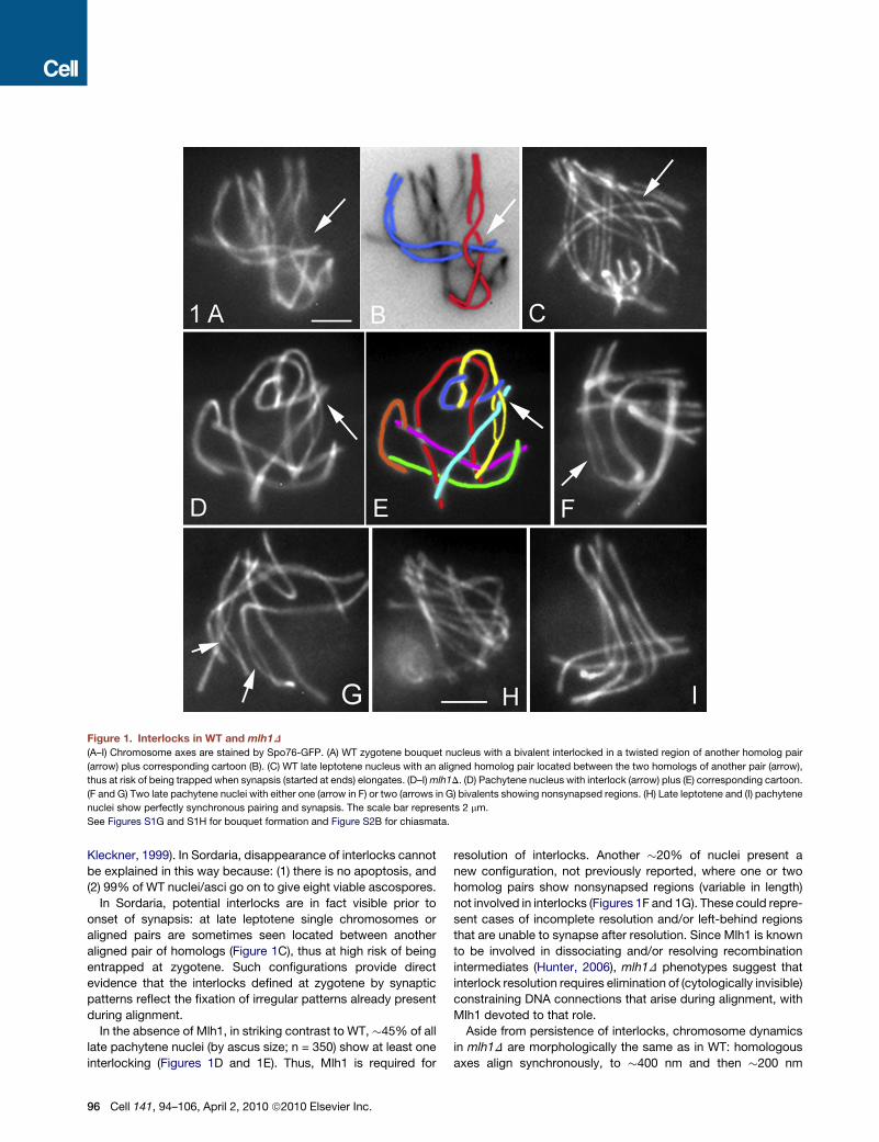
Figure 1. Interlocks in WT and mlh1D
(A–I) Chromosome axes are stained by Spo76-GFP. (A) WT zygotene bouquet nucleus with a bivalent interlocked in a twisted region of another homolog pair
(arrow) plus corresponding cartoon (B). (C) WT late leptotene nucleus with an aligned homolog pair located between the two homologs of another pair (arrow),
thus at risk of being trapped when synapsis (started at ends) elongates. (D–I) mlh1D. (D) Pachytene nucleus with interlock (arrow) plus (E) corresponding cartoon.
(F and G) Two late pachytene nuclei with either one (arrow in F) or two (arrows in G) bivalents showing nonsynapsed regions. (H) Late leptotene and (I) pachytene
nuclei show perfectly synchronous pairing and synapsis. The scale bar represents 2 mm.
See Figures S1G and S1H for bouquet formation and Figure S2B for chiasmata.
Kleckner, 1999). In Sordaria, disappearance of interlocks cannot
be explained in this way because: (1) there is no apoptosis, and
(2) 99% of WT nuclei/asci go on to give eight viable ascospores.
In Sordaria, potential interlocks are in fact visible prior to
onset of synapsis: at late leptotene single chromosomes or
aligned pairs are sometimes seen located between another
aligned pair of homologs (Figure 1C), thus at high risk of being
entrapped at zygotene. Such configurations provide direct
evidence that the interlocks defined at zygotene by synaptic
patterns reflect the fixation of irregular patterns already present
during alignment.
In the absence of Mlh1, in striking contrast to WT, �45% of all
late pachytene nuclei (by ascus size; n = 350) show at least one
interlocking (Figures 1D and 1E). Thus, Mlh1 is required for
96 Cell 141, 94–106, April 2, 2010 ª2010 Elsevier Inc.
resolution of interlocks. Another �20% of nuclei present a
new configuration, not previously reported, where one or two
homolog pairs show nonsynapsed regions (variable in length)
not involved in interlocks (Figures 1F and 1G). These could repre-
sent cases of incomplete resolution and/or left-behind regions
that are unable to synapse after resolution. Since Mlh1 is known
to be involved in dissociating and/or resolving recombination
intermediates (Hunter, 2006), mlh1D phenotypes suggest that
interlock resolution requires elimination of (cytologically invisible)
constraining DNA connections that arise during alignment, with
Mlh1 devoted to that role.
Aside from persistence of interlocks, chromosome dynamics
in mlh1D are morphologically the same as in WT: homologous
axes align synchronously, to �400 nm and then �200 nm
(Figure 1H), with normal accompanying numbers and patterns of
localization for Rad51 foci, which mark the sites of DSBs, and for
Mer3 foci (data not shown). Importantly, however, while the
period of alignment is rather short in WT (10%–20% of prophase
nuclei show fully aligned homologs), in mlh1D, 35% of nuclei
(n = 300 for each) show this configuration (Figure 1H). Mlh1 is
therefore functionally relevant even prior to its involvement in
interlock resolution. The synaptic period is also greatly pro-
longed in mlh1D: while �10% of WT nuclei are at zygotene,
25% of mutant nuclei exhibit early or intermediate extents of
synapsis (n = 300 for each strain). Even so, ultimately, all homo-
logs synchronously synapse (Figure 1I).
Notably, three mlh1D phenotypes emerge at zygotene, thus
one stage earlier than the defined pachytene role of Mlh1
in recombination (Hunter, 2006): delayed onset of synapsis,
delayed bouquet release (above) and persistence of interlocks
(resolved during zygotene in WT).
Msh4 Is Required to Establish CorrectAlignment DistanceLike mlh1D, msh4D homologs have no problem with initiation or
occurrence of presynaptic alignment. An alteration in alignment
is nonetheless apparent: msh4D homolog axes initially align at
a distance of �600-800 nm (Figures 2A and 2B), rather than
the �400 nm seen in WT (Figure 2C). This unique phenotype
points to direct participation of the recombination complex in
determining interaxis distances. Further, Msh4, like Mlh1, plays
a role in homolog pairing at leptotene, thus one stage prior to
the time of its defined role in formation of recombinants (at zygo-
tene; Introduction).
Once synapsis begins, msh4D exhibits also dramatic per-chro-
mosome and per-region defects (Figure 2D). In WT meiosis, all
seven chromosome-pairs progress synchronously and efficiently
from alignment (Figure 2C) to zygotene (Figure 2E) and synapsis
(Figure 2F). In msh4D, post-alignment nuclei show a mixture of
bivalents at different stages of synapsis (Figures 2G). Finally,
95% of the nuclei that would be in pachytene in a WT ascus of cor-
responding size still show a mixture of fully and partially synapsed
homologs (n = 100; Figure 2H). Since synapsis is tightly coupled
to CO-designation and progression during the leptotene/zygo-
tene transition, these defects likely reflect the known role of
Msh4 in recombination during this period (Hunter, 2006).
Despite the complexity of synaptic patterns, the dynamics of
Rad51 and Mer3 foci remain relatively unperturbed in msh4D.
Both types of foci appear with WT timing plus location (Figures
S3A–S3G) and number (respectively 52 ± 8 and 110 ± 9; n = 90).
At pachytene (by ascus size) both types of foci remain present in
unsynapsed regions while disappearing in synapsed regions in
the same nucleus (e.g., Figure 2I for Rad51), as normally seen
during synchronous progression through zygotene in WT (below).
These results emphasize the tight functional linkage between
recombinational progression and pairing/synapsis not only
globally, but also locally, on a region-by-region basis.
Mer3-Dependent Avoidance of Entanglementsduring AlignmentIn WT, at midleptotene, all homolog pairs of every nucleus exhibit
visible alignment (Figure 3A). At the corresponding ascus size in
mer3D, chromosomes are still separated and highly ‘‘inter-
woven’’ (Figures 3B–3D). Further, when alignment does finally
occur, at ascus sizes corresponding to zygotene in WT, even
more dramatic ‘‘interweaving’’ is seen (Figures 3E and 3F).
This phenotype reveals that pairing normally includes specific
features that preclude or minimize the formation of ‘‘interwoven’’
chromosomes’’ and, further, that such features require the pres-
ence of Mer3. To distinguish this phenotype from interlocks
(above), which involve SC formation, we refer to these presyn-
aptic configurations as ‘‘entanglements.’’ Occurrence of entan-
glements is specifically distinct from defects in the process of
alignment per se. Sordaria ski8 mutants exhibit reduced levels
of DSBs and reduced levels of alignment but do not show entan-
glements (Tesse et al., 2003). Moreover, in mer3D, topological
aberrancy is observed in conjunction with a preceding delay in
onset of alignment, raising the possibility of a cause-and-effect
relationship between these two defects. Avoidance of entangle-
ments may be specifically dependent upon tight temporal
coupling between initial identification of a homolog/partner
contact and ensuing juxtaposition at that site, with Mer3 being
required for such coupling (Discussion).
mer3D defects in alignment arise after, and do not reflect
defects in, DSB formation. Rad51 foci appear on axes at early
leptotene (by ascus size) in a WT-like number (52 ± 7, n = 90).
Rad51 morphologies do become aberrant thereafter, with elon-
gated signals occurring and persisting along chromosome axes
(Figures 3G and 3H) throughout synapsis, likely related to unre-
paired DSBs.
Additional mer3D chromosomal defects are notable. (1) Chro-
mosome ends are often bent and curled around neighbor chro-
mosome ends (Figures 3I and 3J). (2) Contrary to WT where all
seven homologs align and synapse synchronously, all mer3D
nuclei show per-region asynchrony and inefficiency of align-
ment-related events, including mixtures of partially aligned
homologs and completely separated chromosomes (Figures
3K and 3L versus 3M). Unsynapsed or partially synapsed pairs
persist through late pachytene by ascus size (Figures 3N and
3O). (3) mer3D and msh4D both affect leptotene alignment, but
in different ways. Neither the alignment phenotype of mer3D
nor the pattern of Mer3 foci (described below) is altered by
msh4D (Figures S3A–S3H), implying that Mer3 acts indepen-
dently of Msh4. The severity of the mer3D phenotype precludes
assessment of whether Msh4 is still relevant when Mer3 is
absent.
Since mer3D confers delayed onset of homolog pairing at
early leptotene, followed by aberrant alignment configurations,
Mer3, like Msh4 and Mlh1, plays a role in homolog pairing one
stage earlier (leptotene) than its role in promoting progression
of recombination (leptotene/zygotene; Introduction).
Entanglement Avoidance Requires Mer3ATPase/Helicase ActivityMer3 is a meiosis-specific helicase. In budding yeast, specific
abrogation of this helicase activity confers defective progression
of CO-fated, but not NCO-fated, DSBs (Nakagawa and Kolodner,
2002). In light of our finding that Sordaria Mer3 is already required
during leptotene alignment, we tested whether this earlier role
also requires helicase activity. We mutated a conserved amino
Cell 141, 94–106, April 2, 2010 ª2010 Elsevier Inc. 97

Figure 2. Prophase Phenotypes of msh4D Compared to WT
(A–I) All chromosome axes are stained by Spo76-GFP. (A–C) Midleptotene: in msh4D homologs are aligned at a greater distance (arrows in A and B) than in WT
(arrow in C). (D) Late leptotene of msh4D: one pair of homologs (arrowhead) is aligned at 200 nm when others (arrow) remain at 600 nm. (E and F) Contrary to
msh4D, WT homologs pair (C), start synapsis (E) and synapse (F) synchronously. (G and H) Synapsis is completely asynchronous in msh4D. At zygotene (G)
some homologs are half synapsed (arrowhead) while another pair is only synapsed at the telomere region the remainder of the axes remaining widely separated
but aligned (arrows). At early pachytene (H) several or, as shown, one pair (arrow) remain(s) unsynapsed. (I) At late pachytene Rad51-RFP foci (arrow) remain
visible on nonsynapsed regions of msh4D (arrow). The scale bars represent 2 mm.
See Figures S1E and S1F for bouquet formation, Figure S2C for chiasmata, and Figures S3A–S3G for Mer3 foci patterns in msh4D.
acid in the motif I of the Mer3 helicase domain (mer3K297A),
shown to be required for ATPase activity (and thus helicase
activity) in the budding yeast protein (Nakagawa and Kolodner,
2002). This mutant yeast-protein behaves identically to the WT
protein in purification (R. Kolodner, personal communication),
and the Sordaria Mer3KA-GFP protein localizes to chromosomes
nearly identically to WT Mer3-GFP (see below). Thus, the mutant
protein is apparently unaltered with respect to functions other
than ATPase/helicase per se.
98 Cell 141, 94–106, April 2, 2010 ª2010 Elsevier Inc.
A strain carrying the mer3KA gene in a mer3D background
shows exactly the same pairing defects as the mer3D mutant
alone: onset of alignment is delayed relative to appearance of
Rad51 foci; and when alignment occurs, at ascus sizes corre-
sponding to late leptotene and zygotene in WT, chromosomes
are highly ‘‘interwoven’’ (Figures 3P and 3Q). Further, at ascus
sizes that should be pachytene in WT, nuclei contain entangled
chromosomes (Figure 3R) or/plus a mixture of aligned and
partially synapsed homologs, and one or two pairs that are not
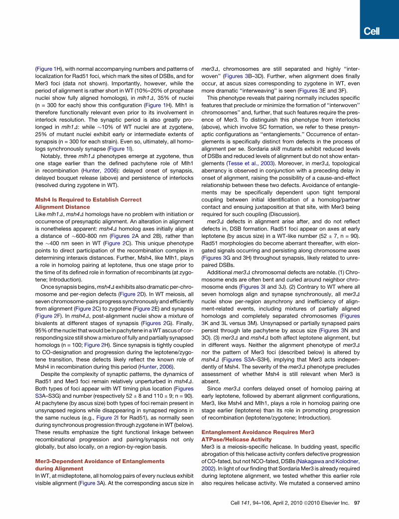
Figure 3. Prophase Phenotypes of mer3D and mer3KA Mutants
(A) In WT midleptotene, all homologs are aligned.
(B–D) Example of interwoven chromosomes of mer3D at midleptotene shown as primary data (B); cartoon (C); and southeast shadowing by Image J (D).
(E) Alignment (arrow) yields even more interweaving.
(F) Corresponding cartoon.
(G) Rad51 foci form elongated lines (arrowhead) from late leptotene through zygotene (by ascus size).
(H) Corresponding DAPI.
(I) Telomere regions are often bent and curled around neighbor chromosomes (arrow).
(J) Drawing of three pairs sorted by length.
(K–M) (K and L) Alignment (arrows) and close pairing (arrowheads) are both highly discoordinated in mer3D versus WT (M) where all homologs align synchronously
(arrow points to one pair).
(N and O) Mixture of synapsing and still unsynapsed (arrows) homologs seen in all late pachytenes of mer3D.
(P–S) mer3KA. (P and Q) Late leptotene chromosomes are highly interwoven and entanglements persist at zygotene (R) and pachytene (S) where all nuclei contain
a mixture of paired (arrowheads) and widely aligned (arrows) homologs. The scale bar represents 2 mm.
See Figures S1C and S1D for bouquet formation and Figure S2D for chiasmata in mer3D.
aligned (Figure 3S). Thus, abrogation of helicase activity is suffi-
cient to generate the mer3D alignment and synapsis defects.
This result shows that the biochemical activity of Mer3 plays
a role in homolog alignment long before it is required to ensure
that CO-designated interactions proceed efficiently.
However, Sordaria mer3KA exhibits more normal 8-spored
asci (5%, 2 to 8 in each perithecium thus among 150 asci) than
mer3D alone (<0.1%; one in 4 perithecia). A similar difference
is reported for yeast (Nakagawa and Kolodner, 2002). Thus,
Mer3 likely has additional roles not strictly dependent on heli-
case activity.
Mer3 Foci Are Evenly Spaced and Occur Transientlyin Matched Pairs along Aligned Homologous AxesMer3 appears in foci at early leptotene (Figure 4A), before chro-
mosome alignment (Figure 4B). Costaining with Rad51-RFP
(Figure 4C) shows that the two proteins appear at the same
time and mostly colocalize (80%–90%, n = 100; Figure 4D).
Cell 141, 94–106, April 2, 2010 ª2010 Elsevier Inc. 99

Figure 4. Mer3 and Mer3KA Localization
(A–D) Colocalization of Mer3-GFP (A) and Rad51-RFP (C) foci at early leptotene with corresponding DAPI (B); (D) Merge of (A) and (C).
(E) Mer3 foci occur at matching sites (arrows) in the two aligned pairs of homologs seen in this optical section. (F) Corresponding pairs (arrows) stained by DAPI.
(G) Double Mer3 foci (arrow) among single foci; arrowhead points to the corresponding ‘‘empty’’ matching site; (H) Cartoon of foci (in red).
(I and J) Another example of Mer3 foci at matching sites with cartoon.
(K–M) Three examples of even-spaced Mer3 foci along leptotene chromosomes. At early leptotene (K and L) all foci are uniformly bright; from midleptotene on (M)
some foci (arrows) are less bright than others.
(N) At early zygotene, costaining with Spo76-GFP shows Mer3 foci (arrows) localized between axes in synapsed regions.
(O) Even-spaced Mer3KA foci at mid leptotene.
(P) mer3KA foci occur at matching sites (arrow) in aligned pair of homologs; (Q) corresponding DAPI. The scale bar represents 2 mm.
See Figures S3A–3G for patterns of Mer3 foci in msh4D.
100 Cell 141, 94–106, April 2, 2010 ª2010 Elsevier Inc.

Thus, Mer3, like Rad51, localizes to sites of axis-associated
DSBs. Further, Mer3 foci appear at the time corresponding to
the alignment defect seen in mer3D, and thus well before the
Mer3 biochemically defined role in progression of CO interac-
tions at zygotene (above).
By the time homolog axes are aligned at �200 nm, foci often
appear to occur at matching sites (Figures 4E and 4F). We
confirmed this visual impression (Figures 4G–4J) statistically.
We compared the differences in the measured positions of
foci along the two axes of 61 pairs of aligned homologs with
those predicted by the null hypothesis H0 that foci are posi-
tioned independently along the two axes. In all pairs, foci
exhibit variable distances on the two homologs and distances
between two foci fluctuate from 0.45 to 0.78 mm. Our 107
data sets under H0 were obtained by taking the individual focus
patterns along the (61 3 2) homolog axes of the experimental
data set, and shuffling and repairing them as new randomly
chosen partners. Experimental and shuffled data sets were
then compared for the extent to which focus positions along
partner pairs were (or were not) similar (Experimental Proce-
dures). The probability of H0 (random occurrence) is extremely
low (p value of 0.0026), thus excluding the possibility that Mer3
foci are positioned independently along the two axes of
a homolog pair. Since Mer3 is thought to interact specifically
with DNA structures at DSB ends (Hunter, 2006), the occur-
rence of matched pairs of Mer3 foci could imply that the two
ends of a DSB are associated with the two-homolog axes
(Discussion).
In further support of this interpretation, at leptotene, the total
number of Mer3 foci is double that of Rad51 foci (136 ± 10 versus
58 ± 6, n = 200 leptotene nuclei, respectively). Further, the
number of Mer3-focus pairs (half of total number, thus 68) and
of Rad51 foci (58 ± 6) are three times higher than the number
of chiasmata in Sordaria (21 ± 3) and thus correspond to the
estimated number of total interactions (Zickler et al., 1992). In
some cases (8%–10%, n = 50) two Mer3 signals are located
one on top of the other along a single axis, with each focus
less bright than single foci (arrow in Figures 4G and 4H). Double
foci never have a matching partner on the homolog (arrowhead
in Figure 4G) and may thus reflect cases in which both DSB
ends remain associated with the ‘‘donor’’ homolog and eventu-
ally undergo intersister recombination.
Mer3 foci exhibit another striking feature: they are evenly
spaced along the chromosomes at early leptotene, thus before
coalignment. The clear visual impression to this effect (Figures
4K–4M) is confirmed quantitatively by measurements of interfo-
cus distances (Experimental Procedures). Among 360 interfocus
distances (measured along 90 five-foci chromosome segments),
minimum and maximum distances vary between 0.54 and
0.7 mm with a mean of 0.62 ± 0.03 mm. The statistics of interval
lengths between adjacent foci were fit to a gamma distribution
(Experimental Procedures), leading to n > 200, where n is the
standard ‘‘shape’’ parameter for this distribution. This high value
indicates even spacing and reflects the very small relative
fluctuations in the interval lengths (reducing the standard devia-
tion by a factor of two corresponds to an increase of n by a factor
of four). Since Mer3 foci mark the sites of total recombinational
interactions (above), these findings further suggest that even
spacing arises irrespective of, and likely prior to, CO/NCO differ-
entiation and CO interference.
Mer3 focus patterns further evolve from late leptotene through
early zygotene. First, a subset of foci remains bright, while the
other foci ‘‘fade’’ at the onset of synapsis (Figures 4M). Then,
in early-synapsed regions, Mer3 foci are delocalized from the
axes toward the space between the homologs, at which point
they are single, not double (Figure 4N). As synapsis progresses,
foci disappear from longer synapsed regions and are finally
completely absent by midpachytene.
The number (122 ± 15, n = 50) and patterns of C-terminally
GFP-tagged-Mer3KA protein are indistinguishable from those
of Mer3-GFP in both WT and mer3D backgrounds (compare
Figure 4O with Figure 4K and Figures 4P and 4Q with 4E
and 4F). The only discernible difference between Mer3KA-GFP
and Mer3-GFP foci is a reduction in brightness at all stages
(compare Figure 4P with 4E), perhaps reflecting a role for
helicase activity in promoting fully normal loading. Thus, Mer3
helicase activity is not required for loading of Mer3 onto chromo-
some axes. Furthermore, the presence of normal numbers of
foci at all stages implies that Mer3 helicase activity is not
required for establishment of total recombinational interactions
at the DNA level, but only for (ensuing) timely and topologically
regular axis juxtaposition during alignment.
Msh4 Foci Appear after Alignment and Are EvenlySpaced along Synapsed HomologsMsh4 foci emerge later than Mer3 foci, first appearing at the tran-
sition from alignment to synapsis both on aligned axes (arrows
in Figure 5A) and in early-synapsed regions (arrowheads in
Figure 5A). They are always single. Further, open regions at sites
of interlocks (above) are devoid of foci, although foci do occur on
the flanking synapsed regions (arrows in Figure 5B). Thus, Msh4
foci and synapsis are tightly correlated, locally, on a per-region
basis. Moreover, appearance of Msh4 foci is closely correlated
temporally with the leptotene/zygotene transition and spatially
with the presynaptic transition from 200 nm to 100 nm axis
distance. These features correspond temporally to a role of
Msh4 in recombination at leptotene/zygotene (Introduction).
However, foci appear much later than the time at which we first
detect msh4D defects (above), implying that important roles are
executed by undetectable quantities of protein during alignment.
Msh4 foci are dramatically evenly spaced along synapsed
chromosomes at early pachytene (Figures 5C–5G). Correspond-
ingly, for 280 interfocus distances measured along 70 chromo-
some segments (e.g., Figures 5D and 5E), minimum and
maximum distances vary between 0.56 and 0.69 mm with a
mean of 0.61 ± 0.03 mm and Gamma distribution analysis (Exper-
imental Procedures) leads to n > 200. Even spacing is already
obvious on longer synapsed regions at late zygotene (Fig-
ure 5A), remains through early pachytene (Figures 5C–5G) and
is still seen at midpachytene when foci ‘‘fade’’ before disappear-
ing (Figure 5H).
Msh4 foci are maximal in number at late zygotene and early
pachytene (81 ± 8; n = 200), diminish in number through
midpachytene (52 ± 8; n = 100) and are essentially absent from
midpachytene on. Further, the maximal number corresponds
to 3-4 times the number of chiasmata in Sordaria (21 ± 3) and
Cell 141, 94–106, April 2, 2010 ª2010 Elsevier Inc. 101
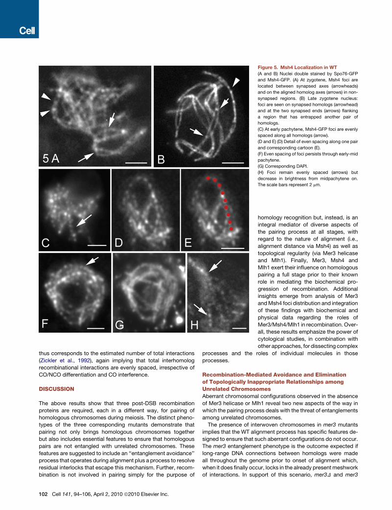
Figure 5. Msh4 Localization in WT
(A and B) Nuclei double stained by Spo76-GFP
and Msh4-GFP. (A) At zygotene, Msh4 foci are
located between synapsed axes (arrowheads)
and on the aligned homolog axes (arrows) in non-
synapsed regions. (B) Late zygotene nucleus:
foci are seen on synapsed homologs (arrowhead)
and at the two synapsed ends (arrows) flanking
a region that has entrapped another pair of
homologs.
(C) At early pachytene, Msh4-GFP foci are evenly
spaced along all homologs (arrow).
(D and E) (D) Detail of even spacing along one pair
and corresponding cartoon (E).
(F) Even spacing of foci persists through early-mid
pachytene.
(G) Corresponding DAPI.
(H) Foci remain evenly spaced (arrows) but
decrease in brightness from midpachytene on.
The scale bars represent 2 mm.
thus corresponds to the estimated number of total interactions
(Zickler et al., 1992), again implying that total interhomolog
recombinational interactions are evenly spaced, irrespective of
CO/NCO differentiation and CO interference.
DISCUSSION
The above results show that three post-DSB recombination
proteins are required, each in a different way, for pairing of
homologous chromosomes during meiosis. The distinct pheno-
types of the three corresponding mutants demonstrate that
pairing not only brings homologous chromosomes together
but also includes essential features to ensure that homologous
pairs are not entangled with unrelated chromosomes. These
features are suggested to include an ‘‘entanglement avoidance’’
process that operates during alignment plus a process to resolve
residual interlocks that escape this mechanism. Further, recom-
bination is not involved in pairing simply for the purpose of
102 Cell 141, 94–106, April 2, 2010 ª2010 Elsevier Inc.
homology recognition but, instead, is an
integral mediator of diverse aspects of
the pairing process at all stages, with
regard to the nature of alignment (i.e.,
alignment distance via Msh4) as well as
topological regularity (via Mer3 helicase
and Mlh1). Finally, Mer3, Msh4 and
Mlh1 exert their influence on homologous
pairing a full stage prior to their known
role in mediating the biochemical pro-
gression of recombination. Additional
insights emerge from analysis of Mer3
and Msh4 foci distribution and integration
of these findings with biochemical and
physical data regarding the roles of
Mer3/Msh4/Mlh1 in recombination. Over-
all, these results emphasize the power of
cytological studies, in combination with
other approaches, for dissecting complex
processes and the roles of individual molecules in those
processes.
Recombination-Mediated Avoidance and Eliminationof Topologically Inappropriate Relationships amongUnrelated ChromosomesAberrant chromosomal configurations observed in the absence
of Mer3 helicase or Mlh1 reveal two new aspects of the way in
which the pairing process deals with the threat of entanglements
among unrelated chromosomes.
The presence of interwoven chromosomes in mer3 mutants
implies that the WT alignment process has specific features de-
signed to ensure that such aberrant configurations do not occur.
The mer3 entanglement phenotype is the outcome expected if
long-range DNA connections between homologs were made
all throughout the genome prior to onset of alignment which,
when it does finally occur, locks in the already present meshwork
of interactions. In support of this scenario, mer3D and mer3

Figure 6. Interlock Avoidance and Recom-
bination-Mediated Axis Alignment
(A) In WT, formation of a connection between
homologs at the DNA level (top; black line, left) is
rapidly followed by local axis alignment at the
corresponding position. This accelerates estab-
lishment of connection/alignment nearby (black
bars), resulting in propagation of alignment
along each chromosome. In mer3D (bottom):
connections are made throughout the chromo-
somes (black lines) prior to alignment which,
when it does occur, yields highly interwoven
chromosomes.
(B) Top: DNA structures are inferred (leptotene)
and demonstrated (zygotene) respectively (Hunter
and Kleckner, 2001). Bottom: proposed four-
layered dispositions of post-DSB proteins on
leptotene and zygotene DNA structures.
(C) Possible architecture of recombinosome-
mediated homolog pairing. Top: (I) ‘‘First’’ DSB
end (red arrowhead) interacts with an homologous
chromatin loop, thereby nucleating assembly of
a complex containing the four post-DSB recombi-
nation proteins as in (B); ‘‘second’’ DSB end (red
arrowhead) associates with the axis of the DSB
‘‘donor’’ chromosome. (II) The complex formed
with the partner chromosome in (I) becomes
axis-associated, thereby bringing donor and
recipient chromosomes into closer proximity, with asymmetric evolution of the recipient chromosome complex. (III) Alignment juxtaposes axes to �400 nm.
(Middle, Bottom) Corresponding stages (I-III) during Allium alignment as defined by Albini and Jones (1987). At stage IV, recombination ‘‘nodule’’ mediates closer
axis juxtaposition for nucleation of SC, analogous to Sordaria progression (above).
helicase mutants exhibit dramatically delayed onset of align-
ment. By implication, in WT meiosis, regular pairing of homologs
requires tight temporal coupling between the formation of
a nascent DSB/partner contact and onset of homolog alignment
at the corresponding position. Such coupling would favor
neighboring interactions and lead to a per-chromosome pairing
process. Homolog pairs would be drawn one-by-one out of
the ‘‘pairing pool,’’ promoting topologically regular arrange-
ments as well as facilitating pairing of remaining chromosomes
(Figure 6A). Absence of Mer3 helicase activity (e.g., at DSB/
partner ends; below) could abrogate this process by triggering
a global regulatory surveillance response that delays onset of
alignment.
Any ‘‘leftover’’ problems that escape the entanglement avoid-
ance process are efficiently resolved concomitant with synapsis.
Previous considerations of interlock resolution focused on the
need to rectify basic topological whole-chromosome relation-
ships. One model invokes movement of the interlocked chro-
mosome out through the ends of the constraining bivalent
(Kleckner and Weiner, 1993; Wang, C.R. et al., 2009) via move-
ments known to occur at the relevant stages (Scherthan et al.,
2007; Koszul et al., 2008). Another model invokes DNA topoiso-
merase II-mediated passage of the trapped chromosomes
through the encircling one (von Wettstein et al., 1984).
The finding that Mlh1 is required for interlock resolution brings
into focus a completely new aspect. Since Mlh1 is a central
player in a set of interrelated DNA transactions (Hunter, 2006),
its involvement in interlock resolution suggests that this process
requires elimination of constraining DNA connections formed by
the recombination process. A classical interlock could arise if
recombinational interactions in synapsed regions flanking the
entrapped chromosome prevented its movement out the ends
of the encircling bivalent. Further, the ‘‘leftover’’ unsynapsed
regions in otherwise completely synapsed homologs, also seen
at high frequencies in mlh1D (above), might reflect ‘‘DNA inter-
locks’’ in which a stalled recombinational interaction within a
previously interlocked region precludes synapsis.
How might the recombination complex sense the presence of
a constraining DNA connection to trigger Mlh1-mediated dissolu-
tion? The complex might directly sense mechanical tension,
resulting from externally directed movements or ongoing
synapsis, with Mlh1 required to sense tension and/or implement
a biochemical response. Also, constraining connections might
be precluded from normal axis interactions, thus triggering a
regulatory checkpoint response that initiates their disassembly.
Communication among Developing InterhomologInteractionsThroughout the entire alignment process, DSBs are interacting
with homologous partner duplexes, via recombination com-
plexes that are associated with homolog axes (Introduction).
Even spacing of Mer3 and Msh4 foci (above) suggests that these
recombinational interactions are also evenly spaced. This regu-
larity, in turn, implies communication along chromosomes during
the alignment period, independent of and prior to the communi-
cation involved in classical CO interference (e.g., de Boer et al.,
2006). Moreover, even spacing does not seem compatible with
communication occurring independently along each of the two
Cell 141, 94–106, April 2, 2010 ª2010 Elsevier Inc. 103
homologs (e.g., during DSB formation), thus suggesting that
interference occurs at the level of the interhomolog interaction
per se. That is: formation of an interhomolog contact at one posi-
tion disfavors formation of a contact nearby. The existence of
such a process could explain interference of MSH4 and RPA
foci in mouse and human (de Boer et al., 2006; Oliver-Bonet
et al., 2007) and evidence that COs interfere not only with each
other but also with adjacent NCOs in budding yeast (Mancera
et al., 2008). Interference at the DSB level has been seen in
budding yeast (Wu and Lichten, 1995; Robine et al., 2007).
This could be a separate effect; alternatively an interhomolog
interaction at one position might preclude subsequent DSB
formation nearby. The existence of per-chromosome interfer-
ence also fits with, and could potentiate, per-chromosome prop-
agation of pairing (above).
Ends-Apart Association of Recombination Complexeswith Partner AxesColocalization of Mer3 foci with Rad51 foci, plus known
biochemical activities of Mer3 (below), suggest that Mer3 foci
mark the positions of DSB ends. Our finding that Mer3 foci occur
in matching pairs on aligned axes thus suggests that one DSB
end is associated with each of the two-homolog axes. This
‘‘ends-apart’’ configuration would correspond to the proposal
(Hunter, 2006; Oh et al., 2007) that one DSB end becomes asso-
ciated with a homolog chromatid while the other DSB end
remains associated with its sister. Ends-apart axis association
is also supported by observation of matching pairs of axis-asso-
ciated Rad51 and RPA foci (Franklin et al., 1999; Oliver-Bonet
et al., 2007). Early Mer3/Rad51 foci would correspond to forma-
tion of nascent DSB/partner interactions on homolog partner
axes. Matching Mer3 foci at mid- to late-leptotene would result
from ensuing development of similar complexes between the
second DSB end and its sister chromatid. A further implication
of the ‘‘ends-apart’’ scenario is that developing recombinosome
ensembles would be asymmetric, with one DSB end forming a
recombinosome/axis complex de novo with the homolog partner
while the other DSB end remains on its original axis in a complex
presumably evolved from the initiating DSB recombinosome.
Recombination Proteins Could Mediate HomologAlignment and Interlock Resolution via Modulationof Nascent DSB/Partner ContactsIdentification of central roles for Mer3 and Msh4 in early homology
pairing and for Mlh1 in interlock resolution raises two further
questions: how could the known biochemical activities of these
molecules be involved in the observed effects; and why do all
three molecules have roles for pairing that occur one stage prior
to their roles in recombination? Possible answers emerge by
considering the fact that throughout the alignment process, DSB/
partner interactions involve nascent RecA homolog (Rad51)-
promoted DSB/partner contacts (e.g., ‘‘nascent D-loops’’; Intro-
duction) which are then extended to stable strand invasion
intermediates during zygotene as the first molecular landmark
of CO/NCO differentiation (Figure 6B top; Hunter, 2006).
Mer3 and Msh4/5 are required for efficient progression of
CO recombination. In budding yeast, they mediate together the
progression of nascent DSB/partner interactions through exten-
104 Cell 141, 94–106, April 2, 2010 ª2010 Elsevier Inc.
sion of strand invasion during zygotene (e.g., Borner et al., 2004).
The mer3/msh4 recombination defects observed above suggest
that the same is likely true in Sordaria. Our analysis reveals that
Mer3 and Msh4 are required for homolog pairing during lepto-
tene, prior to their ‘‘zygotene role’’ in recombination. Since
alignment initiates with formation of a nascent strand exchange
interaction, Mer3/Msh4 could bind to these interactions as they
arise, mediate alignment during leptotene and then, already in
place, shepherd these intermediates through extension of strand
exchange during zygotene. Consistent with this scenario, Mer3
helicase binds at the 30 single stranded tails of branched struc-
tures and blocks ‘‘backwards’’ migration of the branch (Mazina
et al., 2004; Hunter, 2006) while Msh4/5 encircles and stabilizes
strand exchange junctions (Snowden et al., 2004). Both activities
could imply binding to, and conformational modulation of,
nascent DSB/partner contacts. Indeed, given these activities,
as well as those of other molecules implicated as components
of leptotene interaxis ‘‘bridges’’ (Rad51-mediated strand inva-
sion and RPA binding to single-stranded DNA; Oliver-Bonet
et al., 2007; Hunter, 2006), a specific four-layered architecture
for nascent DSB/partner complexes emerges (Figure 6B bottom).
By this scenario, Mer3 and Msh4/5 would not directly establish
nascent DSB/partner contacts, which are presumably formed by
a RecA homolog (e.g., Rad51) but would place the resulting
complexes in an appropriate conformation suitable for other
events. Such a role is supported by two facts: (i) neither Mer3
nor Msh4 is required for establishment of total DSB/partner inter-
actions for recombination (Borner et al., 2004; Nakagawa and
Kolodner, 2002 and references therein); and (ii) Mer3 and
Mer3KA foci appear prior to alignment and occur along axes in
arrays that correspond in number to total recombinational inter-
actions (above).
Recombinosome Architecture acrossthe Interaxis SpaceAbsence of Msh4 results in alteration of the alignment distance
(above). Thus, Msh4 is clearly present and relevant at this stage,
even though corresponding foci do not appear until later. This
finding is the first identification of a molecule involved in specifi-
cation of interaxis distance and thus raises the general question
of how the ‘‘architecture’’ of DSB-mediated interaxis linkages
might be determined. Intriguingly, RPA (and thus single-stranded
(ss) DNA) can be seen spanning interaxis regions during align-
ment in human (Oliver-Bonet et al., 2007), raising the possibility
that the extent of RPA-bound ssDNA determines bridge length.
Since Msh4 is thought to bind the strand exchange junction
that lies immediately adjacent to single-stranded DNA (above),
Msh4 might concomitantly specify alignment distance by influ-
encing the amount of ssDNA available for RPA binding.
Figure 6C presents a speculative but synthetic description of
DSB-mediated alignment that incorporates existing information
(Figure 6C, top I-III). While evolved from the present findings,
this description turns out to exactly match the progression seen
by EM analysis in Allium (Albini and Jones, 1987; Figure 6C,
bottom, I–III).
After alignment, the leptotene/zygotene transition involves
closer juxtaposition of homolog axes plus transits of recombino-
some foci from on-axis to between-axis localization: Rad51 foci

move from axes to interaxis regions (Tesse et al., 2003); Mer3
foci progress from matched pairs on axes to single foci located
between axes; and Msh4 foci appear transiently on aligned
axes before moving to interaxis localization on synapsed regions
(above). This progression, also mirrored in the Allium data
(Figure 6C bottom, IV), implies a major reorganization of recom-
bination complexes in preparation for (or concomitant with)
CO/NCO differentiation.
New Roles for Mlh1Mlh1 appears to be required for resolution of DNA recombination
intermediates that are constraining interlock resolution (above).
In WT meiosis, Mlh1 is thought to be required for resolution of
double Holiday junction (dHJ) intermediates into CO products
at pachytene (Hunter, 2006). Based on analogies with bacterial
MutL and MutS complexes, Mlh1 is proposed to mediate
removal of Msh4/5 from dHJ intermediates (Snowden et al.,
2004). The same activity might resolve interlocks by disassem-
bling nascent DSB/partner interactions in response to entangle-
ment-mediated stalling. Indeed, Mlh1 is already known to
mediate the redirection of meiotic recombination intermediates
that contain basepair mismatches, which likely occurs at the
same stage (Hunter and Kleckner, 2001; Argueso et al., 2003).
Moreover, mlh1D exhibits a delay in onset of synapsis (above),
and thus presumably in progression from nascent DSB/partner
complexes to strand invasion extension. Therefore Mlh1 is not
recruited de novo to the interlock (or heteroduplex mismatch)
sites but is present and ‘‘relevant’’ to nascent DSB/partner
contacts even in the absence of any irregularity. In summary:
Mlh1, like Mer3 and Msh4/5, is present at the sites of nascent
DSB/partner complexes, serving important auxiliary roles for
interhomolog interactions, well before it is actually required for
efficient/effective progression of recombination per se.
EXPERIMENTAL PROCEDURES
Cloning, Plasmids, and Transformation of Sordaria
Sordaria MER3, MSH4, and MLH1 genes were identified by PCR from an
indexed genomic library based on Neurospora crassa sequences homology
(GenBank accession numbers FJ528581, FJ528583, and FJ528582.1). Their
sequences predict the conserved domains found in the respective genes of
other eukaryotic species. MER3 encodes a predicted protein of 1571 amino
acids with 85% identity with Neurospora crassa (hypothetical NCU09793.3),
34% with budding yeast and about 35% with human, mouse and Arabidopsis
thaliana. MSH4 encodes a predicted protein of 1284 amino acids with 61%
identity with N. crassa (MutS ortholog 4), and about 27% with budding yeast,
human, mouse and A. thaliana. MLH1 encodes a predicted protein of 748
amino acids, 93% identity with N. crassa (MutL ortholog-1), 42% with human,
mouse, and budding yeast, and 38% with A. thaliana.
Deleted Sordaria strains were obtained by single step gene replacement. A
hygromycin resistance cassette replaces the entire ORF in all deletion
mutants. Transformants carrying a deleted allele were selected for hygromycin
resistance and confirmed by Southern blotting and PCR. The mer3KA muta-
tion (K297A) was created by PCR-based mutagenesis and introduced in
Sordaria by cotransformation with a plasmid encoding the nourseothricine
resistance cassette (see details in Supplemental Information).
In all analyzed GFP fusions (RAD51, MER3, mer3KA and MSH4) the GFP
coding sequence (p-EGFP-1, Clontech) was fused just after the last C-terminal
amino acid predicted from the ORF. C-terminally GFP-tagged versions of the
genes, under control of their promoters, were introduced in Sordaria at ectopic
locations into a WT strain. Msh4-GFP and Mer3-GFP proteins are fully func-
tional: they complement all meiotic and sporulation defects of their cognate
null mutants. Further details are provided in Supplemental Information.
Cytology
For cytological analysis, GFP and DAPI (0.5mg/ml) signals were observed,
either on living material or after fixation in 4% paraformaldehyde, with a Zeiss
Axioplan microscope with a CCD Princeton camera as described (Storlazzi
et al., 2003).
Interfocus distances were measured on straight axis segments with 5–12
foci, from the center of the first focus to the center of the next focus (using
public domain software ImageJ: http://rsb.info.nih.gov/ij). The n parameter
of the Gamma distribution fitted against interfocus-distances was computed
using maximum likelihood fitting with the Wessa Gamma Distribution calcu-
lator: http://www.wessa.net/rwasp_fitdistrgamma.wasp/.
Details of the procedures used for measurements of interfocus distances,
Gamma distribution and null hypothesis H0 whereby foci positions on two
chromosomes are not more correlated on homologs than on nonhomologs
are given in Supplemental Information.
SUPPLEMENTAL INFORMATION
Supplemental Information includes Extended Experimental Procedures and
three figures and can be found with this article online at doi:10.1016/j.cell.
2010.02.041.
ACKNOWLEDGMENTS
We thank Stefanie Poggeler for the recombinant ku70 plasmid and Olivier
Martin for his advice in the statistical analyses. D.Z., A.S. and G.R.-R. were
supported by grants from the Centre National de la Recherche Scientifique
(UMR 8621), and from the National Institutes of Health (GM025326 and
GM044794) to N.K. S.G. was supported by Regione campania Legge 28/03/
2002 n.5, and together with A.S. by CNR.
Received: March 31, 2009
Revised: April 28, 2009
Accepted: February 11, 2010
Published: April 1, 2010
REFERENCES
Albini, S.M., and Jones, G.H. (1987). Synaptonemal complex spreading in
Allium cepa and Allium fistulosum I. The initiation and sequence of pairing.
Chromosoma 95, 324–338.
Anderson, L.K., and Stack, S.M. (2005). Recombination nodules in plants.
Cytogenet. Genome Res. 109, 198–204.
Argueso, J.L., Kijas, A.W., Sarin, S., Heck, J., Waase, M., and Alani, E. (2003).
Systematic mutagenesis of the Saccharomyces cerevisiae MLH1 gene reveals
distinct roles for Mlh1p in meiotic crossing over and in vegetative and meiotic
mismatch repair. Mol. Cell. Biol. 23, 873–886.
Blat, Y., Protacio, R.U., Hunter, N., and Kleckner, N. (2002). Physical and func-
tional interactions among basic chromosome organizational features govern
early steps of meiotic chiasma formation. Cell 111, 791–802.
Borner, G.V., Kleckner, N., and Hunter, N. (2004). Crossover/noncrossover
differentiation, synaptonemal complex formation, and regulatory surveillance
at the leptotene/zygotene transition of meiosis. Cell 117, 29–45.
de Boer, E., Stam, P., Dietrich, A.J., Pastink, A., and Heyting, C. (2006). Two
levels of interference in mouse meiotic recombination. Proc. Natl. Acad. Sci.
USA 103, 9607–9612.
Franklin, A.E., McElver, J., Sunjevaric, I., Rothstein, R., Bowen, B., and Cande,
W.Z. (1999). Three-dimensional microscopy of the Rad51 recombination
protein during meiotic prophase. Plant Cell 11, 809–824.
Franklin, F.C., Higgins, J.D., Sanchez-Moran, E., Armstrong, S.J., Osman,
K.E., Jackson, N., and Jones, G.H. (2006). Control of meiotic recombination
Cell 141, 94–106, April 2, 2010 ª2010 Elsevier Inc. 105
in Arabidopsis: role of the MutL and MutS homologues. Biochem. Soc. Trans.
34, 542–544.
Guillon, H., Baudat, F., Grey, C., Liskay, R.M., and de Massy, B. (2005). Cross-
over and Noncrossover pathways in mouse meiosis. Mol. Cell 20, 563–573.
Henderson, K.A., and Keeney, S. (2005). Synaptonemal complex formation:
where does it start? Bioessays 27, 995–998.
Higgins, J.D., Armstrong, S.J., Franklin, F.C., and Jones, G.H. (2004). The
Arabidopsis MutS homolog AtMSH4 functions at an early step in recombina-
tion: evidence for two classes of recombination in Arabidopsis. Genes Dev.
18, 2557–2570.
Hunter, N. (2006). Meiotic recombination. In Molecular Genetics of Recombi-
nation, A. Aguilera and R. Rothstein, eds. (Heidelberg: Springer Berlin),
pp. 381–442.
Hunter, N., and Kleckner, N. (2001). The single-end invasion: an asymmetric
intermediate at the double-strand break to double-holliday junction transition
of meiotic recombination. Cell 106, 59–70.
Jackson, N., Sanchez-Moran, E., Buckling, E., Armstrong, S.J., Jones, G.H.,
and Franklin, F.C. (2006). Reduced meiotic crossovers and delayed prophase
I progression in AtMLH3-deficient Arabidopsis. EMBO J. 25, 1315–1323.
Kleckner, N., and Weiner, B.M. (1993). Potential advantages of unstable inter-
actions for pairing of chromosomes in meiotic, somatic, and premeiotic cells.
Cold Spring Harb. Symp. Quant. Biol. 58, 553–565.
Koszul, R., Kim, K.P., Prentiss, M., Kleckner, N., and Kameoka, S. (2008).
Meiotic chromosomes move by linkage to dynamic actin cables with transduc-
tion of force through the nuclear envelope. Cell 133, 1188–1201.
Mancera, E., Bourgon, R., Brozzi, A., Huber, W., and Steinmetz, L.M. (2008).
High-resolution mapping of meiotic crossovers and non-crossovers in yeast.
Nature 454, 479–485.
Mazina, O.M., Mazin, A.V., Nakagawa, T., Kolodner, R.D., and Kowalczykow-
ski, S.C. (2004). Saccharomyces cerevisiae Mer3 helicase stimulates 30-50
heteroduplex extension by Rad51: implications for crossover control in meiotic
recombination. Cell 117, 47–56.
Moens, P.B., Marcon, E., Shore, J.S., Kochakpour, N., and Spyropoulos, B.
(2007). Initiation and resolution of interhomolog connections: crossover and
non-crossover sites along mouse synaptonemal complexes. J. Cell Sci. 120,
1017–1027.
Nakagawa, T., and Ogawa, H. (1999). The Saccharomyces cerevisiae MER3
gene, encoding a novel helicase-like protein, is required for crossover control
in meiosis. EMBO J. 18, 5714–5723.
Nakagawa, T., and Kolodner, R.D. (2002). Saccharomyces cerevisiae Mer3
is a DNA helicase involved in meiotic crossing over. Mol. Cell. Biol. 22,
3281–3291.
Oh, S.D., Lao, J.P., Hwang, P.Y., Taylor, A.F., Smith, G.R., and Hunter, N.
(2007). BLM ortholog, Sgs1, prevents aberrant crossing-over by suppressing
formation of multichromatid joint molecules. Cell 130, 259–272.
106 Cell 141, 94–106, April 2, 2010 ª2010 Elsevier Inc.
Oliver-Bonet, M., Campillo, M., Turek, P.J., Ko, E., and Martin, R.H. (2007).
Analysis of replication protein A (RPA) in human spermatogenesis. Mol.
Hum. Reprod. 13, 837–844.
Page, S.L., and Hawley, R.S. (2004). The genetics and molecular biology of the
synaptonemal complex. Annu. Rev. Cell Dev. Biol. 20, 525–558.
Robine, N., Uematsu, N., Amiot, F., Gidrol, X., Barillot, E., Nicolas, A., and
Borde, V. (2007). Genome-wide redistribution of meiotic double-strand breaks
in Saccharomyces cerevisiae. Mol. Cell. Biol. 27, 1868–1880.
Scherthan, H., Wang, H., Adelfalk, C., White, E.J., Cowan, C., Cande, W.Z.,
and Kaback, D.B. (2007). Chromosome mobility during meiotic prophase in
Saccharomyces cerevisiae. Proc. Natl. Acad. Sci. USA 104, 16934–16939.
Snowden, T., Acharya, S., Butz, C., Berardini, M., and Fishel, R. (2004).
hMSH4-hMSH5 recognizes Holliday junctions and forms a meiosis-specific
sliding clamp that embraces homologous chromosomes. Mol. Cell 15,
437–451.
Storlazzi, A., Tesse, S., Gargano, S., James, F., Kleckner, N., and Zickler, D.
(2003). Meiotic double strand breaks at the interface of chromosome move-
ment, chromosome remodeling, and reductional division. Genes Dev. 17,
2675–2687.
Storlazzi, A., Tesse, S., Ruprich-Robert, G., Gargano, S., Poggeler, S., Kleck-
ner, N., and Zickler, D. (2008). Coupling meiotic chromosome axis integrity to
recombination. Genes Dev. 22, 796–809.
Tesse, S., Storlazzi, A., Kleckner, N., Gargano, S., and Zickler, D. (2003). Local-
ization and roles of Ski8p protein in Sordaria meiosis and delineation of three
mechanistically distinct steps of meiotic homolog juxtaposition. Proc. Natl.
Acad. Sci. USA 100, 12865–12870.
van Heemst, D., James, F., Poggeler, S., Berteaux-Lecellier, V., and Zickler, D.
(1999). Spo76p is a conserved chromosome morphogenesis protein that links
the mitotic and meiotic programs. Cell 98, 261–271.
von Wettstein, D., Rasmussen, S.W., and Holm, P.B. (1984). The synaptone-
mal complex in genetic segregation. Annu. Rev. Genet. 18, 331–413.
Wang, K., Tang, D., Wang, M., Lu, J., Yu, H., Liu, J., Qian, B., Gong, Z., Wang,
X., Chen, J., et al. (2009). MER3 is required for normal meiotic crossover forma-
tion, but not for presynaptic alignment in rice. J. Cell Sci. 122, 2055–2063.
Wang, C.R., Carlton, P.M., Golubovskaya, I.N., and Cande, W.Z. (2009). Inter-
lock formation and coiling of meiotic chromosome axes during synapsis.
Genetics 183, 905–915.
Wu, T.C., and Lichten, M. (1995). Factors that affect the location and frequency
of meiosis-induced double-strand-breaks in Saccharomyces cerevisiae.
Genetics 140, 55–66.
Zickler, D., Moreau, P.J., Huynh, A.D., and Slezec, A.M. (1992). Correlation
between pairing initiation sites, recombination nodules and meiotic recombi-
nation in Sordaria macrospora. Genetics 132, 135–148.
Zickler, D., and Kleckner, N. (1999). Meiotic chromosomes: integrating struc-
ture and function. Annu. Rev. Genet. 33, 603–754.
Zickler, D. (2006). From early homologue recognition to synaptonemal
complex formation. Chromosoma 115, 158–174.