![Arabidopsis LORELEI, a Maternally Expressed Imprinted · Arabidopsis LORELEI, a Maternally Expressed Imprinted Gene, Promotes Early Seed Development1[OPEN] Yanbing Wang,2 Tatsuya](https://static.fdocuments.us/doc/165x107/5d4de7fe88c99347698b734e/arabidopsis-lorelei-a-maternally-expressed-arabidopsis-lorelei-a-maternally.jpg)
The role of SYN1 in early Arabidopsis meiosisetheses.bham.ac.uk/1341/1/Tiang11PhD.pdf · THE ROLE...
217
THE ROLE OF SYN1 IN EARLY ARABIDOPSIS MEIOSIS by CHOON LIN TIANG A thesis submitted to University of Birmingham For the degree of DOCTOR OF PHILOSOPHY School of Biosciences University of Birmingham September 2010
Transcript of The role of SYN1 in early Arabidopsis meiosisetheses.bham.ac.uk/1341/1/Tiang11PhD.pdf · THE ROLE...

THE ROLE OF SYN1 IN
EARLY ARABIDOPSIS MEIOSIS
by
CHOON LIN TIANG
A thesis submitted to
University of Birmingham
For the degree of
DOCTOR OF PHILOSOPHY
School of Biosciences
University of Birmingham
September 2010

University of Birmingham Research Archive
e-theses repository This unpublished thesis/dissertation is copyright of the author and/or third parties. The intellectual property rights of the author or third parties in respect of this work are as defined by The Copyright Designs and Patents Act 1988 or as modified by any successor legislation. Any use made of information contained in this thesis/dissertation must be in accordance with that legislation and must be properly acknowledged. Further distribution or reproduction in any format is prohibited without the permission of the copyright holder.

ABSTRACT
SYN1 is a meiosis-specific Arabidopsis homologue of yeast REC8. REC8 is an important
component of the meiotic cohesion complex which maintains cohesion between sister
chromatids. Cytological analysis of syn1-/-
has shown chromosome fragmentation at
metaphase I. To determine the basis of chromosome fragmentation in the syn1-/-
, three double
mutants were constructed. I have demonstrated that chromosome fragmentation in syn1 is
AtSPO11-1-dependent. Moreover, I have also shown that SYN1 has a role in DSB repair by
analysing Atdmc1-/-
/syn1-/-
meiocytes. To investigate this further, immunolocalization studies
in wild-type and syn1-/-
were conducted. Distribution of ASY1 and AtZYP1 was affected in
syn1-/-
. Both proteins appeared as aggregates, developing into an abnormal short linear signal
in early prophase I, suggesting that both axis formation and synapsis are compromised.
Distribution of the recombination proteins AtRAD51 and AtMLH1 was also aberrant.
Localization of SYN1 in wild-type nuclei revealed a continuous signal along the chromosome
axes. However, careful inspection revealed that this was accompanied by patches of more
intense signals, possibly corresponding to DSB regions. To investigate this further I analysed
SYN1 distribution in an Atspo11-1-4-/-
mutant. Whilst faint SYN1 signals were apparent along
the axis, no patches of intense signals were visible. Cisplatin-induced DSBs restored AtZYP1
foci in Atspo11-1-4-/-
and also resulted in restoration of intense patches of the SYN1 signals.
This is consistent with the recruitment of SYN1 to DSB sites.

ACKNOWLEDGEMENTS
I would like to thank my supervisors Sue Armstrong and Chris Franklin for providing me
with great support and guidance throughout the duration of this project. I would also like to
thank Eugenio Sanchez-Moran for helpful discussion and support over the years.
I would like to thank friends from Franklin and Franklin-Tong labs, especially Barend de
Graaf and Kim Osman for their help and guidance in molecular biology experiments. I would
like to thank Natalie Poulter for showing me the useful software Image J and Excel, and also I
would like to thank Sarah Smith for sharing her time and knowledge over the years. I thank
Steve Price and Ruth Perry for technical assistance in the lab and to Karen Staples for
assistance in the glass house.
Finally, I would like to thank my Mom, Dad and my fiancee Hoi Chin Hew for their love and
support.

TABLE OF CONTENTS
CHAPTER 1 ......................................................................................................................... 1
GENERAL INTRODUCTION ............................................................................................ 1
1.1 OVERVIEW OF MITOSIS AND MEIOSIS ............................................................................... 1
1.2 SISTER CHROMATID COHESION ....................................................................................... 5
1.2.1 Establishment of sister chromatid cohesion ............................................................ 5
1.2.1.1 Cohesins ............................................................................................................. 7
1.2.1.2 Loading of cohesin on sister chromatids ........................................................... 10
1.2.2 Localization of cohesin on the chromosome in meiosis ........................................ 12
1.2.3 Removal of cohesins ............................................................................................ 15
1.2.3.1 Cleavage of cohesin by separase ....................................................................... 15
1.2.3.2 Protection of centromeric cohesion ................................................................... 18
1.2.4 Arabidopsis cohesins: SYN1, AtSMC1/AtSMC3 and AtSCC3 ............................. 22
1.2.4.1 SYN1 ............................................................................................................... 22
1.2.4.2 AtSMC1 and AtSMC3 ...................................................................................... 23
1.2.4.3 AtSCC3 ............................................................................................................ 24
1.3 COHESINS ARE REQUIRED FOR DOUBLE-STRAND BREAK (DSB) REPAIR .......................... 25
1.4 HOMOLOGUE PAIRING AND SYNAPSIS............................................................................ 26
1.4.1 Homologous chromosome pairing ....................................................................... 26
1.4.2 Formation of the synaptonemal complex. ............................................................. 27
1.5 MEIOTIC RECOMBINATION............................................................................................ 31
1.5.1 Initial events of meiotic recombination: formation of DSB .................................. 31
1.5.2 Processing of meiotic DNA DSB ......................................................................... 33
1.5.3 Crossover and non-crossover pathways ................................................................ 36
1.6 AIMS OF MY PHD PROJECT ........................................................................................... 40
CHAPTER 2 ....................................................................................................................... 42
MATERIALS AND METHODS ........................................................................................ 42
2.1 PLANT MATERIALS AND GROWING CONDITIONS ............................................................. 42
2.1.1 Plant materials ..................................................................................................... 42
2.1.2 Plant growth media .............................................................................................. 42
2.1.3 Plant growth conditions ....................................................................................... 42
2.2 BACTERIAL STRAINS AND CLONING VECTORS ................................................................ 43

2.2.1 Bacterial strains ................................................................................................... 43
2.2.2 Cloning vectors.................................................................................................... 43
2.3 BACTERIA MEDIA, CULTURE AND GROWTH CONDITION .................................................. 44
2.3.1 Bacterial growth media ........................................................................................ 44
2.3.1.1 Lysogeny Broth (LB) ........................................................................................ 44
2.3.1.2 LB agar ............................................................................................................. 44
2.3.2 Bacterial culture .................................................................................................. 45
2.3.3 Bacterial growth condition ................................................................................... 45
2.4 ISOLATION OF NUCLEIC ACID FROM PLANT AND BACTERIA ............................................. 45
2.4.1Buffer for isolation of nucleic acid ........................................................................ 45
2.4.2 Isolation of total RNA from plants ....................................................................... 46
2.4.2.1 DNase I treatment of plant RNA ....................................................................... 46
2.4.3 Isolation of plasmid DNA from bacteria............................................................... 47
2.4.3.1 Boiling Prep method ......................................................................................... 47
2.4.3.2 Wizard Mini prep method (Promega®) ............................................................. 48
2.4.4 Isolation of DNA from plants for genotyping ....................................................... 48
2.5 POLYMERASE CHAIN REACTION (PCR) TECHNIQUES ..................................................... 48
2.5.1 PCR standard condition ....................................................................................... 49
2.5.2 Genotyping PCR.................................................................................................. 49
2.5.3 Single colony PCR .............................................................................................. 49
2.5.4 Reverse Transcriptase-Polymerase Chain Reaction (RT-PCR) .............................. 50
2.5.5 Primer design ...................................................................................................... 58
2.6 NUCLEIC ACID MANIPULATIONS .................................................................................... 59
2.6.1 Estimation of nucleic acid concentration .............................................................. 59
2.6.2 Digestion of DNA with restriction enzymes ......................................................... 59
2.6.3 Agarose gel electrophoresis of DNA and RNA ..................................................... 59
2.6.3.1 Solutions for nucleic acid electrophoresis ......................................................... 60
2.6.4 Extraction of DNA from agarose gels .................................................................. 60
2.6.5 Ligation of DNA fragments into vector DNA....................................................... 60
2.6.6 DNA sequencing .................................................................................................. 61
2.7 TRANSFORMATION OF BACTERIAL CELLS ...................................................................... 61
2.7.1 Transformation of E.coli by heat shock ................................................................ 61

2.8 PROTEIN MANIPULATIONS ............................................................................................ 62
2.8.1 Protein electrophoresis and western blotting solutions ......................................... 62
2.8.2 Protein test expression, extractions and preparations ............................................ 64
2.8.2.1 Protein test expression and extractions from bacteria ........................................ 64
2.8.2.2 Estimating protein concentration ....................................................................... 71
2.8.2.3 SDS-Polyacrylamide gel electrophoresis (SDS-PAGE) ..................................... 71
2.8.2.4 Coomassie staining of protein gels .................................................................... 72
2.9 WESTERN BLOTTING .................................................................................................... 73
2.9.1 Protein transfer .................................................................................................... 73
2.9.2 Antibody probing and protein detection ............................................................... 73
2.10 SLIDE PREPARATION OF MEIOTIC CHROMOSOMES FOR CYTOLOGY ................................ 74
2.10.1 Determination of meiotic stage..............................................................................74
2.10.2 Slide preparation for immunolocalization studies ............................................... 74
2.10.3 Antibody labelling for immunolocalization studies ............................................ 74
2.10.4 Meiotic time course ........................................................................................... 75
2.10.5 Slide preparation for cytogenetics studies .......................................................... 76
2.10.6 Fluorescent in situ hybridisation (FISH)............................................................. 77
2.11 SLIDE IMAGING .......................................................................................................... 78
2.12 CISPLATIN TREATMENT .............................................................................................. 78
2.12.1 Introduction of cisplatin into meiocytes of Atspo11-1-4-/-
................................... 78
2.12.2 Seedling growth on Cisplatin containing MS agar .............................................. 78
CHAPTER 3 ....................................................................................................................... 79
CHARACTERISING A SYN1 NULL MUTANT .............................................................. 79
3.1 INTRODUCTION ............................................................................................................ 79
3.2 RESULTS ..................................................................................................................... 80
3.2.1 Identification of SYN1 T-DNA insertion mutant ................................................... 80
3.2.2 Expression of SYN1 gene ..................................................................................... 84
3.2.3 Cytogenetic analysis of the syn1 mutant............................................................... 88
3.2.3.1 Male meiosis in wild-type and syn1 mutant ....................................................... 88
3.2.3.2 Fluorescence in situ hybridisation (FISH) analysis of chromosome segregation in
wild-type and syn1 male meiocytes .............................................................................. 93
3.2.4 Relationship between SYN1 and other Rec8 homologues. ................................... 97
3.3 DISCUSSION .............................................................................................................. 102
3.3.1 T-DNA insertion line (SALK_091193) is a syn1 null mutant .............................. 102
3.3.2 SYN1 expression is not specific to meiosis ......................................................... 102
3.3.3 Chromatin is disorganised during early meiosis I in syn1 mutants. ..................... 103
3.3.4 SYN1 is important for centromeric cohesion at the first meiotic division. .......... 104
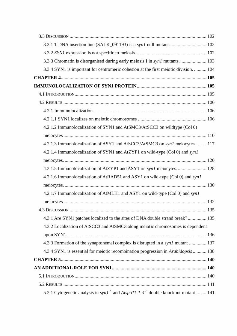
CHAPTER 4 ..................................................................................................................... 105
IMMUNOLOCALIZATION OF SYN1 PROTEIN ........................................................ 105
4.1 INTRODUCTION .......................................................................................................... 105
4.2 RESULTS ................................................................................................................... 106
4.2.1 Immunolocalization ........................................................................................... 106
4.2.1.1 SYN1 localizes on meiotic chromosomes ....................................................... 106
4.2.1.2 Immunolocalization of SYN1 and AtSMC3/AtSCC3 on wildtype (Col 0)
meiocytes ................................................................................................................... 110
4.2.1.3 Immunolocalization of ASY1 and AtSCC3/AtSMC3 on syn1 meiocytes ......... 117
4.2.1.4 Immunolocalization of SYN1 and AtZYP1 on wild-type (Col 0) and syn1
meiocytes. .................................................................................................................. 120
4.2.1.5 Immunolocalization of AtZYP1 and ASY1 on syn1 meiocytes. ....................... 128
4.2.1.6 Immunolocalization of AtRAD51 and ASY1 on wild-type (Col 0) and syn1
meiocytes. .................................................................................................................. 130
4.2.1.7 Immunolocalization of AtMLH1 and ASY1 on wild-type (Col 0) and syn1
meiocytes ................................................................................................................... 132
4.3 DISCUSSION .............................................................................................................. 135
4.3.1 Are SYN1 patches localized to the sites of DNA double strand break? ............... 135
4.3.2 Localization of AtSCC3 and AtSMC3 along meiotic chromosomes is dependent
upon SYN1. ............................................................................................................... 136
4.3.3 Formation of the synaptonemal complex is disrupted in a syn1 mutant .............. 137
4.3.4 SYN1 is essential for meiotic recombination progression in Arabidopsis ........... 138
CHAPTER 5 ..................................................................................................................... 140
AN ADDITIONAL ROLE FOR SYN1 ............................................................................ 140
5.1 INTRODUCTION .......................................................................................................... 140
5.2 RESULTS ................................................................................................................... 141
5.2.1 Cytogenetic analysis in syn1-/-
and Atspo11-1-4-/-
double knockout mutant ......... 141

5.2.2 Immunolocalization of SYN1 on Atspo11-1-4-/-
. ................................................ 146
5.2.3 Immunolocalization of SYN1 on cisplatin-treated Atspo11-1-4-/-
meiocytes ....... 150
5.2.4 Cytogenetic analysis in syn1-/-
and Atrad51c-/-
double knockout mutant ............. 155
5.2.5 Cytogenetic analysis in a syn1-/-
/Atdmc1-/-
double knockout mutant ................... 159
5.2.6 Seeds of wild-type and SYN1 heterozygous plants grew slowly on cisplatin MS
medium. ..................................................................................................................... 163
5.3 DISCUSSION .............................................................................................................. 168
5.3.1 SYN1 is essential for centromeric cohesion at first meiotic segregation ............. 168
5.3.2 Chromosome fragmentation in syn1 is AtSPO11-1-dependent ............................ 171
5.3.3 SYN1 loading is reduced in Atspo11-1-4-/-
meiocytes. ........................................ 171
5.3.4 Some SYN1 loading is dependent on DNA double-strand breaks ....................... 172
5.3.5 SYN1 has a role in DNA DSB repair. ................................................................ 173
5.3.6 SYN1 and AtRAD51c are essential for DNA double strand break repair. ........... 174
5.3.7 Seeds of wild-type and SYN1 heterozygous plants grow slowly in cisplatin MS
medium. ..................................................................................................................... 175
CHAPTER 6 ..................................................................................................................... 178
CONCLUSIONS .............................................................................................................. 178
6.1 INTRODUCTION .......................................................................................................... 178
6.2. IS SYN1 IMPORTANT IN SISTER CHROMATID COHESION? ............................................. 178
6.3 SYN1 IS ESSENTIAL FOR MEIOTIC RECOMBINATION PROGRESSION AND SC
POLYMERIZATION/ ELONGATION. ...................................................................................... 180
6.4 SYN1 PLAYS AN IMPORTANT ROLE DURING DNA DOUBLE STRAND BREAK (DSB) REPAIR
...................................................................................................................................... 181
6.5 FUTURE WORK ........................................................................................................... 185
CHAPTER 7 ..................................................................................................................... 187
REFERENCES ................................................................................................................ 187

LIST OF FIGURES
Figure 1.1 Meiosis and mitosis
2
Figure 1.2 Schematic diagram showing the stages in meiosis I
4
Figure 1.3 The structure of SMC proteins
8
Figure 1.4 A possible model of mitotic and meiotic cohesin complex
9
Figure 1.5 Another two possible models of cohesin complex
11
Figure 1.6 Diagram showing the yeast SCC1 and REC8 during meiosis
13
Figure 1.7 Shugoshin-PP2A protects centromeric cohesin from dissociation
during mitotic prophase
19
Figure 1.8 Shugoshin-PP2A protects centromeric cohesin from separase
cleavage until metaphase I
21
Figure 1.9 Synaptonemal complex (SC) structure
28
Figure 1.10 Early events of meiotic recombination
34
Figure 1.11 Meiotic crossover and non-crossover pathways following D-loop
formation
39
Figure 2.1 The sequence alignment of Arabidopsis SYN1, SYN2, SYN3 and
SYN4 proteins
54
Figure 2.2 Alignment of Arabidopsis SYN1 and DNA fragment
56
Figure 2.3 Nucleotide sequence of the SYN1 insert and pET21b vector
57
Figure 2.4 The translation of SYN1 amino acid sequence
57
Figure 2.5 SDS-PAGE analysis of protein test induction
65
Figure 2.6 Western Blot analysis of E.coli BL21(DE3)pLysS/pET21b-SYN1
(C1) and E.coli BL21(DE3)pLysS/pET21b (Control) in protein test
expression with and without IPTG
66
Figure 2.7 SDS-PAGE (A) and Western Blot (B) analysis of E.coli
BL21(DE3)/pET21b–SYN1 in protein test expression with and
without IPTG
69
Figure 2.8 28 days anti-serum was analysed by western blot
70

Figure 3.1 Nucleotide sequence of the plasmid DNA
81
Figure 3.2 Sequence of A. thaliana genomic DNA
82
Figure 3.3 Map of the SYN1 locus and exon organisation
83
Figure 3.4 SYN1 gene expression in wild-type (Col 0), syn1-/-
and Atspo11-1-4-/-
plants
86
Figure 3.5 Genevestigator expression profile of Arabidopsis SYN1 gene
87
Figure 3.6 Meiotic stages in pollen mother cells of wild-type (Col 0) of A.
thaliana
91
Figure 3.7 Meiotic stages in pollen mother cells of the SALK line 091193 (syn1
mutant) of A. thaliana
92
Figure 3.8 FISH of centromeric probe (pAL38) to pollen mother cells of wild-
type A. thaliana
95
Figure 3.9
FISH of centromeric probe (pAL38) to pollen mother cells of the
SALK line 091193 (syn1 mutant) of A. thaliana
96
Figure 3.10 Alignments of the full length amino acid sequence of the smREC8,
ceREC8 and SYN1
100
Figure 3.11 Alignment of the full length amino acid sequence of AFD1,
OsRAD21-4 and SYN1
101
Figure 4.1 Immunolocalization of SYN1 protein to nuclei of wild-type (Col 0)
108
Figure 4.2 Immunolocalization of ASY1 and SYN1 proteins to wild-type (Col 0)
meiocytes
108
Figure 4.3 Dual immunolocalization of ASY1 (green) and SYN1 (red) to
leptotene (A), zygotene (B), early pachytene (C) and late pachytene
of wild-type (Col 0)
109
Figure 4.4 Dual immunolocalization of SYN1 (green) and AtSMC3 (red) on
meiotic spreads of wild-type Arabidopsis thaliana
112
Figure 4.5 Immunolocalization of SYN1 (green) and AtSMC3 (red) on early
pachytene cell (A) of wild-type
113
Figure 4.6 Dual immunolocalization of SYN1 (green) and AtSCC3 (red) on
meiocytes of wild-type
114

Figure 4.7 Immunolocalization of SYN1 (green) and AtSCC3 (red) on zygotene
cell (A) of wild-type
115
Figure 4.8 Immunolocalization of SYN1 (green) to early pachytene (A) of wild-
type
116
Figure 4.9 Immunolocalization of AtSCC3 (red) and ASY1 (green) to meiocytes
of syn1mutants
118
Figure 4.10 Immunolocalization of AtSMC3 (red) and ASY1 (green) on
meiocytes of syn1 mutants
119
Figure 4.11 Immunolocalization of SYN1 (green) and AtZYP1 (red) on
meiocytes of wild-type Arabidopsis
122
Figure 4.12 Immunolocalization of SYN1 (green) and AtZYP1 (red) to zygotene
(A) of wild-type Arabidopsis
124
Figure 4.13 Immunolocalization of SYN1 (green) and AtZYP1 (red) to pachytene
(A) of wild-type Arabidopsis
125
Figure 4.14 A diagram showing the dynamics of SYN1 and AtZYP1along
chromosomes
126
Figure 4.15 Immunolocalization of AtZYP1 (red) and SYN1 (green) on
meiocytes of syn1 mutants
127
Figure 4.16 Immunolocalization of AtZYP1 (red) and ASY1 (green) to meiocytes
of wild-type (A) and syn1 mutants (B; C)
129
Figure 4.17 Immunolocalization of AtRAD51 (red) and ASY1 (green) to
meiocytes of wild-type (A) and syn1 mutant (B; C; D)
131
Figure 4.18 Immunolocalization of AtMLH1 (red) and ASY1 (green) to
meiocytes of wild-type (A) and syn1 mutant (B; C; D)
133
Figure 5.1 Meiotic stages in pollen mother cells of the Atspo11-1-4 mutant of A.
thaliana
144
Figure 5.2 Meiotic stages in pollen mother cells of the syn1-/-
/ Atspo11-1-4-/-
double knock-out mutant of A. thaliana
145
Figure 5.3
Dual immunolocalization of ASY1 (green) and SYN1 (red) on
prophase I nuclei of wild-type (A) and Atspo11-1-4-/-
(B) mutant.
148
Figure 5.4 BrdU (green) pulse-labelling combined with immunocytological
analysis in wild-type and Atspo11-1-4-/-
meiocytes.
149

Figure 5.5 Dual immunolocalization of ASY1 (green) and SYN1 (red) on
prophase I nuclei of wild-type (A) and untreated Atspo11-1-4-/-
(B)
and cisplatin-treated Atspo11-1-4-/-
mutant (C).
152
Figure 5.6 Signal intensity analysis of SYN1 on prophase I nuclei of wild-type
(Col 0), cisplatin-treated Atspo11-1-4-/-
and untreated Atspo11-1-4-/-
mutants
153
Figure 5.7 Immunolocalization of AtZYP1 (green) on prophase I nuclei of wild-
type (A) and cisplatin-treated Atspo11-1-4-/-
mutant (B; C)
154
Figure 5.8 Meiosis stages in pollen mother cells of the Atrad51c-/- mutant of A.
thaliana
157
Figure 5.9 Meiotic stages in pollen mother cells of syn1-/-
/Atrad51c-/-
of A.
thaliana.
158
Figure 5.10 Meiotic stages in pollen mother cells of the Atdmc1 null mutant of A.
thaliana.
161
Figure 5.11 Meiotic stages in pollen mother cells of the syn1-/-
/Atdmc1--/-
double
knock-out mutant of A. thaliana.
162
Figure 5.12 Cisplatin sensitivity phenotype of wild-type and SYN1(Heter) at 14
days after germination
165
Figure 5.13 Cisplatin sensitivity phenotype of wild-type and SYN1(Heter) at 21
days after germination
166
Figure 5.14 Cisplatin sensitivity of wild-type (Col 0) and SYN1(Heter)
167
Figure 6.1 Loading of SYN1 in two different meiotic stages
184

TABLES
APPENDIX
APPENDIX 5.1 Signal intensity analysis of SYN1 on prophase I nuclei of
wild-type, cisplatin-treated Atspo11-1-4-/-
and cisplatin free
Atspo11-1-4-/-
177
Table 1.1 Names of cohesin subunits in various species
6
Table 1.2. A list of separase cleavage sites in kleisin subunits
17
Table 3.1 Number of chromosome fragments in SALK line 091193
meiocytes
90
Table 3.2 The average number of centromeric FISH signals at
different meiotic stages of wild-type and syn1 mutant
94
Table 4.1 Immunolocalization of various antibodies in meiocytes of
wild-type and syn1 mutant
134
Table 5.1 Number of chromosome fragments at metaphase I in wild-
type (Col 0), syn1-/-
, Atdmc1-/-
and syn1-/ -
/Atdmc1-/-
160
Table 5.2 Summary of mutant phenotype including chromosome
numbers; fragmentation and chromosome segregation
170

LIST OF FREQUENT ABBREVIATIONS
ASY1 Asynaptic 1
At Arabidopsis thaliana
BrdU Bromodeoxyuridine
Col 0 Columbia ecotype
DAPI 4,6-diaminido-2-phenlyinidole
DMC1 Disruption of meiotic control 1
DSB Double strand break
FISH Fluorescent in situ hybridisation
IPTG Isopropylthio-β-D-galactosidase
MLH1 MutL homologue 1
PBS Phosphate buffered saline
PCR Polymerase chain reaction
PMC Pollen mother cell
RAD51 RecA protein homologue
REC8 Abnormal recombination 8
RT-PCR Reverse-transcription polymerase chain reaction
SC Synaptonemal complex
SCC1 Sister-chromatid cohesion protein 1
SCC3 Sister-chromatid cohesion protein 3
SDW Sterile distilled water
SDS-PAGE Sodium-dodecyl-sulfate polyacrylamide gel electrophoresis
SMC1 Structural maintenance of chromosomes 1
SMC3 Structural maintenance of chromosomes 3
SPO11 Sporulation specific protein 11
SYN1 Synaptic 1
T-DNA Transfer-DNA
WT Wild-type
ZYP1 Synaptonemal complex protein 1

1
Chapter 1
General introduction
1.1 Overview of mitosis and meiosis
The process of mitosis involves a single cell division which produces two daughter cells
from a single parent cell (Figure 1.1). Each daughter cell is genetically identical to the original
parent cell (Zickler and Kleckner, 1998; Cnudde and Gerats, 2005). The mitotic cell division
is preceded by the DNA replication which generates one pair of sister chromatids from each
original chromosome. Chromosomes are then maintained by sister chromatid cohesion
throughout the prophase (Nasmyth, 2001). The kinetochores of chromosomes are attached to
spindle fibres that align the chromosomes on the equatorial plate and then separate them to
opposite poles. The separated chromatids form into two diploid daughter cells. In contrast,
meiosis is a process of two cell divisions, meiosis I and meiosis II, which results in the
production of four haploid daughter cells from a single parent cell (Figure 1.1). Each daughter
cell carries half the amount of parental genetic material (Zickler and Kleckner, 1998; Cnudde
and Gerats, 2005). The first meiotic division (meiosis I) is a reductional division, because the
homologous chromosomes segregate into opposite poles. Meiosis I has been divided into a
series of stages based on the appearance of chromosomes under a light microscope. These
stages are prophase I, metaphase I, anaphase I and telophase I. Meiotic prophase I is further
divided into five substages: leptotene, zygotene, pachytene, diplotene and diakinesis (Zickler
and Kleckner, 1999; Pawlowski and Cande, 2005). Leptotene is considered as the first meiotic
stage that can be distinguished after interphase. Although DNA has been replicated after S
phase, the chromosomes are maintained by sister cohesion, which appear as single long thin
threads in the nucleus. During early prophase I (Figure1.2), paternal and maternal

2
Figure 1.1. Meiosis and mitosis.
A diagram showing how a single cell produces two daughter cells via mitotic cell
division and four daughter cells via meiotic cell division. Note: paternal (red) and
maternal (yellow) chromosomes are shown in this diagram.

3
homologous chromosomes search for each other. Once the connection between homologous
chromosomes is formed, structural proteins, the synaptonemal complex (SC), are established
between chromosomes and the chromosomes are synapsed (Higgins et al., 2005). The
synapsis of homologous chromosomes initiates during zygotene and continues through to
pachytene when the synapsis of homologous chromosomes is complete. The fully synapsed
homologous chromosomes or bivalents appear as thick thread-like structures within the
nucleus. At the end of pachytene, the SC proteins begin to disassociate from the
chromosomes, which are gradually separated. However, the homologous chromosomes
remain tightly associated at chiasmata (singular chiasma), where non-sister chromatids have
exchanged genetic material. This stage is defined as diplotene. Further chromosome
condensation then occurs to form short bivalents by diakinesis, which marks the end of
prophase I. During metaphase I, all the bivalents are aligned on the equatorial plate, due to the
pulling forces of the spindle microtubules. The kinetochores of paternal and maternal
homologous chromosomes are attached to spindle fibres from different poles. Therefore,
paternal and maternal chromosomes are pulled to the opposite poles at anaphase I, forming
two sets of chromosomes that remain associated at their centromeres. The second meiotic
division (Meiosis II) is similar to mitosis, because the sister chromatids separate to opposite
poles; it is also called the equational division (Zickler and Kleckner, 1999; Cnudde and
Gerats, 2005; Pawlowski and Cande, 2005).

4
Figure 1.2. Schematic diagram showing the stages in meiosis I.
One pair of homologous chromosomes is shown in this diagram. For more description
of stages see text.
Note: paternal (red) and maternal (yellow) chromosomes are duplicated during S-phase.
5
1.2 Sister chromatid cohesion
Cohesion between sister chromatids is established at the onset of S phase (Guacci et al.,
1994). Sister chromatid cohesion is important for the kinetochores of sister chromatids to
connect to the microtubules from opposite spindle poles, and also it resists the force of the
spindle microtubules while aligned at the equatorial plate. This is essential to accurately
segregate chromosomes at anaphase. Therefore, it is crucial to establish and maintain
cohesion between sister chromatids until chromosome segregation. In mitosis, cohesion is
released completely from chromosome arms and centromeres at the metaphase/anaphase
transition stage to allow sister chromatid separation. In contrast, meiotic cohesion is released
in two stages. Firstly, cohesion is lost at diakinesis stage from the homologous chromosome
arms allowing the chiasmata to be resolved and homologous chromosomes to segregate.
Although cohesion is released from chromosome arms, it is still retained at the sister
centromeres until metaphase II. During anaphase II, cohesion is lost completely from the
sister centromeres, at which point, the sister chromatids separate towards opposite poles (Lee
and Orr-Weaver, 2001; Nasmyth, 2001).
1.2.1 Establishment of sister chromatid cohesion
In yeast, sister chromatid cohesion requires two structural maintenance of chromosomes
(SMC) proteins, SMC1 and SMC3, and two non-SMC proteins, the mitotic cohesin subunit
SCC1 (meiotic cohesin subunit REC8) and SCC3. Together they form a cohesin complex
(Guacci et al., 1997; Michaelis et al., 1997). Homologues of these cohesin proteins are also
found in other organisms (Table 1.1). Recently, another protein PDS5 was found that is
associated with cohesin complex, suggesting that it is essential for maintaining the cohesin on
the chromosomes (Panizza et al., 2000).

6
Table 1.1 Names of cohesin subunits in various species.
species
cohesin
S.
cerevisiae
S.
pombe
C.
elegans
D.
melangaster
H.
sapiens
A.
thaliana
SMC1
subunit
SMC1
PSM1
HIM-1
SMC1
SMC1 α
(SMC1β)*
AtSMC1
SMC3
subunit
SMC3
PSM3
SMC-3
SMC3/CAP
SMC3
AtSMC3
Kleisin
subunit
MCD1/
SCC1
(REC8)*
RAD21
(REC8)*
SCC-1;
COH-1
(REC8;
COH-3)*
RAD21
(C(2)M)*
RAD21
(REC8)*
SYN2;
SYN4
(SYN1)*
SCC3
subunit
SCC3/
IRR1
PSC3
(REC11)*
SCC-3
SA; SA-2
STAG1;
STAG2
(STAG3)*
AtSCC3
PDS5
subunit
PDS5
PDS5 EVL-14
PDS5
PDS5A;
PDS5B
N/C
Meiosis specific cohesin subunits are written in a ( )* below their mitotic counterparts.
According to WormBase, NCBI database and TAIR, most species contain mitotic and meiotic
specific kleisin subunits.
N/C represents that a homologous gene has not been identified.

7
1.2.1.1 Cohesins
SMC1 and SMC3 are both members of the SMC family which share five conserved domain
structures, including amino and carboxyl termini of ATP-binding domains named the Walker
A motif and Walker B motif respectively (Figure 1.3A). These two ATP binding domains are
connected by two coiled-coil domains separated by a hinge domain (Jones and Sgouros,
2001). Biochemical studies (Haering et al., 2002) revealed that each SMC subunit folds back
on itself (Figure 1.3B). As a result the N-terminal domain with Walker A motif and C-terminal
domain with Walker B motif come together to form a potentially functional ATP binding
cassette (ABC) ATPase (Melby et al., 1998; Hopfner et al., 2000; Lowe et al., 2001; Nasmyth,
2005). Thus, each SMC protein forms intramolecular antiparallel coiled-coils connected one
end by the hinge domain and on the other end by an ABC ATPase head domain. Both SMC1
and SMC3 proteins dimerize via interactions between their hinges forming a stable V-shape
heterodimer (Figure1.3C) (Haering et al., 2004). SCC1 is a member of the kleisin (a Greek
word meaning “closure”) family of proteins (Haering and Nasmyth, 2003). All kleisin
proteins are most conserved at their N and C termini (Schleiffer et al., 2003). Mutation of
these conserved termini disrupts the interaction of SCC1 with SMC1/SMC3. Biochemical
analyses showed that the N terminal domain of SCC1 binds to the SMC3 head domain while
the C terminal domain of SCC1 binds to the SMC1 head domain, forming a ring-like structure
that could hold the sister chromatids (Figure 1.4) (Haering et al., 2002; Gruber et al., 2003;
Haering et al., 2004). During meiosis, SCC1 is replaced by a meiotic cohesin subunit REC8,
another member of the Kleisin family. The similarities between SCC1 and REC8 proteins are
restricted to their N and C termini. Biochemical analyses have shown that the C-terminal of
REC8 binds to SMC1 and the N-terminal of REC8 binds to SMC3. These results suggest that
the N and C termini of SCC1 and REC8 are able to bind with the SMC1 and SMC3 heads to

8
Figure 1.3. The structure of SMC proteins.
(A) SMC contains five domains. There are two coiled coil domains, one hinge domain,
N-terminal domain includes a Walker A motif and C-terminal domain includes a Walker
B motif. (B) Each SMC protein folds by antiparallel coiled-coil interactions to form a
hinge domain at one end and a head domain at the other. (B; C) Hinge-hinge interaction
between SMC1 (red) and SMC3 (blue) form a V-shape heterodimer.
Figures, (B) and (C), are modified from Nasmyth (2001).

9
Figure 1.4. A possible model of mitotic and meiotic cohesin complex.
Cohesin complex contains a SMC1-SMC3 heterodimer, kleisin subunit and SCC3
subunit. Biochemical analysis have shown that the N-terminal of kleisin SCC1/REC8
associates with SMC3 head domain and C-terminal of SCC1/REC8 associates with
SMC1 head domain forming a cohesin ring-like structure. SCC3 binds directly to C-
terminal half of SCC1. Currently, PDS5 has been suggested to associate with the hinge
domain. The figure is modified from Nasmyth (2005).

10
form a ring-like cohesin complex (Gruber et al., 2003).The cohesin complex has also been
proposed to form a dimeric ring or a filament structure (Figure 1.5)(Nasmyth, 2005). SCC3 is
the fourth cohesin subunit protein, it has been shown that the yeast SCC3 binds directly to the
C-terminal half of SCC1 (Nasmyth, 2002). A recent report in vertebrate mitotic cells revealed
that SA2 (SCC3-like protein) has a role in cohesin disassociation from chromosome during
mitosis (Hauf et al., 2005). Another protein PDS5 is found to associate with the cohesin
complex. In vivo analysis showed that weak PDS5 signals are detected on the SMC1/SMC3
hinge domain. However, the physical interaction of PDS5 with hinge domain is still not
demonstrated (Mc Intyre et al., 2007).
1.2.1.2 Loading of cohesin on sister chromatids
If cohesin is loaded onto a chromosome during DNA replication, how is the DNA trapped
within the cohesin ring-like structure? In yeast, artificial linkage was built between the SMC1
and SMC3 hinge domains. This showed that the establishment of sister chromatid cohesion
was inhibited. This observation suggests that a temporary dissociation of SMC1 and SMC3
hinge domains allows DNA to enter the cohesin ring (Gruber et al., 2006). The loading of
cohesin onto chromosomes is facilitated by SCC2 together with SCC4. Studies have
confirmed that cohesin is not able to associate with the chromosome arms and centromeres in
the absence of SCC2 or SCC4 (Ciosk et al., 2000). Another protein Eco1 (also called Ctf7) is
also essential for establishing sister chromatid cohesion around S-phase. In yeast, sister
chromatid cohesion is affected severely when the activation of Eco1 (Ctf7) is inhibited before
entering into S-phase. Interestingly, if inactivation of Eco1 (Ctf7) occurs after S-phase is
completed then normal sister chromatid cohesion is observed. This finding indicates that
cohesin appears around S-phase to form sister chromatid cohesion, suggesting that Eco1 is

11
Figure 1.5. Another two possible models of cohesin complex.
Kleisin N terminus associates with a SMC1-SMC3 heterodimer and C terminus
associates with a different SMC1-SMC3 heterodimer, it can possibly form either a
cohesin filament (A) or a dimeric ring (B) structure. Figures are modified from
Nasmyth (2005).

12
required for the formation of cohesive structures between the sister chromatids after cohesin
associates with chromosomes (Skibbens et al., 1999; Toth et al., 1999; Noble et al., 2006).
1.2.2 Localization of cohesin on the chromosome in meiosis
Yeast studies showed that the mitotic kleisin subunit SCC1 is replaced by REC8 during early
meiosis I (Klein et al., 1999). Complementation studies in S. pombe revealed that
overexpression of REC8 can rescue the mitotic sister chromatid cohesion in the absence of
RAD21/SCC1. However, the meiotic cohesion defect could not be restored in a rec8 mutant
by overexpression of RAD21/SCC1. This indicates that REC8 has a specific role during
meiosis that RAD21 cannot support (Watanabe and Nurse, 1999). A yeast antibody to REC8
is observed as a spotty signal on chromosomes at leptotene and zygotene. During pachytene,
REC8 is present at the centromere and adjacent chromosome arms (Klein et al., 1999;
Watanabe and Nurse, 1999). Although SCC1 is largely replaced by REC8 throughout meiosis
I, some SCC1 signals are still detectable on chromosomes (Figure 1.6). Immunolocalization
studies confirmed that SCC1 did not colocalize with REC8 from leptotene to pachytene
(Klein et al., 1999). The localization of REC8/RAD21 in mammals is different from that of
REC8/SCC1 in yeast (S. pombe and S. cerevisiae). Mammalian REC8 is first observed as foci
in premeiotic S phase. During pachytene, both REC8 and RAD21 proteins appear along the
entire the length of the chromosomes, indicating that the mitotic cohesin subunit RAD21 has
a role in meiosis (Prieto et al., 2004). Mammalian RAD21 is lost from chromosome arms but
it is still detectable at the centromeres from metaphase I to metaphase II. During anaphase II,
RAD21 signals were no longer observed at the centromeres, indicating that RAD21 is
released from the centromeres at metaphase II. These observations suggest that the mitotic
cohesin subunit RAD21/SCC1 might have a role in meiotic chromosome cohesion and

13
Figure 1.6. Diagram showing the yeast SCC1 ( ) and REC8 ( ) during meiosis.
In yeast, (A) SCC1 is replaced with the meiotic kleisin subunit REC8 at the onset of
meiotic S-phase. (B) During pachytene, SCC1 is largely replaced by REC8 but a little
SCC1 is still present at the end of the chromosomes. (C) Cohesin is lost from
chromosome arms distal to a chiasma. (D) During the first meiotic division, cohesin is
released along the chromosome arm, whereas REC8 is still present at the sister
centromere ( ) for maintaining the centromeric cohesion until metaphase II (E). (F)
During second meiotic division, REC8 is released at centromere allowing sister
chromatid separation.

14
segregation (Xu et al., 2004). Intriguingly, meiotic recombination and homologous
chromosome synapsis are still affected when cohesin RAD21 is present in a rec8 mutant. This
suggests that kleisin RAD21 cannot simply replace REC8 in meiotic sister cohesion role (Xu
et al., 2005).
In yeast meiocytes, REC8 associates with the V shape SMC1-SMC3 heterodimers forming a
meiotic cohesin complex. Immunolocalization studies revealed that the SMC3 colocalizes
with REC8 but not with SCC1 on chromosomes during early prophase I (Klein et al., 1999).
Substantial signals of SMC3 and REC8 disappear from the chromosome arms after pachytene
stage but persist at the centromeric regions until metaphase II, suggesting that SMC3 is
required for sister chromatid cohesion in meiosis (Klein et al., 1999). In mammals, SMC1β, a
meiotic variant of SMC1, is found and expressed specifically in the testes. Biochemical
analysis showed that SMC1β co-immunoprecipitated with SMC3 from testis extracts but not
in somatic cells. Interestingly mitotic cohesin SMC1 (also called SMC1α) is still present in
meiotic cells, suggesting that SMC1α is partially replaced by SMC1β during meiosis
(Revenkova et al., 2001). Immunolocalization studies revealed that SMC1β and SMC3
colocalize with REC8 during early prophase I but disappears from the chromosome arms at
metaphase I (Eijpe et al., 2003). SMC1β but not SMC1α is present at the centromeres until
metaphase II, suggesting that SMC1β is important for maintaining sister cohesion at
centromeric regions during meiotic segregation (Revenkova et al., 2001). Although
SMC1β/SMC3 colocalize with REC8 on chromosomes, the loading of SMC3 protein is not
affected in the absence of REC8 meiocytes (Xu et al., 2005), indicating that SMC protein
loads on chromosomes before REC8.
In S. cerevisiae, SCC3 is the fourth cohesin subunit protein that interacts with both the

15
mitotic and meiotic cohesin complex (Toth et al., 1999; Nasmyth, 2002). In contrast, S. pombe
contains two SCC3 homologues, named REC11 and PSC3 respectively. REC11 appears at
chromosome arms while PSC3 is present at centromeric region during early meiosis.
Immunostaining results showed that both REC11 and PSC3 proteins colocalize with the
meiotic kleisin subunit REC8 (Tomonaga et al., 2000; Kitajima et al., 2003). Furthermore,
three SCC3 homologues, STAG1, STAG2 and STAG3, were found in mammalian cells but
only STAG3 is classified as a meiotic specific cohesin (Pezzi et al., 2000). Co-
immunoprecipitation analysis showed that STAG3 interacts with SMC1 and SMC3 in
meiocytes but not in somatic cells (Prieto et al., 2002). Immunolocalization studies reveal that
this protein colocalizes with REC8 along the chromosome arms in pachytene stage but not at
the chromosome ends where only REC8 is detectable (Prieto et al., 2002). STAG3 is released
from the chromosome arms and inner part of centromeres during the metaphase-anaphase I
transitional stage, indicating that STAG3 is only active in meiosis I (Prieto et al., 2001; Prieto
et al., 2002). A previous report showed that the mitotic cohesin subunit RAD21 is still
detectable at the centromeres from metaphase I to metaphase II (Xu et al., 2004). It is possible
that mitotic cohesin STAG1 or STAG2 might associate with RAD21 to maintain sister
centromere cohesion until metaphase II.
1.2.3 Removal of cohesins
1.2.3.1 Cleavage of cohesin by separase
In mitosis, cohesion remains on the chromosomes until the metaphase and anaphase
transition stage. At the beginning of anaphase, cohesion is released from chromosomes due to
the cleavage of SCC1 (Uhlmann et al., 1999; Uhlmann et al., 2000). Recently, a TEV-
cleavable RAD21/SCC1 was created in D. melanogaster. Expression of TEV protease

16
(+TEV) showed that chromosomes fail to gather together on equatorial plate. Thus, the
separation of sister chromatids occured prematurely (Pauli et al., 2008), indicating that the
cohesin ring was opened and thereby triggers chromosome separation. In S. cerevisiae, two
related sequence motifs were identified as SCC1 cleavage sites (Table 1.2). Mutation of both
SCC1 cleavage sites prevents the disassociation of SCC1 from sister chromatids (Uhlmann et
al., 1999; Uhlmann et al., 2000). These specific cleavage sites of SCC1 are cleaved by an
endopeptidase protein called separase (ESP1 in S.cerevisiae; Cut1 in S. pombe), initiating the
separation of the sister chromatids (Uhlmann et al., 1999; Uhlmann et al., 2000). Separase is
associated with inhibitory proteins called securin (PDS1 in S. cereviae; Cut2 in S. pombe) and
cyclin B at early mitosis (Yanagida, 2000; Gorr et al., 2005). During metaphase and anaphase
transition, an ubiquitin protein ligase, the anaphase-promoting complex or cyclosome
(APC/C) in conjunction with the Cdc20 protein promotes the ubiquitin-dependent degradation
of both securin and cyclin B (Irniger et al., 1995; Cohen-Fix et al., 1996; Funabiki et al.,
1996; Yamamoto et al., 1996; Uhlmann, 2003; Nasmyth and Haering, 2005). This process
eventually activates the separase allowing this protein to split the kleisin subunit SCC1. Sister
chromatids are then separated to their respective poles by the pulling force of the spindle
microtubules during anaphase (Ciosk et al., 1998; Uhlmann et al., 1999).

17
Table 1.2. A list of separase cleavage sites in kleisin subunits.
Species Kleisin subunit Cleavage site Sequence
S. cerevisiae SCC1 180 174- TSLEVGRRF-182
S. cerevisiae SCC1 268 262-NSVEQGRRL-270
S. cerevisiae REC8 431 425- SSVERGRKR-433
S. cerevisiae REC8 453 447-RSHEYGRKS-455
S. pombe RAD21 179 173-LSIEAGRNA-181
S. pombe RAD21 231 225-I SIEVGRDA-233
S. pombe REC8 384 378-SEVEVGRDV-386
In yeast meiosis SCC1 is largely replaced by the meiotic kleisin subunit REC8 which also
contains two separase cleavage sites. The sequence motifs of REC8 are similar to the two
SCC1 cleavage sites (Table 1.2)(Uhlmann et al., 1999). During the first meiotic division,
separase is activated to cleave the REC8 and thereby, cohesin is removed from the
chromosome arms (Buonomo et al., 2000). Previously, a mutation of separase in mouse
oocytes showed that REC8 is not removed from chromosome arms and chiasmata are not
resolved during anaphase I, indicating that separase is essential for removing cohesin from
bivalents and resolving chiasmata (Kudo et al., 2006). This finding indicates cleavage of the
cohesin kleisin by separase triggers both mitotic division and meiotic division in yeast and
animals. These observations suggest that the molecular mechanism of chromosome
segregation might be universal across different species. However, recent reports in vertebrate
mitotic cells revealed that the cleavage of kleisin subunit SCC1 by separase is not required for
dissociation of cohesin from chromosome arms during prophase, but is essential for sister

18
chromatid separation at anaphase (Hauf et al., 2001; Hauf et al., 2005). The cohesin
dissociation at prophase depends on two mitotic kinases, Polo-like kinase 1 (Plk1) and Aurora
B (Losada et al., 2002; Sumara et al., 2002; Gimenez-Abian et al., 2004). The biochemical
analysis of HeLa cell lines in vitro revealed that Plk1 phosphorylates the cohesin subunits
SA2 (SCC3-like protein), suggesting that it is essential for cohesin dissociation during
prometaphase (Hauf et al., 2005). Phosphorylation of cohesin subunit SA2 by Plk1 is
dissociated but not cleaved at prophase, thereby SA2 and other cohesins relocate to the
chromosomes in the next cell cycle (Ishiguro and Watanabe, 2007).
1.2.3.2 Protection of centromeric cohesion
In vertebrate mitosis, most cohesin is released from chromosome arms by Aurora B and Plk1
but still small amount of cohesin persists around the centromeric region to hold the sister
chromatids. Mitotic cohesin at centromeres is protected from the dissociation of cohesin by
Shugoshin (Sgo) protein (Watanabe, 2005). In human, shugoshin, Sgo1, is identified and is
expressed in HeLa cells (Watanabe and Kitajima, 2005). Immunostaining of Sgo1 in HeLa
cells revealed that this protein appears as a strong signal from prophase until metaphase. By
the end of anaphase, Sgo1 is not detectable (Watanabe and Kitajima, 2005). Furthermore,
studies in the depletion of human Sgo1 by RNAi revealed that SCC1 is displaced from
centromeres before metaphase and separated chromosomes are observed, suggesting that
Sgo1 is essential for protection of sister centromere cohesion during prophase (Watanabe and
Kitajima, 2005). It is clearly observed that cohesin is not secured from separase cleavage by
the Sgo1 but is resistant to the dissociation of cohesin by Aurora B and Plk1 (Hauf et al.,
2001; McGuinness et al., 2005). Biochemical analysis revealed that shugoshin interacts with

19
Figure 1.7 Shugoshin-PP2A protects centromeric cohesin from dissociation during
mitotic prophase
During prometaphase, Polo-like kinase 1 and Aurora B (orange) disassociate cohesins
(blue ring) from chromosome arms but not at centromeric region which is protected by
Shugoshin-PP2A complex (green ring). However, this complex does not protect the
centromeric cohesin from separase (yellow) cleavage. Therefore, sister chromatids are
separated at anaphase.

20
the protein phosphatase 2A (PP2A) forming a complex (Figure 1.7). This Sgo1-PP2A
complex can dephosphorylate cohesin SA2 in vitro, suggesting that this Shugoshin-PP2A
complex protects centromeric cohesin from dissociation triggered by Plk1 and Aurora B
(Kitajima et al., 2006). Meiotic cohesion is released in two steps. During diakinesis, cohesin
is released from the chromosome arms but some still remains at the centromeres until
metaphase II. During anaphase II, residual cohesin completely disappears from the
centromeres allowing separation of sister chromatids. In yeast, mitotic kleisin subunit SCC1 is
still present on meiotic chromosomes during prophase I but the sister chromatid cohesin fails
to persist at the centromeres in the absence of REC8. Therefore, sister chromatids are
separated during early prophase I. A similar result is also observed in a yeast smc3 mutant
(Klein et al., 1999). These observations suggest that SMC3 and REC8 but not SCC1 play a
crucial role in sister chromatid cohesion at centromeres during the first meiotic division. This
cohesin holds the sister centromeres and, thereby, sister chromatids move to the same pole.
What kind of mechanism or protein can retain the cohesin at centromeric region throughout
meiosis I until metaphase II? In yeast, it has been discovered that Shugoshin (Sgo1) protects
centromeric cohesion during meiosis I (Figure 1.8) (Kitajima et al., 2004). Biochemical
analysis also showed that Sgo1 associates with PP2A at the pericentromeric regions during
meiosis, as in mitotic mammalian cells (Figure 1.8) (Riedel et al., 2006). Moreover, mutation
of PP2A showed premature separation of sister chromatids during anaphase I, which is
identical to the sgo1 mutant phenotype. In addition, the PP2A protein can prevent
phosphorylation of REC8 to block the cleavage of REC8 from separase, suggesting that Sgo1-
PP2A complex is essential for protection of centromeric cohesion during meiosis (Riedel et
al., 2006).

21
Figure 1.8 Shugoshin-PP2A protects centromeric cohesin from separase cleavage until
metaphase I. Separase (yellow box) cleaves cohesins (blue ring) from chromosome arms
but not the centromeric cohesin which is protected by Shugoshin-PP2A complex (green
ring) during early meiosis. This allows the sister chromatids moving towards the same
spindle poles. Before metaphase II, Shugoshin-PP2A disassociates from centromeres.
Cohesin without the protection of Shugosin-PP2A is cleaved by separase during
metaphase II. This allows the sister chromatids to separate into opposite poles during
anaphase II.

22
1.2.4 Arabidopsis cohesins: SYN1, AtSMC1/AtSMC3 and AtSCC3
1.2.4.1 SYN1
The Arabidopsis meiotic kleisin subunit SYN1 gene has been cloned and encodes a protein
with similarity to S. pombe RAD21/REC8 and RAD21-like proteins. The N- and C- termini of
the SYN1 protein sequence have similarity to other RAD21/REC8 protein in the GenBank
database (Bai et al., 1999; Bhatt et al., 1999). Complementation studies in yeast have shown
that SYN1 can restore growth in the absence of mitotic kleisin subunit MCD1. This
experiment shows that SYN1 performs as a cohesin (Dong et al., 2001). However, SYN1 is
not crucial for somatic development in Arabidopsis, because vegetative growth appears
normal in the syn1 mutant. This mutant plant is only defective in reproductive growth.
Cytological analysis revealed that abnormal chromosomes appear throughout meiosis. In
addition, chromosome fragments are observed during metaphase I. Chromosome
fragmentation and bridges are seen at anaphase II resulting in the formation of polyads (Bai et
al., 1999; Bhatt et al., 1999). Previously, chromosome fragmentation has also been identified
in mutants of other REC8 homologues including maize afd1, Sordaria sm-rec8, mouse rec8,
worm rec8 and rice OsRAD21-4 depletion line (Yu and Dawe, 2000; Pasierbek et al., 2001;
Xu et al., 2005; Zhang et al., 2006; Storlazzi et al., 2008). Immunolocalization studies using
antibodies against SYN1 showed that SYN1 appears on the arms of meiotic chromosome
from interphase to anaphase I. Interestingly, SYN1 is not detectable at the centromeres
throughout meiosis. Moreover, a substantial amount of the SYN1 protein re-appears in the
nucleus of meiocytes during interkinesis (Cai et al., 2003). In contrast to SYN1, yeast REC8
is present at chromosome arms and centromeric regions at the onset of meiotic S phase.
During diakinesis, REC8 is released from the chromosome arms but it still retained at the
centromeric region until anaphase I (Klein et al., 1999; Watanabe and Nurse, 1999). This

23
suggested that SYN1 is different from the other REC8 homologues and that other proteins
might play a crucial role in centromeric cohesion.
Arabidopsis has another three kleisin genes, SYN2, SYN3 and SYN4, which are expressed
throughout the plant (Dong et al., 2001; Jiang et al., 2007). A previous report suggests that
SYN2 and SYN4 may represent mitotic cohesins (da Costa-Nunes et al., 2006) and SYN3
plays a critical role in the nucleolus of both meiotic and mitotic cells and is also essential for
megagametogenesis (Jiang et al., 2007). Although SYN2 and SYN3 are related to SYN1 with
greatest similarity at the N- and C- termini, SYN2 and SYN3 cannot replace the mitotic
kleisin subunit MCD1 in S. cerevisiae mcd1 mutant line. However, SYN1 is able to
complement in this mutant line, suggesting that SYN2 and SYN3 might not be involved in
mitotic and meiotic cohesion (Dong et al., 2001).
1.2.4.2 AtSMC1 and AtSMC3
Homologues of SMC1 and SMC3 have been identified in Arabidopsis, named AtSMC1 and
AtSMC3 respectively. Both genes are highly expressed in mitotic and meiotic cells. The
expression of AtSMC1 is greates in floral buds than other tissues (Lam et al., 2005). AtSMC1
and AtSMC3 genes encode proteins (approximately 140 kDa) which contain the structures
typical of the SMC family of protein, including the N- and C-termini ATP-binding domains
and hinge region domain. Mutations of AtSMC1 and AtSMC3 showed that embryo and
endosperm development are affected, indicating that both proteins are essential for somatic
development (Liu Cm et al., 2002). Immunolocalization studies using antibodies against
AtSMC3 showed that the protein appears in cytoplasm and the nucleus (Lam et al., 2005). A
signal corresponding to AtSMC3 is detectable at the onset of interphase. It is distributed along

24
the entire length of chromosomes during pachytene. A substantial AtSMC3 signal is also
observed at the microtubule spindle from metaphase I to telophase I, suggesting that AtSMC3
might play a role in spindle assembly during the first meiotic division (Lam et al., 2005). The
AtSMC3 signal is present on meiotic chromosomes in the absence of SYN1, suggesting that
AtSMC3 loading on meiotic chromosomes is independent from SYN1 (Lam et al., 2005).
Currently, it is not clear whether AtSMC3 colocalizes with other cohesin proteins during
meiotic prophase I.
1.2.4.3 AtSCC3
Mammalian cells contain three SCC3 homologues, called STAG1, STAG2 and STAG3
respectively. Two of the three SCC3 homologues, STAG1 and STAG2, are classified as
mitotic cohesins but STAG3 is present on sister chromatids only at early meiosis I (Prieto et
al., 2001; Prieto et al., 2002). In contrast to mammals, only one SCC3 homologue was found
in Arabidopsis, (AtSCC3). According to RT-PCR analysis, this gene is expressed equally in
roots, mature leaves and buds. This gene encodes a protein with 21% identity and 40%
similarity to S. cerevisiae SCC3 (Chelysheva et al., 2005). Immunolocalization of AtSCC3
revealed that this protein is observed on the chromosome from leptotene to metaphase I
(Chelysheva et al., 2005). Mutation of Atscc3 causes vegetative and reproductive tissues to
be smaller than that of wild-type, indicating that it plays an important role in vegetative and
reproductive development. Cytological analysis in an Atscc3 mutant showed low levels of
chromosome fragmentation and bridges at metaphase I. However, AtRAD51 foci were
distributed normally and some bivalents were found in meiocytes. This suggests that AtSCC3
might not play a crucial role in Arabidopsis DNA repair (Chelysheva et al., 2005).
Immunolocalization studies revealed that SYN1 appears as a linear signal along the

25
chromosome in the absence of AtSCC3. An AtSCC3 signal is detectable at interphase in a
syn1 mutant but disassociates from the chromosomes and then disappears at later stages
(Chelysheva et al., 2005). It is still unknown whether AtSCC3 physically associates or
interacts with SYN1.
1.3 Cohesins are required for double-strand break (DSB) repair
Yeast studies showed that mitotic cohesin mutants, S. pombe rad21 and S.cerevisiae rad21,
are sensitive to ionizing irradiation during vegetative growth, suggesting RAD21 plays a role
in DNA repair after DSB formation (Birkenbihl and Subramani, 1992; Heo et al., 1998). Two
yeast groups have used HO endonuclease to induce a single DSB in mitotic cell (Strom et al.,
2004; Unal et al., 2004). These experiments allow observation of protein expression at this
specific site. The result showed that cohesin is accumulated around this DSB site. This
accumulation around the DNA damage site is not just to maintain sister chromatid cohesion
but is also required to stabilize the broken DNA arms to facilitate the DSB repair (Strom et al.,
2004; Unal et al., 2004; Lowndes and Toh, 2005). Further support for the role of cohesin in
DNA repair comes from studies in mammalian cells which have shown that cohesin is
accumulated to the region of DNA damage, created by laser microbeam (Kim et al., 2002).
The accumulation of cohesin to the damage site is dependent on the recombination proteins,
Mre11 and Rad50. The meiosis kleisin subunit REC8 has also been suggested to have a role
in DNA repair. Mutation of REC8 in S. pombe shows a decline in meiotic but not mitotic
recombination around the centromere region (Parisi et al., 1999). In S. cerevisiae, cells
lacking REC8 are deficient in double-strand break repair (Klein et al., 1999). These results
suggest that yeast REC8 is required for meiotic recombination and DNA repair. Previous
complementation studies revealed that S. cerevisiae REC8 can rescue the mitotic cohesion in

26
the absence of MCD1/SCC1, but REC8 is not recruited around the DSB sites. This indicates
that REC8 cannot support DNA repair in mitotic cells (Heidinger-Pauli et al., 2008).
1.4 Homologue pairing and synapsis.
1.4.1 Homologous chromosome pairing
During early prophase I, maternal and paternal chromosomes are brought together by a
chromosome alignment mechanism that has yet to be fully elucidated. It has been suggested
that chromosome morphology, specific DNA sequence or meiotic protein might promote
correct recognition and association of homologous chromosomes (Dawe et al., 1994).
Previously, Scherthan et al. (1996) demonstrated by using fluorescent in situ hybridization
(FISH) that homologous chromosomes become paired during telomere clustering. These
clustered telomeres attach to the nuclear envelope forming a bouquet arrangement. It is
thought that the telomere clustering and bouquet formation might facilitate pairing of
homologous chromosomes by bringing them together (Dawe et al., 1994; Scherthan et al.,
1996; Trelles-Sticken et al., 1999). In Arabidopsis, telomeres associate at the nucleolus rather
than on the nuclear envelope during interphase. The clustered telomeres then pair before
synapsis. Paired telomeres dissociate from the nucleolus during leptotene without forming a
bouquet. This observation suggests that telomere clustering might play an important role in
homologous chromosome pairing in Arabidopsis (Armstrong et al., 2001). In the maize
meiotic mutant pam1 (plural abnormalities of meiosis I), telomeres attach normally to the
nuclear envelope forming several small telomere clusters but fail to form a normal bouquet
(Golubovskaya et al., 2002). The pam1 meiocytes exhibit a dramatic reduction in pairing of
homologous chromosomes and abnormal synapsis. However, the number of foci of
recombination protein RAD51 is normal during zygotene. In addition, some meiocytes of

27
pam1 are observed to proceed through meiosis I and II in a manner that cannot be
distinguished from wild-type (Golubovskaya et al., 2002). Currently, there is no evidence to
show that the bouquet is crucial for chromosome pairing, but the clustering of telomeres is
probably one of the mechanisms to aid the homology search.
1.4.2 Formation of the synaptonemal complex.
Homologous chromosome pairing appears to be stabilized by a protein structure known as
the synaptonemal complex (SC). The SC consists of two lateral elements (LEs) that are
derived from the axial elements (AEs). The two LEs sit at the base of the chromatin loops and
are connected to each other via transverse filaments (Figure1.9A). A previous report in yeast
suggests that AEs are derived from the cohesin complex (Klein et al., 1999). The
immunolocalization of SMC3 and REC8 showed that both proteins colocalize along the axes
of the chromosomes during pachytene. Furthermore, an axial element is not established in the
absence of REC8, suggesting that cohesin is essential for the formation of SC (Klein et al.,
1999). In mammals, it has been suggested that REC8 forms an AE-like structure at the onset
of meiotic S-phase allowing the AE formation (Eijpe et al., 2003). In D. melanogaster, a
distantly related REC8 homologue, C(2)M, is present at the central region of the chromosome
(Anderson et al., 2005). Although C(2)M is unlikely to be involved in sister chromatid
cohesion, this protein still interacts with cohesin subunit SMC3. Moreover, immunogold
labelling studies showed that C(2)M appears at the C-terminal region of transverse filament
protein, indicating that C(2)M interacts with both cohesin subunit SMC3 and transverse
filament protein (Manheim and McKim, 2003; Heidmann et al., 2004; Anderson et al., 2005).
Interestingly, a short SC structure with distinct lateral elements is observed in the syn1 mutant
(Zhao et al., 2006). A similar phenotype is also found in the absence of AFD1, the maize

28
A)
Figure 1.9. Synaptonemal complex (SC) structure
A) Diagram of the SC structure showing that two lateral elements anchor at the base of
chromatin loops and are connected via transverse filaments (TFs).
B) is the same SC structure of (A) from the green circle area, showing that C termini of
TFs associate with the lateral elements. N termini of TFs overlap in the central region of
the SC is called central element.

29
REC8 homologue. In plants expressing a weak afd1 allele, it has been shown that longer SC
structures are formed in early prophase I. This suggests that the cohesin subunit AFD1 is not
required for the establishment of axial element but is essential for the elongation of the axial
elements (Golubovskaya et al., 2006).
Synapsis is defined by the formation of SC. In yeast and mammals, synapsis requires DNA
double strand break (DSB) formation which is catalysed by the SPO11 protein (Lichten, 2001;
Burgess, 2002). As chromosome pairing and synapsis are not found in the Atspo11-1 mutant,
it is apparent that DSBs are essential for synapsis in Arabidopsis (Grelon et al., 2001).
However, the DSB-dependent synapsis is not universal. In C. elegans and D. melanogaster,
homologous chromosome pairing and synapsis occur normally in the absence of DSB
formation. The studies suggest that both species use a different mechanism for synapsis and to
establish SC. Studies in C. elegans revealed that initial chromosome pairing occurs in the
absence of SC. However, crossing over is severely affected in this mutant, suggesting that SC
is essential to stabilize the chromosome pairing and promote crossing over (Dernburg et al.,
1998; McKim et al., 1998; MacQueen et al., 2002; Vazquez et al., 2002).
In Arabidopsis, ASY1 is a protein that is related to the yeast Hop1 protein which contains a
HORMA domain (Caryl et al., 2000). In the asy1 mutant, synapsis of homologous
chromosomes is affected during early prophase I. Most chromosomes appear as univalents at
metaphase I but a few chiasmata are detected (Ross et al., 1997; Sanchez Moran et al., 2001).
ASY1 is observed as punctate foci in meiotic G2 cells. At the leptotene stage, ASY1 appears
to develop into a continuous signal along the full length of the chromosome. This signal is
maintained until the chromosomes desynapse. Electron microscopy studies in Brassica

30
showed that ASY1 protein is closely localized to the chromosome axes but not on the sister
chromatids. This observation suggests that ASY1 protein plays an important role at the
interface of the axis-associated chromatin and the SC protein structure (Armstrong et al.,
2002). Transverse filament proteins have been identified from yeast (ZIP1) and mammals
(SCP1). Both ZIP1 and SCP1 proteins do not share significant primary amino acid sequence
identity but do share similarity in secondary structures. The most common characteristic of
these proteins is a central region comprising a coiled-coil domain allowing the formation of
parallel homodimers with the N-termini from opposing dimers interacting in the central
region of the SC and the C-termini associated with the lateral elements (Figure 1.9B)
(Meuwissen et al., 1992; Sym et al., 1993; Page and Hawley, 2001; MacQueen et al., 2002).
Recently, two transverse filament genes, AtZYP1a and AtZYP1b, in Arabidopsis have been
identified by using previously known TF proteins in mammals, yeast, Drosophila and C
elgans to BLAST search against the Arabidopsis proteome. Moreover, the BLAST resulting
proteins were then compared and screened for number of amino acid residues, mass, pI, coiled
coil structure flanked by N- and C-terminal domains and the C-terminal domain (Higgins et
al., 2005). Immunolocalization of AtZYP1 revealed that the protein is restricted to meiocytes
and is observed as foci at leptotene. These foci appear to lengthen and develop into
continuous signals during pachytene. Dual immunolocalization of AtZYP1 and ASY1 showed
that AtZYP1 is localized between axis-associated protein ASY1, suggesting that AtZYP1
forms at the central region of SC (Higgins et al., 2005).

31
1.5 Meiotic recombination
1.5.1 Initial events of meiotic recombination: formation of DSB
In yeast, meiotic recombination is initiated by DNA DSBs. These meiotic DNA cleavage
activities are catalysed by a topoisomerase type II like protein called SPO11 (Keeney et al.,
1997; Keeney, 2001; Lichten, 2001). SPO11 contains a tyrosine side chain which attacks the
phosphodiester backbone, forming a covalent linkage through a 5‟-phosphodiester bond to a
tyrosine side chain of SPO11 and releasing a free 3‟ OH-terminus. The SPO11 is released
from DNA by either hydrolysis of phosphodiester or a single-strand nucleolytic cleavage,
forming a 5‟ phosphate terminus on the cleaved strand (Keeney et al., 1997). The mechanism
of initiation of meiotic recombination is widely conserved in many organisms, including
yeast, mouse, human and plants. In the Archaeon Sulfolobus shibatae, the DNA
topoisomerase VI-A subunit has been identified and shown to be a member of the SPO11
family (Bergerat et al., 1997). This protein is required, together with topoisomerase VI-B
subunit, during DNA replication to separate newly formed chromosomes. The topoisomerase
VI-A subunit binds and then cleaves to DNA, forming a 5‟-phosphotyrosyl linkage (Bergerat
et al., 1997).
In contrast to yeast and other eukaryotes, Arabidopsis has at least three SPO11 paralogues,
AtSPO11-1, AtSPO11-2 and AtSPO11-3 (Hartung and Puchta, 2000, 2001). AtSPO11 was
found based on the sequence of C. elegans SPO11 (Hartung and Puchta, 2000). The
AtSPO11-1 mutant was the first to be identified by using ethyl-methane sulfonate (EMS)-
induced mutant lines in the Columbia (Col 0) background as well as T-DNA insertion lines in
the ecotype Wassilewskija (WS). These T-DNA and EMS alleles are named Atspo11-1-1 and
Atspo11-1-2 respectively (Grelon et al., 2001). These two lines show normal vegetative

32
growth but reduced fertility, where only a few seeds are produced. A cytological analysis
revealed that few bivalents at metaphase I are observed in male and female meiocytes,
suggesting that either one of the other AtSPO11 homologues could be active in meiocytes to
form few bivalents (Grelon et al., 2001). Alternatively, the Atspo11-1-1 line, where the T-
DNA is inserted in exon 1, is able to produce a truncated partially functional protein
(Sanchez-Moran et al., 2007). Recently, Sanchez-Moran et al., (2007) report showed that the
third mutant allele, Atspo11-1-3, did not produce any bivalent meiocytes, indicating that no
meiotic recombination in an AtSPO11-1 mutant.
The AtSPO11-2 mutant was identified by screening the RIKEN collection and Syngenta
collection. Two alleles Atspo11-2-1 and Atspo11-2-2 were identified (Stacey et al., 2006). The
Atspo11-2 mutant showed a severely reduced fertility phenotype similar to the Atspo11-1
mutant. The cytological analysis showed that meiosis is disrupted in the absence of AtSPO11-
2. No bivalents are observed at the prophase I, suggesting that AtSPO11-2 is required for
meiotic recombination (Stacey et al., 2006). In addition, the cytological analysis in Atspo11-
2xAtrad51 double knock-out mutant showed no chromosome fragmentation in meiosis,
indicating that AtSPO11-2, like AtSPO11-1, is required for DSB induction. It has been
suggested that the AtSPO11-1 and AtSPO11-2 proteins might act as a heterodimer to break
each DNA strand (Hartung et al., 2007). In contrast to AtSPO11-1 and AtSPO11-2, the
AtSPO11-3 protein is one subunit of the topoisomerase VI and is involved in DNA replication
in somatic cells. It has been suggested that the AtSPO11-3 is required for plant cell
enlargement during normal development (Hartung et al., 2002; Hartung et al., 2007).
After the formation of DSBs in yeast, the ends of the broken DNA are resected from 5‟ to 3‟

33
by a MRX complex protein which is composed of MRE11, RAD50 and XRS2/NBS1 to
generate 3‟ single-stranded DNA (ssDNA) tails (Figure 1.10) (Smith and Nicolas, 1998;
Connelly and Leach, 2002). RAD50 and MRE11 homologues have been identified in
Arabidopsis (Gallego et al., 2001; Bundock and Hooykaas, 2002). Interaction of AtRAD50
and AtMRE11 proteins has been confirmed by using co-immunoprecipitation (Daoudal-
Cotterell et al., 2002). In Atrad50 or Atmre11 mutants, chromosome fragmentation is observed
in meiotic prophase I, suggesting that the DNA DSB repair is defective (Bleuyard et al.,
2004b; Puizina et al., 2004). Cytological analysis of meiosis has shown that chromosome
fragmentation in the Atmre11 is rescued by deleting AtSPO11 protein, indicating that
chromosome fragmentation in the Atmre11 is AtSPO11-1 dependent. Recently, an NBS1
orthologue has been identified in Arabidopsis (Akutsu et al., 2007). Studies in AtNBS1 have
shown that this protein interacts with the AtMRE11 in the early meiosis (Waterworth et al.,
2007). These observations suggest that AtRAD50-AtNBS1-AtMRE11 complex protein acts
downstream of AtSPO11-1 in the meiotic recombination pathway.
1.5.2 Processing of meiotic DNA DSB
After single-stranded DNA ends are resected by the MRX/MRN complex a nucleoprotein
filament is formed, this nucleofilament invades homologous non-sister chromatid. This
process is catalyzed by a recombination complex protein containing RAD51 and DMC1
(Figure 1.10), which are homologues of the RecA recombinase. In yeast, RAD51 is required
for both mitotic (intersister) and meiotic (interhomolog) recombination, whereas DMC1 is
required for only meiotic recombination and formation of the synaptonemal complex (Masson
and West, 2001; Krogh and Symington, 2004). In plants, an Arabidopsis homologue of
RAD51, AtRAD51, has been identified (Li et al., 2004). According to cytological studies,

34
Figure 1.10. Early events of meiotic recombination
SPO11 induces DNA double strand break (DSB) to initiate meiotic recombination. The
broken DNA ends are resected by MRX complex forming a single-stranded tails. One of
the 3‟-OH tails invades its equivalent sequence on the homologous chromosomes to
form a displacement loop (D-loop). This process is called single end invasion (SEI)
which is promoted by DMC1 and RAD51 recombination proteins.

35
chromosome pairing and synapsis are affected in the absence of AtRAD51. Furthermore,
chromosome fragmentation is observed in meiocytes of Atrad51 mutant. Cytological studies
in Atspo11-1-/-
xAtrad51-/-
showed that chromosome fragmentation is absent throughout
meiosis I and II, suggesting that AtRAD51 protein is required for DSB repair. Plants deficient
for AtDMC1, the Arabidopsis homologue of DMC1, showed that 10 univalents with no
fragmentation observed in the meiocytes, indicating that the DNA DSBs in Atdmc1 mutant are
repaired during early prophase I (Couteau et al., 1999). This observation suggests that the
function of AtDMC1 is distinct from AtRAD51 but is still crucial for meiotic recombination
and synapsis. Recently, RAD51 paralogs, (RAD51B, RAD51C, RAD51D, XRCC2 and
XRCC3) have been identified in birds and mammals (Shinohara et al., 1993; Tebbs et al.,
1995; Albala et al., 1997; Dosanjh et al., 1998; Liu et al., 1998; Pittman et al., 1998; Schild et
al., 2000). These RAD51 paralogs form two complexes: (1) RAD51B-RAD51C-RAD51D-
XRCC2 complex and (2) RAD51C-XRCC3 complex (Masson et al., 2001; Liu et al., 2002).
These complexes are thought to play an important role in the assembly of RAD51 during the
process of homologous recombination (Sung et al., 2003; Miller et al., 2004). Two RAD51
paralogues, AtRAD51C and AtXRCC3, have been identified in Arabidopsis. Yeast two hybrids
analyses have confirmed that AtXRCC3 interacts with AtRAD51C and also with AtRAD51
(Osakabe et al., 2002). Moreover, chromosome fragmentation is observed in both Atrad51c
and Atxrcc3 mutants. Cytological analyses in Atspo11-1-/-
xAtrad51c-/-
showed that no
chromosome fragmentation is found throughout the meiosis (Li et al., 2005), indicating that
fragmentation in Atrad51c mutant is triggered by AtSPO11-1. In contrast, bridges and
fragmented chromosomes are observed during the meiotic division II in Atspo11-1-/-
xAtxrcc3-/-
(Bleuyard et al., 2004a). This observation indicates that chromosome fragmentation in
Atspo11-1xAtxrcc3 double knockout mutant is caused by the absence of AtXRCC3. This

36
suggests that AtXRCC3 has a role in the post-synapsis and Holliday junction resolution
(Bleuyard et al., 2004a).
1.5.3 Crossover and non-crossover pathways
After the MRX complex protein resects the DNA DSBs, single-stranded DNA ends are
covered by recombination proteins, DMC1 and RAD51, forming a nucleoprotein filament.
This nucleoprotein filament invades one of the non-sister chromatid of the other homologous
chromosome in a process called single-end invasion (SEI). Some SEI occurs and then DNA
synthesis is initiated. However, the freshly synthesized DNA strand is removed to anneal with
the other DSB end. The broken DNA is completely repaired by DNA synthesis, which leads to
a non-crossover event. This process is called the synthesis-dependent strand annealing
(SDSA) pathway (Allers and Lichten, 2001; Bishop and Zickler, 2004; Borner et al., 2004).
Some SEIs form an extensive displacement loop (D-loop). DNA synthesis occurs from the
invading strands and then DNA ligation forms the double-Holliday junction (dHjs). This
eventually leads to a crossover event (Bishop and Zickler, 2004; Borner et al., 2004). Previous
studies in yeast have proposed that at least two separate pathways to yield crossovers (COs)
(de los Santos et al., 2003; Argueso et al., 2004). The first pathway, so called class I COs, is
associated with a complex of proteins referred to as ZMM protein (Zip1, Zip2, Zip3, Mer3,
Msh4 and Msh5) (Borner et al., 2004). Class I COs are subject to crossover interference
(Figure 1.11), a mechanism which ensures that two crossovers do not occur in adjacent region
on a chromosome. Previous studies have revealed that the MutS homologues MSH4 and
MSH5 are to stabilize dHJs (Snowden et al., 2004; Franklin et al., 2006; Higgins et al.,
2008b). The MutL homologues MLH1 and MLH3 then maintain the dHJs that ensure CO
formation (Franklin et al., 2006; Jackson et al., 2006). The second pathway, so called class II

37
COs, does not exhibit interference which is dependent on two interacting proteins, Mus81 and
Mms4/Eme1 (de los Santos et al., 2003; Berchowitz et al., 2007; Higgins et al., 2008a).
Several studies have shown that Arabidopsis, similar to that described in yeast, has two
pathways for crossover formation. Copenhaver et al. (2002) was the first to suggest two
crossover pathways in Arabidopsis by analysing the segregation of molecular markers in the
meiotic tetrads produced in quartet mutant. This report estimated that the proportion of
crossovers without interference is up to 25% (generally is close to 20%) (Copenhaver et al.,
2002). This was experimentally confirmed in a cytological analysis in Atmsh4 mutant showed
that chiasma/crossover frequency is reduced to approximately 15% of wild-type. In addition,
these remaining chiasmata/crossovers in the Atmsh4 mutant are randomly distributed among
chromosomes, indicating that these residual chiasmata in Atmsh4 are interference-insensitive
(Higgins et al., 2004). In conclusion, about 85% of COs/chiasmata in Arabidopsis arise via
the class I interference-dependent pathway and approximately 15% of COs/chiasmata arise
via the class II interference-independent pathway. Recently, cytological analysis in
Atmsh4xAtmus81 double mutant revealed a significant decline in the number of chiasmata
(0.85 per cell) compare to Atmsh4 (1.25 per cell). This result indicates that AtMUS81
contributes to around 1/3 of the 15% AtMSH4-independent pathway. This suggests that other
proteins might be involved in this interference-independent pathway (Higgins et al., 2008a). A
number of proteins have now been described that are essential for class I CO formation. These
include the Arabidopsis homologue of MER3, AtMER3, which is required for the
interference-dependent pathway (Chen et al., 2005; Mercier et al., 2005). The cytological
analysis of Atmer3 mutant shows a reduction in chiasmata and synaptonemal complex
formation. These observations suggest that both crossover and SC formation are compromised
in the absence of AtMER3 (Chen et al., 2005). Recently, SHOC1, an XPF endonuclease-

38
related protein, was identified in Arabidopsis meiocytes (Macaisne et al., 2008). Cytological
analysis in shoc1 mutant showed that chiasma/crossover frequency is reduced to
approximately 15% of wild-type. This result is similar or identical to class I CO mutants, e.g
Atmsh4-/-
and Atmsh5-/-
. Furthermore, the number of chiasma/crossover in shoc1-/-
/Atmsh5-/-
double mutant showed no significant difference compared to single knockout gene shoc1-/-
and Atmsh5-/-
mutants. This observation suggests that SHOC1 acts in the same pathway as
AtMSH5, which is required for class I CO formation in Arabidopsis (Macaisne et al., 2008).

39
Figure 1.11. Meiotic crossover and non-crossover pathways following D-loop formation
DNA synthesis occurs after single end invasion. It extends the invading strand and
enables to anneal to the single stranded tail on the other side of the break, this is called
second end capture (SEC).(A) This SEC leads to the formation of the double Holliday
junction (dHJ), which can be resolved to crossover. This crossover pathway,
interference-dependent pathway, accounts for the majority of meiotic COs (~85%)
which are dependent on ZMM protein complex. (B) The remaining COs (~15%) do not
show interference and some of these COs arise from MUS81-dependent pathway. (C)
The SEI disassociates from the D-loop formation which can generate non-crossover.
This mechanism called synthesis-dependent strand annealing (SDSA). Figure is
modified from (Osman et al., 2009).

40
1.6 Aims of my PhD project
The aims of this project are to study, investigate and clarify the possible role for SYN1 in the
repair of DSBs. Recent evidence has shown that mitotic cohesin is recruited around DNA
DSB sites, suggesting that cohesin has a role in stabilizing broken DNA to facilitate DSB
repair (Strom et al., 2004; Unal et al., 2004; Lowndes and Toh, 2005). However, the role of
meiotic cohesin in DNA DSB repair remains poorly understood. SYN1 is a meiosis-specific
Arabidopsis homologue of yeast REC8, an important component of the meiotic cohesin
complex which maintains the association of sister chromatids (Bai et al., 1999; Bhatt et al.,
1999; Klein et al., 1999). Previous studies of a syn1 mutant revealed that chromosome
fragmentation occurs during metaphase I (Bai et al., 1999; Bhatt et al., 1999). To determine
the basis of chromosome fragmentation in syn1-/-
, a syn1-/-
and Atspo11-1-4-/-
double knockout
mutant was constructed in our laboratory. In addition, two double knock-out mutants, syn1-/-
xAtdmc1-/-
and syn1-/-
xAtrad51c-/-
, were constructed, which will be analysed to confirm
whether the SYN1 has a role during DNA DSB repair.
To further study the function of SYN1, an antibody against SYN1 was developed, which will
be tested for its antibody specificity in chromosome spreads of wild-type (Col 0) and syn1-/-
.
Immunolocalization of various antibodies, ASY1, AtZYP1, AtRAD51 and AtMLH1, in
meiocytes of wild-type and syn1-/-
will also be carried out to investigate the basis of the syn1
fragmentation phenotype.
To study whether SYN1 is recruited on sister chromatids during DSB formation, Atspo11-1-
4-/-
meiocytes will be treated with cisplatin, a platinum chemical complex which reacts with
DNA to create DSBs. Immunolocalization of SYN1 will be examined in meiocytes of
cisplatin-treated Atspo11-1-4-/-
and compared to untreated Atspo11-1-4-/-
and wild-type (Col
0). By analysing the intensity of SYN1 on each meiocyte, I can therefore uncover the role of

41
SYN1 during meiotic DNA DSB.

42
Chapter 2
Materials and methods
2.1 Plant materials and growing conditions
2.1.1 Plant materials
Arabidopsis thaliana ecotype Columbia (0) [Col 0] was chosen in this study for wild-type
analysis. The T-DNA insertions in the SYN1 (SALK_091193, SALK_137095 and
SALK_047995), AtSPO11-1-4 (WiscDsLox_461-464J19), AtDMC1 (Feldmann line 3668)
and AtRAD51C (SALK_021960) were obtained from the SALK Institute via NASC for
mutant analysis.
2.1.2 Plant growth media
½ Murashige and Skoog (MS) agar (Murashige and Skoog, 1962)
2.2g MS basal salts with Gamborg‟s vitamins (Sigma)
pH 5.6-5.8 (with KOH)
10g/l agar (Sigma)
Levington M3 compost/ vermiculite mix
Levington M3 plus peat-based compost mix : silver peat = 3: 1 ratio
2.1.3 Plant growth conditions
Seeds were sterilized in 20% ParazoneTM
bleach on a turning wheel for 15 minutes. The
seeds were rinsed three times in SDW before being placed out to air dry in a laminar flow
cabinet. Cleaned seeds were placed on an 0.5 MS-agar plate and vernalized 48 hours at 4oC

43
before moving the MS plate to a growth chamber (at 22 o
C with a day length of 16 hours).
Alternatively cleaned seeds were vernalized 48 hours at 4oC prior to sowing on Levington
M3 compost/vermiculite mix. Plants were grown in a glasshouse at 18-23 °C with a 16 hour
light cycle.
2.2 Bacterial strains and cloning vectors
2.2.1 Bacterial strains
Escherichia coli DH5α
SupE44, ΔlacU169(φ80lacZΔM15), hsdR17, recA1, endA1, gyrA96, thi-1, relA1.
Escherichia coli BL21(DE3)
B, F-, dcm, ompT, hsdS(r B
–m B
–), gal, λ(DE3)
Escherichia coli BL21(DE3)pLysS
B, F-, dcm, ompT, hsdS(r B
–m B
–), gal, λ(DE3) [pLysS Cam
r]
2.2.2 Cloning vectors
pDrive (QIAGEN)
pDrive is a 3.85 kb linear form cloning vector with an U overhang at each 3‟ end which
ligates with the A overhang of PCR products. This vector has ampicillin and kanamycin
resistance markers and also allows blue / white screening of recombinant colonies in bacteria.
This vector has a large number of unique restriction enzyme recognition sites and universal
sequencing primer sites to facilitate analysis of cloned PCR products.

44
pET21b (Novagen)
A 5.4kb vector is designed to produce quickly a large quantity of protein when activated.
This vector carries a multiple cloning site downstream of a T7 promoter plus a C-terminal
His-Taq sequence. The T7 promoter, which is under the regulation of the IPTG-responsive
Lac operator (lac I), is specific to only T7 RNA polymerase for the transcription of the
desired protein.
2.3 Bacteria media, culture and growth conditions
2.3.1 Bacterial growth media
All media were prepared in distilled water and sterilized by autoclave at 15 psi and 121oC
for 20 minutes.
2.3.1.1 Lysogeny Broth (LB)
5.0% (w/v) bacto-yeast extract (Difco)
10.0% (w/v) bacto-tryptone (Difco)
10.0% (w/v) NaCl
2.3.1.2 LB agar
5.0% (w/v) bacto-yeast extract (Difco)
10.0% (w/v) bacto-tryptone (Difco)
10.0% (w/v) NaCl
15.0% (w/v) bacto-agar

45
2.3.2 Bacterial culture
Antibiotic resistance was selected for by the addition of antibiotics at these respective
concentrations: ampicillin-100μg/ml; kanamycin, 50μg/ml (Sigma).
2.3.3 Bacterial growth conditions
E.coli liquid cultures were grown for approximately 16 hours at 37oC on a rotary shaker at
200 rpm. Agar plates were grown inverted for 20 to 24 hours at 37oC. Inoculation of liquid
culture and agar plates were carried out under aseptic conditions.
2.4 Isolation of nucleic acid from plant and bacteria
2.4.1Buffer for isolation of nucleic acid
Plant Extraction Buffer
2.5ml of 2M Tris (pH9.5)
500μl of 1M EDTA
6.25ml of 1M KCl
40.75ml of sterile water (SDW)
Plant Dilution Buffer
3% of BSA in sterile water
2.4.1 Buffer for isolation of nucleic acid
STET Buffer
8% Sucrose
0.5% Triton x-100

46
50mM NaEOTA (pH8.0)
10μM Tris-HCl (pH8.0)
2.4.2 Isolation of total RNA from plants
Total RNA was extracted from Arabidopsis plant tissue (less than 100mg per sample)
following an RNeasy mini protocol (Qiagen). All equipment was soaked overnight in DEPC
treated water and autoclaved prior to use. Plant tissues were frozen in liquid nitrogen and
ground-up in a pestle and mortar before vortexing with 450μl RLT buffer in a 2ml collection
tube. Plant tissues were lysed by RLT buffer which contained guanidine thiocyanate and β-
mercaptoethanol to denature proteins and RNase. The tissues were homogenized by
centrifugation through a QIAshredder spin column to remove cell debris and reduce
viscosity of the lysate. RNA was adsorbed to the silica membrane of an RNeasy mini
column. The sample was then washed with ethanol, RW1 and RPE buffers. The silica
membrane of column was then dried by centrifugation in an empty collection tube. The RNA
was eluted from the column in RNase free water. Eluted RNA was stored at -70oC.
2.4.2.1 DNase I treatment of plant RNA
Plant total RNA extracts were DNase treated prior to RT-PCR analysis using the following:
10X DNase I buffer (Invitrogen) 2μl
RNAsin (Promega®) 1μl
DNase I (Invitrogen) 1μl
Total RNA 10μl
RNase Free dH2O 6μl
Total mix 20μl

47
DNase I, 10X DNase I buffer, RNase Free dH2O and RNAsin were added to total RNA
samples (total mixture up to 20μl). This mixture was incubated at room temperature for 15
minutes to efficiently degrade genomic DNA in the RNA samples. 2μl of 25mM EDTA was
added in the sample and incubated at 65oC for 10 minutes. The EDTA was added to chelate
ions in the digestion buffer, therefore to allow the DNase I to be efficiently heat inactivated
without loss of RNA. 80μl of RNase-free SDW was added to the sample before RNA
phenol-chloroform extraction. 100μl of RNA phenol (Stratagene) with pH 5.3-5.7 was added
into the sample, then vortexed and centrifuged 5 minutes in room temperature. The upper
aqueous phase of sample was transferred to a fresh tube with the chloroform (200μl)
mixture. After centrifugation, the top layer of sample was transferred to another fresh tube.
RNA was precipitated in 1μl of glycogen (Roche, 20mg/ml), washed in an ethanol series and
eluted in 30μl of RNase-free water. DNase I-treated RNA was stored at -70oC.
2.4.3 Isolation of plasmid DNA from bacteria
Bacterial cultures were grown with shaking at 37oC overnight. Cells were then collected by
centrifugation at 13000rpm for 5 minutes. Plasmid DNA was purified from bacteria using
the Boiling Prep method and Wizard Plus SV miniprep kit (Promega).
2.4.3.1 Boiling Prep method
Cells were harvested by centrifugation and were resuspended in 120μl of STET buffer by
vortexing. The cell wall was permeabilised by adding 12μl of lysozyme (10mg/ml), prior to
placing the sample in boiling water for 45 seconds. Samples were immediately spun at
13000rpm for 10 minutes. The viscous pellet was removed before adding 10μl of 3M sodium
acetate and 500μl of 100% cold ethanol. Sodium acetate allows the nucleic acids to

48
precipitate. The sample was placed at -20oC for 30 minutes before centrifugation at high
speed (13000rpm; 10 minutes). The supernatant was removed. The pellet was washed in 70%
ethanol before another spin down at 13000rpm. The DNA pellet was dried under vacuum for
10 minutes before resuspending in 100μl of sterile distilled water.
2.4.3.2 Wizard Mini prep method (Promega®)
The Wizard Plus SV Miniprep kit allows isolation of plasmid DNA from bacteria by using a
modified alkaline lysis procedure (Birnboim and Doly, 1979). Plasmid DNA was trapped in a
membrane column in the presence of high salt, then washed with ethanol twice and eluted in
sterile distilled water (40μl). This Wizard Plus SV Miniprep DNA purification system
(Promega®) was carried out according to the manufacturer‟s instructions.
2.4.4 Isolation of DNA from plants for genotyping
A fresh leaf disc was taken from the plant and ground with a pipette tip in 40μl of plant
extraction buffer in a tube. The sample was incubated at 95oC for 10 minutes, then
immediately put on ice to prevent evaporation. The sample was mixed with 40μl of plant
dilution buffer and the sample was centrifuged at 13,000rpm for 10 secs. The supernatant
containing the genomic DNA was used for plant genotyping by PCR.
2.5 Polymerase chain reaction (PCR) techniques
Polymerase chain reaction (PCR) was carried out with plasmid DNA, genomic DNA or
RNA to amplify a PCR product. The PCR reaction including single colony PCR and RT-
PCR were carried out using PCR machines manufactured by TECHNE (model: TC-412) and
ThermoHybaid. The ThermoPrime Taq DNA polymerase from ReddyMix PCR Master Mix

49
(Thermo Scientific) and RedTaq® DNA polymerase from ReadyMix
TM PCR reaction Mix
(Sigma) were used for colony PCR and plant genotyping. HotStar Taq DNA polymerase
from Qiagen one-step RT-PCR kit (QIAGEN) was routinely used for gene expression and
gene cloning. These enzymes produce PCR products with an A overhang which allows
direct UA- or TA-cloning. Primers were designed in house and were supplied by MWG
Biotech Company.
2.5.1 PCR standard conditions
The initial PCR reaction template melting stage was carried out at 93oC for 30 seconds. The
annealing temperature depended on the melting temperature of the primers being used. The
temperature was set at 5oC below the melting temperature of the primer pair, this
temperature can be estimated by the following equation: (4XG/C)+(2XA/T). The annealing
stage was carried out for 1 minute. The extension phase of the amplification was carried out
at 72oC. The time for completion of the extension phase was either 30 seconds for ~500bp
products or 1 minute for ~1kb products.
2.5.2 Genotyping PCR
1μl sample of genomic DNA was added to a PCR tube containing 12.5μl ReddyMix (Thermo
Scientific), 9.5μl SDW, 1μl forward and 1μl of reverse primers. The PCR cycling conditions
were carried out as described in the PCR standard conditions (section 2.6.1).
2.5.3 Single colony PCR
A single bacterial colony containing plasmid DNA was picked and added to the ReddyMix
PCR Master Mix (Thermo Scientific) with specific reverse and forward primers. The PCR

50
cycling conditions were carried out as described in PCR standard condition. PCR products
were analysed by gel electrophoresis.
2.5.4 Reverse Transcriptase-Polymerase Chain Reaction (RT-PCR)
RT-PCR is a technique to detect expression of mRNA. RNA was first reverse transcribed into
complementary DNA (cDNA) using the enzyme reverse transcriptase. The cDNA was used
as a template for PCR amplification using specific primers. In this study, RT-PCR was carried
out as one-step RT-PCR by using Qiagen one-step RT-PCR kit (QIAGEN) for the gene
cloning and gene expression studies. Plant total RNA extracts were treated with DNase I
prior to starting the RT-PCR. The DNase I treated RNA was added to a master mix according
to the Qiagen one-step RT-PCR protocol (QIAGEN) which is shown below:
Volume Component______________________
10μl 5x QIAGEN OneStep RT-PCR buffer
2μl dNTP Mix (10mM of each dNTP)
2μl QIAGEN OneStep RT-PCR enzyme mix
1μl RNase inhibitor (Promega)
1μl 10μM Forward Primer
1μl 10μM Reverse Primer
Xμl RNase-free water to make up 50.0μl total
The RT-PCR thermal cycler conditions were started at 50oC for 30 minutes to allow the
reverse transcription of the RNA template. The second step reaction was run at 95oC for 15
minutes to activate the HotStarTaq DNA polymerase and to inactivate the reverse

51
transcriptases (Omniscript and Sensiscript). The cDNA template was also denatured in this
step. For the third cycling step, the annealing temperature was dependent on the melting
temperature of the primers being used. The number of cycles in the RT-PCR reaction was set
at 30 to avoid saturated PCR products. The final extension reaction was completed at 72oC
for 10 minutes. The reactions were held at 10oC until they were removed from the thermal
cycler.
RT-PCR was used to investigate the expression pattern of SYN1 gene. PCR primers (STC
and EX8) were designed at the start codon and exon 8 of SYN1 gene. RT-PCR analysis result
showed that SYN1 transcripts were not detected in all samples. I decided to re-amplify the
RT-PCR products. To do that, 1μl of RT-PCR sample was added with Reddy Mix PCR master
mix (Therma Scientific). The number of cycles in the PCR was set on 20 to avoid saturating
the PCR products.
To produce a specific SYN1 antibody, SYN1 cDNA sequence containing amino acids 207 to
384 was selected (Figure 2.1A). The selected region was located at the centre region of the
SYN1 protein and did not contain the N- and C- termini of REC8 domains. If the cDNA
includes the N and C termini of the SYN1, the antibody will possibly recognize other kleisen
subunits e.g. SYN2, SYN3 and SYN4 (Figure 2.1B and C). It has been reported that both
SYN2 and SYN4 are required for mitotic sister chromatid cohesion, and SYN3 is essential for
gametogenesis (Dong et al., 2001; Jiang et al., 2007; Schubert et al., 2009). To begin the
production of SYN1 recombination protein, RNA was extracted from wild-type (Col 0) buds
using the RNeasy mini protocol (Qiagen) and DNase prior to RT-PCR (QIAGEN) analysis.
RNA was converted to cDNA using the enzyme reverse transcriptase. This cDNA was then
52
used as a template for PCR amplification with S5 forward primer and S6 reverse primer
which contain the NdeI and XhoI restriction sites respectively. These restriction sites allow the
cloning of the amplicon in frame into pET21b expression vector. The RT-PCR product was
first cloned into the pDrive cloning vector and was then transformed into DH5α cells. Plasmid
DNA was isolated by using Wizard prep kit (Promega) and the DNA insert was confirmed by
using nucleotide sequencing reactions. The sequencing showed that three nucleotides were
mutated. Two amino acids at position 35 and 161 were mutated from glutamine (Q) to
arginine (R) and from glutamic acid (E) to valine (V) (Figure 2.2). To clone into pET21b
expression vector, the DNA fragments were released from the pDrive by using the NdeI and
XhoI restriction enzymes. The DNA fragments were gel extracted and cleaned up using the
PCR purification kit (QIAGEN). Purified DNA fragments were cloned into the pET21b
vector and were then transformed into DH5α competent cells. The plasmed DNA was again
isolated using Wizard prep kit and sequenced to confirm the DNA insert containing the start
and stop codons (Figure 2.3). The sequence was also analysed using ExPASy software
(http://us.expasy.org/tools/dna.html) for translating nucleotide sequence to protein sequence
(Figure 2.4). The result showed the expected sequence plus six histidines. The molecular
weight of this SYN1 peptide is 22.2kDa.

53
(A) SYN3 MFYSHTLLARKGPLGTVWCAAHVHQRLKKSQYTSINIPDTVDNIMFPEVP-LALRTSSHL 59
SYN4 MFYSQFILAKKGPLGTIWIAAHLERKLRKNQVADTDIGVSVDSILFPEAP-IALRLSSHL 59
SYN2 MFYSHCLVSRKGPLGAIWVAAYFFKKLKKSQVKATHIPSSVDQILQKELDALTYRVLAYL 60
SYN1 MFYSHQLLARKAPLGQIWMAATLHAKINRKKLDKLDIIQICEEILNPSVP-MALRLSGIL 59
****: ::::*.*** :* ** . ::.:.: .* :.*: . :: * . *
SYN3 LVGVVRIYSKKVDYLYNDWNLLNTWVAKAFVSTQVNLPEDARQAP---PESVTLPQALNL 116
SYN4 LLGVVRIYSRKVNYLFDDCSEALLKVKQAFRSAAVDLPPEESTAP---YHSITLPETFDL 116
SYN2 LLGVVRIYSKKVDFLFDDCNKALIGVKEFVAKERNREKTGVSLPASIECFSIALPERFEL 120
SYN1 MGGVVIVYERKVKLLFDDVNRFLVEINGAWRTKSVPDPTLLPKGK-----THARKEAVTL 114
: *** :*.:**. *::* . : . : : : . *
SYN3 DEFDLEDDTLDME--FDNHTRSEEDITLTDQIP----------------------TGIDP 152
SYN4 DDFELPDNEIFQGNYVDHHVSTKEQITLQDTMDGVVYSTSQFGLDERFGDGDTSQAALDL 176
SYN2 DAFDLGVLEDFHG----GNVKPHEDITLKDGSQ------------------ETERMDMYS 158
SYN1 PENEEADFGDFEQ--------TRNVPKFGNYMD-----------------------FQQT 143
: ..: .: :
SYN3 YVAVTFDEDIIS------------------------------ESIPMDVDQS-------- 174
SYN4 DEAVFQDKDVIGSDDEGVPGIDHNAYLDAAAPGIKDSMEGVSEAMPMDFNEEQVEDLAMN 236
SYN2 MERFDMEEDLLFT---------------------------FHETFSTNHNENKHESFAHD 191
SYN1 FISMRLDESHVNN----------------------------------------------- 156
. ::. :
SYN3 ------------------------------------------------------------
SYN4 NEFIEDAQAPQTPGLVEVPNSSSVREQMACDDHMDVEDLNAEEGIKSSGELNANEMPKRG 296
SYN2 MELDAEN----------------------------------------------------V 199
SYN1 ------------------------------------------------------------
SYN3 -------------TEPVSRHTGEIDVETAHETGPDNE-------------------PRDS 202
SYN4 EDLSSEYNAPESAVTPVEVDKSQIDENVNTQNEPEEERAEHVHVTSPCCSHITTEMEDPG 356
SYN2 RDTTEEASVRVVEAEPLDSNEPSRDHQNASRHREDPESDD--ILLEPQMSEDIRIAQEED 257
SYN1 --------------NPEPEDLGQQFHQADAEN---------------------------I 175
* . . : .
SYN3 NIAFDTGTYSPRNVTEEFTEVQDPRQSN------------LTEERIPNSERNDATSPGTV 250
SYN4 QVMNEAGANVVPDKPDAVPPLETPGEENRDHFAIATEVNQETDSSLQGDEQAYSRPDGQL 416
SYN2 TVRETICTIVQRLVDSHESSGDNLHRDGHTENLESEKTSKKTSCEEMQHDRSLPSECGIP 317
SYN1 TLFEYHGSFQTNNETYDRFERFDIEGDD-------------ETQMNSNPREGAEIPTTLI 222
: : .. .
SYN3 PEIERMRDAAHDLSPTSHPSFAAQQQDVRVERTES------LDETLNEKEP--------- 295
SYN4 NNAHETDEQLGNLTGFTDSDFPPPEKVLAVPNRQGDGNDFMVESTPDKEDPGTCNDDAGN 476
SYN2 EAIHGIEDQPSGATRINGEKEIPEMSTLEKPEPVSVTGSRDLQEGVEKCRDHNEAEMADF 377
SYN1 PSPPRHHDIPEGVNPTSPQRQEQQEN---------------------------------- 248
: . . . .
SYN3 ----------TIPSIDEEMLNSGRHSAFELRSGSPGSAAGSE-------EERADFVHPS- 337
SYN4 NNITGKKRTFTESTLTAESLNSVESVGLIQSKRTADSVPDDDDLLSSILVGKSSFLKMR- 535
SYN2 ELFHGSHKEQSETSEVNLHGSEKGFLSDMTVSKDPSSEFNATDTPVTVTPKTPSRLKISE 437
SYN1 --------------------RRDGFAEQMEEQNIPDKEEHDR------------------ 270
. ...
SYN3 ----PQLVLQPSPPPQPQRRAR-----KRKNFDGVTVLTNKNISERLKDPSDTLRK--RK 386
SYN4 ----PTPVLEPATTKRLRSAPRSTATKRKVLMDDPMVLHGDIIRQQLTNTEDIRRV--RK 589
SYN2 GGTSPQFSIIPTPAAKESSRVS---RKRKCLIDDEVIIPNKVMKEMIEDSSKLLAK--RR 492
SYN1 ----------PQPAKKRARKTA-----TSAMDYEQTIIAGHVYQSWLQDTSDILCRGEKR 315
* .. : :: .. . : :... ::
SYN3 KMPSSKLKFWRMNNQSRKDQNFNEPLFTGFSDDLRNVFEKDYVASKPHLAVSDETLPEPA 446
SYN4 KAPCTVPEIVMLQRQALEDGLFKEPIFTGMSVELVSLHTEPYDLRGIMIIENDDRHASVG 649
SYN2 NVPHTDCPERRTKRFANPFRSFLEPLIQYGSSDLQSLFCQPIKLKNWATTGTPKDTKIAR 552
SYN1 KVRGTIRPDMESFKRANMPPTQLFEKDSSYPPQLYQLWSKNTQVLQTSSSESRHPDLRAE 375
: : . : . :* .: : . .
SYN3 SVS----PTREAEVEINPVSPIPDSTNPDSTVQLSPAQQTEDVLDSAGPRPAHAESVATE 502
SYN4 AVEDNECSVTAVEENKTEESSDPQAHPNDCEEQPGTAHTHPQEEQTINQQEELKDDNELA 709
SYN2 HKE--KSSLDTVRSPGVILSSDQTENTQEIMETPQAAALAGLKVTAGNSNVVSVEMGASS 610
SYN1 QSP----GFVQERMHNHHQTDHHERSDTSSQNLDSPAEILRTVRTGKGASVESMMAGSRA 431
. : . .* .

54
SYN3 AQSPRTFDNDDMGIEHLRDGGFPVYMPSPPPRSSPFRTDDFTTQS--------GNWETES 554
SYN4 EKSDLEVLKEGNGAADEVNLVVIDDVSQIPSEEKLDRVEDLQVEESHENHDGEGGQDVCA 769
SYN2 TTSGTAHQTENAAETPVKPSVIAPETPVRTSEQTVIAPETPVVSEQVEIAPETPVRESMS 670
SYN1 SPETINRQAADINVTPFYSGDDVRSMPSTPSARGAASINNIEISS----------KSRMP 481
. . . .. : .. . .
SYN3 YRTEPSTSTVPEDLPGQRNLG-------------LSPVSERTDEELYFLEVGGN------ 595
SYN4 DPNEKSCTDVIEIAEGDTDINPIFNEMDLKVEDELPHEDEKTDASAEVSELGRDDQTPCD 829
SYN2 KRFFKDPGTCYKKSRPASPFT-----------------SFEEHPSVYYVENRDLDTILMN 713
SYN1 NRKRPNSSPRRGLEPVAEERP-----------------WEHREYEFEFSMLPEKRFTADK 524
. . . .
SYN3 SPVGTPAS----------------------QDSAALTGRARALAQYLKQRSSSSPTTSSH 633
SYN4 NTVGSTETGCLEAGDLSNMALENCNEPLVEANSDGLNPETESYNKYEPHNEMSNEEASMQ 889
SYN2 DEVNADERQ--------------------DLQQETWSSRTRNVAKFLEKTFLEQREREEE 753
SYN1 EILFETAST--------------------QTQKPVCNQSDEMITDSIKSHLKTHFETPGA 564
. : :. . . .
SYN3 PSGD-------------------------------------------------------- 637
SYN4 NALDGEHTSRDGLMGDNDEMDTMENAHDTGFLNVDDDEVDEDHEEDDIQYDDETRLLENS 949
SYN2 EKVS-------------------------------------------------------- 757
SYN1 PQVES------------------------------------------------------- 569
.
SYN3 ---------------------------LSLSEILAGKTRKLAARMFFETLVLKSRGLIDM 670
SYN4 GWSSRTRAVAKYLQTLFDKETENGKNVLVADKLLAGKTRKEASRMFFETLVLKTRDYIQV 1009
SYN2 -----------------------------LLQLCRGRTQKESARLFYETLVLKTKGYVEV 788
SYN1 -----------------------------LNKLAVGMDRNAAAKLFFQSCVLATRGVIKV 600
:: * :: ::::*::: ** ::. :.:
SYN3 QQDRPYGDIALKLMPALFSKVQT 693
SYN4 EQGKPYESIIIKPRPKLTKSIF- 1031
SYN2 KQNHPYSDVFLMRVSRPQKAC-- 809
SYN1 NQAEPYGDILIARGPNM------ 617
:* .** .: : .
Figure 2.1 The sequence alignment of Arabidopsis SYN1, SYN2, SYN3 and SYN4
proteins.
The amino acid sequences of SYN1, SYN2, SYN3 and SYN4 are aligned using
ClustalW (http://www.ebi.ac.uk/Tools/clustalw/).
“*”: the residues in that column are identical in all sequences in the alignment.
“:”: conserved substitutions have been observed.
“.”: semi-conserved substitutions are observed
“-”: a gap.
(A) The amino acid sequences of SYN1, SYN2, SYN3 and SYN4 show many gaps after
N-termimal domain which contains 100 residues. Therefore, the amino acid sequence
of SYN1 from 206 to 384 is selected ( ) and cloned into the pDrive vector.
(B) The alignment of the 100 amino acids from the N-terminal sequences of SYN1,
SYN2, SYN3 and SYN4. The alignment reveals that SYN1 has 44% sequence
identity with SYN4 and 39% with SYN3 and 33% with SYN2.
(C) The alignment of the 100 amino acids from the C-terminal sequences of SYN1,
SYN2, SYN3 and SYN4. From the sequence alignment, SYN1 has low identity with
SYN4 (22%), SYN3 (20%) and SYN2 (17%).

55
(B)
(C)

56
Figure 2.2 Alignment of Arabidopsis SYN1 and DNA fragment.
The amino acid sequences of Arabidopsis SYN1 and DNA fragment are aligned
ClustalW and confirmed in NCBI Blast search (http://blast.ncbi.nlm.nih.gov/Blast.cgi).
“|” represents the identities between the two aligned sequences
“:” represents the conservative replacement
The result shows that the amino acid sequence of the DNA fragment has 98.9%
sequence identity with SYN1. Two conservative substitutions occur at positions 35 and
161, replacing glutamine (Q) with arginine (R) and glutamic acid (E) with valine (V)

57
Figure 2.3 Nucleotide sequence of the SYN1 insert and pET21b vector.
The sequencing result shows that the SYN1-pET21b plasmid contains a start codon in
the Nde1 (CATATG) site and a TAA stop codon downstream of the Xho 1 (CTCGAG)
site.
Figure 2.4 The translation of SYN1 amino acid sequence.
The nucleotide sequence (show in Figure 2.3) was translated into amino acid sequence
with ExPASy web software (http://us.expasy.org/tools/pi_tool.html).
The result shows the expected amino acid of SYN1 sequence plus addition of six
histidine residues. The molecular weight of the SYN1 peptide is 22.2kDa.

58
2.5.5 Primer design
Primers were supplied by MWG Biotech Company. A list of primers show below:
S1 forward primer (5'-CTTCTTAAGGATGGCCGCTAC-3‟)
S2 reverse primer (5‟-AGGTTGGAGAGTTCAAGCCAC-3‟)
DMC1 A primer (5‟-CCTGCAATGGTCTCATGATGCATAC-3‟)
DMC1 B primer (5‟-GATGCAATCGATATCAGCCAATTTTAGAC-3‟)
DMC1 C primer (5‟-AGGTACTCTGTCTCTCAATG-3‟)
DMC1 D primer (5‟-ACTAATCCTTCGCGTCAGCAATGC-3‟)
RAD51C forward primer (5‟-TCACAGAGGAGGAAGCATTTG-3‟)
RAD51C reverse primer (5‟-TTTTTGGCAAGCTTCATGAAC-3‟)
LBa1 forward primer (5‟-TGGTTCACGTAGTGGGCCATCG-3‟)
SPO11 forward primer (5‟-GAGGAT ATCCAG ATGTCT C-3‟)
SPO11 reverse primer (5‟-AGGAGAGCTTACTTCACGAC-3‟)
WISC-LB (5‟-AACGTCCGCAATGTGTTATTAAGTTGTC-3‟)
GAPD forward primer (5‟-CTTGAAGGGTGGTGCCAAGAAGG-3‟)
GARD reverse primer (5‟-CCTGTTGTCGCCAACGAAGTCAG-3‟)
S3 forward primer (5‟-GGAGATGATGAAACACAGATGAACTC-3‟)
S4 reverse primer (5‟-CCAAAGCTG GTAAAGCTGAGGC-3‟)
STC (5‟-GTTTTATTCTCACCAGCTTCTAGCTCG-3‟)
EX8 (5‟-GATGGAACTGCTGTCCAAGATCTTC-„3)
M13 reverse primer (5‟-CAGGAAACAGCTATGAC-3‟)
T7 promoter (5‟-TAATACGACTCACTATAGGG-3‟)
S5 forward primer 5‟-GGCATATGAACTCCAATCCAAGAGAAGG-3‟
S6 reverse primer 5‟-GCCTCGAGTCTCTCCTGAACAAACCCTGG-3‟.

59
2.6 Nucleic acid manipulations
2.6.1 Estimation of nucleic acid concentration
The concentrations of DNA and RNA were estimated by using spectrophotometer (Jenway
6305) to measure the absorbance of 200 fold diluted samples at 260nm. The absorbance of
nucleic acid at 260nm in 1μl of sample was converted to the value to a concentration based
on an OD260nm reading of 1.0 unit correlating to 50μg ml-1
of DNA or 40μg ml-1
of RNA.
2.6.2 Digestion of DNA with restriction enzymes
Restriction enzymes were obtained from New England Biolabs and Invitrogen. Plasmid
DNA was digested at 37oC between 2 and 16 hours, depending on the amount of DNA and
amount of enzyme units that were used. All the digestions were carried out in appropriate
buffers supplied with the enzymes. The DNA products were analysed by gel electrophoresis.
2.6.3 Agarose gel electrophoresis of DNA and RNA
Agarose powder was mixed in 0.5x TBS and heated before pouring on the tank of gel
electrophoresis apparatus. Ethidium bromide (0.5μg ml-1
) was added to the molten agarose
prior to the gel setting. This allows visualization of both DNA and RNA. The gel images
were captured using the FluorS Multi-imager software (BioRad). The concentration of
agarose gel employed is dependent on the size of the expected DNA or RNA. A 0.9% (w/v)
agarose gel was routinely used to visualize large molecular weight DNA. A 1.5% (w/v) gel
was used for DNA with a size less than 300bp. A 1kb DNA ladder marker was used to
estimate the size of DNA and RNA.

60
2.6.3.1 Solutions for nucleic acid electrophoresis
DNA loading buffer:
40% (v/v)glycerol
0.25% bromophenol blue
5x TBE:
0.45 M Tris
0.45 M Orthoboric acid
12.5 mM EDTA
2.6.4 Extraction of DNA from agarose gels
Plasmid DNA was digested and run on an agarose gel. The DNA band was cut from the gel
using minimal UV exposure. The gel was transferred to microcentrifuge tubes and weighed.
The DNA was extracted from RESolve low melting point agarose (Geneflow) gel using the
QIAquick gel extraction kit (QIAGEN). The gel was dissolved in three volumes (w/v) of
buffer QG at 50oC for 10 minutes prior to transferring onto a QIAquick spin column. The
sample was washed with 750μl buffer PE, and was then centrifuged to dry. DNA was eluted
in 30μl nuclease free water. DNA was analysed by gel electrophoresis prior to being used in
ligation reactions.
2.6.5 Ligation of DNA fragments into vector DNA
Two major types of ligation were carried out during this study. “A” overhang ended PCR
products were generated with using RT-PCR kit (QIAGEN) or ReddyMix PCR Master Mix
(Thermo Scientific Reddy Mix). This “A” overhang ended PCR product allows direct

61
ligation into the pDrive cloning vector with an U overhang at each 3‟ end. 3μl PCR product
was added on a tube to 1μl (50ng μl-1
) pDrive cloning vector (QIAGEN) together with 5μl
2X ligation Master Mix and 1μl distilled water to make up total volume 10μl. The ligation-
reaction mixture was incubated at 15oC~17
oC for 16 hours. Alternatively for expression of
SYN1 a ligation was carried out by adding insert DNA to pET21b (Novagen) with T4 DNA
ligase (Invitrogen). The insert DNA and pET21b vector were mixed at a ratio of 3:1 (600ng
insert DNA and 200ng vector) in total a 10μl volume, with 0.5μl T4 DNA ligase and 1μl 10X
DNA T4 ligase buffer. The reaction was incubated at 15oC~17
oC for 16 hours prior to the
transformation.
2.6.6 DNA sequencing
DNA sequencing was carried out using the big dye terminator labelling mix method by the
Functional Genomics Laboratory at The University of Birmingham. This method relies on
fluorescently-labelled di-deoxy-dNTPs that are incorporated during the PCR reaction. Each
different di-deoxy-dNTP is labelled with a different fluorescent tag that was read
automatically by the capillary sequencer ABI3700 (Applied Biosystems). Wizard plasmid
DNA (200-600ng) was added in 4μl of 0.8pmol μl-1
primer (e.g, T7 promoter) and distilled
water to make up total volume of 10μl. The sequencing reagents were added robotically by
the Roboseq 4204s before undergoing thermal cycling. The reactions were then cleaned up
using a silica column method and eluted in diformamide and loaded onto the ABI3700.
2.7 Transformation of Bacterial cells
2.7.1 Transformation of E.coli by heat shock
4μl ligation products were added to a pre-thawed 100μl aliquot of competent cells. The
62
mixture was incubated on ice for 30 minutes before heat-shocked at 42oC for 45 seconds.
The tube was placed on ice before adding 500μl LB broth. The mixture was then incubated
at 37oC for one hour. The incubated mixture was spread onto LB agar plates with appropriate
antibiotics. The LB agar plate was incubated at 37oC for 16 to 18 hours. The plates were
stored at 4oC.
2.8 Protein manipulations
2.8.1 Protein electrophoresis and western blotting solutions
5x SDS loading dye:
67.5% (v/v) Tris-HCL (2M, pH6.8)
10% (w/v) SDS
50% (w/v) glycerol
5% beta-mercaptoethanol
0.005% (w/v) bromophenol blue
5x ELFO buffer:
125 mM Tris
950 mM glycine
0.1% (v/v) SDS
pH8.3
Blocking solution:
10% (v/v) 10 xTBS
5% (w/v) milk powder

63
Protein transfer buffer:
25 mM Tris
190 mM glycine
20% methanol
pH8.0
TBS:
8.8 g/l NaCl
20ml/l 0.5M Tris-HCL (pH8.0)
Coomassie stain
0.1% coomassie blue R-250
45% (v/v) SDW
45% (v/v) methanol
10% glacial acetic acid
Destain solution
30% (v/v) methanol
10% (v/v) acetic acid

64
2.8.2 Protein test expression, extractions and preparations
2.8.2.1 Protein test expression and extractions from bacteria
The pET21b expression vector contains a short DNA sequence called lac operator which is
located downstream of the T7 promoter region. Binding of the lac repressor at lac operator
site interferes with the association of T7 RNA polymerase to its promoter. Therefore, the
target gene is not expressed. Isopropyl β-D-thiogalactopyranoside (IPTG), an artificial
inducer of the lac operon, binds to the lac repressor protein to remove inhibition of the T7
promoter. This allows the T7 RNA polymerase to increase its ability to transcribe the inserted
gene.
The plasmid DNA, pET21b-SYN1, was transformed into E. coli BL21(DE3)pLysS
competent cells and was then used in induction experiments to test expression of the
recombinant protein. Protein expression was induced by addition of IPTG to a final
concentration of 1mM; a culture with only pET21b was also set up at the same time as a
control. Soluble and insoluble protein fractions from induced (IPTG) and non-induced (no
IPTG) culture were resolved by SDS-PAGE (12.5% gel). Coomassie Blue staining showed a
faint band in the insoluble protein fraction between 22kDa and 36kDa (arrows, figure 2.5). To
confirm whether the SYN1 recombinant protein was present in the soluble protein fraction,
samples were analysed using western blotting with an anti-histidine antibody and alkaline
phosphatase. The result showed that bands in IPTG-induced soluble and insoluble fractions
were visualized by alkaline phosphatase detection solution (1X APDB/NET/BCIP). However,
the band in the insoluble protein fraction was stronger than that in the soluble protein fraction,
suggesting that the high levels of SYN1 recombinant protein was located in cytoplasmic
inclusion bodies (figure 2.6).

65
Figure 2.5. SDS-PAGE analysis of protein test induction
Three E.coli BL21(DE3)pLysS /pET21b-SYN1 (C1, C3, C6) and one E coli
BL21(DE3)pLysS/pET21b (Control) are selected for the protein text induction. Each
sample has been treated with and without IPTG. Both insoluble and soluble fractions are
resolved on 12.5% SDS-PAGE. Proteins are stained with Coomassie R250. Note: ( )
arrow shows protein expression.

66
Figure 2.6 Western Blot analysis of E.coli BL21(DE3)pLysS/pET21b-SYN1 (C1)
and E.coli BL21(DE3)pLysS/pET21b (Control) in protein test expression with and
without IPTG.
In the C1 sample, two bands ( ) in between 22 and 36 kDa corresponding to
SYN1 recombinant protein are detected by using anti-his tagged antibody and
alkaline phosphatase. Exquivalent bands are not detected in control samples.

67
As the yield of protein in the test expression was rather low, I decided to transform the
pET21b-SYN1 construct into E.coli BL21(DE3) competent cells as an alternative to using
BL21(DE3)pLysS. The BL21 (DE3) competent cell is a strain for high-level protein
expression, but the leaky expression of T7 polymerase can lead to problems if the
recombinant protein is toxic to the E. coli cell. The BL21(DE3)pLysS competent cells
expresses T7 lysozyme, an inhibitor of T7 RNA polymerase, which prevents any toxicity
problem before the induction with IPTG.
Plasmid DNA, pET21b-SYN1, was transformed into E.coli BL21(DE3) cells and then
grown on LB selective agar plates overnight. A single colony of transformed E. coli was
selected and inoculated into 60ml of LB broth (Ampicillin 100μg/ml) and incubated 16
hours on a rotary shaker (200rpm) in 37oC room. This overnight culture (5ml) was then
inoculated into pre-warned LB broth (50ml). The culture was incubated at 37oC until an
OD600nm reached to 0.6. 1mM IPTG was added into the culture and incubated for another 3
hours. A culture without IPTG was used as a control and set up at the same time. The cells
were harvested by centrifugation (16,000g, 10 minutes, 4oC) and the pellets were then stored
in a freezer. The bacterial pellet was resuspended in 1ml of BugBuster Master Mix
(Novagen) containing BugBuster Protein extraction reagent with Benzonase Nuclease and
rLysozymetm
solution. The suspension was placed on a shaking platform for 20 minutes at
room temperature. The mixture was centrifuged (16,000g) at 4oC for 20 minutes prior to
collecting the supernatant in a fresh tube. This supernatant containing soluble proteins was
resuspended in SDS gel loading buffer. The pellet was resuspended with another 1ml of
BugBuster Master Mix to obtain a high purity preparation by solubilising and removing
contaminating proteins. 6 volumes of 10X diluted BugBuster Master Mix was then added to
the resuspended pellet and vortexed for 1 minute. The suspension was centrifuged at 5,000xg

68
for 15 minutes at 4oC prior to resuspending the pellet with 0.5 volumes of 10X diluted
BugBuster Master Mix. This washing step was repeated two to three times. The suspension
was centrifuged at 16,000g for 15 minutes in 4oC. The pellet comprising protein inclusion
body was collected and resuspended in PBS and SDS gel loading buffer for SDS-PAGE
analysis. Soluble and insoluble protein fractions from induced and non-induced culture were
resolved and analysed by SDS-PAGE with Coomassie Blue staining and Western blotting.
Coomassie Blue staining and Western blotting analysis showed a high level of SYN1
recombinant protein in the IPTG-induced insoluble protein fraction but a low level in the
IPTG-induced soluble protein fraction (Figure 2.7). Compared with protein test expression
using BL21(DE3)pLysS, a high yield of SYN1 recombinant protein was obtained by using
E.coli BL21(DE3). A large scale (110ml) induction was carried out from which a final yield
of 2.5μg per μl of SYN1 protein was obtained. In total a yield of 1.3 mg was produced. The
purified SYN1 recombinant protein was sent to the BioGene Company (Berlin) to generate
anti-SYN1 antibody in rabbit.
2.8.2.1.1 Validation of SYN1 anti-serum
The immune serum was received about 28 days after the first injection. The SYN1 pre-
immune and anti-sera were tested for cross-reactivity against the SYN1 recombinant protein
in western blotting (Figure 2.8). Western blotting analysis revealed that a band corresponding
to the SYN1 recombinant protein with a predicted molecular weight of 22.2kDa was detected
with the SYN1 antiserum. The band was not detected with the pre-immune serum. This
indicates that the SYN1 anti-serum contains antibodies that recognise the SYN1 recombinant
protein. The anti-SYN1 antibody was then further purified by using the Immobilize E. coli
Lysate Kit (PIERCE).

69
Figure 2.7. SDS-PAGE (A) and Western Blot (B) analysis of E.coli
BL21(DE3)/pET21b-SYN1 in protein test expression with and without IPTG..
(A) Samples are treated with and without IPTG. Both insoluble and soluble fractions
are resolved on 12.5% SDS-PAGE. With the new protein test expression protocol and
BL21(DE3) competent cell, high protein expression ( ) is detected in the induced
insoluble sample. (B) All samples in between 22 and 36 kDa corresponding to SYN1
recombinant protein are detected by using anti-his tagged antibody and alkaline
phosphatase.
B A
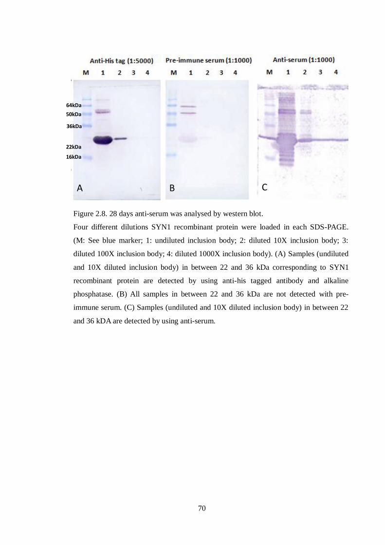
70
Figure 2.8. 28 days anti-serum was analysed by western blot.
Four different dilutions SYN1 recombinant protein were loaded in each SDS-PAGE.
(M: See blue marker; 1: undiluted inclusion body; 2: diluted 10X inclusion body; 3:
diluted 100X inclusion body; 4: diluted 1000X inclusion body). (A) Samples (undiluted
and 10X diluted inclusion body) in between 22 and 36 kDa corresponding to SYN1
recombinant protein are detected by using anti-his tagged antibody and alkaline
phosphatase. (B) All samples in between 22 and 36 kDa are not detected with pre-
immune serum. (C) Samples (undiluted and 10X diluted inclusion body) in between 22
and 36 kDA are detected by using anti-serum.
A B C
22kDa
16kDa
36kDa
50kDa
64kDa

71
2.8.2.2 Estimating protein concentration
The concentration of protein was estimated using the BioRad assay according to
manufacturer‟s instructions. The protein assay reagent containing Coomassie brilliant blue
G-250 was added to diluted protein samples. The mixture was placed at room temperature
for 10 minutes prior to measuring protein concentration. The absorbance was measured at
595nm using a spectrophotometer. Estimation of protein concentration was compared with
bovine albumin (BSA) as a standard.
2.8.2.3 SDS-Polyacrylamide gel electrophoresis (SDS-PAGE)
Proteins were analysed by SDS-PAGE using a BioRad kit. Different percentages of resolving
and stacking gels were cast as follows:
10% Resolving Gel
6.1 ml Sterile distilled water
3.75ml Resolving buffer (1.5M Tris, pH8.8)
150μl 10% Sodium Dodecyl Sulphate (SDS)
5ml Acrylamide (Protogel)
75μl 15% Ammonium per sulphate (APS)
15μl N,N,N,N-tetramethylethylenediamine (TEMED) (Sigma)
12.5% Resolving Gel
4.8 ml Sterile distilled Water
3.75ml Resolving buffer (1.5M Tris, pH8.8)
150μl 10% Sodium Dodecyl Sulphate (SDS)

72
6.3 ml Acrylamide (Protogel)
75μl 15% Ammonium per sulphate (APS)
15μl N,N,N,N-tetramethylethylenediamine (TEMED) (Sigma)
Stacking Gel
3 ml Sterile Distilled Water
1.25ml Stacking buffer (0.5M Tris, pH6.8)
50μl 10% Sodium Dodecyl Sulphate (SDS)
625μl Acrylamide (Protogel)
25μl 15% Ammonium per sulphate (APS)
5μl N,N,N,N-tetramethylethylenediamine (TEMED) (Sigma)
Proteins were mixed with 5X final protein loading buffer before boiling the sample for 5
minutes. Proteins were loaded on a SDS-PAGE and run in 1x ELFO buffer according to the
manufacturer‟s instructions at 80- 150V for 2 to 3 hours.
2.8.2.4 Coomassie staining of protein gels
Gels were stained at room temperature in fresh Coomassie stain for three hours. Stained gel
was destained overnight in destain solution. Gels were washed in SDW and preserved
between cellophane sheets after taking or scanning a picture of the gel.

73
2.9 Western blotting
2.9.1 Protein transfer
Proteins were resolved by SDS-PAGE. The gel was then transferred onto one sheet of
Whatman paper soaked in protein transfer buffer. A nylon transfer membrane (Hybond-C
extra, Amersham) was cut to size, soaked in protein transfer buffer and then laid over the gel.
One more sheet of Whatman paper was soaked in protein transfer buffer and laid on top of
the membrane. This arrangement was placed between two electroblotting pads (BioRad) and
inserted into an electroblotting tank (BioRad). This tank was filled with protein transfer
buffer. A BioRad power pack was used to blot the gel at 400 mA (~1.5 hours, 4oC).
Following this, the nylon transfer membrane was removed and placed in blocking solution
on rotary shaker at 4oC room.
2.9.2 Antibody probing and protein detection
The blot was incubated with primary antibodies, Anti-Histidine Tagged protein and Anti-
SYN1, which was diluted in 1:1000 in milk block solution for 1hr 30 min. After the primary
antibody incubation, the blot was washed 3x 10 min in blocking solution and then incubated
with anti-rabbit alkaline phosphatase (1:10,000 dilution), for 1 hour. The blot was then
washed 3x 10 min in blocking solution. The blot was incubated in the dark with alkaline
phosphatase detection solution (1X APDB/NBT/BCIP) until the target protein was detected.
The blot was washed with 2x 5min in PBS to stop the detection.

74
2.10 Slide preparation of meiotic chromosomes for cytology
2.10.1 Determination of meiotic stage
It is necessary to check the meiotic stage of the material to be studied before analysis. To do
this material was stained in lacto-propionic orcein (1%(w/v) natural orcein (Sigma), 49.5%
(v/v) propionic acid, 49.5% (v/v) lactic acid (LP orcein). Dissected anthers were placed on a
microscope slide and covered with a single drop of lacto-propionic orcein. Material was
covered with a glass cover slip and the cover slip pressed down to release pollen mother
cells (PMCs). Stained preparations were viewed under phase-contrast.
2.10.2 Slide preparation for immunolocalization studies
Arabidopsis buds were dissected from the inflorescence under stereomicroscope. Stems and
large (yellow) buds were removed and those of approximately 0.2-0.6 mm were used for the
immunolocalisation. To each slide, dissected anthers from approximately 10 buds were
placed into 10μl EM digestion medium (0.4% cytohelicase (C8274), 1.5% sucrose, and 1%
polyvinylpyrrolidone) and incubated for 10 minutes at 37oC in a moist chamber. Anthers
were tapped out in digestion mixture by using a brass rod to release pollen mother cells.
Following this, a further 10μl EM digestion mix was added to the tapped solution and placed
on a hotplate at 37 o
C for 2 minutes. 10μl Lipsol spreading medium (1% (v/v) lipsol
detergent to pH9.0 with borate buffer) was added and incubated on a hotplate at 37 o
C for 2
minutes. Materials were fixed on the slide by adding 20μl 4% (v/v) paraformaldehyde and
dried in a laminar flow cabinet for 2 hours (Armstrong et al., 2009).
2.10.3 Antibody labelling for immunolocalization studies
Dried slides were washed in PBS/0.1% Triton for 3X5 minutes. 100μl primary antibody,

75
diluted 1:500 in EM blocking solution (PBS+0.1%Triton+ 1% BSA), was added directly to
the slide which was covered with parafilm. The slides were incubated overnight at 4 o
C in a
moist chamber. Following the next day, the slides were washed in PBS/0.1% Triton for 3x5
minutes. 100μl secondary antibody was added and incubated for 1 hour at room temperature.
Finally the slides were washed in PBS/0.1% Triton for 3X5 minutes and were counterstained
with DAPI in Vectashield antifade mounting medium (Vector Laboratories).
2.10.4 Meiotic time course
Flowering stems were cut under water and transferred to BrdU solution (1x10⁻²
M). The
stems were left for 2 hours for uptake of BrdU via the transpiration stream. This allowed
BrdU incorporated into those cells in S-phase. After 2 hours the stems were transferred from
the BrdU to tap water. Buds were collected at 1hour post-BrdU pulse and 30 hours post-
BrdU. Immediately, anthers were dissected out from the buds for making the slide
preparations as previously described in section (2.10.1). The primary antibody (100μl of
anti-SYN1 raised in rabbit; diluted 1:500 in EM blocking solution) was added to the slides,
covered by parafilm, and incubated overnight at 4 o
C in a moist chamber. The slides were
washed in PBS/0.1% Triton for 3X5 minutes. The secondary antibody (anti-rabbit biotin;
diluted 1:50 in EM blocking buffer) was added to the slides and incubated for 30 minutes at
37oC. The slides were washed in PBS/0.1% Triton for 2X5 minutes and once in PBS for 5
minutes. The slides were treated with the anti-BrdU raised in mouse (Roche) for 30 minutes
at 37oC. Following this, anti-mouse FITC was added to the slide for 30 minutes at 37
oC.
Finally, Cy3 avidin (1:200 in EM blocking solution) was used to conjugate to the biotin. The
material was incubated for 30 minutes at 37oC before washing for 3x5 minutes in PBS/0.1%
Triton. The slides were mounted in 10μl of a solution containing 4, 6-diaminido-2-

76
phenlyinidole (DAPI) at 1 mg/ml in Vectashield antifade mounting medium (Vector
laboratories) (Armstrong and Jones, 2003).
2.10.5 Slide preparation for cytogenetic studies
The slides were prepared as previously described by Armstrong et al. (2009). Fixed
inflorescences were transferred to a black watch-glass containing 1ml 0.01M citrate buffer.
Stems and large buds were removed from the black watch-glass. The remaining buds were
washed in 1ml 0.01M citrate buffer (buffer stock 0.1M citric acid: 0.1M sodium citrate,
diluted 1:10 for a working solution) for 3X5 minutes. The buds were then incubated at 37oC
in a moist chamber with an enzyme mixture (0.3% (w/v) pectolyase, 0.3% (w/v)
cytohelicase, 0.3% (w/v) cellulose (all from Sigma, in citrate buffer) for 1 hour and 15
minutes. Replacing the enzyme mixture with fresh citrate buffer stopped the enzyme
reaction. A single bud was transferred to slide in a drop of water and tapped out using a
needle to produce a cell suspension. One drop of 5μl 60% (v/v) acetic acid was then added to
the cell suspension and the slide was placed on the hotplate at 45oC for 1 minute. A further
5μl acetic acid was added on the suspension to avoid drying. Finally, 100μl fixative
(Absolute ethanol: Glacial acetic acid in 3:1 ratio) was added as a circle around the
suspension on the slide and drained away. The slide was dried with a hair drier.
The slides were washed in 2xSSC at room temperature for 10 minutes. Material on the slide
was digested in 0.01% (w/v) pepsin (Sigma) in 0.01M HCl at 37oC for 90 seconds. Slides
were washed in 2xSSC at room temperature twice for 5 minutes. Material was fixed on the
slide in 4% paraformaldehyde pH8.0 for 10 minutes. Following this, slides were dehydrated
by passing through an alcohol series, 70%, 85% and 100%. The slides were ready for

77
fluorescent in situ hybridisation (FISH) or were counterstained with DAPI in Vectashield
antifade mounting medium (Vector Laboratories).
2.10.6 Fluorescent in situ hybridisation (FISH)
The pAL38 containing a pericentromeric 360bp repeat sequence (Fransz et al., 2000) was
used in cytological study. The pAL38 probe was produced by PCR amplification using the
M13 reverse and forward primers followed by random primer labelling in the presence of
biotin-labelled dUTP (Roche).
20μl of the probe mixture was made as follows:
14μl hybridization mix (5ml deionised formamide, 1ml 20xSSC, 1g dextran sulphate, pH7)
+2μl labelled probe (The pAL38 probe, centromeric probe, was indirectly labelled with
biotin) + SDW made up to 20μl.
20μl of the probe mixture was added on the slide and covered with a coverslip (22x22mm)
and sealed by rubber solution. The slide was heated on a hot plate at 75oC for 4 minutes. The
heated slide was placed in a moist chamber and incubated at 37oC overnight. After the
hybridization, the slide was washed three times, 5 minutes each, in the 50% formamide-
2xSSC and then washed in 2xSSC and afterwards washed in 4x SSC/0.05% Tween20. All
solutions were heated to 45oC. Finally, the slide was washed in 4x SSC/0.05% Tween20 at
room temperature for 5 minutes. 100μl of fluorescent antibodies (Cy3 avidin) was added to
the slide and covered with parafilm. The slide was incubated in a moist chamber at 37 oC in a
dark environment for 30 minutes. Afterwards, the slide was washed in PBS/ 0.1% Triton for
three times, 5 minutes each. The slides were mounted in 10μl of a solution containing 4, 6-

78
diaminido-2-phenlyinidole (DAPI) at 1mg/ml in Vectashield antifade mounting medium
(Vector laboratories)(Armstrong et al., 2009).
2.11 Slide imaging
Slides were examined by fluorescence microscopy using a Nikon Eclipse T300 microscope.
Images were captured and analysed by using Cell Analysis (Olympus) software.
2.12 Cisplatin treatment
2.12.1 Introduction of cisplatin into meiocytes of Atspo11-1-4-/-
Bud stems of Atspo11-1-4-/-
were cut to a length of about 3 to 4cm and immersed into 2.5μM
cisplatin solution. After 2 hours, the bud stems were transferred from the cisplatin solution to
the tap water. At the same time, bud stems of wild-type and Atspo11-1-4-/-
without cisplatin
treatment were immersed in tap water as positive and negative controls. Immunolocalization
studies were carried out on spread preparations with anti-SYN1 (rabbit) and anti-ASY1 (rat)
antibodies.
2.12.2 Seedling growth on Cisplatin containing MS agar
One hundred seeds were placed on Murashige and Skoog (MS) agar medium with different
concentrations of cisplatin (0 μM; 12.5 μM; 25 μM; 50 μM). These were incubated at 4oC in
a chamber for two days prior to transfer to a 22oC growth chamber. After growing for three
weeks, seedlings were collected and weighed for statistical analysis.

79
Chapter 3
Characterising a syn1 null mutant
3.1 Introduction
Budding yeast (S. cerevisiae) REC8, a meiotic kleisin subunit of the cohesin complex, is
essential for maintaining sister chromatid cohesion during meiosis (Parisi et al., 1999;
Haering and Nasmyth, 2003; Haering et al., 2004). During metaphase I, REC8 is released by
separase from the chromosome arms but remains at the centromeres, ensuring the sister
chromatids are paired at the centromeric region. This ensures they move towards the same
pole during the first meiotic division. When REC8 is released from the centromeric region,
sister chromatids are able to segregate towards the opposite poles during the second meiotic
division. This indicates that REC8 plays a crucial role in chromosome segregation throughout
meiosis. SYN1 has been reported to be the Arabidopsis homologue of REC8 (Bai et al., 1999;
Bhatt et al., 1999). However, the function of SYN1 in meiosis is not well understood. Several
studies have reported chromosome fragmentation in plant rec8 mutants. These include the rice
OsRAD21-4 deficient line, maize afd1 and Arabidopsis syn1 (Bai et al., 1999; Bhatt et al.,
1999; Yu and Dawe, 2000; Zhang et al., 2006). In the afd1 mutant, low frequencies of small
fragments are observed during meiotic prophase I, whereas severe chromosome
fragmentations from the syn1 mutant and OsRAD21-4 deficient line are detectable throughout
the meiosis I and II. However, a detailed analysis of chromosome fragmentation in the
absence of cohesin has yet to be conducted. To investigate this chromosome fragmentation
phenotype, a syn1 mutant line was identified. In this chapter, a molecular and cytological
analysis to characterise this syn1 mutant line is described.

80
3.2 Results
3.2.1 Identification of SYN1 T-DNA insertion mutant
A T-DNA insertion line (SALK_091193) was obtained from the SALK Institute via NASC
that contained an insertion in the SYN1 gene (At5g05490). The seeds were grown together
with a wild type (Col 0) in the glasshouse. To identify homozygous knockout lines, genomic
DNA was isolated from plant leaves and subjected to PCR genotyping using specific primers.
Two primers (S1 and S2) were designed to identify the wild type SYN1 gene. Another two
primers (S2 and LBa1) were designed to check for the presence of an insert. To determine the
exact site of the T-DNA insertion, the PCR fragments of a homozygous plant were cloned into
pDrive cloning vector (Qiagen) prior to nucleotide sequencing. The sequencing result
confirmed that the plasmid contained a DNA fragment comprising a T-DNA sequence and an
Arabidopsis genomic DNA sequence (Figure 3.1). This genomic DNA was analysed by using
NCBI blast search and TAIR SeqViewer (Figure 3.2). The result revealed that the genomic
DNA sequence was the gene At5g05490 on chromosome 5. The T-DNA insertion site is
located at exon 6 (nucleotide position 1625566; Figure 3.3) of the SYN1 gene.
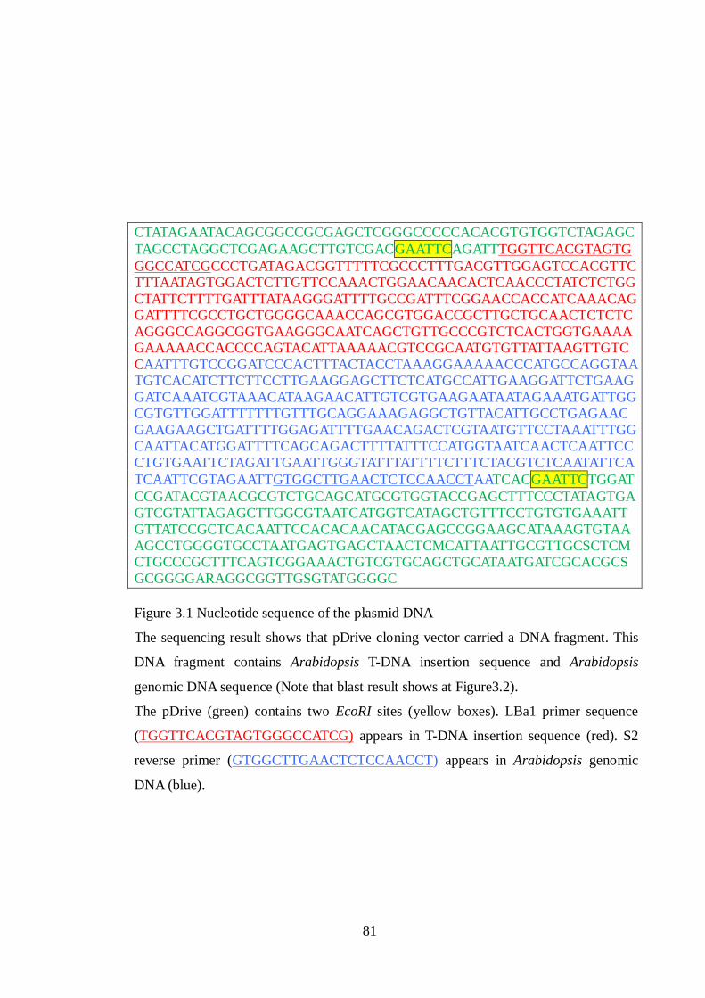
81
CTATAGAATACAGCGGCCGCGAGCTCGGGCCCCCACACGTGTGGTCTAGAGC
TAGCCTAGGCTCGAGAAGCTTGTCGACGAATTCAGATTTGGTTCACGTAGTG
GGCCATCGCCCTGATAGACGGTTTTTCGCCCTTTGACGTTGGAGTCCACGTTC
TTTAATAGTGGACTCTTGTTCCAAACTGGAACAACACTCAACCCTATCTCTGG
CTATTCTTTTGATTTATAAGGGATTTTGCCGATTTCGGAACCACCATCAAACAG
GATTTTCGCCTGCTGGGGCAAACCAGCGTGGACCGCTTGCTGCAACTCTCTC
AGGGCCAGGCGGTGAAGGGCAATCAGCTGTTGCCCGTCTCACTGGTGAAAA
GAAAAACCACCCCAGTACATTAAAAACGTCCGCAATGTGTTATTAAGTTGTC
CAATTTGTCCGGATCCCACTTTACTACCTAAAGGAAAAACCCATGCCAGGTAA
TGTCACATCTTCTTCCTTGAAGGAGCTTCTCATGCCATTGAAGGATTCTGAAG
GATCAAATCGTAAACATAAGAACATTGTCGTGAAGAATAATAGAAATGATTGG
CGTGTTGGATTTTTTTGTTTGCAGGAAAGAGGCTGTTACATTGCCTGAGAAC
GAAGAAGCTGATTTTGGAGATTTTGAACAGACTCGTAATGTTCCTAAATTTGG
CAATTACATGGATTTTCAGCAGACTTTTATTTCCATGGTAATCAACTCAATTCC
CTGTGAATTCTAGATTGAATTGGGTATTTATTTTCTTTCTACGTCTCAATATTCA
TCAATTCGTAGAATTGTGGCTTGAACTCTCCAACCTAATCACGAATTCTGGAT
CCGATACGTAACGCGTCTGCAGCATGCGTGGTACCGAGCTTTCCCTATAGTGA
GTCGTATTAGAGCTTGGCGTAATCATGGTCATAGCTGTTTCCTGTGTGAAATT
GTTATCCGCTCACAATTCCACACAACATACGAGCCGGAAGCATAAAGTGTAA
AGCCTGGGGTGCCTAATGAGTGAGCTAACTCMCATTAATTGCGTTGCSCTCM
CTGCCCGCTTTCAGTCGGAAACTGTCGTGCAGCTGCATAATGATCGCACGCS
GCGGGGARAGGCGGTTGSGTATGGGGC
Figure 3.1 Nucleotide sequence of the plasmid DNA
The sequencing result shows that pDrive cloning vector carried a DNA fragment. This
DNA fragment contains Arabidopsis T-DNA insertion sequence and Arabidopsis
genomic DNA sequence (Note that blast result shows at Figure3.2).
The pDrive (green) contains two EcoRI sites (yellow boxes). LBa1 primer sequence
(TGGTTCACGTAGTGGGCCATCG) appears in T-DNA insertion sequence (red). S2
reverse primer (GTGGCTTGAACTCTCCAACCT) appears in Arabidopsis genomic
DNA (blue).

82
dbj|AB005241.1| Arabidopsis thaliana genomic DNA, chromosome 5, P1 clone:MOP10
Length=57892
Score = 747 bits (404), Expect = 0.0 Identities = 409/411 (99%), Gaps = 1/411 (0%)
Strand=Plus/Plus
Query 1 AATTTG-TCCGGATCCCACTTTACTACCTAAAGGAAAAACCCATGCCAGGTAATGTCACA 59
||| || |||||||||||||||||||||||||||||||||||||||||||||||||||||
Sbjct 4251 AATCTGTTCCGGATCCCACTTTACTACCTAAAGGAAAAACCCATGCCAGGTAATGTCACA 4310
Query 60 TCTTCTTCCTTGAAGGAGCTTCTCATGCCATTGAAGGATTCTGAAGGATCAAATCGTAAA 119
||||||||||||||||||||||||||||||||||||||||||||||||||||||||||||
Sbjct 4311 TCTTCTTCCTTGAAGGAGCTTCTCATGCCATTGAAGGATTCTGAAGGATCAAATCGTAAA 4370
Query 120 CATAAGAACATTGTCGTGAAGAATAATAGAAATGATTGGCGTGTTGGATTTTTTTGTTTG 179
||||||||||||||||||||||||||||||||||||||||||||||||||||||||||||
Sbjct 4371 CATAAGAACATTGTCGTGAAGAATAATAGAAATGATTGGCGTGTTGGATTTTTTTGTTTG 4430
Query 180 CAGGAAAGAGGCTGTTACATTGCCTGAGAACGAAGAAGCTGATTTTGGAGATTTTGAACA 239
||||||||||||||||||||||||||||||||||||||||||||||||||||||||||||
Sbjct 4431 CAGGAAAGAGGCTGTTACATTGCCTGAGAACGAAGAAGCTGATTTTGGAGATTTTGAACA 4490
Query 240 GACTCGTAATGTTCCTAAATTTGGCAATTACATGGATTTTCAGCAGACTTTTATTTCCAT 299
||||||||||||||||||||||||||||||||||||||||||||||||||||||||||||
Sbjct 4491 GACTCGTAATGTTCCTAAATTTGGCAATTACATGGATTTTCAGCAGACTTTTATTTCCAT 4550
Query 300 GGTAATCAACTCAATTCCCTGTGAATTCTAGATTGAATTGGGTATTTATTTTCTTTCTAC 359
||||||||||||||||||||||||||||||||||||||||||||||||||||||||||||
Sbjct 4551 GGTAATCAACTCAATTCCCTGTGAATTCTAGATTGAATTGGGTATTTATTTTCTTTCTAC 4610
Query 360 GTCTCAATATTCATCAATTCGTAGAATTGTGGCTTGAACTCTCCAACCTAA 410
|||||||||||||||||||||||||||||||||||||||||||||||||||
Sbjct 4611 GTCTCAATATTCATCAATTCGTAGAATTGTGGCTTGAACTCTCCAACCTAA 4661
Figure 3.2. Sequence of A. thaliana genomic DNA
410bp of Arabidopsis genomic DNA is analysed by using NCBI blast search. The blast
result shows that the genomic DNA locates to the short arm of Chromosome 5. This
genomic DNA contains two SYN1 mRNA sequences: (1) Sequence of genomic DNA
from 1 to 49 (green colour) is a SYN1 exon 6 sequence. This genomic DNA associates
with T-DNA sequence (Figure 3.1). (2) Sequence of genomic DNA from 180 to 300 (red
colour) is a full length sequence of exon 7.

83
Figure 3.3. Map of the SYN1 locus and exon organisation.
The 4.4kb open reading frame of SYN1 located on chromosome 5. The sequence
analysis shows the T-DNA insertion in exon 6 of the SYN1 gene. The position of the T-
DNA insertion is represented as an inverted triangle (blue). Exons are represented in red
blocks.

84
3.2.2 Expression of the SYN1 gene
To investigate the expression pattern of SYN1 and to determine if its expression was
knocked out in SALK_091193, I analysed bud and leaf tissues of wild-type (Col 0), Atspo11-
1-4-/-
and SALK_091193 by using reverse transcription PCR (RT-PCR). The primers (S3 and
S4) were designed downstream of the T-DNA insertion and were run by the PCR to show the
relative abundance of the SYN1 transcripts in tissues of wild-type and mutants, using the
housekeeping gene GAPD as a control. The RT-PCR analysis showed that SYN1 transcripts
were detected in bud and leaf of all samples (data not shown). SYN1 transcripts were detected
in leaf and bud tissues from wild-type (Col 0), Atspo11-1-4-/-
and SALK_091193, but the
SYN1 level transcripts from the bud samples were higher than that of the leaf. This result
suggested that the SYN1 transcripts in SALK_091193 could be triggered by the activity of a
promoter within the T-DNA insertion sequence.
To ensure full length SYN1 transcripts were not found in the T-DNA insertion line
(SALK_091193), two PCR primers (STC and EX8) were designed at the start codon and exon
8 of the SYN1 gene, which were used to amplify products from wild-type (Col 0), Atspo11-1-
4-/-
and SALK_091193. The amplification analysis showed that SYN1 transcript was not
detectable in all samples. Therefore, second rounds of amplification of SYN1 were required to
detect expression. 1μl of RT-PCR sample was added in Reddy Mix PCR master mix (Therma
Scientific). 20 cycles was sufficient for the PCR reaction to avoid saturating the PCR
products. The re-amplification analysis revealed that SYN1 was expressed at the expected size
503bp in bud and leaf samples from the wild-type (Col 0) and Atspo11-1-4 mutant (Figure
3.4), indicating that SYN1 is expressed in both reproductive and vegetative tissues. No product
was detected in bud and leaf samples from the SALK_091193 line, suggesting that the T-DNA

85
insertion sequence interrupted expression of the SYN1 gene. This result implied that
SALK_091193 is likely a syn1 null mutant. In addition, the amplification analysis showed
that the level of transcription of the SYN1 gene is relatively low compared with the
housekeeping gene GAPD.
Data from Genevestigator (https://www.genevestigator.com/gv/index.jsp) (Hruz et al., 2008),
a microarray database and analysis system which allows average expression levels of given
genes across a variety of tissues to be visualized, showed that SYN1 gene (At5g05490) was
expressed in cell culture, flower, stamen, pollen, endosperm, stem, root hair zone and root
stele (Figure 3.5), This supports our experimental observation that SYN1 is expressed in both
reproductive and vegetative tissues.
.
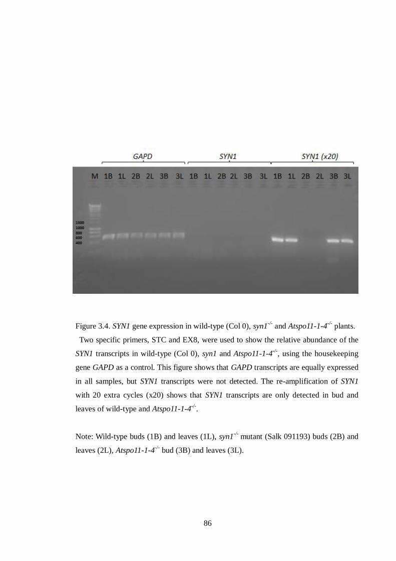
86
Figure 3.4. SYN1 gene expression in wild-type (Col 0), syn1-/-
and Atspo11-1-4-/-
plants.
Two specific primers, STC and EX8, were used to show the relative abundance of the
SYN1 transcripts in wild-type (Col 0), syn1 and Atspo11-1-4-/-
, using the housekeeping
gene GAPD as a control. This figure shows that GAPD transcripts are equally expressed
in all samples, but SYN1 transcripts were not detected. The re-amplification of SYN1
with 20 extra cycles (x20) shows that SYN1 transcripts are only detected in bud and
leaves of wild-type and Atspo11-1-4-/-
.
Note: Wild-type buds (1B) and leaves (1L), syn1-/-
mutant (Salk 091193) buds (2B) and
leaves (2L), Atspo11-1-4-/-
bud (3B) and leaves (3L).

87
Figure 3.5. Genevestigator expression profile of Arabidopsis SYN1 gene.
The microarray analysis displays the expression of SYN1 genes in cell culture, flower,
stamen, pollen, endosperm, stem, root hair zone and root stele. For each tissue, we can
find out how many arrays are taken into account to calculate the mean value.

88
3.2.3 Cytogenetic analysis of the syn1 mutant
3.2.3.1 Male meiosis in wild-type and syn1 mutant
It was previously reported that chromosome fragmentation is observed in the absence of
SYN1 protein (Bai et al., 1999; Bhatt et al., 1999). To confirm the chromosome fragmentation
phenotype of syn1, I analysed the male meiosis in the wild-type and T-DNA insertion line
(SALK_091193) by using chromosome spreads stained with 4, 6-diamidino-2-phenylindole
(DAPI).
In wild-type (Col 0) plants, chromosomes stained with DAPI showed chromosomes as
normal thin thread structures at leptotene (Figure 3.6A). Synapsed and unsynapsed
chromosome regions were observed at the early zygotene (Figure 3.6B). Following further
chromosome pairing and synapsis, homologous chromosomes were fully synapsed at the end
of pachytene. Therefore, thick and heavily stained homologous chromosomes were observed
(Figure 3.6C). As chromosome condensation continued, desynapsis of the homologous
chromosome regions increased during diplotene and diakinesis stages. Homologous
chromosomes appeared to repel each other slightly thus revealing the chiasmata (Figure 3.6D;
E). At metaphase I, five short and thick bivalents were seen to be aligned on the equatorial
plate prior to the first meiotic segregation (Figure 3.6F). 10 chromosomes were observed at
anaphase I due to the nonsister chromatids of homologous chromosomes moving to opposite
poles forming two haploid sets (Figure 3.6G). Two groups of chromosomes were seen to be
separated by a band of organelles during prophase II (Figure 3.6H). At the late prophase II,
tetrads contained 20 chromosomes arranged in groups of 5 forming four haploid nuclei were
observed (Figure 3.6I).

89
In the T-DNA insertion line (SALK_091193), chromosome abnormalities were found
throughout the meiotic program. This made it difficult to identify the early meiotic stages.
DAPI showed chromosomes as abnormal thin thread structures (Figure 3.7A; B).
Chromosomes were also observed as tangled structures revealing unpaired homologous
chromosomes (Figure 3.7C; D). Chromosome fragments were aligned on the equatorial plate
(Figure 3.7E). The numbers of chromosome fragments ranged from 7 to 17; Mean=10.17
(Table 3.1; n=53), whereas wild-type meiocytes had the expected five bivalents at metaphase
I. During anaphase I, chromosome fragments separated to the opposite poles. Many
chromosome fragments remained at centre of cell, the number of chromosome fragments on
the equatorial plate ranged from 2 to 22 (Mean=13.1; n=14). The total number of
chromosome fragments increased significantly from 10.17 at metaphase I to 32.36 at
anaphase I (t-test, p<0.001; Table 3.1) due to the absence of SYN1. The chromosome mis-
segregation occurred throughout the meiosis II (Figure3.7G; H). The number of chromosome
fragments showed that there is no significant difference between anaphase I and prophase II
(t-test, p=0.3756; Table 3.1), indicating that chromosome fragmentation occurred mainly at
meiosis I. This result demonstrated that the chromosome fragmentation likely leads to sterility
in syn1 plants. The cytological analysis also showed that the meiocytes of the T-DNA
insertion line (SALK line 091193) is identical to the phenotype of syn1 (Bai et al., 1999; Bhatt
et al., 1999). In the following chapter, the SALK line 091193 will be referred to as syn1
mutant.

90
Meiotic stages
chromosome
fragments
Metaphase I
Anaphase I
Prophase II
Mean (Range) 10.17 (7-17) 32.36 (19-51) 29.68 (17-46)
Number of cells 53 14 16
Standard Deviation 2.465526 7.879936 8.292316
Standard Error 0.337256 2.10637 2.073079
Table 3.1: Number of chromosome fragments in SALK line 091193 meiocytes.
Number of chromosome fragments at metaphase I is significantly lower than that at anaphase
I (t-test, p<0.001) and prophase II (t-test, p<0.001). The number of chromosome fragments is
not significantly different between anaphase I and prophase II (t-test, p=0.3756).

91
Figure 3.6 Meiotic stages in pollen mother cells of wild-type (Col 0) of A. thaliana.
(A) Thin thread chromosomes appear in the early leptotene, (B) Zytogene stage:
unsynaped (arrow) and synapsed homologous chromosomes, (C) Pachytene stage: fully
synapsed homologous chromosomes, (D) Diplotene stage: non-sister chromatids of each
bivalent repel each other forming chiasmata, (E) Diakinesis stage: paired homologous
chromosomes are short and thick, (F) Metaphase I: Five condensed bivalents align on
the equatorial plate, (G) Anaphase I: chromosomes migrate towards the same pole. (H)
Prometaphase II: two groups of chromosomes are separated from a band of organelle,
(I) Telophase I or Tetrad stage: four haploid nuclei. Bars 10μm.

92
Figure 3.7 Meiotic stages in pollen mother cells of the SALK line 091193 (syn1 mutant)
of A. thaliana.
(A) Loose chromatin structures and (B) thin thread chromosomes appear in the early
meiotic prophase I, (C, D) tangled chromosomes appear in early prophase I, (E)
Metaphase1: chromosome fragments appear on the equatorial plate, (F) Anaphase 1:
chromosome fragments remain on equatorial plate after chromosomes migrate towards
each pole, (G) Early anaphase II: chromosome fragments mis-segregate, (H) Late
anaphase II: segregated chromosome fragments are separated from a band of organelles,
(I) Tetrad stage: chromosome fragments in four nuclei. Bars 10μm.

93
3.2.3.2 Fluorescence in situ hybridisation (FISH) analysis of chromosome segregation in
wild-type and syn1 male meiocytes
To investigate whether SYN1 is essential to maintain cohesion between sister chromatids, I
analysed centromere movement in wild-type and syn1 mutant meiocytes by using the FISH
probe, pAL38, a tandemly repeated DNA sequence that localises to the centromeric region of
all ten Arabidopsis chromosomes. FISH analysis in wild-type meiocytes showed that there are
an average of 6.55 (n=20) centromeric signals at early prophase I cell (Table 3.2; Figure 3.8A
and B). Ten centromeric FISH signals were detected in diakinesis cells because the non-sister
chromatids repelled each other revealing chiamata (Figure 3.8 B). Following further
condensation, five bivalents were aligned on the equatorial plate. Two centromeric signals
were detected in each bivalent pointing opposite directions (Table 3.2; Figure 3.8C). After
chromosome segregation, non-paired sister chromatids moved towards the opposite poles.
There are an average of 10.5 (n=4) and 11.2 (n=5) FISH signals at anaphase I and
prometaphase II (Figure 3.8D; E; Table 3.2). Finally, an average of 19 (n=6) FISH signals per
cell were detected after sister chromatids segregated to opposite poles (Table 3.2; Figure
3.8F).
In syn1 meiocytes, an average of 5.65 (n=26) FISH signals were observed on the loose and
tangled chromosomes during prophase I (Table 3.2; Figure 3.9 A; B). Following the
chromosome condensation, an average of 14.57 (n=7) FISH signals were detected due to the
increasing number of chromosome fragments (Table 3.2; Figure 3.9C) during metaphase I.
Abnormal centromeric signals were seen distributed on the chromosome fragments. During
the first meiotic division, some delay in segregation was apparent (Figure 3.9D). In syn1
mutant, an average of 19 (n=2) FISH signals were detected at anaphase I whereas in wild-type

94
(Col 0) an average of 10.5 (n=4) centromeric FISH signals were observed (Table 3.2). The
FISH analysis of chromosome segregation in syn1 meiocytes showed that sister centromeres
were not aligned together during anaphase I. Approximately twenty (average=19.14; n=7)
centromeric signals were detected at prometaphase II (Figure 3.9E). However, these twenty
(average=20.33; n=6) FISH signals were not distributed equally in tetrads due to the
chromosome mis-segregation (Figure 3.9F; Table 3.2).
Table 3.2: The average number of centromeric FISH signals at different meiotic stages of
wild-type and syn1 mutant
Meiotic stage
Early
prophase I
Metaphase I Anaphase I Prometaphase
II
Tetrad
Wild-type
(Col 0)
6.55
(3-10)
*n=20
10
(10)
*n=6
10.5
(10-12)
*n=4
11.2
(10-14)
*n=5
19
(17-20)
*n=6
syn1 mutant 5.65
(4-8)
*n=26
14.57
(13-16)
*n=7
19
(18-20)
*n=2
19.14
(14-21)
*n=7
20.33
(20-21)
*n=6
Note: The number of cells examined at each stage is shown next to the *. The range of
centromeric FISH signals is shown within brackets

95
Figure 3.8. FISH of centromeric probe (pAL38) to pollen mother cells of wild-type A.
thaliana. (A) Five centromeric FISH signals appeared in pachytene stage, (B) Ten
centromeric FISH signals were detected in diakinesis, (C) Two centromeric FISH
signals were detected in each bivalent during metaphase I, (D) One centromeric FISH
signal was detected in sister chromatids during first meiotic segregation, (E) Ten
centromeric FISH signals were observed in prometaphase II, (F) After sister chromatids
segregation, twenty centromeric FISH signals were detectable in tetrad.
Note: FISH signal is red colour and DAPI is blue colour. Bars 10μM.

96
Figure 3.9. FISH of centromeric probe (pAL38) to pollen mother cells of the SALK line
091193 (syn1 mutant) of A. thaliana. (A) and (B) Five centromeric FISH signals were
detected in loose chromatin and tangled chromosome during the early prophase I, (C)
16 centromeric FISH signals were detected in metaphase I. (D) 10 centromeric FISH
signals were detected in each pole during anaphase I. (E) and (F) 20 centromeric FISH
signals were detected at prometaphase II and tetrad. Bars 10μm.

97
3.2.4 Relationship between SYN1 and other Rec8 homologues.
Cytological analysis of syn1 mutant showed chromosome fragmentation during
metaphase I. However, severe fragmentation is rarely detected in non-plant species. It is
essential to evaluate the relationship between SYN1 and other REC8 homologues. Six species
Saccharomyces cerevisiae, Caenorhabditis elegans, Sordaria macrospora, Oryza sativa, Zea
mays and Arabidopsis thaliana were chosen for protein sequence alignment. The amino acid
sequences were analysed with ClustalW (http://www.ebi.ac.uk/Tools/emboss/align/) using the
Blosum62 comparison table (Thompson et al., 1994).
REC8 has been identified in S. cerevisiae (Parisi et al., 1999). Currently, chromosome
fragmentation has not been found in a yeast rec8 mutant. The yeast REC8 contains 680 amino
acids whereas A. thaliana SYN1 has 617 amino acids. REC8 showed only 18% identity to
SYN1. Another two REC8 homologues have been identified in C. elegans, CeREC8 and S.
macrospora, SmREC8. It has been reported that chromosome fragments are found in cerec8
and smrec8 mutants (Pasierbek et al., 2001; Storlazzi et al., 2008). Both ceREC8 and
smREC8 contain 781 and 763 amino acids (Figure 3.10). The analysis showed that the amino
acid sequence identity between ceREC8 and SYN1 is lower (18.2% identity) than that
between smREC8 and SYN1 proteins (20.7% identity). All REC8 proteins are rather
conserved at N- and C- termini. Therefore, this result showed that SYN1 has low identity in
full length amino acid sequence with others.
Recently, chromosome fragmentation was identified in other plant REC8 mutants including
Z. mays, AFD1 (Yu and Dawe, 2000) and O. sativa, OsRad21-4 (Zhang et al., 2006). AFD1
and OsRad21-4 contain 602 and 608 amino acids respectively. The levels of amino acid

98
sequence identity between AFD1 and SYN1 are the same (41% identity) to those between
OsRad21-4 and SYN1 protein (41% identity). However, the amino acid sequence identity
between AFD1 and OsRad21-4 protein is higher (71% identity; Figure 3.11).

99
SYN1 MFYSHQLLARKAP-LGQIWMAATL-----HAKINRKKLDKLDIIQICEEILNPS------ 48
smREC8 MFYSHEILTNQQYGVATVWLVSTFGLRSSNRKISRKAIQGVNVRKACETILQPG------ 54
ceREC8 MVVSAEVIR-KDAVFHVAWILGTG----DSKKLSRREILDQNLPELCHSIIEMVPERHRG 55
*. * ::: : . *: .* *:.*: : :: : *. *::
SYN1 --VPMALRLSGILMGGVVIVYERKVKLLFDDVNRFLVEINGAWRTKSVPDPTLLPKGKTH 106
smREC8 --APIALRLQGSLLYGVSRVFSQQCSYVLTDAEKIHMHMRCFYNVLGGSGNALDPQAGKA 112
ceREC8 SATKTGLYLLSLLTYGTVLIHQVQVDFLKRDVEKLKELMKKKSFILLMAERFDRNQELQR 115
. .* * . * *. :.. : . : *.::: :. . :
SYN1 ARKEAVTLPE--------------NEEADFG-DFEQTRNVPKFGNYMDFQQTFISMRLDE 151
smREC8 KRNQLILPDDPDFEVNMGLPAFHFDEEGNLATQFQSQASRKTSSQFSPLDRFNLTPDGNG 172
ceREC8 KEDKFARLRSKPIMCVEELDRVDLAHLQAIGDELGINGNPGDFIMMDALPNMNQWIDNNS 175
..: . . :. :: . : . :
SYN1 SHVN--NNPEPEDLGQQFHQADAENITLFEYHG------SFQTNNETYDRFERF-DIEGD 202
smREC8 SFIIGLDLPQSSPTNPLYSQHSSYSLGPLSQHKPDDDMMQFGEPEANAEIFGDW-GIEID 231
ceREC8 ELNAIYGCVEPYLREKEITMHSTFVEGNGSNEHNKERRNDAVIADFSQLLFPEIPEITLG 235
. . :. .: . . . : . * * .
SYN1 DETQMNSNPREGAEIPTTLIPSPPRHHDIPEGVNPTSP---------------------- 240
smREC8 ADGNVMATVDE-PELPILPRPEEERAYATASGQVHDEPEFHFDDQDDLLINGGTAPPPEA 290
ceREC8 EKFPIDVDSRKRSAILQEEQEEALQLPKEASEIVQEEP-------------------TKF 276
. : : . : . : .. .*
SYN1 -------QRQEQQENRRDGFAEQMEEQNIPDKE--EHDRP-QPAKKRARKTATSAMDYEQ 290
smREC8 DEVPGLVPDQVEQAQENQVMAEAEEEQVIAEEEPVAEASA-APVRRRQRRRVVLAPD-ED 348
ceREC8 VSIALLPSETVEQPAPQEPIQEPIQPIIEEPAPQLELPQPELPPQLDAIDLVTIPASQQD 336
:* .: : * : . * : .. . . ::
SYN1 TIIAGHVYQSWLQDTSDILCRGEKRKVR-------------------------GTIRPDM 325
smREC8 TKISRHELKSWSNDYLANAERTNNAARRAAATNPAAAKKHAYDLVFGRGVGGVGSFDDGL 408
ceREC8 MVVEYLQLINDLPDDENSRLPPLPKDLELFEDVILPPPAKKSKVEEEEDALERARRRPSS 396
: . * . . .
SYN1 ESFKRANMPPTQLFEKDSSYPPQLYQLWSKNTQVLQTSSSESRHP------DLRAEQSPG 379
smREC8 NSGNHRSHPLAALFAG-VDFAAEVLDIDIDIDEGHEAGGRRGRRRSALEALELEEEDAER 467
ceREC8 RPVTPINQTDLTDLHSTVRPEDPSFAIDSQIHDVLPQRKKSKRNLPIIHSDDLEIDEA-- 454
.. . . . : : . : *. :*. :::
SYN1 FVQERMHNHHQTDHHERSDTSSQNLDSPAEILRTVRTGKGASVESMMAGSRASPETINRQ 439
smREC8 RVRRRLSDENEEEHHAQSQQNVAEPQAEAGRGLLTGEAEEDAEIGRRDGSALPDPDIPSD 527
ceREC8 -VQKVLQADYSSLVRKKEDVIAKIPPKTDAVAVLMNLPEPVFSIGYRLPPEVR--DMFKA 511
*:. : . . : :.: : . . :
SYN1 AADINVTPFYSGDDVRSMPSTPSARGAASINNIEISS--KSRMPNRKRPNSS-------P 490
smREC8 APWNRAPSLVPGSSVKGDSHMPGSSRQVSASPLHSRGSHLAHLPPIDRFSDTGSYAPLLH 587
ceREC8 CYNQAVGSPVSDDEEDEDEEEEEEYKYAKVCLLSPNRIVEDTLLLEEQPRQPEEFPSTDN 571
. . . .... .. : : .: ..
SYN1 RRGLEPVAEERPWEHREYEF-------EFSMLPEKRFTADKEILFETASTQTQKPVCNQS 543
smREC8 SGGVPDFSSDPVMPPGEDEP-------ELPHLPGGNHGSQLLRRSPSSEAGAATEVMAET 640
ceREC8 INPPRQLQENPVFENLEYEAPPHPIRTARTPTPIKDLKYSVISLFPTPEKRRETSIIAEL 631
. .: * * . * . :.. . : :
SYN1 --DEMITDSIKSHLKTHFETPGAPQVESLNKLAVG------------------------- 576
smREC8 --SQVMKDALDRDGRNFLTYVNMVAKTRGETRSLGGGTPSQAVGDGDGNRRLRQWVAFDE 698
ceREC8 NLDPIPVEEIDP-LLTMRTEEELENVRRRQKSSLGVQFMRTDDLEEDTRRN-------RL 683
. : : :. . :. ::*
SYN1 -------MDRNAAAKLFFQSCVLATRGVIKVN------------------QAEPYGDILI 611
smREC8 LLDEPRDRTRQVATQAFYNVLVLATKNAIKVEQDME--------------DFQPFGEIRV 744
ceREC8 FEDEERTRDAREDELFFYSSGSLLPNNRLNIHKELLNEAEARYPEWVNFNEFTADHDRKK 743
. *:. * ... :::. : . :
SYN1 ARGPNM-------------------------------- 617
smREC8 GVGVSEAEMLAMMEEDGDA------------------- 763
ceREC8 AATAFEGLLLSLKNMKVEAKQEDPYFPILVRHISHEEM 781

100
Figure 3.10 Alignments of the full length amino acid sequence of the smREC8, ceREC8
and SYN1.
The SYN1 amino acid sequence is shown aligned with smREC8 (S. macrospora) and
ceREC8 (C. elegans). Sequences are aligned using the ClustalW method with the
Blosum62 comparison table. The degree (%) of identity between the amino acid
sequence of the smREC8 and SYN1 (20.7%) is very close to those between ceREC8
and SYN1 (18.2%) and between ceREC8 and smREC8 (19.5%).
Note:
“*” marks residues in the column that are identical in all sequences in the alignment
“:” marks conserved substitutions
“.” marks semi-conserved substitutions
“-” marks a gap

101
AFD1 MFYSHQLLARKAPLGQIWMAATLHSKINRKRLDKLDIIKICEEILNPSVPMALRLSGILM 60
OsRAD21-4 MFYSHQLLARKAPLGQIWMAATLHSKINRKRLDKLDIIKICEEILNPSVPMALRLSGILM 60
SYN1 MFYSHQLLARKAPLGQIWMAATLHAKINRKKLDKLDIIQICEEILNPSVPMALRLSGILM 60
************************:*****:*******:*********************
AFD1 GGVVIVYERKVKLLYTDVSRLLTEINEAWRIKPVTDPTVLPKGKTQAKYEAVTLPE--IN 118
OsRAD21-4 GGVAIVYERKVKALYDDVSRFLIEINEAWRVKPVADPTVLPKGKTQAKYEAVTLPENIMD 120
SYN1 GGVVIVYERKVKLLFDDVNRFLVEINGAWRTKSVPDPTLLPKGKTHARKEAVTLPE---N 117
***.******** *: **.*:* *** *** *.*.***:******:*: ******* :
AFD1 MVVEQPMFFSEPDGAKFR-RMGLED------LDEQYVQVNLDDDDFSHADDRHQAKAVNI 171
OsRAD21-4 MDVEQPMLFSEADTTRFR-GMRLED------LDDQYINVNLDDDDFSRAENHHQADAENI 173
SYN1 EEADFGDFEQTRNVPKFGNYMDFQQTFISMRLDESHVNNNPEPEDLG--QQFHQADAENI 175
.: : . : .:* * ::: **:.::: * : :*:. :: ***.* **
AFD1 TLVDNFESGLAETDLFNHFERFDIA-DDETTVNITPDEYPQVPSTLIPSPPRQEDIPQQE 230
OsRAD21-4 TLADNFGSGLGETDVFNRFERFDIT-DDDATFNVTPDGHPQVPSNLVPSPPRQEDSPQQQ 232
SYN1 TLFEYHGSFQTNNETYDRFERFDIEGDDETQMNSNPREGAEIPTTLIPSPPRHHDIPEGV 235
** : . * :.: :::****** **:: .* .* .::*:.*:*****:.* *:
AFD1 EPYYAAPSPVHGEPQQGGP---EDQEEQKMK--QPPKASKRKARWEVPRVIMDNNQMMIP 285
OsRAD21-4 ENHHAASSPLHEEAQQGGASVKNEQEQQKMKGQQPAKSSKRKKRRKDDEVMMDNDQIMIP 292
SYN1 NPTSPQRQEQQENRRDGFAEQMEEQNIPDKEEHDRPQPAKKRAR-KTATSAMDYEQTIIA 294
: . . : : ::* . ::*: . : : .:.:*:: * : ** :* :*.
AFD1 GNIYQTWLKDASSLVSKRRKLNSNFNFIRSTKISDLMHIPPVALIS-HDNLFSELCYPKP 344
OsRAD21-4 GNVYQTWLKDPSSLITKRHRINSKVNLIRSIKIRDLMDLPLVSLISSLEKSPLEFYYPKE 352
SYN1 GHVYQSWLQDTSDILCRGEKRKVRGTIRPDMESFKRANMPPTQLFE------KDSSYPPQ 348
*::**:**:*.*.:: : .: : . .: . : . .:* . *:. : **
AFD1 LMQLWKDCTEVKSTKA-SSGGQRSSSQEPQPKNSPPQAGG-----EYEMETGGLPMDLTD 398
OsRAD21-4 LMQLWKECTEVKSPKAPSSGGQQSSSPEQQQRNLPPQAFPTQPQVDNDREMGFHPVDFAD 412
SYN1 LYQLWSKNTQVLQTSS--SESRHPDLRAEQSPGFVQERMHNHHQTDHHERSDTSSQNLDS 406
* ***.. *:* ...: * .::.. * . : : . . . . :: .
AFD1 GIEKLRANMSAKYD-------------RAYNILHSDHSVTPGSPGLSRRSASSSGGSGSA 445
OsRAD21-4 DIEKLRGNTSGEYG-------------RDYDAFHSDHSVTPGSPGLSRRSASSSGGSGRG 459
SYN1 PAEILRTVRTGKGASVESMMAGSRASPETINRQAADINVTPFYSGDDVRSMPSTPSARGA 466
* ** :.: . : :* .*** .* . ** .*: .: .
AFD1 FIQLDPEVQLPSGSGRSKRGQHSSARSLGNLDTVEEDFPLEQEVRDFKMRRLSD--YVPT 503
OsRAD21-4 FTQLDPEVQLPSG--RSKR-QHSSGKSFGNLDPVEEEFPFEQELRDFKMRRLSD--VGPT 514
SYN1 ASINNIEISSKSRMPNRKRPNSSPRR---GLEPVAEERPWEHREYEFEFSMLPEKRFTAD 523
: *:. * . ** : *. : .*:.* *: * *:. :*:: *.: .
AFD1 PDLLEETEPTQTPYERRSNPMDKITETIQSHLKLHFDTPGVPQSESLSHLAHGMTKARAA 563
OsRAD21-4 PDLLEEIEPTQTPYEKKSNPIDQVTQSIHSYLKLHFDTPGASQSESLSQLAHGMTTAKAA 574
SYN1 KEILFETASTQTQKPVCNQSDEMITDSIKSHLKTHFETPGAPQVESLNKLAVGMDRNAAA 583
::* * .*** .:. : :*::*:*:** **:***..* ***.:** ** **
AFD1 RLFYQIAVLATCDYIKVTQLERKGDELYGDILISRGLKM 602
OsRAD21-4 RLFYQACVLATHDFIKVNQLEP-----YGDILISRGPKM 608
SYN1 KLFFQSCVLATRGVIKVNQAEP-----YGDILIARGPNM 617
:**:* .**** . ***.* * ******:** :*
Figure 3.11 Alignment of the full length amino acid sequence of AFD1, OsRAD21-4
and SYN1.
The degree (%) of identity between the amino acid sequence of the AFD1 and SYN1
(41%) is similar to that between OsRAD21-4 and SYN1 (41%) but is lower than that
between AFD1 and OsRAD21-4 (71%).
Note: “-” marks a gap.
“*” marks residues in the column that are identical in all sequences in the alignment.
“:” marks conserved substitutions. “.” marks semi-conserved substitutions.

102
3.3 Discussion
3.3.1 T-DNA insertion line (SALK_091193) is a syn1 null mutant
Despite few published studies on SYN1, we still do not know very much about the function
of this protein. To investigate the role of SYN1, I began to search for a syn1 mutant line for
my study. A T-DNA insertion line (SALK_091193) was obtained from the SALK institute. I
confirmed that the T-DNA sequence was inserted at exon 6 of SYN1 gene by sequencing
selected DNA fragment. Furthermore, SYN1 transcripts were not detected in this T-DNA
insertion line by using RT-PCR analysis, whereas SYN1 was expressed in wild-type buds and
leaves. The cytological analysis showed that abnormal tangled chromosome and chromosome
fragmentation appeared during meiotic prophase I. All examination revealed that the T-DNA
insertion line (SALK_091193) is a syn1 null mutant.
3.3.2 SYN1 expression is not specific to meiosis
According to previous reports (Bai et al., 1999; Bhatt et al., 1999), SYN1 gene transcripts are
detected in wild-type floral buds and seedlings. In my work, the RT-PCR analysis confirmed
that SYN1 was expressed in both floral buds and mature leaves at the same levels. In addition,
the microarray database and analysis from genevestigator showed that SYN1 was expressed in
cell culture, flower, stamen, pollen, endosperm, stem, root hair zone and root stele. These
suggest that SYN1 expression is not limited in reproductive tissues. In other species, AFD1
expression is abundant in leaves, tassel and ear of maize (Golubovskaya et al., 2006) but in
mouse REC8 expression appears to be confined to testes and ovary (Lee et al., 2002).
Recently, human cancer studies showed that REC8 is expressed in radiation-induced
endopolyploid tumour cells. Furthermore, REC8 localizes to sister centromeres in these
tumour cells (Erenpreisa et al., 2009). It is possible that mitotic cells express SYN1 but this

103
protein is not playing a crucial role in vegetative growth. In conclusion, the expression of
SYN1 gene is not meiosis specific, although the defective phenotype of syn1 mutant is
restricted to reproductive organs.
3.3.3 Chromatin is disorganised during early meiosis I in syn1 mutants.
It has been reported that the chromosome fragmentation is found in the absence of SYN1 in
plants (Bai et al., 1999; Bhatt et al., 1999). I have used the T-DNA insertion line
(SALK_091193) to investigate this phenotype. Abnormal thin thread-like and tangled
chromosomes were detected throughout the early prophase I. It is very difficult to identify
individual chromosome and specific meiotic stages until chromosome fragments were aligned
on the metaphase plate. These chromosome fragments appeared throughout meiosis I and II
and the number of fragments increased from 10.17 (at metaphase I) to 29.68 (at prophase II).
Unlike the syn1 mutant, abnormal loose chromatin and tangled thin thread-like chromosomes
were not found in rec8 mutants of other species (Pasierbek et al., 2001; Xu et al., 2005;
Golubovskaya et al., 2006; Zhang et al., 2006). Chromosome fragments are found at much
early stages, e.g. pachytene in O. sativa OsRad21-4 deficient lines and diakinesis in C.
elegans REC8 depleted lines (Pasierbek et al., 2001; Zhang et al., 2006). As we know, severe
chromosome fragmentation appears in both the syn1 mutant and OsRad21-4 deficient lines
(Zhang et al., 2006). Interestingly, only low frequencies of small fragments are observed in
maize afd1 mutant (Yu and Dawe, 2000). According to sequence analysis the amino acid
sequence identity between AFD1 and OsRad21-4 protein is higher (70% identity) than that
between OsRad21-4 and SYN1 protein (41% identity). Surprisingly, the cytological outcomes
were rather different. Despite the striking differences in the chromosome phenotypes of the
three mutants, i.e. syn1, afd1 and OsRad21-4 depletion lines, chromosome segregation is

104
affected in all of them, regardless of the species. This chromosome mis-segregation is likely
to be the basis of plant sterility.
3.3.4 SYN1 is important for centromeric cohesion at the first meiotic division.
In budding yeast, REC8 persists at centromeres to maintain centromeric sister chromatid
cohesion throughout meiosis I and disappears at anaphase II. As a result, sister chromatids are
separated during the second meiotic division. In the absence of REC8, the cohesion of
centromeres is lost during early meiosis I (Klein et al., 1999), whereas in Arabidopsis syn1, an
average of 5.65 (n=26) FISH centromeric signals was found at prophase I cell, suggesting that
centromeric cohesion is still present during early meiosis I. FISH analysis showed that an
average of 19 (n=2) centromeric signals was detected at anaphase I whereas in wild-type (Col
0) an average of 10.5 (n=4) centromeric signals was observed, suggesting that loss of
cohesion in the centromeric region during anaphase I stage. This phenotype is consistent with
fission yeast rec8 mutant (Watanabe and Nurse, 1999). This result suggests that centromeric
cohesion was maintained during early prophase I by other cohesin in the absence of SYN1.
However centromeric cohesion was lost during metaphase and anaphase I transition stage, as
a result centromeres were not aligned together. This suggests that SYN1 is essential for
centromeric sister chromatid cohesion during the first meiotic segregation.

105
Chapter 4
Immunolocalization of SYN1 protein
4.1 Introduction
Previous reports have indicated that the SYN1 appears along chromosome arms but not at
centromeric regions in early meiosis I. The protein is then released from chromosome arms
during the first meiotic division. Interestingly, substantial SYN1 signals are detected in the
nucleus of prophase II cells (Cai et al., 2003; Chelysheva et al., 2005; Zhao et al., 2006). This
observation is inconsistent with the distribution of yeast/mammalian REC8 (Watanabe and
Nurse, 1999; Shonn et al., 2002; Lee et al., 2006), where REC8 appears along chromosome
arms and centromeric regions in early meiosis I. Centromeric REC8 persists throughout
meiosis I and is released by separase at anaphase II. The localization of SYN1 in relation to
other cohesin proteins has not been well described in Arabidopsis. Therefore, it was decided
to raise an anti-SYN1 antibody in order to further investigate the role of SYN1. Recombinant
SYN1 protein was expressed in E. coli and was then injected into rabbits. The anti-SYN1
antibody was tested by western blotting and examined in spread preparations of pollen mother
cells (PMCs) from wild-type (Col 0) and the syn1 mutant. Meiocytes of wild-type (Col 0) and
the syn1 mutant were also examined using antibodies against cohesins, an axis-associated
protein and meiotic recombination proteins. The results provide new insights into the roles of
SYN1 protein in meiosis.

106
4.2 Results
4.2.1 Immunolocalization
4.2.1.1 SYN1 localizes on meiotic chromosomes
To investigate the distribution of SYN1 in Arabidopsis wild-type (Col 0) meiocytes,
chromosome spread preparations of wild-type pollen mother cells (PMCs) were examined by
using anti-SYN1 antibody. At the same time, an antibody against the axis-associated protein
ASY1 was used on a different slide as a control. Both primary antibodies, anti-SYN1 and
anti-ASY1, were raised in rabbit. Anti-rabbit FITC was then used to detect bound antibodies.
Immunolocalization studies revealed that the SYN1 appeared as foci in preleptotene
meiocytes (Figure 4.1). According to BrdU time course experiment, immunostaining showed
that SYN1 signals were detectable in wild-type meiocytes at G2 stage (Figure 5.4A). During
leptotene/zygotene stage, a strong punctate signal was present along the chromosomes (Figure
4.2B) but was not detectable in mitotic cells (Figure 4.1), suggesting that anti-SYN1 antibody
detects meiosis specific cohesin SYN1. Unlike SYN1, ASY1 appeared as a continuous signal
along the chromosome axes during leptotene/zygotene stage (Figure 4.2A).
To further investigate the distribution of SYN1 at early prophase I, chromosome spread
preparations of wild-type pollen mother cells (PMCs) were examined by using anti-SYN1
(rabbit) antibody in conjunction with anti-ASY1 (rat) antibody. Immunostaining of SYN1 and
ASY1 showed strong signals in the wild-type nuclei at early prophase I. Both proteins were
first detectable as foci in the nucleus during early leptotene (Fig 4.3A). As the homologous
chromosomes continue to pair and synapse through zygotene, SYN1 appeared as patchy
signals running along the chromosome axes. In contrast, ASY1 appeared as rather more
defined linear signals along the chromosome axes (Figure 4.3B; C). During early pachytene,

107
SYN1 was still detectable as a strong patchy signal distributing along the chromosome axes
while ASY1 appeared as a thick linear signal. Following further synapsis, SYN1 patchy
signals gradually extended to form a continuous signal which colocalized with ASY1 along
the chromosome axes during late pachytene stage (Figure 4.3D). Previous reports have only
shown SYN1 linear signal running along the chromosome at meiotic prophase I (Cai et al.,
2003; Zhao et al., 2006). Therefore, our finding suggested that the distribution of SYN1 along
the chromosomes during early meiosis may be slightly different than previously described.

108
Figure 4.1. Immunolocalization of SYN1 protein to nuclei of wild-type (Col 0). The
result shows that the localization of SYN1 (green) appears in meiocyte (Pre-leptotene
stage) but not in mitotic cell (white arrow). The chromosomes are counterstained with
DAPI (blue). Bar 5μm.
Figure 4.2. Immunolocalization of ASY1 and SYN1 proteins to wild-type (Col 0)
meiocytes. The result shows that ASY1 (green) appears as a linear signal (A) and SYN1
(green) appears as numerous punctate foci (B) along the homologous chromosomes in
the early prophase I. Bars 5μm.
A B C
DAPI/ SYN1 SYN1 DAPI
A B
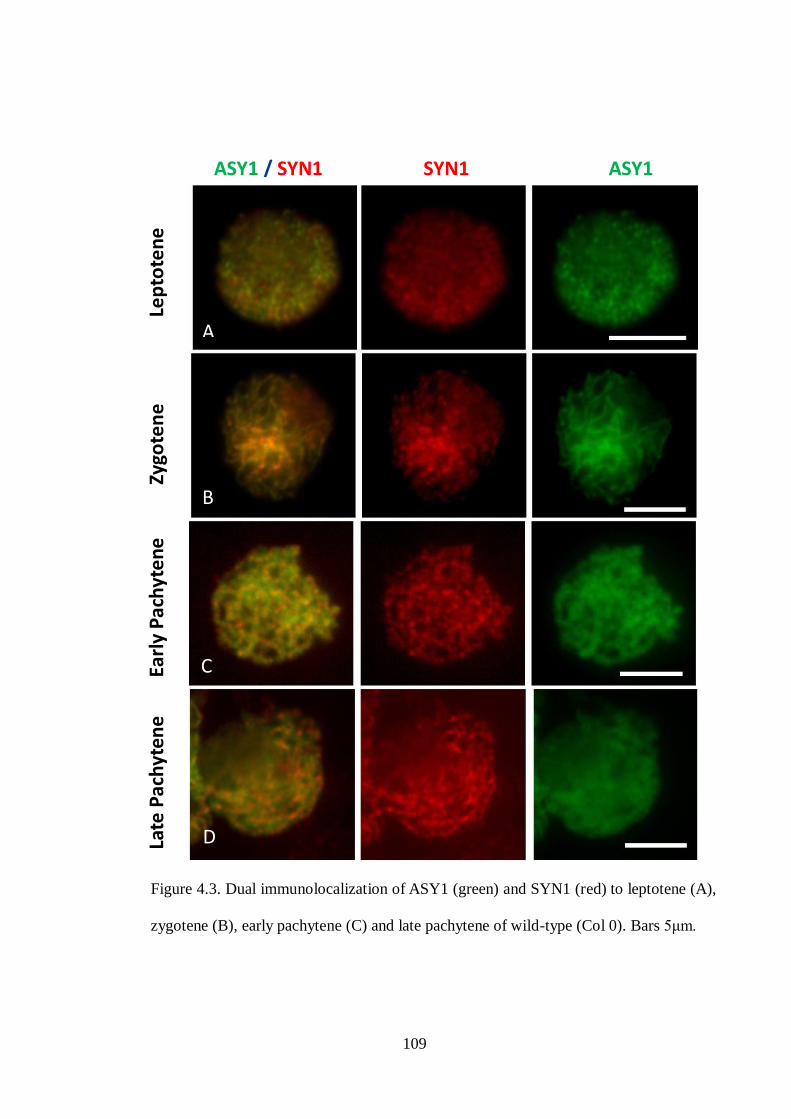
109
Figure 4.3. Dual immunolocalization of ASY1 (green) and SYN1 (red) to leptotene (A),
zygotene (B), early pachytene (C) and late pachytene of wild-type (Col 0). Bars 5μm.
Late
Pac
hyt
ene
Ear
ly P
ach
yte
ne
Z
ygo
ten
e
Le
pto
ten
e ASY1 / SYN1 SYN1 ASY1
C
D
A
B

110
4.2.1.2 Immunolocalization of SYN1 and AtSMC3/AtSCC3 on wildtype (Col 0)
meiocytes
Studies in yeast have shown that REC8 associates with SMC1 and SMC3 forming a cohesin
complex to pair the sister chromatids together (Gruber et al., 2003). However, the association
of SYN1 and SMC proteins in Arabidopsis is not well understood. To investigate the
distribution of SYN1 and AtSMC3, an Arabidopsis SMC3 homolog (Lam et al., 2005),
chromosome spread preparations of wild-type PMCs were examined by using anti-AtSMC3
(rat) and anti-SYN1 (rabbit). The immunostaining result showed that both SYN1 and
AtSMC3 were first detectable as foci at early leptotene (Figure 4.4A). A substantial
proportion of the SYN1 signals co-localized with AtSMC3 as the chromosomes continue to
pair and synapse during zygotene/pachytene stages (Figure 4.4B). I observed that SYN1
partially co-localized with AtSMC3 at the pre-synaptic chromosome regions (Figure 4.4C;
4.5). By late pachytene, both proteins appeared as linear signals and colocalized along the
entire length of chromosomes (Figure 4.4D).
To further investigate the distribution of SYN1 and another cohesin, wild-type (Col 0)
meiocytes were examined by using anti-SYN1 and anti-AtSCC3 (rat), an Arabidopsis
homologue of yeast SCC3. SCC3 is another yeast cohesin subunit that associates with the
cohesin complex (Toth et al., 1999; Nasmyth, 2002). Immunostaining showed that both SYN1
and AtSCC3 proteins appeared as foci at early leptotene (Figure 4.6A), developing into a
strong patchy signal on chromosomes during zygotene. At this stage, the colocalization of
SYN1 and AtSCC3 was detectable on homologous chromosomes but both proteins were less
colocalized on non-synaptic regions (Figure 4.6B). I observed that strong AtSCC3 signals
appeared at pre-synaptic regions while SYN1 patchy signals were distributed evenly on

111
chromosomes (Figure 4.7). Importantly, SYN1 patches appeared not to be in register on
synapsed homologous chromosomes during early pachytene (Figure 4.6C; 4.8). As the
chromosome condensed further, both SYN1 and AtSCC3 appeared as continuous signals
along chromosomes at the end of pachytene (Figure 4.6D). The observation revealed that
substantial proportion of the SYN1 signal co-localized with AtSMC3 along the chromosomes
during zygotene, while colocalization of SYN1 and AtSCC3 increased during pachytene. It is
possible that these observations reflect differences in the dynamics of localization of the
proteins. However, I cannot exclude the possibility that the availability of the protein epitopes
may have influenced these observations.

112
Figure 4.4. Dual immunolocalization of SYN1 (green) and AtSMC3 (red) on meiotic
spreads of wild-type Arabidopsis thaliana. (A) Leptotene; (B) Zygotene; (C) Early
Pachytene; (D) Late Pachytene. Bars 5μm.
SYN1 / AtSMC3 SYN1 AtSMC3 La
te P
ach
yten
e
Ear
ly P
ach
yte
ne
Zy
gote
ne
E
arly
Le
pto
ten
e

113
Figure 4.5. Immunolocalization of SYN1 (green) and AtSMC3 (red) on early pachytene
cell (A) of wild-type.
(C; D; E) are the same cell of (A) from the boxed area. The results show that a few
SYN1 patches do not co-localize with AtSMC3 on the chromosome pre-synaptic region
(white arrow). (B) is the same cell as (A) with DAPI staining. White bars 5μm; red bar
2.5μm.
B A
D E C

114
Figure 4.6. Dual immunolocalization of SYN1 (green) and AtSCC3 (red) on meiocytes
of wild-type. (A) Leptotene; (B) Zygotene; (C) Early Pachytene; (D) Late Pachytene.
Note: Presynaptic region (white arrow) details show in figure 4.7; synapsed
homologous chromosome (yellow arrow) details show in figure 4.8. Bars 5μm.
A
B
C
D
A
Lat
e P
ach
yte
ne
Ea
rly
Pac
hyt
en
e
Zyg
ote
ne
L
ep
tote
ne
SYN1 / AtSCC3 SYN1 AtSCC3

115
Figure 4.7. Immunolocalization of SYN1 (green) and AtSCC3 (red) on zygotene cell
(A) of wild-type.
(B; C; D) are enlargement of cell (A) from the box area. The results show that the SYN1
and AtSCC3 distribution are not similar in the pre-synaptic region (white arrow). (E) is
the same cell of (A) with DAPI staining.Bars 5μm.
C
B
D
E
A

116
Figure 4.8. Immunolocalization of SYN1 (green) to early pachytene (A) of wild-type.
SYN1 patches appear on synapsed homologous chromosome (white arrow). (C; D) are
enlargement of cell (A) from the highlighted area, showing that SYN1 patches do not
appear to be in register. (B) is the same cell of (A) with DAPI staining. White bars 5μm;
red bar 1.25μm.
Note: the patches do not
appear to be in register
C
A B
D

117
4.2.1.3 Immunolocalization of ASY1 and AtSCC3/AtSMC3 on syn1 meiocytes
I have shown that, in wild-type meiocytes, SYN1 fully colocalized with other cohesins
during pachytene. To further investigate the role of SYN1 in the cohesin complex,
chromosome spread preparations of syn1 mutant PMCs were examined by using anti-AtSCC3
(rat) antibody in conjunction with antibody against axis-associated protein, ASY1 (rabbit), to
identify the meiotic stages. Immunostaining showed that ASY1 was loaded abnormally
throughout meiosis. This protein appeared aggregate in early prophase I (Figure 4.9A),
forming into abnormal stretches and short linear signals. By later stages, abnormal thick
continuous signals of ASY1 were observed throughout the meiosis of the syn1 mutant (Figure
4.9B; C). Therefore, it is difficult to identify the early meiotic stages based on the abnormal
distribution of ASY1 in syn1 mutant. Interestingly, cohesin subunit AtSCC3 was not
detectable in the syn1 meiocytes. This result showed that the loading of AtSCC3 on
chromosomes was affected severely in the absence of SYN1 protein, suggesting that
association of AtSCC3 with the cohesin complex is dependent upon SYN1 during early
meiosis I.
To further investigate other cohesins in the syn1 mutant, meiocytes of syn1 were examined
using antibodies against AtSMC3 (rat) and ASY1 (rabbit). Immunostaining of AtSMC3
revealed that the protein was detectable as foci and fuzzy signals on chromosomes during the
early prophase I. This protein appeared to continue developing into fuzzy linear signals which
co-localized with ASY1 (Figure 4.10). This observation suggests that the AtSMC3
polymerization was affected by the absence of SYN1. In conclusion, the appearances of
AtSCC3 and AtSMC3 were affeacted severely in the absence of SYN1. This suggests that the
meiotic cohesin subunit SYN1 is essential for forming the meiotic cohesin complex.

118
Figure 4.9. Immunolocalization of AtSCC3 (red) and ASY1 (green) to meiocytes of
syn1 mutants. ASY1 appears as aggregates in the early meiosis of syn1 mutant,
developing into abnormal linear signals. AtSCC3 is not detectable in meiocytes of syn1
mutant. Bars 5μm.
A
B
C
ASY1 / AtSCC3 ASY1 AtSCC3

119
Figure 4.10. Immunolocalization of AtSMC3 (red) and ASY1 (green) on meiocytes of
syn1 mutants. AtSMC3 appears as fuzzy linear signals. These AtSMC3 fuzzy linear
signals co-localize with ASY1. Bars 5μm.
A
B
ASY1 / AtSMC3 ASY1 AtSMC3

120
4.2.1.4 Immunolocalization of SYN1 and AtZYP1 on wild-type (Col 0) and syn1
meiocytes.
Studies in maize have reported that AFD1/REC8 is a component of the synaptonemal
complex (SC) associating with axial and lateral elements at early meiosis I (Golubovskaya et
al., 2006). To investigate the distribution of SYN1 and the SC protein AtZYP1,
immunolocalization studies were carried out on spread preparations of wild-type pollen
mother cells using anti-SYN1 (rabbit) and anti-ZYP1 (rat). Immunostaining revealed that
SYN1 was detectable throughout the nucleus at leptotene stage, while AtZYP1 was present at
synapsed chromosome regions (Figure 4.11A). During zygotene, AtZYP1 continued to
polymerize along the chromosomes to form short linear signals which colocalized only
partially with SYN1 patches (Figure 4.11B). I found that two AtZYP1 signals appeared next
to a short stretch of AtZYP1 (Figure 4.12; red arrow). This observation suggests that two
AtZYP1 signals were first present side by side on the pre-synaptic chromosomal region
before forming a strong stretch of AtZYP1 (Figure 4.14; Zone B). During pachytene stage,
AtZYP1 appeared as a continuous signal and co-localized with majority of SYN1 running
along the chromosome axes (Figure 4.13; curly braket). I also observed a SYN1-AtZYP1
non-overlapping region, in which two separate SYN1 appeared side by side with AtZYP1
signal (Figure 4.11C and 4.13; white arrow). This observation suggests that a stretch of
AtZYP1 forms at the initiating synaptic/premature synaptic region while SYN1 is still not
recruited to chromosome core region (Figure 4.14 (Zone C)). AtZYP1 continued to
polymerize and colocalized with SYN1 at the mature synapsed region, indicating that the two
separate SYN1 signals come together into a single chromosome axis associated signal. This
could be a reflection of the formation of synaptonemal complex bringing the homologous
chromosomes closer together. Therefore, SYN1 is recruited to the core of chromosomes

121
(Figure 4.14 (Zone D).
To further investigate whether SYN1 is a component of the SC, meiocytes of syn1 were
examined using anti-AtZYP1 and anti-SYN1. Immunostaining results showed that AtZYP1
appeared as aggregates in early meiosis I (Figure 4.15A), developing into very short linear
signals (Figure 4.15B), while SYN1 signal was not detectable. Continuous signal of AtZYP1
was not seen in the syn1 meiocytes. The observation revealed that AtZYP1 signal was present
at the early meiosis I but the polymerization of SC was affected by the absence of SYN1.
.
A

122
Figure 4.11. Immunolocalization of SYN1 (green) and AtZYP1 (red) on meiocytes of
wild-type Arabidopsis. (A) AtZYP1 appears at the leptotene stage. (B) Two AtZYP1
foci (red arrow) are detected in the presynaptic region. One strong AtZYP1 signal (blue
arrow) is detected in the synaptic region. Detail shown in figure 4.12. (C) At pachytene,
AtZYP1 appears as linear signals but two distinct SYN1 signals (white arrow) are
detected between AtZYP1. Bars 5μm.
C
B
A
SYN1 / AtZYP1 SYN1 AtZYP1
Pac
hyt
en
e
Zyg
ote
ne
Lep
tote
ne

123
C B
A
D D”

124
Figure 4.12 Immunolocalization of SYN1 (green) and AtZYP1 (red) to zygotene (A) of
wild-type Arabidopsis. (B, C, D, D”) are enlargement of (A) from the highlighted area.
(B) AtZYP1 signal partially co-localizes with SYN1 some region (blue and red arrows).
It shows that AtZYP1 sits in the middle of two SYN1. (C) Two rows of SYN1 are
detectable on chromosomes during zygotene, indicating that one row of SYN1 patches
appear at one pair of sister chromatids. (D)Two AtZYP1 foci (red arrow) are detected in
pre-synaptic region and one strong AtZYP1 signal (blue arrow) is detected in the
synapsed region. The AtZYP1 signals (red and blue arrow) partially colocalize with
SYN1 (B and C). (D”), an identical picture from (D), is to show that the two AtZYP1
foci ( ) appear next to a strong signal or short stretch of AtZYP1 ( ). White bar 5μm;
red bar 2.5μm.
C B
A
D
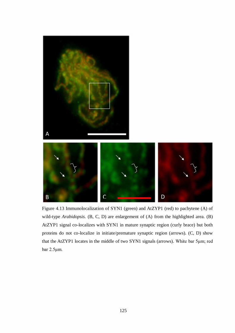
125
Figure 4.13 Immunolocalization of SYN1 (green) and AtZYP1 (red) to pachytene (A) of
wild-type Arabidopsis. (B, C, D) are enlargement of (A) from the highlighted area. (B)
AtZYP1 signal co-localizes with SYN1 in mature synaptic region (curly brace) but both
proteins do not co-localize in initiate/premature synaptic region (arrows). (C, D) show
that the AtZYP1 locates in the middle of two SYN1 signals (arrows). White bar 5μm; red
bar 2.5μm.
C B
A
D

126
Figure 4.14. A diagram showing the dynamics of SYN1 and AtZYP1along
chromosomes.
Zone A: SYN1 signal ( ) is detectable at the non-synaptic chromosomes but not
AtZYP1 ( ). Note: Chromosome axes (Blue).
Zone B: Two AtZYP1 signals appear at the pre-synaptic chromosome region (detail
shown at Figure 4.12; red arrow).
Zone C: Two separate SYN1 appear side by side with an AtZYP1 at initiate synaptic/
premature synapsed region (detail shown at Figure 4.11C (white arrow) and 4.12 (blue
arrow) and 4.13 (white arrow)).
Zone D (Mature fully synapsed region): AtZYP1 appears as a continuous signal and
completely co-localizes with SYN1 running along the chromosome axes.

127
Figure 4.15. Immunolocalization of AtZYP1 (red) and SYN1 (green) on meiocytes of
syn1 mutants. AtZYP1 appears as aggregates (A) at the beginning of SC formation,
developing into stretches (B). SYN1 is not detected in meiocytes of the syn1 mutant. The
chromosomes are counterstained with DAPI (blue). Bars 5μm.
A
B
AtZYP1/SYN1 /DAPI AtZYP1 SYN1 DAPI

128
4.2.1.5 Immunolocalization of AtZYP1 and ASY1 on syn1 meiocytes.
Immunostaining studies in syn1 meiocytes revealed that AtZYP1 was present in early
prophase I. It is highly possible that the axis-associated protein ASY1 might co-localize with
AtZYP1 in the syn1 mutant. To investigate the distribution of ASY1 and AtZYP1 proteins in
more detail, immunolocalization studies were carried out on spread preparations of PMCs
from wild-type (Col 0) and the syn1 mutant. The result, in wild-type (Col 0) as a control,
showed that ASY1 and AtZYP1 appeared as a continuous signal running along the
chromosome axis during early prophase I (Figure 4.16A). In contrast, the distribution of
ASY1 and AtZYP1 along the chromosome was affected in syn1 meiocytes. Both proteins
appeared as aggregates in early prophase I. Interestingly, ASY1 was seen developing into
abnormal thick and linear signals, while AtZYP1 appeared as very thick and short linear
signals or stretches in the later stages (Figure 4.16B; C). The signals of AtZYP1 either
completely or partially co-localized with ASY1 in syn1 meiocytes. This result showed that
both axis formation and synapsis are compromised in the absence of SYN1.
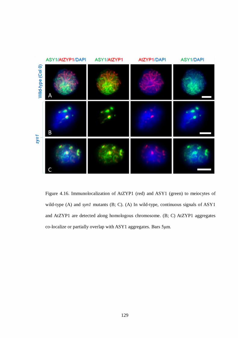
129
Figure 4.16. Immunolocalization of AtZYP1 (red) and ASY1 (green) to meiocytes of
wild-type (A) and syn1 mutants (B; C). (A) In wild-type, continuous signals of ASY1
and AtZYP1 are detected along homologous chromosome. (B; C) AtZYP1 aggregates
co-localize or partially overlap with ASY1 aggregates. Bars 5μm.
A
B
C

130
4.2.1.6 Immunolocalization of AtRAD51 and ASY1 on wild-type (Col 0) and syn1
meiocytes.
Synapsis is defined by the formation of synaptonemal complex (SC). In yeast and
mammals, synapsis requires the formation of DNA double strand breaks (DSBs), which are
catalysed by SPO11 protein, to initiate meiotic recombination (Lichten, 2001; Burgess, 2002).
The previous immunostaining result showed that the distribution of SC protein, AtZYP1, was
abnormal in the syn1 mutant. This suggests that meiotic recombination might be affected in
the absence of SYN1. To investigate how the meiotic recombination proteins behave in syn1
mutant, antibodies were used against the axis-associated protein ASY1 and recombination
protein AtRAD51, an Arabidopsis homolog of yeast RAD51 (Doutriaux et al., 1998; Li et al.,
2004). AtRAD51 is essential for chromosome pairing, synapsis and the repair of DNA DSBs
(Li et al., 2004). Immunostaining in wild-type (Col 0) as a control showed that ASY1
appeared as linear signals along chromosomes while foci of AtRAD51 were widely
distributed throughout the nucleus at early prophase I (Figure 4.17A). In contrast,
immunolocalization of ASY1 and AtRAD51 showed aberrant distribution in syn1 meiocytes.
Both proteins appeared as aggregates in early prophase I (Figure 4.17B). Interestingly,
substantial aggregates of AtRAD51 accumulated around ASY1 (Figure 4.17C). In later stages,
ASY1 developed into abnormal linear signals along the chromosomes while AtRAD51
signals reduced significantly (Figure 4.17D). This data showed that AtRAD51 foci were not
widely present along the homologous chromosomes of syn1 mutant, suggesting that the
chromosome pairing, synapsis and DSB repair are disturbed by the loss of SYN1.

131
Figure 4.17. Immunolocalization of AtRAD51 (red) and ASY1 (green) to meiocytes of
wild-type (A) and syn1 mutant (B; C; D). (A) In wild-type, AtRAD51foci distribute
evenly on linear ASY1 signals. (B; C) In syn1 mutant, substantial AtRAD51 aggregates
are found around ASY1. (D) Few AtRAD51 foci appear on linear ASY1. Bars 5μm.
A
B
C
D
132
4.2.1.7 Immunolocalization of AtMLH1 and ASY1 on wild-type (Col 0) and syn1
meiocytes
The immunolocalization result showed that the distribution of AtRAD51 was affected by the
absence of SYN1. To investigate the impact of loss of SYN1 on recombination in more detail,
immunolocalization was carried out using antibodies against ASY1 and the recombination
proteins AtMLH1, an Arabidopsis homologue of the E. coli MutL (Jean et al., 1999). In yeast,
MLH1 acts as a heterodimer with MLH3, which plays an important role in crossovers of
meiocytes (Wang et al., 1999). In Arabidopsis, AtMLH1, in wild-type meiocytes, colocalizes
with AtMLH3 at pachytene. The number of foci is ~10 which is close to the number of COs.
AtMLH1 is found abnormally in the nucleolus of the Atmlh3 mutant. Analysis of chiasma
frequency reveals an approximately 60% reduction in crossovers occurs in the absence of
AtMLH3 (Jackson et al., 2006), suggesting that AtMLH1 and AtMLH3 play an important role
in meiotic crossing over. Immunostaining studies in wild-type meiocytes showed that both
ASY1 and AtMLH1 proteins were detectable in early prophase I. ASY1 appeared as linear
signals, while AtMLH1 foci were widely distributed in the nucleus of zygotene cell (Figure
4.18A). In contrast to wild-type, immunostaining of AtMLH1 showed abnormal distribution
on ASY1 in meiocytes of syn1 mutant. AtMLH1 appeared as aggregates throughout meiosis I.
The majority of AtMLH1 signals co-localized with the ASY1 aggregates (Figure 4.18B).
After ASY1 elongation, substantial AtMLH1 signal still remained on the ASY1 aggregates
(Figure 4.18C; D). This result revealed that the distribution of the recombination protein
AtMLH1 was affected in syn1 mutant.

133
Figure 4.18. Immunolocalization of AtMLH1 (red) and ASY1 (green) to meiocytes of
wild-type (A) and syn1 mutant (B; C; D). (A) In wild-type, AtMLH1 distributes evenly
on linear ASY1. (B; C; D) In syn1 mutant, the majority of AtMLH1 aggregates co-
localize with ASY1 aggregates. Bars 5μm.
A
B
C
D

134
Table 4.1 Immunolocalization of various antibodies in meiocytes of wild-type and syn1
mutant.
Antibodies
Meiocytes
Anti-
SYN1
Anti-
ASY1
Anti-
AtSMC3
Anti-
AtSCC3
Anti-
AtZYP1
Anti-
AtRAD51
Anti-
AtMLH1
Wild- type
(Col 0)
√
√
√
√
√
√
√
syn1 mutant
X
√*
√*
X
√*
√*
√*
Note: √: protein signal appears normal in meiocytes
√*: protein signal appears abnormal in meiocytes
X: protein signal is not detectable in meiocytes

135
4.3 Discussion
To study the distribution of SYN1 in early meiosis, I raised an anti-SYN1 antibody against
the central region of SYN1 (amino acid 207 to 384). This central region was chosen due to
lack of similarity to kleisins SYN2, SYN3 and SYN4 which are involved in cohesion during
mitosis (Dong et al., 2001; Jiang et al., 2007; Schubert et al., 2009). The immunocytological
analysis of SYN1 showed that SYN1 appeared in meiocytes but not in mitotic cells.
Furthermore, SYN1 signals were not detected in the syn1 mutant (Table 4.1). This result
indicates that this antibody is specific for the meiotic cohesin SYN1.
4.3.1 Are SYN1 patches localized to the sites of DNA double strand break?
A previous immunolocalization report has suggested that SYN1 appears on chromosome
arms but not at the centromere during meiotic prophase I (Cai et al., 2003). Immunostaining
of SYN1 revealed that the protein appeared as foci in early meiosis, forming into a more
linear, yet patchy pattern during zygotene. Continuous signals of SYN1 were detectable
during late pachytene. It is difficult to see any gap in SYN1 linear signals, suggesting that
SYN1 could be also located in the centromeres.
A recent report in maize has shown that AFD1/REC8 protein appears as foci in early
meiosis, developing into discontinuous short stretches and spots with varying intensities
(Wang et al., 2009). A similar result was seen in Arabidopsis; SYN1 appeared as foci in early
meiosis, forming patches during zygotene stage. These patches did not appear to be in
register. Studies in yeast mitotic cells have shown that cohesin is recruited to DNA double-
strand break sites (Strom et al., 2004; Lowndes and Toh, 2005). Furthermore, the meiotic
kleisin subunit REC8 is expressed in human tumour cells after gamma irradiation (Erenpreisa

136
et al., 2009). It is possible that SYN1 might have a role during DNA DSB. Therefore, I
suggest that SYN1 might accumulate on the site of DNA double-strand break forming a
domain or patchy signal.
4.3.2 Localization of AtSCC3 and AtSMC3 along meiotic chromosomes is dependent
upon SYN1.
In yeast meiocytes, the cohesion of sister chromatids is essential for chromosome
segregation during anaphase I. This cohesion is established by cohesins which include SMC1,
SMC3, SCC3 and REC8 proteins (Gruber et al., 2003). In Arabidopsis, immunostaining of
SYN1 and AtSMC3 revealed that the proteins appeared as foci in early leptotene. SYN1 and
AtSMC3 colocalization on meiotic chromosomes increased during zygotene and pachytene. I
suggest that SYN1 replaces the mitotic kleisin subunit SYN2 and SYN4 to associate with
SMC proteins. Therefore, substantial SYN1 signal is colocalized with SMC3 during zygotene
and pachytene. Data from yeast showed that most SCC1/RAD21 is replaced by REC8 during
pre-meiotic DNA replication (Klein et al., 1999; Gruber et al., 2003). However, few SCC1
signals are still detectable in pachytene cells (Klein et al., 1999). Currently, we do not have
SYN2 and SYN4 antibodies to examine the appearance of mitotic kleisin cohesins in early
meiosis.
Few publications are available for cohesin subunit SCC3. It has been reported that yeast
SCC3 binds to the COOH-terminal half of SCC1 (Nasmyth, 2002). This binding probably is
to stabilize the cohesin complex throughout mitotic cells. In Arabidopsis, a recent report
showed that AtSCC3 is involved in both mitotic and meiotic divisions (Chelysheva et al.,
2005). Unfortunately, the co-localization of AtSCC3 and kleisin subunits, SYN1, SYN2 and

137
SYN4, is not understood. I have used antibodies against AtSCC3 and SYN1 in chromosome
spreads of wild-type meiocytes. The immunostaining result showed that a small amount of
SYN1 and AtSCC3 co-localized on the pre-synaptic chromosome region. During pachytene,
colocalization of SYN1 and AtSCC3 proteins increased rapidly along the full length of
chromosomes. These observations reflect differences in the dynamics of localization of the
proteins. However, I showed that AtSCC3 was not observed in syn1 meiocytes. This result
revealed that AtSCC3 loading on chromosomes is dependent upon SYN1. In contrast, fuzzy
AtSMC3 signals were found in syn1 meiocytes. I suggest that AtSMC3 appears on
chromosome before SYN1. However, the SMC proteins could not form a stable cohesin
complex in the absence of SYN1. As a consequence, abnormal loose chromatin structures
appear at early meiosis.
4.3.3 Formation of the synaptonemal complex is disrupted in a syn1 mutant
A report in rat revealed that REC8 appears on chromosomes and forms axial-element
structures (REC8-AEs) during premeiotic S-phase. It has been suggested that REC8 provides
a platform for the axial element assembly without requiring SMC1, SMC3, SCP2 and SCP3
(Eijpe et al., 2003). Interestingly, immunostaining of SYN1 and ASY1, an axis-associated
protein required for synapsis and crossover formation, in wild-type showed that SYN1
appeared as patches running along ASY1 linear signals at zygotene . By late pachytene, SYN1
co-localized with ASY1 along the chromosomes. This result did not show that SYN1 provides
a basis for axis-associated protein assembly. In maize afd1/rec8 mutant, stretches and short
lines of ASY1 are observed during meiosis. It has been suggested that AFD1/REC8 is
required for controlling the ASY1 elongation during early meiosis (Golubovskaya et al.,
2006). However, ASY1 in the syn1 mutant appeared as an aggregate in early meiosis,

138
developing into abnormal linear signals in later stages. This result showed that SYN1 does not
affect the localization and normal elongation of ASY1. Therefore, I suggest that SYN1 is
required for maintenance of axis-associated protein on chromosomes during axial-element
assembly and formation.
Once the axis-associated proteins on chromosomes are stabilized by SYN1, the
synaptonemal complex (SC) protein AtZYP1 is loaded on the axial elements of the
homologous chromosomes. I have detected two AtZYP1 signals side by side in the pre-
synaptic region, developing into one patchy signal which was surrounded by two regions of
SYN1. Finally, AtZYP1 co-localized with SYN1 at the synaptic site. It has been suggested
that cohesins are essential for the formation of the synaptonemal complex (Klein et al., 1999;
Eijpe et al., 2000; Pelttari et al., 2001). Interestingly, AtZYP1 appeared as aggregates and
short stretches in syn1 meiocytes. I suggest that SYN1 is not essential for the formation of SC
but is required for SC polymerization and elongation. In conclusion, the distribution of both
ASY1 and AtZYP1 proteins was abnormal in the syn1 mutant. This indicates that both axis
formation and synapsis are compromised in the absence of SYN1.
4.3.4 SYN1 is essential for meiotic recombination progression in Arabidopsis
Our immunostaining of AtZYP1 showed that the protein appeared as aggregates in the syn1
mutant and co-localized with ASY1 aggregates during early prophase I. Recently,
transmission electron microscopy (TEM) analysis showed that abnormal short stretches of SC
and polycomplex structures associated with chromatin are detected in syn1 mutant (Zhao et
al., 2006). Clearly, this report is entirely consistent with our finding.

139
According to a previous report, it was suggested that ASY1 plays an important role in
coordinating the activity of the meiotic recombinase, AtDMC1, to promote interhomologue
recombination (Sanchez-Moran et al., 2007). I believe that abnormal ASY1 in syn1 might
affect the distribution of recombination proteins. Our data showed aberrant distribution of
meiotic recombination proteins, AtRAD51 and AtMLH1, in the syn1 meiocytes. AtRAD51,
an Arabidopsis protein that is required for synapsis and DNA DSB repair (Doutriaux et al.,
1998; Li et al., 2004), appeared as aggregates and accumulated around ASY1. This indicates
that DSB repair was disrupted in the syn1 mutant. A similar result was seen in maize; RAD51
aggregates were observed in the absence of AFD1/REC8 (Golubovskaya et al., 2006).
Another recombination protein, AtMLH1, an Arabidopsis protein which localizes to late
recombination nodules at late pachytene and marks crossover sites (Franklin et al., 2006;
Jackson et al., 2006), appeared as aggregates and co-localized with ASY1 aggregates. This
observation suggests that both AtRAD51 and AtMLH1 proteins co-localize as large
aggregates together with ASY1, indicating that the processes of meiotic recombination are
affected in the absence of SYN1. As a consequence, this can lead to chromosome
fragmentation. I suggest that SYN1 is essential for meiotic recombination progression and
DSB repair.

140
Chapter 5
An additional role for SYN1
5.1 Introduction
The initial event of meiotic recombination is triggered by the induction of DSBs by the
protein SPO11 (Keeney et al., 1997). The DSBs are resected from the 5‟ to 3‟ end by the
recombination proteins RAD50 and MRE11 (Smith and Nicolas, 1998). A complex of
recombination proteins containing two RecA homologs RAD51 and DMC1 binds to single-
stranded DNA ends created by RAD50 and MRE11, forming a nucleoprotein filament
(Bishop, 1994). This filament invades homologous chromosome DNA. This process is called
single-strand invasion. RAD51 is also required for DNA double strand break repair. Recently,
two Arabidopsis RAD51 parologous genes, AtXRCC3 and AtRAD51C, have been reported to
be involved in meiotic recombination and DSB repair (Bleuyard et al., 2004a; Li et al., 2005).
In the absence of several meiotic recombination proteins, DNA DSBs are not repaired. As a
result, chromosome fragmentation is observed in Atxrcc3 and Atrad51c mutants. Interestingly,
chromosome fragmentation is also found in the absence of cohesin proteins including maize
afd1, Sordaria sm-rec8, mouse rec8, Arabidopsis syn1, worm rec8 and rice OsRAD21-4
depletion line (Bai et al., 1999; Bhatt et al., 1999; Yu and Dawe, 2000; Pasierbek et al., 2001;
Xu et al., 2005; Zhang et al., 2006; Storlazzi et al., 2008).
Recent studies in yeast cells have shown that mitotic cohesin accumulates at DNA double
strand break (DSB) sites where it might facilitate DSB repair (Lowndes and Toh, 2005;
Kugou et al., 2009), but the association of SYN1 and DSB site has not been reported. To
investigate whether SYN1 plays an important role during DNA double-strand breaks, three

141
double gene knockout mutants were constructed, syn1-/-
xAtspo11-1-4-/-
; syn1-/-
xAtdmc1-/-
and
syn1-/-
xAtrad51c-/-
. The double knockout plants were identified by using PCR to determine the
T-DNA insertion sites. Cytological studies conducted on male meiocytes in wild-type (Col 0),
mutants and double knockout mutants using chromosome spread preparations stained with
4,6-diamidino-2-phenylindole (DAPI). The cytological analyses of wild-type and syn1-/-
have
been reported in chapter 3. In this chapter, the data from three mutants, Atspo11-1-4-/-
;
Atdmc1-/-
; Atrad51c-/-
; and three double gene knockout mutants, syn1-/-
x Atspo11-1-4-/-
; syn1-/-
x Atdmc1-/-
and syn1-/-
xAtrad51c-/-
are presented. To further investigate whether SYN1 is
recruited at the DSB sites, I analysed the distribution of SYN1 in Atspo11-1-4-/-
. In addition,
the DSB induction using cisplatin was carried out in meiocytes of Atspo11-1-4-/-
and syn1-/-
to
examine the role of the SYN1 protein during DNA DSBs.
5.2 Results
5.2.1 Cytogenetic analysis of syn1-/-
and Atspo11-1-4-/-
double knockout mutant
To determine the basis of chromosome fragmentation in the syn1 mutant, I constructed a
double knockout mutant of SYN1 and AtSPO11-1 genes. AtSPO11-1 is one of the three SPO11
paralogues in Arabidopsis that in conjunction with AtSPO11-2 induces DSBs to initiate
meiotic recombination (Grelon et al., 2001; Stacey et al., 2006). A previous report
(Chelysheva et al., 2005) showed that there is no chromosome fragmentation in the meiocytes
of a double gene knockout of syn1-/-
/Atspo11-1-1-/-
, but little information was provided
regarding early meiotic prophase 1. Recent evidence showed that a few bivalents are found in
an Atspo11-1-1 mutant (Grelon et al., 2001; Chelysheva et al., 2005). This suggested that
Atspo11-1-1 plants are able to produce a truncated SPO11-1 protein which is partly functional
(Sanchez-Moran et al., 2007), which led to search for another mutant. A T-DNA insertion line

142
(WiscDsLox461-464J19) was obtained from the NASC stock. The T-DNA insertion locates in
Exon 14 of the AtSPO11-1 gene (At3g13170). It has been confirmed that the T-DNA insertion
line (WiscDsLox461-464J19) is a null mutant (PhD thesis of Nicola Robert at The University
of Birmingham UK., 2009). The WiscDsLox461-464J19 line referred to as Atspo11-1-4-/-
. A
syn1-/-
and Atspo11-1-4-/-
double knockout mutant was constructed for analysis.
Cytological analysis of Atspo11-1-4-/-
showed normal thin thread-like chromosomes at early
prophase I (Figure 5.1A). Short univalents were observed during chromosome condensation
at a diakinesis-like stage (Figure 5.1B). At metaphase I, 10 univalents were aligned at the
equatorial plate for the first meiotic division (Figure 5.1C). At anaphase I, the 10 univalents
moved randomly to the poles (Figure 5.1D). At anaphase II, the sister chromatids were pulled
apart and sister chromosomes moved toward the opposite poles (Figure 5.1E).
In the syn1-/-
/ Atspo11-1-4-/-
double knockout mutant, loose and tangled chromosome
structures appeared in early prophase I (Figure 5.2A). Upon further condensation, the
chromosomes appeared phenotypically similar to that of Atspo11-1-4-/-
at a diakinesis-like
stage (Figure 5.1B; 5.2B). At metaphase I, ten univalents were aligned at the centre of each
meiocyte in readiness for the first meiotic division (Figure 5.2C). The sister chromatids of
each pair were pulled apart to opposite pole of the cell at anaphase I. A total of 20 separated
sister chromatids per meiocyte were observed during the end of first meiotic division (Figure
5.2D; E). There was no evidence of fragmentation and each pole contained 10 sister
chromatids. During second meiotic division, the sister chromatids are randomly segregated.
This result showed that the Atspo11-1-4-/-
mutation suppressed the chromosome fragmentation
phenotype of syn1-/-
plants, indicating that the chromosome fragmentation in syn1-/-
is

143
AtSPO11-1-dependent.

144
Figure 5.1. Meiotic stages in pollen mother cells of the Atspo11-1-4 mutant of A.
thaliana.
(A) Thread-like and unsynapsed chromosomes appear at early meiotic prophase I. (B)
condensed univalents at a diakinesis-like stage. (C) Metaphase I showing 10 univalents.
(D) 9 univalents move randomly to the poles one univalent is seen lagging on middle of
the cell at anaphase I. (E) Sister chromatids are pulled apart during anaphase II (F)
Sister chromatids are segregated to produce four sets of chromosome at telophase II.
The number of chromosomes in each set is not equal to the haploid set of chromosomes.
Bars 10μm.

145
Figure 5.2. Meiotic stages in pollen mother cells of the syn1-/-
/ Atspo11-1-4-/-
double
knock-out mutant of A. thaliana. (A) Abnormal loose and tangled chromosome
structures appear before chromosome condensation. (B) Chromosomes condense at a
diakinesis-like stage (C) Ten univalents appear in metaphase I (D)Sister chromatids are
pulled apart to opposite pole at early anaphase I (E). Chromosomes are segregated
randomly at anaphase II (F). Four sets of chromosome in tetrads. One of the set contains
four chromosomes. Bars 10μm.

146
5.2.2 Immunolocalization of SYN1 on Atspo11-1-4-/-
.
To investigate SYN1 distribution in the absence of double strand breaks, wild-type (Col 0)
and Atspo11-1-4-/-
meiocytes were examined using antibodies against ASY1 (rat) and SYN1
(rabbit). In order to allow direct comparison between both meiocytes, the image capture
conditions were set up with the same exposure time and index of brightness and contrast.
Immuno-localization of ASY1 and SYN1 showed that both proteins appeared as strong
signals in wild-type meiocytes. Patchy SYN1 signals were distributed on the continuous
ASY1 signals in early prophase I (Figure 5.3). In contrast, weak SYN1 foci and strong ASY1
linear signals were detected on Atspo11-1-4-/-
meiocytes. This observation showed that the
SYN1 loading was affected in the Atspo11-1-4-/-
mutant.
To confirm the decrease of SYN1 signal in Atspo11-1-4-/-
, wild-type and Atspo11-1-4-/-
mutant were treated with Bromodeoxyuridine (BrdU). BrDU is a thymidine analogue that can
be incorporated into the DNA during the S-phase of the cell cycle. The incorporation of BrdU
can be detected immunocytologically by anti-BrdU antibodies. The BrdU labelling method
has been described and published as a useful tool for analysing the timing and relationships of
meiotic events (Armstrong et al., 2003). Immunolocalization of SYN1 was carried out in
chromosome preparations from wild-type and Atspo11-1-4-/-
meiocytes following BrdU pulse-
labelling. The immunostaining result showed that strong SYN1 signals were detectable in
wild-type meiocytes at one hour post-S phase (G2 stage). According to a meiotic time-course
report, axis-associated protein ASY1 is first detectable at five hour post-S phase (Armstrong
et al., 2003), indicating that SYN1 appears before ASY1 in meiocytes of wild-type. Under the
same conditions, the SYN1 signals were substantially reduced in Atspo11-1-4-/-
meiocytes
(Figure 5.4 A; B). At 30 hours post-S phase (late pachytene), strong SYN1 signals were still

147
apparent in the wild-type meiocytes but weak SYN1 signals were detected in Atspo11-1-4-/-
(Figure 5.4 C; D). These results show that SYN1 loading was reduced at the onset of G2 stage
until pachytene in the Atspo11-1-4-/-
mutant. According a report in yeast mitotic cells, cohesin
is recruited to DNA DSB sites where it might facilitate DNA DSB repair (Lowndes and Toh,
2005; Kugou et al., 2009). These suggest that SYN1 loading is affected due to the absence of
DNA DSBs.

148
Figure 5.3. Dual immunolocalization of ASY1 (green) and SYN1 (red) on
prophase I nuclei of wild-type (A) and Atspo11-1-4-/-
(B) mutant. Substantial
SYN1 foci are localized to chromosomes in wild-type (A) but not in Atspo11-1-4-/-
.Bars 5μm.
D
A
B
SYN1/ASY1 ASY1 SYN1 A
tsp
o1
1-1
-4-/
-
W
ild-t
ype
(C
ol 0
)

149
Figure 5.4. BrdU (green) pulse-labelling combined with immunocytological analysis in
wild-type and Atspo11-1-4-/-
meiocytes. Strong SYN1 signal (red) in wild-type
meiocytes is shown at 1h (A) and 30 h (C) post-BrdU pulse but weak SYN1 signal is
shown in Atspo11-1-4-/-
at 1h (B) and 30h (D) post-BrdU pulse. The chromosomes are
counterstained with DAPI (blue). Bars 5μm.
A
B
C
D
Ats
po
11
-1-4
-/-
W
ild-t
ype
(Co
l 0)
Ats
po
11-1
-4-/
-
W
ild-t
ype
(C
ol 0
)
SYN1/BrdU/DAPI BrdU SYN1
SYN1/BrdU/DAPI BrdU SYN1
1 hour post-BrdU pulse
30 hours post-BrdU pulse

150
5.2.3 Immunolocalization of SYN1 on cisplatin-treated Atspo11-1-4-/-
meiocytes
Previously, I have shown that SYN1 signal was reduced in Atspo11-1-4-/-
meiocytes by
using BrdU pulse-labelling combined with immunocytological analysis. To investigate
whether SYN1 loads on sister chromatids during DSB formation, meiocytes of Atspo11-1-4-/-
were treated with cisplatin. Cisplatin (cis-diaminedichloridoplatinum II) forms platinum
chemical complexes which react with DNA to form intrastrand and interstrand crosslinks. The
excision of the DNA crosslink creates a DSB (Olive and Banath, 2009). In this study,
inflorescences of Atspo11-1-4-/-
were immersed in cisplatin solution at concentrations of
2.5μM and 5.0μM for a 2 hour pulse. At the same time, inflorescences from wild-type and
Atspo11-1-4-/-
were immersed in tap water as positive and negative controls. Buds were
collected and dissected at 28 hour post-cisplatin pulse. Cisplatin-treated buds with high
concentration (0.5 μM) were excluded in the following examination because the buds become
pale and wilted. Immunolocalization studies were carried out on spread preparations with
anti-SYN1 (rabbit) and anti-ASY1 (rat) antibodies. The pixel intensity of meiocytes was
analysed by using ImageJ (Appendix 5.1). Immuno-localization analysis showed (Figure 5.5
and 5.6) that the intensity of SYN1 was different between wild-type and untreated Atspo11-1-
4-/-
. The wild-type signal was much stronger than the untreated Atspo11-1-4-/-
. The statistical
analysis showed that there is significant difference between the wild-type and untreated
Atspo11-1-4-/-
(t-test, p<0.001; n=50). Furthermore, the pixel intensity of SYN1 was also
significantly different between cisplatin-treated Atspo11-1-4-/-
and untreated Atspo11-1-4-/-
(t-
test, p<0.001; n=50). The signal in cisplatin-treated Atspo11-1-4-/-
was stronger than the
untreated Atspo11-1-4-/-
. Although SYN1 signal increased after cisplatin treatment in Atspo11-
1-4-/-
, the wild-type signal was still stronger than the cisplatin-treated Atspo11-1-4-/-
. The
statistical analysis showed that there is significant difference in the intensity between wild-

151
type and cisplatin-treated Atspo11-1-4-/-
(t-test, p=0.0105; n=50). These results show that the
SYN1 signal is increased after cisplatin treatment in Atspo11-1-4-/-
, indicating that SYN1
loading can be restored in the absence of AtSPO11-1 by artificially inducing DSBs with
cisplatin. It is also important to investigate whether meiotic recombination can be triggered in
the Atspo11-1-4-/-
mutant by using cisplatin. Therefore, the meiocytes of cisplatin-treated
Atspo11-1-4-/-
were examined by using antibodies against the synaptonemal complex protein
AtZYP1. Immunolocalization of AtZYP1 revealed that the protein appeared as strong foci and
short linear signals in cisplatin-treated Atspo11-1-4 meiocytes (Figure 5.7). This result showed
that SC formation can be initiated in the absence of AtSPO11-1 protein by using cisplatin.

152
Figure 5.5. Dual immunolocalization of ASY1 (green) and SYN1 (red) on prophase I
nuclei of wild-type (A) and untreated Atspo11-1-4-/-
(B) and cisplatin-treated
Atspo11-1-4-/-
mutant (C). Strong patchy signals of SYN1 appeared on chromosome
in wild-type but weak SYN1 signals in Atspo11-1-4-/-
. After Atspo11-1-4-/-
treated
with cisplatin, strong signals of SYN1 are observed on chromosomes. The
chromosomes are counterstained with DAPI (blue). Bars 5μm.
A
B
C
SYN1/ DAPI ASY1 SYN1
Wild
-typ
e (C
ol 0
)
Ats
po
11
-1-4
-/-
wit
ho
ut
cisp
lati
n
trea
tmen
t
Ats
po
11
-1-4
-/-
wit
h c
isp
lati
n
trea
tmen
t

153
Figure 5.6. Signal intensity analysis of SYN1 on prophase I nuclei of wild-type (Col 0),
cisplatin-treated Atspo11-1-4-/-
and untreated Atspo11-1-4-/-
mutants.
(A) Signal intensity of Atspo11-1-4-/-
is significantly lower than that of wild-type
(p<0.001; n=50). After cisplatin treatment in Atspo11-1-4-/-
, the SYN1 signal is
significantly higher than that of cisplatin free Atspo11-1-4-/-
(p<0.001; n=50) but is still
lower than that of wild-type (0.01<p<0.05; n=50). The corresponding p values are
calculated in the table (B) and Appendix 5.1.
Wild-type (Col 0) Atspo11-1-4-/-
with
cisplatin treatment
Atspo11-1-4-/-
with
cisplatin free
Total (n=50) 3052.509 2620.908 915.52
Mean 61.05018 52.41816 18.3104
Standard
Deviation
19.887303 12.37164835 11.70871021
Standard Error 2.81251633 1.749632068 1.655877558
A)
B)
Pix
al in
ten
sity
of
SYN
1

154
Figure 5.7. Immunolocalization of AtZYP1 (green) on prophase I nuclei of wild-
type (A) and cisplatin-treated Atspo11-1-4-/-
mutant (B; C). The chromosomes are
counterstained with DAPI (blue). Continuous signals of AtZYP1 are observed in
meiocytes of wild-type (A). Few strong patchy (B) and short linear (C) signals are
detectable in cisplatin-treated Atspo11-1-4-/-
meiocytes. Bars 5μm.
A
B
C
Ats
po
11-1
-4-/
-
cisp
lati
n t
reat
me
nt
DAPI/AtZYP1 AtZYP1 DAPI
Wild
-typ
e (
Co
l 0)

155
5.2.4 Cytogenetic analysis in syn1-/-
and Atrad51c-/-
double knockout mutant
Recently, research in rat showed that RAD51 and DMC1 coimmunoprecipitate with REC8. It
has been proposed that the cohesins provide a platform for the assembly of recombination
proteins after S phase (Eijpe et al., 2003). To test the relationship between SYN1 and
recombination proteins during the formation of DSB, I chose AtRAD51C, an Arabidopsis
RAD51C ortholog. It has been suggested that AtRAD51C plays an important role in DSB
repair (Li et al., 2005). Cytological analysis of Atrad51c-/-
showed thin threads and
unsynapsed chromosomes during the early prophase I (Figure 5.8A). Following the
chromosome condensation, the chromosome structure was seriously disrupted. Tangled
chromosome fragments were observed, which aligned at the equatorial plate in preparation for
the first meiotic segregation (Figure 5.8B). Chromosome bridges and fragments were
observed at early anaphase I (Figure 5.8C). A substantial number of chromosome fragments
remained in the middle of the meiocyte after five chromosomes migrated toward each pole
(Figure 5.8D). Chromosome fragmentation and mis-segregation appeared throughout
prophase II (Figure 5.8 E; F).
A syn1-/-
and Atrad51c-/-
double knockout mutant was constructed. The cytological analysis
showed that chromosome abnormalities were found throughout meiosis. During early
prophase I, chromosomes can be seen as loose and tangled structures (Figure 5.9A; B). The
tangled chromosomes were aligned at the equatorial plate and segregated towards opposite
poles during the first meiotic segregation (Figure 5.9C). Some chromosome fragments
remained in the middle of meiocytes and other chromosome fragments moved to the two
poles at anaphase I (Figure 5.9D). Chromosome fragmentation and mis-segregation appeared
from metaphase I to prophase II, which is very similar to both Atrad51-/-
and syn1-/-
mutants.

156
Cytological analysis showed that chromosome phenotype at early prophase I is significantly
different between syn1-/-
/Atrad51c-/-
and Atrad51c-/-
mutants. Currently, it is impossible to
draw a conclusion whether SYN1 and AtRAD51C interact during early meiosis I.

157
Figure 5.8. Meiosis stages in pollen mother cells of the Atrad51c-/-
mutant of A.
thaliana.
(A) Thin and unsynapsed chromosomes appear during early prophase I. (B) Abnormal
condensed chromosomes and bridges are seen at metaphase I. (C) Chromosome
fragments are randomly segregated at early anaphase I. (D) Each pole of the cell
contains 4 or 5 chromosome fragments but a large number of chromosome fragments
remain in the middle of the meiocytes during late anaphase I. (E) Chromosomes are
randomly segregated at anaphase II (F) Chromosome fragments distribute randomly at
telophase II. Bars 10μm.

158
Figure 5.9. Meiotic stages in pollen mother cells of syn1-/-
/ Atrad51c-/-
of A. thaliana.
(A) Loose and tangled chromosomes appear during early prophase I. (B) Tangled
chromosomes appear after chromosome condensation. (C) Bridges and fused
chromosomes are aligned on equatorial plate at metaphase I (D) During first meiotic
division, 9 or 10 chromosome fragments locate in each pole and many chromosome
fragments still remain in the middle of meiocyte. (E) Chromosome fragments are
randomly segregated at anaphase II. (F) Chromosome fragments distribute unevenly in
four set of haploid products. Bars 10μm.

159
5.2.5 Cytogenetic analysis in a syn1-/-
/Atdmc1-/-
double knockout mutant
To find out whether SYN1 has a role in DNA double strand break (DSB) repair, a syn1-/-
and
Atdmc1 -/-
double knockout mutant was constructed. AtDMC1, an Arabidopsis homologue of
DMC1, promotes interhomologue recombination following AtSPO11-induced DSB formation
(Bishop et al., 1992; Couteau et al., 1999; Sanchez-Moran et al., 2007). Cytological analysis
of Atdmc1 showed normal thin thread-like, unsynapsed chromosomes at early prophase I
(Figure 5.10A). Condensed univalents were observed at the diakinesis-like stage (Figure
5.10B). 10 univalents were aligned at the equatorial plate at metaphase I (Figure 5.10C; Table
5.1) and each univalent moved to the same pole during the first meiotic segregation. There
was no evidence of chromosome fragmentation (Figure 5.10D). The sister chromatids were
separated to the opposite pole during the second meiotic division (Figure 5.10E; F). This
result showed that despite the lack of CO formation, the DSBs induced by AtSPO11-1 were
fully repaired in Atdmc1.
In the syn1 / Atdmc1 double knockout mutant, abnormal chromosomes appeared throughout
the process of meiosis. Diffuse and tangled chromosomes were observed at early meiotic
prophase I (Figure 5.11A). This phenotype resembles the meiocytes of the syn1 single mutant.
Condensed and fused chromosome fragments with bridges were observed to align on the
equatorial plate (Figure 5.11C). The number of chromosome fragments at metaphase I ranged
from 6 to 13 (mean=8.59; n=32; Table 5.1). Chromosome fragments persisted throughout
meiotic prophase I and II (Figure 5.11D; E). In conclusion, analysis of syn1-/-
/Atdmc1-/-
demonstrated that SYN1 is essential for DSB repair during meiosis.

160
Table 5.1: Number of chromosome fragments at metaphase I in wild-type (Col 0), syn1-/-
,
Atdmc1-/-
and syn1-/ -
/ Atdmc1-/-
Col 0 & mutants Metaphase I (Mean) Number of fragments Number of cells
Wild-type (Col 0) 5 bivalents - n=25
syn1-/-
10.17 fragments; range from 7 to 17 n=53
Atdmc1-/-
10 univalents - n=22
syn1-/ -
/ Atdmc1-/-
8.59 fragments range from 6 to 13 n=32

161
Figure 5.10. Meiotic stages in pollen mother cells of the Atdmc1 null mutant of A.
thaliana.
(A) Thin thread-like and unsynapsed chromosomes appear at early prophase I. (B) 10
condensed chromosomes in diakinesis-like stage. (C) 10 univalents at metaphase I. (D)
9 univalents move randomly to the poles at anaphase I, one univalent is seen lagging on
middle of the cell. (E) Separated sister chromatids move randomly to opposite pole but
some sister chromatids delay separation during anaphase II. (F) Sister chromatids are
separated to produce four sets of chromosomes in tetrads. Bars 10μm.

162
Figure 5.11. Meiotic stages in pollen mother cells of the syn1-/-
/Atdmc1-/-
double knock-
out mutant of A. thaliana.
(A) Loose chromatin and tangled structures appear at early prophase I. This phenotype
resembles the meiocytes of the syn1 mutant. (B) Abnormal condensed chromosome
fragment and univalent at prophase I. (C) Chromosome fragments and bridges appear at
metaphase I. (D) chromosome fragments distribute around meiocytes at prophase II. (E)
Sister chromatids and chromosome fragments are randomly separated in anaphase II.
Bars 10μm.

163
5.2.6 Seeds of wild-type and SYN1 heterozygous plants grew slowly on cisplatin MS
medium.
Previous studies reported that an Atrad51c-/-
mutant has a normal vegetative phenotype.
Seedlings of the Atrad51c-/-
are highly sensitive to cisplatin, suggesting that homologous
recombination plays a role in mitotic cells during the repair of DNA lesions arising from DNA
cross-linking. A similar experiment was also carried out in a mammalian xrcc3-/-
mutant (De
Silva et al., 2002; Abe et al., 2005). Therefore, it is of interest to examine the role of SYN1 in
mitotic cells by using cisplatin. In the experiment, cisplatin sensitivity was measured based on
the weight of three week old seedlings grown on Murashige and Skoog (MS) agar medium
with different concentrations of cisplatin (0 μM; 12.5 μM; 25 μM; 50 μM). One hundred
seeds from a SYN1 heterozygote, SYN1+/-
, plant were placed on one MS agar plate and then
incubated in a 22oC growth chamber, while seeds of wild-type (Col 0) plant were grown in
parallel as a control. The result showed that all the seedlings of wild-type and SYN1+/-
grew
rapidly producing true leaves at 14 days on cisplatin-free MS medium (Figure 5.12).
However, only 50% of wild-type and 30% of SYN1+/-
seedlings produced true leaves on
12.5μM cisplatin medium. Furthermore, at high concentrations of cisplatin (25μM and
50μM), both the wild-type and SYN1+/-
seedlings grew slowly producing only primary leaves.
After 21 days (Figure 5.13), all the wild-type and SYN1+/-
seedlings in cisplatin-free MS
medium grew rapidly and developed normal size leaves. In contrast, only 81% of wild-type
and 53% of SYN1+/-
seedlings produced true leaves which varied in size on 12.5μM cisplatin
medium. Only 11% of wild-type and 4% of SYN1+/-
seedlings produced true leaves on 25μM
cisplatin medium, while all the seedlings of wild-type and SYN1+/-
on 50μM cisplatin medium
failed to grow beyond cotyledon stage, many of which turned brown. This observation
revealed that the growth of both wild-type and SYN1+/-
seedlings were affected by cisplatin.

164
All the three-week old seedlings were collected and weighed for statistical analysis. This
analysis showed that there is no significant difference between the wild-type and SYN1+/-
seedlings on cisplatin free MS medium (t-test, p=0.8286; n=3; figure 5.14). Moreover, there is
no significant difference between the wild-type and SYN1+/-
seedlings on 12.5μM cisplatin
MS medium (t-test, p=0.0837; n=3); 25μM cisplatin MS medium (t-test, p=0.4175; n=3) and
50μM cisplatin MS medium (t-test, p=0.5636; n=3). This indicates that seedlings from
SYN1+/-
plants were not more sensitive to cisplatin than wild-type.

165
Figure 5.12 Cisplatin sensitivity phenotype of wild-type and SYN1(Heter) at 14 days after
germination. Seeds of wild-type (Col 0) and SYN1 heterozygous plants were grown on plates
containing cisplatin (0μM; 12.5μM; 25μM; 50μM). Note: 1) SYN1(Heter): seeds from SYN1
heterozygous plants contain three genotypes SYN1+/-
, SYN1-/-
, SYN+/+
; 2) Each plate contains
100 seeds.

166
Figure 5.13. Cisplatin sensitivity phenotype of wild-type and SYN1(Heter) at 21 days after
germination. Seeds of wild-type (Col 0) and SYN1 heterozygous plants were grown on plates
containing cisplatin (0μM; 12.5μM; 25μM; 50μM). (Note: SYN1(Heter): seeds from SYN1
heterozygous plants contain three genotypes SYN1+/-
, SYN1-/-
, SYN+/+
).

167
Figure 5.14. Cisplatin sensitivity of wild-type (Col 0) and SYN1(Heter).
Seeds of wild-type and SYN1 heterozygous plants were grown for 21 days on cisplatin
(0μM; 12.5μM; 25μM; 50μM) treated MS medium. Weight of wild-type seedings is
compared to SYN1(Heter) seedlings for each cisplatin concentration. Statistical analysis
shows that weight of wild-type is not significantly different to that of SYN1(Heter) in
the same cisplatin condition (0μM; 12.5μM; 25μM; 50μM ).
Note: SYN1(Heter): seeds from SYN1 heterozygous plants contain three genotypes
SYN1+/-
, SYN1-/-
, SYN+/+
.

168
5.3 Discussion
5.3.1 SYN1 is essential for centromeric cohesion at first meiotic segregation
We obtained a T-DNA insertion line (WiscDsLox461-464J19) which we referred to as
Atspo11-1-4-/-
. Cytological analysis of Atspo11-1-4 mutant showed 10 univalents during
diakinesis-like and metaphase I stages. Bridges between chromosomes were not observed
during first meiotic division. Other mutant alleles such as Atspo11-1-1-/-
, produce a few
bivalents in meiocytes (Grelon et al., 2001; Chelysheva et al., 2005). The reports have also
shown that these bivalents are pulled apart forming bridges between chromosomes during first
meiotic division (Grelon et al., 2001; Chelysheva et al., 2005), indicating that the Atspo11-1-
1-/-
line produces a truncated partially functional protein. Thus, in contrast to Atspo11-1-1-/-
our data showed that Atspo11-1-4-/-
is a null mutant.
A syn1-/-
/Atspo11-1-4-/-
double knockout mutant was constructed. The cytological analysis
of syn1-/-
/Atspo11-1-4-/-
showed that loose and tangled chromosomes appeared at early
prophase I as seen in syn1. Furthermore chromosome condensation, 10 univalents were
observed to align at the equatorial plate in the meiocytes, showing that sister chromatids were
aligned during meiotic prophase I. A similar result has been observed in Sordaria sm-rec8-/-
/spo11-/-
, this double knockout mutant contains 14 univalents, indistinguishable from the
phenotype as spo11 mutant (Storlazzi et al., 2008). In contrast to the phenotype of
Arabidopsis and Sordaria, the C.elegans sister chromatids are separated at early prophase I
after REC8 depletion in a spo11 mutant. This observation clearly suggests that cohesion is lost
when REC8 is depleted in C. elegans (Pasierbek et al., 2001). Currently, we do not know what
maintains the sister chromatid cohesion in syn1-/-
/Atspo11-1-4-/-
and spo11-/-
/sm-rec8-/-
.
According to a report in yeast, a large quantity of the mitotic kleisin subunit SCC1 is replaced

169
by REC8 when the cells enter meiotic S phase. Interestingly, SCC1 does not disappear
entirely before the first meiotic division (Klein et al., 1999). In mammals, REC8 and RAD21
proteins appear along the chromosomes during pachytene stage (Prieto et al., 2004),
suggesting that mitotic kleisin subunit RAD21/SCC1 might have a role in meiosis. It seems
likely that this also applies to Arabidopsis. Therefore, I suggest that the cohesion of sister
chromatids in syn1-/-
/Atspo11-1-4-/-
is maintained by the mitotic kleisin subunits SYN2 and/or
SYN4 during early prophase I.
Cytological analysis of Atspo11-1-4-/-
showed 10 univalents at metaphase I. Each univalent
moved to the same pole at anaphase I. In syn1-/-
/Atspo11-1-4-/-
, 10 univalents were located at
the equatorial plate during metaphase I and the sister chromatids were then pulled apart
during anaphase I (Table 5.2). This result indicated that the centromeric cohesion was lost
during meiotic first division. Although I suggest that the mitotic kleisin subunits SYN2 and/or
SYN4 maintain the sister chromatid cohesion at early meiosis I, these proteins must be
released from sister chromatid arms and centromeres by separase during metaphase I. As a
result, the sister chromatids are separated in the absence of both mitotic and meiotic kleisin
proteins. This indicates that SYN1 plays a crucial role in centromeric cohesion during first
meiotic segregation.

170
Table 5.2 Summary of mutants phenotype including chromosome numbers; fragmentation and
chromosome segregation.
Meiosis I
Mutant
Metaphase I Anaphase I
5 bivalents 10 univalents Chromosome
fragmentation
Reductional/Equational
segregation
Wild-type (Col 0) √ X X Reductional segregation
syn1-/-
X X √ NC
Atspo11-1-4-/-
X √ X Reductional segregation
Atspo11-1-4 -/-
/syn1-/-
X √ X Equational segregation
Atdmc1-/-
X √ X Reductional segregation
Atdmc1-/-
/syn1-/-
X √ √ NC
Atrad51c-/-
X X √ NC
Atrad51c-/-
/syn1-/-
X X √ NC
Note: NC represents not classified.

171
5.3.2 Chromosome fragmentation in syn1 is AtSPO11-1-dependent
In the absence of SYN1, chromosome fragmentation was observed at metaphase I. The
number of chromosome fragments increased from 10.17 to 32.70 during the first meiotic
division (Table 3.1). When AtSPO11-1 was deleted in a syn1 mutant, background
chromosome fragments were not seen at any stage of the meiotic program, indicating that the
chromosome fragmentation was rescued by the Atspo11-1-4 mutant. Chromosome
fragmentation is also observed in C. elegans rec8 and S. macrospora sm-rec8 mutants. The
cytological evidence showed that spo11 mutation suppresses the chromosome fragmentation
of REC8 depletion line/sm-rec8 mutant (Pasierbek et al., 2001; Storlazzi et al., 2008). In
conclusion, this observation suggests that chromosome fragmentation in syn1 mutant is
triggered by AtSPO11-1.
5.3.3 SYN1 loading is reduced in Atspo11-1-4-/-
meiocytes.
I have studied the cytology of the syn1-/-
/Atspo11-1-4-/-
double knock-out mutant, showing
that the chromosome fragmentation in the syn1-/-
mutant is AtSPO11-1-dependent. To further
investigate the interaction of AtSPO11-1 and SYN1 in early meiosis, I discovered that the
SYN1 signal was reduced in the meiocytes of the Atspo11-1-4-/-
mutant. Statistical analysis
confirmed that the signal of SYN1 in wild-type was stronger than that observed in Atspo11-1-
4-/-
meiocytes (p<0.001; n=50). In addition, the BrdU pulse-labelling combined with
immunocytological analysis in Atspo11-1-4-/-
meiocytes revealed that the SYN1 signal was
reduced at G2. These results showed that SYN1 loading was affected severely before
leptotene in Atspo11-1-4-/-
meiocytes, suggesting that SYN1 might interact with AtSPO11-1
during S phase. A similar reduction of REC8 is observed in the yeast spo11-/-
meiocytes (Beth
Weiner, Personal communication). According to yeast studies, the mitotic cohesin subunit

172
SCC1 is replaced by REC8, which is expressed around the time of meiotic S phase (Klein et
al., 1999). During this stage, a large amount of SPO11 associates at the REC8 binding sites
(Kugou et al., 2009). Therefore, it is highly possible that the AtSPO11-1 protein associates
with some of the SYN1 binding sites at the time of DNA replication. This association might
be the reason that a large number of SYN1 protein persists on chromosomes throughout
meiosis I. I suggest that some SYN1 loading on the chromosomes is regulated by AtSPO11-1.
5.3.4 Some SYN1 loading is dependent on DNA double-strand breaks
In yeast, SPO11 protein specifically regulates the length of S phase (Cha et al., 2000). It has
been reported that this protein firstly binds to centromeric regions and then relocalizes to the
chromosome arms during premeiotic DNA replication and persists throughout meiotic
prophase I. Interestingly, the initially accumulation of SPO11 around centromeric regions
depends on REC8 protein (Kugou et al., 2009). If SPO11 loading depends on REC8 during
meiotic S phase, then why is REC8/SYN1 loading on the chromosome affected in the absence
of SPO11/AtSPO11-1? To investigate this, I introduced artificial DNA DSBs with cisplatin in
the Atspo11-1-4-/-
mutant. I observed that SYN1 patchy signals reappeared in cisplatin-treated
Atspo11-1-4-/-
. Furthermore, the statistical analysis confirmed that the signal of SYN1 in
cisplatin-treated Atspo11-1-4-/-
was stronger than that observed in untreated Atspo11-1-4-/-
(p<0.001; n=50). This result showed that loading of SYN1 can be restored in the absence of
AtSPO11-1 by artificially inducing DSB formation with cisplatin. I suggest that some SYN1
loading is dependent on DNA DSB induced by AtSPO11-1. Recent studies in yeast revealed
that mitotic cohesin is recruited to the DNA DSB sites (Strom et al., 2004; Lowndes and Toh,
2005). Therefore, I hypothesize that the SYN1 associates with chromosomes at the early S
phase and thereafter some SYN1 protein re-localizes to the DNA DSB sites to form

173
domains/patches (Figure 4.16) along the chromosomes during the initial event of meiotic
recombination.
5.3.5 SYN1 has a role in DNA DSB repair.
The induction of DNA DSB is triggered by AtSPO11-1 (Grelon et al., 2001; Stacey et al.,
2006). These DSBs are not completely repaired in the absence of SYN1. Therefore,
chromosome fragments were found at metaphase I. Previous studies have focussed on the role
of SYN1 in sister chromatid cohesion. To investigate this other aspect of SYN1, I studied the
role it plays in an AtDMC1 mutant. AtDMC1 is an Arabidopsis orthologue of yeast DMC1.
DMC1 promotes interhomologue recombination which is an essential process for the
formation of the synaptonemal complex during early prophase I (Bishop et al., 1992; Couteau
et al., 1999; Sanchez-Moran et al., 2007). In an Atdmc1 mutant, chromosome fragmentation
was not seen throughout the meiosis program. It has been suggested that either DSBs are not
formed in the absence of AtDMC1, or AtSPO11-1 induced DSBs are fully repaired in the
Atdmc1 mutant (Couteau et al., 1999). The cytological analysis of syn1-/-
/Atdmc1-/-
showed
that chromosome fragments were found during early prophase I. This indicates that the DSBs
are formed in the Atdmc1 mutant. These DSBs are then repaired early in the meiotic
recombination in the presence of SYN1, resulting in 10 univalents aligned on metaphase plate
without chromosome fragments. Thus, I suggest that SYN1 plays an important role in DSB
repair. Immunolocalization of SYN1 has shown that loading of SYN1 can be restored in
cisplatin treated Atspo11-1-4-/-
, suggesting that some SYN1 loading is dependent on DNA
DSBs induced by AtSPO11-1. I hypothesize that the meiotic cohesin subunit SYN1
accumulates on the DSB site to stabilize the broken DNA. This stabilization allows the DNA
repair proteins to load correctly on the DSB sites.

174
5.3.6 SYN1 and AtRAD51c are essential for DNA double strand break repair.
I propose that SYN1 is essential for DNA DSB repair, but we still have no idea whether
SYN1 associates with recombination proteins during the initial event of meiotic
recombination. To investigate the interaction between SYN1 and recombination protein, I
selected AtRAD51C, an Arabidopsis RAD51C orthologue. Cytological analysis of Atrad51c-/-
showed thin threads and unsynapsed chromosomes during early prophase I. In contract,
tangled and less condensed chromatin structure was observed in Atrad51c-/-
/syn1-/-
, which
resembles to the meiocytes of syn1-/-
, indicating that chromatin structure is affected during
early prophase I when SYN1 is deleted from Atrad51c-/-
mutant. Furthermore, fused and
fragmented chromosomes in Atrad51c-/-
were found during the first meiotic division. A
previous report (Li et al., 2005) has shown that the chromosome fragmentation of Atrad51c is
rescued in the Atspo11-1 mutant. It has been suggested that AtRAD51C plays an important
role in DNA DSB repair (Bleuyard and White, 2004; Li et al., 2004; Bleuyard et al., 2005). In
syn1-/-
/Atrad51c-/-
, tangled and fragmented chromosomes were also found during metaphase I
and anaphase I. This phenotype resembles the meiocytes of Atrad51c-/-
or syn1-/-
. Currently, I
could not draw a conclusion based on this cytological analysis. Biochemical methods such as
chromatin immunoprecipitation (ChIP) and yeast two hybrid (Y2H) could be used to
investigate the interaction of SYN1 and AtRAD51C during DNA DSB repair.

175
5.3.7 Seeds of wild-type and SYN1 heterozygous plants grow slowly in cisplatin MS
medium.
I have shown that the SYN1 gene transcript was detected in both flower buds and mature
leaves. According to previous reports (Bai et al., 1999; Bhatt et al., 1999), SYN1 is also
expressed in seedlings of wild-type. This indicates that the SYN1 expression is in both
reproductive and vegetative tissues. It is important to examine whether SYN1 protein has a
role in mitotic cells during DNA double stand breaks. To do so, seeds of SYN1 heterozygous,
SYN1+/-
, plants were grown on different concentrations of cisplatin (0μM; 12.5μM; 25μM;
50μM) while seeds of wild-type (Col 0) plants were grown in parallel as a control. The three
week old seedlings were collected and weighed for statistical analysis. The analysis showed
no significant difference between the wild-type and SYN1+/-
growing in the same
concentration of cisplatin. This indicates that the seedlings from the SYN1+/-
plants were not
affected dramatically in cisplatin/MS medium compared to the seedlings of wild-type.
However, I cannot conclude that SYN1 is not required in mitotic cells during DNA DSB
repair because the results from this experiment are inconclusive. This is due to 75% of the
seeds from SYN1+/-
plant carrying at least a single copy of a functional SYN1 gene which is
enough for them to function as wild-type. For example, 50% wild-type seedlings produced
true leaves at 14 day after germination on 12.5μM cisplatin MS medium. In the same
condition, 30% of SYN1+/-
seedlings produced true leaves, which is very close to half of the
75% seedlings from SYN1+/-
plant (75% seeds from SYN1+/-
are divided by 2=37.5%).
Therefore, it is biased to make a conclusion in this experiment. Recently, a report showed that
the meiotic cohesin REC8 is expressed in human tumour cells after gamma irradiation
(Erenpreisa et al., 2009). It is necessary to re-investigate the role of SYN1 in mitotic cells.
Future studies will examine cisplatin sensitivity in cultured cells derived from wild-type and

176
syn1-/-
. Indeed, this experimental design will provide more convincing data.

177
APPENDIX 5.1. Signal intensity analysis of SYN1 on prophase I nuclei of wild-type, cisplatin-
treated Atspo11-1-4-/-
and cisplatin free Atspo11-1-4-/-
.

178
Chapter 6
Conclusions
6.1 Introduction
SYN1 has been reported to be the Arabidopsis homologue of REC8 (Bai et al., 1999; Bhatt
et al., 1999). However, the function of SYN1 in meiosis remains poorly understood. The aim
of this research was to confirm whether SYN1 has a role in sister chromatid cohesion and
DNA DSB repair during early meiosis.
6.2. Is SYN1 important in sister chromatid cohesion?
Previously, SYN1 gene has been cloned and encodes a protein with similarity to S. pombe
RAD21/REC8 and RAD21-like proteins including frog RAD21, fission yeast RAD21 and
Human RAD21 (Bai et al., 1999). This report revealed that SYN1 is most similar (18%
identity) to frog RAD21 with great similarity at their N- and C-termini but little similarity at
the middle region of the protein. According to the current BLAST database search, I have
confirmed that SYN1 is most similar (41% identity) to maize and rice REC8 homologues,
AFD1 and OsRAD21-4 (Figure 3.11). Our cytological analysis of syn1 mutant showed that
abnormal thin thread structure forming less condensed chromatin was observed during early
prophase I (Figure 3.7A and B; Figure 3.9B), that has not been found and described in
previous work (Bai et al., 1999; Bhatt et al., 1999). Chromosomes were also observed as
tangled structures revealing unpaired homologous chromosomes (Figure 3.7C; D). If SYN1
has a role in sister chromatid cohesion then I expect to see a less condensed chromatin
structure. However, I discovered that centromeres of sister chromatids were associated during
early prophase I but separated apart at metaphase I (Figure 3.9), indicating that a centromeric

179
cohesion is present in the syn1 mutant during early prophase I. This finding was carried out
with using centromeric FISH probes. Does SYN1 have a role in sister chromatid cohesion? To
answer this question, I obtained the cytological evidence from Atspo11-1-4-/-
xsyn1-/-
showing
that 10 univalents were found during diakinesis-like and metaphase I stages (Figure 5.2). This
indicates that cohesion is still present between sister chromatids. However, cohesion is lost at
sister centromeres during anaphase I. Therefore, sister chromatids are separated into opposite
spindle poles. This observation suggests that SYN1 plays a crucial role at sister centromere
cohesion during the first meiotic division. Currently, we do not know which proteins, in
Atspo11-1-4-/-
xsyn1-/-
, maintain the sister chromatid cohesion during early prophase I.
According to the yeast and mammalian reports, the mitotic kleisin subunit SCC1/RAD21 and
meiotic kleisin subunit REC8 coexist at the sister chromatids in wild-type meiocytes (Klein et
al., 1999; Prieto et al., 2004). I believe that this finding also applies to Arabidopsis. I propose
that the cohesion of sister chromatids in Atspo11-1-4-/-
xsyn1-/-
is maintained by the mitotic
kleisin subunit SYN2 and SYN4 during early prophase I. Therefore, 10 pairs of sister
chromatids are detectable.
Does SYN1 affect other cohesin subunits? I have evidence to show that AtSCC3 signal was
not detectable in the syn1 meiocytes, indicating that loading of AtSCC3 on chromosomes is
affected (Figure 4.17). Interestingly, abnormal fuzzy signal of AtSMC3 was observed in the
syn1 meiocytes (Figure 4.18). It is thought that AtSMC3 appears on chromosome before
SYN1. However, the SMC3 could not form a stable cohesin complex when SYN1 was absent.
As a consequence, fuzzy AtSMC3 signal was found during prophase I. These observations
suggest that loading of cohesins, AtSCC3 and AtSMC3, on the chromosome is dependent on
SYN1.

180
6.3 SYN1 is essential for meiotic recombination progression and SC polymerization/
elongation.
Mutation of yeast REC8 revealed that axial elements are not formed, suggesting that cohesin
is essential for the formation of SC (Klein et al., 1999). In mammals, it has been proposed that
a platform for axial element assembly is provided by REC8 forming a so-called AE-like
structure during meiotic S-phase (Eijpe et al., 2003). Interestingly, axis-associated protein
ASY1, in the syn1 mutant, appeared as aggregates in early meiosis, developing into abnormal
linear signals in later stages. Furthermore, short SC protein, AtZYP1, colocalized with ASY1
(Figure 4.24), indicating that both axis formation and synapsis are compromised in the
absence of SYN1. Previously, transmission electron microscopy (TEM) analysis revealed that
short SC structure with distinct lateral elements is observed in the syn1 mutant (Zhao et al.,
2006). This report is entirely consistent with our finding. A similar phenotype is also found in
the absence of AFD1, maize REC8 homologue (Golubovskaya et al., 2006). These
observations suggest that SYN1 is not essential for formation of axial/lateral elements but is
required for SC polymerization and elongation. A previous report suggests that ASY1 plays a
role in coordinating the activity of meiotic recombination protein (Sanchez-Moran et al.,
2007). Abnormal ASY1 in the syn1 mutant might affect the distribution of recombination
proteins. Our finding showed that both recombination proteins AtRAD51 and AtMLH1
appear as aggregates, which predominately co-localises with ASY1 (Figure 4.25; 4.26). This
indicates that the processes of meiotic recombination including synapsis and DNA repair are
affected in the absence of SYN1. This may be why short SC and chromosome fragmentation
occur in the syn1 mutant.

181
6.4 SYN1 plays an important role during DNA double strand break (DSB) repair
Although I propose that mitotic and meiotic kleisin cohesins coexist during early prophase I,
the mitotic kleisin cohesin cannot function as SYN1 does, because severe chromosome
fragmentation appears in the absence of SYN1. This fragmentation phenotype is also found in
other REC8 mutants including maize afd1, Sordaria sm-rec8, mouse rec8, worm rec8 and rice
OsRAD21-4 depletion line (Yu and Dawe, 2000; Pasierbek et al., 2001; Xu et al., 2005;
Zhang et al., 2006; Storlazzi et al., 2008). To determine the basis of chromosome
fragmentation in the syn1 mutant, I studied the double knockout mutant of SYN1 and
AtSPO11-1 genes. Cytological analysis showed that background chromosome fragments were
not seen at any stage of meiosis, indicating that chromosome fragmentation of syn1 was
rescued by the Atspo11-1-4 mutant (Figure 5.2). In C. elegans and S. macrospora reports also
showed that spo11 mutation suppresses chromosome fragmentation of REC8 depletion
line/sm-rec8 mutant (Pasierbek et al., 2001; Storlazzi et al., 2008). This indicates that
chromosome fragmentation in the syn1 mutant is triggered by AtSPO11.
To further investigate the role of SYN1 in meiosis, I studied the phenotype of mutated
AtDMC1, an Arabidopsis ortholog of yeast DMC1. DMC1 promotes interhomologue
recombination, an essential process for the formation of the synaptonemal complex during
meiosis I (Bishop et al., 1992; Couteau et al., 1999; Sanchez-Moran et al., 2007). Cytological
analysis in Atdmc1 mutant showed that chromosome fragmentation was not seen throughout
the meiosis program (Figure 5.10). In contrast to Atdmc1-/-
, less condensed and tangled
chromosome structure and fragments were found in the syn1-/-
x Atdmc1 -/-
double mutant
(Figure 5.11). This phenotype resembles to those seen in the syn1-/-
meiocytes, indicating that
DSBs induced by AtSPO11-1 are restored in the absence of AtDMC1, but they are not fully

182
repaired when SYN1 was deleted in the Atdmc1 mutant. This observation suggests that SYN1
plays an important role during DNA DSB repair.
I have shown that chromosome fragmentation in the syn1 mutant is triggered by AtSPO11-1.
It is important to investigate the distribution of SYN1 in the absence of AtSPO11-1.
Immunolocalization result showed that signal of SYN1 was substantially reduced in Atspo11-
1-4-/-
(Figure 5.5; 5.6). A similar reduction of REC8 is also observed in the yeast spo11-/-
meiocytes (Beth Weiner, Personal communication). According to yeast studies, SPO11
associates at the REC8 binding sites around the time of meiotic S phase (Kugou et al., 2009).
It is highly possible that the AtSPO11-1 associates to some of the SYN1 binding sites at the
time of DNA replication. This association might be the reason that a large amount of SYN1
persists on chromosomes throughout meiosis I. Therefore, some SYN1 loading on the
chromosomes might be regulated by AtSPO11-1.
To further investigate the loading of SYN1, Atspo11-1-4-/-
meiocytes were treated with
cisplatin, a platinum chemical complex which reacts with DNA to create a DSB.
Immunolocalization studies showed that signal of SYN1 was increased on chromosomes of
cisplatin-treated Atspo11-1-4-/-
, compared with untreated Atspo11-1-4-/-
(Figure 5.6). This
indicates that loading of SYN1 can be restored in Atspo11 mutant by artificially inducing
DSB formation with cisplatin. This observation suggests that some SYN1 loading is
dependent on DNA DSB induced by AtSPO11-1. Studies in yeast mitotic cells have shown
that cohesin is recruited to DSB sites where it might facilitate DSB repair (Strom et al., 2004;
Lowndes and Toh, 2005). I hypothesize that the meiotic kleisin subunit SYN1 loads on the
chromosome at two different stages. (1) SYN1 gradually replaces mitotic kleisin cohesins and
183
then loads on sister chromatids during meiotic S-phase. Some mitotic kleisin subunit is still
retained at the sister chromatids but it cannot function as SYN1 does. (2) During leptotene
and zygotene stages, SYN1 is recruited to the DSB site forming patchy domains to stabilize
the broken DNA. This stabilization allows the DNA repair proteins to load correctly on the
DSB sites (Figure 6.1).

184
Figure 6.1. Loading of SYN1 in two different meiotic stages
(A) During premeiotic S-phase, mitotic kleisin cohesins ( ) appear on the sister
chromatids.
(B) Mitotic kleisin cohesins are gradually replaced by meiotic kleisin cohesin
SYN1( ) during S phase.
(C) SYN1 is recruited to the DNA DSB site forming a domain to stabilize the DNA
during leptotene and zygotene stages.

185
6.5 Future work (Further experiments to verify the role of SYN1)
A previous report suggested that SYN1 does not appear at centromere during early prophase
I (Cai et al., 2003). However, immunostaining of SYN1 revealed that protein was detectable
as a continuous signal along the chromosome during pachytene, suggesting that SYN1 could
be present at centromeres. In the future, it is necessary to combine the immunostaining of
SYN1 with FISH using centromeric probes to confirm the appearance of SYN1 in the
centromeric region.
Cytological analysis of double knock-out, syn1-/-
x Atdmc1 -/-
and syn1-/-
x Atspo11-1-4-/-
revealed that SYN1 plays a crucial role in DNA DSB repair during early prophase I.
However, I have no evidence to suggest that SYN1 is directly involved with DNA repair
proteins to restore the DNA broken region. In the future, biochemistry methods such as
chromatin immunoprecipitation (ChIP) and yeast two hybrid (Y2H) can be used to study the
relationship between SYN1 and DNA repair proteins during the formation of DSB. These
experiments will provide new insights into the role of SYN1 during meiotic DNA DSB repair.
Does SYN1 have a role in mitosis? Our gene expression studies showed that SYN1 gene
transcript was detected in both flower buds and mature leaves. Previous reports (Bai et al.,
1999; Bhatt et al., 1999) showed that SYN1 is also expressed in seedlings of wild-type,
suggesting that SYN1 is expressed in mitotic and meiotic cells. Recent report showed that the
meiotic cohesin REC8 is expressed in human tumour cells after gamma irradiation
(Erenpreisa et al., 2009). Therefore, it is necessary to investigate the role of SYN1 in mitotic
cells during DNA DSB. I do not support using seeds from heterozygous plants in these
experiments because approximately 75% seeds contain wild-type SYN1 gene. Therefore, the

186
result would be misleading. Cultured cells derived from wild-type and syn1 mutant will be
examined in MS agar medium with different concentration of cisplatin to test their cisplatin
sensitivity. This experiment will provide more convincing data.

187
Chapter 7
References
Abe, K., Osakabe, K., Nakayama, S., Endo, M., Tagiri, A., Todoriki, S., Ichikawa, H.,
and Toki, S. (2005). Arabidopsis RAD51C gene is important for homologous
recombination in meiosis and mitosis. Plant Physiol 139, 896-908.
Akutsu, N., Iijima, K., Hinata, T., and Tauchi, H. (2007). Characterization of the plant
homolog of Nijmegen breakage syndrome 1: Involvement in DNA repair and
recombination. Biochem Biophys Res Commun 353, 394-398.
Albala, J.S., Thelen, M.P., Prange, C., Fan, W., Christensen, M., Thompson, L.H., and
Lennon, G.G. (1997). Identification of a novel human RAD51 homolog, RAD51B.
Genomics 46, 476-479.
Allers, T., and Lichten, M. (2001). Differential timing and control of noncrossover and
crossover recombination during meiosis. Cell 106, 47-57.
Anderson, L.K., Royer, S.M., Page, S.L., McKim, K.S., Lai, A., Lilly, M.A., and Hawley,
R.S. (2005). Juxtaposition of C(2)M and the transverse filament protein C(3)G within
the central region of Drosophila synaptonemal complex. Proc Natl Acad Sci U S A
102, 4482-4487.
Argueso, J.L., Wanat, J., Gemici, Z., and Alani, E. (2004). Competing crossover pathways
act during meiosis in Saccharomyces cerevisiae. Genetics 168, 1805-1816.
Armstrong, S.J., and Jones, G.H. (2003). Meiotic cytology and chromosome behaviour in
wild-type Arabidopsis thaliana. J Exp Bot 54, 1-10.
Armstrong, S.J., Franklin F.C.H., and Jones G.H. (2003). A meiotic time-course for
Arabidopsis thaliana. Sex Plant Reprod 16:141-149.
Armstrong, S.J., Franklin, F.C., and Jones, G.H. (2001). Nucleolus-associated telomere
clustering and pairing precede meiotic chromosome synapsis in Arabidopsis thaliana.
J Cell Sci 114, 4207-4217.
Armstrong, S.J., Sanchez-Moran, E., and Franklin, F.C. (2009). Cytological analysis of
Arabidopsis thaliana meiotic chromosomes. Methods Mol Biol 558, 131-145.
Armstrong, S.J., Caryl, A.P., Jones, G.H., and Franklin, F.C. (2002). Asy1, a protein
required for meiotic chromosome synapsis, localizes to axis-associated chromatin in
Arabidopsis and Brassica. J Cell Sci 115, 3645-3655.
Bai, X., Peirson, B.N., Dong, F., Xue, C., and Makaroff, C.A. (1999). Isolation and

188
characterization of SYN1, a RAD21-like gene essential for meiosis in Arabidopsis.
Plant Cell 11, 417-430.
Berchowitz, L.E., Francis, K.E., Bey, A.L., and Copenhaver, G.P. (2007). The role of
AtMUS81 in interference-insensitive crossovers in A. thaliana. PLoS Genet 3, e132.
Bergerat, A., de Massy, B., Gadelle, D., Varoutas, P.C., Nicolas, A., and Forterre, P.
(1997). An atypical topoisomerase II from Archaea with implications for meiotic
recombination. Nature 386, 414-417.
Bhatt, A.M., Lister, C., Page, T., Fransz, P., Findlay, K., Jones, G.H., Dickinson, H.G.,
and Dean, C. (1999). The DIF1 gene of Arabidopsis is required for meiotic
chromosome segregation and belongs to the REC8/RAD21 cohesin gene family. Plant
J 19, 463-472.
Birkenbihl, R.P., and Subramani, S. (1992). Cloning and characterization of rad21 an
essential gene of Schizosaccharomyces pombe involved in DNA double-strand-break
repair. Nucleic Acids Res 20, 6605-6611.
Birnboim, H.C., and Doly, J. (1979). A rapid alkaline extraction procedure for screening
recombinant plasmid DNA. Nucleic Acids Res 7, 1513-1523.
Bishop, D.K. (1994). RecA homologs Dmc1 and Rad51 interact to form multiple nuclear
complexes prior to meiotic chromosome synapsis. Cell 79, 1081-1092.
Bishop, D.K., and Zickler, D. (2004). Early decision; meiotic crossover interference prior to
stable strand exchange and synapsis. Cell 117, 9-15.
Bishop, D.K., Park, D., Xu, L., and Kleckner, N. (1992). DMC1: a meiosis-specific yeast
homolog of E. coli recA required for recombination, synaptonemal complex
formation, and cell cycle progression. Cell 69, 439-456.
Bleuyard, J.Y., and White, C.I. (2004). The Arabidopsis homologue of Xrcc3 plays an
essential role in meiosis. EMBO J 23, 439-449.
Bleuyard, J.Y., Gallego, M.E., and White, C.I. (2004a). The atspo11-1 mutation rescues
atxrcc3 meiotic chromosome fragmentation. Plant Mol Biol 56, 217-224.
Bleuyard, J.Y., Gallego, M.E., and White, C.I. (2004b). Meiotic defects in the Arabidopsis
rad50 mutant point to conservation of the MRX complex function in early stages of
meiotic recombination. Chromosoma 113, 197-203.
Bleuyard, J.Y., Gallego, M.E., Savigny, F., and White, C.I. (2005). Differing requirements
for the Arabidopsis Rad51 paralogs in meiosis and DNA repair. Plant J 41, 533-545.
Borner, G.V., Kleckner, N., and Hunter, N. (2004). Crossover/noncrossover differentiation,
synaptonemal complex formation, and regulatory surveillance at the
leptotene/zygotene transition of meiosis. Cell 117, 29-45.

189
Bundock, P., and Hooykaas, P. (2002). Severe developmental defects, hypersensitivity to
DNA-damaging agents, and lengthened telomeres in Arabidopsis MRE11 mutants.
Plant Cell 14, 2451-2462.
Buonomo, S.B., Clyne, R.K., Fuchs, J., Loidl, J., Uhlmann, F., and Nasmyth, K. (2000).
Disjunction of homologous chromosomes in meiosis I depends on proteolytic cleavage
of the meiotic cohesin Rec8 by separin. Cell 103, 387-398.
Burgess, S.M. (2002). Homologous chromosome associations and nuclear order in meiotic
and mitotically dividing cells of budding yeast. Adv Genet 46, 49-90.
Cai, X., Dong, F., Edelmann, R.E., and Makaroff, C.A. (2003). The Arabidopsis SYN1
cohesin protein is required for sister chromatid arm cohesion and homologous
chromosome pairing. J Cell Sci 116, 2999-3007.
Caryl, A.P., Armstrong, S.J., Jones, G.H., and Franklin, F.C. (2000). A homologue of the
yeast HOP1 gene is inactivated in the Arabidopsis meiotic mutant asy1. Chromosoma
109, 62-71.
Cha, R.S., Weiner, B.M., Keeney, S., Dekker, J., and Kleckner, N. (2000). Progression of
meiotic DNA replication is modulated by interchromosomal interaction proteins,
negatively by Spo11p and positively by Rec8p. Genes Dev 14, 493-503.
Chelysheva, L., Diallo, S., Vezon, D., Gendrot, G., Vrielynck, N., Belcram, K., Rocques,
N., Marquez-Lema, A., Bhatt, A.M., Horlow, C., Mercier, R., Mezard, C., and
Grelon, M. (2005). AtREC8 and AtSCC3 are essential to the monopolar orientation of
the kinetochores during meiosis. J Cell Sci 118, 4621-4632.
Chen, C., Zhang, W., Timofejeva, L., Gerardin, Y., and Ma, H. (2005). The Arabidopsis
ROCK-N-ROLLERS gene encodes a homolog of the yeast ATP-dependent DNA
helicase MER3 and is required for normal meiotic crossover formation. Plant J 43,
321-334.
Ciosk, R., Zachariae, W., Michaelis, C., Shevchenko, A., Mann, M., and Nasmyth, K.
(1998). An ESP1/PDS1 complex regulates loss of sister chromatid cohesion at the
metaphase to anaphase transition in yeast. Cell 93, 1067-1076.
Ciosk, R., Shirayama, M., Shevchenko, A., Tanaka, T., Toth, A., and Nasmyth, K. (2000).
Cohesin's binding to chromosomes depends on a separate complex consisting of Scc2
and Scc4 proteins. Mol Cell 5, 243-254.
Cnudde, F., and Gerats, T. (2005). Meiosis: inducing variation by reduction. Plant Biol
(Stuttg) 7, 321-341.
Cohen-Fix, O., Peters, J.M., Kirschner, M.W., and Koshland, D. (1996). Anaphase
initiation in Saccharomyces cerevisiae is controlled by the APC-dependent
degradation of the anaphase inhibitor Pds1p. Genes Dev 10, 3081-3093.

190
Connelly, J.C., and Leach, D.R. (2002). Tethering on the brink: the evolutionarily conserved
Mre11-Rad50 complex. Trends Biochem Sci 27, 410-418.
Copenhaver, G.P., Housworth, E.A., and Stahl, F.W. (2002). Crossover interference in
Arabidopsis. Genetics 160, 1631-1639.
Couteau, F., Belzile, F., Horlow, C., Grandjean, O., Vezon, D., and Doutriaux, M.P. (1999). Random chromosome segregation without meiotic arrest in both male and
female meiocytes of a dmc1 mutant of Arabidopsis. Plant Cell 11, 1623-1634.
da Costa-Nunes, J.A., Bhatt, A.M., O'Shea, S., West, C.E., Bray, C.M., Grossniklaus, U.,
and Dickinson, H.G. (2006). Characterization of the three Arabidopsis thaliana
RAD21 cohesins reveals differential responses to ionizing radiation. J Exp Bot 57,
971-983.
Daoudal-Cotterell, S., Gallego, M.E., and White, C.I. (2002). The plant Rad50-Mre11
protein complex. FEBS Lett 516, 164-166.
Dawe, R.K., Sedat, J.W., Agard, D.A., and Cande, W.Z. (1994). Meiotic chromosome
pairing in maize is associated with a novel chromatin organization. Cell 76, 901-912.
de los Santos, T., Hunter, N., Lee, C., Larkin, B., Loidl, J., and Hollingsworth, N.M.
(2003). The Mus81/Mms4 endonuclease acts independently of double-Holliday
junction resolution to promote a distinct subset of crossovers during meiosis in
budding yeast. Genetics 164, 81-94.
De Silva, I.U., McHugh, P.J., Clingen, P.H., and Hartley, J.A. (2002). Defects in
interstrand cross-link uncoupling do not account for the extreme sensitivity of ERCC1
and XPF cells to cisplatin. Nucleic Acids Res 30, 3848-3856.
Dernburg, A.F., McDonald, K., Moulder, G., Barstead, R., Dresser, M., and Villeneuve,
A.M. (1998). Meiotic recombination in C. elegans initiates by a conserved mechanism
and is dispensable for homologous chromosome synapsis. Cell 94, 387-398.
Dong, F., Cai, X., and Makaroff, C.A. (2001). Cloning and characterization of two
Arabidopsis genes that belong to the RAD21/REC8 family of chromosome cohesin
proteins. Gene 271, 99-108.
Dosanjh, M.K., Collins, D.W., Fan, W., Lennon, G.G., Albala, J.S., Shen, Z., and Schild,
D. (1998). Isolation and characterization of RAD51C, a new human member of the
RAD51 family of related genes. Nucleic Acids Res 26, 1179-1184.
Doutriaux, M.P., Couteau, F., Bergounioux, C., and White, C. (1998). Isolation and
characterisation of the RAD51 and DMC1 homologs from Arabidopsis thaliana. Mol
Gen Genet 257, 283-291.
Eijpe, M., Heyting, C., Gross, B., and Jessberger, R. (2000). Association of mammalian

191
SMC1 and SMC3 proteins with meiotic chromosomes and synaptonemal complexes. J
Cell Sci 113 ( Pt 4), 673-682.
Eijpe, M., Offenberg, H., Jessberger, R., Revenkova, E., and Heyting, C. (2003). Meiotic
cohesin REC8 marks the axial elements of rat synaptonemal complexes before
cohesins SMC1beta and SMC3. J Cell Biol 160, 657-670.
Erenpreisa, J., Cragg, M.S., Salmina, K., Hausmann, M., and Scherthan, H. (2009). The
role of meiotic cohesin REC8 in chromosome segregation in gamma irradiation-
induced endopolyploid tumour cells. Exp Cell Res 315, 2593-2603.
Franklin, F.C., Higgins, J.D., Sanchez-Moran, E., Armstrong, S.J., Osman, K.E.,
Jackson, N., and Jones, G.H. (2006). Control of meiotic recombination in
Arabidopsis: role of the MutL and MutS homologues. Biochem Soc Trans 34, 542-
544.
Funabiki, H., Yamano, H., Kumada, K., Nagao, K., Hunt, T., and Yanagida, M. (1996).
Cut2 proteolysis required for sister-chromatid seperation in fission yeast. Nature 381,
438-441.
Gallego, M.E., Jeanneau, M., Granier, F., Bouchez, D., Bechtold, N., and White, C.I.
(2001). Disruption of the Arabidopsis RAD50 gene leads to plant sterility and MMS
sensitivity. Plant J 25, 31-41.
Gimenez-Abian, J.F., Sumara, I., Hirota, T., Hauf, S., Gerlich, D., de la Torre, C.,
Ellenberg, J., and Peters, J.M. (2004). Regulation of sister chromatid cohesion
between chromosome arms. Curr Biol 14, 1187-1193.
Golubovskaya, I.N., Harper, L.C., Pawlowski, W.P., Schichnes, D., and Cande, W.Z.
(2002). The pam1 gene is required for meiotic bouquet formation and efficient
homologous synapsis in maize (Zea mays L.). Genetics 162, 1979-1993.
Golubovskaya, I.N., Hamant, O., Timofejeva, L., Wang, C.J., Braun, D., Meeley, R., and
Cande, W.Z. (2006). Alleles of afd1 dissect REC8 functions during meiotic prophase
I. J Cell Sci 119, 3306-3315.
Gorr, I.H., Boos, D., and Stemmann, O. (2005). Mutual inhibition of separase and Cdk1 by
two-step complex formation. Mol Cell 19, 135-141.
Grelon, M., Vezon, D., Gendrot, G., and Pelletier, G. (2001). AtSPO11-1 is necessary for
efficient meiotic recombination in plants. EMBO J 20, 589-600.
Gruber, S., Haering, C.H., and Nasmyth, K. (2003). Chromosomal cohesin forms a ring.
Cell 112, 765-777.
Gruber, S., Arumugam, P., Katou, Y., Kuglitsch, D., Helmhart, W., Shirahige, K., and
Nasmyth, K. (2006). Evidence that loading of cohesin onto chromosomes involves
opening of its SMC hinge. Cell 127, 523-537.

192
Guacci, V., Hogan, E., and Koshland, D. (1994). Chromosome condensation and sister
chromatid pairing in budding yeast. J Cell Biol 125, 517-530.
Guacci, V., Koshland, D., and Strunnikov, A. (1997). A direct link between sister chromatid
cohesion and chromosome condensation revealed through the analysis of MCD1 in S.
cerevisiae. Cell 91, 47-57.
Haering, C.H., and Nasmyth, K. (2003). Building and breaking bridges between sister
chromatids. Bioessays 25, 1178-1191.
Haering, C.H., Lowe, J., Hochwagen, A., and Nasmyth, K. (2002). Molecular architecture
of SMC proteins and the yeast cohesin complex. Mol Cell 9, 773-788.
Haering, C.H., Schoffnegger, D., Nishino, T., Helmhart, W., Nasmyth, K., and Lowe, J.
(2004). Structure and stability of cohesin's Smc1-kleisin interaction. Mol Cell 15, 951-
964.
Hartung, F., and Puchta, H. (2000). Molecular characterisation of two paralogous SPO11
homologues in Arabidopsis thaliana. Nucleic Acids Res 28, 1548-1554.
Hartung, F., and Puchta, H. (2001). Molecular characterization of homologues of both
subunits A (SPO11) and B of the archaebacterial topoisomerase 6 in plants. Gene 271,
81-86.
Hartung, F., Angelis, K.J., Meister, A., Schubert, I., Melzer, M., and Puchta, H. (2002).
An archaebacterial topoisomerase homolog not present in other eukaryotes is
indispensable for cell proliferation of plants. Curr Biol 12, 1787-1791.
Hartung, F., Wurz-Wildersinn, R., Fuchs, J., Schubert, I., Suer, S., and Puchta, H. (2007). The catalytically active tyrosine residues of both SPO11-1 and SPO11-2 are
required for meiotic double-strand break induction in Arabidopsis. Plant Cell 19,
3090-3099.
Hauf, S., Waizenegger, I.C., and Peters, J.M. (2001). Cohesin cleavage by separase
required for anaphase and cytokinesis in human cells. Science 293, 1320-1323.
Hauf, S., Roitinger, E., Koch, B., Dittrich, C.M., Mechtler, K., and Peters, J.M. (2005).
Dissociation of cohesin from chromosome arms and loss of arm cohesion during early
mitosis depends on phosphorylation of SA2. PLoS Biol 3, e69.
Heidinger-Pauli, J.M., Unal, E., Guacci, V., and Koshland, D. (2008). The kleisin subunit
of cohesin dictates damage-induced cohesion. Mol Cell 31, 47-56.
Heidmann, D., Horn, S., Heidmann, S., Schleiffer, A., Nasmyth, K., and Lehner, C.F.
(2004). The Drosophila meiotic kleisin C(2)M functions before the meiotic divisions.
Chromosoma 113, 177-187.

193
Heo, S.J., Tatebayashi, K., Kato, J., and Ikeda, H. (1998). The RHC21 gene of budding
yeast, a homologue of the fission yeast rad21+ gene, is essential for chromosome
segregation. Mol Gen Genet 257, 149-156.
Higgins, J.D., Armstrong, S.J., Franklin, F.C., and Jones, G.H. (2004). The Arabidopsis
MutS homolog AtMSH4 functions at an early step in recombination: evidence for two
classes of recombination in Arabidopsis. Genes Dev 18, 2557-2570.
Higgins, J.D., Buckling, E.F., Franklin, F.C., and Jones, G.H. (2008a). Expression and
functional analysis of AtMUS81 in Arabidopsis meiosis reveals a role in the second
pathway of crossing-over. Plant J 54, 152-162.
Higgins, J.D., Sanchez-Moran, E., Armstrong, S.J., Jones, G.H., and Franklin, F.C.
(2005). The Arabidopsis synaptonemal complex protein ZYP1 is required for
chromosome synapsis and normal fidelity of crossing over. Genes Dev 19, 2488-2500.
Higgins, J.D., Vignard, J., Mercier, R., Pugh, A.G., Franklin, F.C., and Jones, G.H.
(2008b). AtMSH5 partners AtMSH4 in the class I meiotic crossover pathway in
Arabidopsis thaliana, but is not required for synapsis. Plant J 55, 28-39.
Hopfner, K.P., Karcher, A., Shin, D.S., Craig, L., Arthur, L.M., Carney, J.P., and Tainer,
J.A. (2000). Structural biology of Rad50 ATPase: ATP-driven conformational control
in DNA double-strand break repair and the ABC-ATPase superfamily. Cell 101, 789-
800.
Hruz, T., Laule, O., Szabo, G., Wessendorp, F., Bleuler, S., Oertle, L., Widmayer, P.,
Gruissem, W., and Zimmermann, P. (2008). Genevestigator v3: a reference
expression database for the meta-analysis of transcriptomes. Adv Bioinformatics 2008,
420747.
Irniger, S., Piatti, S., Michaelis, C., and Nasmyth, K. (1995). Genes involved in sister
chromatid separation are needed for B-type cyclin proteolysis in budding yeast. Cell
81, 269-278.
Ishiguro, K., and Watanabe, Y. (2007). Chromosome cohesion in mitosis and meiosis. J Cell
Sci 120, 367-369.
Jackson, N., Sanchez-Moran, E., Buckling, E., Armstrong, S.J., Jones, G.H., and
Franklin, F.C. (2006). Reduced meiotic crossovers and delayed prophase I
progression in AtMLH3-deficient Arabidopsis. EMBO J 25, 1315-1323.
Jean, M., Pelletier, J., Hilpert, M., Belzile, F., and Kunze, R. (1999). Isolation and
characterization of AtMLH1, a MutL homologue from Arabidopsis thaliana. Mol Gen
Genet 262, 633-642.
Jiang, L., Xia, M., Strittmatter, L.I., and Makaroff, C.A. (2007). The Arabidopsis cohesin
protein SYN3 localizes to the nucleolus and is essential for gametogenesis. Plant J 50,
1020-1034.

194
Jones, S., and Sgouros, J. (2001). The cohesin complex: sequence homologies, interaction
networks and shared motifs. Genome Biol 2, RESEARCH0009.
Keeney, S. (2001). Mechanism and control of meiotic recombination initiation. Curr Top Dev
Biol 52, 1-53.
Keeney, S., Giroux, C.N., and Kleckner, N. (1997). Meiosis-specific DNA double-strand
breaks are catalyzed by Spo11, a member of a widely conserved protein family. Cell
88, 375-384.
Kim, J.S., Krasieva, T.B., LaMorte, V., Taylor, A.M., and Yokomori, K. (2002). Specific
recruitment of human cohesin to laser-induced DNA damage. J Biol Chem 277,
45149-45153.
Kitajima, T.S., Kawashima, S.A., and Watanabe, Y. (2004). The conserved kinetochore
protein shugoshin protects centromeric cohesion during meiosis. Nature 427, 510-517.
Kitajima, T.S., Yokobayashi, S., Yamamoto, M., and Watanabe, Y. (2003). Distinct
cohesin complexes organize meiotic chromosome domains. Science 300, 1152-1155.
Kitajima, T.S., Sakuno, T., Ishiguro, K., Iemura, S., Natsume, T., Kawashima, S.A., and
Watanabe, Y. (2006). Shugoshin collaborates with protein phosphatase 2A to protect
cohesin. Nature 441, 46-52.
Klein, F., Mahr, P., Galova, M., Buonomo, S.B., Michaelis, C., Nairz, K., and Nasmyth,
K. (1999). A central role for cohesins in sister chromatid cohesion, formation of axial
elements, and recombination during yeast meiosis. Cell 98, 91-103.
Krogh, B.O., and Symington, L.S. (2004). Recombination proteins in yeast. Annu Rev
Genet 38, 233-271.
Kudo, N.R., Wassmann, K., Anger, M., Schuh, M., Wirth, K.G., Xu, H., Helmhart, W.,
Kudo, H., McKay, M., Maro, B., Ellenberg, J., de Boer, P., and Nasmyth, K. (2006). Resolution of chiasmata in oocytes requires separase-mediated proteolysis.
Cell 126, 135-146.
Kugou, K., Fukuda, T., Yamada, S., Ito, M., Sasanuma, H., Mori, S., Katou, Y., Itoh, T.,
Matsumoto, K., Shibata, T., Shirahige, K., and Ohta, K. (2009). Rec8 guides
canonical Spo11 distribution along yeast meiotic chromosomes. Mol Biol Cell 20,
3064-3076.
Lam, W.S., Yang, X., and Makaroff, C.A. (2005). Characterization of Arabidopsis thaliana
SMC1 and SMC3: evidence that AtSMC3 may function beyond chromosome
cohesion. J Cell Sci 118, 3037-3048.
Lee, J., Yokota, T., and Yamashita, M. (2002). Analyses of mRNA expression patterns of
cohesin subunits Rad21 and Rec8 in mice: germ cell-specific expression of rec8

195
mRNA in both male and female mice. Zoolog Sci 19, 539-544.
Lee, J., Okada, K., Ogushi, S., Miyano, T., Miyake, M., and Yamashita, M. (2006). Loss
of Rec8 from chromosome arm and centromere region is required for homologous
chromosome separation and sister chromatid separation, respectively, in mammalian
meiosis. Cell Cycle 5, 1448-1455.
Lee, J.Y., and Orr-Weaver, T.L. (2001). The molecular basis of sister-chromatid cohesion.
Annu Rev Cell Dev Biol 17, 753-777.
Li, W., Chen, C., Markmann-Mulisch, U., Timofejeva, L., Schmelzer, E., Ma, H., and
Reiss, B. (2004). The Arabidopsis AtRAD51 gene is dispensable for vegetative
development but required for meiosis. Proc Natl Acad Sci U S A 101, 10596-10601.
Li, W., Yang, X., Lin, Z., Timofejeva, L., Xiao, R., Makaroff, C.A., and Ma, H. (2005).
The AtRAD51C gene is required for normal meiotic chromosome synapsis and
double-stranded break repair in Arabidopsis. Plant Physiol 138, 965-976.
Lichten, M. (2001). Meiotic recombination: breaking the genome to save it. Curr Biol 11,
R253-256.
Liu Cm, C.M., McElver, J., Tzafrir, I., Joosen, R., Wittich, P., Patton, D., Van
Lammeren, A.A., and Meinke, D. (2002). Condensin and cohesin knockouts in
Arabidopsis exhibit a titan seed phenotype. Plant J 29, 405-415.
Liu, N., Schild, D., Thelen, M.P., and Thompson, L.H. (2002). Involvement of Rad51C in
two distinct protein complexes of Rad51 paralogs in human cells. Nucleic Acids Res
30, 1009-1015.
Liu, N., Lamerdin, J.E., Tebbs, R.S., Schild, D., Tucker, J.D., Shen, M.R., Brookman,
K.W., Siciliano, M.J., Walter, C.A., Fan, W., Narayana, L.S., Zhou, Z.Q.,
Adamson, A.W., Sorensen, K.J., Chen, D.J., Jones, N.J., and Thompson, L.H.
(1998). XRCC2 and XRCC3, new human Rad51-family members, promote
chromosome stability and protect against DNA cross-links and other damages. Mol
Cell 1, 783-793.
Losada, A., Hirano, M., and Hirano, T. (2002). Cohesin release is required for sister
chromatid resolution, but not for condensin-mediated compaction, at the onset of
mitosis. Genes Dev 16, 3004-3016.
Lowe, J., Cordell, S.C., and van den Ent, F. (2001). Crystal structure of the SMC head
domain: an ABC ATPase with 900 residues antiparallel coiled-coil inserted. J Mol Biol
306, 25-35.
Lowndes, N.F., and Toh, G.W. (2005). DNA repair: the importance of phosphorylating
histone H2AX. Curr Biol 15, R99-R102.

196
Macaisne, N., Novatchkova, M., Peirera, L., Vezon, D., Jolivet, S., Froger, N.,
Chelysheva, L., Grelon, M., and Mercier, R. (2008). SHOC1, an XPF endonuclease-
related protein, is essential for the formation of class I meiotic crossovers. Curr Biol
18, 1432-1437.
MacQueen, A.J., Colaiacovo, M.P., McDonald, K., and Villeneuve, A.M. (2002). Synapsis-
dependent and -independent mechanisms stabilize homolog pairing during meiotic
prophase in C. elegans. Genes Dev 16, 2428-2442.
Manheim, E.A., and McKim, K.S. (2003). The Synaptonemal complex component C(2)M
regulates meiotic crossing over in Drosophila. Curr Biol 13, 276-285.
Masson, J.Y., and West, S.C. (2001). The Rad51 and Dmc1 recombinases: a non-identical
twin relationship. Trends Biochem Sci 26, 131-136.
Masson, J.Y., Stasiak, A.Z., Stasiak, A., Benson, F.E., and West, S.C. (2001). Complex
formation by the human RAD51C and XRCC3 recombination repair proteins. Proc
Natl Acad Sci U S A 98, 8440-8446.
Mc Intyre, J., Muller, E.G., Weitzer, S., Snydsman, B.E., Davis, T.N., and Uhlmann, F.
(2007). In vivo analysis of cohesin architecture using FRET in the budding yeast
Saccharomyces cerevisiae. EMBO J 26, 3783-3793.
McGuinness, B.E., Hirota, T., Kudo, N.R., Peters, J.M., and Nasmyth, K. (2005).
Shugoshin prevents dissociation of cohesin from centromeres during mitosis in
vertebrate cells. PLoS Biol 3, e86.
McKim, K.S., Green-Marroquin, B.L., Sekelsky, J.J., Chin, G., Steinberg, C., Khodosh,
R., and Hawley, R.S. (1998). Meiotic synapsis in the absence of recombination.
Science 279, 876-878.
Melby, T.E., Ciampaglio, C.N., Briscoe, G., and Erickson, H.P. (1998). The symmetrical
structure of structural maintenance of chromosomes (SMC) and MukB proteins: long,
antiparallel coiled coils, folded at a flexible hinge. J Cell Biol 142, 1595-1604.
Mercier, R., Jolivet, S., Vezon, D., Huppe, E., Chelysheva, L., Giovanni, M., Nogue, F.,
Doutriaux, M.P., Horlow, C., Grelon, M., and Mezard, C. (2005). Two meiotic
crossover classes cohabit in Arabidopsis: one is dependent on MER3,whereas the
other one is not. Curr Biol 15, 692-701.
Meuwissen, R.L., Offenberg, H.H., Dietrich, A.J., Riesewijk, A., van Iersel, M., and
Heyting, C. (1992). A coiled-coil related protein specific for synapsed regions of
meiotic prophase chromosomes. EMBO J 11, 5091-5100.
Michaelis, C., Ciosk, R., and Nasmyth, K. (1997). Cohesins: chromosomal proteins that
prevent premature separation of sister chromatids. Cell 91, 35-45.
Miller, K.A., Sawicka, D., Barsky, D., and Albala, J.S. (2004). Domain mapping of the
Rad51 paralog protein complexes. Nucleic Acids Res 32, 169-178.

197
Nasmyth, K. (2001). Disseminating the genome: joining, resolving, and separating sister
chromatids during mitosis and meiosis. Annu Rev Genet 35, 673-745.
Nasmyth, K. (2002). Segregating sister genomes: the molecular biology of chromosome
separation. Science 297, 559-565.
Nasmyth, K. (2005). How might cohesin hold sister chromatids together? Philos Trans R Soc
Lond B Biol Sci 360, 483-496.
Nasmyth, K., and Haering, C.H. (2005). The structure and function of SMC and kleisin
complexes. Annu Rev Biochem 74, 595-648.
Noble, D., Kenna, M.A., Dix, M., Skibbens, R.V., Unal, E., and Guacci, V. (2006).
Intersection between the regulators of sister chromatid cohesion establishment and
maintenance in budding yeast indicates a multi-step mechanism. Cell Cycle 5, 2528-
2536.
Olive, P.L., and Banath, J.P. (2009). Kinetics of H2AX phosphorylation after exposure to
cisplatin. Cytometry B Clin Cytom 76, 79-90.
Osakabe, K., Yoshioka, T., Ichikawa, H., and Toki, S. (2002). Molecular cloning and
characterization of RAD51-like genes from Arabidopsis thaliana. Plant Mol Biol 50,
71-81.
Osman, K., Sanchez-Moran, E., Mann, S.C., Jones, G.H., and Franklin, F.C. (2009).
Replication protein A (AtRPA1a) is required for class I crossover formation but is
dispensable for meiotic DNA break repair. EMBO J 28, 394-404.
Page, S.L., and Hawley, R.S. (2001). c(3)G encodes a Drosophila synaptonemal complex
protein. Genes Dev 15, 3130-3143.
Panizza, S., Tanaka, T., Hochwagen, A., Eisenhaber, F., and Nasmyth, K. (2000). Pds5
cooperates with cohesin in maintaining sister chromatid cohesion. Curr Biol 10, 1557-
1564.
Parisi, S., McKay, M.J., Molnar, M., Thompson, M.A., van der Spek, P.J., van Drunen-
Schoenmaker, E., Kanaar, R., Lehmann, E., Hoeijmakers, J.H., and Kohli, J. (1999). Rec8p, a meiotic recombination and sister chromatid cohesion phosphoprotein
of the Rad21p family conserved from fission yeast to humans. Mol Cell Biol 19, 3515-
3528.
Pasierbek, P., Jantsch, M., Melcher, M., Schleiffer, A., Schweizer, D., and Loidl, J.
(2001). A Caenorhabditis elegans cohesion protein with functions in meiotic
chromosome pairing and disjunction. Genes Dev 15, 1349-1360.
Pauli, A., Althoff, F., Oliveira, R.A., Heidmann, S., Schuldiner, O., Lehner, C.F., Dickson,
B.J., and Nasmyth, K. (2008). Cell-type-specific TEV protease cleavage reveals

198
cohesin functions in Drosophila neurons. Dev Cell 14, 239-251.
Pawlowski, W.P., and Cande, W.Z. (2005). Coordinating the events of the meiotic prophase.
Trends Cell Biol 15, 674-681.
Pelttari, J., Hoja, M.R., Yuan, L., Liu, J.G., Brundell, E., Moens, P., Santucci-Darmanin,
S., Jessberger, R., Barbero, J.L., Heyting, C., and Hoog, C. (2001). A meiotic
chromosomal core consisting of cohesin complex proteins recruits DNA
recombination proteins and promotes synapsis in the absence of an axial element in
mammalian meiotic cells. Mol Cell Biol 21, 5667-5677.
Pezzi, N., Prieto, I., Kremer, L., Perez Jurado, L.A., Valero, C., Del Mazo, J., Martinez,
A.C., and Barbero, J.L. (2000). STAG3, a novel gene encoding a protein involved in
meiotic chromosome pairing and location of STAG3-related genes flanking the
Williams-Beuren syndrome deletion. FASEB J 14, 581-592.
Pittman, D.L., Weinberg, L.R., and Schimenti, J.C. (1998). Identification, characterization,
and genetic mapping of Rad51d, a new mouse and human RAD51/RecA-related gene.
Genomics 49, 103-111.
Prieto, I., Suja, J.A., Pezzi, N., Kremer, L., Martinez, A.C., Rufas, J.S., and Barbero, J.L. (2001). Mammalian STAG3 is a cohesin specific to sister chromatid arms in meiosis I.
Nat Cell Biol 3, 761-766.
Prieto, I., Tease, C., Pezzi, N., Buesa, J.M., Ortega, S., Kremer, L., Martinez, A.,
Martinez, A.C., Hulten, M.A., and Barbero, J.L. (2004). Cohesin component
dynamics during meiotic prophase I in mammalian oocytes. Chromosome Res 12,
197-213.
Prieto, I., Pezzi, N., Buesa, J.M., Kremer, L., Barthelemy, I., Carreiro, C., Roncal, F.,
Martinez, A., Gomez, L., Fernandez, R., Martinez, A.C., and Barbero, J.L. (2002). STAG2 and Rad21 mammalian mitotic cohesins are implicated in meiosis.
EMBO Rep 3, 543-550.
Puizina, J., Siroky, J., Mokros, P., Schweizer, D., and Riha, K. (2004). Mre11 deficiency in
Arabidopsis is associated with chromosomal instability in somatic cells and Spo11-
dependent genome fragmentation during meiosis. Plant Cell 16, 1968-1978.
Revenkova, E., Eijpe, M., Heyting, C., Gross, B., and Jessberger, R. (2001). Novel
meiosis-specific isoform of mammalian SMC1. Mol Cell Biol 21, 6984-6998.
Riedel, C.G., Katis, V.L., Katou, Y., Mori, S., Itoh, T., Helmhart, W., Galova, M.,
Petronczki, M., Gregan, J., Cetin, B., Mudrak, I., Ogris, E., Mechtler, K.,
Pelletier, L., Buchholz, F., Shirahige, K., and Nasmyth, K. (2006). Protein
phosphatase 2A protects centromeric sister chromatid cohesion during meiosis I.
Nature 441, 53-61.

199
Robert, N.Y. (2009). Invesitgating the control of homologous chromosome pairing and
crossover formation in meiosis of Arabidopsis thaliana. A thesis submitted to The
University of Birmingham (U.K.) for the degree of Doctor of Philosophy.
Ross, K.J., Fransz, P., Armstrong, S.J., Vizir, I., Mulligan, B., Franklin, F.C., and Jones,
G.H. (1997). Cytological characterization of four meiotic mutants of Arabidopsis
isolated from T-DNA-transformed lines. Chromosome Res 5, 551-559.
Sanchez-Moran, E., Santos, J.L., Jones, G.H., and Franklin, F.C. (2007). ASY1 mediates
AtDMC1-dependent interhomolog recombination during meiosis in Arabidopsis.
Genes Dev 21, 2220-2233.
Sanchez Moran, E., Armstrong, S.J., Santos, J.L., Franklin, F.C., and Jones, G.H. (2001).
Chiasma formation in Arabidopsis thaliana accession Wassileskija and in two meiotic
mutants. Chromosome Res 9, 121-128.
Scherthan, H., Weich, S., Schwegler, H., Heyting, C., Harle, M., and Cremer, T. (1996).
Centromere and telomere movements during early meiotic prophase of mouse and
man are associated with the onset of chromosome pairing. J Cell Biol 134, 1109-1125.
Schild, D., Lio, Y.C., Collins, D.W., Tsomondo, T., and Chen, D.J. (2000). Evidence for
simultaneous protein interactions between human Rad51 paralogs. J Biol Chem 275,
16443-16449.
Schleiffer, A., Kaitna, S., Maurer-Stroh, S., Glotzer, M., Nasmyth, K., and Eisenhaber, F.
(2003). Kleisins: a superfamily of bacterial and eukaryotic SMC protein partners. Mol
Cell 11, 571-575.
Schubert, V., Weissleder, A., Ali, H., Fuchs, J., Lermontova, I., Meister, A., and Schubert,
I. (2009). Cohesin gene defects may impair sister chromatid alignment and genome
stability in Arabidopsis thaliana. Chromosoma 118, 591-605.
Shinohara, A., Ogawa, H., Matsuda, Y., Ushio, N., Ikeo, K., and Ogawa, T. (1993).
Cloning of human, mouse and fission yeast recombination genes homologous to
RAD51 and recA. Nat Genet 4, 239-243.
Shonn, M.A., McCarroll, R., and Murray, A.W. (2002). Spo13 protects meiotic cohesin at
centromeres in meiosis I. Genes Dev 16, 1659-1671.
Skibbens, R.V., Corson, L.B., Koshland, D., and Hieter, P. (1999). Ctf7p is essential for
sister chromatid cohesion and links mitotic chromosome structure to the DNA
replication machinery. Genes Dev 13, 307-319.
Smith, K.N., and Nicolas, A. (1998). Recombination at work for meiosis. Curr Opin Genet
Dev 8, 200-211.
Snowden, T., Acharya, S., Butz, C., Berardini, M., and Fishel, R. (2004). hMSH4-hMSH5
recognizes Holliday Junctions and forms a meiosis-specific sliding clamp that

200
embraces homologous chromosomes. Mol Cell 15, 437-451.
Stacey, N.J., Kuromori, T., Azumi, Y., Roberts, G., Breuer, C., Wada, T., Maxwell, A.,
Roberts, K., and Sugimoto-Shirasu, K. (2006). Arabidopsis SPO11-2 functions with
SPO11-1 in meiotic recombination. Plant J 48, 206-216.
Storlazzi, A., Tesse, S., Ruprich-Robert, G., Gargano, S., Poggeler, S., Kleckner, N., and
Zickler, D. (2008). Coupling meiotic chromosome axis integrity to recombination.
Genes Dev 22, 796-809.
Strom, L., Lindroos, H.B., Shirahige, K., and Sjogren, C. (2004). Postreplicative
recruitment of cohesin to double-strand breaks is required for DNA repair. Mol Cell
16, 1003-1015.
Sumara, I., Vorlaufer, E., Stukenberg, P.T., Kelm, O., Redemann, N., Nigg, E.A., and
Peters, J.M. (2002). The dissociation of cohesin from chromosomes in prophase is
regulated by Polo-like kinase. Mol Cell 9, 515-525.
Sung, P., Krejci, L., Van Komen, S., and Sehorn, M.G. (2003). Rad51 recombinase and
recombination mediators. J Biol Chem 278, 42729-42732.
Sym, M., Engebrecht, J.A., and Roeder, G.S. (1993). ZIP1 is a synaptonemal complex
protein required for meiotic chromosome synapsis. Cell 72, 365-378.
Tebbs, R.S., Zhao, Y., Tucker, J.D., Scheerer, J.B., Siciliano, M.J., Hwang, M., Liu, N.,
Legerski, R.J., and Thompson, L.H. (1995). Correction of chromosomal instability
and sensitivity to diverse mutagens by a cloned cDNA of the XRCC3 DNA repair
gene. Proc Natl Acad Sci U S A 92, 6354-6358.
Thompson, J.D., Higgins, D.G., and Gibson, T.J. (1994). CLUSTAL W: improving the
sensitivity of progressive multiple sequence alignment through sequence weighting,
position-specific gap penalties and weight matrix choice. Nucleic Acids Res 22, 4673-
4680.
Tomonaga, T., Nagao, K., Kawasaki, Y., Furuya, K., Murakami, A., Morishita, J., Yuasa,
T., Sutani, T., Kearsey, S.E., Uhlmann, F., Nasmyth, K., and Yanagida, M. (2000).
Characterization of fission yeast cohesin: essential anaphase proteolysis of Rad21
phosphorylated in the S phase. Genes Dev 14, 2757-2770.
Toth, A., Ciosk, R., Uhlmann, F., Galova, M., Schleiffer, A., and Nasmyth, K. (1999).
Yeast cohesin complex requires a conserved protein, Eco1p(Ctf7), to establish
cohesion between sister chromatids during DNA replication. Genes Dev 13, 320-333.
Trelles-Sticken, E., Loidl, J., and Scherthan, H. (1999). Bouquet formation in budding
yeast: initiation of recombination is not required for meiotic telomere clustering. J Cell
Sci 112 ( Pt 5), 651-658.

201
Uhlmann, F. (2003). Separase regulation during mitosis. Biochem Soc Symp, 243-251.
Uhlmann, F., Lottspeich, F., and Nasmyth, K. (1999). Sister-chromatid separation at
anaphase onset is promoted by cleavage of the cohesin subunit Scc1. Nature 400, 37-
42.
Uhlmann, F., Wernic, D., Poupart, M.A., Koonin, E.V., and Nasmyth, K. (2000). Cleavage
of cohesin by the CD clan protease separin triggers anaphase in yeast. Cell 103, 375-
386.
Unal, E., Arbel-Eden, A., Sattler, U., Shroff, R., Lichten, M., Haber, J.E., and Koshland,
D. (2004). DNA damage response pathway uses histone modification to assemble a
double-strand break-specific cohesin domain. Mol Cell 16, 991-1002.
Vazquez, J., Belmont, A.S., and Sedat, J.W. (2002). The dynamics of homologous
chromosome pairing during male Drosophila meiosis. Curr Biol 12, 1473-1483.
Wang, C.J., Carlton, P.M., Golubovskaya, I.N., and Cande, W.Z. (2009). Interlock
formation and coiling of meiotic chromosome axes during synapsis. Genetics 183,
905-915.
Wang, T.F., Kleckner, N., and Hunter, N. (1999). Functional specificity of MutL homologs
in yeast: evidence for three Mlh1-based heterocomplexes with distinct roles during
meiosis in recombination and mismatch correction. Proc Natl Acad Sci U S A 96,
13914-13919.
Watanabe, Y. (2005). Shugoshin: guardian spirit at the centromere. Curr Opin Cell Biol 17,
590-595.
Watanabe, Y., and Nurse, P. (1999). Cohesin Rec8 is required for reductional chromosome
segregation at meiosis. Nature 400, 461-464.
Watanabe, Y., and Kitajima, T.S. (2005). Shugoshin protects cohesin complexes at
centromeres. Philos Trans R Soc Lond B Biol Sci 360, 515-521, discussion 521.
Waterworth, W.M., Altun, C., Armstrong, S.J., Roberts, N., Dean, P.J., Young, K., Weil,
C.F., Bray, C.M., and West, C.E. (2007). NBS1 is involved in DNA repair and plays
a synergistic role with ATM in mediating meiotic homologous recombination in
plants. Plant J 52, 41-52.
Xu, H., Beasley, M.D., Warren, W.D., van der Horst, G.T., and McKay, M.J. (2005).
Absence of mouse REC8 cohesin promotes synapsis of sister chromatids in meiosis.
Dev Cell 8, 949-961.
Xu, H., Beasley, M., Verschoor, S., Inselman, A., Handel, M.A., and McKay, M.J. (2004).
A new role for the mitotic RAD21/SCC1 cohesin in meiotic chromosome cohesion
and segregation in the mouse. EMBO Rep 5, 378-384.

202
Yamamoto, A., Guacci, V., and Koshland, D. (1996). Pds1p, an inhibitor of anaphase in
budding yeast, plays a critical role in the APC and checkpoint pathway(s). J Cell Biol
133, 99-110.
Yanagida, M. (2000). Cell cycle mechanisms of sister chromatid separation; roles of
Cut1/separin and Cut2/securin. Genes Cells 5, 1-8.
Yu, H.G., and Dawe, R.K. (2000). Functional redundancy in the maize meiotic kinetochore. J
Cell Biol 151, 131-142.
Zhang, L., Tao, J., Wang, S., Chong, K., and Wang, T. (2006). The rice OsRad21-4, an
orthologue of yeast Rec8 protein, is required for efficient meiosis. Plant Mol Biol 60,
533-554.
Zhao, D., Yang, X., Quan, L., Timofejeva, L., Rigel, N.W., Ma, H., and Makaroff, C.A. (2006). ASK1, a SKP1 homolog, is required for nuclear reorganization, presynaptic
homolog juxtaposition and the proper distribution of cohesin during meiosis in
Arabidopsis. Plant Mol Biol 62, 99-110.
Zickler, D., and Kleckner, N. (1998). The leptotene-zygotene transition of meiosis. Annu
Rev Genet 32, 619-697.
Zickler, D., and Kleckner, N. (1999). Meiotic chromosomes: integrating structure and
function. Annu Rev Genet 33, 603-754.