Recent advances on polyproline II.
24
HAL Id: inserm-01573940 https://www.hal.inserm.fr/inserm-01573940 Submitted on 11 Aug 2017 HAL is a multi-disciplinary open access archive for the deposit and dissemination of sci- entific research documents, whether they are pub- lished or not. The documents may come from teaching and research institutions in France or abroad, or from public or private research centers. L’archive ouverte pluridisciplinaire HAL, est destinée au dépôt et à la diffusion de documents scientifiques de niveau recherche, publiés ou non, émanant des établissements d’enseignement et de recherche français ou étrangers, des laboratoires publics ou privés. Recent advances on polyproline II. Tarun Jairaj Narwani, Hubert Santuz, Nicolas Shinada, Akhila Melarkode Vattekatte, Yassine Ghouzam, Narayanasamy Srinivasan, Jean-Christophe Gelly, Alexandre de Brevern To cite this version: Tarun Jairaj Narwani, Hubert Santuz, Nicolas Shinada, Akhila Melarkode Vattekatte, Yassine Ghouzam, et al.. Recent advances on polyproline II.. Amino Acids, Springer Verlag, 2017, 49 (4), pp.705-713. 10.1007/s00726-017-2385-6. inserm-01573940
Transcript of Recent advances on polyproline II.

HAL Id: inserm-01573940https://www.hal.inserm.fr/inserm-01573940
Submitted on 11 Aug 2017
HAL is a multi-disciplinary open accessarchive for the deposit and dissemination of sci-entific research documents, whether they are pub-lished or not. The documents may come fromteaching and research institutions in France orabroad, or from public or private research centers.
L’archive ouverte pluridisciplinaire HAL, estdestinée au dépôt et à la diffusion de documentsscientifiques de niveau recherche, publiés ou non,émanant des établissements d’enseignement et derecherche français ou étrangers, des laboratoirespublics ou privés.
Recent advances on polyproline II.Tarun Jairaj Narwani, Hubert Santuz, Nicolas Shinada, Akhila MelarkodeVattekatte, Yassine Ghouzam, Narayanasamy Srinivasan, Jean-Christophe
Gelly, Alexandre de Brevern
To cite this version:Tarun Jairaj Narwani, Hubert Santuz, Nicolas Shinada, Akhila Melarkode Vattekatte, YassineGhouzam, et al.. Recent advances on polyproline II.. Amino Acids, Springer Verlag, 2017, 49 (4),pp.705-713. �10.1007/s00726-017-2385-6�. �inserm-01573940�
Preprint – Amino Acids 2017 - PolyProline II
1
Recent advances on PolyProline II
Tarun Jairaj Narwani1,2,3,4, Hubert Santuz1,2,3,4, Nicolas Shinada1,2,3,4,5, Akhila Melarkode Vattekatte1,2,3,4,6, Yassine Ghouzam1,2,3,4, Narayanasamy Srinivasan7,
Jean-Christophe Gelly1,2,3,4,* & Alexandre G. de Brevern1,2,3,4,*
1 INSERM, U 1134, DSIMB, F-75739 Paris, France. 2 Univ Paris Diderot, Sorbonne Paris Cité, UMR_S 1134, F-75739 Paris, France. 3 Institut National de la Transfusion Sanguine (INTS), F-75739 Paris, France. 4 Laboratoire d'Excellence GR-Ex, F-75739 Paris, France. 5 Discngine, SAS - Paris, 75012 France. 6 Univ de La Réunion, DSIMB, UMR-S S1134, F-97744 Saint Denis, La Réunion, France. 7 Molecular Biophysics Unit, Indian Institute of Science, Bangalore, 560012, India
Short title: PolyProline II * Corresponding authors: Mailing address: Dr. Jean-Christophe Gelly & Dr. Alexandre G. de Brevern, INSERM UMR_S 1134, DSIMB, Université Paris Diderot, Institut National de Transfusion Sanguine (INTS), 6, rue Alexandre Cabanel, 75739 Paris cedex 15, France e-mails : [email protected], [email protected]
Preprint – Amino Acids 2017 - PolyProline II
2
Abstract
About half of the globular proteins are composed of regular secondary structures, α-
helices and β-sheets while the rest are constituted of irregular secondary structures such as
turns or coil conformations. Other regular secondary structures are often ignored, despite their
importance in biological processes. Among such structures, the polyproline II helix (PPII) has
interesting behaviours. PPIIs are not usually associated with conventional stabilizing
interactions and recent studies have observed that PPIIs are more frequent than anticipated. In
addition, it is suggested that they may have an important functional role, particularly in
protein-protein or protein-nucleic acid interactions and recognition.
Residues associated to PPII conformations represent nearly 5% of the total residues, but
the lack of PPII assignment approaches prevents their systematic analysis. This short review
will present current knowledge and recent research in PPII area. In a first step, the different
methodologies able to assign PPII are presented. In the second step, recent studies that have
shown new perspectives in PPII analysis in term of structure and function are underlined with
three cases: (i) PPII in protein structures. For instance, the first crystal structure of an
oligoproline adopting an all-trans polyproline II (PPII) helix had been presented, (ii) the
involvement of PPII in different diseases and drug design, and (iii) an interesting extension of
PPII study in the protein dynamics. For instance, PPII are often linked to disorder region
analysis and the precise analysis of a potential PPII helix in hypogonadism shows
unanticipated PPII formations in the patient mutation while it is not observed in the wild type
form of KISSR1 protein.
Key words: secondary structure / sequence structure relationship / structural alphabet / local protein conformations / frameworks.

Preprint – Amino Acids 2017 - PolyProline II
3
Introduction
Protein sequences encompass the information needed to provide the right protein folding
pathways to the biologically active protein fold. Nonetheless, it is the protein functions at
atomistic level that direct their structures, i.e. the biological functions need to find the proper
set of local protein conformations to perform its activity. Three-dimensional structure
information is usually described as a simple succession of repetitive structures (see Figure 1),
namely the α-helix and the β-sheet, connected by “random” coil (Eisenberg 2003; Pauling and
Corey 1950). Helical structures are locally stabilized by hydrogen bond patterns of backbone
atoms (between residue i and i+4) (Pauling et al. 1951), while extended structures are
maintained also by hydrogen bonds but at longer distances (Pauling and Corey 1951a). They
represent 1/3rd and 1/5th of the total residues, respectively. A third defined state, called β-
turns, is characterized by the reversal of polypeptide chain and is stabilized by a hydrogen
bond between the first and the last residue (Richardson 1981; Rose 1978; Venkatachalam
1968). 25% of the residues are associated to such structures (Bornot and de Brevern 2006).
However, another common repetitive conformation exists, characterized before the β-
turns in the 1950s, but often forgotten, namely Poly-L-proline-II helices II (PPII) helix
(Cowan et al. 1955; Pauling and Corey 1951b) (see Figure 1B). It can be characterized as a
left-handed helical structure with dihedral angles characteristic to that of β-strands and with
an overall shape resembling a triangular prism (Arnott and Dover 1968; Sasisekharan 1959)
(see Figure 2 for a comparison with other local structure conformations). The PPII helix has
distinct trans isomers of peptide bonds with dihedral angles of [-750, +1500]. The rise per
residue of PPII helix is 3.1 Å with 3 residues per turn. Thus, this distinct helical structure rises
at 9.3 Å per turn compared 6.0 Å pitch of a 310 helix. The primary reason for such open and
Preprint – Amino Acids 2017 - PolyProline II
4
relatively elongated geometry of PPII is absence of H-donor atoms due to the cyclic side
chain of proline residues. Therefore, the PPII conformation is highly acceptable of H-donor
atoms from its environment or third party moieties enhancing its solvation energy. PPII is
observed commonly in the collagen triple helix and hence was deemed confined to fibrous
proteins (Bochicchio and Tamburro 2002; Soman and Ramakrishnan 1983; Sreerama and
Woody 1994; Sreerama and Woody 2003). It would be found through circular dichroïsm
studies that PPII is present in folded proteins and in other structural folding contexts as well.
Later, Creamer et al (Whittington et al. 2005) demonstrated the existence of PPII in denatured
proteins while NMR studies (Toal and Schweitzer-Stenner 2014) established PPII as a
favoured local structure over α−helices in denatured states. Interestingly, presence of proline
residues is not a strict requirement for a PPII and that indeed establishes PPII as a distinct
class in secondary structures. Rather, it has been advocated since 1993 (Adzhubei and
Sternberg 1993) to include PPII in mainstream secondary structures like α−helices and β-
sheets. A striking fact is that residues associated to PPII conformations represent nearly 5% of
the total residues in a structure (Mansiaux et al. 2011), but the lack of popular PPII
assignment approaches prevents their systematic analysis.
The structural properties of PPII make it highly suitable for partnered interactions. Since
the backbone of PPII lacks any intra-hydrogen bonding it requires external partners for
hydration. This unique property is a reason for PPII conformation to interact with SH3
domain and thus playing a regulating role in crucial signalling pathways and cell recognition
involving SH3. The distinctive structural properties like open, elongated structure suggests
PPII to be involved in interaction with nucleic acids. PPII has also been observed to be
involved in amyloid fibrillar pathologies like, Parkinson’s. Since the PPII helix is relatively
small and flexible, it is highly useful in design of cell penetrating peptides (CPP). The current

Preprint – Amino Acids 2017 - PolyProline II
5
review aims at covering the different definitions of PPII based on contexts and the various
methodologies that assign PPII helix. Later it also reviews the role of PPII in protein-protein
and protein-DNA interactions, involvement of PPII conformation in pathologies and recent
advances made in PPII scaffold applications.
Developments in PPII Structural Assignment.
PPII dihedral angles are quite particular. The most classical way to analyse them is to
use Ramachandran map (Ramachandran et al. 1963) as seen in Figure 3. The map is based on
calculations of dihedral angles between the two adjacent planes of protein backbone, hinged
at Cα atoms. The dihedral rotation of the planes is restricted by the steric clashes that define
the disallowed regions on the map. Therefore, the map is a very powerful tool to assess the
stability of a structure based on the local analysis of degrees of freedom for dihedral planes.
Further evolution of the map lead to the marking of areas for specific secondary structures
namely α-helix, β-strands and later β-turns (see Figure 3A). Lately, allowed region for PPII
was assigned from the north-western quadrant of the map, allowed for β-strands (see Figure
3B). A recent review catalogues the evolution of Ramachandran map very efficiently (Carugo
and Djinovic-Carugo 2013). It is however, very distinctive observation that Prof
Ramachandran incepted the idea based on the collagen hydrogen bonding argument (Bella et
al. 1994; Rich and Crick 1955), which arose due to presence of Hydroxyproline.
More than 20 secondary structure assignment methods (SSAM) had been published in
30 years (Aksianov and Alexeevski 2012; Cao et al. 2016; Carter et al. 2003; Cubellis et al.
2005b; Dupuis et al. 2004; Fodje and Al-Karadaghi 2002; Frishman and Argos 1995;
Hosseini et al. 2008; Hutchinson and Thornton 1996; Kabsch and Sander 1983; King and
Johnson 1999; Kneller and Hinsen 2015; Labesse et al. 1997; Law et al. 2014; Majumdar et

Preprint – Amino Acids 2017 - PolyProline II
6
al. 2005; Martin et al. 2005; Oluwatobi Salawu 2016; Parisien and Major 2005; Park et al.
2011; Richards and Kundrot 1988; Sklenar et al. 1989; Zacharias and Knapp 2014). They
have been defined with various criteria (Offmann et al. 2007): the most popular SSAM use
backbone hydrogen bonding pattern-based methods (Carter et al. 2003; Fodje and Al-
Karadaghi 2002; Frishman and Argos 1995; Kabsch and Sander 1983; Zhang and Sagui
2015).
Nonetheless, very few SSAM assign PPII to the protein coordinates. Only 5 SSAMs, to
be more precise, include the assignment of PPII conformations. The first available approach
was XTLSSTR (King and Johnson 1999) where a structure is assigned based on a simple
approach similar to the visual inspection of secondary structures. It calculates three distances
and two angles based on the backbone geometry and then searches for amide-amide
interactions. It successfully assigns α-Helix, 310 Helix, Extended β−strand, hydrogen bonded
and non-hydrogen bonded turns and Polyproline (type-II) helices.
SEGNO (Cubellis et al. 2005b) makes assignment based on distance and torsion angles
calculation. For assigning PPII, it uses dihedral angles between the two-peptide planes
separated by one and two residues, respectively named diheco and diheco2. An important
observation is that PPII is assigned when a residue is not defined as β-strand by SEGNO and
lies within predefined values of Φ and Ψ angles. Later, taking into account the range of the
four diheco angles (220-270 and 100-140), the PPII helical conformation is assigned to the
residue. These thresholds are relaxed for the termini of PPII with a minimum length of the
helix to be 3 residues and the overall shape of PPII is deemed to be like a triangular prism.
PROSS (Srinivasan and Rose 1999) uses the concept of mesostates from a torsional grid
for the assignments. The grid is described as the unit squares covering all areas in a
Ramachandran plot. The grids are of two kinds based on their unit area, smaller unit square:
Preprint – Amino Acids 2017 - PolyProline II
7
Fine-grid, broader unit square: coarse grid. Based on the type, each unit grid is referred to as a
coarse/fine mesostate. Therefore, in principle, the Ramachandran plot is converted into a Φ/Ψ
grid with marked regions (allowed, favourable, disallowed) covering more than one
mesostates. In a very similar approach related to SEGNO, PROSS also does not directly
assign PPII conformation rather resolute it out after β-strand leftovers.
DSSP-PPII (Mansiaux et al. 2011) is an extension of DSSP with included dihedral
angles parameters for PPII assignment, thus isolating PPII from coils. Kabsch and Sander's
DSSP (Kabsch and Sander 1983) has been the most widely used method. It is based on
detection of hydrogen bonds defined under an electrostatic criterion. It makes an elaborate 8
state SSA: α-Helix, 310 Helix, π-helix, β-turn, bend, extended strand, β-bridge and coil. DSSP
has been implemented in numerous databases and softwares, e.g. PDB (Berman et al. 2000;
Bernstein et al. 1977) and GROMACS (Pronk et al. 2013; Van Der Spoel et al. 2005).
Although being widely used and treated as a gold standard methodology, DSSP does not
assign PPII. DSSP-PPII (Mansiaux et al. 2011) uses dihedral space (Φ and Ψ, −75° and
+145°) to define the core of PPII while increasing by ε radiating out at 1 degree. The value of
ε is chosen as an equilibrium between the number of amino acids assigned as PPII by the
three previous approaches (with an extra constraints, two consecutive dihedral angles should
be assigned as PPII. One of the major features of this method is to use DSSP that is already an
established and trusted method for other secondary structure elements (SSE). Therefore, the
code can be adapted to apparently any other assignment method, if and when required. A
specific database had been proposed to the scientific community (Chebrek et al. 2014).
ASSP (Kumar and Bansal 2015), an extension of helical geometry calculation program,
HELANAL-plus (Bansal et al. 2000) that is used to calculate the local helical structure
parameters: twist, rise, virtual torsion and radii. ASSP uses the difference between these

Preprint – Amino Acids 2017 - PolyProline II
8
parameters calculated over two or more adjacent Cα windows of 4 residues. Later in the
protocol, the overlaps are resolved based on the established minimum lengths of helices, α(4),
310(3), π(5) and PPII(3). Therefore, PPII conformations are assigned based on the helical
geometry of the local region. Since it uses HELANAL, which further is based on Sugeta &
Miyazawa, and Shakarji methods for helical geometry, ASSP tends to assign β-sheets with
less efficiency (Shakarji ; Sugeta and Miyazawa 1967). They applied their SSAM to analyse
in details the PPII (Kumar and Bansal 2016) and found that near 3/4 of PPIIs occur in
conjunction with α-helices and β-strands, and serve as linkers as well. They also underline a
large number of CH…O H-bonds.
All these methods are well designed for PPII assignments. However, the number of
PPII assignment approaches is still limited compared to SSAM for other secondary structure
elements, and remains a limitation for the use by scientific community.
Survey of amino acids in PPII conformation
The Adzhubei and Sternberg paper in 1993 (Adzhubei and Sternberg 1993) had
refreshed the interest in PPII as a mainstream secondary structures like α−helices and β-
sheets, but also underlined the non-obligation of PPII to be constituted with only Proline
residues. Numerous mutational studies, e.g. SH3 domain - PPII peptide binding analysis
provided a desired assertion that PPII conformations are favourable in denatured space
(Creamer 1998; Ferreon and Hilser 2003). Impact of residue level mutations on PPII
concludes that PPII conformation is retained even after successive changes of proline with
alanine or glycine residues, implying that PPII are not constituted by a succession of proline
residues alone. Therefore, PPII should rather be understood as a structural conformation
found with different residue propensities in folded and unfolded state. Others experiments
Preprint – Amino Acids 2017 - PolyProline II
9
further establish PPII as a separate structural class (Adzhubei et al. 2013; Stapley and
Creamer 1999).
Apart from these studies, restricted coiled library analysis performed by Jha et al,
explores the influence of neighbours on the residues having favourable PPII propensities (Jha
et al. 2005). Examination of bias-free coiled library sets reveals dominant PPII conformation
for ten of amino acid residues (Pro, Ala, Met, Glu, Leu, Asn, Cys, Gln, Lys, Gly and Tyr).
Another proposal of similar propensities comes from Cubellis and coworkers which analyse
position specific propensities in 5700 PPII helices and classified data with peptide lengths
(Cubellis et al. 2005a). Thus, residues like Ala, Met, Lys, Thr and Leu favour PPII
conformation in longer peptides while Asp, Ile, Glu adopts the conformation in shorter
peptides (< 3 res). Trp, Phe and Gly do not favour PPII, however interestingly Gly is present
in a repetitive motif in Collagen triple helix while Trp and Phe have been crystallized in
interaction with PPII-hydrophobic motif interactions. Thus supposedly these residues could
stabilize and mark the terminus of a PPII helix (Cubellis et al. 2005a). In the most recent
survey, Kumar and Bansal show that 40% of PPIIs contain no Pro residues. Besides, aromatic
amino acids are avoided within the helix, while Gly, Asn and Asp residues are preferred in the
proximal flanking regions (Kumar and Bansal 2016).
Based on hard-sphere Monte-Carlo simulations, the propagation of the PPII helix is
logically explained by the interaction between the prolyl ring and the backbone (Cβ) of
previous residue. However, this logic breaks when a poly-Alanine adopts a PPII conformation
and therefore a better explanation could be the neighbouring environment and presence of
polar residues (Creamer 1998). PPII does not have characteristic main chain H-bonding
pattern, thus arguably, Ser, Thr, Gln and other polar residues can stabilize the PPII helix by
non-local hydrogen bonding with the backbone (Creamer 1998; Cubellis et al. 2005a). The
Preprint – Amino Acids 2017 - PolyProline II
10
overall survey of amino acid propensities reveals that propensities of amino acids in PPII are
highly context based. They seem to deviate according to the presence of PPII in fibrous or
globular protein context.
Role of PPII in protein-protein (PPI) and DNA-protein interactions
The distinct feature of polyproline helices is that unlike other SSE they do not have
intra-hydrogen bonding, making the backbone, as well as the side chains, highly solvent
accessible. Such conformations would be hankering for finding partners for hydrogen bonding
and stabilization. Therefore, the sequence and structural characteristics of PPII makes it worth
to be probed for partnered interactions. One of the important tools to study the PPII role in
protein-protein and DNA-protein interactions are the SH3 domain models. SH3 (Src
homology 3) domains are small yet important structural domains in proteins involved in cell
signalling and regulation, e.g. Tyrosine kinases. SH3 domains are also well known to interact
with PPII conformations (Agrawal and Kishan 2002). Hence, host-pathogen models designed
with SH3 domains are critical to understand interaction space of PPII conformation with
respect to proteins and/or nucleic acids. Many such studies focusing on signal transduction,
cell-cell recognition have been explored for potential PPII-protein and PPII-Nucleic acid
interactions (Hicks and Hsu 2004; Williamson 1994). For instance, C-terminus of Synapsin-I,
a protein regulating synaptic vesicle transport in neurons, is proline rich region. Synapsin-I
interacts with the cytoplasmic Polyproline region of membrane protein, Vesicle-associated
membrane protein 1(VAMP-I) (Williamson 1994). Phosphorylation of a serine residue
upstream of C-terminus PPII helix regulates the secretion of a synaptic vesicle while VAMP-I
helps in recognition. Similarly, in Ras-GTP signalling pathway the SH3 domains of the

Preprint – Amino Acids 2017 - PolyProline II
11
adaptor protein binds to the polyproline region of SoS protein (xPxxPPPψxPx) leading to
exchange of GTP. Another set of interactions (Booker et al. 1992) is in vacuolar sorting
where SH3 domain of phosphatidylinositol-3 kinase binds to the GTP-binding protein
dynamin. Structurally, it is acknowledged that the PPII helix-binding region of SH3 domain is
a smooth hydrophobic surface flanked by conserved charged residues (Booker et al. 1992).
The PPII interactions also have a significant structural-functional role in transcription, as
many transcription factors have proline rich terminals (Koleske et al. 1992). This could also
indicate points to the role of PPII interactions in multimeric complex formation during
transcription. A well-characterized case of PPII-protein interaction is the RNA polymerase II
(RNApolII). C-terminus of RNApolII has multiple copies of conserved motif YSPTSPS,
which further is a two fold SPXX motif. SPXX is a DNA binding motif found in DNA
binding domains (Suzuki 1989; Suzuki et al. 1990). Further, Hicks and Hsu (2004)
investigated the structural aspects of PPII in DNA binding and recognition (Hicks and Hsu
2004). Exemplifying with three DNA interacting proteins; viz. third K homology domain of
NOVA-2 (see Figure 4 (Lewis et al. 2000)), the Epstein-Barr nuclear antigen-1, and the
Drosophila paired protein homeodomain, they quantify the binding of PPII to the nucleotides’
minor groove and underline the specificity and non-specificity of recognition. The optimal
size and specific recognition offered by PPII backbone residues strongly advocate to
recognize PPII as a nucleic acid binding motif (Hicks and Hsu 2004).
Functional role of Polyproline in diseases
Role of PPII in protein-protein, DNA-protein interactions and role in sorting and
transport mechanisms has been investigated for its involvement in pathologies and diseases.
KISS-1 Receptor (KISS1R) has in its intracellular domain three triplets of Proline - Arginine -
Preprint – Amino Acids 2017 - PolyProline II
12
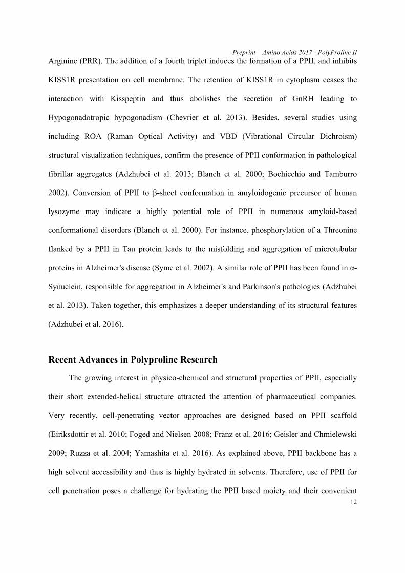
Arginine (PRR). The addition of a fourth triplet induces the formation of a PPII, and inhibits
KISS1R presentation on cell membrane. The retention of KISS1R in cytoplasm ceases the
interaction with Kisspeptin and thus abolishes the secretion of GnRH leading to
Hypogonadotropic hypogonadism (Chevrier et al. 2013). Besides, several studies using
including ROA (Raman Optical Activity) and VBD (Vibrational Circular Dichroism)
structural visualization techniques, confirm the presence of PPII conformation in pathological
fibrillar aggregates (Adzhubei et al. 2013; Blanch et al. 2000; Bochicchio and Tamburro
2002). Conversion of PPII to β-sheet conformation in amyloidogenic precursor of human
lysozyme may indicate a highly potential role of PPII in numerous amyloid-based
conformational disorders (Blanch et al. 2000). For instance, phosphorylation of a Threonine
flanked by a PPII in Tau protein leads to the misfolding and aggregation of microtubular
proteins in Alzheimer's disease (Syme et al. 2002). A similar role of PPII has been found in α-
Synuclein, responsible for aggregation in Alzheimer's and Parkinson's pathologies (Adzhubei
et al. 2013). Taken together, this emphasizes a deeper understanding of its structural features
(Adzhubei et al. 2016).
Recent Advances in Polyproline Research
The growing interest in physico-chemical and structural properties of PPII, especially
their short extended-helical structure attracted the attention of pharmaceutical companies.
Very recently, cell-penetrating vector approaches are designed based on PPII scaffold
(Eiriksdottir et al. 2010; Foged and Nielsen 2008; Franz et al. 2016; Geisler and Chmielewski
2009; Ruzza et al. 2004; Yamashita et al. 2016). As explained above, PPII backbone has a
high solvent accessibility and thus is highly hydrated in solvents. Therefore, use of PPII for
cell penetration poses a challenge for hydrating the PPII based moiety and their convenient

Preprint – Amino Acids 2017 - PolyProline II
13
uptake in hydrophobic membranes (Franz et al. 2016). Chmielewski’s group (Fillon et al.
2005) addressed this by designing and introducing cationic and hydrophobic moieties on the
PPII backbone and observe no structural change. The compactness and inherent flexibility of
the PPII conformation is the key to their adaptability and accompanied by cationic and
hydrophobic moieties, they becomes highly suitable for a cell-penetrating vector (Foged and
Nielsen 2008). The study observes a tremendous increase in PPII based Cell-Penetrating
Peptide (CPP) uptake compared to the traditional ones. Another important difference is the
claimed reduction in toxicity. This is based on the observations that PPII scaffold based CPP:
Sweet Arrow Peptides- SAP(E) obtains a net negative charge unlike the traditional CPP
which are positively charged (Franz et al. 2016; Geisler and Chmielewski 2009; Li et al.
2010).
Conclusion and Perspectives
Polyproline II helix is arguably a distinct member in secondary structure elements, based on
its geometry, sequence and structure. PPII has a left-handed geometry compared to right-
handedness of popular protein helices (see Figure 2). Its sequence composition varies based
on the presence in a globular or fibrous protein environment. It is quite an interesting
observation that proline, a major α-helix breaker/kink, when in succession adapts a distinct
helical form itself. Moreover, it dominates the α-helical form in denatured space. Such
examples can be appreciated in light of the expanse of 2nd structural space. Although PPII
conformation represents only 5% of the conformational space we highly advocate for it to be
considered in regular secondary structures. Besides, its representation is equivalent if not
more than the 310 helices. The involvement of PPII-protein and PPII-nucleic acid interactions
in different pathologies, structural applications and drug carriers makes it even more viable

Preprint – Amino Acids 2017 - PolyProline II
14
candidate to be included in the main regular secondary structures. Its potential role in
Alzheimer's and Parkinson's could not be ignored, given recent publications on the subject.
The presence of PPII in regular, ordered and disordered regions while establishes its
distinctiveness is not sufficient to seize the complete structural space of PPII conformations.
Therefore, more assignment approaches and coiled library experiments are needed to explore
such conformations. This review addresses the neglect on conformations like PPII and bias
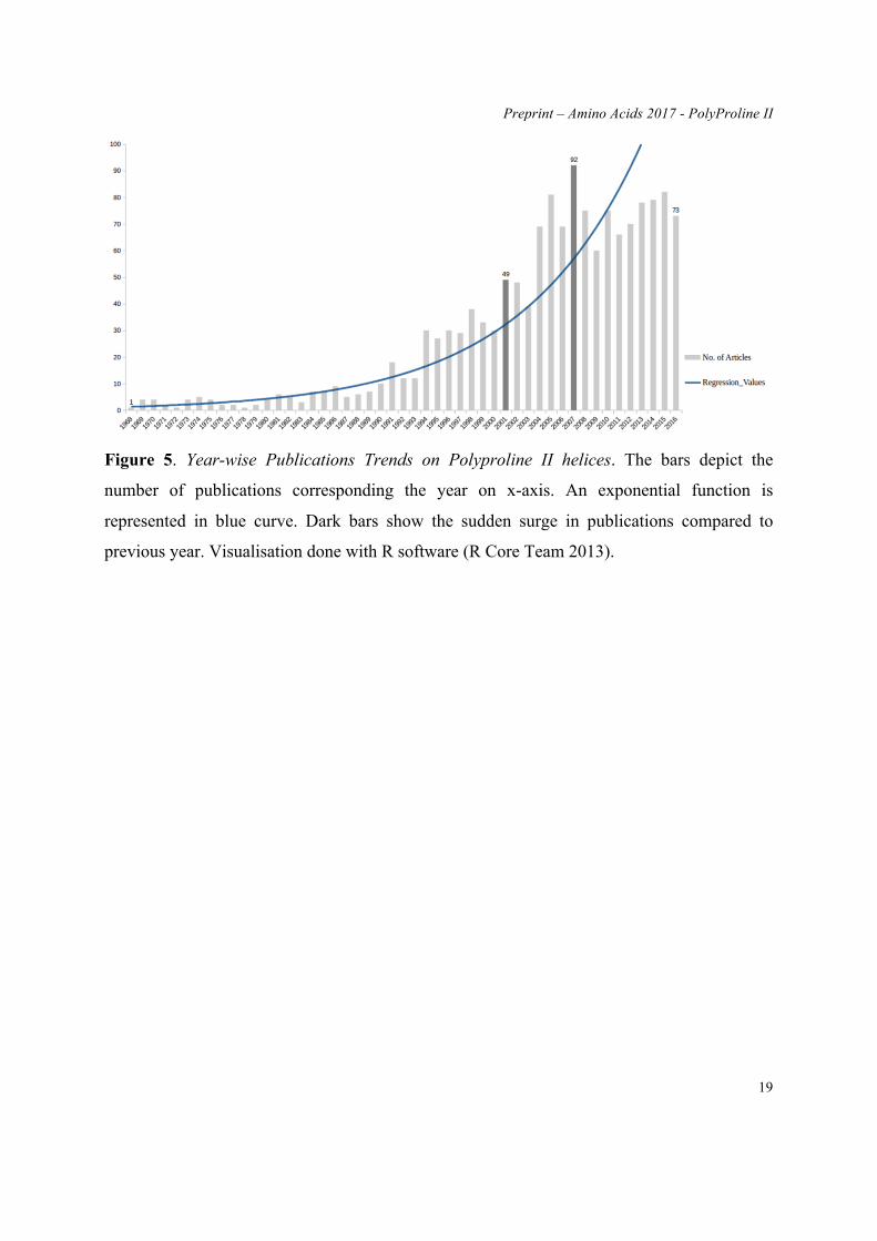
towards "regular" secondary structures. Figure 5 shows the number of publications about PPII
since 1968. The increase is clear, but remains limited. The number of papers had never been
higher than 100 papers/per year. In regards to the interest of this “lost” secondary structure,
we can expect a better representation in the future.
Acknowledgments
We would like to thank Catherine Etchebest for fruitful discussions. This work was
supported by grants from the Ministry of Research (France), University Paris Diderot,
Sorbonne, Paris Cité (France), National Institute for Blood Transfusion (INTS, France),
National Institute for Health and Medical Research (INSERM, France) and labex GR-Ex. The
labex GR-Ex, reference ANR-11-LABX-0051 is funded by the program “Investissements
d’avenir” of the French National Research Agency, reference ANR-11-IDEX-0005-02. TjrN,
NSr and AdB acknowledge to Indo-French Centre for the Promotion of Advanced Research /
CEFIPRA for collaborative grant (number 5302-2). NSh acknowledges support from ANRT.
Conflict of Interest: The authors declare that they have no conflict of interest.

Preprint – Amino Acids 2017 - PolyProline II
15
Legends
Figure 1. The structural characteristics of three secondary structures. A) Right handed α-
helix, B) Left handed PPII, C) Three β-strands forming sheet. The cartoon representation
highlights the structural geometry while ball and stick represents the atomic arrangements of
the three secondary structures. The proline rings can be observed in (B) and the comparison of
oxygen (red) and nitrogen (blue) clearly indicates the absence of intra H-bonding in PPII. In
(A) and (C), the close proximity of oxygen and nitrogen atoms makes it favourable for intra
H-bonding. High helical rise of the PPII and lack of intra H-bonding makes its backbone
highly solvent accessible. Visualisation done with PyMOL software (Delano 2013).
Preprint – Amino Acids 2017 - PolyProline II
16
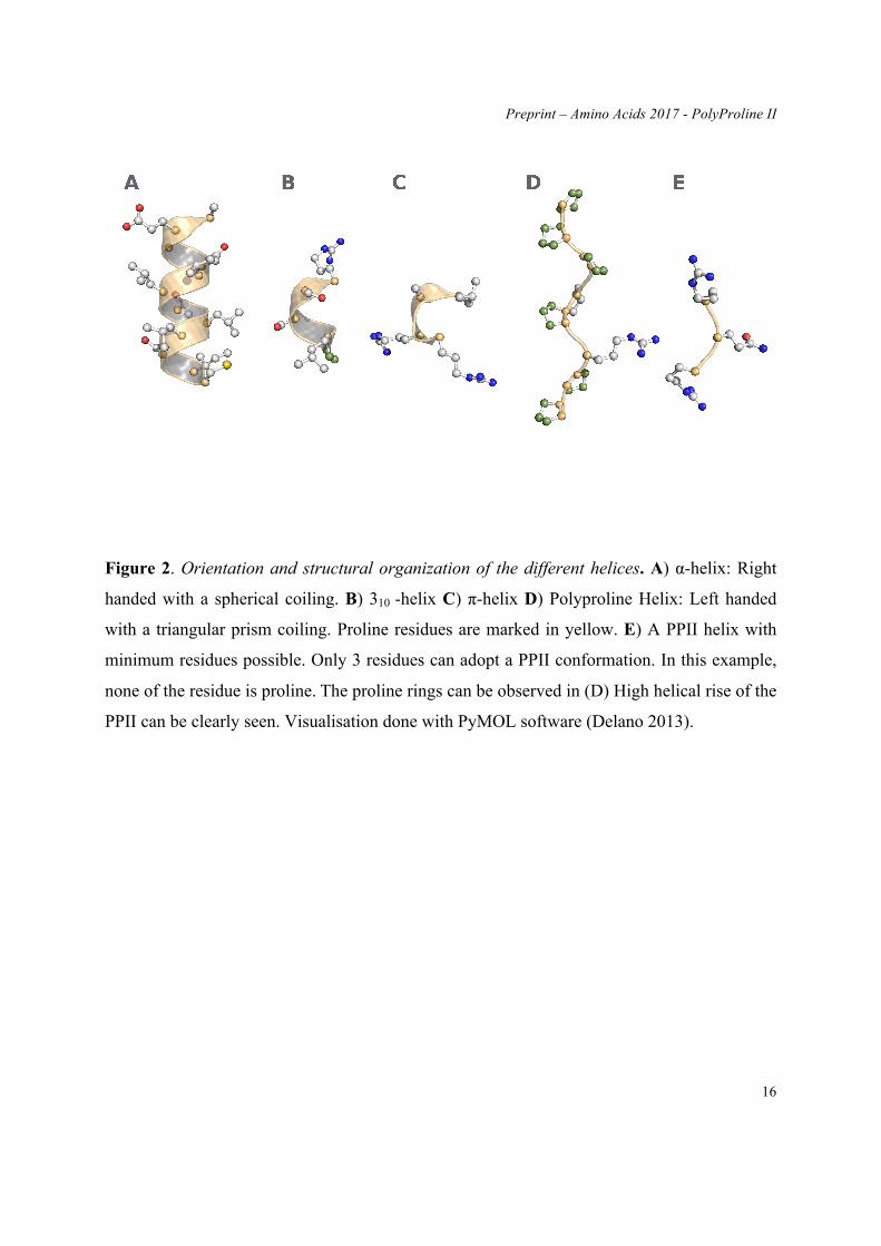
Figure 2. Orientation and structural organization of the different helices. A) α-helix: Right
handed with a spherical coiling. B) 310 -helix C) π-helix D) Polyproline Helix: Left handed
with a triangular prism coiling. Proline residues are marked in yellow. E) A PPII helix with
minimum residues possible. Only 3 residues can adopt a PPII conformation. In this example,
none of the residue is proline. The proline rings can be observed in (D) High helical rise of the
PPII can be clearly seen. Visualisation done with PyMOL software (Delano 2013).
Preprint – Amino Acids 2017 - PolyProline II
17
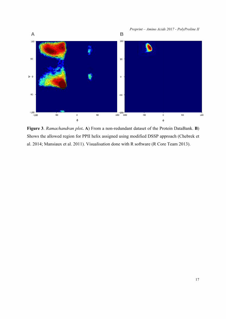
Figure 3. Ramachandran plot. A) From a non-redundant dataset of the Protein DataBank. B)
Shows the allowed region for PPII helix assigned using modified DSSP approach (Chebrek et
al. 2014; Mansiaux et al. 2011). Visualisation done with R software (R Core Team 2013).

Preprint – Amino Acids 2017 - PolyProline II
18
Figure 4. Interaction of Nova protein K homology domain with RNA hairpin (PDB id:
1ec6_A (Lewis et al. 2000)). The conserved motif of the variable loop is colour in yellow. The
two PPII helices are coloured in magenta. The occurrence of C-term helix is reported to be the
difference between RNA bound and unbound form. Visualisation done with PyMOL software
(Delano 2013).
Preprint – Amino Acids 2017 - PolyProline II
19
Figure 5. Year-wise Publications Trends on Polyproline II helices. The bars depict the
number of publications corresponding the year on x-axis. An exponential function is
represented in blue curve. Dark bars show the sudden surge in publications compared to
previous year. Visualisation done with R software (R Core Team 2013).
Preprint – Amino Acids 2017 - PolyProline II
20
References
Adzhubei AA, Anashkina AA, Makarov AA (2016) Left-handed polyproline-II helix revisited: proteins causing proteopathies J Biomol Struct Dyn:1-13 doi:10.1080/07391102.2016.1229220
Adzhubei AA, Sternberg MJ (1993) Left-handed polyproline II helices commonly occur in globular proteins J Mol Biol 229:472-493 doi:10.1006/jmbi.1993.1047
Adzhubei AA, Sternberg MJ, Makarov AA (2013) Polyproline-II helix in proteins: structure and function J Mol Biol 425:2100-2132 doi:10.1016/j.jmb.2013.03.018
Agrawal V, Kishan KV (2002) Promiscuous binding nature of SH3 domains to their target proteins Protein Pept Lett 9:185-193
Aksianov E, Alexeevski A (2012) SheeP: A tool for description of beta-sheets in protein 3D structures J Bioinform Comput Biol 10:1241003 doi:10.1142/S021972001241003X
Arnott S, Dover SD (1968) The structure of poly-L-proline II Acta Crystallogr B 24:599-601 Bansal M, Kumar S, Velavan R (2000) HELANAL: a program to characterize helix geometry in proteins J
Biomol Struct Dyn 17:811-819 doi:10.1080/07391102.2000.10506570 Bella J, Eaton M, Brodsky B, Berman HM (1994) Crystal and molecular structure of a collagen-like peptide at
1.9 A resolution Science 266:75-81 Berman HM et al. (2000) The Protein Data Bank Nucleic Acids Res 28:235-242 Bernstein FC et al. (1977) The Protein Data Bank: a computer-based archival file for macromolecular structures
J Mol Biol 112:535-542 Blanch EW, Morozova-Roche LA, Cochran DA, Doig AJ, Hecht L, Barron LD (2000) Is polyproline II helix the
killer conformation? A Raman optical activity study of the amyloidogenic prefibrillar intermediate of human lysozyme J Mol Biol 301:553-563 doi:10.1006/jmbi.2000.3981
Bochicchio B, Tamburro AM (2002) Polyproline II structure in proteins: identification by chiroptical spectroscopies, stability, and functions Chirality 14:782-792 doi:10.1002/chir.10153
Booker GW, Breeze AL, Downing AK, Panayotou G, Gout I, Waterfield MD, Campbell ID (1992) Structure of an SH2 domain of the p85 alpha subunit of phosphatidylinositol-3-OH kinase Nature 358:684-687 doi:10.1038/358684a0
Bornot A, de Brevern AG (2006) Protein beta-turn assignments Bioinformation 1:153-155 Cao C, Wang G, Liu A, Xu S, Wang L, Zou S (2016) A New Secondary Structure Assignment Algorithm Using
Calpha Backbone Fragments Int J Mol Sci 17:333 doi:10.3390/ijms17030333 Carter P, Andersen CA, Rost B (2003) DSSPcont: Continuous secondary structure assignments for proteins
Nucleic Acids Res 31:3293-3295 Carugo O, Djinovic-Carugo K (2013) Half a century of Ramachandran plots Acta Crystallogr D Biol Crystallogr
69:1333-1341 doi:10.1107/S090744491301158X Chebrek R, Leonard S, de Brevern AG, Gelly JC (2014) PolyprOnline: polyproline helix II and secondary
structure assignment database Database (Oxford) 2014 doi:10.1093/database/bau102 Chevrier L, de Brevern A, Hernandez E, Leprince J, Vaudry H, Guedj AM, de Roux N (2013) PRR repeats in
the intracellular domain of KISS1R are important for its export to cell membrane Mol Endocrinol 27:1004-1014 doi:10.1210/me.2012-1386
Cowan PM, McGavin S, North AC (1955) The polypeptide chain configuration of collagen Nature 176:1062-1064
Creamer TP (1998) Left-handed polyproline II helix formation is (very) locally driven Proteins 33:218-226 Cubellis MV, Caillez F, Blundell TL, Lovell SC (2005a) Properties of polyproline II, a secondary structure
element implicated in protein-protein interactions Proteins 58:880-892 doi:10.1002/prot.20327 Cubellis MV, Cailliez F, Lovell SC (2005b) Secondary structure assignment that accurately reflects physical and
evolutionary characteristics BMC Bioinformatics 6 Suppl 4:S8 doi:10.1186/1471-2105-6-S4-S8 Delano WL (2013) The PyMOL Molecular Graphics System on World Wide Web. http://www.pymol.org Dupuis F, Sadoc JF, Mornon JP (2004) Protein secondary structure assignment through Voronoi tessellation
Proteins 55:519-528 doi:10.1002/prot.10566 Eiriksdottir E, Konate K, Langel U, Divita G, Deshayes S (2010) Secondary structure of cell-penetrating
peptides controls membrane interaction and insertion Biochim Biophys Acta 1798:1119-1128 doi:10.1016/j.bbamem.2010.03.005
Eisenberg D (2003) The discovery of the alpha-helix and beta-sheet, the principal structural features of proteins Proc Natl Acad Sci U S A 100:11207-11210
Ferreon JC, Hilser VJ (2003) The effect of the polyproline II (PPII) conformation on the denatured state entropy
Preprint – Amino Acids 2017 - PolyProline II
21
Protein Sci 12:447-457 doi:10.1110/ps.0237803 Fillon YA, Anderson JP, Chmielewski J (2005) Cell penetrating agents based on a polyproline helix scaffold J
Am Chem Soc 127:11798-11803 doi:10.1021/ja052377g Fodje MN, Al-Karadaghi S (2002) Occurrence, conformational features and amino acid propensities for the pi-
helix Protein Eng 15:353-358 Foged C, Nielsen HM (2008) Cell-penetrating peptides for drug delivery across membrane barriers Expert Opin
Drug Deliv 5:105-117 doi:10.1517/17425247.5.1.105 Franz J, Lelle M, Peneva K, Bonn M, Weidner T (2016) SAP(E) - A cell-penetrating polyproline helix at lipid
interfaces Biochim Biophys Acta 1858:2028-2034 doi:10.1016/j.bbamem.2016.05.021 Frishman D, Argos P (1995) Knowledge-based protein secondary structure assignment Proteins 23:566-579
doi:10.1002/prot.340230412 Geisler I, Chmielewski J (2009) Cationic amphiphilic polyproline helices: side-chain variations and cell-specific
internalization Chem Biol Drug Des 73:39-45 doi:10.1111/j.1747-0285.2008.00759.x Hicks JM, Hsu VL (2004) The extended left-handed helix: a simple nucleic acid-binding motif Proteins 55:330-
338 doi:10.1002/prot.10630 Hosseini SR, Sadeghi M, Pezeshk H, Eslahchi C, Habibi M (2008) PROSIGN: a method for protein secondary
structure assignment based on three-dimensional coordinates of consecutive C(alpha) atoms Comput Biol Chem 32:406-411 doi:10.1016/j.compbiolchem.2008.07.027
Hutchinson EG, Thornton JM (1996) PROMOTIF--a program to identify and analyze structural motifs in proteins Protein Sci 5:212-220 doi:10.1002/pro.5560050204
Jha AK, Colubri A, Zaman MH, Koide S, Sosnick TR, Freed KF (2005) Helix, sheet, and polyproline II frequencies and strong nearest neighbor effects in a restricted coil library Biochemistry 44:9691-9702 doi:10.1021/bi0474822
Kabsch W, Sander C (1983) Dictionary of protein secondary structure: pattern recognition of hydrogen-bonded and geometrical features Biopolymers 22:2577-2637 doi:10.1002/bip.360221211
King SM, Johnson WC (1999) Assigning secondary structure from protein coordinate data Proteins 35:313-320 Kneller GR, Hinsen K (2015) Protein secondary-structure description with a coarse-grained model Acta
Crystallogr D Biol Crystallogr 71:1411-1422 doi:10.1107/S1399004715007191 Koleske AJ, Buratowski S, Nonet M, Young RA (1992) A novel transcription factor reveals a functional link
between the RNA polymerase II CTD and TFIID Cell 69:883-894 Kumar P, Bansal M (2015) Identification of local variations within secondary structures of proteins Acta
Crystallogr D Biol Crystallogr 71:1077-1086 doi:10.1107/S1399004715003144 Kumar P, Bansal M (2016) Structural and functional analyses of PolyProline-II helices in globular proteins J
Struct Biol 196:414-425 doi:10.1016/j.jsb.2016.09.006 Labesse G, Colloc'h N, Pothier J, Mornon JP (1997) P-SEA: a new efficient assignment of secondary structure
from C alpha trace of proteins Comput Appl Biosci 13:291-295 Law SM, Frank AT, Brooks CL, 3rd (2014) PCASSO: a fast and efficient Calpha-based method for accurately
assigning protein secondary structure elements J Comput Chem 35:1757-1761 doi:10.1002/jcc.23683 Lewis HA, Musunuru K, Jensen KB, Edo C, Chen H, Darnell RB, Burley SK (2000) Sequence-specific RNA
binding by a Nova KH domain: implications for paraneoplastic disease and the fragile X syndrome Cell 100:323-332
Li L, Geisler I, Chmielewski J, Cheng JX (2010) Cationic amphiphilic polyproline helix P11LRR targets intracellular mitochondria J Control Release 142:259-266 doi:10.1016/j.jconrel.2009.10.012
Majumdar I, Krishna SS, Grishin NV (2005) PALSSE: a program to delineate linear secondary structural elements from protein structures BMC Bioinformatics 6:202 doi:10.1186/1471-2105-6-202
Mansiaux Y, Joseph AP, Gelly JC, de Brevern AG (2011) Assignment of PolyProline II conformation and analysis of sequence--structure relationship PLoS One 6:e18401 doi:10.1371/journal.pone.0018401
Martin J, Letellier G, Marin A, Taly JF, de Brevern AG, Gibrat JF (2005) Protein secondary structure assignment revisited: a detailed analysis of different assignment methods BMC Struct Biol 5:17 doi:10.1186/1472-6807-5-17
Offmann B, Tyagi M, de Brevern AG (2007) Local Protein Structures Current Bioinformatics 3:165-202 Oluwatobi Salawu E (2016) RaFoSA: Random forests secondary structure assignment for coarse-grained and
all-atom protein systems Cogent Biology 2: 1214061 Parisien M, Major F (2005) A new catalog of protein beta-sheets Proteins 61:545-558 doi:10.1002/prot.20677 Park SY, Yoo MJ, Shin J, Cho KH (2011) SABA (secondary structure assignment program based on only alpha
carbons): a novel pseudo center geometrical criterion for accurate assignment of protein secondary
Preprint – Amino Acids 2017 - PolyProline II
22
structures BMB Rep 44:118-122 doi:10.5483/BMBRep.2011.44.2.118 Pauling L, Corey RB (1950) Two hydrogen-bonded spiral configurations of the polypetide chain J Am Chem
Soc 72:5349-5349 Pauling L, Corey RB (1951a) The pleated sheet, a new layer configuration of polypeptide chains Proc Natl Acad
Sci U S A 37:251-256 Pauling L, Corey RB (1951b) The structure of fibrous proteins of the collagen-gelatin group Proc Natl Acad Sci
U S A 37:272-281 Pauling L, Corey RB, Branson HR (1951) The structure of proteins; two hydrogen-bonded helical configurations
of the polypeptide chain Proc Natl Acad Sci U S A 37:205-211 Pronk S et al. (2013) GROMACS 4.5: a high-throughput and highly parallel open source molecular simulation
toolkit Bioinformatics 29:845-854 doi:10.1093/bioinformatics/btt055 R Core Team (2013) R: A language and environment for statistical computing. R Foundation for Statistical
Computing, Vienna, Austria. Ramachandran GN, Ramakrishnan C, Sasisekharan V (1963) Stereochemistry of polypeptide chain
configurations J Mol Biol 7:95-99 Rich A, Crick FH (1955) The structure of collagen Nature 176:915-916 Richards FM, Kundrot CE (1988) Identification of structural motifs from protein coordinate data: secondary
structure and first-level supersecondary structure Proteins 3:71-84 doi:10.1002/prot.340030202 Richardson JS (1981) The anatomy and taxonomy of protein structure Adv Protein Chem 34:167-339 Rose GD (1978) Prediction of chain turns in globular proteins on a hydrophobic basis Nature 272:586-590 Ruzza P, Calderan A, Guiotto A, Osler A, Borin G (2004) Tat cell-penetrating peptide has the characteristics of a
poly(proline) II helix in aqueous solution and in SDS micelles J Pept Sci 10:423-426 doi:10.1002/psc.558
Sasisekharan V (1959) Structure of poly-L-proline II Acta Crystallogr 12:897-903 Shakarji CM Least-Squares Fitting Algorithms of the NIST Algorithm Testing System Journal of Research of
the National Institute of Standards and Technology 103:633 Sklenar H, Etchebest C, Lavery R (1989) Describing protein structure: a general algorithm yielding complete
helicoidal parameters and a unique overall axis Proteins 6:46-60 doi:10.1002/prot.340060105 Soman KV, Ramakrishnan C (1983) Occurrence of a single helix of the collagen type in globular proteins J Mol
Biol 170:1045-1048 Sreerama N, Woody RW (1994) Poly(pro)II helices in globular proteins: identification and circular dichroic
analysis Biochemistry 33:10022-10025 Sreerama N, Woody RW (2003) Structural composition of betaI- and betaII-proteins Protein Sci 12:384-388
doi:10.1110/ps.0235003 Srinivasan R, Rose GD (1999) A physical basis for protein secondary structure Proc Natl Acad Sci U S A
96:14258-14263 Stapley BJ, Creamer TP (1999) A survey of left-handed polyproline II helices Protein Sci 8:587-595
doi:10.1110/ps.8.3.587 Sugeta H, Miyazawa T (1967) General method for calculating helical parameters of polymer chains from bond
lengths, bond angles, and internal-rotation angles Biopolymers 5:673-679 Suzuki M (1989) SPXX, a frequent sequence motif in gene regulatory proteins J Mol Biol 207:61-84 Suzuki M, Sohma H, Yazawa M, Yagi K, Ebashi S (1990) Histone H1 kinase specific to the SPKK motif J
Biochem 108:356-364 Syme CD, Blanch EW, Holt C, Jakes R, Goedert M, Hecht L, Barron LD (2002) A Raman optical activity study
of rheomorphism in caseins, synucleins and tau. New insight into the structure and behaviour of natively unfolded proteins Eur J Biochem 269:148-156
Toal S, Schweitzer-Stenner R (2014) Local order in the unfolded state: conformational biases and nearest neighbor interactions Biomolecules 4:725-773 doi:10.3390/biom4030725
Van Der Spoel D, Lindahl E, Hess B, Groenhof G, Mark AE, Berendsen HJ (2005) GROMACS: fast, flexible, and free J Comput Chem 26:1701-1718 doi:10.1002/jcc.20291
Venkatachalam CM (1968) Stereochemical criteria for polypeptides and proteins. V. Conformation of a system of three linked peptide units Biopolymers 6:1425-1436
Whittington SJ, Chellgren BW, Hermann VM, Creamer TP (2005) Urea promotes polyproline II helix formation: implications for protein denatured states Biochemistry 44:6269-6275 doi:10.1021/bi050124u
Williamson MP (1994) The structure and function of proline-rich regions in proteins Biochem J 297 ( Pt 2):249-260

Preprint – Amino Acids 2017 - PolyProline II
23
Yamashita H et al. (2016) Development of a Cell-penetrating Peptide that Exhibits Responsive Changes in its Secondary Structure in the Cellular Environment Sci Rep 6:33003 doi:10.1038/srep33003
Zacharias J, Knapp EW (2014) Protein secondary structure classification revisited: processing DSSP information with PSSC J Chem Inf Model 54:2166-2179 doi:10.1021/ci5000856
Zhang Y, Sagui C (2015) Secondary structure assignment for conformationally irregular peptides: comparison between DSSP, STRIDE and KAKSI J Mol Graph Model 55:72-84 doi:10.1016/j.jmgm.2014.10.005