Recent Advances in Pediatric Otolaryngology - downloads - Hindawi
70
International Journal of Pediatrics Recent Advances in Pediatric Otolaryngology Guest Editors: Jeffrey A. Koempel, Tomislav Baudoin, Alan T. L. Cheng, Debra M. Don, and Ajoy M. Varghese
Transcript of Recent Advances in Pediatric Otolaryngology - downloads - Hindawi

International Journal of Pediatrics
Recent Advances in Pediatric OtolaryngologyGuest Editors: Jeffrey A. Koempel, Tomislav Baudoin, Alan T. L. Cheng, Debra M. Don, and Ajoy M. Varghese

Recent Advances in Pediatric Otolaryngology
International Journal of Pediatrics
Recent Advances in Pediatric Otolaryngology
Guest Editors: Jeffrey A. Koempel, Tomislav Baudoin,Alan T. L. Cheng, Debra M. Don, and Ajoy M. Varghese

Copyright © 2012 Hindawi Publishing Corporation. All rights reserved.
This is a special issue published in “International Journal of Pediatrics.” All articles are open access articles distributed under the CreativeCommons Attribution License, which permits unrestricted use, distribution, and reproduction in any medium, provided the originalwork is properly cited.

Editorial Board
Ian T. Adatia, USAUri S. Alon, USALaxman Singh Arya, IndiaErle H. Austin, USAAnthony M. Avellino, USASylvain Baruchel, CanadaAndrea Biondi, ItalyJulie Blatt, USACatherine Bollard, USAP. D. Brophy, USARonald T. Brown, USAS. Burdach, GermanyLavjay Butani, USAWaldemar A. Carlo, USAJoseph M. Croffie, USASteven M. Donn, USATai Fai Fok, Hong KongMasahiro Fukuzawa, Japan
Eduardo H. Garin, USAMyron Genel, USAMark A. Gilger, USARalph A. Gruppo, USAEva C. Guinan, USASandeep Gupta, USAPamela S. Hinds, USAThomas C. Hulsey, USAGeorge Jallo, USAR. W. Jennings, USAEunice John, USARichard A. Jonas, USAMartin Kaefer, USAF. J. Kaskel, USAEmmanuel Katsanis, USAPraveen Kumar, USAHans Juergen Laws, GermanyEdward Y. Lee, USA
Steven E. Lipshultz, USADoff B. McElhinney, USASamuel Menahem, AustraliaKannan Laksmi Narasimhan, IndiaRoderick Nicolson, UKAlberto Pappo, USASeng Hock Quak, SingaporeR. Rink, USAJoel R. Rosh, USAMinnie M. Sarwal, USACharles L. Schleien, USAElizabeth J. Short, USAV. C. Strasburger, USADharmapuri Vidyasagar, USAFrans J. Walther, The NetherlandsMiles Weinberger, USAR. Wyatt, USANamik Yasar Ozbek, Turkey

Contents
Recent Advances in Pediatric Otolaryngology, Jeffrey A. Koempel, Tomislav Baudoin, Alan T. L. Cheng,Debra M. Don, and Ajoy M. VargheseVolume 2012, Article ID 535016, 2 pages
Hemangiomas and Vascular Malformations: Current Theory and Management, Gresham T. Richter andAdva B. FriedmanVolume 2012, Article ID 645678, 10 pages
Laryngomalacia: Disease Presentation, Spectrum, and Management, April M. Landry andDana M. ThompsonVolume 2012, Article ID 753526, 6 pages
Sleep Endoscopy in the Evaluation of Pediatric Obstructive Sleep Apnea, Aaron C. Lin and Peter J. KoltaiVolume 2012, Article ID 576719, 6 pages
Cortical Auditory Evoked Potentials in Children with a Hearing Loss: A Pilot Study, Amineh Koravand,BenoAmineh Koravand, Benoıt Jutras, and Maryse Lassondet Jutras, and Maryse LassondeVolume 2012, Article ID 250254, 8 pages
Neonatal Stridor, Matija Daniel and Alan ChengVolume 2012, Article ID 859104, 5 pages
Juvenile Angiofibroma: Evolution of Management, Piero Nicolai, Alberto Schreiber,and Andrea Bolzoni VillaretVolume 2012, Article ID 412545, 11 pages
Surgical and Pathological Characteristics of Papillary Thyroid Cancer in Children and Adolescents,Davor DzepinaVolume 2012, Article ID 125389, 6 pages
Chronic Rhinosinusitis in Children, Hassan H. RamadanVolume 2012, Article ID 573942, 5 pages
Troublesome Tinnitus in Children: Epidemiology, Audiological Profile, and Preliminary Results ofTreatment, G. Bartnik, A. Stepien, D. Raj-Koziak, A. Fabijanska, I. Niedziałek, and H. SkarzynskiVolume 2012, Article ID 945356, 5 pages
Hindawi Publishing CorporationInternational Journal of PediatricsVolume 2012, Article ID 535016, 2 pagesdoi:10.1155/2012/535016
Editorial
Recent Advances in Pediatric Otolaryngology
Jeffrey A. Koempel,1, 2 Tomislav Baudoin,3 Alan T. L. Cheng,4
Debra M. Don,1, 2 and Ajoy M. Varghese5
1 Division of Otolaryngology-Head and Neck Surgery, Children’s Hospital, Los Angeles, CA 90027, USA2 Department of Otolaryngology-Head and Neck Surgery, Keck School of Medicine of the University of Southern California,Los Angeles, CA 90089, USA
3 Referral Center for Pediatric Otolaryngology, Sisters of Charity University Hospital, Ministry of Health and Social Welfare of theRepublic of Croatia, 10000 Zagreb, Croatia
4 Department of Pediatric Otolaryngology, The Sydney Children’s Hospital Network—Westmead Campus,The University of Sydney, Sydney NSW 2145, Australia
5 Department of ENT-2, Christian Medical College, Vellore 632004, India
Correspondence should be addressed to Jeffrey A. Koempel, [email protected]
Received 11 April 2012; Accepted 11 April 2012
Copyright © 2012 Jeffrey A. Koempel et al. This is an open access article distributed under the Creative Commons AttributionLicense, which permits unrestricted use, distribution, and reproduction in any medium, provided the original work is properlycited.
Ear, nose, and throat problems comprise a significant portionof patient visits to the primary care physicians’ offices, urgentcare facilities, emergency rooms, and children’s hospitals.Since its beginnings in the 1970s, the specialty of pediatricotolaryngology has developed significantly and even moreso in the last five to ten years. The aim of this special issueis to offer our pediatrician colleagues an opportunity tolearn about recent advances in both diagnostic methods andtherapeutic procedures that are now available to assist in thecare of children with ear, nose, and throat disorders.
This special issue is comprised of both original clinicalresearch and review articles from all areas of pediatricotolaryngology such as otologic disease and hearing loss,sinonasal disorders, airway issues, and head and neckmasses. These papers emphasize how methods of diagnosishave improved for certain conditions, how existing surgicalprocedures have been modified to be less invasive and bettertolerated, and finally, how new technology and operationsallow clinicians to treat otolaryngologic disorders that werewithout treatment in the past.
The paper “Troublesome tinnitus in children: epidemiol-ogy, audiological profile, and preliminary Results of Treatment”by G. Bartnik et al. offers a very readable and practicalapproach to a common otologic problem in children whilemost pediatric textbooks often have no information on thissubject. This group offers their experience with a treatment
called “Tinnitus Retraining Therapy.” A. Koravand, B. Jutras,and M. Lassonde present their original research using thediagnostic tools of cortical auditory evoked potentials andmismatch responses (MMRs) to identify patterns of neuralactivity in the central auditory system of children withhearing loss.
The care of patients with nose and sinus complaints canbe frustrating to the primary care physician. H. Ramadan’spaper “Chronic rhinosinusitis in children” provides a simple,straightforward approach to the pediatric patient with thesesymptoms including the use of “real-life” examples toillustrate the important aspects of the history and physicalexam that are necessary in the care of these patients. Afterreading this article, any health care practitioner will feelmore confident about their decision-making in patientswith sinus disease. Although juvenile angiofibroma is arare clinical problem, P. Nicolai, A. Schreiber, and A. B.Villaret describe new, less invasive approaches to treatmentof this sinonasal mass. This is in addition to a succinctreview of the pathophysiology and recommended work-up including the relative advantages and disadvantages ofvarious radiographic imaging techniques.
The airway section of this special issue will be of greatvalue to the primary care practitioner. Included are threepapers on stridor in the infant, one dedicated entirely tolaryngomalacia, and lastly, a paper on a new diagnostic tool

2 International Journal of Pediatrics
called “sleep endoscopy.” M. Daniel and A. Cheng discussthe approach to an infant with noisy breathing or stridor;separating the material into three sections that allow for easyrecall and use by the pediatrician. Novel approaches suchas the EXIT procedure to treat disorders of the fetal airwayare presented. A. M. Landry and D. M. Thompson’s reviewarticle on laryngomalacia will be equally relevant. The readerwill find it to be complete yet easily digestible. Finally, anyhealth care practitioner will be familiar with the frustrationof seeing patients for persistent difficulty breathing at nightafter adenotonsillectomy for upper airway obstruction orobstructive sleep apnea. Although polysomnography candetermine the presence and severity of any degree ofobstruction, it cannot identify the location of obstruction.The paper by A. C. Lin and P. J. Koltai describe a newdiagnostic tool called “sleep endoscopy” that will allowotolaryngologists the opportunity to identify such areas ofobstruction and perhaps offer treatment options which werenot obvious in the past.
The approach to a patient with a vascular malformationcan be confusing. G. T. Richter’s review article on this clinicalentity will be very helpful to the primary care practitioner.Nomenclature, relevant parts of the history and physicalexam, diagnostic tools including radiographic imaging stud-ies, and medical and surgical therapies are discussed. D.Dzepina’s paper on papillary thyroid carcinoma in childrendescribes their approach and results with total thyroidectomyincluding neck dissection for lymphatic dissemination whichis common in this type of thyroid cancer.
The editors of this special issue have worked hard withthe authors to provide primary care practitioners withinformation that is relevant to their practice and, at the sametime, very easy to read and understand. We hope the papersin this special issue will be of great help to primary carepractitioners as they see pediatric patients with ear, nose, andthroat problems.
Jeffrey A. KoempelTomislav BaudoinAlan T. L. Cheng
Debra M. DonAjoy M. Varghese
Hindawi Publishing CorporationInternational Journal of PediatricsVolume 2012, Article ID 645678, 10 pagesdoi:10.1155/2012/645678
Review Article
Hemangiomas and Vascular Malformations:Current Theory and Management
Gresham T. Richter and Adva B. Friedman
Division of Pediatric Otolaryngology, Department of Otolaryngology-Head and Neck Surgery,University of Arkansas for Medical Sciences, Arkansas Children’s Hospital, 1 Children’s Way, Little Rock, AR 72202, USA
Correspondence should be addressed to Gresham T. Richter, [email protected]
Received 2 August 2011; Revised 17 November 2011; Accepted 17 January 2012
Academic Editor: Ajoy M. Varghese
Copyright © 2012 G. T. Richter and A. B. Friedman. This is an open access article distributed under the Creative CommonsAttribution License, which permits unrestricted use, distribution, and reproduction in any medium, provided the original work isproperly cited.
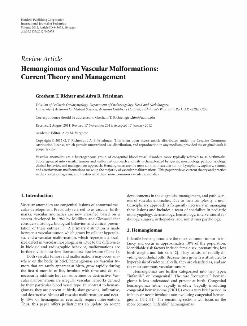
Vascular anomalies are a heterogeneous group of congenital blood vessel disorders more typically referred to as birthmarks.Subcategorized into vascular tumors and malformations, each anomaly is characterized by specific morphology, pathophysiology,clinical behavior, and management approach. Hemangiomas are the most common vascular tumor. Lymphatic, capillary, venous,and arteriovenous malformations make up the majority of vascular malformations. This paper reviews current theory and practicein the etiology, diagnosis, and treatment of these more common vascular anomalies.
1. Introduction
Vascular anomalies are congenital lesions of abnormal vas-cular development. Previously referred to as vascular birth-marks, vascular anomalies are now classified based on asystem developed in 1982 by Mulliken and Glowacki thatconsiders histology, biological behavior, and clinical presen-tation of these entities [1]. A primary distinction is madebetween a vascular tumor, which grows by cellular hyperpla-sia, and a vascular malformation, which represents a local-ized defect in vascular morphogenesis. Due to the differencesin biologic and radiographic behavior, malformations arefurther divided into slow-flow and fast-flow lesions (Table 1).
Both vascular tumors and malformations may occur any-where on the body. In brief, hemangiomas are vascular tu-mors that are rarely apparent at birth, grow rapidly duringthe first 6 months of life, involute with time and do notnecessarily infiltrate but can sometimes be destructive. Vas-cular malformations are irregular vascular networks definedby their particular blood vessel type. In contrast to heman-giomas, they are present at birth, slow growing, infiltrative,and destructive. Almost all vascular malformations and near-ly 40% of hemangiomas eventually require intervention.Thus, this paper offers pediatricians an update on recent
developments in the diagnosis, management, and pathogen-esis of vascular anomalies. Due to their complexity, a mul-tidisciplinary approach is frequently necessary in managingthese lesions and includes a team of specialists in pediatricotolaryngology, dermatology, hematology, interventional ra-diology, surgery, orthopedics, and sometimes psychology.
2. Hemangiomas
Infantile hemangiomas are the most common tumor in in-fancy and occur in approximately 10% of the population.Identifiable risk factors include female sex, prematurity, lowbirth weight, and fair skin [2]. They consist of rapidly di-viding endothelial cells. Because their growth is attributed tohyperplasia of endothelial cells, they are classified as, and arethe most common, vascular tumors.
Hemangiomas are further categorized into two types:“infantile” or “congenital.” The rare “congenital” heman-gioma is less understood and present at birth. Congenitalhemangiomas either rapidly involute (rapidly involutingcongenital hemangioma (RICH)) over a very brief period ininfancy or never involute (noninvoluting congenital heman-gioma; (NICH)). The remaining sections will focus on themore common “infantile” hemangiomas.
2 International Journal of Pediatrics
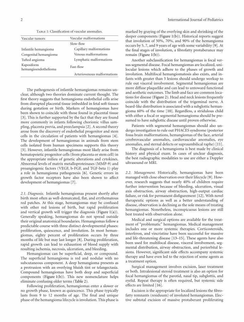
Table 1: Classification of vascular anomalies.
Vascular tumors Vascular malformations
Slow-flow
Infantile hemangioma Capillary malformations
Congenital hemangioma Venous malformations
Tufted angioma Lymphatic malformations
Kaposiformhemangioendothelioma
Fast-flow
Arteriovenous malformations
The pathogenesis of infantile hemangiomas remains un-clear, although two theories dominate current thought. Thefirst theory suggests that hemangioma endothelial cells arisefrom disrupted placental tissue imbedded in fetal soft tissuesduring gestation or birth. Markers of hemangiomas havebeen shown to coincide with those found in placental tissue[3]. This is further supported by the fact that they are foundmore commonly in infants following chorionic villus sam-pling, placenta previa, and preeclampsia [2]. A second theoryarose from the discovery of endothelial progenitor and stemcells in the circulation of patients with hemangiomas [4].The development of hemangiomas in animals from stemcells isolated from human specimens supports this theory[5]. However, infantile hemangiomas most likely arise fromhematopoietic progenitor cells (from placenta or stem cell) inthe appropriate milieu of genetic alterations and cytokines.Abnormal levels of matrix metalloproteinases (MMP-9) andproangiogenic factors (VEGF, b-FGF, and TGF-beta 1) playa role in hemangioma pathogenesis [6]. Genetic errors ingrowth factor receptors have also been shown to affectdevelopment of hemangiomas [7].
2.1. Diagnosis. Infantile hemangiomas present shortly afterbirth most often as well-demarcated, flat, and erythematousred patches. At this stage, hemangiomas may be confusedwith other red lesions of birth, but rapid proliferationand vertical growth will trigger the diagnosis (Figure 1(a)).Generally speaking, hemangiomas do not spread outsidetheir original anatomical boundaries. Hemangiomas follow apredictable course with three distinct developmental phases:proliferation, quiescence, and involution. In most heman-giomas, eighty percent of proliferation occurs by threemonths of life but may last longer [8]. During proliferation,rapid growth can lead to exhaustion of blood supply withresulting ischemia, necrosis, ulceration, and bleeding.
Hemangiomas can be superficial, deep, or compound.The superficial hemangioma is red and nodular with nosubcutaneous component. A deep hemangioma presents asa protrusion with an overlying bluish tint or telangectasia.Compound hemangiomas have both deep and superficialcomponents (Figure 1(b)). This new nomenclature helpseliminate confusing older terms (Table 2).
Following proliferation, hemangiomas enter a slower orno growth phase, known as quiescence. This phase typicallylasts from 9 to 12 months of age. The final and uniquephase of the hemangioma lifecycle is involution. This phase is
marked by graying of the overlying skin and shrinking of thedeeper components (Figure 1(b)). Historical reports suggestthat involution of 50%, 70%, and 90% of the hemangiomaoccurs by 5, 7, and 9 years of age with some variability [9]. Atthe final stages of involution, a fibrofatty protuberance mayremain (Figure 1(b)).
Another subclassification for hemangiomas is focal ver-sus segmental disease. Focal hemangiomas are localized, uni-locular lesions which adhere to the phases of growth andinvolution. Multifocal hemangiomatosis also exists, and in-fants with greater than 5 lesions should undergo workup torule out visceral involvement. Segmental hemangiomas aremore diffuse plaquelike and can lead to untoward functionaland aesthetic outcomes. The limb and face are common loca-tions for disease (Figure 2). Head and neck lesions frequentlycoincide with the distribution of the trigeminal nerve. Abeard-like distribution is associated with a subglottic heman-gioma 60% of the time [10]. Regardless, a stridulous childwith either a focal or segmental hemangioma should be pre-sumed to have subglottic disease until proven otherwise.
Patients with segmental hemangiomas should also un-dergo investigation to rule out PHACES syndrome (posteriorfossa brain malformations, hemangiomas of the face, arterialcerebrovascular anomalies, cardiovascular anomalies, eyeanomalies, and sternal defects or supraumbilical raphe) [11].
The diagnosis of a hemangioma is best made by clinicalhistory and physical exam. In cases of unclear diagnosis,the best radiographic modalities to use are either a Dopplerultrasound or MRI.
2.2. Management. Historically, hemangiomas have beenmanaged with close observation over their lifecycle [9]. How-ever, research suggests that nearly 40% of children requirefurther intervention because of bleeding, ulceration, visualaxis obstruction, airway obstruction, high-output cardiacfailure, or risk for permanent disfigurement [12]. With noveltherapeutic options as well as a better understanding ofdisease, observation is declining as the sole means of treatinghemangiomas. Nonetheless, inconspicuous lesions are stillbest treated with observation alone.
Medical and surgical options are available for the treat-ment of “problematic” hemangiomas. Medical managementincludes one or more systemic therapies. Corticosteroids,interferon, and vincristine have been successful for massiveand life-threatening disease [13–15]. These agents have alsobeen used for multifocal disease, visceral involvement, seg-mental distribution, airway obstruction, and periorbital le-sions. However, significant side effects accompany systemictherapy and have even led to the rejection of some agents asa treatment option.
Surgical management involves excision, laser treatmentor both. Intralesional steroid treatment is also an option forfocal hemangiomas of the parotid, nasal tip, subglottis, andeyelid. Repeat therapy is often required, but systemic sideeffects are limited [16].
Excision is the appropriate for localized lesions the fibro-fatty remnants (residuum) of involuted hemangiomas. Elec-tive subtotal excision of massive protuberant proliferating

International Journal of Pediatrics 3
(a) (b)
Figure 1: (a) Proliferating hemangioma at 3 months of age. (b) Same hemangioma at involution at 4 years of age.
(a) (b)
Figure 2: (a) Segmental hemangioma in trigeminal (V3) distribution. (b) Same hemangioma after 2 months of therapy with propranolol(2 mg/kg divided tid).
Table 2: Old versus current nomenclature for describing heman-gioma types.
Old nomenclature New nomenclature
Strawberry or capillary hemangioma Superficial hemangioma
Cavernous hemangioma Deep hemangioma
Capillary cavernous hemangioma Compound hemangioma
hemangiomas can be employed in order to maintain aes-thetic facial boundaries. Small remnants of disease are thenleft for involution. Residual erythema and telangiectasiasfrequently remain in involuted hemangiomas and are besttreated by selective photothermolysis using the flash pulsedye laser (FPDL). Similarly, ulcerative lesions during prolif-eration can be treated with FPDL to induce healing and newepidermal growth.
2.3. Propranolol. A paradigm shift has occurred regardingthe treatment of hemangiomas over the past few years. In2008, propranolol, a nonselective β-adrenergic antagonist,was serendipitously discovered to cause regression of prolif-erating hemangiomas in newborns receiving treatment forcardiovascular disease [17]. Numerous studies demonstrat-ing the success of propranolol for shrinking hemangiomas
have followed suit [17–19]. In fact, over ninety percentof patients have dramatic reduction in the size of theirhemangiomas as early as 1-2 weeks following the first doseof propranolol (Figure 2(b)). Dosing for propranolol intreating hemangiomas is recommended to be 2-3 mg/kgseparated into two or three-times-a-day regimens [20]. Thesedoses are dramatically below the concentration employedfor cardiovascular conditions in children. Thus, reportedside effects of propranolol for hemangiomas have beenminimal. Nonetheless, serious concerns for hypoglycemiaand lethargy that can occur with this medicine should notbe brushed aside [21, 22]. To address these concerns, parentsare instructed to give propranolol with meals, report anyunusual sleepiness, and not administer it during infections.Early and frequent visits to assess vital signs are recom-mended in young infants while on therapy. Exacerbationof gastroesophageal reflux may result due to beta-receptorblockade at the lower esophageal sphincter [18].
Monitoring the administration of propranolol variesamong institutions and practitioners. A unified approachhas not yet been determined. However, elective admissionwith cardiovascular monitoring may be necessary. Outpa-tient administration with close monitoring has also beensuccessfully performed [23]. Nonetheless, an electrocardio-gram must be reviewed by a pediatric cardiologist prior to

4 International Journal of Pediatrics
(a) (b) (c)
Figure 3: (a) Macrocystic lymphatic malformation (LM) of right neck in toddler. (b) Microcystic lip LM displaying mucosal vesicles. (c)Microcystic LM in older patient with bone involvement and mandibular hypertrophy.
administration. Cardiopulmonary conditions at risk for pro-pranolol therapy such as heart block or reactive airway dis-ease should draw careful consideration before administering.Consensus on patient monitoring and best dose regimensremains to be determined, but prospective research is under-way.
Propranolol is currently employed for “problematic”hemangiomas, those that would have received either surgicalor some other systemic therapy to prevent untoward sideeffects. Subglottic, periorbital, and massive hemangiomasseem to respond well [24]. Despite the success of propranololin reducing hemangioma size, adjuvant therapy may benecessary in up to 50% of patients [17]. Propranolol’s mech-anism on treating hemangiomas remains unclear but mayinvolve the regulation of vascular growth factors and hemo-dynamic cytokines.
3. Vascular Malformations Overview
Vascular malformations are rare vascular anomalies com-posed of inappropriately connected vasculature. Any bloodvessel type, or a combination thereof, can be affected ina vascular malformation. These lesions infiltrate normaltissue which makes them very difficult to manage. The mostcommon vascular malformations include lymphatic mal-formations (LMs), capillary-venular malformations (CM),venous malformations (VMs) and arteriovenous malforma-tions (AVMs) which have been selected to be covered in thispaper (Table 1). While different in their biologic and clinicalprofile, as a whole, vascular malformations do not regressand continue to expand with time. Periods of rapid growth,infiltration, and soft tissue destruction will spur therapeuticapproaches that depend upon the malformation involved.
4. Lymphatic Malformations
Lymphatic malformations (LMs) are composed of dilatedlymphatic vessels with inappropriate communication, linedby endothelial cells and filled with lymphatic fluid. Theirincidence is approximated to be 1 in 2000 to 4000 live births[25]. Lesions are classified as macrocystic (single or multiplecysts >2 cm3), microcystic (<2 cm3), or mixed [1]. Previous
terminology, that is no longer used, has included “cystichygroma” and “lymphangioma” to describe these entities.
The etiology of LM is unclear. Although most are con-genital, there have been reports of LM occurring after traumaor infection. Receptors involved in the formation of lym-phatic vascular channels, such as VEGFR3 and Prox-1, mayplay a role in the development of this disease [26].
4.1. Diagnosis. Lymphatic malformations may be macrocys-tic, microcystic, or mixed. Gradual growth and expansion istypical. Approximately half of the lesions are present at birthand 80–90% by 2 years of age. Local infections approximat-ing the course of lymphatic drainage will cause LM to swell,protrude, and sometimes become painful. This is a hallmarkof a LM versus other vascular anomalies that do not presentin this fashion.
Clinically, the appearance of macrocystic disease differsfrom that of microcystic. Macrocystic LMs present as a soft,fluid-filled swelling beneath normal or slightly discoloredskin (Figure 3(a)). Intracystic bleeding or a mixed lymphaticvenous malformation may result in blue discoloration of theoverlying skin. Microcystic LMs are soft and noncompress-ible masses with an overlying area of small vesicles involvingthe skin or mucosa. These vesicles can weep and at timescause pain or minor bleeding (Figure 3(b)).
LM can occur anywhere on the body, and symptoms aredetermined by the extent of disease. Most LMs are foundin the cervicofacial region and extend to involve the oralcavity or airway, especially when mixed or microcystic [27].Symptoms secondary to bulky disease often include pain,dysphagia, odynophagia, impaired speech, or in severe cases,airway obstruction. When involving the skeletal frameworkin this area, LMs often cause osseous hypertrophy leading todental or extremity abnormalities (Figure 3(c)).
Although these malformations can usually be diagnosedby physical examination, MRI is used to confirm diagnosis,identify cystic architecture, and determine extent of disease.
4.2. Management. An ideal option for treatment of LM doesnot exist. Several interventions may be required. There havebeen rare cases of sporadic resolution of a lesion althoughthe majority of these malformations continue to enlarge withage [27]. Macrocystic lesions are more amenable to treatment
International Journal of Pediatrics 5
and have a better prognosis. Swelling from acute infection isbest controlled with a short course of systemic steroids andantibiotics. Definitive treatment is delayed until resolution.
LM may be detected on prenatal ultrasound and mayrequire special interventions during delivery. The EXIT (exutero intrapartum treatment) procedure provides good air-way control of the infant if compromise is suspected to occurat birth.
Sclerotherapy is frequently employed for lymphatic mal-formations, especially if deep seated and difficult to accesssurgically. It involves injection of a sclerosing agent directlyinto the lesion leading to fibrosis and ultimately regressionof the cysts. Several treatments are usually required, andswelling is expected following therapy. Macrocystic lesionsare more easily treated in this fashion, but there have beenreports of success in microcystic lesions [28]. Several agentshave been utilized for lymphatic malformations includingethanol, bleomycin, OK-432, and doxycycline [29, 30]. Com-plications include skin breakdown, pain, and swelling. Severeswelling can at times occur and may lead to airway obstruc-tion requiring intensive care [31]. Risks to local nerves arealso real but usually result in only transient loss of function.
Carbon dioxide laser therapy may also be employed inlimited disease of the airway and oral mucosa [32]. Macro-cystic disease is often cured with surgical extirpation. Surgi-cal excision is also frequently employed for microcystic dis-ease although it is more aggressive, invasive, and difficult tocontrol [33, 34]. Infiltration of normal soft tissue and boneby extensive microcystic LM requires massive resections andlocal or free-flap reconstruction. Failure to completely excisemicrocystic LM often leads to recurrence. Surgery is alsoemployed in the correction of secondary deformities causedby LM such as bony overgrowth of the facial skeleton [34].
Overall, treatment for LM should be aimed at completeelimination of disease. When this is not feasible, multipletreatment modalities are combined to control disease andprovide satisfactory functional outcomes.
5. Capillary Malformations
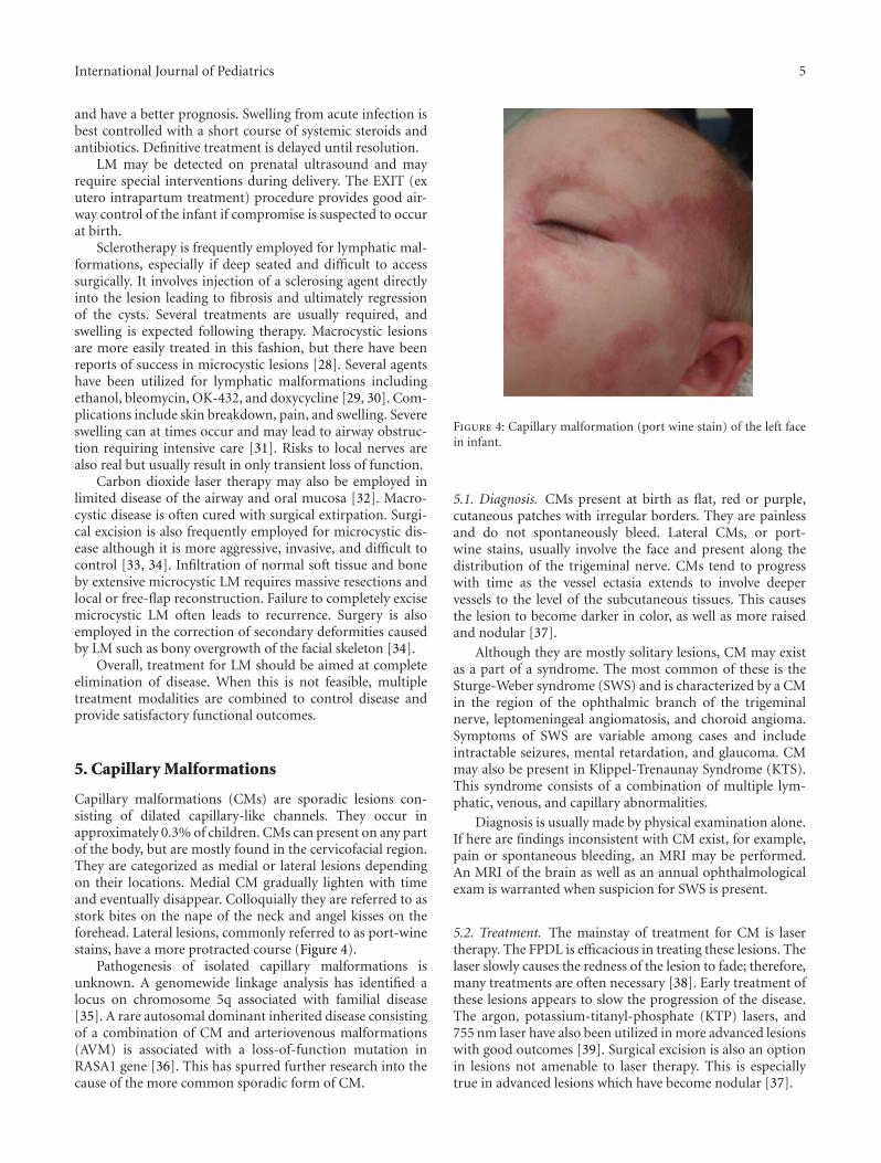
Capillary malformations (CMs) are sporadic lesions con-sisting of dilated capillary-like channels. They occur inapproximately 0.3% of children. CMs can present on any partof the body, but are mostly found in the cervicofacial region.They are categorized as medial or lateral lesions dependingon their locations. Medial CM gradually lighten with timeand eventually disappear. Colloquially they are referred to asstork bites on the nape of the neck and angel kisses on theforehead. Lateral lesions, commonly referred to as port-winestains, have a more protracted course (Figure 4).
Pathogenesis of isolated capillary malformations isunknown. A genomewide linkage analysis has identified alocus on chromosome 5q associated with familial disease[35]. A rare autosomal dominant inherited disease consistingof a combination of CM and arteriovenous malformations(AVM) is associated with a loss-of-function mutation inRASA1 gene [36]. This has spurred further research into thecause of the more common sporadic form of CM.
Figure 4: Capillary malformation (port wine stain) of the left facein infant.
5.1. Diagnosis. CMs present at birth as flat, red or purple,cutaneous patches with irregular borders. They are painlessand do not spontaneously bleed. Lateral CMs, or port-wine stains, usually involve the face and present along thedistribution of the trigeminal nerve. CMs tend to progresswith time as the vessel ectasia extends to involve deepervessels to the level of the subcutaneous tissues. This causesthe lesion to become darker in color, as well as more raisedand nodular [37].
Although they are mostly solitary lesions, CM may existas a part of a syndrome. The most common of these is theSturge-Weber syndrome (SWS) and is characterized by a CMin the region of the ophthalmic branch of the trigeminalnerve, leptomeningeal angiomatosis, and choroid angioma.Symptoms of SWS are variable among cases and includeintractable seizures, mental retardation, and glaucoma. CMmay also be present in Klippel-Trenaunay Syndrome (KTS).This syndrome consists of a combination of multiple lym-phatic, venous, and capillary abnormalities.
Diagnosis is usually made by physical examination alone.If here are findings inconsistent with CM exist, for example,pain or spontaneous bleeding, an MRI may be performed.An MRI of the brain as well as an annual ophthalmologicalexam is warranted when suspicion for SWS is present.
5.2. Treatment. The mainstay of treatment for CM is lasertherapy. The FPDL is efficacious in treating these lesions. Thelaser slowly causes the redness of the lesion to fade; therefore,many treatments are often necessary [38]. Early treatment ofthese lesions appears to slow the progression of the disease.The argon, potassium-titanyl-phosphate (KTP) lasers, and755 nm laser have also been utilized in more advanced lesionswith good outcomes [39]. Surgical excision is also an optionin lesions not amenable to laser therapy. This is especiallytrue in advanced lesions which have become nodular [37].

6 International Journal of Pediatrics
(a) (b)
Figure 5: Cervicofacial venous malformation involving the right neck (a) and oropharyngeal mucosa (b).
6. Venous Malformations
Venous malformations (VMs) are slow-flow vascular anoma-lies composed of ectatic venous channels. These aberrantvenous connections lead to venous congestion, thrombosis,and gradual expansion of these lesions. As a result, VMspersist and progress until therapeutic intervention. The inci-dence of VMs is approximately 1 in 10,000 [40]. VMs morecommonly occur sporadically, but research into multifocaldisease and familial patterns has helped discover suspectedgenetic loci involved in their development. There are inher-ited forms of VMs, the cause of which has been localized tochromosome 9p [41]. Recently a loss-of-function mutationwas discovered on the angiopoetin receptor gene TIE2/TEKin many solitary and multiple sporadic venous malfor-mations [42]. In addition, upregulation of several factorsincluding tissue growth factor beta (TGF-beta) and basicfibroblast growth factor (beta-FGF) has been discoveredin patients with venous malformations [26]. Progesteronereceptors have been discovered in venous malformations.This likely explains their tendency to grow rapidly duringhormonal changes [43].
6.1. Diagnosis. Venous malformations are often visible atbirth but may present as a deep mass. Protrusion may bethe only presenting symptom. They are known to grow pro-portionately with the child with sudden expansion in adult-hood. Rapid growth may occur during puberty, pregnancy,or traumatic injury. VM can be either well localized orextensive. The overlying skin may appear normal or possessa bluish discoloration. With more cutaneous involvement,the lesions appear darker blue or purple (Figure 5(a)).Upper aerodigestive involvement is common, and VM areparticularly evident when mucosa is affected (Figure 5(b)).
VMs are compressible and swell when the region isdependent or there is an increase in hydrostatic pressure suchas during a valsalva maneuver. With time, pain and swellingwill occur with the formation of phleboliths (calcified throm-bi), or small clots, secondary to trauma or venous stasis.For very large lesions with significant thrombosis the risk ofdistal emboli remains low but real. D-dimers may be elevatedand a marker of disease [44]. When isolated, VM are gen-erally benign with slow growth. They expand secondary to
venous stasis and elastic vascular expansion. Airway obstruc-tion, snoring, and sleep apnea may also be present withrecumbence [45]. VM can occur anywhere in the body butoften are found in the head and neck where they involvethe oral cavity, airway, or cervical musculature. MRI is theimaging modality of choice when diagnosing VM and offerssuperior delineation of disease for treatment planning [46].
6.2. Treatment. No single treatment modality is favored inthe treatment of VMs and often more than one modality isutilized [47]. Surgery, Nd : YAG laser therapy, and sclerother-apy (directed vascular injury) are all options for treating VM.
Conservative observation of small VM in children maybe an option with the knowledge that growth is imminent.Elevating the involved area can decrease hydrostatic pressureand vascular expansion and may impede growth. In largelesions, elevation also decreases swelling and improves painand airway obstruction. Similarly, compression garmentsare the initial treatment of choice for advanced limblesions allowing risks from other treatment options to beavoided. Low-molecular-weight heparin can improve painfrom thrombosis [44].
Treatment of larger airway and multifocal disease is oftenwarranted. Symptom-directed therapy is the goal for theselesions. Management techniques typically aim to relieve air-way symptoms, pain, and/or disfigurement. Surgical resec-tion and sclerotherapy alone can, at times, be curative forsmaller lesions. Local recurrence may occur years after treat-ment.
Laser therapy provides good control of VM [48]. Use ofthe Nd : Yag and KTP lasers has been described [47, 49]. TheNd : Yag laser can be used via a fiber attached to an endoscopeto treat intraoral and airway venous malformations. Directinjury to deep venous malformations may also be performedby passing the laser directly into the lesion (interstitialtherapy). The laser causes shrinking of the lesion alongwith thrombosis. Serial treatment with these lasers offersreduction and control of disease [48]. Nerve injury mayoccur with interstitial laser.
Sclerotherapy, as described above, has been used exten-sively for treatment of VM [50]. The sclerosants mostcommonly used include ethanol and sotradecol [51]. Com-plications of sclerotherapy include skin and mucosal injury,

International Journal of Pediatrics 7
swelling leading to airway compromise, infection, and nerveinjury. In addition, each sclerosant has its own risk profile.Cardiovascular shock can occur with ethanol, shock-likesymptoms with OK-432, interstitial pneumonia or pul-monary fibrosis with bleomycin, and tooth discoloration orelectrolyte abnormalities with doxycycline [33].
Surgery remains one of the most superior treatment op-tions and may offer a cure for localized VM. Excision ofcomplex lesions remains difficult secondary to intraoperativebleeding. Preoperative sclerosant can be used prior to exci-sion (24–48 hours) to decrease surgical risk. Patients withextensive disease will often require combined modality ther-apy. Cure is not common, but disease control for many yearsis often achieved.
7. Arteriovenous Malformations
Arteriovenous malformations (AVMs) are congenital high-flow vascular malformations composed of anomalous cap-illary beds shunting blood from the arterial system to thevenous system. They are often misdiagnosed at birth asother vascular lesions because of the delay in presentation ofcharacteristic signs of the malformation. Puberty and traumatrigger the growth of the lesion and manifestation of itstroublesome symptoms [52]. They are infiltrative causingdestruction of local tissue and often life-threatening sec-ondary to massive bleeding. Extracranial AVMs are differentfrom their intracranial counterpart and are found in severalareas in the cervicofacial region.
Little is known about the origin and pathogenesis ofAVM. A defect in vascular stabilization is thought to causeAVM, but it remains unclear whether these lesions are pri-marily congenital in origin. Most AVM, are present at birth,but there are several case reports of these lesions presentingafter trauma in adults. Defects in TGF-beta signaling anda genetic two-hit hypothesis are the prevailing theories tothe pathogenesis [53, 54]. Progesterone receptors have beenisolated in AVMs explaining their expansion during puberty[43].
7.1. Diagnosis. Diagnosis of AVM is based upon clinicalexamination and imaging. A growing hypervascular lesionmay have been present as a slight blush at birth. AVMs areoften quiescent for many years and grow commensurate withthe child. Intermittent expansion will suggest the diagnosis[52]. Hormonal changes are thought to influence growth[43]. The distinguishing characteristics of an AVM will bepalpable warmth, pulse, or thrill due to its high vascularflow [55]. The overlying skin may have a well-demarcatedblush with elevated temperature relative to adjacent skin(Figure 6).
The natural course of AVM is early quiescence, lateexpansion, and ultimately infiltration and destruction oflocal soft tissue and bone. Common sites for occurenceare the midface, oral cavity, and limbs [52]. Oral lesionscan present early due to gingival involvement, disruption ofdeciduous teeth, and profuse periodontal bleeding. Althoughboth focal (small vessel) and diffuse lesions exist, AVMs areby far the most difficult vascular anomaly to manage due to
Figure 6: Evidence of skin involvement in limb AVM. Patchy ery-thematous areas are palpably warmer and pulsatile relative to adja-cent skin.
the replacement of normal tissue by disease vessels and veryhigh recurrence rates [55, 56].
Imaging is essential in identifying the extent of AVM.MRI may be useful, but MRA and CTA can give a superioroutline of these lesions [57]. Numerous hypolucent arterialflow voids are the hallmark of AVM by MRI. CTA allows eval-uation of surrounding tissues and bones. Individual arterialfeeders can be visualized with this imaging as well [58]. Anarteriogram, the time-tested approach to diagnosing AVM,will provide good definition of central “nidus” of affectedvessels and provide access for intravascular treatment whennecessary [59].
7.2. Treatment. Treatment of AVM consists of embolization,surgical extirpation, or a combination of these modalities.Treatment and timing are often individualized to the patientand the extent of disease. For example, small-vessel AVMs areknown to be localized and can be resected with good long-term outcomes [60]. Historically, young children were closelyobserved until disease expansion with the concept that thetreatment should not be worse than the disease. However,this approach is currently being challenged due to the highrecurrence rates experienced with AVM [61]. Diffuse lesionsare a lifelong problem. Long-term followup with a dedicatedmultidisciplinary team is important for AVM management.
Intravascular embolization of AVM can be used aloneor in combination with surgical excision. Absolute ethanol,polyvinyl alcohol, and ONYX have been employed as AVMembolization materials [62]. These agents selectively ob-struct and destroy the arteries treated. Complications of thisapproach include local skin ulceration, soft tissue necrosis,mucosal sloughing, or nerve injury. Embolization providestemporary control of disease, but recurrence is high [61].This is theoretically due to collateralization and recruitmentof new vessels to support an undetected portion of the“nidus.” Frequent serial embolizations may improve patientoutcomes.
In general, surgical management of AVMs requires pre-operative supraselective embolization, judicious removal oftissue, and complex reconstructive techniques. In focal le-sions, surgical excision has been shown to cure AVM [56, 63].However, diffuse AVMs have recurrence rates as high as 93%[61]. Excision is preformed 24–48 hours after embolization.This helps control blood loss and define surgical marginsof the lesion. Close postoperative observation with expected

8 International Journal of Pediatrics
management of local recurrence is required. Recruitment ofnew vessels occurs after excision as well. In essence, AVMsare debilitating vascular malformations that are often mis-diagnosed early in life. Despite successful initial therapy,these lesions may recur many years later making vigilantmanagement necessary.
8. Conclusions
Vascular anomalies embody a myriad of blood vessels abnor-malities that are thought to occur perinatally. Correct diag-nosis is imperative for appropriate treatment. The mostcommon vascular anomalies in order of presentation includehemangiomas, lymphatic malformations, capillary malfor-mations (port-wine stains), venous malformations, and arte-riovenous malformations. Treatment of vascular anomaliesis complex and often involves multiple disciplines and ther-apeutic options. Referral to a vascular anomalies team isrecommended when considering therapy for “problematic”hemangiomas and vascular malformations.
References
[1] J. B. Mulliken and J. Glowacki, “Hemangiomas and vascularmalformations in infants and children: a classification basedon endothelial characteristics,” Plastic and ReconstructiveSurgery, vol. 69, no. 3, pp. 412–422, 1982.
[2] A. N. Haggstrom, B. A. Drolet, E. Baselga et al., “Prospectivestudy of infantile hemangiomas: demographic, prenatal, andperinatal characteristics,” Journal of Pediatrics, vol. 150, no. 3,pp. 291–294, 2007.
[3] P. E. North, M. Waner, and M. C. Brodsky, “Are infantilehemangiomas of placental origin?” Ophthalmology, vol. 109,no. 4, pp. 633–634, 2002.
[4] Y. Yu, A. F. Flint, J. B. Mulliken, J. K. Wu, and J. Bischoff,“Endothelial progenitor cells in infantile hemangioma,” Blood,vol. 103, no. 4, pp. 1373–1375, 2004.
[5] Z. A. Khan, E. Boscolo, A. Picard et al., “Multipotential stemcells recapitulate human infantile hemangioma in immunod-eficient mice,” Journal of Clinical Investigation, vol. 118, no. 7,pp. 2592–2599, 2008.
[6] J. Chang, D. Most, S. Bresnick et al., “Proliferative heman-giomas: analysis of cytokine gene expression and angiogene-sis,” Plastic and Reconstructive Surgery, vol. 103, no. 1, pp. 1–9,1999.
[7] M. L. Calicchio, T. Collins, and H. P. Kozakewich, “Identi-fication of signaling systems in proliferating and involutingphase infantile hemangiomas by genome-wide transcriptionalprofiling,” American Journal of Pathology, vol. 174, no. 5, pp.1638–1649, 2009.
[8] L. C. Chang, A. N. Haggstrom, B. A. Drolet et al., “Growthcharacteristics of infantile hemangiomas: implications formanagement,” Pediatrics, vol. 122, no. 2, pp. 360–367, 2008.
[9] F. Ronchese, “The spontaneous involution of cutaneousvascular tumors,” The American Journal of Surgery, vol. 86, no.4, pp. 376–386, 1953.
[10] S. J. Orlow, M. S. Isakoff, and F. Blei, “Increased risk ofsymptomatic hemangiomas of the airway in association withcutaneous hemangiomas in a “beard” distribution,” Journal ofPediatrics, vol. 131, no. 4, pp. 643–646, 1997.
[11] D. Metry, G. Heyer, C. Hess et al., “Consensus statement ondiagnostic criteria for PHACE syndrome,” Pediatrics, vol. 124,no. 5, pp. 1447–1456, 2009.
[12] A. N. Haggstrom, B. A. Drolet, E. Baselga et al., “Prospectivestudy of infantile hemangiomas: clinical characteristics pre-dicting complications and treatment,” Pediatrics, vol. 118, no.3, pp. 882–887, 2006.
[13] N. M. Bauman, D. K. Burke, and R. J. H. Smith, “Treatmentof massive or life-threatening hemangiomas with recombinantα2a-interferon,” Otolaryngology—Head and Neck Surgery, vol.117, no. 1, pp. 99–110, 1997.
[14] J. Perez, J. Pardo, and C. Gomez, “Vincristine—an effec-tive treatment of corticoid-resistant life-threatening infantilehemangiomas,” Acta Oncologica, vol. 41, no. 2, pp. 197–199,2002.
[15] E. Pope, B. R. Krafchik, C. Macarthur et al., “Oral versus high-dose pulse corticosteroids for problematic infantile heman-giomas: a randomized, controlled trial,” Pediatrics, vol. 119,no. 6, pp. e1239–e1247, 2007.
[16] L. M. Buckmiller, C. L. Francis, and R. S. Glade, “Intralesionalsteroid injection for proliferative parotid hemangiomas,”International Journal of Pediatric Otorhinolaryngology, vol. 72,no. 1, pp. 81–87, 2008.
[17] C. Leaute-Labreze, E. D. de la Roque, T. Hubiche, F. Boralevi,J. B. Thambo, and A. Taieb, “Propranolol for severe heman-giomas of infancy,” The New England Journal of Medicine, vol.358, no. 24, pp. 2649–2651, 2008.
[18] L. M. Buckmiller, P. D. Munson, U. Dyamenahalli, Y. Dai,and G. T. Richter, “Propranolol for infantile hemangiomas:early experience at a tertiary vascular anomalies center,”Laryngoscope, vol. 120, no. 4, pp. 676–681, 2010.
[19] N. Leboulanger, P. Fayoux, N. Teissier et al., “Propranolol inthe therapeutic strategy of infantile laryngotracheal heman-gioma: a preliminary retrospective study of French experi-ence,” International Journal of Pediatric Otorhinolaryngology,vol. 74, no. 11, pp. 1254–1257, 2010.
[20] L. Bagazgoitia, A. Torrelo, J. C. L. Gutierrez et al., “Propranololfor infantile hemangiomas,” Pediatric Dermatology, vol. 28, no.2, pp. 108–114, 2011.
[21] M. de Graaf, J. M. P. J. Breur, M. F. Raphael, M. Vos, C.C. Breugem, and S. G. M. A. Pasmans, “Adverse effects ofpropranolol when used in the treatment of hemangiomas: acase series of 28 infants,” Journal of the American Academy ofDermatology, vol. 65, no. 2, pp. 320–327, 2011.
[22] K. E. Holland, I. J. Frieden, P. C. Frommelt, A. J. Mancini, D.Wyatt, and B. A. Drolet, “Hypoglycemia in children takingpropranolol for the treatment of infantile hemangioma,”Archives of Dermatology, vol. 146, no. 7, pp. 775–778, 2010.
[23] S. L. Cushing, R. J. Boucek, S. C. Manning, R. Sidbury,and J. A. Perkins, “Initial experience with a multidisciplinarystrategy for initiation of propranolol therapy for infantilehemangiomas,” Otolaryngology—Head and Neck Surgery, vol.144, no. 1, pp. 78–84, 2011.
[24] K. M. Haider, D. A. Plager, D. E. Neely, J. Eikenberry, andA. Haggstrom, “Outpatient treatment of periocular infantilehemangiomas with oral propranolol,” Journal of AmericanAssociation for Pediatric Ophthalmology and Strabismus, vol.14, no. 3, pp. 251–256, 2010.
[25] J. A. Perkins, S. C. Manning, R. M. Tempero et al., “Lymphaticmalformations: current cellular and clinical investigations,”Otolaryngology—Head and Neck Surgery, vol. 142, no. 6, pp.789–794, 2010.
[26] K. A. Pavlov, E. A. Dubova, A. I. Shchyogolev, and O. D.Mishnyov, “Expression of growth factors in endotheliocytes in
International Journal of Pediatrics 9
vascular malformations,” Bulletin of Experimental Biology andMedicine, vol. 147, no. 3, pp. 366–370, 2009.
[27] J. A. Perkins, C. Maniglia, A. Magit, M. Sidhu, S. C. Manning,and E. Y. Chen, “Clinical and radiographic findings in chil-dren with spontaneous lymphatic malformation regression,”Otolaryngology—Head and Neck Surgery, vol. 138, no. 6, pp.772–777, 2008.
[28] Y. Bai, J. Jia, X. X. Huang, M. J. Alsharif, J. H. Zhao, and Y. F.Zhao, “Sclerotherapy of microcystic lymphatic malformationsin oral and facial regions,” Journal of Oral and MaxillofacialSurgery, vol. 67, no. 2, pp. 251–256, 2009.
[29] M. C. Smith, M. B. Zimmerman, D. K. Burke, N. M. Bauman,Y. Sato, and R. J. H. Smith, “Efficacy and safety of OK-432immunotherapy of lymphatic malformations,” Laryngoscope,vol. 119, no. 1, pp. 107–115, 2009.
[30] D. Nehra, L. Jacobson, P. Barnes, B. Mallory, C. T. Albanese,and K. G. Sylvester, “Doxycycline sclerotherapy as primarytreatment of head and neck lymphatic malformations inchildren,” Journal of Pediatric Surgery, vol. 43, no. 3, pp. 451–460, 2008.
[31] H. Ravindranathan, J. Gillis, and D. J. E. Lord, “Intensivecare experience with sclerotherapy for cervicofacial lymphaticmalformations,” Pediatric Critical Care Medicine, vol. 9, no. 3,pp. 304–309, 2008.
[32] R. S. Glade and L. M. Buckmiller, “CO2 laser resurfacingof intraoral lymphatic malformations: a 10-year experience,”International Journal of Pediatric Otorhinolaryngology, vol. 73,no. 10, pp. 1358–1361, 2009.
[33] J. A. Perkins, S. C. Manning, R. M. Tempero et al.,“Lymphatic malformations: review of current treatment,”Otolaryngology—Head and Neck Surgery, vol. 142, no. 6, pp.795.e1–803.e1, 2010.
[34] R. S. Zeng, X. Q. Liu, A. X. Wang, D. W. Wang, and J. N.Wang, “Sequential treatment of giant lymphatic malformationof the tongue combined with severe oral and maxillofacialdeformities,” Journal of Oral and Maxillofacial Surgery, vol. 66,no. 11, pp. 2364–2371, 2008.
[35] I. Eerola, L. M. Boon, S. Watanabe, H. Grynberg, J. B.Mulliken, and M. Vikkula, “Locus for susceptibility forfamilial capillary malformation (“port-wine stain”) maps to5q,” European Journal of Human Genetics, vol. 10, no. 6, pp.375–380, 2002.
[36] L. M. Boon, J. B. Mulliken, and M. Vikkula, “RASA1: variablephenotype with capillary and arteriovenous malformations,”Current Opinion in Genetics and Development, vol. 15, no. 3,pp. 265–269, 2005.
[37] K. C. Tark, D. H. Lew, and D. W. Lee, “The fate of long-standing port-wine stain and its surgical management,” Plasticand Reconstructive Surgery, vol. 127, no. 2, pp. 784–791, 2011.
[38] A. M. Chapas, K. Eickhorst, and R. G. Geronemus, “Efficacy ofearly treatment of facial port wine stains in newborns: a reviewof 49 cases,” Lasers in Surgery and Medicine, vol. 39, no. 7, pp.563–568, 2007.
[39] L. Izikson and R. R. Anderson, “Treatment endpoints forresistant port wine stains with a 755 nm laser,” Journal ofCosmetic and Laser Therapy, vol. 11, no. 1, pp. 52–55, 2009.
[40] L. M. Boon, J. B. Mulliken, O. Enjolras, and M. Vikkula,“Glomuvenous malformation (glomangioma) and venousmalformation: distinct clinicopathologic and genetic entities,”Archives of Dermatology, vol. 140, no. 8, pp. 971–976, 2004.
[41] L. M. Boon, J. B. Mulliken, M. Vikkula et al., “Assignmentof a locus for dominantly inherited venous malformations tochromosome 9p,” Human Molecular Genetics, vol. 3, no. 9, pp.1583–1587, 1994.
[42] N. Limaye, V. Wouters, M. Uebelhoer et al., “Somatic muta-tions in angiopoietin receptor gene TEK cause solitary andmultiple sporadic venous malformations,” Nature Genetics,vol. 41, no. 1, pp. 118–124, 2009.
[43] L. J. Duyka, C. Y. Fan, J. M. Coviello-Malle, L. Buckmiller,and J. Y. Suen, “Progesterone receptors identified in vascularmalformations of the head and neck,” Otolaryngology—Headand Neck Surgery, vol. 141, no. 4, pp. 491–495, 2009.
[44] A. Dompmartin, M. Vikkula, and L. M. Boon, “Venousmalformation: update on aetiopathogenesis, diagnosis andmanagement,” Phlebology, vol. 25, no. 5, pp. 224–235, 2010.
[45] L. A. Ohlms, J. Forsen, and P. E. Burrows, “Venous malforma-tion of the pediatric airway,” International Journal of PediatricOtorhinolaryngology, vol. 37, no. 2, pp. 99–114, 1996.
[46] O. Konez, P. E. Burrows, and J. B. Mulliken, “Cervicofa-cial venous malformations: MRI features and interventionalstrategies,” Interventional Neuroradiology, vol. 8, no. 3, pp.227–234, 2002.
[47] R. S. Glade, G. T. Richter, C. A. James, J. Y. Suen, andL. M. Buckmiller, “Diagnosis and management of pediatriccervicofacial venous malformations: retrospective review froma vascular anomalies center,” Laryngoscope, vol. 120, no. 2, pp.229–235, 2010.
[48] R. Glade, K. Vinson, G. Richter, J. Y. Suen, and L. M.Buckmiller, “Endoscopic management of airway venous mal-formations with Nd : YAG laser,” Annals of Otology, Rhinologyand Laryngology, vol. 119, no. 5, pp. 289–293, 2010.
[49] Y. Kishimoto, S. Hirano, N. Kato, A. Suehiro, S. I. Kanemaru,and J. Ito, “Endoscopic KTP laser photocoagulation ther-apy for pharyngolaryngeal venous malformations in adults,”Annals of Otology, Rhinology and Laryngology, vol. 117, no. 12,pp. 881–885, 2008.
[50] B. Berenguer, P. E. Burrows, D. Zurakowski, and J. B. Mulliken,“Sclerotherapy of craniofacial venous malformations: compli-cations and results,” Plastic and Reconstructive Surgery, vol.104, no. 1, pp. 1–11, 1999.
[51] Y. A. Wang, J. W. Zheng, H. G. Zhu, W. M. Ye, Y. He, and Z. Y.Zhang, “Sclerotherapy of voluminous venous malformation inhead and neck with absolute ethanol under digital subtractionangiography guidance,” Phlebology, vol. 25, no. 3, pp. 138–144,2010.
[52] M. P. Kohout, M. Hansen, J. J. Pribaz, and J. B. Mulliken,“Arteriovenous malformations of the head and neck: naturalhistory and management,” Plastic and Reconstructive Surgery,vol. 102, no. 3, pp. 643–654, 1998.
[53] P. Corti, S. Young, C. Y. Chen et al., “Interaction between alk1and blood flow in the development of arteriovenous malfor-mations,” Development, vol. 138, no. 8, pp. 1573–1582, 2011.
[54] H. Kim, H. Su, S. Weinsheimer, L. Pawlikowska, and W. L.Young, “Brain arteriovenous malformation pathogenesis: aresponse-to-injury paradigm,” Acta Neurochirurgica, Supple-mentum, vol. 111, pp. 83–92, 2011.
[55] G. T. Richter and J. Y. Suen, “Clinical course of arteriove-nous malformations of the head and neck: a case series,”Otolaryngology—Head and Neck Surgery, vol. 142, no. 2, pp.184–190, 2010.
[56] J. P. Bradley, B. M. Zide, A. Berenstein, and M. T. Longaker,“Large arteriovenous malformations of the face: aestheticresults with recurrence control,” Plastic and ReconstructiveSurgery, vol. 103, no. 2, pp. 351–361, 1999.
[57] S. Ziyeh, R. Strecker, A. Berlis, J. Weber, J. Klisch, and I. Mader,“Dynamic 3D MR angiography of intra- and extracranialvascular malformations at 3T: a technical note,” AmericanJournal of Neuroradiology, vol. 26, no. 3, pp. 630–634, 2005.

10 International Journal of Pediatrics
[58] Q. Tao, B. Lv, K. S. S. Bhatia, B. Qiao, C. Q. Zheng, and Z. F.Chen, “Three-dimensional CT angiography for the diagnosisand assessment of arteriovenous malformations in the oraland maxillofacial region,” Journal of Cranio-MaxillofacialSurgery, vol. 38, no. 1, pp. 32–37, 2010.
[59] R. Thiex, I. Wu, J. B. Mulliken, A. K. Greene, R. Rahbar,and D. B. Orbach, “Safety and clinical efficacy of onyx forembolization of extracranial head and neck vascular anoma-lies,” American Journal of Neuroradiology, vol. 32, no. 6, pp.1082–1086, 2011.
[60] G. T. Richter, J. Suen, P. E. North, C. A. James, M. Waner,and L. M. Buckmiller, “Arteriovenous malformations of thetongue: a spectrum of disease,” Laryngoscope, vol. 117, no. 2,pp. 328–335, 2007.
[61] J. A. Fearon, “Discussion: extracranial arteriovenous malfor-mations: natural progression and recurrence after treatment,”Plastic and Reconstructive Surgery, vol. 125, no. 4, pp. 1195–1196, 2010.
[62] A. Arat, B. E. Cil, I. Vargel et al., “Embolization of high-flowcraniofacial vascular malformations with Onyx,” AmericanJournal of Neuroradiology, vol. 28, no. 7, pp. 1409–1414, 2007.
[63] A. Visser, T. Fitzjohn, and S. T. Tan, “Surgical management ofarteriovenous malformation,” Journal of Plastic, Reconstructiveand Aesthetic Surgery, vol. 64, no. 3, pp. 283–291, 2011.

Hindawi Publishing CorporationInternational Journal of PediatricsVolume 2012, Article ID 753526, 6 pagesdoi:10.1155/2012/753526
Review Article
Laryngomalacia: Disease Presentation,Spectrum, and Management
April M. Landry1 and Dana M. Thompson2
1 Department of Otolaryngology, Head and Neck Surgery, Mayo Clinic Arizona, Phoenix, AZ 85054, USA2 Division of Pediatric Otolaryngology, Department of Otorhinolaryngology, Head and Neck Surgery, Mayo Clinic Children’s Centerand Mayo Eugenio Litta Children’s Hospital, Mayo Clinic Rochester, 200 First Street SW, Gonda 12, Rochester, MN 55905, USA
Correspondence should be addressed to Dana M. Thompson, [email protected]
Received 10 August 2011; Accepted 23 November 2011
Academic Editor: Jeffrey A. Koempel
Copyright © 2012 A. M. Landry and D. M. Thompson. This is an open access article distributed under the Creative CommonsAttribution License, which permits unrestricted use, distribution, and reproduction in any medium, provided the original work isproperly cited.
Laryngomalacia is the most common cause of stridor in newborns, affecting 45–75% of all infants with congenital stridor. Thespectrum of disease presentation, progression, and outcomes is varied. Identifying symptoms and patient factors that influencedisease severity helps predict outcomes. Findings. Infants with stridor who do not have significant feeding-related symptoms canbe managed expectantly without intervention. Infants with stridor and feeding-related symptoms benefit from acid suppressiontreatment. Those with additional symptoms of aspiration, failure to thrive, and consequences of airway obstruction and hypoxiarequire surgical intervention. The presence of an additional level of airway obstruction worsens symptoms and has a 4.5x riskof requiring surgical intervention, usually supraglottoplasty. The presence of medical comorbidities predicts worse symptoms.Summary. Most with laryngomalacia will have mild-to-moderate symptoms and not require surgical intervention. Those withgastroesophageal reflux and/or laryngopharyngeal reflux have symptom improvement from acid suppression therapy. Those withsevere enough disease to require supraglottoplasty will have minimal complications and good outcomes if multiple medicalcomorbidities are not present. Identifying patient factors that influence disease severity is an important aspect of care provided toinfants with laryngomalacia.
1. Introduction
Laryngomalacia is the most common cause of stridor innewborns, affecting 45–75% of all infants with congenitalstridor [1]. The stridor can be overwhelming to parents andcaregivers. The high-pitched noise of stridor is created byairflow through an area of obstruction. In laryngomalaciathe supraglottic structures collapse into the airway during theinspiratory phase of respiration which produces inspiratorystridor. Most infants with laryngomalacia will have mildsymptoms and a benign disease course that resolves by theage of 12 to 24 months; however, it is important to recognizethat not all cases of laryngomalacia have a benign course[1]. Once the condition is diagnosed and differentiated fromother causes of stridor, most mild cases can be followedexpectantly by their pediatrician and referred back to an oto-laryngology if symptoms worsen. The purpose of this paper
is to review the disease presentation spectrum, highlightingsymptoms and patient factors that predict which infantsmay worsen and require intervention or comanagement withan otolaryngologist. Supraglottoplasty is the mainstay sur-gical management. Tracheotomy to bypass the obstructionis rarely performed and reserved for surgical failures orchildren with multiple medical comorbidities.
2. Presentation
Laryngomalacia presents with inspiratory stridor that typi-cally worsens with feeding, crying, supine positioning, andagitation. The symptoms begin at birth or within the firstfew weeks of life, peak at 6 to 8 months, and typicallyresolve by 12 to 24 months [1]. Laryngomalacia is usuallydiagnosed within the first 4 months of life [2]. Although

2 International Journal of Pediatrics
inspiratory stridor is the classic symptom of laryngomalacia,there are a number of associated symptoms. The mostcommon associated symptoms are related to feeding whichinclude regurgitation, emesis, cough, choking, and slowfeedings. Infants with laryngomalacia may have a difficulttime coordinating the suck swallow breath sequence neededfor feeding as a result of their airway obstruction [3]. Theincreased metabolic demand of coordinating eating andbreathing against the obstruction can be so severe thatit results in weight loss and failure to thrive. Other lesscommon but concerning associated symptoms are tachyp-nea, suprasternal and substernal retractions, cyanosis, pectusexcavatum, and obstructive sleep apnea. Chronic hypoxiafrom airway obstruction can lead to pulmonary hypertensionif not recognized and managed.
It is important for a clinician to differentiate laryngo-malacia from other conditions that cause noisy breathing.All too often the diagnosis of tracheomalacia, asthma,bronchiolitis, and reactive airway disease may precede thecorrect diagnosis of laryngomalacia. Because infants areoften misdiagnosed with these conditions, understandingpatterns and characteristics of breathing will aid the clinicianin differentiating the noisy breathing of laryngomalacia fromothers. Identifying which phase of the respiratory cyclewill also help determine the level of obstruction. Wheez-ing, stertor, and stridor are the types of noisy breathing.Wheezing is typified as a coarse whistling sound heard onthe phase of expiration and is usually due to lung disease.Stertor is a grunting or a snoring sound and is loudestduring inspiration. In children it is typically caused byadenotonsillar disease. The high-pitched noise of stridor canoccur during the respiratory phase of inspiration, expiration,or both (biphasic). Inspiratory stridor is caused by airwayobstruction at the vocal cords or higher. Biphasic stridoris caused by obstruction below the vocal cords. The mostcommon cause of biphasic stridor in children is viral croup.Expiratory stridor is caused by obstruction in the trachea.The most common cause of expiratory stridor in childrenis tracheomalacia. Infants and children who have chronicstridor should be referred to an otolaryngologist for accuratediagnosis.
3. Diagnosis
The diagnosis of laryngomalacia is suspected by the typicalclinical history but is confirmed by flexible laryngoscopy inan awake infant. Flexible laryngoscopy is easily preformed inthe otolaryngology office with the help of a caregiver. Theinfant is held in the caregivers lap in an upright or semire-clined position, and a flexible laryngoscope is passed throughthe nose, pharynx, and positioned above the larynx. Theotolaryngologist is able to examine the dynamic movementof the laryngeal structures during spontaneous respirationand differentiate laryngomalacia from other cause of inspi-ratory stridor such as vocal cord paralysis or a laryngealcyst. Supraglottic tissue collapse and obstruction duringinspiration is the hallmark of laryngomalacia. The epiglottis,false vocal cords, arytenoids, ventricle, and aryepiglottic folds
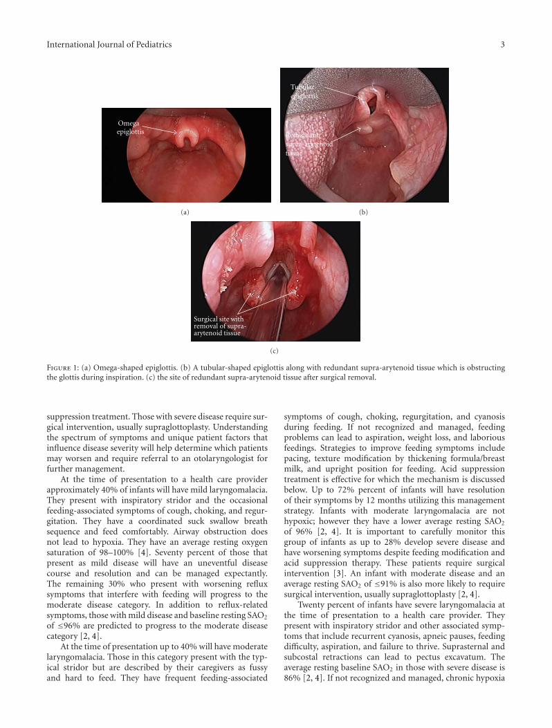
are the structures making up the supraglottis. As seen inFigures 1(a) and 1(b), the common findings seen on examare prolapse of the posteriorly positioned arytenoid cartilagesand mucosa into the airway during inspiration, shorteningof the distance between the arytenoid and epiglottis, and an“omega-shaped” or retroflexed epiglottis.
4. Etiology
The exact etiology of laryngomalacia is unknown and con-tinues to be an area of great interest and research. Theories ofetiology include the anatomic, cartilaginous, and neurologictheories. The anatomic theory proposes that there is anabnormal placement of flaccid tissue resulting in stridor. Thechallenge with the anatomic theory is there are infants whohave the typical anatomic laryngeal findings of laryngoma-lacia who do not have symptoms of airway obstruction. Thecartilaginous theory proposes that the cartilages of the larynxare immature and abnormally pliable. This theory has beenrefuted by the finding of histologically normal cartilage ininfants with symptomatic laryngomalacia. The neurologictheory is the best supported by the literature and as a resultis the prevailing etiologic theory [2].
The neurologic theory recognizes that laryngomalaciamay be a consequence of an underdeveloped or abnormallyintegrated CNS system, particularly the peripheral nervesand brainstem nuclei responsible for breathing and airwaypatency. As the infant matures laryngomalacia likely resolvessecondary to the maturation of the CNS system. Thelaryngeal adductor reflex is a vagal nerve reflex responsiblefor laryngeal function and tone. The afferent activationof the reflex is mediated by the superior laryngeal nervewhich is located in the aryepiglottic fold [2]. Sensoryinformation from this nerve is then transmitted to thebrainstem nuclei that regulate respiration and swallowing.A motor response to sensory stimulation is mediated bythe vagus nerve resulting in glottic closure, inhibition ofrespiration, and swallow. An alteration in this pathway hasa role in the etiology of laryngomalacia and the associatedfeeding symptoms. Laryngeal sensory testing in infants withlaryngomalacia has demonstrated that the sensory stimulusthreshold needed to elicit the typical motor response iselevated in those with moderate-to-severe disease versusthose with mild disease. This testing supports the notion ofan underdeveloped or abnormally integrated peripheral andcentral nervous system mechanism of laryngeal function andtone [2].
5. Spectrum of Disease
Laryngomalacia has a disease spectrum that can be dividedinto mild, moderate, and severe categories [2]. These cate-gories are not based on the quantity of stridor but ratherby the associated feeding and obstructive symptoms. Thosewith mild disease usually have inconsequential inspiratorystridor. Those with moderate disease usually have stridorwith feeding-related symptoms and often improve on acid
International Journal of Pediatrics 3
Omega epiglottis
(a)
Tubular epiglottis
Redundantsupra-arytenoidtissue
(b)
Surgical site with removal of supra-arytenoid tissue
(c)
Figure 1: (a) Omega-shaped epiglottis. (b) A tubular-shaped epiglottis along with redundant supra-arytenoid tissue which is obstructingthe glottis during inspiration. (c) the site of redundant supra-arytenoid tissue after surgical removal.
suppression treatment. Those with severe disease require sur-gical intervention, usually supraglottoplasty. Understandingthe spectrum of symptoms and unique patient factors thatinfluence disease severity will help determine which patientsmay worsen and require referral to an otolaryngologist forfurther management.
At the time of presentation to a health care providerapproximately 40% of infants will have mild laryngomalacia.They present with inspiratory stridor and the occasionalfeeding-associated symptoms of cough, choking, and regur-gitation. They have a coordinated suck swallow breathsequence and feed comfortably. Airway obstruction doesnot lead to hypoxia. They have an average resting oxygensaturation of 98–100% [4]. Seventy percent of those thatpresent as mild disease will have an uneventful diseasecourse and resolution and can be managed expectantly.The remaining 30% who present with worsening refluxsymptoms that interfere with feeding will progress to themoderate disease category. In addition to reflux-relatedsymptoms, those with mild disease and baseline resting SAO2
of ≤96% are predicted to progress to the moderate diseasecategory [2, 4].
At the time of presentation up to 40% will have moderatelaryngomalacia. Those in this category present with the typ-ical stridor but are described by their caregivers as fussyand hard to feed. They have frequent feeding-associated
symptoms of cough, choking, regurgitation, and cyanosisduring feeding. If not recognized and managed, feedingproblems can lead to aspiration, weight loss, and laboriousfeedings. Strategies to improve feeding symptoms includepacing, texture modification by thickening formula/breastmilk, and upright position for feeding. Acid suppressiontreatment is effective for which the mechanism is discussedbelow. Up to 72% percent of infants will have resolutionof their symptoms by 12 months utilizing this managementstrategy. Infants with moderate laryngomalacia are nothypoxic; however they have a lower average resting SAO2
of 96% [2, 4]. It is important to carefully monitor thisgroup of infants as up to 28% develop severe disease andhave worsening symptoms despite feeding modification andacid suppression therapy. These patients require surgicalintervention [3]. An infant with moderate disease and anaverage resting SAO2 of ≤91% is also more likely to requiresurgical intervention, usually supraglottoplasty [2, 4].
Twenty percent of infants have severe laryngomalacia atthe time of presentation to a health care provider. Theypresent with inspiratory stridor and other associated symp-toms that include recurrent cyanosis, apneic pauses, feedingdifficulty, aspiration, and failure to thrive. Suprasternal andsubcostal retractions can lead to pectus excavatum. Theaverage resting baseline SAO2 in those with severe disease is86% [2, 4]. If not recognized and managed, chronic hypoxia
4 International Journal of Pediatrics
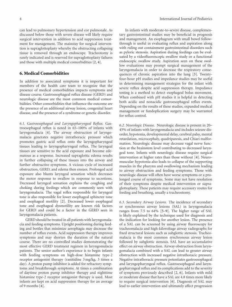
can lead to pulmonary hypertension and cor pulmonale. Asdiscussed below those with severe disease will likely requiresurgical intervention in addition to acid suppression treat-ment for management. The mainstay for surgical interven-tion is supraglottoplasty whereby the obstructing collapsingtissue is removed through an endoscope. Tracheotomy israrely indicated and is reserved for supraglottoplasty failuresand those with multiple medical comorbidities [2, 4].
6. Medical Comorbidities
In addition to associated symptoms it is important formembers of the health care team to recognize that thepresence of medical comorbidities impacts symptoms anddisease course. Gastroesophageal reflux disease (GERD) andneurologic disease are the most common medical comor-bidities. Other comorbidities that influence the outcome arethe presence of an additional airway lesion, congenital heartdisease, and the presence of a syndrome or genetic disorder.
6.1. Gastroesophageal and Laryngopharyngeal Reflux. Gas-troesophageal reflux is noted in 65–100% of infants withlaryngomalacia [4]. The airway obstruction of laryngo-malacia generates negative intrathoracic pressure whichpromotes gastric acid reflux onto the laryngopharyngealtissues leading to laryngopharyngeal reflux. The laryngealtissues are sensitive to the acid exposure and become ede-matous as a response. Increased supraglottic edema resultsin further collapsing of these tissues into the airway andfurther obstructive symptoms. A vicious cycle of increasedobstruction, GERD, and edema then ensues. Prolonged acidexposure also blunts laryngeal sensation which decreasesthe motor response to swallow in response to secretions.Decreased laryngeal sensation explains the coughing andchoking during feedings which are commonly seen withlaryngomalacia. The vagal reflex responsible for laryngealtone is also responsible for lower esophageal sphincter toneand esophageal motility [2]. Decreased lower esophagealtone and esophageal dysmotility are known risk factorsfor GERD and could be a factor in the GERD seen inlaryngomalacia patients.
GERD should be treated in all patients with laryngomala-cia and feeding symptoms. Upright positioning during feed-ing and bottles that minimize aerophagia may decrease thenumber of reflux events. Acid suppression therapy improvessymptoms and may shorten the duration of the naturalcourse. There are no controlled studies demonstrating themost effective GERD treatment regimen in laryngomalaciapatients. The senior author’s experience is to begin infantswith feeding symptoms on high-dose histamine type-2receptor antagonist therapy (ranitidine 3 mg/kg, 3 times aday). A proton pump inhibitor is added for refractory symp-toms and breakthrough symptoms. At times a combinationof daytime proton pump inhibitor therapy and nighttimehistamine type-2 receptor antagonist therapy is used. Mostinfants are kept on acid suppression therapy for an averageof 9 months [4].
In infants with moderate-to-severe disease, complemen-tary gastrointestinal studies may be beneficial in prognosisand management. An esophagram with small bowel follow-through is useful in evaluating reflux and aspiration alongwith ruling out containment gastrointestinal disorders suchas pyloric stenosis. Aspiration during feedings can be eval-uated by a videofluoroscopic swallow study or a functionalendoscopic swallow study. Aspiration seen on these swal-low evaluations may prompt surgical management of thelaryngomalacia in order to decrease the respiratory conse-quences of chronic aspiration into the lung [3]. Twenty-four-hour pH studies and impedance studies may be usefulin determining management strategies for the infant withsevere reflux despite acid suppression therapy. Impedancetesting is a method to detect esophageal bolus movement.When combined with pH studies it is helpful in detectingboth acidic and nonacidic gastroesophageal reflux events.Depending on the results of these studies, expanded medicalmanagement or fundoplication surgery may be warrantedfor reflux control.
6.2. Neurologic Disease. Neurologic disease is present in 20–45% of infants with laryngomalacia and includes seizure dis-order, hypotonia, developmental delay, cerebral palsy, mentalretardation, microcephaly, quadriparesis, and Chiari malfor-mation. Neurologic disease may decrease vagal nerve func-tion at the brainstem level contributing to decreased laryn-geal tone. Infants with neurologic disease require surgicalintervention at higher rates than those without [4]. Neuro-muscular hypotonia also leads to collapse of the supportingmuscles in the pharynx and swallowing mechanism leadingto airway obstruction and feeding symptoms. Those withneurologic disease will often have worse symptoms or a pro-longed course of symptoms. Some may not have resolutionof their symptoms despite medical intervention or supra-glottoplasty. These patients may require accessory routes forfeeding and breathing, usually a tracheostomy.
6.3. Secondary Airway Lesions. The incidence of secondaryor synchronous airway lesions (SAL) in laryngomalaciaranges from 7.5 to 64% [5–9]. The higher range of SALis likely explained by the technique used for diagnosis andthe indication for looking for another lesion. The presenceof a SAL can be screened by using airway fluoroscopy fortracheomalacia and high-kilovoltage airway radiographs forfixed structural lesions such as subglottic stenosis. Tracheo-malacia is the most common synchronous airway lesionfollowed by subglottic stenosis. SAL have an accumulativeeffect on airway obstruction. Airway obstruction from laryn-gomalacia combined with a SAL can lead to greater airwayobstruction with increased negative intrathoracic pressure.Negative intrathoracic pressure potentiates gastroesophagealand laryngopharyngeal reflux. Gastroesophageal and laryn-gopharyngeal reflux and its complications add to the severityof symptoms previously described [2, 6]. Infants with mildor moderate disease that have a SAL are 4.8 times more likelyto require surgical intervention [6]. Diagnosis of SAL maylead to earlier intervention and ultimately affect progression
International Journal of Pediatrics 5
of disease. By surgically addressing laryngomalacia, the re-sultant effect of SAL on the airway may become less signif-icant. If a SAL is suspected on screening radiographs, theinfant will benefit from a referral to an otolaryngologist forclinical correlation.
6.4. Congenital Heart Disease. Congenital heart disease isreported in 10% of infants with laryngomalacia. Theseinfants are more likely to have moderate-to-severe diseaseat the time of presentation. The additive effect of airwayobstruction on compromised cardiovascular function likelytips these infants towards worsening symptoms. Up to 34%of infants with both laryngomalacia and congenital heartdisease will require surgical management [2].
6.5. Congenital Anomalies/Syndromes/Genetic Disorders. Con-genital anomalies and genetic disorders occur with an esti-mated incidence of 8–20% [2, 10, 11]. The incidence is ashigh as 40% of infants with severe laryngomalacia thatrequire surgical intervention [2, 12]. Infants with congenitalanomalies and genetic disorders often have other medicalcomorbidities such as synchronous airway lesions, cardiacdisease, and neurologic disease that confound oxygenationand breathing; this makes any degree of airway obstructionmore of problematic for these patients. Infants with severelaryngomalacia, an isolated anomaly or syndrome, and min-imal comorbidities can be managed successfully with suprag-lottoplasty [2, 12]. Of those infants, Down syndrome appearsto be the most commonly reported associated geneticdisorder with laryngomalacia. Fifty percent of those thathave respiratory symptoms also have laryngomalacia [13–15]. The senior author’s experience with supraglottoplastyin Down syndrome children is that if no coexisting cardiacdisease or neurologic disease is present, they do well withaggressive acid suppression therapy and supraglottoplastyeven if a synchronous airway lesion is present. Those withcardiac disease, neurologic disease, and synchronous airwaylesions often fail supraglottoplasty and may require a tra-cheostomy until cardiac disease is treated.
Those with laryngomalacia and syndromes associatedwith micrognathia such as CHARGE association and PierreRobin sequence will do worse due to the retrodisplacementof the tongue base. The retrodisplaced tongue base collapseson the epiglottis in addition to supra-arytenoid tissueredundancy and short aryepiglottic folds. Supraglottoplastyor epiglottic suspension procedures usually are unsuccessful[16], and most with severe airway obstruction and laryngo-malacia will require a tracheostomy until they grow into themicrognathia or surgical intervention is performed to correctit. Laryngomalacia and variants of 22q11.2 microdele-tion syndrome are described to have severe upper airwayobstruction [17–19] and can be successfully managed withsupraglottoplasty [17]. Because cervical vertebral anomaliesare common in this patient population, cervicomedullarycompression of the brainstem should be investigated asa potentiating cause of symptoms. A recent case seriesdescribes a child who had laryngomalacia symptom reversal
and improvement in laryngeal tone after brainstem decom-pression and did not require supraglottoplasty [19].
If micrognathia is not present, a syndrome or anomalyshould not preclude supraglottoplasty in those with severelaryngomalacia that require intervention. The rates of failureand tracheostomy placement however may be higher inthese patients and should be taken into consideration whencounseling parents and managing this unique group ofinfants.
7. Surgical Management
Surgical management is indicated in those with severedisease. The most common indications for surgery arestridor with respiratory compromise and feeding difficultieswith failure to thrive [1]. Severe airway obstruction withsignificant retractions, pectus excavatum, cor pulmonale,pulmonary hypertension, and hypoxia are all consideredabsolute indications for surgery. The relative indications areaspiration with recurrent pneumonia, weight loss withouttrue failure to thrive, and a difficult to feed child who hasnot responded to acid suppression therapy. The decisionto operate is individualized and based on the trend of theinfants overall health and development. Supraglottoplastyis the mainstay of surgical treatment for laryngomalacia.The patient is anesthetized with a combination of mask andintravenous anesthesia. The airway is first evaluated by rigidendoscopy (microdirect laryngoscopy and bronchoscopy) torule out secondary lesions of the subglottis and trachea. Thesupraglottis is visualized during spontaneous respiration,and the major areas of collapse are noted. The larynx isthen exposed with operating laryngoscopes, and the su-praglottoplasty is performed focusing on removal of theredundant arytenoid mucosa. As seen in Figure 1(c), theprocedure is tailored to the patient’s areas of obstruction, andcare is taken to preserve mucosa in areas prone to stenosis.The success of supraglottoplasty approximates 94% and hasa low complication rate [1]. Revision supraglottoplasty ortracheostomy will be required in 19–45% of infants andis directly influenced by the number and type of medicalcomorbidities [2]. Tracheostomy is reserved for patients whocontinue to have life-threatening airway obstruction andwho fail to improve after supraglottoplasty.
8. Conclusion
Laryngomalacia is a common disease of infancy where thediagnosis is suspected by primary care providers based onhistory. Those with mild disease can be managed expectantly.Continued monitoring of the symptoms is necessary assymptoms can progress over the natural course of the disease.Recognizing patient factors and symptoms associated withmoderate and severe disease helps determine which infantswill benefit from otolaryngology consultation. Identifyingpatient factors that influence disease severity and outcomes isan important aspect of counseling care givers and providingcare to infants with laryngomalacia.

6 International Journal of Pediatrics
References
[1] G. T. Richter and D. M. Thompson, “The Surgical Man-agement of Laryngomalacia,” Otolaryngologic Clinics of NorthAmerica, vol. 41, no. 5, pp. 837–864, 2008.
[2] D. M. Thompson, “Abnormal sensorimotor integrative func-tion of the larynx in congenital laryngomalacia: a new theoryof etiology,” Laryngoscope, vol. 117, no. 6, supplement, pp. 1–33, 2007.
[3] G. T. Richter, C. T. Wootten, M. J. Rutter, and D. M. Thomp-son, “Impact of supraglottoplasty on aspiration in severelaryngomalacia,” Annals of Otology, Rhinology and Laryngol-ogy, vol. 118, no. 4, pp. 259–266, 2009.
[4] D. M. Thompson, “Laryngomalacia: factors that influence dis-ease severity and outcomes of management,” Current Opinionin Otolaryngology and Head and Neck Surgery, vol. 18, no. 6,pp. 564–570, 2010.
[5] S. R. Cohen, M. S. Desmond, R. D. Eavey, and B. C. May,“Endoscopy and tracheotomy in the neonatal period. A 10-year review,” Annals of Otology, Rhinology and Laryngology,vol. 86, no. 5, pp. 577–583, 1977.
[6] J. M. Dickson, G. T. Richter, J. Meinzen-Derr, M. J. Rutter, andD. M. Thompson, “Secondary airway lesions in infants withlaryngomalacia,” Annals of Otology, Rhinology and Laryngol-ogy, vol. 118, no. 1, pp. 37–43, 2009.
[7] J. W. Schroeder, N. D. Bhandarkar, and L. D. Holinger, “Syn-chronous airway lesions and outcomes in infants with severelaryngomalacia requiring supraglottoplasty,” Archives of Oto-laryngology, vol. 135, no. 7, pp. 647–651, 2009.
[8] E. Krashin, J. Ben-Ari, C. Springer, A. DeRowe, A. Avital, andY. Sivan, “Synchronous airway lesions in laryngomalacia,”International Journal of Pediatric Otorhinolaryngology, vol. 72,no. 4, pp. 501–507, 2008.
[9] H. W. Yuen, H. K. K. Tan, and A. Balakrishnan, “Synchronousairway lesions and associated anomalies in children withlaryngomalacia evaluated with rigid endoscopy,” InternationalJournal of Pediatric Otorhinolaryngology, vol. 70, no. 10, pp.1779–1784, 2006.
[10] I. B. Masters, A. B. Chang, L. Patterson et al., “Series of laryn-gomalacia, tracheomalacia, and bronchomalacia disorders andtheir associations with other conditions in children,” PediatricPulmonology, vol. 34, no. 3, pp. 189–195, 2002.
[11] D. R. Olney, J. H. Greinwald, R. J. H. Smith, and N. M. Bau-man, “Laryngomalacia and its treatment,” Laryngoscope, vol.109, no. 11, pp. 1770–1775, 1999.
[12] S. R. Hoff, J. W. Schroeder, J. C. Rastatter, and L. D. Holinger,“Supraglottoplasty outcomes in relation to age and comorbidconditions,” International Journal of Pediatric Otorhinolaryn-gology, vol. 74, no. 3, pp. 245–249, 2010.
[13] P. Bertrand, H. Navarro, S. Caussade, N. Holmgren, and I.Sanchez, “Airway anomalies in children with Down syndrome:endoscopic findings,” Pediatric Pulmonology, vol. 36, no. 2, pp.137–141, 2003.
[14] R. B. Mitchell, E. Call, and J. Kelly, “Diagnosis and therapy forairway obstruction in children with down syndrome,” Archivesof Otolaryngology, vol. 129, no. 6, pp. 642–645, 2003.
[15] R. B. Mitchell, E. Call, and J. Kelly, “Ear, nose and throatdisorders in children with Down syndrome,” Laryngoscope,vol. 113, no. 2, pp. 259–263, 2003.
[16] Y. Naito, M. Higuchi, G. Koinuma, M. Aramaki, T. Takahashi,and K. Kosaki, “Upper airway obstruction in neonates andinfants with CHARGE sndrome,” American Journal of MedicalGenetics, Part A, vol. 143, no. 16, pp. 1815–1820, 2007.
[17] M. C. Digilio, D. M. McDonald-McGinn, C. Heike et al.,“Three patients with oculo-auriculo-vertebral spectrum andmicrodeletion 22q11.2,” American Journal of Medical Genetics,Part A, vol. 149, no. 12, pp. 2860–2864, 2009.
[18] S. Yu, K. Cox, K. Friend et al., “Familial 22q11.2 duplication:a three-generation family with a 3-Mb duplication and afamilial 1.5-Mb duplication,” Clinical Genetics, vol. 73, no. 2,pp. 160–164, 2008.
[19] R. S. Petersson, N. M. Wetjen, and D. M. Thompson, “Neuro-logic variant laryngomalacia associated with Chiari malforma-tion and cervicomedullary compression: case reports,” Annalsof Otology, Rhinology and Laryngology, vol. 120, no. 2, pp. 99–103, 2011.
Hindawi Publishing CorporationInternational Journal of PediatricsVolume 2012, Article ID 576719, 6 pagesdoi:10.1155/2012/576719
Review Article
Sleep Endoscopy in the Evaluation of PediatricObstructive Sleep Apnea
Aaron C. Lin1 and Peter J. Koltai2
1 Division of Pediatric Otolaryngology, Children’s Hospital of Los Angeles, USC Keck School of Medicine, Los Angeles, CA 90027, USA2 Division of Pediatric Otolaryngology, Lucile Packard Children’s Hospital, Stanford University School of Medicine, Stanford,CA 94304, USA
Correspondence should be addressed to Aaron C. Lin, [email protected]
Received 14 July 2011; Revised 24 October 2011; Accepted 19 November 2011
Academic Editor: Ajoy M. Varghese
Copyright © 2012 A. C. Lin and P. J. Koltai. This is an open access article distributed under the Creative Commons AttributionLicense, which permits unrestricted use, distribution, and reproduction in any medium, provided the original work is properlycited.
Pediatric obstructive sleep apnea (OSA) is not always resolved or improved with adenotonsillectomy. Persistent or complex casesof pediatric OSA may be due to sites of obstruction in the airway other than the tonsils and adenoids. Identifying these areasin the past has been problematic, and therefore, therapy for OSA in children who have failed adenotonsillectomy has often beenunsatisfactory. Sleep endoscopy is a technique that can enable the surgeon to determine the level of obstruction in a sleeping childwith OSA. With this knowledge, site-specific surgical therapy for persistent and complex pediatric OSA may be possible.
1. Introduction
Obstructive sleep apnea (OSA) has been estimated to affect1–4% of all children in the United States [1] and islinked to a number of health-related issues and behavioralproblems such as daytime sleepiness, enuresis, cardiovascularproblems, poor growth, hyperactivity, academic difficulties,and attention issues [2, 3]. Pediatric OSA is characterizedby recurrent obstruction of the airway resulting in snoring,gasping, and apneic events, and OSA in children has beenrecognized as a significant disorder warranting evaluationand management. In many cases, adenotonsillar hypertrophyis the primary cause of OSA in children. Therefore, tonsil-lectomy and adenoidectomy (T&A) is the preferred surgicalmethod to treat OSA in most pediatric patients.
With the increased use of polysomnography (PSG),studies have shown that 15–20% of children will havepersistent OSA with postoperative symptoms of obstruc-tion [4, 5]. Children with comorbidities such as obesity,Down syndrome, and craniofacial abnormalities may haveobstruction unrelated to the tonsils and adenoids; thesepatients are at even higher risk for persistent apnea afterT&A. In addition, healthy children with OSA may havesmall tonsils and adenoids that are unlikely to be completely
responsible for their obstruction [6]. There are a variety ofcauses for persistent and complex cases of OSA; these includeobesity, hypotonia, craniofacial disproportion, lingual ton-sillar hypertrophy, and occult laryngomalacia [7–10]. Onesolution to these difficult cases is the use of continuouspositive airway pressure (CPAP). CPAP is generally effectivein managing persistent OSA after adenotonsillectomy orcomplex OSA unrelated to the tonsils and adenoids, andits use is widely adopted in adults. Unfortunately, the useof CPAP in children can be difficult, and many children donot tolerate it [11]. Tracheotomy may be necessary in somesevere cases of OSA.
The evaluation of children suspected of having complexOSA or persistent OSA after adenotonsillectomy shouldinclude polysomnography to confirm obstruction duringsleep. Once OSA is diagnosed, identifying the site of airwayobstruction during sleep may allow further interventionsbeyond tonsillectomy and adenoidectomy and CPAP. Flex-ible fiberoptic laryngoscopy is a useful tool that can beperformed in the office to look for any potential levelsof airway obstruction. Adenoid regrowth, lingual tonsillarhypertrophy, tongue base prolapsed, or laryngeal causes ofobstruction can be seen with office fiberoptic laryngoscopy.However, this evaluation is limited by the fact that patients

2 International Journal of Pediatrics
are awake and upright when examined; potential sites ofobstruction may be missed.
Sleep endoscopy has been previously described in theadult and pediatric populations for the purpose of evaluatingthe dynamic airway in the supine position during a sleep-likestate [6, 12, 13]. It is an increasingly useful tool in the eval-uation of children with persistent OSA after initial therapy,children with OSA without tonsil or adenoid hypertrophy,or children with significant craniofacial anomalies.
2. Method
Sleep endoscopy is performed in the operating room undergeneral anesthesia and is performed in conjunction withdirect laryngoscopy and bronchoscopy. The child is giveninhalational anesthesia by mask. The IV is then placed,and anesthesia is then maintained with an infusion ofdexmedetomidine at 1-2 mcg/kg/hr without a loading dose,as well as with a concurrent ketamine bolus of 1 mg/kgas our primary anesthetic cocktail. In the past, a propofolinfusion was used to maintain anesthesia. However, we havefound that there is less muscular relaxation and a moresustained respiratory effort with this current technique. Wealso vasoconstrict and anesthetize the nose with a half andhalf mixture of oxymetazoline and 1% xylocaine deliveredon a 1 cm× 4 cm cottonoid pledget. Spontaneous respirationis supported by oxygen (2L/min) delivered via nasal canula.The child should be in the supine position without a shoulderroll, mimicking the position of natural sleep as much aspossible.
Once a rhythmic pattern of respiration is established, aflexible fiberoptic laryngoscope is passed directly into thechild’s nose, passing posteriorly toward the nasopharynx. Forvisualization and documentation, a digital video camera isused with the endoscope.
At the nasopharynx, the adenoids are examined as apotential site of obstruction. The position of the palate anduvula in relation to the posterior pharyngeal wall can beseen. The scope is then passed into the oropharynx wherethe tongue base, lingual tonsils, and pharyngeal tonsils (ifstill present) are examined. The position of the base oftongue, vallecula, and epiglottis in relation to the posteriorpharyngeal wall is noted. In some cases, the tongue basecan be seen collapsed against the posterior pharyngeal wall;visualizing the improvement in airway patency by lifting thetongue base with jaw thrust can be quite dramatic (Figures1(a) and 1(b)). The dynamics of lateral pharyngeal wallmotion can be seen. The scope is then passed under theepiglottis where the dynamics of the supraglottic soft tissues,as well as the motion of the vocal cords, are observed.At the completion of the sleep endoscopy, the scope isremoved. Direct laryngoscopy and bronchoscopy can then beperformed to complete the airway evaluation.
During sleep endoscopy, dynamic airway obstructionpotentially can be observed at several levels. In the nasophar-ynx, the normal velopharyngeal opening should remainpatent during inspiration and expiration. However, adenoidhypertrophy, adenoid regrowth, and midfacial hypoplasia
(a)
(b)
Figure 1: (a) Posteriorly displaced epiglottis secondary to tonguebase collapse. (b) Dramatic improvement of the airway upon liftingthe tongue base anteriorly using jaw thrust.
can obstruct this opening and reduce nasal airflow. In theoropharynx, the airway can be narrowed by the lateralpharyngeal wall and tonsils. This space collapses and expandsduring respiration but normally should not result in obstruc-tion. Not surprisingly, tonsillar hypertrophy can significantlynarrow and obstruct this space (Figures 2(a), 2(b), and 2(c)).Collapse of the lateral pharyngeal walls can occur duringinspiration in children with hypotonia.
The tongue base should not rest on the posterior pha-ryngeal wall or displace the epiglottis posteriorly, allowingair to flow under and around the edges of the epiglottis.Again, hypotonia can result in collapse and obstruction atthe tongue base, displacing the epiglottis against the pos-terior pharyngeal wall (Figure 3). Similarly, lingual tonsillarhypertrophy can crowd the entire vallecula with lymphoidtissue and displace the epiglottis against the posterior

International Journal of Pediatrics 3
(a)
(b)
(c)
Figure 2: (a) Supraglottic larynx with the pharyngeal tonsils seenlaterally. (b) Rapid collapse of the pharyngeal tonsils medially oninspiration. (c) Complete obstruction of the airway by the pharyn-geal tonsils on inspiration. On the expiration, the tonsils returned totheir normal positions as in Figure 2(a).
Figure 3: Tongue base collapsed against the posterior pharyngealwall. The tip of the epiglottis is barely seen.
Figure 4: Lymphoid tissue of the lingual tonsils displacing theepiglottis posteriorly.
pharyngeal wall during inspiration (Figure 4). This is alsoseen in children with micrognathia or retrognathia where thetongue base is displaced posteriorly by the underdevelopedmandible.
If not displaced posteriorly by the tongue base, thenormal supraglottis should remain patent during inspirationand expiration. In infants, airway obstruction may resultfrom laryngomalacia characterized by floppy arytenoidmucosa and short aryepiglottic folds; this is present bothwhile awake and during sleep. In some older children withpersistent OSA, dynamics similar to laryngomalacia can beseen where excessive mucosal folds above the arytenoidsprolapse into the glottis on inspiration. However, as opposedto infantile laryngomalacia, this form of obstruction only
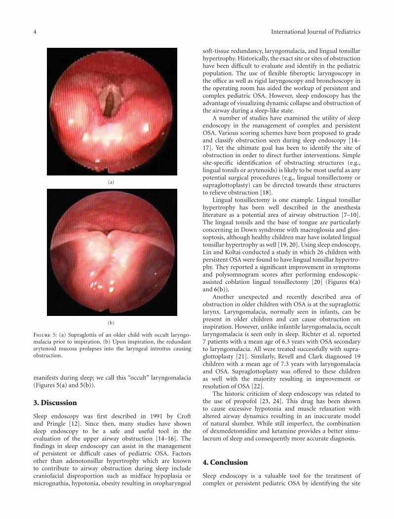
4 International Journal of Pediatrics
(a)
(b)
Figure 5: (a) Supraglottis of an older child with occult laryngo-malacia prior to inspiration. (b) Upon inspiration, the redundantarytenoid mucosa prolapses into the laryngeal introitus causingobstruction.
manifests during sleep; we call this “occult” laryngomalacia(Figures 5(a) and 5(b)).
3. Discussion
Sleep endoscopy was first described in 1991 by Croftand Pringle [12]. Since then, many studies have shownsleep endoscopy to be a safe and useful tool in theevaluation of the upper airway obstruction [14–16]. Thefindings in sleep endoscopy can assist in the managementof persistent or difficult cases of pediatric OSA. Factorsother than adenotonsillar hypertrophy which are knownto contribute to airway obstruction during sleep includecraniofacial disproportion such as midface hypoplasia ormicrognathia, hypotonia, obesity resulting in oropharyngeal
soft-tissue redundancy, laryngomalacia, and lingual tonsillarhypertrophy. Historically, the exact site or sites of obstructionhave been difficult to evaluate and identify in the pediatricpopulation. The use of flexible fiberoptic laryngoscopy inthe office as well as rigid laryngoscopy and bronchoscopy inthe operating room has aided the workup of persistent andcomplex pediatric OSA. However, sleep endoscopy has theadvantage of visualizing dynamic collapse and obstruction ofthe airway during a sleep-like state.
A number of studies have examined the utility of sleependoscopy in the management of complex and persistentOSA. Various scoring schemes have been proposed to gradeand classify obstruction seen during sleep endoscopy [14–17]. Yet the ultimate goal has been to identify the site ofobstruction in order to direct further interventions. Simplesite-specific identification of obstructing structures (e.g.,lingual tonsils or arytenoids) is likely to be most useful as anypotential surgical procedures (e.g., lingual tonsillectomy orsupraglottoplasty) can be directed towards these structuresto relieve obstruction [18].
Lingual tonsillectomy is one example. Lingual tonsillarhypertrophy has been well described in the anesthesialiterature as a potential area of airway obstruction [7–10].The lingual tonsils and the base of tongue are particularlyconcerning in Down syndrome with macroglossia and glos-soptosis, although healthy children may have isolated lingualtonsillar hypertrophy as well [19, 20]. Using sleep endoscopy,Lin and Koltai conducted a study in which 26 children withpersistent OSA were found to have lingual tonsillar hypertro-phy. They reported a significant improvement in symptomsand polysomnogram scores after performing endoscopic-assisted coblation lingual tonsillectomy [20] (Figures 6(a)and 6(b)).
Another unexpected and recently described area ofobstruction in older children with OSA is at the supraglotticlarynx. Laryngomalacia, normally seen in infants, can bepresent in older children and can cause obstruction oninspiration. However, unlike infantile laryngomalacia, occultlaryngomalacia is seen only in sleep. Richter et al. reported7 patients with a mean age of 6.3 years with OSA secondaryto laryngomalacia. All were treated successfully with supra-glottoplasty [21]. Similarly, Revell and Clark diagnosed 19children with a mean age of 7.3 years with laryngomalaciaand OSA. Supraglottoplasty was offered to these childrenas well with the majority resulting in improvement orresolution of OSA [22].
The historic criticism of sleep endoscopy was related tothe use of propofol [23, 24]. This drug has been shownto cause excessive hypotonia and muscle relaxation withaltered airway dynamics resulting in an inaccurate modelof natural slumber. While still imperfect, the combinationof dexmedetomidine and ketamine provides a better simu-lacrum of sleep and consequently more accurate diagnosis.
4. Conclusion
Sleep endoscopy is a valuable tool for the treatment ofcomplex or persistent pediatric OSA by identifying the site
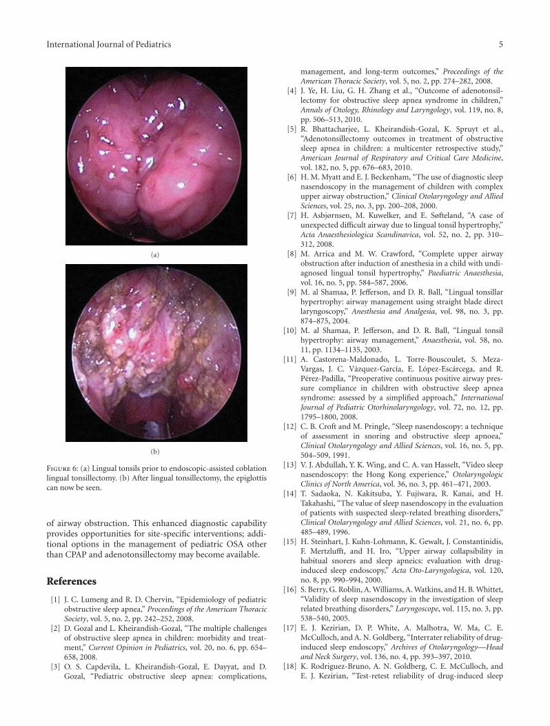
International Journal of Pediatrics 5
(a)
(b)
Figure 6: (a) Lingual tonsils prior to endoscopic-assisted coblationlingual tonsillectomy. (b) After lingual tonsillectomy, the epiglottiscan now be seen.
of airway obstruction. This enhanced diagnostic capabilityprovides opportunities for site-specific interventions; addi-tional options in the management of pediatric OSA otherthan CPAP and adenotonsillectomy may become available.
References
[1] J. C. Lumeng and R. D. Chervin, “Epidemiology of pediatricobstructive sleep apnea,” Proceedings of the American ThoracicSociety, vol. 5, no. 2, pp. 242–252, 2008.
[2] D. Gozal and L. Kheirandish-Gozal, “The multiple challengesof obstructive sleep apnea in children: morbidity and treat-ment,” Current Opinion in Pediatrics, vol. 20, no. 6, pp. 654–658, 2008.
[3] O. S. Capdevila, L. Kheirandish-Gozal, E. Dayyat, and D.Gozal, “Pediatric obstructive sleep apnea: complications,
management, and long-term outcomes,” Proceedings of theAmerican Thoracic Society, vol. 5, no. 2, pp. 274–282, 2008.
[4] J. Ye, H. Liu, G. H. Zhang et al., “Outcome of adenotonsil-lectomy for obstructive sleep apnea syndrome in children,”Annals of Otology, Rhinology and Laryngology, vol. 119, no. 8,pp. 506–513, 2010.
[5] R. Bhattacharjee, L. Kheirandish-Gozal, K. Spruyt et al.,“Adenotonsillectomy outcomes in treatment of obstructivesleep apnea in children: a multicenter retrospective study,”American Journal of Respiratory and Critical Care Medicine,vol. 182, no. 5, pp. 676–683, 2010.
[6] H. M. Myatt and E. J. Beckenham, “The use of diagnostic sleepnasendoscopy in the management of children with complexupper airway obstruction,” Clinical Otolaryngology and AlliedSciences, vol. 25, no. 3, pp. 200–208, 2000.
[7] H. Asbjørnsen, M. Kuwelker, and E. Søfteland, “A case ofunexpected difficult airway due to lingual tonsil hypertrophy,”Acta Anaesthesiologica Scandinavica, vol. 52, no. 2, pp. 310–312, 2008.
[8] M. Arrica and M. W. Crawford, “Complete upper airwayobstruction after induction of anesthesia in a child with undi-agnosed lingual tonsil hypertrophy,” Paediatric Anaesthesia,vol. 16, no. 5, pp. 584–587, 2006.
[9] M. al Shamaa, P. Jefferson, and D. R. Ball, “Lingual tonsillarhypertrophy: airway management using straight blade directlaryngoscopy,” Anesthesia and Analgesia, vol. 98, no. 3, pp.874–875, 2004.
[10] M. al Shamaa, P. Jefferson, and D. R. Ball, “Lingual tonsilhypertrophy: airway management,” Anaesthesia, vol. 58, no.11, pp. 1134–1135, 2003.
[11] A. Castorena-Maldonado, L. Torre-Bouscoulet, S. Meza-Vargas, J. C. Vazquez-Garcıa, E. Lopez-Escarcega, and R.Perez-Padilla, “Preoperative continuous positive airway pres-sure compliance in children with obstructive sleep apneasyndrome: assessed by a simplified approach,” InternationalJournal of Pediatric Otorhinolaryngology, vol. 72, no. 12, pp.1795–1800, 2008.
[12] C. B. Croft and M. Pringle, “Sleep nasendoscopy: a techniqueof assessment in snoring and obstructive sleep apnoea,”Clinical Otolaryngology and Allied Sciences, vol. 16, no. 5, pp.504–509, 1991.
[13] V. J. Abdullah, Y. K. Wing, and C. A. van Hasselt, “Video sleepnasendoscopy: the Hong Kong experience,” OtolaryngologicClinics of North America, vol. 36, no. 3, pp. 461–471, 2003.
[14] T. Sadaoka, N. Kakitsuba, Y. Fujiwara, R. Kanai, and H.Takahashi, “The value of sleep nasendoscopy in the evaluationof patients with suspected sleep-related breathing disorders,”Clinical Otolaryngology and Allied Sciences, vol. 21, no. 6, pp.485–489, 1996.
[15] H. Steinhart, J. Kuhn-Lohmann, K. Gewalt, J. Constantinidis,F. Mertzlufft, and H. Iro, “Upper airway collapsibility inhabitual snorers and sleep apneics: evaluation with drug-induced sleep endoscopy,” Acta Oto-Laryngologica, vol. 120,no. 8, pp. 990–994, 2000.
[16] S. Berry, G. Roblin, A. Williams, A. Watkins, and H. B. Whittet,“Validity of sleep nasendoscopy in the investigation of sleeprelated breathing disorders,” Laryngoscope, vol. 115, no. 3, pp.538–540, 2005.
[17] E. J. Kezirian, D. P. White, A. Malhotra, W. Ma, C. E.McCulloch, and A. N. Goldberg, “Interrater reliability of drug-induced sleep endoscopy,” Archives of Otolaryngology—Headand Neck Surgery, vol. 136, no. 4, pp. 393–397, 2010.
[18] K. Rodriguez-Bruno, A. N. Goldberg, C. E. McCulloch, andE. J. Kezirian, “Test-retest reliability of drug-induced sleep

6 International Journal of Pediatrics
endoscopy,” Otolaryngology—Head and Neck Surgery, vol. 140,no. 5, pp. 646–651, 2009.
[19] K. Nakazawa, D. Ikeda, S. Ishikawa, and K. Makita, “A caseof difficult airway due to lingual tonsillar hypertrophy in apatient with Down’s syndrome,” Anesthesia and Analgesia, vol.97, no. 3, pp. 704–705, 2003.
[20] A. C. Lin and P. J. Koltai, “Persistent pediatric obstructive sleepapnea and lingual tonsillectomy,” Otolaryngology—Head andNeck Surgery, vol. 141, no. 1, pp. 81–85, 2009.
[21] G. T. Richter, M. J. Rutter, A. DeAlarcon, L. J. Orvidas, andD. M. Thompson, “Late-onset laryngomalacia: a variant ofdisease,” Archives of Otolaryngology—Head and Neck Surgery,vol. 134, no. 1, pp. 75–80, 2008.
[22] S. M. Revell and W. D. Clark, “Late-onset laryngomalacia:a cause of pediatric obstructive sleep apnea,” InternationalJournal of Pediatric Otorhinolaryngology, vol. 75, no. 2, pp.231–238, 2011.
[23] A. de Vito, V. Agnoletti, S. Berrettini et al., “Drug-inducedsleep endoscopy: conventional versus target controlled infu-sion techniques-a randomized controlled study,” EuropeanArchives of Oto-Rhino-Laryngology, vol. 268, no. 3, pp. 457–462, 2011.
[24] G. Roblin, A. R. Williams, and H. Whittet, “Target-controlledinfusion in sleep endoscopy,” Laryngoscope, vol. 111, no. 1, pp.175–176, 2001.

Hindawi Publishing CorporationInternational Journal of PediatricsVolume 2012, Article ID 250254, 8 pagesdoi:10.1155/2012/250254
Research Article
Cortical Auditory Evoked Potentials in Children witha Hearing Loss: A Pilot Study
Amineh Koravand,1, 2 Benoıt Jutras,1, 2 and Maryse Lassonde2, 3
1 Ecole d’Orthophonie et d’Audiologie, Universite de Montreal, C.P. 6125, Succursale Centre-Ville, Montreal, Qc, Canada H3C 3J72 Centre de Recherche du CHU Sainte-Justine, 3175 Cote Sainte-Catherine, Montreal, Qc, Canada H3T 1C53 Centre de Recherche en Neuropsychologie et Cognition, Universite de Montreal, C.P. 6125, Succursale Centre-Ville, Montreal,Qc, Canada H3C 3J7
Correspondence should be addressed to Amineh Koravand, [email protected]
Received 7 July 2011; Revised 3 October 2011; Accepted 11 October 2011
Academic Editor: Ajoy M. Varghese
Copyright © 2012 Amineh Koravand et al. This is an open access article distributed under the Creative Commons AttributionLicense, which permits unrestricted use, distribution, and reproduction in any medium, provided the original work is properlycited.
Objective. This study examined the patterns of neural activity in the central auditory system in children with hearing loss. Methods.Cortical potentials and mismatch responses (MMRs) were recorded from ten children aged between 9 and 10 years: five withhearing loss and five with normal hearing in passive oddball paradigms using verbal and nonverbal stimuli. Results. Results indicatea trend toward larger P1 amplitude, a significant reduction in amplitude, and latency of N2 in children with hearing loss comparedto control. No significant group differences were observed for the majority of the MMRs conditions. Conclusions. Data suggest thatthe reduced auditory input affects the pattern of cortical-auditory-evoked potentials in children with a mild to moderately severehearing loss. Results suggest maturational delays and/or deficits in central auditory processing in children with hearing loss, asindicated by the neurophysiological markers P1 and N2. In contrast, negative MMR data suggest that the amplification providedby the hearing aids could have allowed children with hearing loss to develop adequate discriminative abilities.
1. Introduction
Sensory hearing loss often affects speech perception due toa decreased audibility of the signal as well as decreased tem-poral analysis ability [1–3]. Studies have demonstrated theinfluence of hearing loss on auditory temporal ordering,a task which involves the central auditory system [4–6].The lower performance of children with hearing loss in thistask could be caused by central auditory neurophysiologicaldeficits.
Auditory neurophysiological functions have been meas-ured in adults and children with hearing loss [7–11]. Sensoryhearing loss in adults induced a delay in the latency of N1,N2, and a reduction in N2-P2 amplitude [8]. Oates et al. [7]investigated the N1, N2, MMN, and P3, presented at 65 and80 dB SPL, and found a latency prolongation and an ampli-tude reduction of these components in adults with hearingloss compared to those of the control group at both levelsof presentation. However, an earlier study did not reveal
any significant differences in the latencies of N1, P2, andP3 components between adults with hearing loss and theirnormal-hearing controls [11]. Several factors could accountfor these differential findings, such as participants’ age, age atonset of hearing loss, type and/or degree of hearing loss, levelof stimulus presentation, and type of stimuli used.
In children, latency changes in the cortical-auditory-evoked potentials (CAEPs) have been used to document au-ditory system plasticity and recovery from auditory depriva-tion following cochlear implantation [10, 12]. Congenitallydeaf children who are fitted with cochlear implants during asensitive period of early childhood show normal central au-ditory maturation within six months of implant use as dem-onstrated by changes in P1 latency [10, 12]. Interestingly, inchildren with sensory hearing loss and those with auditoryneuropathy, the P1, N1, and P2 components are present onlyin those children exhibiting good speech perception skills[9]. However, the children with and without good speechperception skills were not age-matched and this factor could
2 International Journal of Pediatrics
have influenced the results, since CAEPs show substantialchanges with maturation [13, 14].
To determine CAEPs potential clinical benefits in assess-ing central auditory functions in children with hearing loss,a clear understanding of the effects of a sensory hearing losson CAEPs is needed. The main objective of the present studyis to explore central auditory neurophysiological functions inchildren with hearing loss wearing hearing aids. If CAEPs areaffected by sensory hearing loss, cortical auditory measurescould become neurophysiological markers in clinical audiol-ogy for evaluating young children for whom central auditoryfunctions are difficult to assess behaviourally.
2. Materials and Methods
2.1. Participants. Two groups of nine-to-ten years old femalechildren participated in the study: five with sensory hearingloss (mean age: 9 : 10 years, SD = ±3 mo) and five withnormal hearing (mean age: 9 : 11 years, SD = ±3 mo).Participants with normal hearing had auditory detectionthreshold at 15 dB HL or less between 500 Hz and 8 kHz,bilaterally (re: ANSI, 1996 [15]). Average hearing sensitivitythresholds of children with hearing loss, based on the averageof 500, 1000, and 2000 Hz thresholds, were within thelimits of mild to moderately severe hearing loss (accordingto Clark (1981) classifications [16]). All participants wereright handed, as measured by an adapted protocol to assesslaterality dominance [17]. The study was approved by theEthics Committee of the Sainte-Justine Hospital.
2.2. Stimuli. Three pairs of synthetic stimuli were used forthis study: one verbal and two nonverbal pairs. The ver-bal stimuli consisted of two syllables: /ba/ and /da/. Thesestimuli were selected from the CD-ROM, Speech Production,and Perception I [18]. The first pair of nonverbal stimu-li consisted of synthesized transformations of /ba/-/da/, gen-erated using two softwares, Dr. Speech and Mitsyn [19, 20].Only the second and third formants were used to create thenonverbal stimuli (for more detail on the syllable transfor-mation, see Mody et al. [21]). Using only these two formants,the stimuli are recognized as nonverbal sounds [21]. The sec-ond pair of nonverbal stimuli was the pair of a 1 kHz puretone and a wide-band noise. Stimuli were 250 ms in durationwith 2.2 ms rise and fall times.
The stimuli were presented with a computer (DELL)using Stimaudio (NeuroScan Inc.) and the Stim2 software.They were presented to the right ear through insert-earphone(E-A-RTONE 3A), connected to an audiometer (Interacous-tics, AD229b model), at 70 dB HL for children with normalhearing and between 85 and 105 dB HL for those with hear-ing loss (See Table 1). Stimuli were presented in a passiveoddball paradigm, with standard stimuli (syllable /ba/, non-verbal /ba/ and a 1 kHz pure tone) of 85% probability of oc-currence and deviant stimuli (syllable /da/, nonverbal /da/and wide-band noise) with a 15% probability of occurrence.The interstimulus interval (ISI) was one second. The order ofstimulus presentation was pseudorandomized within a run,with no two deviants occurring in succession and no runbeginning with a deviant stimulus. Any deviant stimulus was
always preceded by at least three standard stimuli. A thou-sand counterbalanced trials for each pair of stimuli were re-corded.
2.3. Electrophysiological Recordings. The cortical responseswere digitally recorded using a high-density system, Scan 4.0software (NeuroScan, Inc., USA), with SynAmps amplifiersand from 128 Ag/AgCl electrodes. Electrophysiological sig-nals were acquired at a sampling rate of 250 Hz, with ananalog online bandpass filtering from 0.1 to 100 Hz usingthe SynAmps amplifiers running on a Dell computer. Anelectrode located on the forehead (Fpz) served as ground andreference was located at the vertex. Electrode impedance waskept under 7 kΩ for mastoid, central, and frontal regions andbelow 15 kΩ for ocular and peripheral regions.
2.4. Procedure. The children were seated in a chair in a dou-ble-walled sound-proof booth. Participants watched a movieor cartoon of their choice on a computer monitor with thesound off. They were told to ignore the auditory input andto focus their attention on the movie. Total testing durationwas approximately between 90 and 120 minutes.
2.5. Data Analysis. Using BrainVision Analyser program onan IBM computer, the data were corrected for eye move-ments using Gratton and Coles algorithm [22]. They werenext digitally filtered using a filter of 1–15 Hz at 24 dB/octave.These data were rereferenced to both mastoids electrodes.Eye movements and epochs with other artefacts were rejectedbased on voltage criteria (±100 μV). The timeframe of analy-sis was from−100 ms to 700 ms. Data were baseline correctedto−50 ms. Auditory cortical components were defined as fol-lowed: P1 and N1 were the first positive and negative wave-forms in the time window of 50–100 ms and 80–120 ms,respectively. They are followed by a positive peak, definedas P2 within the time window of 100–160 ms, and N2, thesecond negative peak at 200–280 ms. Amplitude values weremeasured from baseline to peak for each component, andlatency values were measured relative to the onset of stimuluspresentation.
MMRs were computed according to the following pro-cedure: ERPs evoked by a standard stimulus were subtractedfrom ERPs evoked by the presentation of a deviant stimulusfor each participant. Responses to standard stimuli that im-mediately followed the presentation of deviant stimuli wereexcluded from the standard stimulus average. Two MMRswere observed with the pair 1 kHz pure tone wide-bandnoise; a first negative peak was measured from 115 to 200 msand a positive slope was observed from 200 to 330 ms. How-ever, only one prevalent negative response from 115 to260 ms was observed with the nonverbal and verbal pairs. Foreach participant, the latency of the most negative or positivepeak was measured for the MMRs by using a peak amplitudeautomatic detection.
3. Results
3.1. CAEP Components. Statistical analyses were conductedon the amplitude and latency values of the standard sound
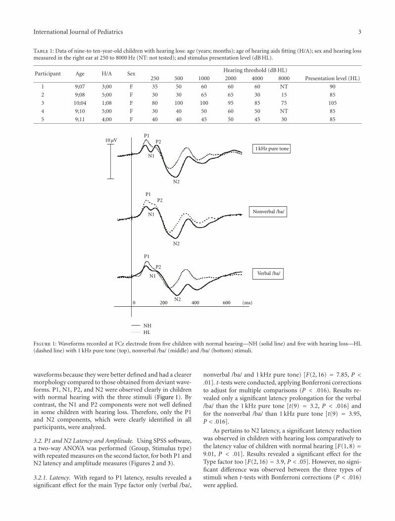
International Journal of Pediatrics 3
Table 1: Data of nine-to ten-year-old children with hearing loss: age (years; months); age of hearing aids fitting (H/A); sex and hearing lossmeasured in the right ear at 250 to 8000 Hz (NT: not tested); and stimulus presentation level (dB HL).
Participant Age H/A SexHearing threshold (dB HL)
250 500 1000 2000 4000 8000 Presentation level (HL)
1 9;07 3;00 F 35 50 60 60 60 NT 90
2 9;08 5;00 F 30 30 65 65 30 15 85
3 10;04 1;08 F 80 100 100 95 85 75 105
4 9;10 5;00 F 30 40 50 60 50 NT 85
5 9;11 4;00 F 40 40 45 50 45 30 85
N2
P1P2
N2
N1
(ms)
1 kHz pure tone
Nonverbal /ba/
Verbal /ba/
NHHL
P1P2
N1
N2
P1
P2
N1
2000 400 600
10 μV
Figure 1: Waveforms recorded at FCz electrode from five children with normal hearing—NH (solid line) and five with hearing loss—HL(dashed line) with 1 kHz pure tone (top), nonverbal /ba/ (middle) and /ba/ (bottom) stimuli.
waveforms because they were better defined and had a clearermorphology compared to those obtained from deviant wave-forms. P1, N1, P2, and N2 were observed clearly in childrenwith normal hearing with the three stimuli (Figure 1). Bycontrast, the N1 and P2 components were not well definedin some children with hearing loss. Therefore, only the P1and N2 components, which were clearly identified in allparticipants, were analyzed.
3.2. P1 and N2 Latency and Amplitude. Using SPSS software,a two-way ANOVA was performed (Group, Stimulus type)with repeated measures on the second factor, for both P1 andN2 latency and amplitude measures (Figures 2 and 3).
3.2.1. Latency. With regard to P1 latency, results revealed asignificant effect for the main Type factor only (verbal /ba/,
nonverbal /ba/ and 1 kHz pure tone) [F(2, 16) = 7.85, P <.01]. t-tests were conducted, applying Bonferroni correctionsto adjust for multiple comparisons (P < .016). Results re-vealed only a significant latency prolongation for the verbal/ba/ than the 1 kHz pure tone [t(9) = 3.2, P < .016] andfor the nonverbal /ba/ than 1 kHz pure tone [t(9) = 3.95,P < .016].
As pertains to N2 latency, a significant latency reductionwas observed in children with hearing loss comparatively tothe latency value of children with normal hearing [F(1, 8) =9.01, P < .01]. Results revealed a significant effect for theType factor too [F(2, 16) = 3.9, P < .05]. However, no signi-ficant difference was observed between the three types ofstimuli when t-tests with Bonferroni corrections (P < .016)were applied.

4 International Journal of Pediatrics
350
300
250
200
150
100
50
0T NV V
P1 N2
NHHL
Late
ncy
(m
s)
T NV V
Figure 2: P1 and N2 mean latency values and standard deviationrecorded of five children with hearing loss (HL) and five childrenwith normal hearing (NH) with 1 kHz pure tone (T), nonverbal /ba/(NV), and verbal /ba/ (V) stimuli.
P1 N210
8
6
4
2
0
−2
−4
−6
−8
−10
Am
plit
ude
(U
v)
NHHL
T NV V T NV V
Figure 3: P1 and N2 mean amplitude values and standard deviationof five children with hearing loss (HL) and five children with normalhearing (NH) with 1 kHz pure tone (T), nonverbal /ba/ (NV), andverbal /ba/ (V) stimuli.
3.2.2. Amplitude. Regarding P1 amplitude, results revealeda significant effect for the Type factor only [F(2, 16) = 5.5,P < .01]. The main Group factor failed to reach significancebut a trend was observed [F(1, 8) = 4.03, P = .08]. For thesignificant Type factor, a t-tests, with Bonferroni corrections(P < .016), revealed a significant greater amplitude for the1 kHz pure tone than the nonverbal /ba/ [t(9) = 3, P < .016]only.
For the N2 amplitude, a significant amplitude reductionwas observed in children with hearing loss comparativelyto the amplitude value of children with normal hearing[F(1, 8) = 5.8, P < .05]. Results also showed a significant
effect for the type factor [F(2, 16) = 3.8, P < .05]. t-tests withBonferroni corrections (P < .016) demonstrated a significantgreater amplitude for the 1 kHz pure tone than the verbal /ba/[t(9) = 3.1, P < .016] only.
3.3. Mismatch Responses (MMRs). A two-way ANOVA wasperformed (Group, Stimulus type) with repeated measureson the second factor, for both the MMR latency and am-plitude measures (Figure 4).
3.3.1. Latency. With regard to negative MMR latency, resultsrevealed a significant effect only for the main Type factor(verbal /ba/-/da/ pair, nonverbal /ba-/da/ pair and 1 kHz puretone and wide-band noise pair) [F(2, 16) = 23.3, P < .001].A t-test with Bonferroni corrections (P < .016) demon-strated a significant latency prolongation for the verbal /ba/-/da/ pair as compared with the 1 kHz pure tone and wide-band noise pair [t(9) = 6.9, P < .016] and also comparativelyto the nonverbal /ba/-/da/ pairs [t(9) = 4.3, P < .016].
3.3.2. Amplitude. Regarding the negative MMR amplitude,results revealed no significant effect for the two main factorsnor for the two-way interaction Group × Type. A positiveMMR component was observed with the pair of 1 kHzpure tone and wide-band noise (Figure 4). A t-test wasconducted on the amplitude and latency values. Resultsrevealed a significant difference between the two groups forthe amplitude value [t(8) = 1.8, P < .05], but not for thelatency value [t(8) = .53, P = .23].
4. Discussion
The aim of the present research was to study the patterns ofthe neurophysiological activity in the central auditory systemin children with hearing loss as compared with children withnormal hearing. Differential findings were observed withregard to the principal cortical components and the MMRresults.
4.1. Cortical Principal Components. P1 amplitude tended tobe greater, N1 and P2 components less defined, and ampli-tude and latency of N2 reduced in children with hearing losscompared with the results of the children with normal hear-ing. These findings will be discussed according to three fac-tors: the presentation level, the maturation of the central au-ditory system, and the deficit in the central auditory system.
4.2. Presentation Level. The stimuli were presented between80 and 105 dB HL for the children with hearing loss andat 70 dB HL for the children with normal hearing. Thehigher level of stimulus presentation (in dB HL) could havecontributed to the large amplitude of P1 and to the shorterlatency of N2. Oates et al. (2002) found that the amplitudeof the N1 and the P300 was larger and their latency shorterat 80 dB SPL compared to 65 dB SPL in adults with hearingloss [7]. In normal-hearing adults, as the intensity increases,peak latencies of P1, N1, P2, and N2 decrease and their peakamplitudes increase [23].
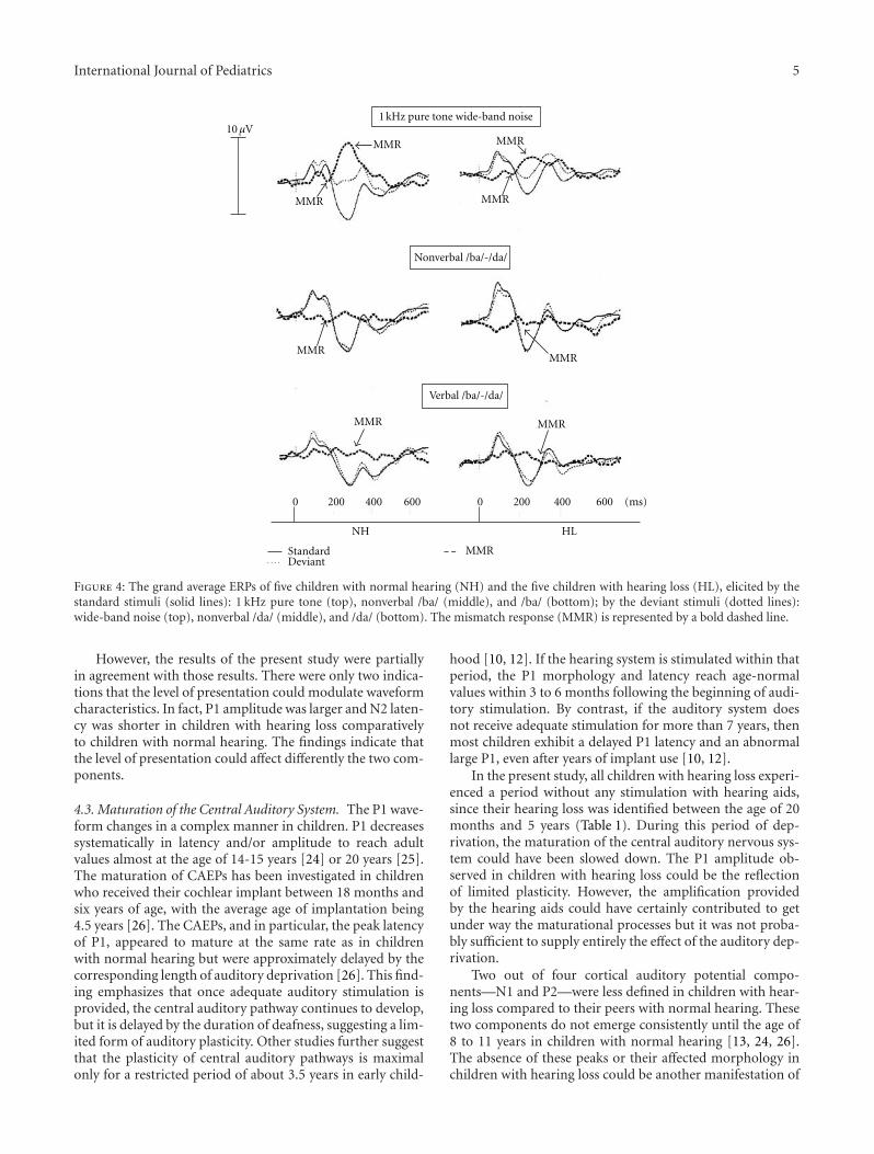
International Journal of Pediatrics 5
1 kHz pure tone wide-band noise
Nonverbal /ba/-/da/
Verbal /ba/-/da/
StandardDeviant
MMR
(ms)200 400 6000
MMR
MMR
MMR
MMR
MMRMMR
MMRMMR
0 200 400 600
NH HL
10 μV
Figure 4: The grand average ERPs of five children with normal hearing (NH) and the five children with hearing loss (HL), elicited by thestandard stimuli (solid lines): 1 kHz pure tone (top), nonverbal /ba/ (middle), and /ba/ (bottom); by the deviant stimuli (dotted lines):wide-band noise (top), nonverbal /da/ (middle), and /da/ (bottom). The mismatch response (MMR) is represented by a bold dashed line.
However, the results of the present study were partiallyin agreement with those results. There were only two indica-tions that the level of presentation could modulate waveformcharacteristics. In fact, P1 amplitude was larger and N2 laten-cy was shorter in children with hearing loss comparativelyto children with normal hearing. The findings indicate thatthe level of presentation could affect differently the two com-ponents.
4.3. Maturation of the Central Auditory System. The P1 wave-form changes in a complex manner in children. P1 decreasessystematically in latency and/or amplitude to reach adultvalues almost at the age of 14-15 years [24] or 20 years [25].The maturation of CAEPs has been investigated in childrenwho received their cochlear implant between 18 months andsix years of age, with the average age of implantation being4.5 years [26]. The CAEPs, and in particular, the peak latencyof P1, appeared to mature at the same rate as in childrenwith normal hearing but were approximately delayed by thecorresponding length of auditory deprivation [26]. This find-ing emphasizes that once adequate auditory stimulation isprovided, the central auditory pathway continues to develop,but it is delayed by the duration of deafness, suggesting a lim-ited form of auditory plasticity. Other studies further suggestthat the plasticity of central auditory pathways is maximalonly for a restricted period of about 3.5 years in early child-
hood [10, 12]. If the hearing system is stimulated within thatperiod, the P1 morphology and latency reach age-normalvalues within 3 to 6 months following the beginning of audi-tory stimulation. By contrast, if the auditory system doesnot receive adequate stimulation for more than 7 years, thenmost children exhibit a delayed P1 latency and an abnormallarge P1, even after years of implant use [10, 12].
In the present study, all children with hearing loss experi-enced a period without any stimulation with hearing aids,since their hearing loss was identified between the age of 20months and 5 years (Table 1). During this period of dep-rivation, the maturation of the central auditory nervous sys-tem could have been slowed down. The P1 amplitude ob-served in children with hearing loss could be the reflectionof limited plasticity. However, the amplification providedby the hearing aids could have certainly contributed to getunder way the maturational processes but it was not proba-bly sufficient to supply entirely the effect of the auditory dep-rivation.
Two out of four cortical auditory potential compo-nents—N1 and P2—were less defined in children with hear-ing loss compared to their peers with normal hearing. Thesetwo components do not emerge consistently until the age of8 to 11 years in children with normal hearing [13, 24, 26].The absence of these peaks or their affected morphology inchildren with hearing loss could be another manifestation of

6 International Journal of Pediatrics
a delayed maturation of the central auditory nervous system.This interpretation is consistent with a study reporting thatN1 and P2 are either delayed in developing or absent in chil-dren with a cochlear implant [26].
Regarding the N2 maturation in children with normalhearing, N2 amplitude has an initial increase between theage of 5 to 11 years [24] followed by a gradual decline fromlate childhood to midadolescence [27, 28] and finally N2 am-plitude reaches adult values by age 17 [24]. However, thereis no general consensus regarding the development of peaklatency, with some studies showing a decline [29], no change,[27] or an increase in latency with age [24]. The maturationeffect was examined at central (Cz, C3, and C4) and at frontal(Fz) electrodes in 118 subjects [24]. The N2 latency increasedsignificantly as a function of age at central electrodes with nomaturational change at the frontal electrode. However, forthe children between 9 and 10 years old, the latency valueswere similar at the four electrode sites [24]. Based on thisstudy [24], the reduction in amplitude and in latency of N2in children with hearing loss in the present set of data couldbe explained by a delay in maturation of the central audi-tory nervous system. Alternately, based on other studies (e.g.,[13, 29]), the reduction of N2 latency could be related toa more mature system. However, it seems counter-intuitivethat the late component (N2) should mature more rapidlyin children with hearing loss than in children with normalhearing. Taking into account the increased P1 amplitude andthe abnormal morphology of N1 and P2, the N2 changeswould rather militate in favor of delayed maturation in chil-dren with hearing loss.
4.4. Deficit in the Central Auditory System. The greater am-plitude of P1 with a concomitant reduction in N2 amplitudeand the less well-defined N1-P2 components could also indi-cate a deficit in central auditory processing. The anomalieshave been reported in central auditory late latency com-ponents in children with language-based learning problems(LPs) [30]. Albeit displaying normal hearing sensitivity, thesechildren had abnormalities in neurophysiological encodingmarked by different patterns in amplitude or latency com-pared to their control peers. In fact, one normal categoryand three atypical categories based on cortical responses ofchildren with LP were found. The atypical category 1 includ-ed children with a delayed P1 latency and no evidence of N1or P2 component. The atypical category 2 was composed ofchildren having normal P1 but delayed N1 and P2 responses.For the atypical category 3, children had generally low-am-plitude responses [30]. Although N2 properties were not spe-cifically examined in this study, observations from their re-sults suggest that N2 amplitude and latency values were ab-normal (low-amplitude and/or delayed latency) for childrenin the three atypical categories. These atypical responsesmight represent a general decrease in synchronous activity,indicating an immature development of the central auditorypathways or slower processing mechanisms [30].
4.5. Mismatch Responses. Similar patterns of results were ob-tained in the two groups of children with the negative mis-match response measured in the 150–200 ms window. These
results suggest that the auditory system can discriminatesounds, being the verbal or nonverbal, and that this patternof discrimination can be found in children with hearing lossas well as in children with normal hearing. They further sug-gest that the amplification provided by the hearing aids couldhave contributed to get under way the maturational proc-esses, allowing the children to develop adequate discrimina-tive abilities.
A positive MMR was measured in the 200–300 ms win-dow with the pair of 1 kHz pure tone and wide-band noiseonly. Results showed that the amplitude of this positiveMMR was significantly smaller in children with hearing lossthan that observed in children without hearing loss. Thisresult may simply be related to the fact that children withhearing loss have, as stated above, a smaller N2 amplitude inresponse to the standard stimuli compared to normal hearingchildren.
The negative MMR was also found to differ accordingto stimulus type. When the stimuli were simple, (the pair1 kHz and wide-band noise), the MMR had an earlier latencycompared to more complex stimuli, such as the nonverbaland verbal /ba/-/da/. The effect of stimulus type on ERP re-sults has also been reported by other studies [31, 32]. Thoseand the present results confirm that simple stimuli aremore rapidly processed within the central auditory systemin comparison to complex stimuli.
5. Conclusion and Clinical Implications
Although obtained in a limited number of children and ina restricted age range, these preliminary findings indicatethat reduced auditory input early in life has an impact onthe development of central auditory functions reflected bythe specific patterns of CAEPs. The interaction and thecombination of at least two factors, delay in maturation anddeficit in the central auditory system, could contribute to thepattern of results obtained in children with hearing loss. Thedata further indicate that sensory hearing loss affects dif-ferently the earlier cortical component P1 compared to thelater component N2. Moreover, the findings suggest thatCAEPs can be more sensitive markers of the effects of senso-ry hearing loss than are mismatch responses in childrenwith mild to moderately severe hearing loss. Measuring P1and N2, as the neurophysiological markers in children withhearing loss, can provide an objective assessment of the mat-uration of their central auditory system. For well-trained au-diologists with CAEPs, results can be easily interpreted. P1and N2 amplitude measured before and after a given au-ditory training program may reflect the efficiency of the pro-gram and confirm the plasticity of the auditory pathways.Also, with these two neurophysiological components, audi-ologists may determine whether appropriate stimulation isbeing provided by a hearing aid or cochlear implant, andbased on the findings, they may adjust the auditory trainingprogram. However, the CAEPs measures should be adaptedbefore being implanted as an assessment tool in clinics andits cost effectiveness has to be assessed. In the near future,studies will take into account the clinical testing conditionsby reducing the number of recording channels (limited to
International Journal of Pediatrics 7
frontal sites) in order to be suitable to clinical equipmentsand also by developing normative data.
Acknowledgments
This research was funded in part by the Canadian Institutesof Health Research, Reseau provincial de recherche en adap-tation-readaptation (REPAR), the Research Centre of CHUSainte-Justine, The Hearing Foundation of Canada, and theCanadian Foundation for Innovation. The authors are grate-ful to the children and their parents who invested consider-able time and effort to participate in this research project.They are very grateful to Phetsamone Vannasing, MarileneGagnon, and Manon Robert for their assistance in thepresent research study.
References
[1] A. Boothroyd, “Speech perception, sensorineural hearing loss,and hearing aids,” in Acoustical Factors Affecting Hearing AidPerformance, G. Studebaker and I. Hochberg, Eds., pp. 277–300, PRO-ED, Austin, Tex, USA, 1993.
[2] P. B. Nelson and S. D. Thomas, “Gap detection as a function ofstimulus loudness for listeners with and without hearing loss,”Journal of Speech, Language, and Hearing Research, vol. 40, no.6, pp. 1387–1394, 1997.
[3] B.-E. Walden, “Speech perception of the hearing-impaired,” inHearing Disorders in Adults, J. Jerger, Ed., pp. 263–309, Col-lege-Hill Press, California, Calif, USA, 1984.
[4] B. Jutras and J. P. Gagne, “Auditory sequential organizationamong children with and without a hearing loss,” Journal ofSpeech, Language, and Hearing Research, vol. 42, no. 3, pp.553–567, 1999.
[5] A. Koravand, B. Jutras, and N. Roumy, “Peripheral hearingloss and auditory temporal ordering ability in children,”International Journal of Pediatric Otorhinolaryngology, vol. 74,no. 1, pp. 50–55, 2010.
[6] M. M. Rose and B. C. J. Moore, “Perceptual grouping of tonesequences by normally hearing and hearing impaired listen-ers,” Journal of the Acoustical Society of America, vol. 102, no.3, pp. 1768–1778, 1997.
[7] P. A. Oates, D. Kurtzberg, and D. R. Stapells, “Effects of sensor-ineural hearing loss on cortical event-related potential and be-havioral measures of speech-sound processing,” Ear and Hear-ing, vol. 23, no. 5, pp. 399–415, 2002.
[8] S. B. Polen, “Auditory event related potentials,” Seminars InHearing, vol. 5, pp. 127–141, 1984.
[9] G. Rance, B. Cone-Wesson, J. Wunderlich, and R. Dowell,“Speech perception and cortical event related potentials inchildren with auditory neuropathy,” Ear and Hearing, vol. 23,no. 3, pp. 239–253, 2002.
[10] A. Sharma, M. F. Dorman, and A. Kral, “The influence of asensitive period on central auditory development in childrenwith unilateral and bilateral cochlear implants,” Hearing Re-search, vol. 203, no. 1-2, pp. 134–143, 2005.
[11] L. G. Wall, S. D. Dalebout, S. A. Davidson, and R. A. Fox, “Ef-fect of hearing impairment on event-related potentials for toneand speech distinctions,” Folia Phoniatrica, vol. 43, no. 6, pp.265–274, 1991.
[12] A. Sharma, M. F. Dorman, and A. J. Spahr, “A sensitive periodfor the development of the central auditory system in children
with cochlear implants: implications for age of implantation,”Ear and Hearing, vol. 23, no. 6, pp. 532–539, 2002.
[13] E. Sussman, M. Steinschneider, V. Gumenyuk, J. Grushko, andK. Lawson, “The maturation of human evoked brain poten-tials to sounds presented at different stimulus rates,” HearingResearch, vol. 236, no. 1-2, pp. 61–79, 2008.
[14] S. Lippe, E. Martinez-Montes, C. Arcand, and M. Lassonde,“Electrophysiological study of auditory development,” Neuro-science, vol. 164, no. 3, pp. 1108–1118, 2009.
[15] ANSI-American National Standard Institute, American Na-tional Standard Specifications for Audiometers, USA Standard,American National Standard Institute, New York, NY, USA,1996.
[16] J. G. Clark, “Uses and abuses of hearing loss classification,”ASHA, vol. 23, no. 7, pp. 493–500, 1981.
[17] M. De Agostini and G. Dellatolas, “Une epreuve simple pourevaluer la preference manuelle chez l’enfant a partir de 3 ans,”Enfance, vol. 41, pp. 139–147, 1988.
[18] Sensimetrics Series in Human Communication: Speech Produc-tion and Perception I. (CD-ROM), Sensimetrics, Edmonds,Wash, USA, 1994.
[19] Tiger DSR Inc., Dr. Speech. (Version 4) [Computer software],Seattle, Wash, USA, 1998.
[20] W. Henke, “Mitsyn [Computer software],” Massachusetts In-stitute of Technology, Boston, Mass, USA, 1993.
[21] M. Mody, M. Studdert-Kennedy, and S. Brady, “Speech per-ception deficits in poor readers: auditory processing or phono-logical coding?” Journal of Experimental Child Psychology, vol.64, no. 2, pp. 199–231, 1997.
[22] G. Gratton, M. G. H. Coles, and E. Donchin, “A new methodfor off-line removal of ocular artifact,” Electroencephalographyand Clinical Neurophysiology, vol. 55, no. 4, pp. 468–484, 1983.
[23] C. J. Billings, K. L. Tremblay, P. E. Souza, and M. A.Binns, “Effects of hearing aid amplification and stimulusintensity on cortical auditory evoked potentials,” Audiologyand Neurotology, vol. 12, no. 4, pp. 234–246, 2007.
[24] C. W. Ponton, J. J. Eggermont, B. Kwong, and M. Don, “Matu-ration of human central auditory system activity: evidencefrom multi-channel evoked potentials,” Clinical Neurophysiol-ogy, vol. 111, no. 2, pp. 220–236, 2000.
[25] A. Sharma, N. Kraus, T. J. McGee, and T. G. Nicol, “Develop-mental changes in P1 and N1 central auditory responses elic-ited by consonant-vowel syllables,” Electroencephalography andClinical Neurophysiology, vol. 104, no. 6, pp. 540–545, 1997.
[26] C. W. Ponton, M. Don, J. J. Eggermont, M. D. Waring, andA. Masuda, “Maturation of human cortical auditory function:differences between normal-hearing children and childrenwith cochlear implants,” Ear and Hearing, vol. 17, no. 5, pp.430–437, 1996.
[27] S. J. Johnstone, R. J. Barry, J. W. Anderson, and S. F. Coyle,“Age-related changes in child and adolescent event-relatedpotential component morphology, amplitude and latency tostandard and target stimuli in an auditory oddball task,” Inter-national Journal of Psychophysiology, vol. 24, no. 3, pp. 223–238, 1996.
[28] R. Ceponiene, T. Rinne, and R. Naatanen, “Maturation of cor-tical sound processing as indexed by event-related potentials,”Clinical Neurophysiology, vol. 113, no. 6, pp. 870–882, 2002.
[29] J. Cunningham, T. Nicol, S. Zecker, and N. Kraus, “Speech-evoked neurophysiologic responses in children with learningproblems: development and behavioral correlates of percep-tion,” Ear and Hearing, vol. 21, no. 6, pp. 554–568, 2000.

8 International Journal of Pediatrics
[30] P. M. Gilley, A. Sharma, M. Dorman, and K. Martin, “Abnor-malities in central auditory maturation in children withlanguage-based learning problems,” Clinical Neurophysiology,vol. 117, no. 9, pp. 1949–1956, 2006.
[31] R. Uwer, R. Albrecht, and W. von Suchodoletz, “Automaticprocessing of tones and speech stimuli in children with specificlanguage impairment,” Developmental Medicine and ChildNeurology, vol. 44, no. 8, pp. 527–532, 2002.
[32] C. Sebastian and I. Yasin, “Speech versus tone processing incompensated dyslexia: discrimination and lateralization witha dichotic mismatch negativity (MMN) paradigm,” Interna-tional Journal of Psychophysiology, vol. 70, no. 2, pp. 115–126,2008.

Hindawi Publishing CorporationInternational Journal of PediatricsVolume 2012, Article ID 859104, 5 pagesdoi:10.1155/2012/859104
Review Article
Neonatal Stridor
Matija Daniel and Alan Cheng
Department of Paediatric Otorhinolaryngology Head & Neck Surgery, The Children’s Hospital at Westmead, Sydney Medical School,The University of Sydney, Hawkesbury Road, Westmead, Sydney, NSW 2145, Australia
Correspondence should be addressed to Alan Cheng, [email protected]
Received 1 August 2011; Accepted 5 October 2011
Academic Editor: Tomislav Baudoin
Copyright © 2012 M. Daniel and A. Cheng. This is an open access article distributed under the Creative Commons AttributionLicense, which permits unrestricted use, distribution, and reproduction in any medium, provided the original work is properlycited.
Neonatal stridor is an important condition, in many cases implying an impending disaster with a very compromised airway. Itis a sign that has to be considered with the rest of the history and examination findings, and appropriate investigations shouldthen be undertaken to confirm the source of the noise. Neonates with stridor should be managed in a multidisciplinary setting,by clinicians familiar with the intricate physiology of these children, and with access to the multitude of medical and surgicalinvestigative and therapeutic options required to provide first-rate care.
1. Introduction
Neonatal life involves the readaptation of gas exchange fromthe intrauterine to extrauterine environment. Stridor in thisperiod reflects a critical airway obstruction which may havebeen anticipated or wholly unexpected. With the advent ofbetter investigations in uterus, greater translation of estab-lished therapeutic practices to the neonatal setting, and tech-nological advances that see more children coming to thisworld at an earlier gestational age, comes the challenge tobe more cognizant of the needs of the neonate and to knowwhen to intervene. In modern neonatal intensive care units(NICUs), infants weighing more than 1000 grams and bornafter 27 weeks of gestation have an approximately 90%chance of survival, and the majority have normal neurologi-cal development [1].
Stridor can be defined as a harsh, grating sound as a resultof partial obstruction of the laryngotracheal airway. In Latin,originally in the 17th century, it meant “to creak.” Stridor in aneonate potentially implies an impending disaster with a verycompromised airway. If seen with significant suprasternal tugand intercostal recession, stridor indicates an airway that maybe less than a millimeter away from complete obstruction.Stridor is, however, a sign that has to be considered withthe rest of the history and examination findings, and
appropriate investigations should then be undertaken toconfirm the source of the noise. Its severity, as well as theseverity of accompanying respiratory distress, determines theurgency with which investigations are required to proceed,ranging from the well neonate with mild stridor, no respira-tory distress, and good feeding, to the one with severe airwaycompromise requiring immediate intervention.
2. Anatomical and Physiological Considerations
The newborn has a much narrower airway in dimensionscompared to the infant, child, or adult. The average diameterof the subglottis is around 4.0 mm, and the impact of anyform of swelling to an airway that size has been reiterated fre-quently by quoting the inverse relation of resistance of flowto the radius in a tubular structure (resistance α 1/r4) [2].
The neonatal respiratory physiology and its impact onairway size highlight the adaptive requirements of the fetusto handle life outside uterus. Their pulmonary blood flow isrelatively restricted in uterus by relative fetal hypoxia, but thisis radically changed when the establishment of respirationcauses improved oxygen content and improved pulmonaryblood flow. This can cause the development of unusual bloodvessels that extrinsically impress on the airway, as is seen inpatients with vascular rings.

2 International Journal of Pediatrics
The chest wall of the neonate stabilizes a very compliantribcage, whilst the neonatal lung, especially in a prematurechild, accounts only for 10–15% of the total lung capacity.To increase the functional residual capacity of the neonatallung, the child uses (1) expiratory braking—the use of activeglottic narrowing during expiration, (2) ongoing active useof inspiratory muscles during expiration, and (3) rapidrespiratory rates [3]. Lack of these reflexes, especially in achild with bilateral vocal cord impairment, produces bipha-sic stridor and rapid decompensation, hence the need forimmediate intubation or possible continuous positive airwaypressure (CPAP) to maintain a patent airway. However, in aneurologically functioning airway, this physiology also allowstubeless anaesthesia with spontaneous respiration, a com-mon practice when most units perform microlaryngoscopyand bronchoscopy.
3. Evaluation of the Neonate
Stridor is but a symptom of an illness that requires a fullhistory and examination. History should cover antenataland perinatal events, breathing difficulties, feeding, andgrowth, and previous intubation or intensive care. This hasto be complemented by looking for tachypnoea, grunting,inward retractions of the chest wall, nasal flaring, and centralcyanosis.
Flexible laryngoscopy is now widely used in the assess-ment of neonatal stridor. The procedure is well tolerated andcan be carried out either via the nose or via the mouth. Itgives good view of the supraglottis and vocal cords, allowingone to make an assessment of the dynamic airway in anawake child. However, flexible laryngoscopy does not allowpalpation, nor visualization of the subglottis and trachea.Therefore, most children with stridor other than simplemild laryngomalacia still require rigid laryngotracheobron-choscopy.
An additional investigation useful when assessing neo-nates with airway obstruction is polysomnography. Manybabies with airway obstruction will suffer with sleep apnoeaas the upper airway musculature relaxes during sleep.Polysomnography can therefore be a useful tool when in-vestigating these neonates and deciding on operative man-agement. It allows the calculation of certain variables such asthe respiratory distress index (RDI), frequency and severityof oxygen desaturations (pO2), and the retention of carbondioxide (pCO2). If a neonate has values such as RDI >20/hr,frequent pO2 desaturations below 90%, or pCO2 levels above50 mmHg, this child may have impending respiratory failure.
The aetiology of stridor in neonates is usually congenital.In a study examining stridor in 219 patients, Holinger con-firmed this, also noting that more than half of those childrenaged under 2.5 years had laryngeal abnormalities [4]. He alsofound that 45.2% of children with stridor had another associ-ated abnormality involving the respiratory tract, promptingthe statement that in evaluating stridor, one does notconclude with laryngoscopy but proceeds with an endoscopicexamination of the entire tracheobronchial tree [4].
In a significant proportion of neonatal illnesses, stridorcomes as a result of an underlying congenital abnormality
probably aggravated by an inflammatory component. It isalso important to evaluate the response of stridor to moreconservative treatment options. These include the responseto adrenaline/epinephrine in croup or subglottic stenosis, theuse of positive end expiratory pressure (PEEP) or continuouspositive airway pressure (CPAP) for tracheomalacia, the useof inhaled and systemic steroids in inflammation, or thechange in body positioning as in mandibular retrognathia.
Narrowing of the laryngotracheal pathway may be at thelevel of the supraglottis, glottis, subglottis, cervical trachea,or thoracic trachea. It may be a condition extrinsic tothese areas, intrinsic to the structures that carry airflowto and from the lungs, or caused by a result of materialwithin the lumen itself. Stridor implies an obstruction atthe laryngotracheal airway and has to be distinguished fromother airway noises; stertor is a pharyngeal-induced noisewhich is often worse when the child is asleep (typifiedby snoring), whilst wheezing is the result of bronchialnarrowing. Stridor tends to be worse when the child is awake,feeding, or upset.
There have been many ways of describing the noise heard,including the quality of the sound, the site of obstruc-tion, and the pathological diagnosis. However, the classicaldescription of its relationship to breathing continues to holdfirm. Stridor may be inspiratory, expiratory, or both. Thelength of the expiratory component often allows one tosurmise where the site of maximal narrowing or where theclosing pressure is most critical. If the sound is purely inspi-ratory, most otolaryngologists will assume the obstructionis likely supraglottic and the differential diagnosis will likelyinclude laryngomalacia, or something causing the supraglot-tic structures to draw in as the child inspires. Stridor dueto obstruction at the level of the vocal cords or subglottisis often biphasic. Obstruction in the trachea will causepredominantly expiratory stridor, with fixed obstructions (asopposed to dynamic ones) having biphasic stridor.
4. Supraglottic Airway Obstruction
Laryngomalacia is the commonest cause of neonatal stridor.It is typified by inspiratory stridor, which worsens with feed-ing, agitation, and supine positioning. Direct visualization ofthe larynx typically shows collapse of the supraglottis, tightaryepiglottic folds, an omega-shaped epiglottis, retroflexedepiglottis, and supra-arytenoid tissue prolapse. The condi-tion is thought to be the result of neuromuscular alterationin laryngeal tone with subsequent collapse of the supraglotticstructures [5]. Many children with laryngomalacia experi-ence reflux [6]; whilst this may be the result of very negativeintrathoracic pressure, reflux itself can contribute to oedemaand thus airway compromise.
Laryngomalacia has also been associated with other con-genital abnormalities such as neurological problems orDown’s syndrome, and up to two thirds may have an asso-ciated second airway lesion.
For most children, laryngomalacia is mild and self-resolving [7], with the diagnosis confirmed on flexible lar-yngoscopy. However, in children with severe or atypical

International Journal of Pediatrics 3
features, rigid airway endoscopy is warranted, with surgicalsupraglottoplasty being used to relieve symptoms with goodresults in vast majority of cases.
In the neonatal practice, the other common cause ofsupraglottic obstruction is the mucus retention cyst in thevallecula. Our unit has seen many of these cases, and thecondition is well described in the literature [8]. They‘oftendisplace the epiglottis posteriorly causing stridor and appar-ent life-threatening episodes, which is readily corrected witha marsupialisation procedure of the cystic lesion.
5. Glottic Airway Obstruction
Glottic obstruction caused by vocal cord motion impairment(VCMI) is the second commonest cause of stridor in aneonate. Whilst bilateral VCMI tends to present with stridorand airway obstruction, patients with unilateral VCMI mayalso have stridor but additionally present with a weak cryor feeding difficulties due to aspiration. It is important toexclude correctable neural abnormalities resulting from thebrainstem, such as the Arnold Chiari malformation, as cor-rection of the pressure to the cerebellar tonsils often resolvesa history of fluctuating stridor and airway obstruction.In practice, many cases of unilateral VCMI are iatrogenic,acquired following life-saving intrathoracic procedures suchas in tracheoesophageal fistula repair or cardiothoracicprocedures for cardiac abnormalities as a neonate. Thedamaged recurrent laryngeal nerve, often involving the leftvocal cord, allows the cord to sit in a paramedian position,which draws in with inspiration. Occasionally, the arytenoidmay rotate medially and the vocal cord may be midline andexertional stridor is heard. A variety of treatments are usedin unilateral VCMI, including speech and language therapy,vocal cord medialisation, and laryngeal reinnervation [9].
The management of bilateral VCMI centers on achieve-ment of a safe airway. Traditionally, this was achieved witha tracheostomy that may be required in approximatelyhalf of patients. However, more recently a variety of openand endoscopic procedures have been used to try andavoid tracheostomy [9]. Botulinum toxin to the cricothyroidmuscle, excision of the cricothyroid muscle, vocal cordlateralisation, and endoscopic posterior cricoid split withcostal cartilage graft have been proposed at the neonatal orearly infancy period, whilst other modalities including vocalcord arytenoidectomy, cordotomy, or recurrent laryngealnerve reinnervation are for older children with establishedtracheotomies needing decannulation. It is likely that intwo-thirds of children movement in at least one vocal cordwill recover in due course, so any aggressive early surgicalinterventions need to be balanced against the fact that theairway may improve spontaneously, especially as any surgicalairway widening may be achieved at the expense of a goodvoice in the future.
An important diagnosis in VCMI is the differentiationbetween palsy/paralysis and fixation of the vocal cords due toposterior glottic stenosis. The latter is an acquired problemthat has become more frequent in our practice with theincrease in the number of extremely premature children
requiring ENT input. These children require neonatalintensive care, often having respiratory distress followingmeconium aspiration, and may require endotracheal in-tubation for a prolonged period of time. Unfortunately, theendotracheal tube may cause significant glottic irritation,and this in turn causes granulation of the posterior glottis.Acquired subglottic stenosis is rare as a result of betterunderstanding of the pathophysiology of this condition,but this has been replaced to some extent by posteriorglottic stenosis. Children born at 24–26 weeks of gestation,weighing less than 1000 g, are renowned for having chroniclung disease, and a small percentage of these neonates appearto react significantly to the presence of the endotrachealtube. It is only with trial extubation that the diagnosis oflaryngeal granulomata is made, and these neonates may goon to develop interarytenoid adhesions and, unfortunately,posterior glottic stenosis.
Neurological dysfunction of the laryngeal airway can alsocome in the form of inflammation at the glottic aperture.The classical description of gastroesophageal reflux disease(GERD) involving the larynx is said to occur when the larynxis bathed in refluxate, and the sensitivity of the larynx isaltered [5]. This can present with abnormal constrictionof the larynx to stimuli causing intermittent spasmodicconstriction of the neonatal larynx, or with lack of constric-tion allowing significant aspiration of the stomach contentsleading to lower respiratory tract symptoms mimickingbronchiolitis or reactive airways disease. Bronchial aspirationconfirming lipid-laden macrophages along with signs ofthe cobblestone tracheal mucosa may assist the pediatricianto consider prolonged antireflux therapy or in some casesfundoplication if maximal therapy fails to prevent ongoingrespiratory issues.
Another uncommon cause of glottic obstruction is thelaryngeal cleft [10]. This can be missed on routine fibre-optic nasendoscopy and even during laryngoscopy underanesthesia. The gold standard would be to perform amicrolaryngoscopy with binocular vision and to spread theposterior laryngeal structures aside and examine the depthof the interarytenoid tissues. Excessive or exuberant tissuesin this area can alert one to the possibility. In additionto aspiration, however, children with laryngeal cleft presentwith stridor when the tissues are more adducted thannormal, and inspiration leads to mild indrawing of the vocalfolds and the offending noise. Some have suggested thatthe incidence of laryngeal clefts is increasing, although thismay simply reflect greater awareness amongst the clinicians.Small clefts can be repaired endoscopically, but longer oneswith a significant cleft between the larynx/trachea and theesophagus require an external approach.
Congenital glottic webs can present as aphonia or a high-pitched cat-like cry along with stridor. Webs can be associ-ated with a genetic alteration as seen with velocardiofacialsyndrome [11]. Rarely the web is thin and confined to thelarynx alone, and the web may end up being disruptedduring intubation, or it can be divided with a sickle knife.More often the web is thick with subglottic extension thatappears like a sail on a lateral radiograph. These cannot betreated with simple division, but may require tracheostomy,

4 International Journal of Pediatrics
open repair, keel placement (or the use of perichondriumto prevent web re-forming), and treatment of associatedsubglottic stenosis. Surgery was previously deemed possiblewhen the child is older, but the improvement of neonatalanaesthesia and microscopic techniques has allowed theperformance of this form of surgery at an earlier age, henceavoiding a tracheotomy [11].
Recurrent respiratory papillomatosis is rarely a cause ofneonatal stridor, but it can present with normal breathing atbirth and then progressive biphasic stridor and loss of voiceeither in infancy or as a toddler. It is hoped that the recentintroduction of vaccines aimed at human papilloma virus(types 6 and 11) will reduce the incidence of this condition[12]. At present, the condition is most commonly treatedwith repeat debulking using a microdebrider, cold steel, orthe CO2 laser.
6. Subglottic Airway Obstruction
Subglottic stenosis (SGS) is nowadays seen relatively infre-quently. If the stenosis is early and soft, a period of laryngealrest may be used (2-week undisturbed intubation), whilstany granulation tissue can be removed, and any subglotticcysts forming due to obstruction of the mucous glandscan be deroofed or removed (using cold steel techniquesto minimize tissue damage). If recurrent granulations area problem, mitomycin C may be of use. Balloon dilationis also useful, but if the oedema is severe or appears to beprogressing, then cricoid split (endoscopic or open) can beused. Once the stenosis is firm and established, tracheostomymay be required, with the surgical options of laryngotrachealreconstruction (LTR) or cricotracheal resection (CTR).
To avoid tracheostomy, early surgery has recently beenadvocated for SGS; a study of patients aged less than12 months undergoing single-stage LTR found that tra-cheostomy was avoided in 9 out of 10 neonates and infants[13]. Interestingly, earlier definitive surgical intervention toavoid tracheostomy has also been proposed in the Robinsequence, where distraction osteogenesis and glossopexyavoided tracheostomy in 6 infants that failed CPAP [14].Similarly, early postnatal surgery has been advocated in chil-dren with masses causing airway obstruction. Whilst recenttrends towards early surgery may be a tempting way to avoidtracheostomy, it is more technically difficult, with increasedphysiological risks including those due to blood loss.
Failed extubation is still a common scenario requiringENT involvement in the neonatal or pediatric intensive careunit. Generally, extubation should be attempted when thechild is relatively well, with a leak around the tube. Whilstit is important to ensure that there is no respiratory reasonfor failed extubation, from the ENT point one should lookfor both intubation-related and other ENT causes. Close co-operation between the different specialties is required.
Subglottic hemangioma is another condition that hasseen its management evolve in the last three years. Thechild presents with a biphasic stridor, and in 50% of cases,there may be a cutaneous lesion as well. A plain X-rayof the tracheal column sees the classical asymmetrical air
column at the subglottis, and they respond fantastically topropranolol such that the operation of tracheotomy for thiscondition is a thing of the past. There is still controversyabout the treatment duration, whether surgical interventionis still required in some cases, and whether simultaneoustreatment with systemic steroids is required. However, thereis consensus that following endoscopic confirmation of thisdiagnosis, the premature or low-birth-weight neonate shouldbe closely monitored for hypoglycemic episodes whilst on thetreatment with propranolol [15].
7. Obstruction in the Trachea
Tracheomalacia is caused by either weakness of the trachealwall due to alterations in the ratio of cartilage to muscle,or due to hypotonia of trachealis muscle causing anteriorprolapsed [16]. It can be primary or secondary to anotherlesion (such as tracheoesophageal fistula or vascular malfor-mation). Tracheomalacia accompanying tracheal esophagealfistula in the neonate is usually expertly managed by the gen-eral pediatric surgeon in our institution. However, childrenwith other associated midline cleft abnormalities such asVATER syndrome need more intensive scrutiny. There havebeen many case reports over the years where these childrenhave developed tracheal diverticulum that continue to causesignificant obstruction despite expert surgical care [17].Surgical management of tracheomalacia varies depending onsite, as the upper tracheomalacia is more amenable to antracheopexy involving the sternum, whilst lower tracheoma-lacia involves attachment to the great vessels or possibly evena slide tracheoplasty with primary anastomosis.
Congenital tracheal stenosis with complete tracheal ringsis often related to the presence of a pulmonary sling. Thissling is a result of the development of a left pulmonaryartery coming off the right pulmonary artery, wrapping itselfaround the trachea at a fetal stage of development. The failureof the development of trachealis or a C-shaped tracheal ringat birth leads to the condition, and if it involves a significantsegment of the trachea, the neonate will go on to developthe washing-machine-type stridor characteristic of thesecases. The surgical management of these cases often involvesrelocation of the left pulmonary artery to the pulmonarytrunk away from the area of narrowing, whilst correctionof the tracheal narrowing can be performed with either aslide tracheoplasty as championed by Grillo [18], or with avariety of other alternatives such as autograft tracheoplasty,pericardial patch tracheoplasty, or stent surgery.
The trachea can also be occluded by vascular abnor-malities [19]. A variety of causes have been described.Common vascular rings, completely encircling the trachea,include a double aortic arch and a right-sided aortic archwith aberrant left subclavian artery. Common vascularslings, exerting noncircumferential pressure, are an aberrantinnominate artery and a pulmonary artery sling producedby an anomalous left pulmonary artery. Surgical correctionof underlying abnormality is often required, but secondarytracheomalacia is also a frequent complication.

International Journal of Pediatrics 5
8. CHAOS and EXIT
Clinical practice has changed much over the last few decadesas advances in ultrasonography have facilitated accurateantenatal diagnosis of airway problems and thus appropriateperinatal management. Typical prenatal ultrasound featuresin congenital high airway obstruction syndrome (CHAOS)include polyhydramnios, dilated trachea and increasedechogenicity of the lungs, flat or inverted diaphragm, andascites [20]. The actual cause of airway obstruction mayalso be seen. In utero MRI is a useful imaging modality inaddition to the ultrasound.
When airway obstruction at delivery is a concern, theprocedure of ex utero intrapartum treatment (EXIT) hasbeen used over the last few years to save lives of these childrenthat would have died previously. In addition to EXIT,fetoscopic surgery has also been advocated [21]: creating aperforation in the obstructed larynx allows release of fluidfrom the obstructed lungs to aid lung development, andalthough EXIT is still required it is hoped that the long-termrespiratory function will be better.
9. Conclusion
Stridor in the neonate implies a very severe airway obstruc-tion which needs emergency management. The approach tomanagement needs to be done by clinicians familiar withthe intricate physiology of these children who may be stillvery immature in their development. There are medical andsurgical options in the investigative armamentarium, and theuse of these newer technological advances may be necessaryto temporalize their condition until they grow out of theircondition. Importantly, this is a multidisciplinary condition,and good communication among the professionals andcarers is likely to achieve long-term successful outcomes forthese very vulnerable members of our society.
References
[1] J. A. Lemons, C. R. Bauer, W. Oh et al., “Very low birthweight outcomes of the National Institute of Child healthand human development neonatal research network, January1995 through December 1996. NICHD Neonatal ResearchNetwork,” Pediatrics, vol. 107, no. 1, p. E1, 2001.
[2] L. D. Holinger, “Evaluation of stridor and wheezing,” inPediatric Laryngology and Bronchoesophagology, L. D. Holingeret al., Ed., Lippincott-Raven, Philadelphia, Pa, USA, 1992.
[3] J. P. Mortola, “Dynamics of breathing in newborn mammals,”Physiological Reviews, vol. 67, no. 1, pp. 187–243, 1987.
[4] L. D. Holinger, “Etiology of stridor in the neonate, infant andchild,” Annals of Otology, Rhinology and Laryngology, vol. 89,no. 5, pp. 397–400, 1980.
[5] D. M. Thompson, “Abnormal sensorimotor integrative func-tion of the larynx in congenital laryngomalacia: a new theoryof etiology,” Laryngoscope, vol. 117, no. 6, supplement 114, pp.1–33, 2007.
[6] B. L. Matthews, J. R. Little, W. F. McGuirt, and J. A. Koufman,“Reflux in infants with laryngomalacia: results of 24-hourdouble-probe pH monitoring,” Otolaryngology, vol. 120, no.6, pp. 860–864, 1999.
[7] D. M. Thompson, “Laryngomalacia: factors that influencedisease severity and outcomes of management,” CurrentOpinion in Otolaryngology and Head and Neck Surgery, vol. 18,no. 6, pp. 564–570, 2010.
[8] N. B. Sands, S. M. Anand, and J. J. Manoukian, “Series ofcongenital vallecular cysts: a rare yet potentially fatal causeof upper airway obstruction and failure to thrive in thenewborn,” Journal of Otolaryngology, vol. 38, no. 1, pp. 6–10,2009.
[9] E. F. King and J. H. Blumin, “Vocal cord paralysis in children,”Current Opinion in Otolaryngology and Head and Neck Surgery,vol. 17, no. 6, pp. 483–487, 2009.
[10] S. Bakthavachalam, J. W. Schroeder, and L. D. Holinger,“Diagnosis and management of type I posterior laryngealclefts,” Annals of Otology, Rhinology and Laryngology, vol. 119,no. 4, pp. 239–248, 2010.
[11] A. T. L. Cheng and E. J. Beckenham, “Congenital anterior glot-tic webs with subglottic stenosis: surgery using perichondrialkeels,” International Journal of Pediatric Otorhinolaryngology,vol. 73, no. 7, pp. 945–949, 2009.
[12] D. Novakovic, A. T. L. Cheng, D. H. Cope, and J. M. L.Brotherton, “Estimating the prevalence of and treatment pat-terns for juvenile onset recurrent respiratory papillomatosis inAustralia pre-vaccination: a pilot study,” Sexual Health, vol. 7,no. 3, pp. 253–261, 2010.
[13] T. E. O’Connor, D. Bilish, D. Choy, and S. Vijayasekaran,“Laryngotracheoplasty to avoid tracheostomy in neonatal andinfant subglottic stenosis,” Otolaryngology, vol. 144, no. 3, pp.435–439, 2011.
[14] A. T. Cheng, M. Corke, A. Loughran-Fowlds, C. Birman, P.Hayward, and K. A. Waters, “Distraction osteogenesis andglossopexy for Robin sequence with airway obstruction,” ANZJournal of Surgery, vol. 81, no. 5, pp. 320–325, 2011.
[15] M. Mahadevan, A. Cheng, and C. Barber, “Treatment ofsubglottic hemangiomas with propranolol: initial experiencein 10 infants,” ANZ Journal of Surgery, vol. 81, no. 6, pp. 456–461, 2011.
[16] J. M. Graham, G. K. Scadding, and P. D. Bull, Pediatric ENT,Springer, Berlin, Germany, 2008.
[17] A. T. L. Cheng and N. Gazali, “Acquired tracheal diverticulumfollowing repair of tracheo-oesophageal fistula: endoscopicmanagement,” International Journal of Pediatric Otorhino-laryngology, vol. 72, no. 8, pp. 1269–1274, 2008.
[18] H. C. Grillo, “Slide tracheoplasty for Long-Segment congenitaltracheal stenosis,” Annals of Thoracic Surgery, vol. 58, no. 3, pp.613–621, 1994.
[19] A. H. Gaafar and K. I. El-Noueam, “Bronchoscopy versusmulti-detector computed tomography in the diagnosis ofcongenital vascular ring,” Journal of Laryngology and Otology,vol. 125, no. 3, pp. 301–308, 2011.
[20] J. L. Roybal, K. W. Liechty, H. L. Hedrick et al., “Predictingthe severity of congenital high airway obstruction syndrome,”Journal of Pediatric Surgery, vol. 45, no. 8, pp. 1633–1639,2010.
[21] T. Kohl, P. Van De Vondel, R. Stressig et al., “Percutaneousfetoscopic laser decompression of congenital high airwayobstruction syndrome (CHAOS) from laryngeal atresia viaa single trocar—current technical constraints and potentialsolutions for future interventions,” Fetal Diagnosis and Ther-apy, vol. 25, no. 1, pp. 67–71, 2009.

Hindawi Publishing CorporationInternational Journal of PediatricsVolume 2012, Article ID 412545, 11 pagesdoi:10.1155/2012/412545
Review Article
Juvenile Angiofibroma: Evolution of Management
Piero Nicolai, Alberto Schreiber, and Andrea Bolzoni Villaret
Department of Otorhinolaryngology, University of Brescia, Piazza Spedali Civili 1, 25123 Brescia, Italy
Correspondence should be addressed to Piero Nicolai, [email protected]
Received 14 July 2011; Accepted 5 September 2011
Academic Editor: Alan T. L. Cheng
Copyright © 2012 Piero Nicolai et al. This is an open access article distributed under the Creative Commons Attribution License,which permits unrestricted use, distribution, and reproduction in any medium, provided the original work is properly cited.
Juvenile angiofibroma is a rare benign lesion originating from the pterygopalatine fossa with distinctive epidemiologic featuresand growth patterns. The typical patient is an adolescent male with a clinical history of recurrent epistaxis and nasal obstruction.Although the use of nonsurgical therapies is described in the literature, surgery is currently considered the ideal treatmentfor juvenile angiofibroma. Refinement in preoperative embolization has provided significant reduction of complications andintraoperative bleeding with minimal risk of residual disease. During the last decade, an endoscopic technique has been extensivelyadopted as a valid alternative to external approaches in the management of small-intermediate size juvenile angiofibromas. Herein,we review the evolution in the management of juvenile angiofibroma with particular reference to recent advances in diagnosis andtreatment.
1. Epidemiology
Juvenile angiofibroma (JA) is a benign vascular neoplasmwhich affects young males between 9 and 19 years of age andaccounts for 0.05% of all head and neck tumors [1]. In USA,this lesion represents the most frequent head and neck tumorof adolescence with one new case per 5000 to 50,000 patientsreferred to an otolaryngologist [2]. Glad and colleagues [3]reported an incidence of JA in Denmark of 0.4 cases permillion inhabitants per year. In the Middle East and India,the incidence seems to be much higher than in Europe [4].
2. Histopathological Aspects and Pathogenesis
Histologically, JA is a pseudocapsulated lesion characterizedby an irregular vascular component composed of numerousblood vessels of different calibers embedded in a fibrousstroma, rich in collagen and fibroblasts. Vessels are slit ordilated, organized in clusters, without elastic fibers in theirwall, and the muscular lining is incomplete in large vessels,and totally absent in the smaller ones. Mitotic figures are rare[5, 6] (Figure 1).
Since the 19th century, there is considerable debateconcerning the fibrous or vascular origin of JA. Because ofits extensive vascularization, several authors have consideredthe hypothesis of vasoproliferative malformation: Sternberg
[7] and Hubbard [8] proposed JA as a specific type ofhemangioma, while the theory of an ectopic proliferatingvascular tissue was forwarded by Schiff in 1959 [9]. Morerecently, immunohistological and electron microscopic stud-ies have suggested that this lesion may be considered avascular malformation (or hamartoma) rather than a tumor[10]. These observations led Schick and colleagues [11] topostulate that JA might be due to incomplete regressionof the first branchial artery, which arises in embryogenesisbetween days 22 and 24 and forms a temporary connectionbetween the ventral and dorsal aorta. This artery commonlyregresses and forms a vascular plexus that either involutesor may leave remnants, potentially leading to developmentof JA. This theory is supported by the finding that JAvessels express laminin alpha-2, which is considered to be amarker for early embryological angiogenesis [12]. Moreover,Gramann et al. [13] demonstrate prominent collagen-typeVI expression in JAs, which is an extracellular matrixcomponent that is attractive for neural crest cells and mightbe involved in the development of JA from plexus remnantsof the first brachial artery.
The observation that JA typically arises in adolescentmales and that the lesion frequently regresses only after fulldevelopment of secondary sex characteristics provided theevidence of hormonal influence on JA growth [14, 15]. Inspite of reports of hormonal disorders in patients with JA and
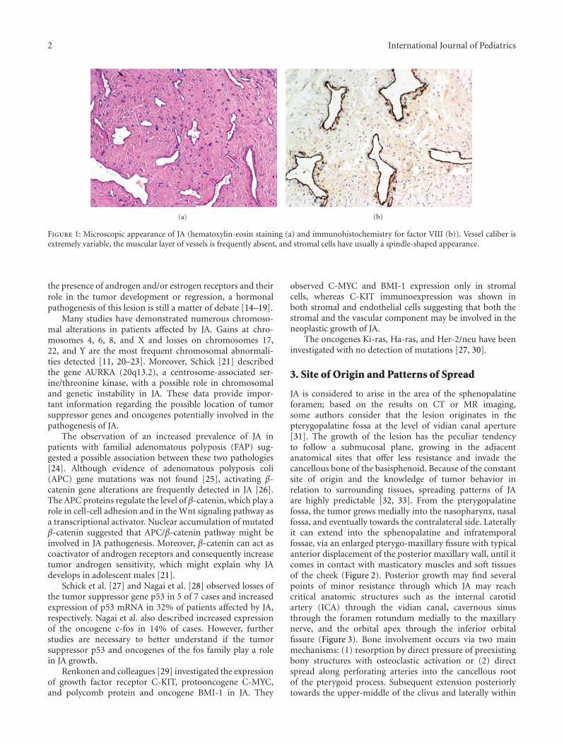
2 International Journal of Pediatrics
(a) (b)
Figure 1: Microscopic appearance of JA (hematoxylin-eosin staining (a) and immunohistochemistry for factor VIII (b)). Vessel caliber isextremely variable, the muscular layer of vessels is frequently absent, and stromal cells have usually a spindle-shaped appearance.
the presence of androgen and/or estrogen receptors and theirrole in the tumor development or regression, a hormonalpathogenesis of this lesion is still a matter of debate [14–19].
Many studies have demonstrated numerous chromoso-mal alterations in patients affected by JA. Gains at chro-mosomes 4, 6, 8, and X and losses on chromosomes 17,22, and Y are the most frequent chromosomal abnormali-ties detected [11, 20–23]. Moreover, Schick [21] describedthe gene AURKA (20q13.2), a centrosome-associated ser-ine/threonine kinase, with a possible role in chromosomaland genetic instability in JA. These data provide impor-tant information regarding the possible location of tumorsuppressor genes and oncogenes potentially involved in thepathogenesis of JA.
The observation of an increased prevalence of JA inpatients with familial adenomatous polyposis (FAP) sug-gested a possible association between these two pathologies[24]. Although evidence of adenomatous polyposis coli(APC) gene mutations was not found [25], activating β-catenin gene alterations are frequently detected in JA [26].The APC proteins regulate the level of β-catenin, which play arole in cell-cell adhesion and in the Wnt signaling pathway asa transcriptional activator. Nuclear accumulation of mutatedβ-catenin suggested that APC/β-catenin pathway might beinvolved in JA pathogenesis. Moreover, β-catenin can act ascoactivator of androgen receptors and consequently increasetumor androgen sensitivity, which might explain why JAdevelops in adolescent males [21].
Schick et al. [27] and Nagai et al. [28] observed losses ofthe tumor suppressor gene p53 in 5 of 7 cases and increasedexpression of p53 mRNA in 32% of patients affected by JA,respectively. Nagai et al. also described increased expressionof the oncogene c-fos in 14% of cases. However, furtherstudies are necessary to better understand if the tumorsuppressor p53 and oncogenes of the fos family play a rolein JA growth.
Renkonen and colleagues [29] investigated the expressionof growth factor receptor C-KIT, protooncogene C-MYC,and polycomb protein and oncogene BMI-1 in JA. They
observed C-MYC and BMI-1 expression only in stromalcells, whereas C-KIT immunoexpression was shown inboth stromal and endothelial cells suggesting that both thestromal and the vascular component may be involved in theneoplastic growth of JA.
The oncogenes Ki-ras, Ha-ras, and Her-2/neu have beeninvestigated with no detection of mutations [27, 30].
3. Site of Origin and Patterns of Spread
JA is considered to arise in the area of the sphenopalatineforamen; based on the results on CT or MR imaging,some authors consider that the lesion originates in thepterygopalatine fossa at the level of vidian canal aperture[31]. The growth of the lesion has the peculiar tendencyto follow a submucosal plane, growing in the adjacentanatomical sites that offer less resistance and invade thecancellous bone of the basisphenoid. Because of the constantsite of origin and the knowledge of tumor behavior inrelation to surrounding tissues, spreading patterns of JAare highly predictable [32, 33]. From the pterygopalatinefossa, the tumor grows medially into the nasopharynx, nasalfossa, and eventually towards the contralateral side. Laterallyit can extend into the sphenopalatine and infratemporalfossae, via an enlarged pterygo-maxillary fissure with typicalanterior displacement of the posterior maxillary wall, until itcomes in contact with masticatory muscles and soft tissuesof the cheek (Figure 2). Posterior growth may find severalpoints of minor resistance through which JA may reachcritical anatomic structures such as the internal carotidartery (ICA) through the vidian canal, cavernous sinusthrough the foramen rotundum medially to the maxillarynerve, and the orbital apex through the inferior orbitalfissure (Figure 3). Bone involvement occurs via two mainmechanisms: (1) resorption by direct pressure of preexistingbony structures with osteoclastic activation or (2) directspread along perforating arteries into the cancellous rootof the pterygoid process. Subsequent extension posteriorlytowards the upper-middle of the clivus and laterally within

International Journal of Pediatrics 3
TMMM
∗∗
Figure 2: Axial contrast enhanced MRI: extensive JA with a typicalpattern of spread into the cancellous bone of the basisphenoidalong the vidian canal (white dotted line); on the contralateralside, black arrows indicate the right vidian nerve. Moreover, thelesion spreads deeply into the pterygomaxillary fossa toward themasticatory muscles, with anterior displacement of the posteriormaxillary wall (white arrowheads). Asterisks indicate the foramenovale bilaterally. TM: temporalis muscle; MM: masseter muscle.
the greater wing of the sphenoid, usually with late erosion ofthe inner table of the middle cranial fossa, may be detected inadvanced cases [34]. Intracranial extension along a canal orresulting from spreading through bone destruction cannotbe considered a rare event, especially in large JA, whileinfiltration of the dura is very rare [35].
4. Clinical and Radiologic Findings
Typical symptoms for JA are progressive unilateral nasalobstruction (80–90%) with rhinorrhea and recurrent unilat-eral epistaxis (45–60%), and thus these complaints in a maleadolescent should immediately generate suspicion. Headache(25%) and facial pain may arise secondarily to the blockageof paranasal sinuses, or impairment of Eustachian tubefunction with unilateral secretory otitis media, respectively.Tumor extension into the sinonasal cavity can cause chronicrhinosinusitis. Proptosis and alteration of the vision clearlyindicate an involvement of the orbit. Swelling of the cheek,neurologic deficits, alteration in olfaction, rhinolalia clausa,and otalgia are also possible [1].
Given the presenting symptoms, the patient should beexamined by nasal endoscopy which usually shows a large,lobulated mass behind the middle turbinate filling thechoana with a smooth surface and clear signs of hypervas-cularization (Figure 4).
Since epidemiologic and endoscopic findings are typical,biopsy is absolutely contraindicated because of a consider-able and undue risk of massive hemorrhage [1].
Imaging techniques after contrast enhancement (MSCTand/or MR) are crucial to confirm the clinical suspicionpattern of vascularization and to assess the extension ofthe lesion. The diagnosis on imaging is based on threefactors: the site of origin, hypervascularization after contrastenhancement, and patterns of growth [31, 32]. The evidence
that up to 96% of JA caused enlargement or erosion of theanterior part of the vidian canal supports the hypothesis ofits typical location in the pterygopalatine fossa at the exitof this canal [32]. After administration of contrast agent, astrong and homogeneous enhancement on MSCT or MR isvisible, with several signal voids within the lesion in both MRT1 and T2 sequences, indicating major intralesional vessels[31]. Moreover, enlargement of the internal maxillary arterycan be detected by MSCT or MR, as well as signs of boneremodeling whereby thinning and anterior displacement ofthe posterior maxillary wall, with bone erosion typically atthe level of the pterygoid root. Without doubt, MR betterdepicts cancellous bone invasion and, in lesions invadingthe middle cranial fossa, the relationship of the lesion withcavernous sinus and dura. Differential diagnosis includesother hypervascularized lesions such as hemangiopericy-toma, lobular capillary hemangioma, and paragangliomawhich have a different gender/age distribution and patternof growth.
Preoperative identification of blood supply is a crucialfinding to select the most appropriate surgical strategy.Although angio-MR may help in the vascular assessment,the complete map of all feeders requires digital subtractionangiography. Typically the JA receives vascular supply viathe external carotid system and particularly from internalmaxillary, ascending pharyngeal, and vidian arteries [1].Vascular components from branches of ICA, such as theinferomedial trunk or inferior hypophyseal artery, may befrequently detected in large lesions involving the skull baseand in contact with ICA. Because of the frequent detectionof bilateral vascular supply, around 36% by Wu et al. [36]in a recent literature review, both carotid systems requireangiographic evaluation.
Preoperative embolization is recommended by mostauthors [37–40] as a standard procedure to reduce blood lossduring surgical resection. Some reports [33, 41] have statedthat this procedure did not affect perioperative bleeding,although some years later Glad et al. [3] observed thatembolization provides a 60–70% reduction in intraoperativebleeding, and the need for blood transfusion is required.Although the modification within the lesion induced by theembolization has been indicated as a contributory causeof incomplete excision [42], refinements in the techniqueand the introduction of new materials have minimized therisk of leaving residual disease. During the last decade,the availability of small particles and microcatheters hasmade it possible to reach collateral and terminal branchesof external carotid artery to avoid the risk of neurologicsequelae following the inadvertent embolization of smallvessels supplied by the ICA. Polyvinyl-alcohol particles arethe most frequently used material for this procedure, whichmust be planned 24–48 hours before surgery to avoid the riskof revascularization [1]. As suggested by Hackman et al. [43],when vessels from both external carotid systems vascularizethe lesion, bilateral embolization of internal maxillary arteryis recommended. To control bleeding arising from vesselssupplied by the ICA in lesions with extensive skull baseinvolvement, Tranbahuy et al. [44] introduced a techniqueof direct embolization through a transnasal or lateral
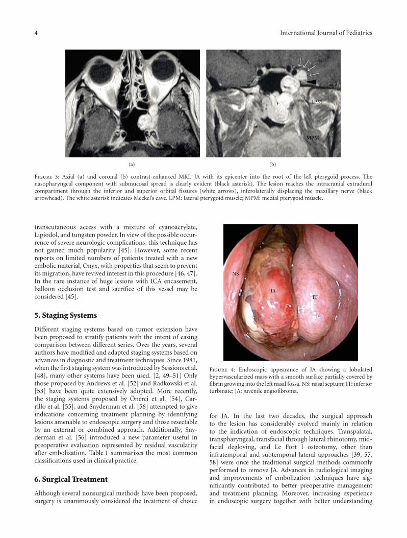
4 International Journal of Pediatrics
∗∗
(a)
∗ LPM
MPM
(b)
Figure 3: Axial (a) and coronal (b) contrast-enhanced MRI. JA with its epicenter into the root of the left pterygoid process. Thenasopharyngeal component with submucosal spread is clearly evident (black asterisk). The lesion reaches the intracranial extraduralcompartment through the inferior and superior orbital fissures (white arrows), inferolaterally displacing the maxillary nerve (blackarrowhead). The white asterisk indicates Meckel’s cave. LPM: lateral pterygoid muscle; MPM: medial pterygoid muscle.
transcutaneous access with a mixture of cyanoacrylate,Lipiodol, and tungsten powder. In view of the possible occur-rence of severe neurologic complications, this technique hasnot gained much popularity [45]. However, some recentreports on limited numbers of patients treated with a newembolic material, Onyx, with properties that seem to preventits migration, have revived interest in this procedure [46, 47].In the rare instance of huge lesions with ICA encasement,balloon occlusion test and sacrifice of this vessel may beconsidered [45].
5. Staging Systems
Different staging systems based on tumor extension havebeen proposed to stratify patients with the intent of easingcomparison between different series. Over the years, severalauthors have modified and adapted staging systems based onadvances in diagnostic and treatment techniques. Since 1981,when the first staging system was introduced by Sessions et al.[48], many other systems have been used. [2, 49–51] Onlythose proposed by Andrews et al. [52] and Radkowski et al.[53] have been quite extensively adopted. More recently,the staging systems proposed by Onerci et al. [54], Car-rillo et al. [55], and Snyderman et al. [56] attempted to giveindications concerning treatment planning by identifyinglesions amenable to endoscopic surgery and those resectableby an external or combined approach. Additionally, Sny-derman et al. [56] introduced a new parameter useful inpreoperative evaluation represented by residual vascularityafter embolization. Table 1 summarizes the most commonclassifications used in clinical practice.
6. Surgical Treatment
Although several nonsurgical methods have been proposed,surgery is unanimously considered the treatment of choice
NS
JAIT
Figure 4: Endoscopic appearance of JA showing a lobulatedhypervascularized mass with a smooth surface partially covered byfibrin growing into the left nasal fossa. NS: nasal septum; IT: inferiorturbinate; JA: juvenile angiofibroma.
for JA. In the last two decades, the surgical approachto the lesion has considerably evolved mainly in relationto the indication of endoscopic techniques. Transpalatal,transpharyngeal, transfacial through lateral rhinotomy, mid-facial degloving, and Le Fort I osteotomy, other thaninfratemporal and subtemporal lateral approaches [39, 57,58] were once the traditional surgical methods commonlyperformed to remove JA. Advances in radiological imagingand improvements of embolization techniques have sig-nificantly contributed to better preoperative managementand treatment planning. Moreover, increasing experiencein endoscopic surgery together with better understanding
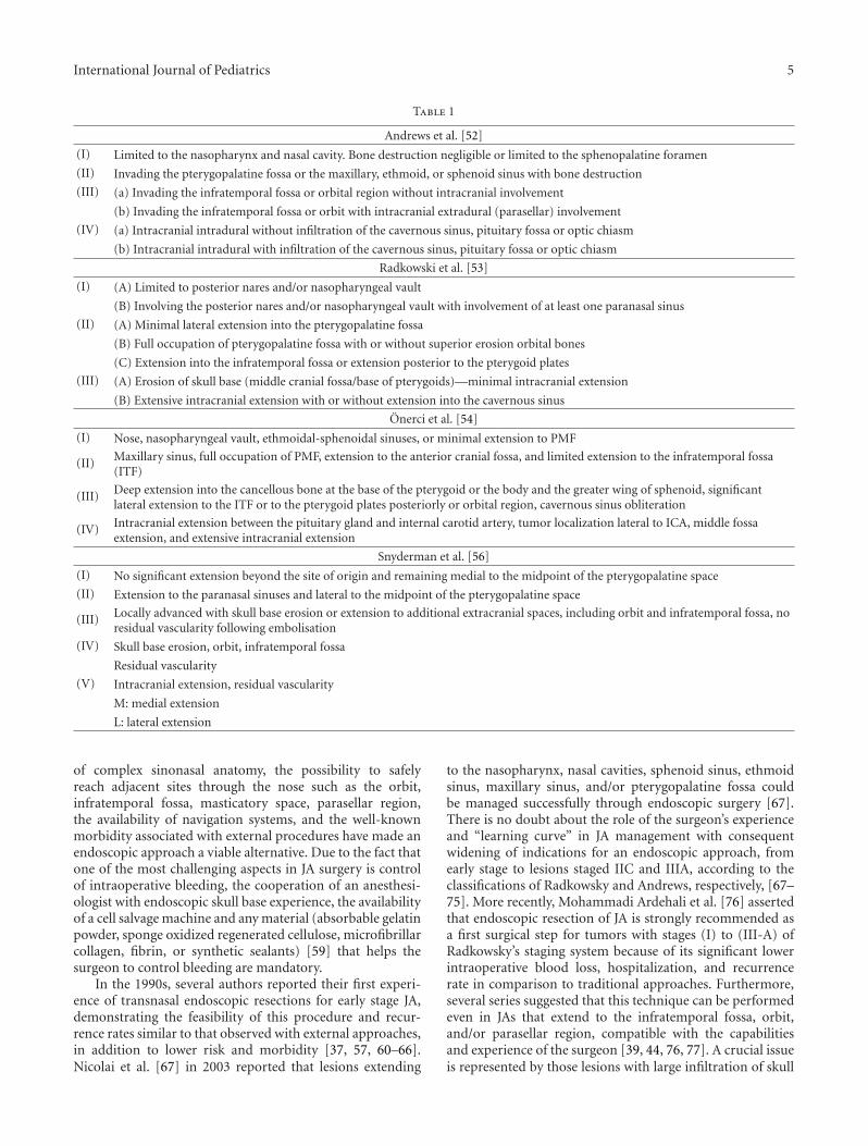
International Journal of Pediatrics 5
Table 1
Andrews et al. [52]
(I) Limited to the nasopharynx and nasal cavity. Bone destruction negligible or limited to the sphenopalatine foramen
(II) Invading the pterygopalatine fossa or the maxillary, ethmoid, or sphenoid sinus with bone destruction
(III) (a) Invading the infratemporal fossa or orbital region without intracranial involvement
(b) Invading the infratemporal fossa or orbit with intracranial extradural (parasellar) involvement
(IV) (a) Intracranial intradural without infiltration of the cavernous sinus, pituitary fossa or optic chiasm
(b) Intracranial intradural with infiltration of the cavernous sinus, pituitary fossa or optic chiasm
Radkowski et al. [53]
(I) (A) Limited to posterior nares and/or nasopharyngeal vault
(B) Involving the posterior nares and/or nasopharyngeal vault with involvement of at least one paranasal sinus
(II) (A) Minimal lateral extension into the pterygopalatine fossa
(B) Full occupation of pterygopalatine fossa with or without superior erosion orbital bones
(C) Extension into the infratemporal fossa or extension posterior to the pterygoid plates
(III) (A) Erosion of skull base (middle cranial fossa/base of pterygoids)—minimal intracranial extension
(B) Extensive intracranial extension with or without extension into the cavernous sinus
Onerci et al. [54]
(I) Nose, nasopharyngeal vault, ethmoidal-sphenoidal sinuses, or minimal extension to PMF
(II) Maxillary sinus, full occupation of PMF, extension to the anterior cranial fossa, and limited extension to the infratemporal fossa(ITF)
(III) Deep extension into the cancellous bone at the base of the pterygoid or the body and the greater wing of sphenoid, significantlateral extension to the ITF or to the pterygoid plates posteriorly or orbital region, cavernous sinus obliteration
(IV) Intracranial extension between the pituitary gland and internal carotid artery, tumor localization lateral to ICA, middle fossaextension, and extensive intracranial extension
Snyderman et al. [56]
(I) No significant extension beyond the site of origin and remaining medial to the midpoint of the pterygopalatine space
(II) Extension to the paranasal sinuses and lateral to the midpoint of the pterygopalatine space
(III) Locally advanced with skull base erosion or extension to additional extracranial spaces, including orbit and infratemporal fossa, noresidual vascularity following embolisation
(IV) Skull base erosion, orbit, infratemporal fossa
Residual vascularity
(V) Intracranial extension, residual vascularity
M: medial extension
L: lateral extension
of complex sinonasal anatomy, the possibility to safelyreach adjacent sites through the nose such as the orbit,infratemporal fossa, masticatory space, parasellar region,the availability of navigation systems, and the well-knownmorbidity associated with external procedures have made anendoscopic approach a viable alternative. Due to the fact thatone of the most challenging aspects in JA surgery is controlof intraoperative bleeding, the cooperation of an anesthesi-ologist with endoscopic skull base experience, the availabilityof a cell salvage machine and any material (absorbable gelatinpowder, sponge oxidized regenerated cellulose, microfibrillarcollagen, fibrin, or synthetic sealants) [59] that helps thesurgeon to control bleeding are mandatory.
In the 1990s, several authors reported their first experi-ence of transnasal endoscopic resections for early stage JA,demonstrating the feasibility of this procedure and recur-rence rates similar to that observed with external approaches,in addition to lower risk and morbidity [37, 57, 60–66].Nicolai et al. [67] in 2003 reported that lesions extending
to the nasopharynx, nasal cavities, sphenoid sinus, ethmoidsinus, maxillary sinus, and/or pterygopalatine fossa couldbe managed successfully through endoscopic surgery [67].There is no doubt about the role of the surgeon’s experienceand “learning curve” in JA management with consequentwidening of indications for an endoscopic approach, fromearly stage to lesions staged IIC and IIIA, according to theclassifications of Radkowsky and Andrews, respectively, [67–75]. More recently, Mohammadi Ardehali et al. [76] assertedthat endoscopic resection of JA is strongly recommended asa first surgical step for tumors with stages (I) to (III-A) ofRadkowsky’s staging system because of its significant lowerintraoperative blood loss, hospitalization, and recurrencerate in comparison to traditional approaches. Furthermore,several series suggested that this technique can be performedeven in JAs that extend to the infratemporal fossa, orbit,and/or parasellar region, compatible with the capabilitiesand experience of the surgeon [39, 44, 76, 77]. A crucial issueis represented by those lesions with large infiltration of skull

6 International Journal of Pediatrics
base, extensive vascular supply from ICA, or encasement ofthe artery itself: an anterior or lateral combined externalapproach according to the relationship of the tumor withICA, and the surgeon’s preference should be planned.Moreover, endoscopic surgery is contraindicated in residualtumors involving critical areas (ICA, optic nerve, cavernoussinus, dura), whereas adhesions due to scar tissue increase therisk of severe uncontrolled complications during dissectionof the lesion [77].
The first surgical step when the surgeon approaches a JAendoscopically is to expose the tumor as extensively as pos-sible through a middle turbinectomy, ethmoidectomy, wideantrostomy and sphenoidotomy, and resection of the poste-rior third of the nasal septum, which enhances the exposureof the nasopharyngeal portion of the lesion. The posteriorwall of the maxillary sinus has to be resected as far lateralas dictated by the lateral extension of the lesion into thepterygopalatine and/or infratemporal fossae. For JA largelyinvolving the infratemporal fossa, the surgeon can improvelateral exposure through a so-called Sturmann-Canfieldmaxillectomy, which provides resection of the anteromedialcorner of the maxillary sinus [77]. An endoscopically assistedantral window approach through the anterior wall of themaxillary sinus, as proposed by Pasquini et al. [72], maybe considered a possible alternative. Another importantprinciple in the resection of large-volume lesions is thefragmentation technique (“piece-meal” resection) that helpsto completely assess the extension without an increased riskof recurrence [69]. During dissection, to maintain a propercleavage plain between the tumor and adjacent tissues, afour-handed technique is highly recommended [77]. Theprocedure is completed by accurate subperiosteal dissectionof the tumor attachment and subsequent extensive drilling ofthe basisphenoid and other bone area where the JA is adheredto remove residual disease, which may not be immediatelyevident, and prevent its regrowth [78].
Because of its high degree of vascularization, bleedingduring surgery is a crucial topic. Some studies comparedthe blood loss between endoscopic and external approaches,showing a lower loss in endoscopic surgery [78, 79]. How-ever, the reliability of these data requires confirmation sinceJAs treated by an open approach usually have a higher stagethan those resected endoscopically. Another question widelydiscussed in literature is the reduction of intraoperativebleeding, thanks to preoperative embolization. Some authorshave correlated the amount of blood loss with the quality ofembolization and with tumor extension [67, 71]. Glad et al.[3] showed a statistically significant decrease in bleedingbetween the nonembolized (650 mL) and the embolizedgroup (1200 mL).
To better control bleeding during the procedure, severalauthors have proposed the use of diode laser, KTP laser, orultrasonic scalpel [70, 77, 80–82].
7. Postoperative Surveillance
Based on the experience by Kania et al. [83], Lund et al.[1], and Nicolai et al. [77] recommended postoperative MRimaging after removal of the nasal packing and until 72 hours
for early identification of any suspicious residual disease.The reason for this is the presence of minor inflammatorychanges, typically observed 3-4 months after treatment,which frequently challenge differentiation between residualJA and active scar tissue. Although this surveillance policyhas to be validated by longer follow-up periods, Nicolai et al.[77] observed that patients with no signs of persistence donot develop any lesion even at subsequent MR examination.Endoscopic examination has limited value in the identifica-tion of residual/recurrence disease because of the submucosalgrowing pattern of JA, which is detected with more precisionby enhanced MR or MSCT. Whatever technique is selected,the examination should be performed every 6–8 monthsfor at least 3 years after surgery. Moreover, depending onsuspicious enhancement, incomplete resection and age ofonset, angio-MR imaging may be scheduled [1]. Closerradiologic survey may be required to better evaluate thegrowth and plan treatment for persistent JA.
8. Outcome
Although comparison between external approaches andendoscopic techniques is biased by the different stagingsystems and follow-up strategy adopted, recently Wang et al.[84] observed no significant difference in the rate ofrecurrence between 11 patients treated endoscopically and 13who underwent transpalatal excision, all staged (I) and (II)according to Chandler classification. Series with a consistentnumber of patients treated with external approaches haveshown a reduction over time in terms of recurrence rateranging from 36–40% [34, 85] reported in the 1990s, to theexcellent results of Danesi et al. [35] in 2008 with 13.5% and18.2% of residual disease in lesions with extracranial andintracranial extension, respectively. At present, endoscopicresection in small/intermediate JA is widely recommendedbecause of the low risk of recurrence demonstrated in severalstudies during the last decade [34, 71, 73, 85–89].
Currently, the results of the two major series (Table 2)of JA resected through an endoscopic approach corroboratethe principle that this modality of treatment can encompassall lesions from stage (I) to (IIIA) or (IIIB) accordingto Radkowsky and Andrews staging systems, respectively[76, 77]. An overall recurrence/residual rate of 8.6% [77]and 19.1% [76], respectively, was reported. As previouslyhighlighted by Howard et al. [78], Nicolai et al. [77] in theirstudy on 46 patients treated with an exclusive endoscopicprocedure observed that all the residual lesions detectedin 4 patients by MR within 24 months after treatmentwere located at the level of the basisphenoid bone, thusemphasizing the need to extend drilling well beyond theapparent margin of tumor infiltration.
In a recent study on 95 JA, Sun et al. [90] identifiedthree predictive factors that may increase the recurrence rate:patient age at diagnosis (under 18 years), tumor size (>4 cm),and stage according to Radkovsky classification.
The management of residual disease, especially whenlocated intracranially or with a relationship with criticalstructures such the ICA, cavernous sinus, and the opticnerve, remains a source of discussion. Certainly, close survey

International Journal of Pediatrics 7
Table 2
MohammadiArdehali et al.
[76]
Nicolai et al.[77]
Number of patients 47 46
Mean age (years) 17.1 (7–37) 17 (10–35)
Previous treatment 16 5
(IA) 5 (IA) 3
(IB) 10 (IB) 1
Stage(Radkowski classification)
(IIA) 3 (IIA) 5
(IIB) 3 (IIB) 10
(IIC) 22 (IIC) 19
(IIIA) 3 (IIIA) 7
(IIIB) 1 (IIIB) 1
Preoperative embolization 5 40
Mean blood loss (mL)1336.2
(300–8500)580 (250–1300)
Mean hospitalization time(days)
3.1 5
Mean followup (months) 33.1 73
Persistence rate (%) 19.1 8.6
with enhanced MR or MSCT is strongly indicated to evaluatethe growth rate, and consequently, the need for surgicalrevision. Onerci et al. [54] prefer an observational strategy toinfratemporal craniofacial resection for intracranial residualdisease. Moreover, some reports [91–93] described thepossibility, in at least a minority of cases, of spontaneousinvolution or reduction in size because of hormonallydependent JA pathogenesis.
9. Nonsurgical Treatments
The use of radiation therapy (RT) in JA is still debated for thereported risk of sarcomatoid transformation [94] or radio-induced neoplasms in the following decades. Some authorsrecommended RT as adjuvant treatment in unresectabletumors, in failure of complete tumor removal, or for exten-sive intracranial extension [53, 95, 96]. Nicolai et al. [77]suggested that RT may be indicated for residual lesions incritical areas that have been demonstrated to increase in size.Lee et al. [97] reported on 27 patients affected by advancedJA treated primarily with RT (30–55 Gy): the recurrencerate was 15%, and long-term complications observed in 4patients included growth retardation, panhypopituitarism,temporal lobe necrosis, cataracts, and radiation keratopathy.More recently, McAfee et al. [98] treated 22 patients affectedby high staged JA with RT (30–36 Gy): in 10 cases asprimary treatment, and in 12 for recurrence. Local controlwas obtained in 90% of patients, with 2 cases of localpersistence. Late complications, which occurred in 7 (32%)cases, included cataracts, transient central nervous systemsyndrome, and cutaneous basal cell carcinoma. The use ofintensity-modulated RT for the treatment of 3 patients with
extensive or persistent JA showed no recurrences and a latetoxicity with epistaxis and chronic rhinitis in 2 cases [99].Although a small number of cases of JA have been treatedby Gamma-Knife radiosurgery [100, 101], the insignificantmorbidity documented may be reasonably correlated tothe optimized irradiation of the target volume by sparinguninvolved structures.
The use of chemotherapy in treatment of JA is sup-ported by only a few reports [102–104]. In the 1980s,Goepfert et al. [102] described successful results withtwo different chemotherapy schedules, including doxoru-bicin and dacarbazine, or vincristine, dactinomycin, andcyclophosphamide.
Several studies on hormone pathogenesis have exten-sively demonstrated the hormonal dependence of this tumor,suggesting a promising role of estrogen or androgen receptorblockers in its treatment [9, 15–19]. Gates et al. administeredflutamide, a potent nonsteroidal androgen receptor blocker,in 5 patients affected by JA and detected an average tumorregression of 44% in four cases [105]. However, in a reporton 7 patients, Labra et al. observed no significant differ-ences between tumor dimensions before or after flutamideadministration, questioning its use in JA [106]. Very recently,flutamide-induced regression in a series of 20 advancedstaged JA was demonstrated only in postpuberal patients[107].
10. Conclusions
Juvenile angiofibroma is a pathology that should be includedin the differential diagnosis of unilateral nasal obstruction,associated or not with epistaxis, especially in young ado-lescent males. The finding at nasal endoscopy, which is thefirst step in the diagnostic algorithm, of a hypervascularizedlesion occupying the posterior half of the nasal fossa shouldimmediately raise suspicion. Morphologic imaging confirmsthe diagnosis. Endoscopic surgery after embolization hasbeen demonstrated to be a viable alternative to externaltechniques for the management of small-intermediate sizeJA. Resorting to external anterior or lateral approaches is stillrecommended in JAs encasing the ICA or with a massivefeeder contribution from it, or in the rare instances ofintradural spread.
References
[1] V. J. Lund, H. Stammberger, P. Nicolai et al., “Europeanposition paper on endoscopic management of tumoursof the nose, paranasal sinuses and skull base,” Rhinology.Supplement, no. 22, pp. 1–143, 2010.
[2] J. R. Chandler, R. Goulding, L. Moskowitz, and R. M.Quencer, “Nasopharyngeal angiofibromas: staging and man-agement,” Annals of Otology, Rhinology and Laryngology, vol.93, no. 4, pp. 322–329, 1984.
[3] H. Glad, B. Vainer, C. Buchwald et al., “Juvenile nasopha-ryngeal angiofibromas in Denmark 1981–2003: diagnosis,incidence, and treatment,” Acta Oto-Laryngologica, vol. 127,no. 3, pp. 292–299, 2007.

8 International Journal of Pediatrics
[4] A. G. D. Maran and V. J. Lund, “Nasal physiology,” in ClinicalRhinology, A. G. D. Maran and V. J. Lund, Eds., p. 5, GeorgThieme, Stuttgart, Germany, 1990.
[5] R. Schuon, J. Brieger, U. R. Heinrich, Y. Roth, W. Szyfter,and W. J. Mann, “Immunohistochemical analysis of growthmechanisms in juvenile nasopharyngeal angiofibroma,”European Archives of Oto-Rhino-Laryngology, vol. 264, no. 4,pp. 389–394, 2007.
[6] D. Stiller and K. Kuttner, “Growth patterns of juvenilenasopharyngeal fibromas. A histological analysis on thebasis of 40 cases,” Zentralblatt fur Allgemeine Pathologie undPathologische Anatomie, vol. 134, no. 4-5, pp. 409–422, 1988.
[7] S. S. Sternberg, “Pathology of juvenile nasopharyngealangiofibroma; a lesion of,” Cancer, vol. 7, no. 1, pp. 15–28,1954.
[8] E. M. Hubbard, “Nasopharyngeal angiofibromas,” A.M.A.Archives of Pathology, pp. 192–204, 1958.
[9] M. Schiff, “Juvenile nasopharyngeal angiofibroma,” Laryngo-scope, vol. 69, pp. 908–1016, 1959.
[10] A. Beham, C. Beham-Schmid, S. Regauer, L. Aubock, andH. Stammberger, “Nasopharyngeal angiofibroma: true neo-plasm or vascular malformation?” Advances in AnatomicPathology, vol. 7, no. 1, pp. 36–46, 2000.
[11] B. Schick, P. K. Plinkert, and A. Prescher, “Aetiology ofangiofibromas: reflection on their specific vascular compo-nent,” Laryngo-Rhino-Otologie, vol. 81, no. 4, pp. 280–284,2002.
[12] V. Starlinger, O. Wendler, M. Gramann, and B. Schick,“Laminin expression in juvenile angiofibroma indicatesvessel’s early developmental stage,” Acta Oto-Laryngologica,vol. 127, no. 12, pp. 1310–1315, 2007.
[13] M. Gramann, O. Wendler, L. Haeberle, and B. Schick,“Prominent collagen type VI expression in juvenile angiofi-bromas,” Histochemistry and Cell Biology, vol. 131, no. 1, pp.155–164, 2009.
[14] M. Schiff, A. M. Gonzalez, M. Ong, and A. Baird, “Juvenilenasopharyngeal angiofibroma contain an angiogenic growthfactor: basic FGF,” Laryngoscope, vol. 102, no. 8, pp. 940–945,1992.
[15] H. Martin, H. E. Ehrlich, and J. C. Abels, “Juvenile nasopha-ryngeal angiofibroma,” Annals of Surgery, vol. 127, pp. 513–536, 1948.
[16] S. Johnsen, J. H. Kloster, and M. Schiff, “The action ofhormones on juvenile nasopharyngeal angiofibroma. A casereport,” Acta Oto-Laryngologica, vol. 61, no. 1, pp. 153–160,1966.
[17] D. A. Lee, B. R. Rao, and J. S. Meyer, “Hormonal receptordetermination in juvenile nasopharyngeal angiofibromas,”Cancer, vol. 46, no. 3, pp. 547–551, 1980.
[18] M. M. Farag, S. E. Ghanimah, A. Ragaie, and T. H.Saleem, “Hormonal receptors in juvenile nasopharyngealangiofibroma,” Laryngoscope, vol. 97, no. 2, pp. 208–211,1987.
[19] H. C. Hwang, S. E. Mills, K. Patterson, and A. M.Gown, “Expression of androgen receptors in nasopharyngealangiofibroma: an immunohistochemical study of 24 cases,”Modern Pathology, vol. 11, no. 11, pp. 1122–1126, 1998.
[20] B. Schick, C. Rippel, C. Brunner, V. Jung, P. K. Plinkert,and S. Urbschat, “Numerical sex chromosome aberrationsin juvenile angiofibromas: genetic evidence for an androgen-dependent tumor?” Oncology reports, vol. 10, no. 5, pp. 1251–1255, 2003.
[21] B. Schick, “Specific aspects of juvenile angiofibromas,” HNO,vol. 55, no. 1, pp. 17–20, 2007.
[22] U. R. Heinrich, J. Brieger, J. Gosepath et al., “Frequentchromosomal gains in recurrent juvenile nasopharyngealangiofibroma,” Cancer Genetics and Cytogenetics, vol. 175, no.2, pp. 138–143, 2007.
[23] C. Brunner, S. Urbschat, V. Jung, M. Praetorius, B. Schick,and P. K. Plinkert, “Chromosomal alterations in juvenileangiofibromas,” HNO, vol. 51, no. 12, pp. 981–985, 2003.
[24] C. M. Coutinho-Camillo, M. M. Brentani, and M. A. Nagai,“Genetic alterations in juvenile nasopharyngeal angiofibro-mas,” Head & Neck, vol. 30, no. 3, pp. 390–400, 2008.
[25] R. Valanzano, M. C. Curia, G. Aceto et al., “Genetic evidencethat juvenile nasopharyngeal angiofibroma is an integral FAPtumour,” Gut, vol. 54, no. 7, pp. 1046–1047, 2005.
[26] S. C. Abraham and T. T. Wu, “Nasopharyngeal angiofi-broma,” Human Pathology, vol. 32, no. 4, article 455, 2001.
[27] B. Schick, B. Veldung, S. Wemmert et al., “p53 and Her-2/neuin juvenile angiofibromas,” Oncology Reports, vol. 13, no. 3,pp. 453–457, 2005.
[28] M. A. Nagai, O. Butugan, A. Logullo, and M. M. Brentani,“Expression of growth factors, proto-oncogenes, and p53 innasopharyngeal angiofibromas,” Laryngoscope, vol. 106, no.2, pp. 190–195, 1996.
[29] S. Renkonen, V. Hayry, P. Heikkila et al., “Stem cell-related proteins C-KIT, C-MYC and BMI-1 in juvenilenasopharyngeal angiofibroma-do they have a role?” VirchowsArchiv, vol. 458, no. 2, pp. 189–195, 2010.
[30] C. M. Coutinho, A. S. Bassini, L. G. Gutierrez et al.,“Genetic alterations in Ki-ras and Ha-ras genes in juvenilenasopharyngeal angiofibromas and head and neck cancer,”Sao Paulo Medical Journal, vol. 117, no. 3, pp. 113–120, 1999.
[31] R. Maroldi and P. Nicolai, Imaging in Treatment Planning forSinonasal Diseases, Springer, New York, NY, USA, 2004.
[32] G. Lloyd, D. Howard, V. J. Lund, and L. Savy, “Imaging forjuvenile angiofibroma,” Journal of Laryngology and Otology,vol. 114, no. 9, pp. 727–730, 2000.
[33] B. Schick and G. Kahle, “Radiological findings in angiofi-broma,” Acta Radiologica, vol. 41, no. 6, pp. 585–593, 2000.
[34] G. Lloyd, D. Howard, P. Phelps, and A. Cheesman, “Juvenileangiofibroma: the lessons of 20 years of modern imaging,”Journal of Laryngology and Otology, vol. 113, no. 2, pp. 127–134, 1999.
[35] G. Danesi, D. T. Panciera, R. J. Harvey, and C. Agostinis,“Juvenile nasopharyngeal angiofibroma: evaluation and sur-gical management of advanced disease,” Otolaryngology—Head and Neck Surgery, vol. 138, no. 5, pp. 581–586, 2008.
[36] A. W. Wu, S. E. Mowry, F. Vinuela, E. Abemayor, and M. B.Wang, “Bilateral vascular supply in juvenile nasopharyngealangiofibromas,” Laryngoscope, vol. 121, no. 3, pp. 639–643,2011.
[37] R. L. Carrau, C. H. Snyderman, A. B. Kassam, and C. A.Jungreis, “Endoscopic and endoscopic-assisted surgery forjuvenile angiofibroma,” Laryngoscope, vol. 111, no. 3, pp.483–487, 2001.
[38] D. J. Enepekides, “Recent advances in the treatment ofjuvenile angiofibroma,” Current Opinion in Otolaryngologyand Head and Neck Surgery, vol. 12, no. 6, pp. 495–499, 2004.
[39] R. Midilli, B. Karci, and S. Akyildiz, “Juvenile nasopharyngealangiofibroma: analysis of 42 cases and important aspectsof endoscopic approach,” International Journal of PediatricOtorhinolaryngology, vol. 73, no. 3, pp. 401–408, 2009.
[40] J. R. Li, J. Qian, X. Z. Shan, and L. Wang, “Evaluation ofthe effectiveness of preoperative embolization in surgery fornasopharyngeal angiofibroma,” European Archives of Oto-Rhino-Laryngology, vol. 255, no. 8, pp. 430–432, 1998.

International Journal of Pediatrics 9
[41] D. M. da Costa, G. L. Franche, R. P. Gessinger, D. Strachan,and G. Nawara, “Surgical experience with juvenile naso-pharyngeal angiofibroma,” Annales d’Oto-Laryngologie et deChirurgie Cervico-Faciale, vol. 109, no. 5, pp. 231–234, 1992.
[42] A. McCombe, V. J. Lund, and D. J. Howard, “Recurrence injuvenile angiofibroma,” Rhinology, vol. 28, no. 2, pp. 97–102,1990.
[43] T. Hackman, C. H. Snyderman, R. Carrau, A. Vescan,and A. Kassam, “Juvenile nasopharyngeal angiofibroma: theexpanded endonasal approach,” American Journal of Rhino-logy and Allergy, vol. 23, no. 1, pp. 95–99, 2009.
[44] P. Tranbahuy, M. Borsik, P. Herman, M. Wassef, andA. Casasco, “Direct intratumoral embolization of juvenileangiofibroma,” American Journal of Otolaryngology, vol. 15,no. 6, pp. 429–435, 1994.
[45] A. Casasco, E. Houdart, A. Biondi et al., “Major complica-tions of percutaneous embolization of skull-base tumors,”American Journal of Neuroradiology, vol. 20, no. 1, pp. 179–181, 1999.
[46] M. Lehmann, S. Ulrich, U. Reineke, U. Hamberger, U. Diet-rich, and H. Sudhoff, “Intratumoral Onyx� embolisation inthe management of juvenile nasopharyngeal angiofibroma,”HNO, vol. 58, no. 8, pp. 853–857, 2010.
[47] B. Herman, M. Bublik, J. Ruiz, and R. Younis, “Endoscopicembolization with onyx prior to resection of JNA: a newapproach,” International Journal of Pediatric Otorhinolaryn-gology, vol. 75, no. 1, pp. 53–56, 2010.
[48] R. B. Sessions, R. N. Bryan, R. M. Naclerio, and B. R. Alford,“Radiographic staging of juvenile angiofibroma,” Head andNeck Surgery, vol. 3, no. 4, pp. 279–283, 1981.
[49] U. Fisch, “The infratemporal fossa approach for nasopharyn-geal tumors,” Laryngoscope, vol. 93, no. 1, pp. 36–44, 1983.
[50] J. W. Bremer, H. B. Neel, L. W. DeSanto, and G. C. Jones,“Angiofibroma: treatment trends in 150 patients during 40years,” Laryngoscope, vol. 96, no. 12, pp. 1321–1329, 1986.
[51] A. R. Antonelli, J. Cappiello, C. A. Donajo, D. Di Lorenzo,P. Nicolai, and A. Orlandini, “Diagnosis, staging, andtreatment of juvenile nasopharyngeal angiofibroma (JNA),”Laryngoscope, vol. 97, no. 11, pp. 1319–1225, 1987.
[52] J. C. Andrews, U. Fisch, A. Valavanis, U. Aeppli, and M. S.Makek, “The surgical management of extensive nasopharyn-geal angiofibromas with the infratemporal fossa approach,”Laryngoscope, vol. 99, no. 4, pp. 429–437, 1989.
[53] D. Radkowski, T. McGill, G. B. Healy, L. Ohlms, and D.T. Jones, “Angiofibroma: changes in staging and treatment,”Archives of Otolaryngology—Head and Neck Surgery, vol. 122,no. 2, pp. 122–129, 1996.
[54] M. Onerci, O. Ogretmenoglu, and T. Yucel, “Juvenile naso-pharyngeal angiofibroma: a revised staging system,” Rhinol-ogy, vol. 44, no. 1, pp. 39–45, 2006.
[55] J. F. Carrillo, F. Maldonado, O. Albores, M. C. Ramırez-Ortega, and L. F. Onate-Ocana, “Juvenile nasopharyngealangiofibroma: clinical factors associated with recurrence, andproposal of a staging system,” Journal of Surgical Oncology,vol. 98, no. 2, pp. 75–80, 2008.
[56] C. H. Snyderman, H. Pant, R. L. Carrau, and P. Gardner, “Anew endoscopic staging system for angiofibromas,” Archivesof Otolaryngology—Head and Neck Surgery, vol. 136, no. 6,pp. 588–594, 2010.
[57] A. W. Scholtz, E. Appenroth, K. Kammen-Jolly, L. U. Scholtz,and W. F. Thumfart, “Juvenile nasopharyngeal angiofibroma:management and therapy,” Laryngoscope, vol. 111, no. 4, pp.681–687, 2001.
[58] I. Yiotakis, A. Eleftheriadou, D. Davilis et al., “Juvenilenasopharyngeal angiofibroma stages I and II: a comparativestudy of surgical approaches,” International Journal of Pedi-atric Otorhinolaryngology, vol. 72, no. 6, pp. 793–800, 2008.
[59] C. A. Solares, Y. K. Ong, and C. H. Snyderman, “Transnasalendoscopic skull base surgery: what are the limits?” CurrentOpinion in Otolaryngology and Head and Neck Surgery, vol.18, no. 1, pp. 1–7, 2010.
[60] M. Jorissen, P. Eloy, P. Rombaux, C. Bachert, and J. Daele,“Endoscopic sinus surgery for juvenile nasopharyngealangiofibroma,” Acta Oto-Rhino-Laryngologica Belgica, vol.54, no. 2, pp. 201–219, 2000.
[61] R. H. Kamel, “Transnasal endoscopic surgery in juvenilenasopharyngeal angiofibroma,” Journal of Laryngology andOtology, vol. 110, no. 10, pp. 962–968, 1996.
[62] M. T. Mitskavich, R. L. Carrau, C. H. Snyderman, J. L.Weissman, and J. J. Fagan, “Intranasal endoscopic excisionof a juvenile angiofibroma,” Auris Nasus Larynx, vol. 25, no.1, pp. 39–44, 1998.
[63] S. D. Newlands and E. A. Weymuller, “Endoscopic treatmentof juvenile nasopharyngeal angiofibroma,” American Journalof Rhinology, vol. 13, no. 3, pp. 213–219, 1999.
[64] G. Roger, P. T. B. Huy, P. Froehlich et al., “Exclusively endo-scopic removal of juvenile nasopharyngeal angiofibroma:trends and limits,” Archives of Otolaryngology—Head andNeck Surgery, vol. 128, no. 8, pp. 928–935, 2002.
[65] R. Sarria, A. Capitan, C. Sprekelsen, E. Viviente, G. Cuervo,and A. Ferran, “Endoscopic surgery of nasopharyngealangiofibroma by double embolization,” Acta Otorrinolaringo-logica Espanola, vol. 51, no. 3, pp. 259–262, 2000.
[66] H. Z. Tseng and W. Y. Chao, “Transnasal endoscopicapproach for juvenile nasopharyngeal angiofibroma,” Amer-ican Journal of Otolaryngology, vol. 18, no. 2, pp. 151–154,1997.
[67] P. Nicolai, M. Berlucchi, D. Tomenzoli et al., “Endoscopicsurgery for juvenile angiofibroma: when and how,” Laryngo-scope, vol. 113, no. 5, pp. 775–782, 2003.
[68] M. Bernal-Sprekelsen, H. Massegur Solench, and M. TomasBarberan, “Paediatric endoscopic sinus surgery (PESS):review of the indications,” Revue de Laryngologie OtologieRhinologie, vol. 124, no. 3, pp. 145–150, 2003.
[69] T. Hofmann, M. Bernal-Sprekelsen, W. Koele, P. Reittner,E. Klein, and H. Stammberger, “Endoscopic resection ofjuvenile angiofibromas—long term results,” Rhinology, vol.43, no. 4, pp. 282–289, 2005.
[70] E. A. Mair, A. Battiata, and J. D. Casler, “Endoscopic laser-assisted excision of juvenile nasopharyngeal angiofibromas,”Archives of Otolaryngology—Head and Neck Surgery, vol. 129,no. 4, pp. 454–459, 2003.
[71] T. M. Onerci, O. T. Yucel, and O. Ogretmenoglu, “Endo-scopic surgery in treatment of juvenile nasopharyngealangiofibroma,” International Journal of Pediatric Otorhino-laryngology, vol. 67, no. 11, pp. 1219–1225, 2003.
[72] E. Pasquini, V. Sciarretta, G. Frank et al., “Endoscopictreatment of benign tumors of the nose and paranasalsinuses,” Otolaryngology—Head and Neck Surgery, vol. 131,no. 3, pp. 180–186, 2004.
[73] S. G. Pryor, E. J. Moore, and J. L. Kasperbauer, “Endo-scopic versus traditional approaches for excision of juvenilenasopharyngeal angiofibroma,” Laryngoscope, vol. 115, no. 7,pp. 1201–1207, 2005.

10 International Journal of Pediatrics
[74] V. Sciarretta, E. Pasquini, G. Farneti, G. Frank, D. Maz-zatenta, and F. Calbucci, “Endoscopic sinus surgery forthe treatment of vascular tumors,” American Journal ofRhinology, vol. 20, no. 4, pp. 426–431, 2006.
[75] P. J. Wormald and A. Van Hasselt, “Endoscopic removalof juvenile angiofibromas,” Otolaryngology—Head and NeckSurgery, vol. 129, no. 6, pp. 684–691, 2003.
[76] M. Mohammadi Ardehali, S. H. Samimi Ardestani, N.Yazdani, H. Goodarzi, and S. Bastaninejad, “Endoscopicapproach for excision of juvenile nasopharyngeal angiofi-broma: complications and outcomes,” American Journal ofOtolaryngology, vol. 31, no. 5, pp. 343–349, 2010.
[77] P. Nicolai, A. B. Villaret, D. Farina et al., “Endoscopic surgeryfor juvenile angiofibroma: a critical review of indicationsafter 46 cases,” American Journal of Rhinology and Allergy, vol.24, no. 2, pp. e67–e72, 2010.
[78] D. J. Howard, G. Lloyd, and V. Lund, “Recurrence and itsavoidance in juvenile angiofibroma,” Laryngoscope, vol. 111,no. 9, pp. 1509–1511, 2001.
[79] P. Borghei, M. H. Baradaranfar, S. H. Borghei, and F. Sokhan-don, “Transnasal endoscopic resection of juvenile nasopha-ryngeal angiofibroma without preoperative embolization,”Ear, Nose and Throat Journal, vol. 85, no. 11, pp. 740–746,2006.
[80] H. Nakamura, M. Kawasaki, Y. Higuchi, S. Seki, and S. Taka-hashi, “Transnasal endoscopic resection of juvenile naso-pharyngeal angiofibroma with KTP laser,” European Archivesof Oto-Rhino-Laryngology, vol. 256, no. 4, pp. 212–214, 1999.
[81] K. Ochi, S. Watanabe, and S. Miyabe, “Endoscopic transnasalresection of a juvenile angiofibroma using an ultrasonicallyactivated scalpel,” ORL, vol. 64, no. 4, pp. 290–293, 2002.
[82] P. Hazarika, D. R. Nayak, R. Balakrishnan, G. Raj, andS. Pillai, “Endoscopic and KTP laser-assisted surgery forjuvenile nasopharyngeal angiofibroma,” American Journal ofOtolaryngology, vol. 23, no. 5, pp. 282–286, 2002.
[83] R. E. Kania, E. Sauvaget, J. P. Guichard, R. Chapot, P. T. B.Huy, and P. Herman, “Early postoperative CT scanning forjuvenile nasopharyngeal angiofibroma: detection of residualdisease,” American Journal of Neuroradiology, vol. 26, no. 1,pp. 82–88, 2005.
[84] Q. Y. Wang, H. H. Chen, and Y. Y. Lu, “Comparison oftwo approaches to the surgical management of juvenilenasopharyngeal angiofibroma stages I and II,” Journal ofOtolaryngology—Head and Neck Surgery, vol. 40, no. 1, pp.14–18, 2011.
[85] P. J. Gullane, J. Davidson, T. O’Dwyer, and V. Forte, “Juvenileangiofibroma: a review of the literature and a case seriesreport,” Laryngoscope, vol. 102, no. 8, pp. 928–933, 1992.
[86] F. Tosun, C. Ozer, M. Gerek, and S. Yetiser, “Surgicalapproaches for nasopharyngeal angiofibroma: comparativeanalysis and current trends,” Journal of Craniofacial Surgery,vol. 17, no. 1, pp. 15–20, 2006.
[87] N. A. Andrade, J. A. Pinto, M. de Oliveira Nobrega,J. E. P. Aguiar, T. F. A. P. Aguiar, and E. S. Vinhaes,“Exclusively endoscopic surgery for juvenile nasopharyngealangiofibroma,” Otolaryngology—Head and Neck Surgery, vol.137, no. 3, pp. 492–496, 2007.
[88] A. K. Gupta, M. G. Rajiniganth, and A. K. Gupta, “Endo-scopic approach to juvenile nasopharyngeal angiofibroma:our experience at a tertiary care centre,” Journal of Laryngol-ogy and Otology, vol. 122, no. 11, pp. 1185–1189, 2008.
[89] B. S. Bleier, D. W. Kennedy, J. N. Palmer, A. G. Chiu,J. D. Bloom, and B. W. O’Malley, “Current management
of juvenile nasopharyngeal angiofibroma: a tertiary centerexperience 1999–2007,” American Journal of Rhinology andAllergy, vol. 23, no. 3, pp. 328–330, 2009.
[90] X. C. Sun, D. H. Wang, H. P. Yu, F. Wang, W. Wang, and J. J.Jiang, “Analysis of risk factors associated with recurrence ofnasopharyngeal angiofibroma,” Journal of Otolaryngology—Head and Neck Surgery, vol. 39, no. 1, pp. 56–61, 2010.
[91] J. E. Dohar and A. J. Duvall, “Spontaneous regression ofjuvenile nasopharyngeal angiofibroma,” Annals of Otology,Rhinology and Laryngology, vol. 101, no. 6, pp. 469–471, 1992.
[92] K. A. Reddy, W. M. Mendenhall, R. J. Amdur, S. P. Stringer,and N. J. Cassisi, “Long-term results of radiation therapy forjuvenile nasopharyngeal angiofibroma,” American Journal ofOtolaryngology, vol. 22, no. 3, pp. 172–175, 2001.
[93] P. M. Spielmann, R. Adamson, K. Cheng, and R. J. Sanderson,“Juvenile nasopharyngeal angiofibroma: spontaneous resolu-tion,” Ear, Nose and Throat Journal, vol. 87, no. 9, pp. 521–523, 2008.
[94] M. S. Makek, J. C. Andrews, and U. Fisch, “Malignant trans-formation of a nasopharyngeal angiofibroma,” Laryngoscope,vol. 99, no. 10, pp. 1088–1092, 1989.
[95] K. Ungkanont, R. M. Byers, R. S. Weber, D. L. Callender,P. F. Wolf, and H. Goepfert, “Juvenile nasopharyngealangiofibroma: an update of therapeutic management,” Head& Neck, vol. 18, no. 1, pp. 60–66, 1996.
[96] G. C. Jones, L. W. DeSanto, J. W. Bremer, and H. B. Neel,“Juvenile angiofibromas. Behavior and treatment of extensiveand residual tumors,” Archives of Otolaryngology—Head andNeck Surgery, vol. 112, no. 11, pp. 1191–1193, 1986.
[97] J. T. Lee, P. Chen, A. Safa, G. Juillard, and T. C. Calcaterra,“The role of radiation in the treatment of advanced juvenileangiofibroma,” Laryngoscope, vol. 112, no. 7, pp. 1213–1220,2002.
[98] W. J. McAfee, C. G. Morris, R. J. Amdur, J. W. Werning,and W. M. Mendenhall, “Definitive radiotherapy for juvenilenasopharyngeal angiofibroma,” American Journal of ClinicalOncology, vol. 29, no. 2, pp. 168–170, 2006.
[99] R. B. Kuppersmith, B. S. Teh, D. T. Donovan et al., “Theuse of intensity modulated radiotherapy for the treatment ofextensive and recurrent juvenile angiofibroma,” InternationalJournal of Pediatric Otorhinolaryngology, vol. 52, no. 3, pp.261–268, 2000.
[100] C. K. Park, D. G. Kim, S. H. Paek, H. T. Chung, and H.W. Jung, “Recurrent juvenile nasopharyngeal angiofibromatreated with gamma knife surgery,” Journal of Korean MedicalScience, vol. 21, no. 4, pp. 773–777, 2006.
[101] A. O. Dare, K. J. Gibbons, G. M. Proulx et al., “Resection fol-lowed by radiosurgery for advanced juvenile nasopharyngealangiofibroma: report of two cases,” Neurosurgery, vol. 52, no.5, pp. 1207–1211, 2003.
[102] H. Goepfert, A. Cangir, A. G. Ayala, and F. Eftekhari,“Chemotherapy of locally aggressive head and neck tumorsin the pediatric age group. Desmoid fibromatosis andnasopharyngeal angiofibroma,” American Journal of Surgery,vol. 144, no. 4, pp. 437–444, 1982.
[103] H. Goepfert, A. Cangir, and Y. Y. Lee, “Chemotherapy foraggressive juvenile nasopharyngeal angiofibroma,” Archives ofOtolaryngology, vol. 111, no. 5, pp. 285–289, 1985.
[104] B. Schick, G. Kahle, R. Haßler, and W. Draf, “Chemotherapyof juvenile angiofibromas—an alternative course for treat-ment?” HNO, vol. 44, no. 3, pp. 148–152, 1996.
[105] G. A. Gates, D. H. Rice, C. F. Koopmann, and D. E.Schuller, “Flutamide-induced regression of angiofibroma,”Laryngoscope, vol. 102, no. 6, pp. 641–644, 1992.

International Journal of Pediatrics 11
[106] A. Labra, R. Chavolla-Magana, A. Lopez-Ugalde, J. Alanis-Calderon, and A. Huerta-Delgado, “Flutamide as a preopera-tive treatment in juvenile angiofibroma (JA) with intracranialinvasion: report of 7 cases,” Otolaryngology—Head and NeckSurgery, vol. 130, no. 4, pp. 466–469, 2004.
[107] A. Thakar, G. Gupta, A. S. Bhalla et al., “Adjuvant therapywith flutamide for presurgical volume reduction in juvenilenasopharyngeal angiofibroma,” Head & Neck, vol. 33, no. 12,pp. 1747–1753, 2011.

Hindawi Publishing CorporationInternational Journal of PediatricsVolume 2012, Article ID 125389, 6 pagesdoi:10.1155/2012/125389
Research Article
Surgical and Pathological Characteristics of Papillary ThyroidCancer in Children and Adolescents
Davor Dzepina
Department of ENT—Head and Neck Surgery, Sisters of Charity University Hospital, Vinogradska 29, 10000 Zagreb, Croatia
Correspondence should be addressed to Davor Dzepina, [email protected]
Received 14 June 2011; Revised 8 September 2011; Accepted 22 September 2011
Academic Editor: Tomislav Baudoin
Copyright © 2012 Davor Dzepina. This is an open access article distributed under the Creative Commons Attribution License,which permits unrestricted use, distribution, and reproduction in any medium, provided the original work is properly cited.
Background. Thyroid carcinoma is a relatively rare pediatric pathology, comprising around 3% of all childhood tumors. Weinvestigated parameters of tumor aggressiveness, multicentricity, and locoregional metastatic spread patterns in patients up to18 years of age and made comparison with the older group. All patients were operated upon with total thyroidectomy, withor without lymph-node neck dissection. Results. Patients with papillary carcinoma present with more advanced stage, largerprimary tumor, and more commonly present with palpable thyroid and/or neck node. Overall, papillary cancer demonstratedpathological aggressiveness as defined by our criteria in 60%, multicentricity in 40%, and locoregional metastatic foci in 77%of cases. Multicentric tumor foci in both thyroid lobes and tumor aggressiveness were identified as a risk factor for metastaticdevelopment. Conclusion. By observing clinicopathological parameters, we demonstrated that papillary thyroid cancer behavesmore aggressively in the younger group. We recommend total thyroidectomy with careful intraoperative exploration of thyroidbed and lateral neck in search for possible metastatic spread. In case of positive findings, it is obligatory to perform a standardneck dissection, keeping in mind that neck lymphonodes are primary site of locoregional recurrence. With meticulous attentionto technical aspects of operation, perioperative morbidity should be minimal.
1. Introduction
It is generally accepted that papillary thyroid carcinoma inchildren and adolescent population has a different clinicalpresentation and course than in adults [1]. Initial diagnosticpresentation in pediatric patients tends to be in moreadvanced stage, namely, the larger primary tumor, the higherincidence of primary tumor multicentricity and higher inci-dence of locoregional metastatic spread. More specifically,palpable neck lymphonodes are most commonly presentingsites in children (up to 90%). In addition, significant primarytumor aggressiveness has been demonstrated in young age[1]. It is already demonstrated that primary thyroid tumoraggressiveness (as measured by nuclear atypia, tumor necro-sis, and lymphovascular invasion) correlates with metastaticdisease, independently from tumor size [2]. Consequently,histological grade should be set as prognostically importantcomponent and included in any classification system ofdifferentiated thyroid cancer [3].
Despite these aggressive characteristics, specific prog-nosis in pediatric age is better than in adults. As a con-sequence, several recommendations have been made thatpropose less aggressive therapeutic approach (less than totalthyroidectomy and no neck dissection). On the contrary,some researchers advise more aggressive therapy, includingtotal thyroidectomy, neck lymphonode dissection, and post-operative radioiodine ablation [4–6]. However, recurrentdisease and locoregional spread still present major clinicalconcern regarding optimal extent of the initial surgicaltherapy, in pediatric population specifically. Several majorstudies attempted to evaluate influence of patient and tumorcharacteristics, as well as treatment factors, but withoutclear recommendations regarding possible risk factors fordeveloping recurrent disease [7–11].
In this paper, we show our retrospective clinical materialin 16 patients up to 18 years of age, who underwent surgicaltherapy at our institution for diagnosis of thyroid papillarycarcinoma. The aim of the presentation is to give insight into

2 International Journal of Pediatrics
clinical and selected pathological characteristics (tumor sizeand aggressiveness, multicentricity, and locoregional spread)of the disease in pediatric age, assess their role in cancerrisk, and identify possible critical parameters for developingmetastatic disease.
2. Material and Methods
This study is a retrospective analysis of data from 16 pediatricpatients with papillary thyroid cancer, ages from 10–18. Allpatients were operated upon at the Department of ENT—Head and Neck Surgery, University Hospital “Sisters ofCharity”, Zagreb, Croatia during the 28-year period (1980–2008). Patients’ data are summarized in Table 1.
All patients underwent total thyroidectomy with orwithout neck dissection (paratracheal or some type oflateral neck dissection). We used Robbins et al.’s neckdissection classification data from Consensus Statement onthe Classification and Terminology of Neck Dissection [12].Medical data were collected from patient documentation(intraoperative reports, recurrences, FNAB results, and addi-tional thyroid diagnosis) and final pathology reports, aswell as from the Hospital Registry for Thyroid Diseaseswere reviewed. Postoperatively, we followed Diagnostic andTherapeutic Guidelines for Differentiated Thyroid Cancer,issued by Croatian Thyroid Society; postoperative diagnosticscintigraphy was performed with 1–3 mCi 131I. High-riskpatients were put to 100–200 mCi 131I ablation without L-T4. Posttherapeutic whole body scintigraphy was performed5–8 days after 131I. Six to twelve months later, the patientswere followed up with the exploration of neck ultrasound,FT4, TSH, Tg, and TgA (without L-T4) measurements andafterwards yearly.
All patients were assigned to groups for variables ofage (≤18 and comparison group >18 years), gender, size(diameter) and pathological parameters of primary tumoraggressiveness, multicentricity, type of neck dissection per-formed, and presence of locoregional metastatic foci in neck.Grading of pathological aggressiveness, multicentricity, andlocoregional spread is demonstrated in Table 2. We appliedstatistical analysis of differences with χ2 test and model ofmultivariate logistic regression for risk factors for metastaticlocoregional spread. Statistical significance level was set atP ≤ 0.05.
3. Results
From the total of 699 cases with papillary thyroid cancersof all ages, 16 patients (2.3%) with age up to 18 yearsfulfilled inclusion criteria, 13 females, and 3 males. Theaverage age at the moment of diagnosis was 16, with arange of 10 to 18 years. Mean tumor size was 2.2 cm.Majority of patients presented with palpable thyroid mass,12/16 or 75%, and palpable neck node in 8/16, or 50%.There were only 4 microcarcinoma cases (25%), which is insharp contrast to older population (45% microcarcinoma).Distribution of parameters of pathological aggressiveness,multicentricity, neck dissection, and metastatic spread and
comparison by age group (≤18 and >18 years) are shownin Table 3. Results showed that 60% of pediatric tumorswere aggressive, according to our chosen criteria. Fortypercent of tumors were multicentric, with foci in bothlobes twice as often as in the single lobe (13% versus27%). Total thyroidectomy was the operation of choice inall cases. Neck dissection was performed in 76.5% patients(35% paratracheal, 41% lateral). There were 77% positivemetastatic tumors: paratracheal metastases in 23% andlateral neck metastases in 54%. Bilateral metastatic neckinvolvement was demonstrated in 50% of cases.
Overall, no age differences were found for pathologicalaggressiveness (P = 0.19) and multicentricity (P =0.89) even though younger group had significantly wideraggressiveness (34% versus 16%). Younger patients hadsignificantly more neck dissections performed (P = 0.005)and more metastatic spread as well (P = 0.000). Therewere no cases of postoperative recurrent nerve paralysis onpostoperative indirect laryngoscopy. We had two cases oftemporary early postoperative hypocalcaemia, which weresuccessfully controlled by calcium carbonate and Rocaltrolp. o. There was one case of permanent hypocalcaemia(recurrent case).
There were two cases of disease recurrence, both locore-gional: one patient with single neck recurrence (lateral neckmetastatic foci after initial TT and paratracheal ND) and onepatient with two recurrences (both in lateral neck regionsafter initial TT and paratracheal ND). Both recurrenceswere detected on follow up by neck palpation and ultra-sonography with cytology, and pathologic Tg; both caseswere amenable to surgical therapy and were put to furtherradioiodine treatment. No patients with N0 neck developedrecurrence. In all patients, postoperative and postablativeaverage follow up was at least 5 years, and they were alive andfree of disease. Multivariate analysis revealed the presenceof multicentric foci in contralateral lobe and higher tumoraggressiveness (group III) to be an independent risk factorfor regional metastatic spread (OR 3.119 and 2.591, resp.).Male gender was identified as an additional risk factor (OR1.919), while older age was associated with lower risk formetastatic development (OR 0.537). Lymphomatous goiterbears lower risk (OR 0.633) (Table 4).
4. Discussion
Different potential prognostic factors of papillary thyroidcancer have been revealed so far, most importantly patientage, tumor grade, and extension (extrathyroid invasion,distant metastasis, and less frequently regional). Papillarycancer characteristics and behavior in children and adoles-cent population is subject of several controversies, mainlyabout the most appropriate surgical therapy, the use ofpostoperative iodine ablation and TSH suppression, as wellas follow-up modalities [1, 13]. It is well demonstrated thatclinical course of this disease in children and adolescentpopulation is significantly different: the worse initial clinicalpresentation in children and, paradoxically, the better diseaseprognosis than in adult population [14, 15]. Children tend

International Journal of Pediatrics 3
Table 1: Epidemiologic, clinical, and pathological data of 16 children with papillary thyroid carcinoma, operated upon with totalthyroidectomy w/o neck dissection.
Caseno.
GenderAge
(years)Tm size
(cm)Thyroid glandmulticentricity
Aggresivenesss1 Neckdissection2
Metastaticspread3
Recurrenceno.
Additional dx
(1) F 10 2,7 Contralateral lobe 1 2 2 0 —
(2) F 13 0,9 — 0 1 0 0 —
(3) F 13 5 Contralateral lobe 2 2 2 1 —
(4) F 15 1,1 — 0 0 0 0 —
(5) F 15 2,8 — 0 1 1 2 —
(6) F 15 1,2 — 1 1 0 0 —
(7) M 16 1,9 Contralateral lobe 1 2 2 0 —
(8) F 16 2,3 — 2 2 2 0 —
(9) F 17 0,4 — 0 1 0 0Lymphomatous
goiter
(10) F 18 2,1 — 1 1 1 0 —
(11) F 18 0,5 — 0 1 1 0Lymphomatous
goiter
(12) F 18 1 Ipsilateral lobe 0 0 0 0Lymphomatous
goiter
(13) M 18 5,5 Contralateral lobe 1 2 2 0 —
(14) M 18 2 Contralateral lobe 2 2 2 0 —
(15) F 18 2,6 Ipsilateral lobe 2 2 2 0 —
(16) F 18 3 — 2 0 0 0 —1Aggresivity: (1) Sharply demarcated; encapsulated tumor; (2) No clear tumor border; tumor capsule invasion; (3) Thyroid capsule invasion, perivascular,
perineural spread, penetration of adjacent soft tissues, fat tissue, muscle or cartilage invasion.2Neck dissection (ND): (1) Not done; (2) Paratracheal ND; (3) Paratracheal and lateral ND.3Metastatic spread: (1) No metastatic spread; (2) Paratracheal region involved; (3) Paratracheal and lateral regions involved.
Table 2: Study variables: gradation by severity of pathohistological features of tumor aggressiveness, intraglandular dissemination, andlocoregional metastatic spread.
GradationVariables
Pathological aggressiveness ofprimary tumor
Intraglandular dissemination(multicentricity)
Neck dissection∗Locoregionalmetastatic spread
Grade I Sharply demarcated; encapsulated No multicentric foci Not done No metastatic spread
Grade IINo clear tumor border; tumorcapsule invasion
Ipsilateral lobe spread Paratracheal Paratracheal
Grade IIIPerivascular, perineural spread;penetration of adjacent soft tissues;fat tissue, muscle, cartilage invasion
Bilateral/Contralateral lobe spread Paratracheal and LateralParatracheal andLateral
∗Neck dissection classification from: Robbins et al. [12].
to present with larger primary tumors, greater incidenceof neck lymphonode metastasis, and distant metastasis aswell [16]. This presentation/prognosis discrepancy is not yetfully understood. Possible explanations are more commonoccurrence of well-differentiated forms of tumor and moreeffective response to postoperative TSH suppression withthyroid hormone [1].
In our series, we demonstrated aggressive characteristicsof papillary cancer in 60% of cases. Most aggressive casescame in the form of a wider tumor aggressiveness, that is,thyroid capsule invasion, penetration of adjacent thyroidtissues, and/or perivascular/perineural spread. Additionally,mean tumor size in younger age group was significantlylarger than in adult population. The role of tumor size
was commented recently in the review of Sherman, whoemphasized that smaller size of thyroid gland in children canlead to earlier thyroid and extrathyroid spread of disease [17].
Papillary thyroid cancer multicentricity is a well-described feature of this tumor, with estimated frequencyrange from 22% to 49% [1]. There are major disagreementsabout the importance of papillary cancer multicentricity; itsetiology and clinical significance are not yet fully understood[6, 18–20]. As a consequence, many authors propose totalthyroidectomy as an adequate surgical approach, claimingthat more extensive operation decreases the likelihoodof recurrence [21]. There are other different reports aswell. Our study revealed that 40% of papillary carcinomasdeveloped multicentric foci, with most of them appearing

4 International Journal of Pediatrics
Table 3: Comparison of chosen pathological parameters by age groups (≤18 years, ≥18 years).
CharacteristicAge ≤ 18 y, n = 16
(2,3%)Age ≥ 18 y,
n = 683 (97,7%)P
Pathologic aggressiveness 9/15 (60%) 302/651 (46,4%) P = 0,19
Capsule invasion/no clear border 4 (26%) 198 (30,4%)
Wider aggression 5 (34%) 104 (16%)
Multicentricity 6/15 (40%) 222/645 (34,4%) P = 0,89
Ipsilateral lobe 2 (13%) 81 (12,5%)
Bilateral/contralateral lobe 4 (27%) 141 (21,9%)
Neck dissection 13/17 (76,5%) 300/674 (44,5%) P = 0,005
Paratracheal 6 (35,3%) 200 (29,7%)
Paratracheal and lateral 7 (41,2%) 100 (14,8%)
Metastatic spread 10/13 (77%) 180/671 (26,8%) P = 0,0000
Paratracheal 3 (23%) 92 (13,7%)
Paratracheal and lateral 7 (54%) 88 (13,1%)
Table 4: Risk factors for regional metastatic spread, a logistic regression model.
OR95% CL
PUpper Lower
Gender
Male 1,919 1,30 2,84 0,012
Female 0,591 0,35 0,77 0,012
Older Age 0,537 0,38 0,75 0,0003
Multicentricity
Ipsilateral 1,062 0,63 1,78 0,8187
Bilateral/contralateral 3,119 2,12 4,59 0,0000
Expansive tm growth
No expansion 0,540 0,38 0,77 0,0007
Capsule invasion/no clear border 1,022 0,71 1,47 0,9056
Wider aggression 2,591 1,69 3,97 0,0000
Additional diagnosis
Lymphomatous goiter 0,633 0,40 1,00 0,0485
synchronously in both thyroid lobes. Thus, multicentricity,when it occurred, was bilateral in more than two-thirdsof multicentric cases. Further, on multivariate analysismulticentricity was found to be the most important riskfactor for development of metastatic spread (OR 3.119).Interpretations of these findings strongly signify the need foran implementation of more aggressive surgery of the primarytumor, incorporating complete removal of the contralateralthyroid lobe in the same act as thyroidectomy.
Positive neck lymphonodes are a frequent presenting signin children/adolescent population. Locoregional metastaticspread is identified as one of the most important risk factorspreceding distant dissemination. Neck metastases occur in15%–60% and with meticulous search can be found in upto 90% of cases [22, 23]. However, even for distant spread,chances of long-term survival are significant, particularly inyounger age group [24]. Half of all our cases had palpableneck node on presentation. On final pathology, there were77% positive metastatic tumors: paratracheal metastases in23%, and lateral neck metastases in 54%. Bilateral metastatic
neck involvement was demonstrated in 50% of cases. It hasbeen demonstrated that metastatic disease, locoregional anddistant, seems to be most important prognostic factor forthe good response to therapy [25]. Data from present studyclearly demonstrate the significance of detailed preoperativeneck examination and careful intraoperative exploration,with special emphasis on lateral neck regions.
Presently, total or less commonly subtotal thyroidectomyis considered optimal surgical therapy for papillary thy-roid cancer in all preoperatively diagnosed cases [18, 26].Conducted studies showed that more extensive operation(total thyroidectomy) usually leads to significant reductionof recurrence [9, 13, 27]. A more conservative surgicalapproach (lobectomy) is advocated by some authors aswell, emphasizing less risk of surgical and postoperativemorbidity (temporary or permanent hypoparathyroidismand recurrent laryngeal nerve injury) [11, 28, 29].
Thyroid cancer recurrence is more common in extremesof age, most notably in patients less than 20 years of age, aswell as older than 60 [4, 30]. Generally, studies performed

International Journal of Pediatrics 5
on all age populations revealed higher chances for recur-rence in procedures including less than total thyroidectomy.This higher aggressiveness of thyroid cancer demonstratedin younger population does not necessarily bear higherrisk for fatal outcome, which is more common in olderpopulation [14, 15]. Our both recurrence cases had a largeprimary tumor in the thyroid gland; one had aggressivecharacteristics (thyroid capsule invasion and perivascular,perilymphatic spread), and both had initially neck metastaticdisease.
Regarding intraoperative and early postoperative compli-cations rate, in our material, we have not found significantdifferences in perioperative complication rates in childrenversus older patients, with extent of surgery being the sameas for adults. We had two cases of temporary and one caseof permanent hypocalcaemia. In recurrent cases, reoperativesurgery, which consisted of lateral neck dissection, wasperformed without any complications. However, in thiscohort of patients, we advise that strong emphasis should beput on minimizing morbidity related issues, bearing in mindexcellent long-term disease prognosis.
In conclusion, in this study, we investigated parametersof papillary thyroid cancer aggressiveness, multicentricity,and locoregional metastatic spread in children/adolescentpopulation. Overall, papillary thyroid cancer demonstratedaggressiveness in 60%, intraglandular dissemination in 40%,and metastasized locoregionally in 77% of cases. By observ-ing clinicopathological parameters and their distributionacross selected groups, we demonstrated that papillarycancer behaves more aggressively in younger age group.Multicentric foci in both thyroid lobes and wider tumoraggressiveness were identified as a risk factor for metastaticdevelopment. We support the need for total thyroidectomyand meticulous intraoperative exploration of the thyroid bedand lateral neck, with surgical extirpation of all potentialmicroscopic disease foci. For positive regional metastaticdisease, standard paratracheal and/or modified radical neckdissection is obligatory part of surgery, in the same act astotal thyroidectomy, keeping in mind that neck lymphonodesare primary sites of locoregional recurrence. With meticu-lous attention to technical aspects of surgery, perioperativemorbidity of parathyroid glands and recurrent laryngealnerve should be minimal.
Conflict of Interests
The author declares that he has no conflict of interests.
References
[1] P. W. Grigsby, A. Gal-or, J. M. Michalski, and G. M. Doherty,“Childhood and adolescent thyroid carcinoma,” Cancer, vol.95, no. 4, pp. 724–729, 2002.
[2] R. E. Gardner, R. M. Tuttle, K. D. Burman et al., “Prognosticimportance of vascular invasion in papillary thyroid carci-noma,” Archives of Otolaryngology—Head and Neck Surgery,vol. 126, no. 3, pp. 309–312, 2000.
[3] L. A. Akslen and V. A. LiVolsi, “Prognostic significanceof histologic grading compared with subclassification of
papillary thyroid carcinoma,” Cancer, vol. 88, no. 8, pp. 1902–1908, 2000.
[4] E. L. Mazzaferri and S. M. Jhiang, “Long-term impact ofinitial surgical and medical therapy on papillary and follicularthyroid cancer,” American Journal of Medicine, vol. 97, no. 5,pp. 418–428, 1994.
[5] R. W. Tsang, J. D. Brierley, W. J. Simpson, T. Panzarella, M.K. Gospodarowicz, and S. B. Sutcliffe, “The effects of surgery,radioiodine, and external radiation therapy on the clinicaloutcome of patients with differentiated thyroid carcinoma,”Cancer, vol. 82, no. 2, pp. 375–388, 1998.
[6] I. D. Hay, E. J. Bergstralh, J. R. Goellner et al., “Predictingoutcome in papillary thyroid carcinoma: development of areliable prognostic scoring system in a cohort of 1779 patientssurgically treated at one institution during 1940 through1989,” Surgery, vol. 114, no. 6, pp. 1050–1058, 1993.
[7] R. Vassilopoulou-Sellin, P. N. Schultz, and T. P. Haynie, “Clin-ical outcome of patients with papillary thyroid carcinomawho have recurrence after initial radioactive iodine therapy,”Cancer, vol. 78, no. 3, pp. 493–501, 1996.
[8] D. Simon, P. E. Goretzki, J. Witte, and H. D. Roher, “Incidenceof regional recurrence guiding radicality in differentiatedthyroid carcinoma,” World Journal of Surgery, vol. 20, no. 7,pp. 860–866, 1996.
[9] C. S. Grant, I. D. Hay, I. R. Gough et al., “Local recurrencein papillary thyroid carcinoma: is extent of surgical resectionimportant?” Surgery, vol. 104, no. 6, pp. 954–962, 1988.
[10] S. K. G. Grebe and I. D. Hay, “Thyroid cancer nodal metas-tases: biologic significance and therapeutic considerations,”Surgical Oncology Clinics of North America, vol. 5, no. 1, pp.43–63, 1996.
[11] E. Gemsenjager, P. U. Heitz, and B. Martina, “Selectivetreatment of differentiated thyroid carcinoma,” World Journalof Surgery, vol. 21, no. 5, pp. 546–552, 1997.
[12] K. T. Robbins, A. R. Shaha, J. E. Medina et al., “Consensusstatement on the classification and terminology of neck dis-section,” Archives of Otolaryngology—Head and Neck Surgery,vol. 134, no. 5, pp. 536–538, 2008.
[13] N. Hod, P. Hagag, M. Baumer, J. Sandbank, and T. Horne,“Differentiated thyroid carcinoma in children and youngadults: evaluation of response to treatment,” Clinical NuclearMedicine, vol. 30, no. 6, pp. 387–390, 2005.
[14] K. D. Newman, T. Black, G. Heller et al., “Differentiatedthyroid cancer: determinants of disease progression in patients< 21 years of age at diagnosis: a report from the SurgicalDiscipline Committee of the Children’s Cancer Group,” Annalsof Surgery, vol. 227, no. 4, pp. 533–541, 1998.
[15] D. Landau, L. Vini, R. A’Hern, and C. Harmer, “Thyroidcancer in children: the Royal Marsden Hospital experience,”European Journal of Cancer, vol. 36, no. 2, pp. 214–220, 2000.
[16] D. Zimmerman, I. D. Hay, I. R. Gough et al., “Papillary thyroidcarcinoma in children and adults: long-term follow-up of 1039patients conservatively treated at one institution during threedecades,” Surgery, vol. 104, no. 6, pp. 1157–1166, 1988.
[17] S. I. Sherman, “Thyroid carcinoma,” Lancet, vol. 361, no. 9356,pp. 501–511, 2003.
[18] S. I. Sherman, “Staging of thyroid carcinoma—reply,” Cancer,vol. 83, no. 5, pp. 848–850, 1998.
[19] N. Sato, M. Oyamatsu, Y. Koyama, I. Emura, Y. Tamiya, and K.Hatakeyama, “Do the level of nodal disease according to theTNM classification and the number of involved cervical nodesreflect prognosis in patients with differentiated carcinoma ofthe thyroid gland?” Journal of Surgical Oncology, vol. 69, no. 3,pp. 151–155, 1998.

6 International Journal of Pediatrics
[20] J. D. Lin, T. C. Chao, C. Hsueh, and S. F. Kuo, “High recurrentrate of multicentric papillary thyroid carcinoma,” Annals ofSurgical Oncology, vol. 16, no. 9, pp. 2609–2616, 2009.
[21] E. Mirallie, J. Visset, C. Sagan, A. Hamy, M. F. Le Bodic, and J.Paineau, “Localization of cervical node metastasis of papillarythyroid carcinoma,” World Journal of Surgery, vol. 23, no. 9,pp. 970–974, 1999.
[22] P. W. Grigsby, R. M. Reddy, J. F. Moley, and B. L. Hall,“Contralateral papillary thyroid cancer at completion thy-roidectomy has no impact on recurrence or survival afterradioiodine treatment,” Surgery, vol. 140, no. 6, pp. 1043–1049, 2006.
[23] A. R. Shaha, “Implications of prognostic factors and riskgroups in the management of differentiated thyroid cancer,”Laryngoscope, vol. 114, no. 3, pp. 393–402, 2004.
[24] F. Vaisman, D. A. Bulzico, C. H. Pessoa et al., “Prognosticfactors of a good response to initial therapy in children andadolescents with differentiated thyroid cancer,” Clinics, vol. 66,no. 2, pp. 281–286, 2011.
[25] D. S. Tyler, A. R. Shaha, R. A. Udelsman et al., “Thyroid cancer:1999 update,” Annals of Surgical Oncology, vol. 7, no. 5, pp.376–398, 2000.
[26] B. H. H. Lang, C. Y. Lo, W. F. Chan, K. Y. Lam, and K. Y. Wan,“Staging systems for papillary thyroid carcinoma: a review andcomparison,” Annals of Surgery, vol. 245, no. 3, pp. 366–378,2007.
[27] L. E. Sanders and B. Cady, “Differentiated thyroid cancer:reexamination of risk groups and outcome of treatment,”Archives of Surgery, vol. 133, no. 4, pp. 419–425, 1998.
[28] W. B. Farrar, M. Cooperman, and A. G. James, “Surgicalmanagement of papillary and follicular carcinoma of thethyroid,” Annals of Surgery, vol. 192, no. 6, pp. 701–704, 1980.
[29] A. Stojadinovic, M. Shoup, A. Nissan et al., “Recurrentdifferentiated thyroid carcinoma: biological implications ofage, method of detection, and site and extent of recurrence,”Annals of Surgical Oncology, vol. 9, no. 8, pp. 789–798, 2002.
[30] R. W. Tsang, J. D. Brierley, W. J. Simpson, T. Panzarella, M.K. Gospodarowicz, and S. B. Sutcliffe, “The effects of surgery,radioiodine, and external radiation therapy on the clinicaloutcome of patients with differentiated thyroid carcinoma,”Cancer, vol. 82, no. 2, pp. 375–388, 1998.

Hindawi Publishing CorporationInternational Journal of PediatricsVolume 2012, Article ID 573942, 5 pagesdoi:10.1155/2012/573942
Review Article
Chronic Rhinosinusitis in Children
Hassan H. Ramadan
Department of Otolaryngology, West Virginia University, P.O. Box 9200, Morgantown, WV 26506, USA
Correspondence should be addressed to Hassan H. Ramadan, [email protected]
Received 6 July 2011; Accepted 7 August 2011
Academic Editor: Alan T. L. Cheng
Copyright © 2012 Hassan H. Ramadan. This is an open access article distributed under the Creative Commons AttributionLicense, which permits unrestricted use, distribution, and reproduction in any medium, provided the original work is properlycited.
Rhinosinusitis is a very common disease worldwide and specifically in the US population. It is a common disease in children butmay be underdiagnosed. Several reasons may account to the disease being missed in children. The symptoms in children are limitedand can be very similar to the common cold or allergic symptoms. Cough and nasal discharge may be the only symptoms presentin children. A high index of suspicion is necessary to make the diagnosis of rhinosinusitis in these children. The majority of thosechildren are treated medically. Only a few number will require surgical intervention when medical treatment fails. Complicationsof rhinosinusitis, even though rare, can carry a high morbidity and mortality rate.
1. Introduction
Rhinosinusitis (RS) is a common disease in children that issometimes overlooked. Children average 6–8 upper respira-tory viral illness with 0.5–5% of these progressing to acuterhinosinusitis (ARS). An undefined number of these childrenwill progress to have chronic rhinosinusitis (CRS) [1]. Thedisease has great impact on the health care system and thenational economy as a whole [2].
The clinical symptoms of ARS in children include nasalstuffiness, colored nasal discharge, and cough with resultantsleep disturbance. Facial pain/headache can be present inolder children. ARS is defined as symptoms lasting up to 4weeks, subacute is when symptoms are between 4 weeks and12 weeks, and CRS is when symptoms have been present formore than 12 weeks [3].
Rhinosinusitis is defined as a symptomatic inflammatorycondition of mucosa of the nasal cavity and paranasal si-nuses, the fluids within these sinuses, and/or the underlyingbone [4]. The term “sinusitis” has been supplanted by “rhi-nosinusitis” due to evidence that the nasal mucosa is almostuniversally involved in the disease process [5].
2. Etiology and Pathogenesis
The etiology of CRS is a subject of much debate and ongoingresearch. The current hypothesis is that of a multifactorial
pathogenesis. The paranasal sinuses are a group of paired,aerated cavities that drain into the nasal cavity via the sinusostia. Several ostia drain in the middle meatus leading tothe “osteomeatal complex” (OMC) as the focus of pathol-ogy [6]. Though the true anatomic role of the paranasalsinuses is uncertain, their ability to clear normal mucoussecretions depends on three major factors: ostial patency,ciliary function, and mucous consistency [4, 7]. Any varietyof inciting factors may irritate the sinus mucosa leadingto inflammation, edema, bacterial proliferation, outflowobstruction, and mucociliary dysfunction.
The association between CRS and allergic conditions,specifically rhinitis, has been well studied. Studies haveshown that patients with persistent or seasonal allergicrhinitis had more significant radiographic findings of sinusdisease [8, 9]. Further, CRS patients with concomitantallergic rhinitis have a significantly decreased rate of long-term success following surgical treatment [10].
Microbes have a controversial role in CRS. Thoughviral infections are known to precede episodes of viralrhinosinusitis [11], viral infections are not usually targetedas a part of CRS treatment. The use of antibacterialagents, however, has remained a first-line treatment formany practitioners despite the questionable role of bacteria.The paranasal sinuses, normally considered sterile, house acharacteristic set of bacteria in CRS. A recent study showedthat greater than half of CRS patients studied produced

2 International Journal of Pediatrics
polymicrobial flora. The most common pathogens werethose found in ARS. Staphylococcus aureus and coagulase-negative staphylococci were noted in those cultures fromchildren with chronic disease. Anaerobes have been shownto be present in higher percentage in children with CRS[12]. The literature is replete with studies showing favorablepatient response to treatment with antibiotics targeting thesespecies, suggesting that there is some role for bacteria in CRSetiology [13].
The role of inflammatory mediators in the pathogenesisof CRS in children is still vague. In adults, emphasis is madeon inflammatory response to the presence of bacteria ratherthan the action of microbes themselves. The finding of asinus mucosal infiltrate of eosinophils, plasma cells, and lym-phocytes suggests a process of “bacterial allergy.” In reality,there is likely a spectrum of illness ranging from an infectiousetiology to a purely noninfectious inflammation [14].
Systemic factors can predispose to the development ofCRS. Cystic fibrosis patients develop chronic mucosal in-flammation and nasal polyps causing mechanical obstruc-tion of sinus ostia [15]. Primary ciliary dyskinesia, thoughuncommon, is an example of CRS caused by a defect ina specific element of mucociliary clearance [16]. Whenclinically suspected, testing for the presence of allergy orany of the other above conditions will assist in tailoring atreatment regimen.
3. Case Study
3.1. Chief Complaint. A 5-year-old girl presents to her pri-mary care physician complaining of nasal stuffiness, colorednasal discharge, and cough for the past few weeks.
3.2. History. The patient has been stuffy and has been havinga cough mainly at night that is keeping parents and childawake. This has been going on for some time over the pastyear. She does get treated with antibiotics; she seems to getbetter but then it starts again. Parents have been frustratedbecause of that. She was diagnosed with asthma and has beenon inhalers and medications but with frequent exacerbations.She was tested for allergies, and she does not seem to haveany, despite that she has been on nasal steroid sprays as wellas oral antihistamines over the past several months. Thiscurrent episode started a couple of weeks ago and has notresolved despite the oral antihistamines, nasal steroid sprays,and cold medicine.
Her past medical history is negative. The child snoreswhen sick, but no history of sore throats or ear infections.There is also no prior history of surgery. Grandparents dosmoke but not near her.
3.3. Physical Examination. The patient is a healthy-appear-ing girl, very cooperative with normal vital signs. Heartand lung sounds are normal. Examination of the headand neck reveals a nasal septum deviated to the right.Anterior rhinoscopy shows bilateral hypertrophy of theinferior turbinates with mucopurulent discharge. Ears areclear with mobile tympanic membranes. Oral cavity is clear
with no enlargement of the tonsils. No submucous cleft isnoted.
4. Diagnosis
CRS in children is a clinical diagnosis. It is based on clinicalpresentation as well as duration of symptoms. In the first 7–10 days it is usually a viral one except if symptoms worsenand a complication develops. If symptoms persist and arenot improving by 10 days, then acute rhinosinusitis needs tobe entertained [17]. CRS is when symptoms persist beyond12 weeks. Sometimes acute exacerbations of these symptomscan occur.Allergic rhinitis can present with a similar clinicalpicture and must be distinguished based on timing ofsymptoms, as well as presence or absence of purulence.
The physical examination of a patient suspected to haveCRS involves direct visualization of the nasal cavity and as-sociated structures. Anterior rhinoscopy with an otoscope iseasy in children and should be a part of the initial evaluation.Attention is given to the nasal septum, all visible turbinates,and the presence of colored discharge. Nasal endoscopy inthe older or more cooperative child may be very helpful.
Some authors have reported on the use of laboratorytests, including sedimentation rate, white blood cell counts,and C-reactive protein levels, to help diagnose acute sinusitis.These tests appear to add little to the predictive value ofclinical findings in the diagnosis [18].
Imaging studies are not necessary when the probabilityof sinusitis is either high or low but may be useful whenthe diagnosis is in doubt, based upon a thorough historyand physical examination. Plain sinus radiographs maydemonstrate mucosal thickening, air-fluid levels, and sinusopacification. A CT scan is necessary when a complicationis suspected, in children with polyps, or in those childrenwho failed medical therapy and are considered for surgery(Figure 1) [1].
5. Treatment
The case presented meets the diagnostic criteria for CRSbased on her complaints of nasal stuffiness, colored nasaldischarge, and cough with acute exacerbations for greaterthan 12 weeks, as well as a finding of colored discharge onanterior rhinoscopy.
The goal of treatment in rhinosinusitis is to reducethe mucosal inflammation thus relieving the blockage ofthe ostia and impairment of mucociliary flow that is thehallmark of the disease.
Empiric treatment with a course of orally administeredantibiotics has been a mainstay of treatment. For ARS a 10–14 days course of oral amoxicillin is first line of treatment.If the patient dose not get better in 48–72 hours, thenantibiotic should be changed to amoxicillin with clavulanicacid. Three to four weeks of amoxicillin with clavulanic acidis a first-line choice for CRS or those children with acuteexacerbation of CRS because of adequate penetration of thesinus mucosa and efficacy against S. aureus and anaerobes[19]. Antibiotic choice is largely guided by patient tolerance.

International Journal of Pediatrics 3
However, in adult cases of treatment failure, culture andsensitivity from sinus secretions can guide antibiotic choice[20]. Since this cannot be performed in children in theoffice, performing those cultures in the operating roomcan be an option. Obtaining cultures is not the standardof care for initial therapy, however. There is a lack ofevidence comparing randomized head-to-head efficacy ofthe various antibiotic classes or demonstrating a benefit ofmultiantibiotic regimens.
Initial medical management also includes a regimen oftopically applied corticosteroids. Fluticasone, beclometha-sone, budesonide, and mometasone are popular choices.Topical corticosteroids have been shown to downregulate theinflammatory cytokine profile of sinus mucosa and improvesubjective patient symptoms [21–23]. Though uncommon,patients should be aware of local side effects of mucosaldrying and bleeding. Often, the choice of agent simplydepends on local practice patterns. Duration of therapy is upto 3 months, and patient response is unlikely before 2 weeksof use. Systemic absorption of topical agents is minimal,but there is evidence that using metered-dose inhalers,rather than spray bottles, prevents accidental overdose andsubsequent adrenal suppression [21]. Systemic steroids inburst or taper are generally avoided due to side effects. Theydo have a role in patients with significant polyposis, as thismay physically prevent the delivery of topical agents to thesite of action. Saline nasal irrigation with a 2-3% solutionhas been found to be helpful. Daily irrigation has been shownto significantly decrease symptoms and improve QOL scores[24, 25]. Relief is likely due to improved mucous outflow anda decrease in secretions and load of inflammatory mediators.
Some clinicians prescribe the use of nasal decongestantsfor symptomatic use. These agents, however, should notbe used for longer than 3-4 days because they are rela-tively short acting and can cause rebound congestion withchronic use. There is also ongoing research into the use ofantileukotrienes. Although they are theoretically effective insituations of eosinophilic inflammation, substantial evidenceis lacking [16]. Future directives in medical managementinclude the use of immunotherapy to decrease inflammation,especially in patients with recalcitrant disease and those withconcurrent allergic disease.
6. Treatment Failures
In cases of treatment failure, a referral to an otolaryn-gologist can be helpful for further treatment and possiblysurgical intervention. Other indications for referral includethe presence of severe signs including bleeding, orbitalsymptoms, facial swelling, uncertainty regarding diagnosis,or the presence of nasal polyposis.
Nasal endoscopy in a cooperative child and the olderchild in the office can be very informative. Anatomical ab-normalities, nasal polyps, and adenoid hypertrophy arefindings that will help with further management of the child(Figure 2).
Based on the exam findings, a CT scan can be obtained.It is important that the scan is obtained after at least
Figure 1: Lateral plain X-ray showing air-fluid level in the maxillarysinus.
Figure 2: An endoscopic view using a 2.7 mm zero-degree rigidscope showing enlarged adenoids.
3-week course of antibiotics. Plain films of the sinuses are nothelpful because of low sensitivity and specificity. MRI doeshave a place in the evaluation of complications, soft tissueinvolvement, and suspected neoplasia.
After referral for further evaluation, our patient had mu-copurulence bilaterally. After treatment with 20 days ofantibiotics and topical nasal steroids with nasal saline washes,a CT scan was obtained, and it showed blockage of herosteomeatal complex area with mucosal thickening in bothmaxillary sinuses (Figure 3).
7. Surgical Management
Surgical management for CRS that failed medical therapyin adults is ESS [26]. In children, however, we have severalsurgical options to consider prior to ESS. Most advocate anadenoidectomy as an initial step in the surgical managementof those children. The success rate however is in the 50–60% [27, 28] range which is less than the 87% successrate with ESS [29]. Interestingly, a recent study comparingbiofilm presence of adenoid samples from children with CRSfound an incidence of 95% biofilm present in those samplescompared to only 2% in adenoid samples of children with
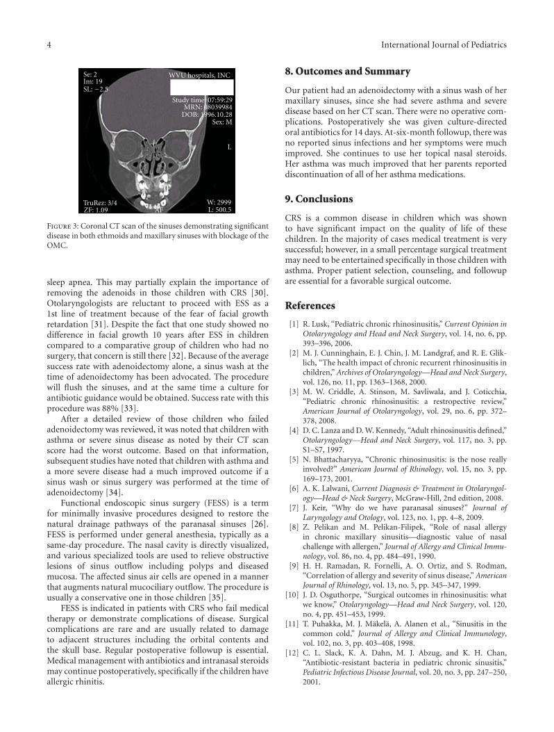
4 International Journal of Pediatrics
Se: 2Im: 19
TruRez: 3/4
WVU hospitals, INC
MRN: 08039984
Sex: M
L
W: 2999AF
SL: −2.5
ZF: 1.09 L: 500.5
DOB: 1996.10.28
Study time: 07:59:29
Figure 3: Coronal CT scan of the sinuses demonstrating significantdisease in both ethmoids and maxillary sinuses with blockage of theOMC.
sleep apnea. This may partially explain the importance ofremoving the adenoids in those children with CRS [30].Otolaryngologists are reluctant to proceed with ESS as a1st line of treatment because of the fear of facial growthretardation [31]. Despite the fact that one study showed nodifference in facial growth 10 years after ESS in childrencompared to a comparative group of children who had nosurgery, that concern is still there [32]. Because of the averagesuccess rate with adenoidectomy alone, a sinus wash at thetime of adenoidectomy has been advocated. The procedurewill flush the sinuses, and at the same time a culture forantibiotic guidance would be obtained. Success rate with thisprocedure was 88% [33].
After a detailed review of those children who failedadenoidectomy was reviewed, it was noted that children withasthma or severe sinus disease as noted by their CT scanscore had the worst outcome. Based on that information,subsequent studies have noted that children with asthma anda more severe disease had a much improved outcome if asinus wash or sinus surgery was performed at the time ofadenoidectomy [34].
Functional endoscopic sinus surgery (FESS) is a termfor minimally invasive procedures designed to restore thenatural drainage pathways of the paranasal sinuses [26].FESS is performed under general anesthesia, typically as asame-day procedure. The nasal cavity is directly visualized,and various specialized tools are used to relieve obstructivelesions of sinus outflow including polyps and diseasedmucosa. The affected sinus air cells are opened in a mannerthat augments natural mucociliary outflow. The procedure isusually a conservative one in those children [35].
FESS is indicated in patients with CRS who fail medicaltherapy or demonstrate complications of disease. Surgicalcomplications are rare and are usually related to damageto adjacent structures including the orbital contents andthe skull base. Regular postoperative followup is essential.Medical management with antibiotics and intranasal steroidsmay continue postoperatively, specifically if the children haveallergic rhinitis.
8. Outcomes and Summary
Our patient had an adenoidectomy with a sinus wash of hermaxillary sinuses, since she had severe asthma and severedisease based on her CT scan. There were no operative com-plications. Postoperatively she was given culture-directedoral antibiotics for 14 days. At-six-month followup, there wasno reported sinus infections and her symptoms were muchimproved. She continues to use her topical nasal steroids.Her asthma was much improved that her parents reporteddiscontinuation of all of her asthma medications.
9. Conclusions
CRS is a common disease in children which was shownto have significant impact on the quality of life of thesechildren. In the majority of cases medical treatment is verysuccessful; however, in a small percentage surgical treatmentmay need to be entertained specifically in those children withasthma. Proper patient selection, counseling, and followupare essential for a favorable surgical outcome.
References
[1] R. Lusk, “Pediatric chronic rhinosinusitis,” Current Opinion inOtolaryngology and Head and Neck Surgery, vol. 14, no. 6, pp.393–396, 2006.
[2] M. J. Cunninghain, E. J. Chin, J. M. Landgraf, and R. E. Glik-lich, “The health impact of chronic recurrent rhinosinusitis inchildren,” Archives of Otolaryngology—Head and Neck Surgery,vol. 126, no. 11, pp. 1363–1368, 2000.
[3] M. W. Criddle, A. Stinson, M. Savliwala, and J. Coticchia,“Pediatric chronic rhinosinusitis: a restropective review,”American Journal of Otolaryngology, vol. 29, no. 6, pp. 372–378, 2008.
[4] D. C. Lanza and D. W. Kennedy, “Adult rhinosinusitis defined,”Otolaryngology—Head and Neck Surgery, vol. 117, no. 3, pp.S1–S7, 1997.
[5] N. Bhattacharyya, “Chronic rhinosinusitis: is the nose reallyinvolved?” American Journal of Rhinology, vol. 15, no. 3, pp.169–173, 2001.
[6] A. K. Lalwani, Current Diagnosis & Treatment in Otolaryngol-ogy—Head & Neck Surgery, McGraw-Hill, 2nd edition, 2008.
[7] J. Keir, “Why do we have paranasal sinuses?” Journal ofLaryngology and Otology, vol. 123, no. 1, pp. 4–8, 2009.
[8] Z. Pelikan and M. Pelikan-Filipek, “Role of nasal allergyin chronic maxillary sinusitis—diagnostic value of nasalchallenge with allergen,” Journal of Allergy and Clinical Immu-nology, vol. 86, no. 4, pp. 484–491, 1990.
[9] H. H. Ramadan, R. Fornelli, A. O. Ortiz, and S. Rodman,“Correlation of allergy and severity of sinus disease,” AmericanJournal of Rhinology, vol. 13, no. 5, pp. 345–347, 1999.
[10] J. D. Osguthorpe, “Surgical outcomes in rhinosinusitis: whatwe know,” Otolaryngology—Head and Neck Surgery, vol. 120,no. 4, pp. 451–453, 1999.
[11] T. Puhakka, M. J. Makela, A. Alanen et al., “Sinusitis in thecommon cold,” Journal of Allergy and Clinical Immunology,vol. 102, no. 3, pp. 403–408, 1998.
[12] C. L. Slack, K. A. Dahn, M. J. Abzug, and K. H. Chan,“Antibiotic-resistant bacteria in pediatric chronic sinusitis,”Pediatric Infectious Disease Journal, vol. 20, no. 3, pp. 247–250,2001.

International Journal of Pediatrics 5
[13] J. B. Anon, “Acute bacterial rhinosinusitis in pediatric medi-cine: current issues in diagnosis and management,” PediatricDrugs, vol. 5, no. 1, pp. 25–33, 2003.
[14] D. L. Hamilos, “Chronic sinusitis,” Journal of Allergy andClinical Immunology, vol. 106, no. 2, pp. 213–227, 2000.
[15] D. Babinski and M. Trawinska-Bartnicka, “Rhinosinusitis incystic fibrosis: not a simple story,” International Journal ofPediatric Otorhinolaryngology, vol. 72, no. 5, pp. 619–624,2008.
[16] F. M. Baroody, “Mucociliary transport in chronic rhinosinusi-tis,” Clinical Allergy and Immunology, vol. 20, pp. 103–119,2007.
[17] E. R. Wald, N. Guerra, and C. Byers, “Upper respiratory tractinfections in young children: duration of and frequency ofcomplications,” Pediatrics, vol. 87, no. 2, pp. 129–133, 1991.
[18] J. A. Stankiewicz and J. M. Chow, “A diagnostic dilemmafor chronic rhinosinusitis: definition accuracy and validity,”American Journal of Rhinology, vol. 16, no. 4, pp. 199–202,2002.
[19] P. B. Dinis, M. C. Monteiro, M. L. Martins, N. Silva, andA. Gomes, “Sinus tissue pharmacokinetics after oral admin-istration of amoxicillin/clavulanic acid,” Laryngoscope, vol.110, no. 6, pp. 1050–1055, 2000.
[20] H. Cincik and B. J. Ferguson, “The impact of endoscopiccultures on care in rhinosinusitis,” Laryngoscope, vol. 116, no.9, pp. 1562–1568, 2006.
[21] R. S. Patel, S. R. Shaw, A. M. Wallace, and G. W. McGarry,“Efficacy and systemic tolerability of mometasone furoate andbetamethasone sodium phosphate,” Journal of Laryngologyand Otology, vol. 118, no. 11, pp. 866–871, 2004.
[22] A. Parikh, G. K. Scadding, Y. Darby, and R. C. Baker,“Topical corticosteroids in chronic rhinosinusitis: a random-ized, double-blind, placebo-controlled trial using fluticasonepropionate aqueous nasal spray,” Rhinology, vol. 39, no. 2, pp.75–79, 2001.
[23] R. Giger, P. Pasche, C. Cheseaux et al., “Comparison ofonce- versus twice-daily use of beclomethasone dipropionateaqueous nasal spray in the treatment of allergic and non-allergic chronic rhinosinusitis,” European Archives of Oto-Rhino-Laryngology, vol. 260, no. 3, pp. 135–140, 2003.
[24] R. Harvey, S. A. Hannan, L. Badia, and G. Scadding, “Nasalsaline irrigations for the symptoms of chronic rhinosinusitis,”Cochrane Database of Systematic Reviews, no. 3, Article IDCD006394, 2007.
[25] D. Rabago, T. Pasic, A. Zgierska, M. Mundt, B. Barrett, andR. Maberry, “The efficacy of hypertonic saline nasal irrigationfor chronic sinonasal symptoms,” Otolaryngology—Head andNeck Surgery, vol. 133, no. 1, pp. 3–8, 2005.
[26] B. A. Senior, D. W. Kennedy, J. Tanabodee, H. Kroger,M. Hassab, and D. Lanza, “Long-term results of functionalendoscopic sinus surgery,” Laryngoscope, vol. 108, no. 2, pp.151–157, 1998.
[27] S. J. Vandenberg and D. G. Heatley, “Efficacy of adenoidec-tomy in relieving symptoms of chronic sinusitis in children,”Archives of Otolaryngology—Head and Neck Surgery, vol. 123,no. 7, pp. 675–678, 1997.
[28] H. H. Ramadan, “Adenoidectomy vs endoscopic sinussurgery for the treatment of pediatric sinusitis,” Archives ofOtolaryngology—Head and Neck Surgery, vol. 125, no. 11, pp.1208–1211, 1999.
[29] R. L. Hebert II and J. P. Bent III, “Meta-analysis of outcomes ofpediatric functional endoscopic sinus surgery,” Laryngoscope,vol. 108, no. 6, pp. 796–799, 1998.
[30] G. Zuliani, M. Carron, J. Gurrola et al., “Identificationof adenoid biofilms in chronic rhinosinusitis,” InternationalJournal of Pediatric Otorhinolaryngology, vol. 70, no. 9, pp.1613–1617, 2006.
[31] E. A. Mair, W. E. Bolger, and E. A. Breisch, “Sinus and facialgrowth after pediatric endoscopic sinus surgery,” Archives ofOtolaryngology—Head and Neck Surgery, vol. 121, no. 5, pp.547–552, 1995.
[32] M. R. Bothwell, J. F. Piccirillo, R. P. Lusk, and B. D. Ride-nour, “Long-term outcome of facial growth after functionalendoscopic sinus surgery,” Otolaryngology—Head and NeckSurgery, vol. 126, no. 6, pp. 628–634, 2002.
[33] H. H. Ramadan and J. L. Cost, “Outcome of adenoidectomyversus adenoidectomy with maxillary sinus wash for chronicrhinosinusitis in children,” Laryngoscope, vol. 118, no. 5, pp.871–873, 2008.
[34] H. H. Ramadan and J. Tiu, “Failures of adenoidectomy forchronic rhinosinusitis in children: for whom and when do theyfail?” Laryngoscope, vol. 117, no. 6, pp. 1080–1083, 2007.
[35] H. H. Ramadan, “Surgical management of chronic sinusitis inchildren,” Laryngoscope, vol. 114, no. 12, pp. 2103–2109, 2004.
Hindawi Publishing CorporationInternational Journal of PediatricsVolume 2012, Article ID 945356, 5 pagesdoi:10.1155/2012/945356
Clinical Study
Troublesome Tinnitus in Children: Epidemiology, AudiologicalProfile, and Preliminary Results of Treatment
G. Bartnik, A. Stepien, D. Raj-Koziak, A. Fabijanska, I. Niedziałek, and H. Skarzynski
Tinnitus Clinic, Institute of Physiology and Pathology of Hearing, AK Kampinos 1, 01-943 Warsaw, Poland
Correspondence should be addressed to G. Bartnik, [email protected]
Received 26 January 2011; Revised 12 April 2011; Accepted 4 May 2011
Academic Editor: Alan T. L. Cheng
Copyright © 2012 G. Bartnik et al. This is an open access article distributed under the Creative Commons Attribution License,which permits unrestricted use, distribution, and reproduction in any medium, provided the original work is properly cited.
Introduction. Although tinnitus often has a significant impact on individual’s life, there are still few reports relating to tinnitus inchildren. In our tinnitus clinic, children with distressing tinnitus constitute about 0,5% of all our patients. Objectives. The aim ofthis study was to analyse children with troublesome tinnitus as regards epidemiology, audiological profile, and preliminary effectsof the therapy. Methods. A retrospective study was carried out involving the cases of 143 children consulted in our Tinnitus Clinicin 2009. The selected group with troublesome tinnitus was evaluated and classified for proper category of Tinnitus RetrainingTherapy (TRT). Results. The study showed that 41.3% of the children suffered from bothersome tinnitus. In this group 44.1%of the patients demonstrated normal hearing. The success of the therapy after 6 months was estimated on 81.4% of significantimprovement. Conclusions. It is recommended that a questionnaire include an inquiry about the presence of tinnitus duringhearing screening tests.
1. Introduction
Tinnitus is perception of sound like buzzing, ringing, roar-ing, clicking, pulsations, and other noises in the ear, ears, orin the head occurring without an outside acoustic stimulus.Tinnitus is a symptom, not a disease, and as such canarise as a result of inappropriate activity at any point inthe auditory pathway. The neural plasticity, responsible fortinnitus, appears as an adaptation or compensation, at acentral nervous system level, following peripheral damage.Tinnitus may be caused by a variety of pathologies, illnesses,medications, allergies, dietary changes, stresses, or traumaticevents. It is often caused by pathologies related to the ear,head, and neck area like: head trauma or whiplash. One ofthe most common causes of tinnitus involves noise exposure.It can be both: long term and produced by brief loud sound.Long-term exposure to noise or acoustic trauma can producesubstantial and often irreparable damage to the delicatestructures of the inner ear—especially outer hair cells. In themajority of cases, tinnitus is clearly related to some changesin the inner ear. At the same time tinnitus is not necessarilyassociated with hearing loss. About 40% of tinnitus patients
have also the symptoms of decreased loudness tolerancecalled hyperacusis [1].
The prevalence of tinnitus increases with age. In olderpeople, tinnitus occurs most often in conjunction with thehearing problems connected with ageing or it can be asso-ciated with general diseases. About 5% of adult Poles sufferfrom constant tinnitus [2]. Research into the prevalence oftinnitus in a pediatric population in Poland on large scale hasbeen continued by the Institute of Physiology and Pathologyof Hearing for last years. The initial results show that 12.8%of seven-year-old children have tinnitus in our country [3].There are few epidemiological studies on the prevalence oftinnitus in children in the literature. In a group of childrenwith normal hearing, the prevalence of tinnitus has beenreported to be between 6% and 36%. In a study by Nodar[4], there was 15% of those patients. Other researchers,Mills et al. [5] estimated that 29% of normally hearingchildren complain about tinnitus, and only 9,6% reportedthe troublesome tinnitus [5]. The latest paper by Holgers[6] highlights about 13% of pediatric patients with normalhearing and tinnitus [6]. In children with hearing loss, theprevalence of tinnitus is reported to be as high as 55% [7].
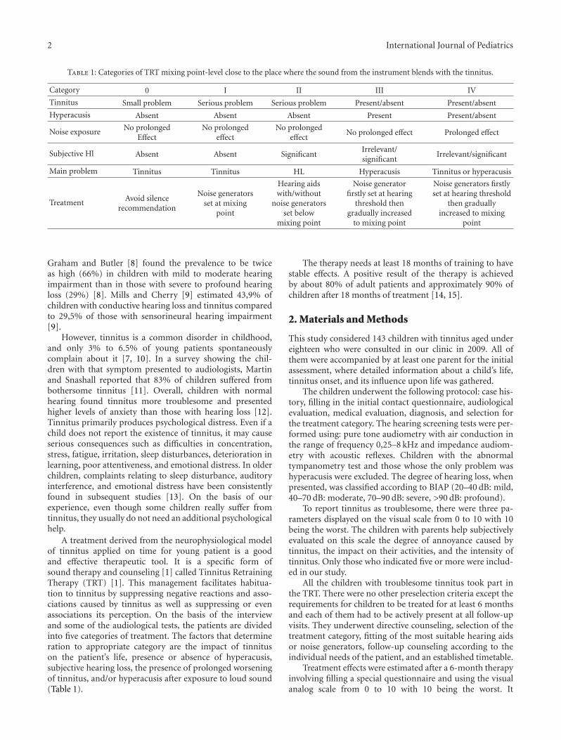
2 International Journal of Pediatrics
Table 1: Categories of TRT mixing point-level close to the place where the sound from the instrument blends with the tinnitus.
Category 0 I II III IV
Tinnitus Small problem Serious problem Serious problem Present/absent Present/absent
Hyperacusis Absent Absent Absent Present Present/absent
Noise exposure No prolongedEffect
No prolongedeffect
No prolongedeffect
No prolonged effect Prolonged effect
Subjective Hl Absent Absent SignificantIrrelevant/significant
Irrelevant/significant
Main problem Tinnitus Tinnitus HL Hyperacusis Tinnitus or hyperacusis
Treatment Avoid silencerecommendation
Noise generatorsset at mixing
point
Hearing aidswith/without
noise generatorsset below
mixing point
Noise generatorfirstly set at hearing
threshold thengradually increased
to mixing point
Noise generators firstlyset at hearing threshold
then graduallyincreased to mixing
point
Graham and Butler [8] found the prevalence to be twiceas high (66%) in children with mild to moderate hearingimpairment than in those with severe to profound hearingloss (29%) [8]. Mills and Cherry [9] estimated 43,9% ofchildren with conductive hearing loss and tinnitus comparedto 29,5% of those with sensorineural hearing impairment[9].
However, tinnitus is a common disorder in childhood,and only 3% to 6.5% of young patients spontaneouslycomplain about it [7, 10]. In a survey showing the chil-dren with that symptom presented to audiologists, Martinand Snashall reported that 83% of children suffered frombothersome tinnitus [11]. Overall, children with normalhearing found tinnitus more troublesome and presentedhigher levels of anxiety than those with hearing loss [12].Tinnitus primarily produces psychological distress. Even if achild does not report the existence of tinnitus, it may causeserious consequences such as difficulties in concentration,stress, fatigue, irritation, sleep disturbances, deterioration inlearning, poor attentiveness, and emotional distress. In olderchildren, complaints relating to sleep disturbance, auditoryinterference, and emotional distress have been consistentlyfound in subsequent studies [13]. On the basis of ourexperience, even though some children really suffer fromtinnitus, they usually do not need an additional psychologicalhelp.
A treatment derived from the neurophysiological modelof tinnitus applied on time for young patient is a goodand effective therapeutic tool. It is a specific form ofsound therapy and counseling [1] called Tinnitus RetrainingTherapy (TRT) [1]. This management facilitates habitua-tion to tinnitus by suppressing negative reactions and asso-ciations caused by tinnitus as well as suppressing or evenassociations its perception. On the basis of the interviewand some of the audiological tests, the patients are dividedinto five categories of treatment. The factors that determineration to appropriate category are the impact of tinnituson the patient’s life, presence or absence of hyperacusis,subjective hearing loss, the presence of prolonged worseningof tinnitus, and/or hyperacusis after exposure to loud sound(Table 1).
The therapy needs at least 18 months of training to havestable effects. A positive result of the therapy is achievedby about 80% of adult patients and approximately 90% ofchildren after 18 months of treatment [14, 15].
2. Materials and Methods
This study considered 143 children with tinnitus aged undereighteen who were consulted in our clinic in 2009. All ofthem were accompanied by at least one parent for the initialassessment, where detailed information about a child’s life,tinnitus onset, and its influence upon life was gathered.
The children underwent the following protocol: case his-tory, filling in the initial contact questionnaire, audiologicalevaluation, medical evaluation, diagnosis, and selection forthe treatment category. The hearing screening tests were per-formed using: pure tone audiometry with air conduction inthe range of frequency 0,25–8 kHz and impedance audiom-etry with acoustic reflexes. Children with the abnormaltympanometry test and those whose the only problem washyperacusis were excluded. The degree of hearing loss, whenpresented, was classified according to BIAP (20–40 dB: mild,40–70 dB: moderate, 70–90 dB: severe, >90 dB: profound).
To report tinnitus as troublesome, there were three pa-rameters displayed on the visual scale from 0 to 10 with 10being the worst. The children with parents help subjectivelyevaluated on this scale the degree of annoyance caused bytinnitus, the impact on their activities, and the intensity oftinnitus. Only those who indicated five or more were includ-ed in our study.
All the children with troublesome tinnitus took part inthe TRT. There were no other preselection criteria except therequirements for children to be treated for at least 6 monthsand each of them had to be actively present at all follow-upvisits. They underwent directive counseling, selection of thetreatment category, fitting of the most suitable hearing aidsor noise generators, follow-up counseling according to theindividual needs of the patient, and an established timetable.
Treatment effects were estimated after a 6-month therapyinvolving filling a special questionnaire and using the visualanalog scale from 0 to 10 with 10 being the worst. It

International Journal of Pediatrics 3
Table 2: The questionnaire that was used to estimate results of treatment.
Measure the following parameters using visual analog scale (VAS): VAS (10 means the worst)
(1) The impact of tinnitus with/without hyperacusis on various everyday activites 0-1-2-3-4-5-6-7-8-9-10
(2) Percentage of time of being aware of tinnitus 0-1-2-3-4-5-6-7-8-9-10
(3) The degree of annoyance 0-1-2-3-4-5-6-7-8-9-10
(4) The impact of tinnitus on your life 0-1-2-3-4-5-6-7-8-9-10
(5) The intensity of tinnitus 0-1-2-3-4-5-6-7-8-9-10
(6) The level of distress caused by tinnitus 0-1-2-3-4-5-6-7-8-9-10
0
2
4
6
8
10
12
14
16
<1/2 >5
Nu
mbe
rof
pati
ents
First counselling (years)
1/2-1 1-2 2-3 3-4 4-5
Figure 1: Time from the onset of tinnitus to first counseling.
Normal hearing
Sensorineural HL
Mixed HL
Conductive HL
5 10 15 20 25 30 35
Number of patients
0
Figure 2: Hearing level in the study group. Hearing loss (HL).
comprised the parameters, which were presented in the formof questions to be answered by the child before, during, andafter 6 months (Table 2).
The data acquired from the questionnaire results of thetherapy were evaluated by means of two criteria: significantimprovement and no improvement or deterioration. The cri-terion of significant improvement was the decrease of at leastthree of the above-mentioned parameters by a minimum of20% and the liberation of at least one of the everyday lifeactivities previously impaired by tinnitus.
3. Results
Troublesome tinnitus was present in 59 children (41,3%), 31girls (52,5%) and 28 boys (47,5%), at the average age of 14,4(min. 7, max. 17). Some of them complained about hyper-acusis (n = 22, 37,3%). There were significantly more girlswith this symptom (n = 14, 63,6%) than boys (n = 8,
Mild SNHL (14)
Moderate SNHL (5)
Severe SNHL (4)
Profound SNHL (8)
Figure 3: Number of children with tinnitus and different degrees ofseverity of sensorineural hearing loss (SNHL).
0
10
20
30
40
50
60
(%)
Vir
us
infe
ctio
n
Noi
set r
aum
a
Mid
dle
e ar
path
olog
y
Vac
c in
atio
n
He a
dtr
a um
a
Bra
intu
mor
Rad
io- ,
chem
oth
erap
y
Un
know
nFigure 4: The suspected etiology of tinnitus in the study group.
36,4%). Despite bothersome tinnitus, only 19 children(32,3%) had first audiological counseling within a year afterthe onset of tinnitus (Figure 1). In all, 44,1% of children (n =26) demonstrated normal hearing while 55,9% (n = 33)had hearing impairment (Figures 2 and 3). According to thesuspected etiology of tinnitus, the most common one was aclinical history of a virus infection (18,6%), and in 49,1% ofcases etiology was unknown. In Figure 4, there is a detailedsummary of the causes percentage distribution.
As far as the specific characteristics of tinnitus wereconcerned, 56 children described their tinnitus as continuousin 20 cases (33,9%), as intermittent in 36 cases (61%). Therewere 3 children that did not specify duration of tinnitus. Thepitch of tinnitus was low (<2 kHz) in 14 children (23,7%),high (>4 kHz) in 40 children (67,8%), pulsating in 1 case(1,7%), and undefined in 3 cases (5,1%).
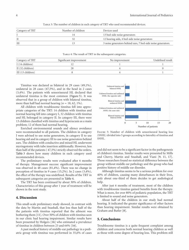
4 International Journal of Pediatrics
Table 3: The number of children in each category of TRT who used recommended devices.
Category of TRT Number of children Devices used
I 14 13 bed-side noise generators
II 32 12 hearing aids, 4 bed-side noise generators
III 13 3 noise generators behind ears, 7 bed-side noise generators.
Table 4: The result of TRT in the subsequent categories.
Category of TRT Significant improvement No improvement Undefined result
I (14 children) 12 1 1
II (32 children) 28 4 0
III (13 children) 8 4 1
Tinnitus was declared as bilateral in 29 cases (49,1%),unilateral in 28 cases (47,5%), and in the head in 2 cases(3,4%). The patients with sensorineural HL declared thatunilateral tinnitus is the most common (Figure 5). It wasobserved that in a group of children with bilateral tinnitus,more than half had normal hearing (n = 18, 62, 1%).
All children with troublesome tinnitus fell into appro-priate categories of the TRT: 14 children with tinnitus andnormal hearing fell into category I, 32 children with tinnitusand HL belonged in category II. In category III, there were13 children classified with tinnitus and hyperacusis as a mainproblem; 12 of them had normal hearing.
Enriched environmental sounds and silence avoidancewere recommended to all patients. The children in categoryI were advised to use noise generators, in category II to usehearing aid and in category III to use noise generators behindears. The children with conductive and mixed HL underwentmyringotomy with tube insertion additionally. However, lessthan half of the patients ( 47,5%) strictly observed the orders.Table 3 shows how many children in each category usedrecommended devices.
The preliminary results were evaluated after 6 monthsof therapy. Management success: significant improvementwas observed in 48 cases (81,4%). There was no change inperception of tinnitus in 9 cases (15,2%). In 2 cases (3,4%),the effect of the therapy was undefined. Results of the TRT insubsequent categories are presented in Table 4.
The TRT has been continued by about 30% of children.Characteristics of this group after 1 year of treatment will beshown in the next study.
4. Discussion
This small-scale preliminary study showed, in contrast withthe data by Martin and Snashall, that less than half of thethe patients with tinnitus reported that the ailment wasbothering them [11]. Over 50% of children with tinnitus seenin our clinic had hearing impairment. Similar results havebeen presented by Holgers [16] who estimated a prevalenceof tinnitus in children between 23 and 64% [16].
A past medical history of middle-ear pathology in a pedi-atric group with tinnitus was performed in 35,6% of cases
0 5 10 15 20
Bilateral tinnitus andbilateral SNHL
Unilateral tinnitus and unilateralSNHL (the same side)
Bilateral tinnitus andunilateral SNHL
Unilateral tinnitus and unilateralSNHL (the opposite side)
Unilateral tinnitus andbilateral SNHL
Number of patients
Figure 5: Number of children with sensorineural hearing loss(SNHL) divided into 5 groups according to laterality of tinnitus andSNHL.
and did not seem to be a significant factor in the pathogenesisof children’s tinnitus. Similar results were presented by Millsand Cherry, Martin and Snashall, and Viani [9, 11, 17].Those researchers found no statistical difference between thegroup without middle ear pathology and the group who hadpositive history of middle ear disorder.
Although tinnitus seems to be a serious problem for over40% of children, causing many disturbances in their lives,only about one-third of them decides to get audiologicalhelp.
After just 6 months of treatment, most of the childrenwith troublesome tinnitus gained benefits from the therapy.What is more, for over 80% of pediatric population, the TRTis limited to sound and noise generators.
About half of the children in our study had normalhearing. It indicated the greater significance of other factorsthan hearing impairment. Similar results were obtained byGraham and Butler [8].
5. Conclusions
Troublesome tinnitus is a quite frequent complaint amongchildren and concerns both normal hearing children as wellas those with some degree of hearing loss. This problem still

International Journal of Pediatrics 5
needs careful attention and research. Until we gain moreknowledge on the development of bothersome tinnitus inpediatric population, audiologists have to rely on the reportsbased on adults. It is recommended that an inquiry about thepresence of tinnitus during hearing screening tests at schoolshould be included in the questionnaire. Treatment strategiesshould be applied individually and involve both the parentsand children.
References
[1] P. J. Jastreboff, “Tinnitus Habituation Therapy (THT) andTinnitus Retraining Therapy (TRT),” in Tinnitus Handbook,R. Tyler, Ed., pp. 357–376, Singular Thomson Learning, SanDiego, Calif, USA, 2000.
[2] A. Fabijanska, M. Rogowski, G. Bartnik, and H. Skarzynski,“Epidemiolody of tinnitus and hyperacusis in Poland,” inProceedings of the 6th International Tinnitus Seminar, pp. 569–572, Cambridge, UK, September 1999.
[3] D. Raj-Koziak, A. Piłka, G. Bartnik, A. Fabijanska, H.Skarzynski, and K. Kochanek, “The prevalence of tinnitusin 7 year old children in the eastern of Poland,” The PolishOtolaryngology. In press.
[4] R. H. Nodar, “Tinnitus aurium in school age children,” Journalof Auditory Research, vol. 12, pp. 133–135, 1972.
[5] R. P. Mills, D. M. Albert, and C. E. Brain, “Tinnitus inchildhood,” Clinical Otolaryngology and Allied Sciences, vol. 11,no. 6, pp. 431–434, 1986.
[6] K. M. Holgers, “Tinnitus in 7-year-old children,” in Proceed-ings of the 6th International Tinnitus Seminar, J. W. P. Hazell,Ed., pp. 218–219, London, UK, 1999.
[7] R. H. Nodar and M. H. W. Lezak, “Paediatric tinnitus: a thesisrevised,” Journal of Laryngology and Otology, supplement 9,pp. 234–235, 1984.
[8] J. Graham and J. Butler, “Tinnitus in children,” Journal ofLaryngology and Otology, vol. 97, 9, pp. 236–241, 1984.
[9] R. P. Mills and J. R. Cherry, “Subjective tinnitus in childrenwith otological disorders,” International Journal of PediatricOtorhinolaryngology, vol. 7, no. 1, pp. 21–27, 1984.
[10] M. Savastano, “Characteristics of tinnitus in childhood,” Eu-ropean Journal of Pediatrics, vol. 166, no. 8, pp. 797–801, 2007.
[11] K. Martin and S. Snashall, “Children presenting with tinnitus:a retrospective study,” British Journal of Audiology, vol. 28,no. 2, pp. 111–115, 1994.
[12] J. L. Stouffer et al., “Tinnitus in normal hearing and hearing-impaired children,” in Proceedings of the 6th InternationalTinnitus Seminar, pp. 255–258, Kugler, Bordeaux, France,1991.
[13] L. Sanchez and D. Stephens, “A tinnitus problem questionnairein a clinic population,” Ear & Hearing, vol. 18, no. 3, pp. 210–217, 1997.
[14] G. Bartnik, A. Fabijanska, and M. Rogowski, “Our experiencein treatment of patients with tinnitus and/or hyperacusis usingthe habituation method,” in Proceedings of the Proceedings ofthe 6th International Tinnitus Seminar, pp. 415–418, Cam-bridge, UK, September 1999.
[15] G. Bartnik, A. Fabijanska, D. Raj-Koziak, and M. Rogowski,“Algorytm postepowania u dzieci z szumami usznymi wKlinice Szumow Usznych,” Instytutu Fizjologii i PatologiiSłuchu Nowa Pediatria, vol. 3, no. 6, pp. 23–26, 1999.
[16] K. M. Holgers, “Tinnitus in 7-year-old children,” EuropeanJournal of Pediatrics, vol. 162, no. 4, pp. 276–278, 2003.
[17] L. G. Viani, “Tinnitus in children with hearing loss,” Journalof Laryngology and Otology, vol. 103, no. 12, pp. 1142–1145,1989.