Re Vista
14
MULTIMODALITY TREATMENT OF BRAIN ARTERIOVENOUS MALFORMATIONS WITH MICROSURGERY AFTER EMBOLIZATION WITH ONYX: SINGLE-CENTER EXPERIENCE AND TECHNICAL NUANCES OBJECTIVE: To report our experience with the treatment of brain arteriovenous mal- formations (AVM) with microsurgical resection after embolization with Onyx liquid embolic agent (eV3, Irvine, CA). METHODS: Between August 2005 and December 2006, 28 patients were treated by the same surgical-endovascular team. Twenty-eight AVMs were embolized preoperatively in 55 sessions (71 pedicles) with Onyx. We analyzed the AVM size, volume, number of embolization sessions, degree of preoperative obliteration, time to embolization and resection after the bleed, intraprocedural complications, intraoperative blood loss, other complications, and postoperative outcome at 6 months. Technical nuances of the emboliza- tion and surgical resection of the embolized AVMs are illustrated in illustrative cases. RESULTS: The average size and volume of AVMs treated with Onyx were 3.56 cm (largest, 7.6 cm), and 13.03 ml, respectively. The average Spetzler-Martin grade was 2.75. The average preoperative volumetric obliteration was 74.1%. The average blood loss during resection of embolized AVMs was 348 ml. Complications related to emboliza- tion were stuck microcatheter (two patients), proximal vessel perforation (one patient), and anterior choroidal territory stroke (one patient). Surgical complications included wound infection (one patient), residual AVM nidus (one patient), normal pressure per- fusion breakthrough with worsening of neurological deficit caused by embolization (one patient), and new-onset motor deficits in five patients. At the time of the 6-month follow-up examination, four patients with new-onset motor deficits had recovered com- pletely or nearly completely, and one patient was disabled. One patient died, never recovering from the initial poor condition due to the bleed. Pathological examination of resected AVMs showed angionecrosis in 42.9%, foreign body giant cells in 39.3%, and evidence of recanalization of Onyx-embolized vessels in 14.3% of specimens. CONCLUSION: Multimodality treatment with microsurgery is safe and effective after embolization with Onyx. High occlusion rates and low complication rates were observed after Onyx embolization and were comparable to those in previous reports. Superselective intranidal or perinidal catheter positions and slow, controlled injections that protect the draining veins make the therapy safe even in complex AVMs and critical locations. We recommend resection of the AVM despite apparently complete embolization with Onyx. Team work and coordination between the surgeon and the interventional neu- roradiologist are important to achieve a good outcome. KEY WORDS: Brain arteriovenous malformation, Embolization, Microsurgery, Onyx, Outcomes, Recanalization, Technique Neurosurgery 62:1213–1226, 2008 DOI: 10.1227/01.NEU.0000316860.35705.AA www.neurosurgery-online.com NEUROSURGERY VOLUME 62 | NUMBER 6 | JUNE 2008 | 1213 CLINICAL STUDIES Sabareesh K. Natarajan, M.D., M.S. Department of Neurological Surgery, University of Washington, Seattle, Washington Basavaraj Ghodke, M.D. Departments of Neurological Surgery and Neuroradiology, University of Washington, Seattle, Washington Gavin W. Britz, M.D. Departments of Neurological Surgery and Neuroradiology, University of Washington, Seattle, Washington Donald E. Born, M.D. Department of Neuropathology, University of Washington, Seattle, Washington Laligam N. Sekhar, M.D. Department of Neurological Surgery, University of Washington, Seattle, Washington Reprint requests: Laligam N. Sekhar, M.D., Department of Neurological Surgery, Harborview Medical Center, Box 359766, Seattle, WA 98104. Email: [email protected] Received, May 11, 2007. Accepted, February 13, 2008. M odern treatment of arteriovenous malformations (AVM) of the brain includes the following interven- tions alone or in combination: endovascular emboliza- tion, microsurgical resection, and radiosurgery (6, 15, 23, 30, 35, 42). Microsurgical resection is preferred for treatment of AVMs in resectable areas (6, 30, 36). Radiosurgical treatment of small (3 cm) AVMs has also shown good obliteration rates at 3 years (41). Embolization is mainly used to reduce the size of a
-
Upload
carolina-guisella -
Category
Documents
-
view
212 -
download
0
description
revista
Transcript of Re Vista

MULTIMODALITY TREATMENT OF BRAINARTERIOVENOUS MALFORMATIONS WITHMICROSURGERY AFTER EMBOLIZATION WITH ONYX:SINGLE-CENTER EXPERIENCE AND TECHNICAL NUANCES
OBJECTIVE: To report our experience with the treatment of brain arteriovenous mal-formations (AVM) with microsurgical resection after embolization with Onyx liquidembolic agent (eV3, Irvine, CA).METHODS: Between August 2005 and December 2006, 28 patients were treated by thesame surgical-endovascular team. Twenty-eight AVMs were embolized preoperatively in55 sessions (71 pedicles) with Onyx. We analyzed the AVM size, volume, number ofembolization sessions, degree of preoperative obliteration, time to embolization andresection after the bleed, intraprocedural complications, intraoperative blood loss, othercomplications, and postoperative outcome at 6 months. Technical nuances of the emboliza-tion and surgical resection of the embolized AVMs are illustrated in illustrative cases.RESULTS: The average size and volume of AVMs treated with Onyx were 3.56 cm(largest, 7.6 cm), and 13.03 ml, respectively. The average Spetzler-Martin grade was2.75. The average preoperative volumetric obliteration was 74.1%. The average bloodloss during resection of embolized AVMs was 348 ml. Complications related to emboliza-tion were stuck microcatheter (two patients), proximal vessel perforation (one patient),and anterior choroidal territory stroke (one patient). Surgical complications includedwound infection (one patient), residual AVM nidus (one patient), normal pressure per-fusion breakthrough with worsening of neurological deficit caused by embolization(one patient), and new-onset motor deficits in five patients. At the time of the 6-monthfollow-up examination, four patients with new-onset motor deficits had recovered com-pletely or nearly completely, and one patient was disabled. One patient died, neverrecovering from the initial poor condition due to the bleed. Pathological examinationof resected AVMs showed angionecrosis in 42.9%, foreign body giant cells in 39.3%,and evidence of recanalization of Onyx-embolized vessels in 14.3% of specimens.CONCLUSION: Multimodality treatment with microsurgery is safe and effective afterembolization with Onyx. High occlusion rates and low complication rates were observedafter Onyx embolization and were comparable to those in previous reports. Superselectiveintranidal or perinidal catheter positions and slow, controlled injections that protectthe draining veins make the therapy safe even in complex AVMs and critical locations.We recommend resection of the AVM despite apparently complete embolization withOnyx. Team work and coordination between the surgeon and the interventional neu-roradiologist are important to achieve a good outcome.
KEY WORDS: Brain arteriovenous malformation, Embolization, Microsurgery, Onyx, Outcomes,Recanalization, Technique
Neurosurgery 62:1213–1226, 2008 DOI: 10.1227/01.NEU.0000316860.35705.AA www.neurosurgery-online.com
NEUROSURGERY VOLUME 62 | NUMBER 6 | JUNE 2008 | 1213
CLINICAL STUDIES
Sabareesh K. Natarajan, M.D.,M.S.Department of Neurological Surgery,University of Washington,Seattle, Washington
Basavaraj Ghodke, M.D.Departments of Neurological Surgeryand Neuroradiology,University of Washington,Seattle, Washington
Gavin W. Britz, M.D.Departments of Neurological Surgeryand Neuroradiology,University of Washington,Seattle, Washington
Donald E. Born, M.D.Department of Neuropathology,University of Washington,Seattle, Washington
Laligam N. Sekhar, M.D.Department of Neurological Surgery,University of Washington,Seattle, Washington
Reprint requests:Laligam N. Sekhar, M.D.,Department of Neurological Surgery,Harborview Medical Center,Box 359766,Seattle, WA 98104.Email: [email protected]
Received, May 11, 2007.
Accepted, February 13, 2008.
Modern treatment of arteriovenous malformations(AVM) of the brain includes the following interven-tions alone or in combination: endovascular emboliza-
tion, microsurgical resection, and radiosurgery (6, 15, 23, 30, 35,
42). Microsurgical resection is preferred for treatment of AVMsin resectable areas (6, 30, 36). Radiosurgical treatment of small(�3 cm) AVMs has also shown good obliteration rates at 3years (41). Embolization is mainly used to reduce the size of a

large AVM, to enhance the safety of surgery, or to make theAVM amenable to radiosurgery (10, 11, 18, 45). Some smallAVMs have been reported to have been cured by embolizationalone (5, 10, 28, 29, 54, 55). Resection of AVMs, especially largeones and those in or around critical regions, requires consider-able technical expertise even after embolization. Before 2005,the most commonly used embolic agent for AVM treatmentwas the fast polymerizing liquid adhesive n-butylcyanoacrylate(NBCA; Cordis Neurovascular, Inc., Miami, FL). The use ofNBCA in brain AVMs requires experience and skill becauseintranidal flow and polymerization of NBCA are quick andlargely unpredictable (21, 28, 51). Recently, a new liquidembolic agent has become available: Onyx liquid embolic sys-tem (eV3, Irvine, CA). Onyx is less adhesive, polymerizesslowly, and has advantages over NBCA. In this article, we out-line the technical aspects of Onyx embolization and microsur-gical resection of AVMs. We describe our experience withmicrosurgical resection of AVMs after Onyx embolization.Technical nuances of the embolization and surgical resection ofthe embolized AVMs are illustrated with illustrative cases.
PATIENTS AND METHODS
Between August 2005 and December 2006, 28 patients with AVMs(14 with acute hemorrhage) were managed by the cerebrovascular serv-ice (LNS, GWB, BG) at the University of Washington HarborviewMedical Center by embolization and microsurgical resection (does notinclude radiosurgical cases). The clinical data were collected and ana-lyzed by a single surgeon (SKN). Twenty-eight AVMs were embolizedpreoperatively in 55 sessions with Onyx and subsequently resected.The following data were collected: admission World Federation ofNeurological Societies (WFNS) grade, AVM diameter, volume, numberof embolization sessions, degree of preoperative obliteration, time toembolization after the bleed, time to surgical resection after the bleed,intraprocedural complications, intraoperative blood loss, other compli-cations, and postoperative outcome (modified Rankin Scale [mRS]score) at 6 months. The postoperative outcomes were verified by tele-phone interviews conducted by a single author (SKN). AVM size wasobtained from the angiographic images, and AVM volume was calcu-lated retrospectively by the method described by Pasqualin et al. (V �width � height � length � 0.52) (38).
Treatment Strategy for AVMsThe treatment strategy for AVMs was decided after joint evaluation
of the magnetic resonance imaging (MRI) and angiography studies bythe neurosurgeons and endovascular neuroradiologists. AVM sizewas measured from MRI and angiography, and all AVMs were clas-sified according to the Spetzler-Martin grading system. AVM factorsconsidered important in the evaluation process by the treating physi-cians included size, deep feeders, eloquent or deep location, compactversus racemose nidus, and aneurysms (distant or intranidal). Whentreatment was considered to be clinically indicated, AVMs larger than3 cm and those in surgically accessible areas were preferentially man-aged by microsurgical resection after staged embolization. Radio-surgery was preferentially used for small (�3 cm) AVMs in surgicallyinaccessible areas and in some cases of Spetzler-Martin Grade IV andV AVMs considered to be inappropriate for surgical resection. Inpatients undergoing preoperative embolization, Onyx was used todecrease the arterial blood supply of the AVM and to facilitate intra-
operative localization in AVMs, particularly near eloquent areas.Embolization was not used as the only treatment modality.
Onyx Liquid Embolic AgentOnyx is a less adhesive liquid embolic agent and is supplied in
ready-to-use vials. Each vial contains ethylene vinyl alcohol copolymermixed with tantalum and dimethyl sulfoxide (DMSO) as a solvent forthe ethylene vinyl alcohol. Ethylene vinyl alcohol copolymer is formedof 48 mol/L ethylene and 52 mol/L vinyl alcohol. Micronized tantalumpowder (35% wt/vol) is added for radiopacity. The vials are kept on ashaker (Vortex Genie; Scientific Industries, Bohemia, NY) for at least 20minutes to ensure proper mixing of the tantalum powder. The lowerthe concentration of the copolymer, the less viscous the agent and themore distal the penetration that can be achieved. The polymer is dis-solved in DMSO and is prepared in two different concentrations: 6.0and 8.0% (Onyx 18 and Onyx 34) (viscosity 18 and 34 cP [centipoise,unit of viscosity], respectively). Generally, Onyx 18 is used forembolization of a plexiform nidus, and Onyx 34 is used for emboliza-tion of large arteriovenous shunts in the AVM. DMSO is potentiallyangiotoxic, but this effect is considered negligible if used at the recom-mended infusion rates (1). When Onyx comes into contact with water-containing liquids such as blood, precipitation of the polymer is initi-ated by diffusion of DMSO. This process begins on the surface whilethe core is still liquid, resulting in a soft, nonadherent mass. Therefore,Onyx has a lava-like flow pattern within blood vessels without anyfragmentation during the injection. Due to these properties andbecause Onyx is not absorbable, it is capable of producing occlusion ofthe nidus of the AVM (33). In addition, prolonged injections of theembolic agent into the feeding artery can be performed, and the Onyxflows along newer areas of the AVM around areas of previous solidifi-cation. It can also flow backward (reflux into the artery) and blockimportant branch arteries.
Catheterization of Nidus and Assessmentof AVM for Onyx
All embolization procedures were performed with the patient undergeneral anesthesia in a biplane angiographic unit. Systolic blood pres-sure during the procedure was controlled at less than 120 mmHg.Postembolization, the blood pressure was kept 10% lower than the base-line systolic pressure in children or less than 120 mmHg in adults afterthe procedure for 24 hours. Catheterization was performed by a trans-femoral approach using standard coaxial techniques. The patient washeparinized before embolization to maintain activated clotting time at300 seconds. The heparin was not reversed at the end of the procedureunless there was intraoperative perforation of the vessel or rupture ofAVM. The guiding catheter was constantly flushed via a pressure bagwith saline containing 5 U of heparin/ml. A DMSO-compatible micro-catheter (Marathon 1.3 F; eV3) was navigated to the nidus of the AVMwith aid of a 0.008-inch microguidewire (Mirage; eV3). Once micro-catheterization as close as possible to the nidus was achieved, anangiogram was obtained to check the position of the microcatheter. Thecourse of the microcatheter within the vasculature (redundancy releasedby withdrawing the catheter), the caliber of the feeders, the distancefrom the catheter tip to the nidus (in case of perinidal catheterization),and origin of normal brain-supplying vessels were assessed. Dependingon the morphological features of the superselective contrast injectiondescribed above, the most appropriate Onyx type was selected forembolization, and the maximum possible reflux to avoid embolizationof normal brain vessels was estimated. (It is important to have themicrocatheter tip as close to the nidus as possible and to have a gooddistance between the catheter tip and any normal branch proximal to it
1214 | VOLUME 62 | NUMBER 6 | JUNE 2008 www.neurosurgery-online.com
NATARAJAN ET AL.

[reflux distance] [Figs. 1 and 2]). The guidewire was then pulled out, anda biplane supraselective arteriogram to analyze the anatomy of thenidus segment was obtained via the microcatheter.
Technical Nuances of Onyx Injection(see video at web site)
First PenetrationBefore Onyx injection, a blank road map (subtracted fluoroscopy)
was obtained. Oblique views were obtained to see the tip of thecatheter clearly, so that the reflux could be appreciated readily. Weneeded to decide ahead of time the safe distance of reflux that could beallowed during injection. Once the microcatheter tip was in the desiredposition, the injection of Onyx was carried out as follows: the micro-catheter was flushed with 10 ml of normal saline; 0.23 ml DMSO was
injected into the Marathon microcatheter to fill the dead space. DMSOwas also used to wash the hub of the syringe to avoid the polymeriza-tion of Onyx (when it comes in contact with water). Onyx was aspi-rated into a 1-ml syringe. Meniscus-to-meniscus connection was madebetween the Onyx (in the syringe) and the DMSO (in the catheter hub).Onyx was injected slowly at a flow rate of 0.1 ml/s to fill the micro-catheter and replace the DMSO in the dead space. The embolic agentwas released at the tip of the microcatheter under free-flow conditionsand filled the directly dependent nidus compartment antegradely, andsubsequently refluxed into the feeding artery beyond the tip of themicrocatheter (first penetration). The goal was to form a cast of Onyxaround the tip of the microcatheter over a short distance, so that whenOnyx was injected, it would flow forward into the AVM and not retro-grade into the feeding vessel. The injection procedure was then inter-rupted for as long as 1 minute to allow the cast to form, and small vol-umes of the Onyx were injected per cycle until there was enough refluxto form an attenuated cast for a second penetration of the nidus. Themaximum safe distance of reflux was usually approximately 2 cm backor at least 1 cm distal to a cortical branch of the feeding artery.
Monitoring Onyx PenetrationOne must have a good idea of the perimeter of the AVM by angiog-
raphy; angiograms in two views were stored in the monitor forrepeated viewing. Repeated biplanar angiography was performed viathe guiding catheter during injection of Onyx to look for remnants ofthe nidus (Fig. 3). Intermittent new road maps (with the previous Onyxcast subtracted) were obtained to observe where subsequent Onyx dep-osition occurred. When reflux occurred in other feeding arteries of theAVM or in normal brain areas, the injection was stopped for a minuteand started again to avoid these areas. Avoiding venous embolizationis very important. When passing through a vein, Onyx tends to lami-nate initially along the venous wall. Venous deposition was suspectedif there was pooling of Onyx or if Onyx traveled in a straight line with-out branching. The operator must also have a good understanding ofwhere the veins are inside the AVM and in its periphery to identify andavoid venous embolization.
NEUROSURGERY VOLUME 62 | NUMBER 6 | JUNE 2008 | 1215
MICROSURGICAL RESECTION OF BRAIN AVMS AFTER EMBOLIZATION WITH ONYX
FIGURE 1. Position of the microcatheter for Onyx (eV3, Irvine, CA)embolization. Note the reflux of Onyx around the tip of the microcatheterto form a plug.
FIGURE 2. Patterns for identifying nidal injection and venous injectionduring Onyx injection.
FIGURE 3. The direct feeder with the longest possible reflux distanceshould be preferably chosen for Onyx injection.

When to Stop?When to end an embolization session is a matter of great importance
and individual judgment. Typically with large AVMs, we decided howmuch and which portions were to be embolized in each session. Theconsiderations were to embolize large AVMs (�3 cm) in a staged fash-ion to achieve maximal but safe reduction of the AVM volume and toembolize areas that were surgically difficult. We always started withdirect feeders to the AVM (not en passage feeders) and progressed tomore surgically difficult areas. Initial embolization was usually per-formed through one pedicle, chosen for direct nidal access with a pos-sibility of maximum reflux distance (greatest distance from normalbranches) (Fig. 1). In subsequent sessions, the focus was on areas of theAVM that were difficult for surgical resection, such as perimotor ordeep-seated areas. In the final session 2 to 3 days before surgical exci-sion, a larger volume of the AVM was usually embolized, and thepatient remained in the hospital until the surgical resection. Although itwas our goal, we found it difficult to embolize the apex of the AVM inmost cases. The embolization session was stopped when: 1) the desiredvolume of embolization had been reached, 2) there was repeated opaci-fication of draining veins, 3) the maximum safe distance for reflux wasexceeded, or 4) despite a moderate injection pressure, there was no for-ward flow of Onyx.
Microcatheter RemovalThe microcatheter was then removed at the end of the procedure as
follows: after aspirating with the Onyx syringe, the microcatheter waspulled back slowly, increasing the tension on the tip during with-drawal, holding the tension for a few seconds, and repeating thismaneuver a few times until the microcatheter pulled out of the cast ofOnyx around it. We have observed that it is easier to withdraw thecatheter if it is not steam-shaped. Catheter retrieval was performed inall but two patients (one with a steam-shaped catheter and one with-out a steam-shaped catheter). Both were subsequently removed at sur-gery. During retrieval, the anesthetist kept protamine ready for rever-sal of anticoagulation should an accidental rupture occur, but we neverhad to use it in our series of patients. Immediately after retrieval of themicrocatheter, the position of the guiding catheter must be checkedbecause it may migrate cranially. The guiding catheter should be thor-oughly aspirated to remove any clots before performing postproce-dural angiography.
Management of Vessel PerforationPerforation of a vessel by the microguidewire or the microcatheter
may occur during the initial catheterization. It may be recognized byan unexplained position (from the arterial anatomy) of the wire orcatheter and in extreme cases, by changes in the patient’s vital signs.It can be confirmed by a small-volume contrast injection through themicrocatheter or by angiography through the guiding catheter. If aperforation is identified, the anesthesiologist should immediatelyreverse the heparin with protamine. The microcatheter is left in placethrough the perforation while the protamine is reversed, and a smallamount of Onyx is injected through the catheter as it is withdrawn toseal the perforation and the perforated artery. If the feeding artery iswell sealed and the extravasation is minimal, then embolization can becontinued through another feeding vessel. If a large extravasation isnoticed or there are changes in vital signs, then the procedure shouldbe stopped. In patients who were in poor condition after hemorrhagefrom the AVM, we frequently performed early embolization followedby surgical resection (average length from bleed to first embolization,15 d; 7 of 14 [50%] were performed within 1 wk) rather than wait forthe clot to clear.
Technical Nuances of Surgical Resection
Timing of Surgery, Anesthesia, andIntraoperative Monitoring
After Onyx embolization, the surgery was frequently performed withina few days after the last embolization session because a much greaterAVM volume was embolized and there was concern about the prospectof hemorrhage from the AVM while awaiting surgery. Total intravenousanesthesia was used for the AVM surgery to allow the monitoring ofmotor evoked potentials. A slack brain, mild hypotension during AVMresection, and adequate replacement of blood or blood products wereimportant elements for the anesthesia. Somatosensory and motor evokedpotentials were usually monitored during the surgery. Direct electrodelocalization of the sensorimotor areas was also used in some patients.
Craniotomy and Localization of AVMWhile planning the craniotomy, the surgeon should pay careful atten-
tion to which areas have been completely embolized and which areasremain unembolized. The exposure of the AVM takes into account theentire AVM as well as the important feeding arteries. This review shouldalso include evaluation of the original angiogram (before embolization)to fully understand the extent of the AVM. This is important becauseOnyx embolization may fragment areas of an AVM such that a smallregion of active AVM still lies outside an angiographically well-embolized area. However, after Onyx embolization, the surgeon canfocus attention on areas of the AVM that are still active. In some patientswith hemispheric AVMs who still have a major supply from a deepartery, one may expose the artery by an additional basal approach, if itis practical. A temporary clip is placed on that vessel with the benefit ofsomatosensory evoked and motor evoked potentials monitoring. At theconclusion of AVM resection, the temporary clip is removed or replacedwith a permanent clip. With small AVMs, frameless stereotactic guid-ance is frequently used to locate the AVM. The presence of Onyx in theperinidal arteries also helps in localization of small AVMs.
Dissection of the AVMResection of the AVM was performed as follows (37). After exposing
the AVM, arachnoidal dissection was performed circumferentially.Temporary clips were placed on the feeding arteries. The AVM was dis-connected circumferentially. Small vessels entering or exiting the AVMwere interrupted by low power (nonsticking or irrigating) bipolarcautery or small clips. The dissection proceeded circumferentiallyaround the AVM, going from superficial to deep, keeping the field dryat all times. The black color of the Onyx-embolized vessels and the firmconsistency of the embolized AVM assist in identifying the plane ofresection. One must cut through feeding arteries that have an Onyxcast. This can be done easily, and these arteries are left behind (they willbe visible in postoperative studies). When the nidus is diffuse or race-mose, we recommend that the resection margin be somewhat awayfrom the main nidus, if this is possible. This is also true of areas of neo-vascularization. Although Onyx embolization is advantageous in gen-eral, extensive embolization does make it difficult to shrink and retractthe AVM, in contrast to cases in which no embolization is performed.
Retractors are rarely placed on the brain, but rather on the AVM ifnecessary. As always, the most difficult area of AVM resection is near theapex. The most important draining vein is ligated at the end of AVMdisconnection, only after it turns blue. Troublesome bleeding from anarea usually indicates residual AVM in that region. Most AVMs extendinto the ventricle or the periventricular region. Bleeding may occur fromarterialized periventricular veins until the deep blood supply is inter-rupted. Sometimes small feeding arteries may lie deep to a large drain-
1216 | VOLUME 62 | NUMBER 6 | JUNE 2008 www.neurosurgery-online.com
NATARAJAN ET AL.

ing vein and may be a source of persistent bleeding from an AVM afterthe remaining part of the AVM is disconnected. Intraoperative angiog-raphy is usually performed to verify the AVM resection. Occasionally,intraprocedural angiography may be necessary to see whether there isa remaining major feeding vessel or a nidus in case of persisting rednessof the draining vein.
Collaboration between the Surgeon and InterventionistClose collaboration between the surgeon and interventionist was
maintained in all our patients’ cases. The neurosurgeon either per-formed or was present during all embolization procedures. The inter-ventional neuroradiologist was frequently present during some aspectsof the AVM resection to observe the ease or difficulty of resection. Thisprocess allowed the team to learn from the experience.
Pathological ExaminationThe specimens of resected AVMs were examined. Resected speci-
mens were fixed in 10% buffered formalin and embedded in paraffinin a standard fashion. Sections from the specimens were preparedfor histological examination with hematoxylin and eosin stain andVerhoeff–van Gieson stain. Specimens were evaluated for the pres-ence of Onyx, inflammation, angionecrosis, and evidence of recanal-ization (endothelium-lined spaces within a fibrosed Onyx cast) by aneuropathologist.
RESULTS
Patient CharacteristicsTwenty-eight patients with brain AVMs were managed by
microsurgical excision after preoperative Onyx embolizationfrom July 2005 to December 2006 (Table 1). Fourteen AVMs(50%) were located in eloquent brain regions. The average agewas 45.6 years and the male-to-female ratio was 4:3. The mostcommon presentation was hemorrhage (50% of patients).Fourteen patients were managed after an acute bleed. Theaverage AVM diameter was 3.56 � 5.25 cm and the averagevolume was 13.03 � 13.81 ml. Ten AVMs treated were largerthan 4 cm, of which four were larger than 6 cm. The largestAVM was 7.6 cm in diameter. The average Spetzler-MartinGrade was 2.75 � 1.04. There were seven patients withSpetzler-Martin Grade IV and V. Fifteen (53.6%) patients withdeep venous drainage were treated. Seven (25%) patients hadassociated arterial or venous aneurysms; five had venousaneurysms, two patients had distant arterial aneurysms man-aged by clipping, one had an intranidal aneurysm, and onehad a feeding artery aneurysm coiled before embolization.
EmbolizationSeventy-one arterial pedicles were embolized in 28 patients in
55 sessions. The average bleed-to-embolization time was 29.2 �27.6 days (range, 1–63 d). The average time from bleed to firstembolization was 15 days. The average injection time with a sin-gle intranidal catheterization was 16 minutes. The average finalpercentage of preoperative nidal obliteration was 74.11 � 18.21%.Six (21.4%) AVMs (2.0, 2.1, 3.0, 3.1, 3.5, and 4.8 cm) could be oblit-erated completely with Onyx by angiographic criteria. All theseAVMs were resected after embolization and were found to have
active remnants (the draining vein was red) during surgery, eventhough the angiograms showed complete obliteration.
Complications of EmbolizationThere were four complications after embolization. One
patient had left-sided hemiplegia 8 hours after embolization ofa Spetzler-Martin Grade V AVM in the occipitotemporal regionthrough feeders from the anterior choroidal artery. She had aninfarct in the right occipitoparietal and thalamic region on MRI.A second patient had an arterial perforation by the microgu-idewire that was managed by immediate reversal of heparinwith protamine and termination of the procedure. She was suc-cessfully embolized the next day and had complete microsur-gical resection after 2 more days. She had no neurologicaldeficits. Two patients had stuck microcatheters after theembolizations that were left in situ (Fig. 4); they were identifiedintraoperatively and were extracted in both cases.
SurgeryAll 28 patients had surgical resection after embolization. The
average blood loss was 348 ml. In one patient, persistent intraop-erative bleeding from the AVM led to intraoperative angiographyand identification of a remaining feeding artery. After this ves-sel was occluded, the remaining AVM was excised. One patientwith a Spetzler-Martin Grade V AVM had normal pressure per-fusion breakthrough bleeding at the end of the AVM resection.After Gelfoam (Pfizer, Inc., New York, New York) hemostasis,removal of the bone flap, and sedation with systolic blood pres-sure less than 110 mmHg for 5 days, her condition stabilized.One patient with a premotor AVM (who did not have intraop-erative angiography performed because of logistical reasons)was found to have a residual AVM nidus. He underwent reop-eration, and the AVM was removed completely without deficit.
Six patients had new motor deficits after embolization fol-lowed by surgery (one after embolization and five after sur-gery). Five of these six patients had complete recovery of motorfunction within 6 months. One patient who had an anteriorchoroidal territory stroke and hemiplegia after embolizationhas been recovering but has permanent hemiparesis andrequires assistance with walking and activities of daily living.
Immediate postoperative angiography (in addition to intra-operative angiography) and 6 months postoperative angiogra-phy were performed in all patients. In two patients, the postop-erative angiogram showed some vessels suggesting abnormalarteries or residual AVM. On 6-month angiography, these ves-sels had disappeared in one patient. In a second patient, a fewvessels persisted, and this patient was treated with radio-surgery. One patient experienced wound infection and anotherhad pneumonia and hydrocephalus postoperatively.
PathologyAngionecrosis was observed in the specimens of 12 of the 28
(42.9%) Onyx-embolized patients, presumably owing to the sol-vent DMSO. Chronic foreign body giant cells were seen in 11 ofthe 28 (39.3%) specimens. There was evidence of recanalization(endothelium-lined spaces within a fibrosed Onyx cast) in Onyx-
NEUROSURGERY VOLUME 62 | NUMBER 6 | JUNE 2008 | 1217
MICROSURGICAL RESECTION OF BRAIN AVMS AFTER EMBOLIZATION WITH ONYX

1218 | VOLUME 62 | NUMBER 6 | JUNE 2008 www.neurosurgery-online.com
NATARAJAN ET AL.
aSM
, Spe
tzle
r-M
artin
; WFN
S, W
orld
Fed
erat
ion
of N
euro
logi
cal S
ocie
ties;
Obt
, obl
itera
tion;
mR
S, m
odifi
ed R
anki
n Sc
ale;
ICH
, int
racr
ania
l hem
orrh
age.
TAB
LE 1
. Cha
ract
eris
tics
, tre
atm
ent,
and
out
com
e of
art
erio
veno
us m
alfo
rmat
ions
a
Endo
vas-
Surg
ical
Pati
ent
Size
Vol
ume
Loca
tion
Rup
ture
SMW
FNS
Obt
cula
r co
m-
com
plic
atio
nM
orta
lity
mR
S sc
ore
no.
(cm
)(m
l)gr
ade
grad
e%
plic
atio
nan
d re
cove
ryat
6 m
o
12.
73.
58C
ereb
ellu
mYe
sI
5; c
oma
with
80H
emip
ares
is r
ecov
ered
in 3
d1
flexi
on w
ithdr
awal
22.
13.
56Pa
riet
o-oc
cipi
tal
No
I1
100
0
34.
514
.51
Pari
eto-
occi
pita
lN
oII
150
0
41.
52.
34M
id-f
ront
al, m
otor
No
II1
600
52.
71.
35C
ereb
ellu
mYe
sII
2; IC
H, s
eizu
re60
1
64.
521
.72
Fron
tal
No
II1
65Pe
rfor
atio
nW
ound
infe
ctio
n0
73.
63.
28O
ccip
ital
No
II1
651
82
3.74
Occ
ipita
l,Ye
sII
2; IC
H, h
eada
che,
700
calc
arin
eri
ght h
emia
nops
ia
94.
621
.84
Pari
etal
Yes
II5;
com
a w
ith fl
ex-
801
ion
with
draw
al
102
0.53
Fron
topa
riet
al,
Yes
II4;
ICH
, hea
dach
e,80
0m
otor
righ
t hem
ipar
esis
113.
13.
93Te
mpo
ral
No
II1
100
0
122
3.87
Fron
tal,
mot
orYe
sII
4; IC
H, h
eada
che,
100
0ri
ght h
emip
ares
is
133
4.98
Cer
ebel
lum
Yes
II5;
com
a w
ith e
xten
sor
100
ICH
afte
r6
post
urin
g2
wk
141.
52.
86Te
mpo
ral,
Bro
caYe
sIII
2; s
eizu
re, a
phas
ia30
1
151
1Pa
riet
al, m
otor
No
III1
501
164.
719
.72
Occ
ipita
lN
oIII
155
0
173.
214
.76
Occ
ipita
lYe
sIII
2; m
igra
ine,
hea
dach
e,75
1vi
sual
fiel
d de
ficit
184.
521
.68
Fron
tal,
prem
otor
Yes
III2;
left
uppe
r ex
trem
ity75
0w
eakn
ess
191.
60.
73Fr
onta
l, pr
emot
orN
oIII
180
Res
idua
l AV
M r
e-re
sect
ed0
203.
33.
51C
ereb
ellu
mYe
sIII
4; IC
H, h
eada
che,
100
1vo
miti
ng, d
iplo
pia
213
4.82
Occ
ipita
lN
oIII
110
00
224
18Te
mpo
ral,
Bro
caYe
sIV
155
Cat
hete
r H
emip
ares
is r
ecov
ered
com
plet
ely
at 6
mo;
2
stuc
kse
vere
cog
nitiv
e de
ficits
236
32.8
4Fr
onta
lN
oIV
170
Hem
ipar
esis
; rec
over
ed in
6 m
o1
245.
519
.25
Fron
tal
No
IV1
60H
emip
ares
is; r
ecov
ered
at 3
mo
0
257
38.6
7Fr
onta
l, pr
emot
orYe
sIV
5; c
oma
with
ext
enso
r75
Cat
hete
rR
ecov
ered
to h
ave
left
leg
wea
knes
s af
ter
1po
stur
ing
stuc
ksu
rger
y an
d re
cove
red
at 3
mo
264.
522
.68
Fron
tal,
prem
otor
No
V1;
ting
ling
and
epis
odic
80H
emip
ares
is; n
earl
y co
mpl
etel
y re
cove
red
at1
left
side
wea
knes
s6
mo
(pow
er 4
+/5
in u
pper
and
low
er li
mbs
)
276.
139
.22
Fron
topa
riet
al, m
otor
Yes
V2
750
287.
650
.32
Occ
ipito
tem
pora
l, N
oV
1; ti
nglin
g an
d ep
isod
ic85
Left
hem
iple
-N
orm
al p
ress
ure
perf
usio
n br
eakt
hrou
gh;
4ca
lcar
ine
righ
t sid
e w
eakn
ess
gia
afte
r 8
hpe
rman
ent l
eft h
emip
ares
is (p
ower
2/5
in u
pper
lim
b, 4
/5 in
low
er li
mb)

embolized vessels in four of the 28 (14.3%) specimens resected42, 46, 70, and 81 days after the first embolization session.
OutcomesOutcomes were measured by 6-month mRS score (Table 1).
Twenty-one patients had Spetzler-Martin Grade I to III AVMs.Of these, one had new motor deficit after surgery but recoveredcompletely within 3 days. Twelve had an mRS score of 0, andeight had an mRS score of 1 at 6 months. One patient who wasin poor neurological condition from the initial hemorrhageremained unchanged after the AVM resection. The familydecided to withdraw supportive care 2 weeks after surgery, andthe patient died. One patient had transient reversible alopecia ofthe scalp, probably due to higher radiation exposure.
Four patients had Spetzler-Martin Grade IV AVMs. Of thesefour, three had new motor deficits after surgery but recoveredcompletely within 6 months. One had an mRS score of 2because of severe cognitive dysfunction, and the other threerecovered, with mRS scores of 0 or 1.
Three patients had Spetzler-Martin Grade V AVMs. Two hadmotor deficits after treatment, one after embolization and
another after surgery. Thepatient who had hemiparesisafter surgery recovered nearlycompletely (power 4+/5)within 6 months and has anmRS score of 1. The secondpatient who had hemiparesisafter embolization is improv-ing but has not completelyrecovered and has an mRSscore of 4 (power 2/5 in upperlimb and 4/5 in lower limb,able to walk with assistance).This patient had an occipi-totemporal AVM and hadhemiplegia secondary toembolization of the anteriorchoroidal artery and, sub-sequently, normal pressureperfusion breakthrough phe-nomenon. Six months aftersurgery, the patient has nor-mal cognition and speech, isable to walk with assistance,but has minimal recovery ofleft upper limb function. Thethird patient had no motordeficits and has an mRS scoreof 0 at the time of a recent fol-low-up examination.
Illustrative Cases
(see video at web site)
Patient 1: Feeder to the Apex Could Not Be Embolized;Operative Approach Modified to Occlude the Feeder
A 17-year-old boy presented with disabling headaches and vertigoand was diagnosed as having a Spetzler-Martin Grade IV AVM meas-uring 5.5 � 3.5 � 2.0 cm in size in the mesial frontal lobe borderingthe ventricle (Fig. 5). The AVM was supplied predominantly by theright anterior cerebral artery, both the callosomarginal and perical-losal vessels. In addition, there was a very large lenticulostriate per-forator that was recurrent and was supplying the apex of the AVM.Preoperative embolization of the patient’s AVM was performed inthree stages using Onyx. However, we were unable to catheterize thelenticulostriate recurrent vessel and the apex of the AVM could not beembolized (Fig. 6). A frontal craniotomy up to the midline was per-formed to expose the AVM, but a frontotemporal craniotomy and atranssylvian approach was added to expose the feeding artery (Video2). The lenticulostriate feeding artery was temporarily occluded, andwe looked for any changes in motor evoked potentials. When nonewas observed for more than 15 minutes, the vessel was permanentlyoccluded. The AVM was removed completely (Fig. 7). Postoperatively,the patient had mild left hemiparesis, more so on the lower limb. Hewas discharged to rehabilitation, recovered completely from thehemiparesis in 3 months, and returned to school with normal per-formance (mRS score, 0).
NEUROSURGERY VOLUME 62 | NUMBER 6 | JUNE 2008 | 1219
MICROSURGICAL RESECTION OF BRAIN AVMS AFTER EMBOLIZATION WITH ONYX
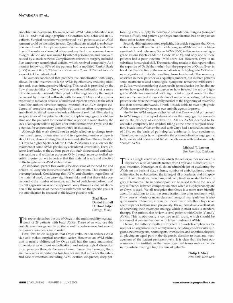
FIGURE 4. Histological examination with hematoxylin and eosin stain showing angionecrosis (arrow) in a vesselfilled with Onyx intermixed with blood and fibrin (A) and foreign body giant cells (arrow) in an Onyx cast (B). C,cut section of a catheter stuck (arrow) in the Onyx plug (unfilled spaces in these vessels represent blood/fibrin lostduring tissue processing and not recanalization). D, histological examination with Verhoeff–van Gieson stain show-ing a partly recanalized vessel filled with Onyx embedded in a fibrosed thrombus interspersed with a proliferation ofnew vessels (endothelium-lined spaces) with blood cells (arrows and inset). Original magnification, �20 (A and B),�10 (C and D), and �60 (insets in D).
A B
C D

Patient 2: Young Girl with a Large AVMBordering the Motor Area
(see video at web site)An 8-year-old girl presented with recurrent tingling sensations on the
left side of the body and weakness of the left arm and leg. She wasfound to have a Spetzler-Martin Grade V AVM measuring 7.0 � 4.5 �2.8 cm in size, involving the motor area (Fig. 8). Embolization was aimedat parts of the AVM that were more difficult to manage with surgery: theapex and the posterior part bordering the sensorimotor cortex. Onyxembolization was performed in four sessions over 3 weeks (Fig. 9). Theposterior portion of AVM bordering the rolandic fissure was embolized,but the apex could not be embolized. Resection was elected after mul-tiple discussions with the parents and the patient and other treatingphysicians because of her young age, which has a greater potential forcerebral plasticity. The AVM was excised completely 4 days after the laststage of embolization. Surgery was still quite difficult, taking approxi-mately 12 hours (Video 3). During the procedure, an intraoperativeangiogram was obtained because the surgeon could not be sure abouta persisting feeder from a branch of the anterior cerebral artery and theapical vessels supplying the AVM. She had significant left hemiparesispostoperatively. The postoperative angiogram showed a questionableremnant/neovascularization (Fig. 10A). We decided to observe this. Shereceived postoperative rehabilitation, and at follow-up examination 9months after the surgery, angiography showed complete removal ofthe AVM (Fig. 10, B and C) with regression of the questionable arteries.
Power in her left arm and leg was 4+/5. She had returned to school andto normal activities including sports (mRS score, 1).
Patient 3: Large AVM in the Basal Ganglia and Deep Frontal Lobe
(see video at web site)A 52-year-old woman presented with recurrent severe headaches.
She was found to have a Spetzler-Martin Grade IV AVM in the rightsylvian fissure, the basal ganglia, and the frontal lobe (Figs. 11 and 12).Embolization was performed with Onyx in four sessions over 4 weeks,aiming at the medial portion of the AVM in the basal ganglia (Fig. 13).Surgery was performed 2 days after the last embolization, and theAVM was removed completely (Video 4). The patient had left hemipare-sis postoperatively and had inpatient rehabilitation. At a follow-upexamination 8 months after surgery, power in all four limbs was nor-mal, and she had returned to her normal activities and work (mRSscore, 1). Angiography 8 months after surgery showed complete oblit-eration of the AVM (Fig. 14).
1220 | VOLUME 62 | NUMBER 6 | JUNE 2008 www.neurosurgery-online.com
NATARAJAN ET AL.
FIGURE 6. Patient 1. Angio-grams showing the embolizedOnyx cast of the AVM. The lentic-ulostriate feeders could not becatheterized, and the apex of theAVM could not be embolized.
FIGURE 7. Patient 1. Angiograms obtained at the time of the follow-upexamination showing complete obliteration of the AVM.
FIGURE 5. Patient 1. T2- (A) and T1-weighted (B) magnetic resonanceimaging (MRI) scans showing a Spetzler-Martin Grade IV arteriovenousmalformation (AVM) in the mesial frontal lobe bordering the ventricle.Lateral (C) and oblique (D) angiograms showing feeders predominantlyfrom the anterior cerebral branches and a recurrent lenticulostriate vessel.
A B
C D

DISCUSSION
Treatment DecisionsThe principal goal of AVM treatment is the elimination of
bleeding risk. Improvement of other symptoms such asheadache or seizures is also an additional benefit. Treatmentresults have to be considered in comparison with the natural
history of AVMs and have to take into account the results ofthe entire AVM management, including embolization andmicrosurgery or radiosurgery. In this series of patients, 53% ofthe patients presented with hemorrhage. The indications fortreatment were self-evident in most patients. However, thephilosophy for treatment of Grade IV and V AVMs is contro-versial (2, 13, 14, 19, 20). Several authors believe that the risksof treatment outweigh those of the natural history. In ourseries, we treated four Grade IV and two Grade V AVMs. In allbut one Grade V AVM, the outcome was excellent or good. Thetwo patients with a Grade V AVM were not treated, as theywere young and, in one, the AVM was located in the temporo-occipital region.
Advantages of Embolization before SurgeryThe goal of embolization in a patient who is to undergo sur-
gical resection is to make the surgery easier. This may beachieved by reduction of intraoperative time or bleeding or bythe elimination of difficult surgical areas such as perimotor/sensory and apical regions. By the gradual reduction of theAVM nidus, embolization is also thought to reduce the risk ofnormal pressure perfusion breakthrough (22, 25, 27, 33, 39, 45,56). Elimination of difficult areas of the AVM by embolizationallows the surgeon to focus on other regions. However, the idealgoal of embolizing the apical regions of the AVM was notachieved in most cases.
Advantages and Disadvantages of Onyx EmbolizationMultimodality therapy with Onyx embolization and
microsurgical excision allowed us to safely treat large AVMs.The slow-flowing characteristic of Onyx and the surgeon-interventionist collaboration allowed more specific areas of anAVM to be treated. Superselective intranidal or perinidalcatheter positions and slow, controlled injections that protectthe draining veins make the therapy safe in complex AVMs
NEUROSURGERY VOLUME 62 | NUMBER 6 | JUNE 2008 | 1221
MICROSURGICAL RESECTION OF BRAIN AVMS AFTER EMBOLIZATION WITH ONYX
FIGURE 8. Patient 2. A, MRI scan showing a 7-cm AVM in the frontallobe bordering the sensorimotor cortex. B, angiograms showing a Spetzler-Martin Grade V AVM.
FIGURE 9. Patient 2. Angiograms showing Onyx cast obliterating theposterior portion of the AVM bordering the rolandic fissure.
FIGURE 10. Patient 2. Immedi-ate postoperative angiogram (A)showing questionable remnant ofthe AVM/neovascularization. Band C, angiograms at 9 monthsshowing complete removal of theAVM.
A B
C
A B

and critical locations, even in less experienced hands. In ourseries, the bleed to embolization time was much shorter thanpreviously described. This difference may be attributable to achange in the treating physician’s philosophy or to the abilityto perform safer embolization earlier after a bleed. The seniorauthor (LNS) believes that there is no advantage during surgi-cal resection after Onyx when compared with NBCA, exceptthat it helps to localize the AVM and identify the brain-AVMinterface better. There was no difference in the handling char-acteristic of Onyx when compared with other embolic agents.A partially obliterated AVM has the same dimensions from theperspective of the surgeon, and the process of resection afterOnyx embolization is still complex.
Embolization of intracranial AVMs with ethylene vinylalcohol-containing embolic agents was first described by Takiet al. (46) and Terada et al. (48) in the early 1990s. The previousreports of treatment of AVMs with Onyx (Table 2) show that ourclinical complication and mortality rates are comparable.
The disadvantage of Onyx is the potential for angiotoxicity ofDMSO, which is dangerous and sometimes lethal. There wasevidence of angiotoxicity in the pathological analysis of ourspecimens. The potential for dangerous or lethal angiotoxicityis less if the volume of DMSO injected in milliliters is less thanhalf the weight of the patient in kilograms and the speed ofDMSO injection is kept at less than 0.1 ml/min. There arepotential complications of longer exposure to radiation due tolonger injection times. One patient in our group had transientalopecia after Onyx embolization.
1222 | VOLUME 62 | NUMBER 6 | JUNE 2008 www.neurosurgery-online.com
NATARAJAN ET AL.
FIGURE 13. Patient 3.Angiograms (A and B)and plain x-ray (C) show-ing the obliteration of themedial portion of the AVMby Onyx cast.
A B
FIGURE 14. Patient 3. Angiograms obtained at the time of follow-upshowing complete obliteration of the AVM nidus.
C
FIGURE 11. Patient 3. MRI scans showing an AVM in the right sylvianfissure and frontal lobe.
FIGURE 12. Patient 3.Angiograms showing aSpetzler-Martin Grade IVAVM with deep venousdrainage.

Complete Obliteration of the AVM Nidus with OnyxEmbolization: Is Surgical Resection Necessary?
Although some interventionists in Europe believe thatAVMs can be cured by using Onyx, this agent is not approvedfor such use in the United States. In our series, six patientshad complete obliteration of the AVM nidus after emboliza-tion by angiographic criteria. All six patients had their AVMssurgically removed, and the AVMs were found to have resid-ual filling during surgery. This implies that we are overesti-mating the efficacy of embolization and that AVMs thatappear cured on angiography may still require resection.There have been recent series (32, 51, 54) from Europe inwhich Onyx was the only modality for curative emboliza-tions. In one short-term series (54) with Onyx, two recurrencesshown on angiography were evident at the 3-month follow-up examination. Recanalization after embolization withacrylics (4, 8, 11, 28, 33, 54) has been described at the time oflong-term follow-up evaluations. Because of lack of long-termfollow-up data, no conclusions can be drawn regarding thepermanence of occlusion using Onyx. No long-term studieshave been performed with regard to toxicity or carcinogenic-ity of Onyx.
In our pathological study, there was evidence of chronic for-eign body giant cells and angionecrosis in 39 and 43% of spec-imens, respectively, suggesting that Onyx is not inert. Therewas evidence of recanalization in 18% of specimens, suggestingthat there may be angiographic evidence of recurrence if thesepatients had been followed without surgical resection. Becauseof these factors, we recommend resection of an AVM despite anapparently complete embolization.
Complications of AVM EmbolizationImmediate postembolization complications occurred in four
of 55 (7.3%) embolization sessions (four of 28 [14.3%] patients).Three patients had vascular complications with no neurologi-
cal deficit, and one patient had a permanent neurologicaldeficit. Other investigators (3, 9, 15, 16, 21, 24, 26, 31, 34, 35, 44,47, 49, 50, 53) have reported that death occurred in 0 to 3% andpermanent disability in 2 to 20% of patients undergoingembolization. Comparing our own experience with emboliza-tion using NBCA, embolization using Onyx has comparablecomplication rates but allows more controlled embolizationof the AVM, even in less experienced hands.
Complications of Multimodality Treatment of AVMsThe complication rate of multimodality treatment of surgery
followed by embolization was eight of 28 (28.6%) patients. Ofthese, five patients had new neurological deficits after surgeryand one patient had worsening of deficit caused by emboliza-tion (six of 28 [21.4%]). At the time of the 6-month follow-upexamination, one patient (3.6%) had a severe deficit and fivepatients (17.9%) had recovered completely or nearly com-pletely. The mortality rate was one in 34 (3%), and it was attrib-utable to the insult of the initial hemorrhage. In reviewing theliterature (3, 4, 7, 12, 21, 26, 31, 35, 51–53) on AVMs, we founda complication rate for a severe deficit of 5.5% and a mortalityrate of 2.3% after embolization and surgical resection.
CONCLUSION
With knowledge of the morphological characteristics ofAVMs, high occlusion rates and low complication rates are fea-sible with Onyx and comparable with those of other previousembolic agents. Superselective intranidal or perinidal catheterpositions and slow, controlled injections that protect the drain-ing veins make the therapy safe in complex AVMs and criticallocations, even in less experienced hands. Large AVMs can beadequately reduced in size for surgical treatment. We recom-mend resection of an AVM despite apparently completeembolization with Onyx.
NEUROSURGERY VOLUME 62 | NUMBER 6 | JUNE 2008 | 1223
MICROSURGICAL RESECTION OF BRAIN AVMS AFTER EMBOLIZATION WITH ONYX
TABLE 2. Previously reported series of Onyx/ethylene vinyl alcohol for embolization of arteriovenous malformationsa
Embolizing Angiographic Permanent neurological Mortality,Series (ref. no.) Patients
agent cure, no. (%) deficit, no. (%) no. (%)
Hamada et al., 2002 (12) 57 EVOH � alcohol 0 3 (5.3) 0
Simonetti et al., 2001 (40) 2 Onyx 1 (50) NA NA
Florio et al., 2003 (7) 10 Onyx 2 (20) 1 (10) NA
Song et al., 2004 (43) 3 Onyx 0 1 (33.3) 0
He et al., 2005 (17) 22 Onyx 3 (13.6) 0 0
Jahan et al., 2001 (21) 23 Onyx 0 9 (4) 0
van Rooij et al., 2007 (51) 44 Onyx 7 (16) 2 (4.6) 1 (2.3)
Weber et al., 2007 (54) 94 Onyx 19 (20); 2 recur- 9 (9) 0rences in 3 mo
Mounayer et al., 2007 (32) 53 Onyx � NBCA 26 (49) 5 (8.5) 3 (5.7)
Current study 28 Onyx 6 (21.4) 1 (3.6) 0
a EVOH, ethylene vinyl alcohol; NA, not available; NBCA, n-butylcyanoacrylate.

Multimodality treatment with microsurgery appears to be safeand feasible after Onyx. Despite treatment of large AVMs in deeplocations, the outcomes and complication rates are low and com-parable with those reported in previous series. Teamwork andcoordination between the surgeon and the interventional neuro-radiologist are important to plan strategies and to have goodoutcomes with fewer complications in these complex lesions.
REFERENCES
1. Chaloupka JC, Huddle DC, Alderman J, Fink S, Hammond R, Vinters HV: Areexamination of the angiotoxicity of superselective injection of DMSO in theswine rete embolization model. AJNR Am J Neuroradiol 20:401–410, 1999.
2. Chang SD, Marcellus ML, Marks MP, Levy RP, Do HM, Steinberg GK:Multimodality treatment of giant intracranial arteriovenous malformations.Neurosurgery 53:1–13, 2003.
3. Debrun GM, Aletich V, Ausman JI, Charbel F, Dujovny M: Embolization of thenidus of brain arteriovenous malformations with n-butyl cyanoacrylate.Neurosurgery 40:112–121, 1997.
4. DeMeritt JS, Pile-Spellman J, Mast H, Moohan N, Lu DC, Young WL, Hacein-Bey L, Mohr JP, Stein BM: Outcome analysis of preoperative embolizationwith N-butyl cyanoacrylate in cerebral arteriovenous malformations. AJNRAm J Neuroradiol 16:1801–1807, 1995.
5. Deveikis JP: Endovascular therapy of intracranial arteriovenous malforma-tions. Materials and techniques. Neuroimaging Clin N Am 8:401–424, 1998.
6. Fleetwood IG, Steinberg GK: Arteriovenous malformations. Lancet359:863–873, 2002.
7. Florio F, Lauriola W, Nardella M, Strizzi V, Vallone S, Trossello MP:Endovascular treatment of intracranial arterio-venous malformations withOnyx embolization: Preliminary experience [in English and Italian]. RadiolMed (Torino) 106:512–520, 2003.
8. Fournier D, TerBrugge KG, Willinsky R, Lasjaunias P, Montanera W:Endovascular treatment of intracerebral arteriovenous malformations:Experience in 49 cases. J Neurosurg 75:228–233, 1991.
9. Frizzel RT, Fisher WS 3rd: Cure, morbidity, and mortality associated withembolization of brain arteriovenous malformations: A review of 1246 patientsin 32 series over a 35-year period. Neurosurgery 37:1031–1040, 1995.
10. Gobin YP, Laurent A, Merienne L, Schlienger M, Aymard A, Houdart E,Casasco A, Lefkopoulos D, George B, Merland JJ: Treatment of brain arteri-ovenous malformations by embolization and radiosurgery. J Neurosurg85:19–28, 1996.
11. Gruber A, Ungersböck K, Reinprecht A, Czech T, Gross C, Bednar M, RichlingB: Evaluation of cerebral vasospasm after early surgical and endovasculartreatment of ruptured intracranial aneurysms. Neurosurgery 42:258–268,1998.
12. Hamada J, Kai Y, Morioka M, Kazekawa K, Ishimaru Y, Iwata H, Ushio Y: Amixture of ethylene vinyl alcohol copolymer and ethanol yielding a nonad-hesive liquid embolic agent to treat cerebral arteriovenous malformations:Initial clinical experience. J Neurosurg 97:881–888, 2002.
13. Han PP, Ponce FA, Spetzler RF: Intention-to-treat analysis of Spetzler-Martingrades IV and V arteriovenous malformations: Natural history and treat-ment paradigm. J Neurosurg 98:3–7, 2003.
14. Harbaugh RE, Heros RC, Hadley MN: More on ISAT. Lancet 361:783–784,2003.
15. Hartmann A, Pile-Spellman J, Stapf C, Sciacca RR, Faulstich A, Mohr JP,Schumacher HC, Mast H: Risk of endovascular treatment of brain arteriove-nous malformations. Stroke 33:1816–1820, 2002.
16. Haw CS, terBrugge K, Willinsky R, Tomlinson G: Complications of emboliza-tion of arteriovenous malformations of the brain. J Neurosurg 104:226–232,2006.
17. He HW, Jiang CH, Liu HB, Li YX, Zhang JB, Wu ZX: Endovascular treatmentof cerebral arteriovenous malformations with Onyx embolization. Chin MedJ (Engl) 118:2041–2045, 2005.
18. Henkes H, Nahser HC, Berg-Dammer E, Weber W, Lange S, Kuhne D:Endovascular therapy of brain AVMs prior to radiosurgery. Neurol Res20:479–492, 1998.
19. Hernesniemi JA, Keränen T: Microsurgical treatment of arteriovenous malfor-mations of the brain in a defined population. Surg Neurol 33:384–390, 1990.
20. Heros RC, Korosue K, Diebold PM: Surgical excision of cerebral arteriove-nous malformations: Late results. Neurosurgery 26:570–578, 1990.
21. Jahan R, Murayama Y, Gobin YP, Duckwiler GR, Vinters HV, Viñuela F:Embolization of arteriovenous malformations with Onyx: Clinicopathologicalexperience in 23 patients. Neurosurgery 48:984–997, 2001.
22. Jizong Z, Shuo W, Jingsheng L, Dali S, Yuanli Z, Yan Z: Combination of intra-operative embolisation with surgical resection for treatment of giant cerebralarteriovenous malformations. J Clin Neurosci 7 [Suppl 1]:54–59, 2000.
23. Jung HW, Yoo H, Paek SH, Choi KS: Long-term outcome and growth rate ofsubtotally resected petroclival meningiomas: Experience with 38 cases.Neurosurgery 46:567–575, 2000.
24. Kim LJ, Albuquerque FC, Spetzler RF, McDougall CG: Postembolization neu-rological deficits in cerebral arteriovenous malformations: Stratification byarteriovenous malformation grade. Neurosurgery 59:53–59, 2006.
25. Kinouchi H, Mizoi K, Takahashi A, Ezura M, Yoshimoto T: Combinedembolization and microsurgery for cerebral arteriovenous malformation.Neurol Med Chir (Tokyo) 42:372–379, 2002.
26. Liu HM, Huang YC, Wang YH: Embolization of cerebral arteriovenous mal-formations with n-butyl-2-cyanoacrylate. J Formos Med Assoc 99:906–913,2000.
27. Luessenhop AJ, Rosa L: Cerebral arteriovenous malformations. Indications forand results of surgery, and the role of intravascular techniques. J Neurosurg60:14–22, 1984.
28. Lundqvist C, Wikholm G, Svendsen P: Embolization of cerebral arteriovenousmalformations: Part II—Aspects of complications and late outcome.Neurosurgery 39:460–469, 1996.
29. Martin NA, Khanna R, Doberstein C, Bentson J: Therapeutic embolization ofarteriovenous malformations: The case for and against. Clin Neurosurg46:295-318, 2000.
30. Mattle HP, Schroth G, Seiler RW: Dilemmas in the management of patientswith arteriovenous malformations. J Neurol 247:917–928, 2000.
31. Meisel HJ, Mansmann U, Alvarez H, Rodesch G, Brock M, Lasjaunias P:Effect of partial targeted N-butyl-cyano-acrylate embolization in brain AVM.Acta Neurochir (Wien) 144:879–888, 2002.
32. Mounayer C, Hammami N, Piotin M, Spelle L, Benndorf G, Kessler I, MoretJ: Nidal embolization of brain arteriovenous malformations using Onyx in 94patients. AJNR Am J Neuroradiol 28:518–523, 2007.
33. Murayama Y, Malisch T, Guglielmi G, Mawad ME, Viñuela F, Duckwiler GR,Gobin YP, Klucznick RP, Martin NA, Frazee J: Incidence of cerebralvasospasm after endovascular treatment of acutely ruptured aneurysms:Report on 69 cases. J Neurosurg 87:830–835, 1997.
34. Nakstad PH, Nornes H: Superselective angiography, embolisation and sur-gery in treatment of arteriovenous malformations of the brain. Neuro-radiology 36:410–413, 1994.
35. n-BCA Trial Investigators: N-butyl cyanoacrylate embolization of cerebralarteriovenous malformations: Results of a prospective, randomized, multi-center trial. AJNR Am J Neuroradiol 23:748–755, 2002.
36. Ogilvy CS, Stieg PE, Awad I, Brown RD Jr, Kondziolka D, Rosenwasser R,Young WL, Hademenos G; Special Writing Group of the Stroke Council,American Stroke Association: AHA Scientific Statement: Recommendationsfor the management of intracranial arteriovenous malformations: A state-ment for healthcare professionals from a special writing group of the StrokeCouncil, American Stroke Association. Stroke 32:1458–1471, 2001.
37. Oliveira E, Sekhar LN: General Techniques of Arteriovenous MalformationSurgery. New York, Thieme, 1999.
38. Pasqualin A, Barone G, Cioffi F, Rosta L, Scienza R, Da Pian R: The relevanceof anatomic and hemodynamic factors to a classification of cerebral arteriove-nous malformations. Neurosurgery 28:370–379, 1991.
39. Pasqualin A, Scienza R, Cioffi F, Barone G, Benati A, Beltramello A, Da PianR: Treatment of cerebral arteriovenous malformations with a combination ofpreoperative embolization and surgery. Neurosurgery 29:358–368, 1991.
40. Simonetti L, Cenni P, de Santis F, Stunale C, Andreoli AF, Calbucci A,Fioravanti A, Leonardi M: Prime esperienze di embolizzazione di MAV cere-brali con Onyx [in Italian]. Riv Neuroradiol 14 [Suppl 3]:251–256, 2001.
41. Sirin S, Kondziolka D, Niranjan A, Flickinger JC, Maitz AH, Lunsford LD:Prospective staged volume radiosurgery for large arteriovenous malforma-
1224 | VOLUME 62 | NUMBER 6 | JUNE 2008 www.neurosurgery-online.com
NATARAJAN ET AL.

tions: Indications and outcomes in otherwise untreatable patients.Neurosurgery 58:17–27, 2006.
42. Söderman M, Andersson T, Karlsson B, Wallace MC, Edner G: Managementof patients with brain arteriovenous malformations. Eur J Radiol 46:195–205,2003.
43. Song DL, Leng B, Zhou LF, Gu YX, Chen XC: Onyx in treatment of large andgiant cerebral aneurysms and arteriovenous malformations. Chin Med J(Engl) 117:1869–1872, 2004.
44. Sorimachi T, Koike T, Takeuchi S, Minakawa T, Abe H, Nishimaki K, Ito Y,Tanaka R: Embolization of cerebral arteriovenous malformations achievedwith polyvinyl alcohol particles: Angiographic reappearance and complica-tions. AJNR Am J Neuroradiol 20:1323–1328, 1999.
45. Spetzler RF, Martin NA, Carter LP, Flom RA, Raudzens PA, Wilkinson E:Surgical management of large AVM’s by staged embolization and operativeexcision. J Neurosurg 67:17–28, 1987.
46. Taki W, Yonekawa Y, Iwata H, Uno A, Yamashita K, Amemiya H: A new liq-uid material for embolization of arteriovenous malformations. AJNR Am JNeuroradiol 11:163–168, 1991.
47. Taylor CL, Dutton K, Rappard G, Pride GL, Replogle R, Purdy PD, White J,Giller C, Kopitnik TA Jr, Samson DS: Complications of preoperative emboliza-tion of cerebral arteriovenous malformations. J Neurosurg 100:810–812, 2004.
48. Terada T, Nakamura Y, Nakai K, Tsuura M, Nishiguchi T, Hayashi S, Kido T,Taki W, Iwata H, Komai N: Embolization of arteriovenous malformationswith peripheral aneurysms using ethylene vinyl alcohol copolymer. Report ofthree cases. J Neurosurg 75:655–660, 1991.
49. Toda N, Ozaki T, Ohta T: Cerebrovascular sensitivity to vasoconstrictingagents induced by subarachnoid hemorrhage and vasospasm in dogs. JNeurosurg 46:296–303, 1977.
50. Valavanis A, Yasargil MG: The endovascular treatment of brain arteriovenousmalformations. Adv Tech Stand Neurosurg 24:131–214, 1998.
51. van Rooij WJ, Sluzewski M, Beute GN: Brain AVM embolization with Onyx.AJNR Am J Neuroradiol 28:172–178, 2007.
52. Vinters HV, Lundie MJ, Kaufmann JC: Long-term pathological follow-up ofcerebral arteriovenous malformations treated by embolization with bucrylate.N Engl J Med 314:477–483, 1986.
53. Wallace RC, Flom RA, Khayata MH, Dean BL, McKenzie J, Rand JC,Obuchowski NA, Zepp RC, Zabramski JM, Spetzler RF: The safety and effec-tiveness of brain arteriovenous malformation embolization using acrylic andparticles: The experiences of a single institution. Neurosurgery 37:606–618,1995.
54. Weber W, Kis B, Siekmann R, Kuehne D: Endovascular treatment of intracra-nial arteriovenous malformations with onyx: Technical aspects. AJNR Am JNeuroradiol 28:371–377, 2007.
55. Yakes WF, Krauth L, Ecklund J, Swengle R, Dreisbach JN, Seibert CE, BakerR, Miller M, VanderArk G, Fullagar T, Prenger E: Ethanol endovascular man-agement of brain arteriovenous malformations: Initial results. Neurosurgery40:1145–1154, 1997.
56. Zhao M, Charbel FT, Alperin N, Loth F, Clark ME: Improved phase-contrastflow quantification by three-dimensional vessel localization. Magn ResonImaging 18:697–706, 2000.
COMMENTS
Natarajan et al. report on a series of 28 patients treated by endovas-cular occlusion with Onyx followed by surgical excision. In the 28
patients, they had four complications related to the endovascular pro-cedure, with one patient being left with a left-sided hemiplegia. Twopatients experienced adherence of the microcatheters to the cerebralvessel, which were left in situ and identified intraoperatively andextruded at the time of the surgery.
What I am surprised about is that all patients had surgery despiteangiographic obliteration. In 21% of their series, or six patients,endovascular therapy resulted in angiographic occlusion; despite this,the patients underwent surgical resection. I submit that it is very rea-sonable to bring the patient back in 4 to 6 weeks to repeat theangiogram, and if there is shunting, then certainly surgery or radiation
can be contemplated. I see no rationale for operating on all patientsdespite angiographic cure. Clearly, if these patients were cured angio-graphically, perhaps the overall management outcome would haveeven been better than in the current series.
There is no question that Onyx is a relatively safe embolic materialand that it does not require the skill and experience as is needed for theuse of other liquid acrylic agents. It clearly has gained a role in thearmamentarium of multimodality management of brain arteriovenousmalformations (AVMs).
Robert H. RosenwasserPhiladelphia, Pennsylvania
Natarajan et al. present a consecutive series of 28 patients with cere-bral AVMs treated with preoperative Onyx embolization and sur-
gical resection. Their series includes four Grade IV AVMs and twoGrade V AVMs. Patients in this series did extremely well: there werefour transient and one permanent neurological deficit and a woundinfection. Six lesions were completely obliterated angiographically afterOnyx embolization; although intraoperatively, all lesions were found tohave residual filling.
This series contributes to the growing body of evidence supportingthe safety and efficacy of Onyx embolization for AVMs. At our institu-tion, we too have found that the slow-flow characteristics of Onyxallow our endovascular team to be more aggressive in embolizing largeAVMs, thereby making subsequent resections safer. Moreover, we agreewith the authors in underscoring the fact that angiographic obliterationwith Onyx embolization is not equivalent to a cure and that surgicalresection may still be necessary. More studies with longer follow-up arenecessary to further clarify this issue.
Brendan KilloryRobert F. SpetzlerPhoenix, Arizona
Natarajan et al. present a single center series of cerebral AVMs thatwere adjunctively embolized with Onyx before surgical resection.
Twenty-eight lesions were embolized in 55 sessions with a complica-tion rate of 7.3%. Permanent endovascular morbidity was 1.8%. Thesurgical morbidity was 3.6% at 6 months.
The strength of the article is the emphasis on a multidisciplinaryapproach, morbidities comparable with those in other studies, the sur-gical evaluation of “angiographically occult” lesions, and the patholog-ical evaluations of resected lesions. At our center, we have also adoptedOnyx as a first-line adjunctive therapy. We have also produced someangiographic “cures.” The comments of Natarajan et al. on the need toresect “angiographically occult” lesions are well timed and supportedby the presence of “red veins” during surgical evaluation. A point todiscuss is the effect of angiographic cures on those patients who pres-ent only with seizures.
Although the outcomes reported are comparable with those of otherOnyx series, it would be helpful to read a similar report from the samecenter on lesions treated with n-butylcyanoacrylate. It is important torealize that Onyx is yet another tool and not a replacement for othertreatment modalities in the adjunctive therapy of AVMs.
Babu G. WelchDuke S. SamsonDallas, Texas
Natarajan et al. report their experience with 28 patients who hadintracranial AVMs treated by preoperative embolization with
Onyx followed by microsurgical resection. A total of 71 pedicles were
NEUROSURGERY VOLUME 62 | NUMBER 6 | JUNE 2008 | 1225
MICROSURGICAL RESECTION OF BRAIN AVMS AFTER EMBOLIZATION WITH ONYX
embolized in 55 sessions. The average final AVM nidus obliteration was74.11%, and total angiographic obliteration was achieved in sixpatients. Surgical resection was performed in all patients, and an AVMremnant could be identified in each. Complications related to emboliza-tion were found in four patients, one of which was caused by emboliza-tion of the anterior choroidal artery and resulted in a permanent neu-rological deficit, one was caused by arterial perforation, and two werecaused by a stuck catheter. Complications related to surgery includedfive temporary neurological deficits, which resolved completely. At 6months follow-up, 86% of the patients had a modified Rankin Scale(mRS) score of 0 or 1, 7% had a mRS score of 2, and 3.5% had an mRSscore of 4. One patient died.
The authors concluded that preoperative embolization with Onyxallows for safe treatment of large AVMs by effectively reducing nidalsize and, thus, intraoperative bleeding. This result is provided by theflow characteristics of Onyx, which permit embolization of a moreintricate vascular network. They point out the angiotoxicity that mightbe caused by dimethyl sulfoxide with the use of Onyx and a greaterexposure to radiation because of increased injection times. On the otherhand, the authors advocate surgical resection of an AVM despite evi-dence of complete angiographic obliteration after preoperativeembolization. Stated reasons include evidence of residual filling duringsurgery in six of the patients who had complete angiographic obliter-ation and the potential for recanalization reported in some studies, thelack of adequate follow-up data after embolization with Onyx, and thepotential for angiotoxicity demonstrated in this study.
Although this work should not be solely relied on to change treat-ment paradigms, it does seem to add to a growing number of reportsabout Onyx, demonstrating that it is safe and effective. The applicationof Onyx to higher Spetzler-Martin Grade AVMs may also allow for thetreatment of some AVMs previously considered untreatable. There aresome drawbacks, as the authors point out, such as increased emboliza-tion sessions and radiation exposure. Only through more controlled sci-entific inquiry can we be certain that this material is safe and effectivein the long-term for AVM embolization.
An important part of this work is the discussion of the need for, andbenefit of, surgeon-interventionalist collaboration. This cannot beoveremphasized. Considering that AVM embolization, regardless ofthe material used, does carry significant risks and that these risks cor-respond to the number of sessions, number of pedicles embolized, andoverall aggressiveness of the approach, only through close collabora-tion of the members of the neurovascular team can the specific goals ofthe embolization be realized at the lowest possible risk.
Ziad HageDaniel SurdellH. Hunt BatjerChicago, Illinois
This report describes the use of Onyx in the multimodality manage-ment of 28 patients with brain AVMs. Those of us who use this
embolic agent are generally excited about its performance, but severalcautionary comments are in order.
First, this article suggests that Onyx embolization reduces AVMsize and makes surgical resection easier. However, an AVM nidusthat is nearly obliterated by Onyx still has the same anatomicaldimensions as without embolization, and microsurgical dissectionmust progress through the same tissue planes. Furthermore, thereare many other important factors besides size that influence the safetyand ease of resection, including AVM location, eloquence, deep per-
forating artery supply, hemorrhagic presentation, margins (compactversus diffuse), and patient age. Onyx embolization has no impact onthese other factors either.
Second, although not stated explicitly, this article suggests that Onyxembolization will enable us to tackle tougher AVMs and still achieveexcellent clinical outcomes. Seven AVMs (25%) in this series were high-grade lesions (Spetzler-Martin Grade IV or V), and only one of thesepatients had a poor outcome (mRS score <2). However, Onyx is nosubstitute for surgical skill. The outstanding results in this report reflectthe expertise of Dr. Sekhar rather than the properties of Onyx. Even inhis skilled hands, five of the seven patients with high-grade AVMs hadnew, significant deficits resulting from treatment. The recoveryobserved in these patients was equally significant, but in three patientssome treatment-related neurological symptoms remained (mRS score 1or 2). It is worth considering these results to emphasize the fact that nomatter how good the neurosurgeon or how injected the nidus, high-grade AVMs are associated with significant surgical morbidity thatmay not be counted in our calculus of outcome reporting but leavespatients who were neurologically normal at the beginning of treatmentless than normal afterwards. I think it is advisable to treat high-gradeAVMs conservatively, even as our embolic agents improve.
Finally, for AVM surgeons who feared that Onyx would put an endto AVM surgery, this report demonstrates that angiography overesti-mates the efficacy of embolization. All six AVMs deemed to beoccluded completely had residual filling at the time of surgical resec-tion. In addition, AVMs embolized with Onyx had a recanalization rateof 14%, on the basis of pathological evidence in four specimens.Therefore, no matter how impressive the postembolization angiogramslook, we should operate and finish the job, even with endovascularly“cured” AVMs.
Michael T. LawtonSan Francisco, California
This is a single center study in which the senior author reviews hisexperience with 28 patients treated with Onyx and subsequent sur-
gical resection. Natarajan et al. do an admirable job of evaluating theAVMs on the basis of size, volume, number of embolizations, percentobliteration by embolization, the timing of all procedures, and intrapro-cedural complications, blood loss, and complications related to the sur-gery at 6 months. The important points to be raised include the lack ofany difference between complication rates when n-butylcyanoacrylateor Onyx is used. We all recognize that Onyx is a more user-friendlyagent. In addition to this, the complication rate after treatment withOnyx versus n-butylcyanoacrylate and surgical manipulation is alsoquite similar. Therefore, it remains unclear as to whether Onyx is anagent superior to those used previously. The authors do an excellent jobof describing their treatment strategy, which in most cases is standardtherapy. The authors also review several patients with Grade IV and VAVMs. This is obviously a controversial topic, which should beaddressed at centers that deal with large numbers of AVMs.
Overall, the authors’ results are excellent. This article emphasizes theneed for an organized team of physicians including endovascular sur-geons, neurosurgeons, neurologists, intensivists, and anesthesiologists,all playing an equal part in the diagnosis, decision to treat, and man-agement of the patient perioperatively. It is clear that the best out-comes occur in institutions that have organized teams such as the onein this article treating a high volume of patients.
Philip E. StiegNew York, New York
1226 | VOLUME 62 | NUMBER 6 | JUNE 2008 www.neurosurgery-online.com
NATARAJAN ET AL.