

Rania H. Younis Assistant professor Oncology and Diagnostic Sciences OPATH528 SGS 1. 21. 2015.
38
Rania H. Younis Assistant professor Oncology and Diagnostic Sciences OPATH528 SGS 1. 21. 2015
-
Upload
sharon-robertson -
Category
Documents
-
view
215 -
download
0
Transcript of Rania H. Younis Assistant professor Oncology and Diagnostic Sciences OPATH528 SGS 1. 21. 2015.

Rania H. Younis
Assistant professor
Oncology and Diagnostic Sciences
OPATH528 SGS1. 21. 2015

A 45-year-old male was seen because of severe sore mouth and sore throat of 4 months’ duration. Treatment with penicillin, nystatin, and vitamin C was ineffective. The patient had “colitis” 3 years ago, which apparently was under medical control.
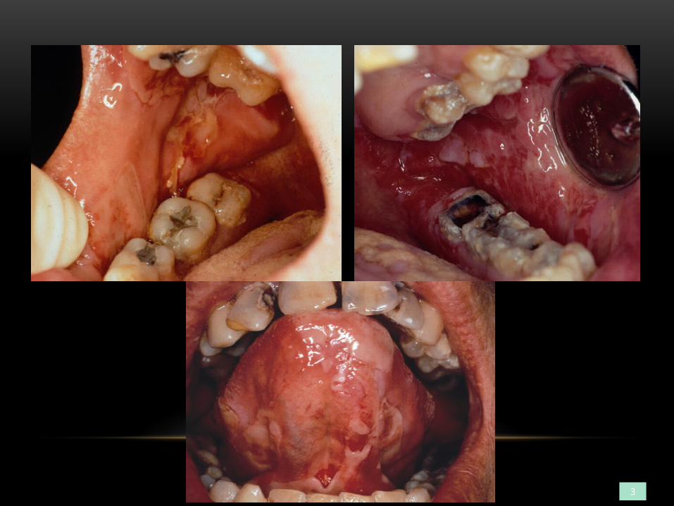
3
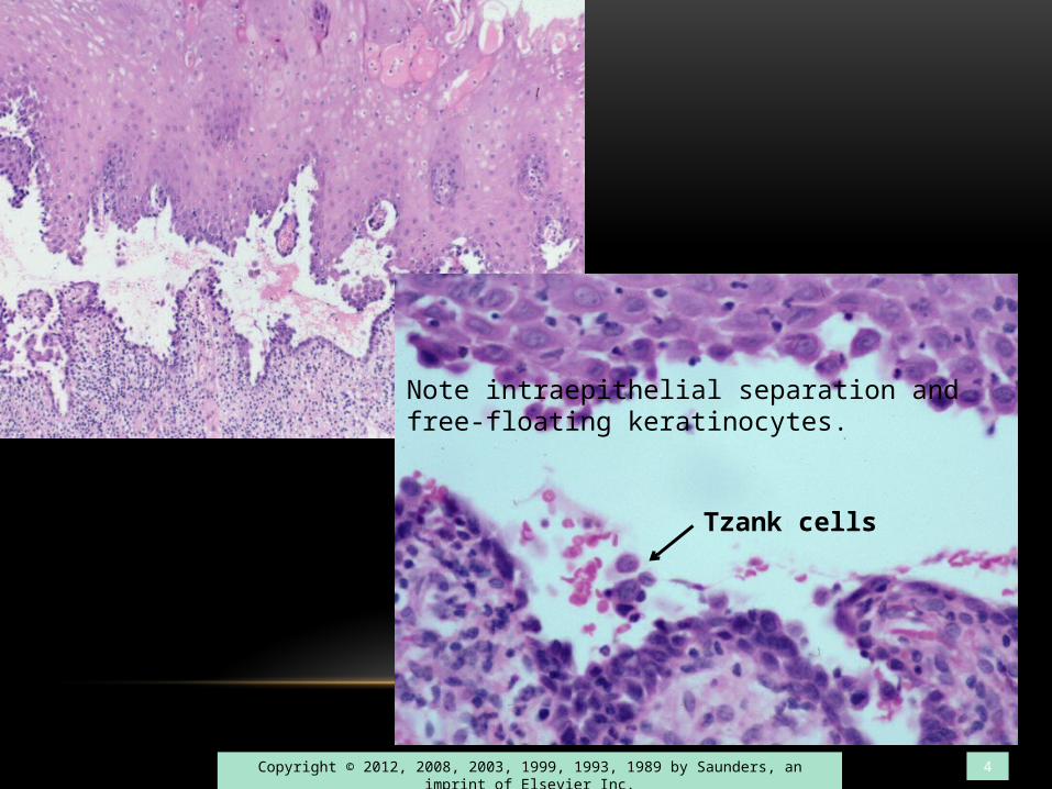
Copyright © 2012, 2008, 2003, 1999, 1993, 1989 by Saunders, an imprint of Elsevier Inc. 4
Note intraepithelial separation and free-floating keratinocytes.
Tzank cells

Pemphigus vulgaris
Diagnosis
Copyright © 2012, 2008, 2003, 1999, 1993, 1989 by Saunders, an imprint of Elsevier Inc. 5
What other pathological conditions show Tzank cells? Does it have any other systemic signs and symptoms?

Direct immunofluorescence (DIF)
The presence of microscopic fluorescence is positive confirmation.
The fluorescent pattern for pemphigus is intraepithelial, and for pemphigoid is subepithelial (the sites where autoantibody is
located for each disease).
HOW COULD THIS DIAGNOSIS BE CONFIRMED?
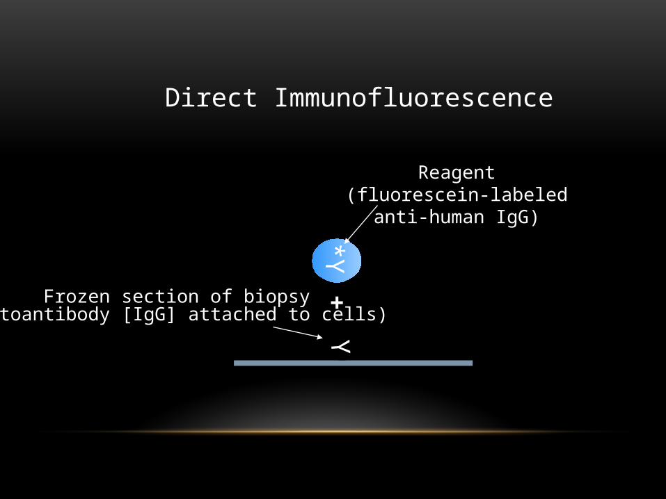
Y*Y
Direct Immunofluorescence
Frozen section of biopsy(autoantibody [IgG] attached to cells)
Reagent(fluorescein-labeled
anti-human IgG)
+
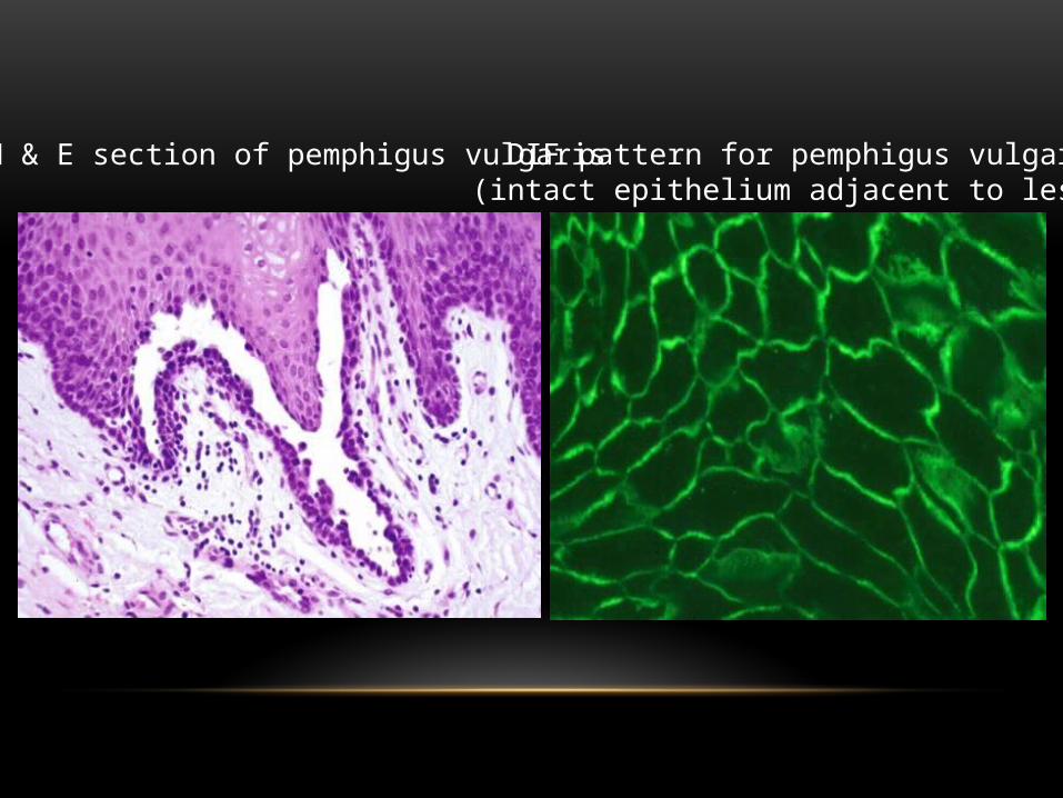
DIF pattern for pemphigus vulgaris(intact epithelium adjacent to lesion)
H & E section of pemphigus vulgaris

No.Primary disease is associated with systemic signs and symptoms and gingival lesions.Secondary disease consists of tiny ulcers in the hard palate and/or gingiva only.Also, both primary and secondary are self-limiting and are not likely to continue for a month, as noted in this patient’s history.
Could this be a primary or secondary herpes simplex infection?

A 53-year-old male developed painful gingival lesions. He had no other oral, ocular, genital, or skin lesions. He was being treated for chronic alcoholism.
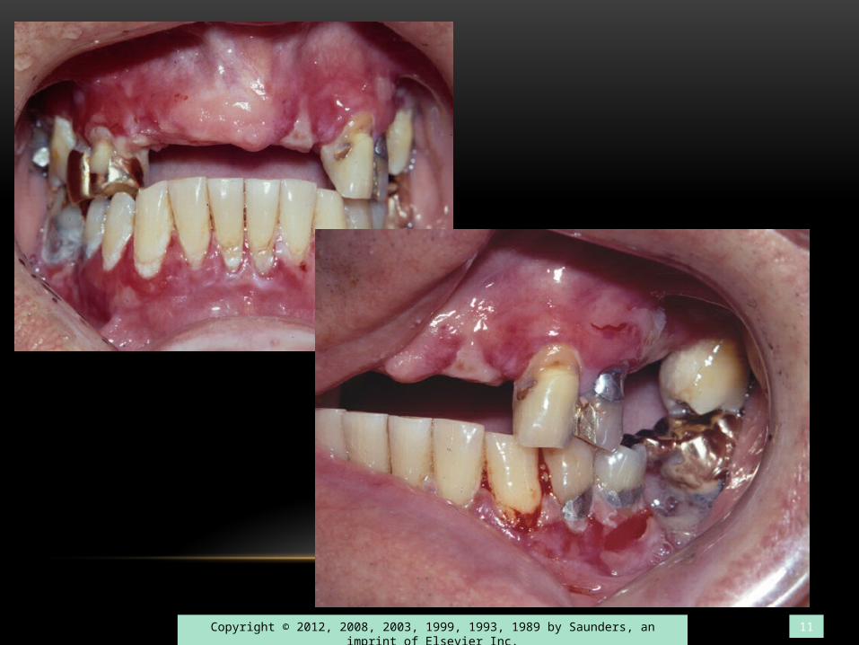
Copyright © 2012, 2008, 2003, 1999, 1993, 1989 by Saunders, an imprint of Elsevier Inc. 11

Note subepithelial separation and negligible infiltrate.
Copyright © 2012, 2008, 2003, 1999, 1993, 1989 by Saunders, an imprint of Elsevier Inc. 12
pemphigoid

H&E section of pemphigoid DIF pattern for pemphigoid

In what vesiculobullous disease are conjunctivitis, blunting of the eyelids, and symblepharon seen?
Mucous Membrane Pemphigoid
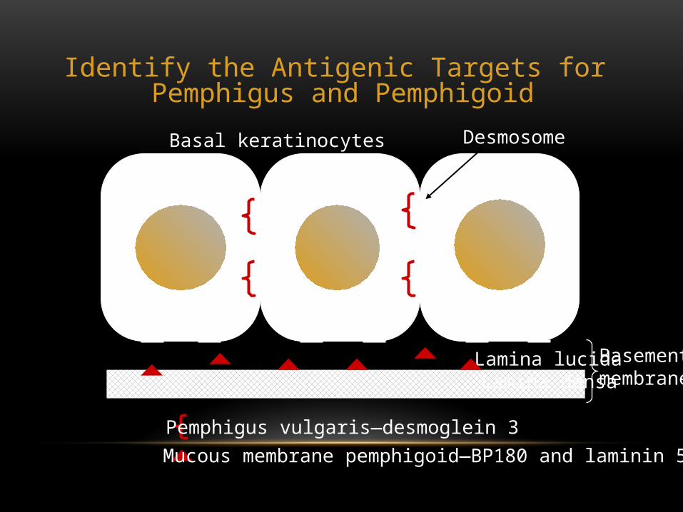
Identify the Antigenic Targets for Pemphigus and Pemphigoid
Lamina lucidaLamina densa
Basal keratinocytes
Pemphigus vulgaris—desmoglein 3Mucous membrane pemphigoid—BP180 and laminin 5
Desmosome
Basementmembrane

Acantholysis resulting from desmosome weakening by autoantibodies is the disease mechanism attributed to which of the following?
A. Epidermolysis bullosaB. Mucous membrane pemphigoidC. Lichen planusD. Herpes simplex infectionE. None of the above
Answer: E(This occurs in pemphigus vulgaris.)

This 24-year-old female presents with headache, fatigue, and elevated temperature. What does she most likely have?
Primary herpes simplex infection
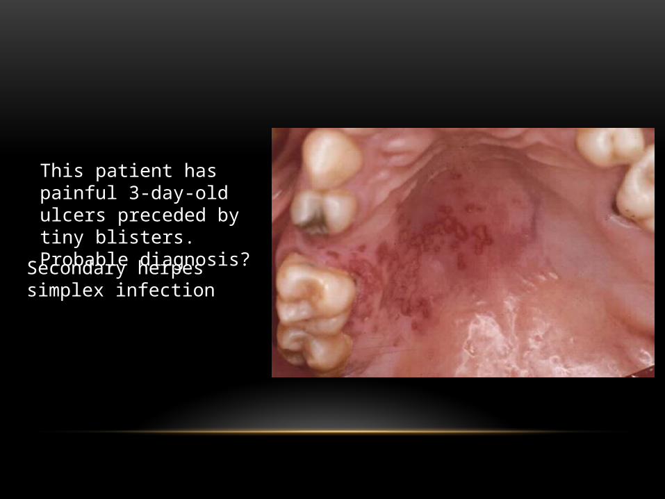
This patient has painful 3-day-old ulcers preceded by tiny blisters. Probable diagnosis?
Secondary herpes simplex infection

A 22-year-old male has forehead ulceration, right eye conjunctivitis, ulceration of right nasal tip, and ulcer of mucosa of right lower lip of 2 days’ duration. He is otherwise well. Probable diagnosis?
Herpes zoster

An otherwise normal 24-year-old female developed multiple painful vesicles of the vermilion of her lower lip. This has happened before in the same site following exposure to sunlight. She has no eye, genital, or cutaneous lesions. She most likely has which of the following?
A. Primary herpes simplexB. Secondary herpes simplex
C. Herpetiform aphthous ulcers D. Mucous membrane pemphigoid
Answer: B

• During routine examination of a 34-year-old male, a lesion was discovered in the floor of his mouth. He was unaware of the lesion. He did not smoke and was a “light drinker.” He had no history of an associated traumatic event or habit that might result in irritation to the area

Note thickened keratin layer and atypical basilar cells.
22
Dx:Hyperkeratosis with dysplasia

A 31-year-old dental hygiene student developed a sore throat during final exams. Other than being under stress, she felt well. She had no systemic problems and was taking no medications.
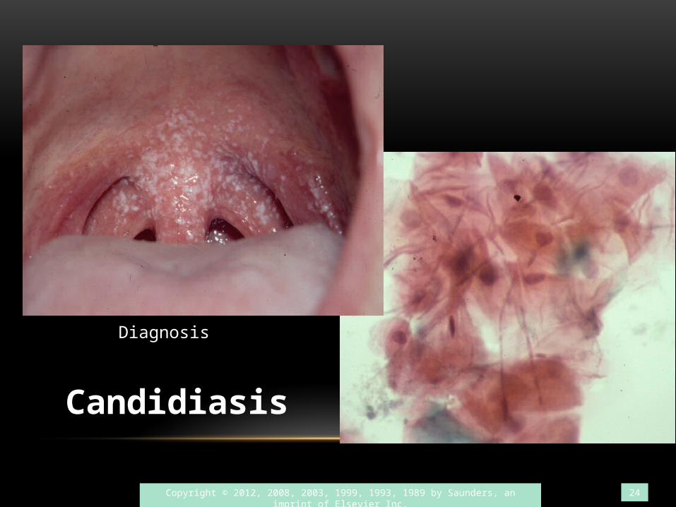
Copyright © 2012, 2008, 2003, 1999, 1993, 1989 by Saunders, an imprint of Elsevier Inc. 24
Candidiasis
Diagnosis

A 40-year-old patient went to his dentist to seek relief from constant oral pain (2 months’ duration). He had been unable to eat and subsequently lost 10 pounds. He had no skin, eye, or genital lesions.

Copyright © 2012, 2008, 2003, 1999, 1993, 1989 by Saunders, an imprint of Elsevier Inc. 26
MAJOR APHTHOUS ULCERS

Note that ulcers are not found on the attached gingivae or hard palate.
Copyright © 2012, 2008, 2003, 1999, 1993, 1989 by Saunders, an imprint of Elsevier Inc. 27
Aphthus Ulcer

Copyright © 2012, 2008, 2003, 1999, 1993, 1989 by Saunders, an imprint of Elsevier Inc. 28
Aphthus Ulcer

On routine exam, a lesion was found in the floor of the mouth of a 55-year-old female. She had no symptoms and was in good health. She did not smoke or drink alcohol. She took no medications.

Copyright © 2012, 2008, 2003, 1999, 1993, 1989 by Saunders, an imprint of Elsevier Inc. 30
Note atypical keratinocytes throughout the epithelium.
Carcinoma in situ

This is the patient’s only oral lesion. Describe this lesion.

Other examples of squamous cell carcinoma of the floor of mouth

Diagnosis for this nonhealing chronic ulcer?
Squamous cell carcinoma:Note hyperchromatic nuclei in the invading tumor nests, and keratin pearls in the submucosa.
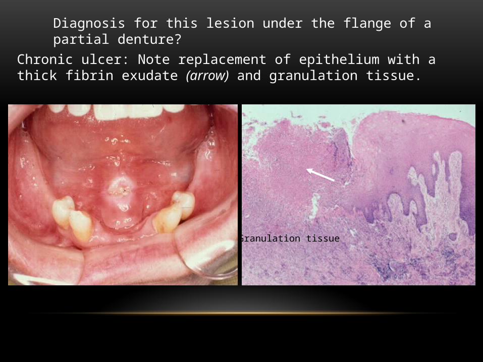
Diagnosis for this lesion under the flange of a partial denture?
Chronic ulcer: Note replacement of epithelium with a thick fibrin exudate (arrow) and granulation tissue.
Granulation tissue

Probable diagnosis for this recurrent painful acute ulcer?
Major aphthous ulcerThe lesion is >0.5 cm in diameter, it is acute and crateriform, it is on nonkeratinized mucosa, and it is recurrent; all support the diagnosis of major aphthous ulcer.

Hand foot and mouth Disease
Herpangina
These patients have fever and malaise

Varicella (pustular)
Measles ( has Koplik’s spots intraorally)
Fever followed by exanthematous rash
What is your differential?