RADIUS AND ULNA 1 right - catagni.it CHAPTER 2 Levels of the anatomical cuts of the upper extremity...
10
11 CHAPTER 2 Levels of the anatomical cuts of the upper extremity RADIUS AND ULNA right CUT 6 CUT 5 CUT 4 CUT 3 CUT 2 CUT 1 Head of Radius Radial Styloid Isolated fixation of the radius is difficult at this level because of the anterolateral vessels and the medial ulna. It can be done with a half pin inserted from postero medial to antero lateral. Fixation of the two bones is done with a wire from antero lateral to postero medial. Isolated ulnar fixation is much simpler and can be done with one transverse wire and a second wire from antero medial to postero lateral, pos- terior to the ulnar nerve. Fixation with half pins can be done posteriorly at an angle of 20° to the sagittal plane. Isolated ulnar fixation can be done with a transverse wire (parallel to the coronal plane) and a half pin from posterior to anterior. Isolated radial fixation can be performed with a wire from anterior to posterior and a half pin from postero- lateral to antero medial, angulated 20° to the sagittal plane. Isolated radial fixation can be carried out with a wire direct- ed from anterior to posterior. A half pin can be inserted from posterolateral to anteromedial at an angle approximating 20 degrees to the coronal plane. Fixation of the ulna can be per- formed with a wire from anteromedial to posterolateral, angulated 20° to the coronal plane, and a half pin from pos- terior to anterior. Isolated radial fixation can be carried out with a wire direct- ed from anterolateral to posteromedial, angulated 30° to the sagittal plane. A half pin can be fixed in a posterolateral position, perpendicular to the previous wire. Fixation of the ulna can be performed with a wire from anteromedial to pos- terolateral, angulated 20° to the coronal plane, and a half pin from posteromedial to anterolateral, angulated 10° to the sagittal plane. Isolated radial fixation can be carried out with a wire direct- ed from anterolateral to posteromedial, angulated 40° to the coronal plane. A half pin can be inserted from a posterolat- eral position, perpendicular to the previous wire. Ulna fixa- tion is performed with a wire from anteromedial to postero- lateral angulated 40° to the coronal plane and a half pin from posteromedial to anterolateral, angulated 15° to the sagittal plane. Ulnar fixation is performed with a wire directed from antero- medial to posterolateral angulated 45° to the sagittal plane and a half pin directed from posteromedial to anterolateral, perpendicular to the previous wire. The radius can be fixed with one wire directed from anterolateral to posteromedial angulated 45° with the coronal plane and a second wire inserted from anterior to posterior, between the flexor carpi radialis and the median nerve, using the open technique. A half pin is inserted from posterolateral to anteromedial, per- pendicular to the first wire. 6 5 4 3 2 1
Transcript of RADIUS AND ULNA 1 right - catagni.it CHAPTER 2 Levels of the anatomical cuts of the upper extremity...

11 CHAPTER 2
Levels of the anatomical cutsof the upper extremity
RADIUS AND ULNAright
CUT 6
CUT 5
CUT 4
CUT 3
CUT 2
CUT 1Head ofRadius
RadialStyloid
Isolated fixation of the radius is difficult at this levelbecause of the anterolateral vessels and the medial ulna. Itcan be done with a half pin inserted from postero medial toantero lateral. Fixation of the two bones is done with a wirefrom antero lateral to postero medial. Isolated ulnar fixationis much simpler and can be done with one transverse wireand a second wire from antero medial to postero lateral, pos-terior to the ulnar nerve. Fixation with half pins can be doneposteriorly at an angle of 20° to the sagittal plane.
Isolated ulnar fixation can be done with a transverse wire(parallel to the coronal plane) and a half pin from posteriorto anterior. Isolated radial fixation can be performed with awire from anterior to posterior and a half pin from postero-lateral to antero medial, angulated 20° to the sagittal plane.
Isolated radial fixation can be carried out with a wire direct-ed from anterior to posterior. A half pin can be inserted fromposterolateral to anteromedial at an angle approximating 20degrees to the coronal plane. Fixation of the ulna can be per-formed with a wire from anteromedial to posterolateral,angulated 20° to the coronal plane, and a half pin from pos-terior to anterior.
Isolated radial fixation can be carried out with a wire direct-ed from anterolateral to posteromedial, angulated 30° to thesagittal plane. A half pin can be fixed in a posterolateralposition, perpendicular to the previous wire. Fixation of theulna can be performed with a wire from anteromedial to pos-terolateral, angulated 20° to the coronal plane, and a half pinfrom posteromedial to anterolateral, angulated 10° to thesagittal plane.
Isolated radial fixation can be carried out with a wire direct-ed from anterolateral to posteromedial, angulated 40° to thecoronal plane. A half pin can be inserted from a posterolat-eral position, perpendicular to the previous wire. Ulna fixa-tion is performed with a wire from anteromedial to postero-lateral angulated 40° to the coronal plane and a half pin fromposteromedial to anterolateral, angulated 15° to the sagittalplane.
Ulnar fixation is performed with a wire directed from antero-medial to posterolateral angulated 45° to the sagittal planeand a half pin directed from posteromedial to anterolateral,perpendicular to the previous wire. The radius can be fixedwith one wire directed from anterolateral to posteromedialangulated 45° with the coronal plane and a second wireinserted from anterior to posterior, between the flexor carpiradialis and the median nerve, using the open technique. Ahalf pin is inserted from posterolateral to anteromedial, per-pendicular to the first wire.
6
5
4
3
2
1

12LEVELS OF THE ANATOMICAL CUTS OF THE UPPER EXTREMITY - RADIUS and ULNA
CUT 1 CUT 4
CUT 5
CUT 6
CUT 2
CUT 3
INSERTION WIRES AND HALF-PINS - RIGHTANTERIOR
ANTERIOR
ANTERIOR
ANTERIOR
ANTERIOR
ANTERIOR

13 CHAPTER 2
Head ofRadius
RadialStyloid
RADIUS AND ULNAleft
Levels of the anatomical cutsof the upper extremity
CUT 6
CUT 5
CUT 4
CUT 3
CUT 2
CUT 1
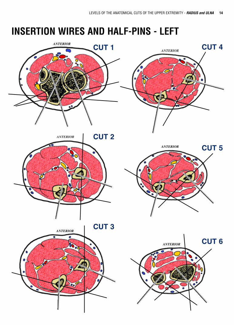
Isolated fixation of the radius is difficult at this levelbecause of the anterolateral vessels and the medial ulna. Itcan be done with a half pin inserted from postero medial toantero lateral. Fixation of the two bones is done with a wirefrom antero lateral to postero medial. Isolated ulnar fixationis much simpler and can be done with one transverse wireand a second wire from antero medial to postero lateral, pos-terior to the ulnar nerve. Fixation with half pins can be doneposteriorly at an angle of 20° to the sagittal plane.
Isolated ulnar fixation can be done with a transverse wire(parallel to the coronal plane) and a half pin from posteriorto anterior. Isolated radial fixation can be performed with awire from anterior to posterior and a half pin from postero-lateral to antero medial, angulated 20° to the sagittal plane.
Isolated radial fixation can be carried out with a wire direct-ed from anterior to posterior. A half pin can be inserted fromposterolateral to anteromedial at an angle approximating 20degrees to the coronal plane. Fixation of the ulna can be per-formed with a wire from anteromedial to posterolateral,angulated 20° to the coronal plane, and a half pin from pos-terior to anterior.
Isolated radial fixation can be carried out with a wire direct-ed from anterolateral to posteromedial, angulated 30° to thesagittal plane. A half pin can be fixed in a posterolateralposition, perpendicular to the previous wire. Fixation of theulna can be performed with a wire from anteromedial to pos-terolateral, angulated 20° to the coronal plane, and a half pinfrom posteromedial to anterolateral, angulated 10° to thesagittal plane.
Isolated radial fixation can be carried out with a wire direct-ed from anterolateral to posteromedial, angulated 40° to thecoronal plane. A half pin can be inserted from a posterolat-eral position, perpendicular to the previous wire. Ulna fixa-tion is performed with a wire from anteromedial to postero-lateral angulated 40° to the coronal plane and a half pin fromposteromedial to anterolateral, angulated 15° to the sagittalplane.
Ulnar fixation is performed with a wire directed from antero-medial to posterolateral angulated 45° to the sagittal planeand a half pin directed from posteromedial to anterolateral,perpendicular to the previous wire. The radius can be fixedwith one wire directed from anterolateral to posteromedialangulated 45° with the coronal plane and a second wireinserted from anterior to posterior, between the flexor carpiradialis and the median nerve, using the open technique. Ahalf pin is inserted from posterolateral to anteromedial, per-pendicular to the first wire.
6
5
4
3
2
1
ANTERIOR
ANTERIOR
14LEVELS OF THE ANATOMICAL CUTS OF THE UPPER EXTREMITY - RADIUS and ULNA
CUT 1
CUT 2
CUT 4
CUT 5
CUT 6
CUT 3
INSERTION WIRES AND HALF-PINS - LEFTANTERIOR
ANTERIOR
ANTERIOR
ANTERIOR

15 CHAPTER 2
CUT 1RADIUS and ULNA left
Head ofRadius
RadialStyloid
CUT 1ANTERIOR
A.V. Brachial
N. Radial
M. Extensor Carpi Radialis (Longus
and Brevis)
N. Median
RADIUS
M. Anconeus
M. Brachialis
M. Pronator Teres
N. Ulnar
ULNA
HUMERUS(TROCHLEA)
M. Flexor Carpi Ulnaris
M. Brachio Radialis
Common Extensor Tendon
Biceps Tendon
In this cross section the ulnar N. runs medial to the ulna at thepoint of confluence of the flexor carpi ulnaris, the flexor digi-
Isolated fixation of the radius is difficult at this level because of the anterolateral vessels and the medial ulna. It can be done witha half pin inserted from postero medial to antero lateral. Fixation of the two bones is done with a wire from antero lateral to pos-tero medial. Isolated ulnar fixation is much simpler and can be done with one transverse wire and a second wire from antero medi-al to postero lateral, posterior to the ulnar nerve. Fixation with half pins can be done posteriorly at an angle of 20° to the sagittalplane.
torum superficialis and the pronator teres. The brachial A. hasmoved laterally and now runs along side the median N.
ANTERIOR
16LEVELS OF THE ANATOMICAL CUTS OF THE UPPER EXTREMITY - RADIUS and ULNA
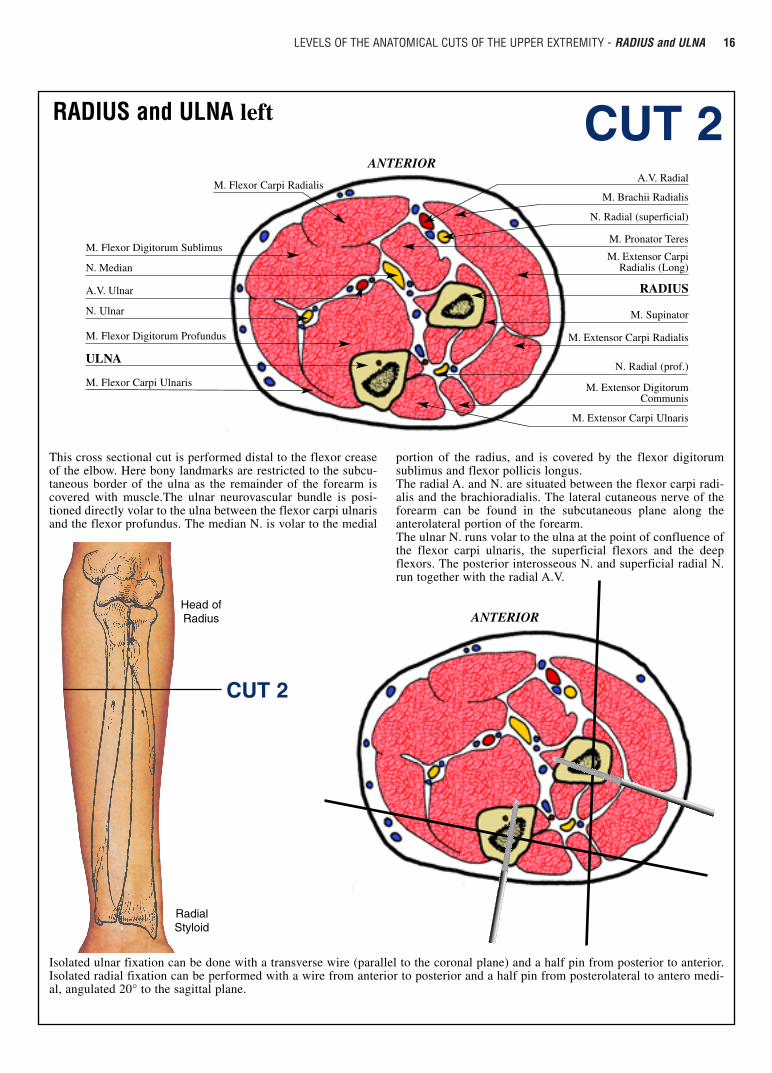
CUT 2RADIUS and ULNA left
ANTERIORHead ofRadius
RadialStyloid
CUT 2
A.V. Radial
M. Brachii Radialis
M. Extensor Carpi Radialis (Long)
N. Radial (superficial)
RADIUS
M. Extensor Carpi Radialis
M. Flexor Carpi Radialis
M. Flexor Digitorum Sublimus
N. Median
N. Ulnar
A.V. Ulnar
ULNA
M. Flexor Digitorum Profundus
M. Flexor Carpi Ulnaris
N. Radial (prof.)
M. Pronator Teres
M. Supinator
M. Extensor DigitorumCommunis
M. Extensor Carpi Ulnaris
This cross sectional cut is performed distal to the flexor creaseof the elbow. Here bony landmarks are restricted to the subcu-taneous border of the ulna as the remainder of the forearm iscovered with muscle.The ulnar neurovascular bundle is posi-tioned directly volar to the ulna between the flexor carpi ulnarisand the flexor profundus. The median N. is volar to the medial
Isolated ulnar fixation can be done with a transverse wire (parallel to the coronal plane) and a half pin from posterior to anterior.Isolated radial fixation can be performed with a wire from anterior to posterior and a half pin from posterolateral to antero medi-al, angulated 20° to the sagittal plane.
portion of the radius, and is covered by the flexor digitorumsublimus and flexor pollicis longus.The radial A. and N. are situated between the flexor carpi radi-alis and the brachioradialis. The lateral cutaneous nerve of theforearm can be found in the subcutaneous plane along theanterolateral portion of the forearm.The ulnar N. runs volar to the ulna at the point of confluence ofthe flexor carpi ulnaris, the superficial flexors and the deepflexors. The posterior interosseous N. and superficial radial N.run together with the radial A.V.
ANTERIOR

17 CHAPTER 2
CUT 3
ANTERIOR
Head ofRadius
RadialStyloid
CUT 3
A.V. Radial
M. Brachii Radialis
M. Extensor Carpi Radialis
N. Radial (superf.)
N. Median
M. Extensor Carpi Radialis
M. Flexor Carpi Radialis
M. Flexor Digitorum (superf.)
N. Ulnar
A.V. Ulnar
ULNA
M. Flexor Carpi Ulnaris
M. Flexor Digitorum (prof.)
M. Flex. Carpi Ulnaris
RADIUS
N. Radial
M. Extensor Com. Digitorum
M. Flex. Digiti Min.
M. Extensor Carpi Ulnaris
M. Pronator Teres
RADIUS and ULNA left
In this cross section each of the three major neurovascular ele-ments have assumed a deep position protected by the overlyingmuscles. The superficial radial N. and radial A. are volar andlateral beneath the brachioradialis. The median N. is volar and
Isolated radial fixation can be carried out with a wire directed from anterior to posterior. A half pin can be inserted from postero-lateral to anteromedial at an angle approximating 20 degrees to the coronal plane. Fixation of the ulna can be performed with awire from anteromedial to posterolateral, angulated 20° to the coronal plane, and a half pin from posterior to anterior.
central between the superficial and deep flexors of the fingers.The ulnar A.V. and N. remain covered by the flexor carpiulnaris. The anterior interosseous artery has maintained itsposition on the volar surface of the interosseous membrane.
ANTERIOR
18LEVELS OF THE ANATOMICAL CUTS OF THE UPPER EXTREMITY - RADIUS and ULNA
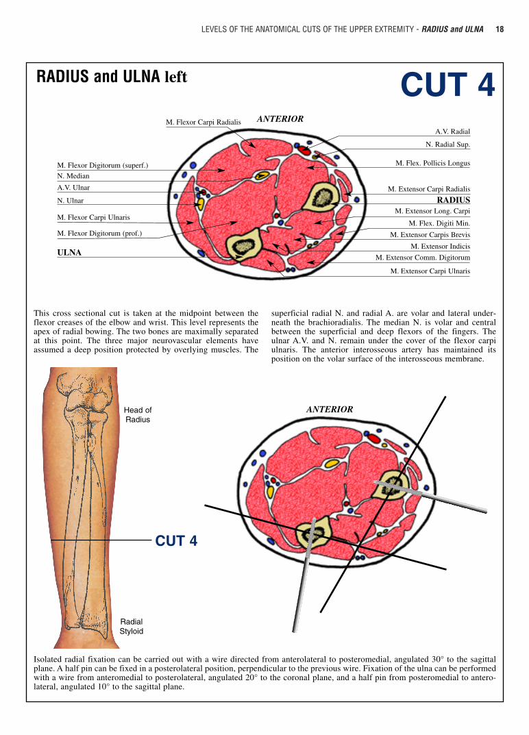
CUT 4ANTERIOR
Head ofRadius
RadialStyloid
CUT 4
A.V. Radial
M. Flex. Pollicis Longus
M. Extensor Carpi Radialis
M. Extensor Long. Carpi
M. Flexor Carpi Radialis
M. Flexor Digitorum (superf.)
N. Ulnar
A.V. Ulnar
N. Median
ULNA
M. Flexor Carpi Ulnaris
M. Flexor Digitorum (prof.)
RADIUS
M. Flex. Digiti Min.
M. Extensor Carpis Brevis
M. Extensor Comm. Digitorum
M. Extensor Carpi Ulnaris
M. Extensor Indicis
N. Radial Sup.
RADIUS and ULNA left
This cross sectional cut is taken at the midpoint between theflexor creases of the elbow and wrist. This level represents theapex of radial bowing. The two bones are maximally separatedat this point. The three major neurovascular elements haveassumed a deep position protected by overlying muscles. The
Isolated radial fixation can be carried out with a wire directed from anterolateral to posteromedial, angulated 30° to the sagittalplane. A half pin can be fixed in a posterolateral position, perpendicular to the previous wire. Fixation of the ulna can be performedwith a wire from anteromedial to posterolateral, angulated 20° to the coronal plane, and a half pin from posteromedial to antero-lateral, angulated 10° to the sagittal plane.
superficial radial N. and radial A. are volar and lateral under-neath the brachioradialis. The median N. is volar and centralbetween the superficial and deep flexors of the fingers. Theulnar A.V. and N. remain under the cover of the flexor carpiulnaris. The anterior interosseous artery has maintained itsposition on the volar surface of the interosseous membrane.
ANTERIOR

19 CHAPTER 2
CUT 5
Head ofRadius
RadialStyloid
CUT 5
ANTERIOR
A.V. Radial
M. Flexor Carpi Radialis
M. Extensor Carpi RadialisLongus and Brevis
N. Median
M. Flexor Digitorum sublimus
M. Pronator Quadratus
A.V. Ulnar
M. Flexor Carpi Ulnaris
N. Ulnar
M. Flexor Digitorum profundus M. Extensor Carpi Brevis
M. Abductor Pollicis
M. Extensor Comm. Digitorum
M. Extensor Digiti Minimi
M. Extensor Indici
RADIUS
N. Radial sup.
ULNA
RADIUS and ULNA left
In this cross sectional cut the superficial radial N. lies subcuta-neously over the abductor pollicis longus. The radial A. liessuperficially and can be found slightly lateral to the flexor carpiradialis. The median N. is on the medial side of the radial wrist flexor,
Isolated radial fixation can be carried out with a wire directed from anterolateral to posteromedial, angulated 40° to the coronalplane. A half pin can be inserted from a posterolateral position, perpendicular to the previous wire. Ulna fixation is performed witha wire from anteromedial to posterolateral angulated 40° to the coronal plane and a half pin from posteromedial to anterolateral,angulated 15° to the sagittal plane.
between it and the flexor digitorum sublimus. The ulnar A.V.and N. are volar and medial under the increasing mass of theflexor carpi ulnaris. The terminal branches of the anteriorinterosseous N. are deeply located on the ulnar border of theradius.
ANTERIOR

20LEVELS OF THE ANATOMICAL CUTS OF THE UPPER EXTREMITY - RADIUS and ULNA
CUT 6
Head ofRadius
RadialStyloid
CUT 6
ANTERIOR
N. Median
M. Flexor Pollicis Longus
M. Flexor Digitorum Superficialis
M. Flexor Carpi Ulnaris
N. Ulnar
A.V. Ulnar
M. Pronator Quadratus
M. Pronator Quadratus
M. Extensor Digiti Minimi
M. Extensor Carpi RadialisLongus and Brevis
M. Extensor Digitorum Communis
M. Longus Extensor Carpi
M. Extensor Indicis
N. Radial Sup.
RADIUS
A.V. Radial
ULNA
M. Flexor Digitorum Profundus
RADIUS and ULNA left
In this cross sectional cut most of the arteries and tendons canbe accurately localized by palpation as they lie superficially.The ulnar A.V. lie volar and medial and are protected by theflexor carpi ulnaris T. The median N. is slightly more radial in
Ulnar fixation is performed with a wire directed from anteromedial to posterolateral angulated 45° to the sagittal plane and a halfpin directed from posteromedial to anterolateral, perpendicular to the previous wire. The radius can be fixed with one wire direct-ed from anterolateral to posteromedial angulated 45° with the coronal plane and a second wire inserted from anterior to posterior,between the flexor carpi radialis and the median nerve, using the open technique. A half pin is inserted from posterolateral toanteromedial, perpendicular to the first wire.
position, being situated between the flexor digitorum superfi-cialis and the flexor carpi radialis. As in 30% of the normal pop-ulation, this diagram shows no palmaris longus T. The radial A.is found between the flexor carpi radialis and the abductor pol-licis longus.
ANTERIOR