Radiobiological Effects of Alpha-Particles from Astatine · PDF fileRadiobiological Effects of...
61
Radiobiological Effects of Alpha-Particles from Astatine-211 From DNA Damage to Cell Death Kristina Claesson Department of Oncology Institute of Clinical Sciences The Sahlgrenska Academy at University of Gothenburg
Transcript of Radiobiological Effects of Alpha-Particles from Astatine · PDF fileRadiobiological Effects of...

Radiobiological Effects of Alpha-Particles from
Astatine-211
From DNA Damage to Cell Death
Kristina Claesson
Department of Oncology
Institute of Clinical Sciences
The Sahlgrenska Academy at University of Gothenburg

ABSTRACT
In recent years, the use of high linear energy transfer (LET) radiation for radiotherapeutic
applications has gained increased interest. Astatine-211 (211At) is an α-particle emitting
radionuclide, promising for targeted radioimmunotherapy of isolated tumor cells and
microscopic clusters. To improve development of safe radiotherapy using 211At it is important
to increase our knowledge of the radiobiological effects in cells. During radiotherapy, both
tumors and adjacent normal tissue will be irradiated and therefore, it is of importance to
understand differences in the radioresponse between proliferating and resting cells. The aim
of this thesis was to investigate effects in fibroblasts with different proliferation status after
irradiation with α-particles from 211At or X-rays, from inflicted DNA damage, to cellular
responses and biological consequences.
Throughout this work, irradiation was performed with α-particles from 211A or X-rays. The
induction and repair of double-strand breaks (DSBs) in human normal fibroblasts were
investigated using pulsed-field gel electrophoresis and fragment analysis. The relative
biological effectiveness (RBE) of 211At for DSB induction varied between 1.4 and 3.1. A
small increase of DSBs was observed in cycling cells compared to stationary cells. The repair
kinetics was slower after 211At and more residual damage was found after 24 h. Comparison
between cells with different proliferation status showed that the repair was inefficient in
cycling cells with more residual damage, regardless of radiation quality. Activation of cell
cycle arrests was investigated using immunofluorescent labeling of the checkpoint kinase
Chk2 and by measuring cell cycle distributions with flow cytometry analysis. After α-particle
irradiation, the average number of Chk2-foci was larger and the cells had a more affected cell
cycle progression for several weeks compared with X-irradiated cells, indicating a more
powerful arrest after 211At. Flow cytometry showed that cycling cells were arrested in G2/M
while stationary cells underwent a delayed entry into S phase after release of contact
inhibition. Radiation-induced chromosomal damage was studied by investigating the
formation of micronuclei after first mitosis post-irradiation. Alpha-particles induced 2.7 and
4.1 times more micronuclei in cycling and stationary cells, respectively, compared with X-
rays.
Induction of DSBs and cell survival after irradiation were also investigated in synchronized
Chinese hamster fibroblasts. The cells were synchronized with mimosine in G1, early, mid
and late S phase and in mitosis and cell survival was determined using the clonogenic assay.
The radioresponse between cell cycle phases varied after both 211At and X-rays, resulting in
variations of RBE for 211At between 1.8 and 3.9 for DSB induction and between 3.1 and 7.9
for 37% survival. The lowest RBE was observed in mitotic cells for both DSB induction and
clonogenic survival.
In summary, for all endpoints studied α-particles from 211At were more detrimental compared
with X-rays. Further, the radioresponse was dependent upon the proliferation status of the
cells at the time of irradiation, after both low- and high-LET radiation, resulting in variations
of the relative biological effects.

LIST OF PAPERS
This thesis is based on the following papers, which will be referred to in the text by their
roman numerals:
I Claesson K, Stenerlöw B, Jacobsson L and Elmroth K. Relative Biological
Effectiveness of the α-Particle Emitter 211At for Double-Strand Break Induction
in Human Fibroblasts. Radiation Research 2007; 167, 312-318.
II Claesson K, Magnander K, Kahu K, Lindegren S, Hultborn R and Elmroth K.
RBE of α-Particles from 211At for Complex DNA Damage and Cell Survival in
Relation to Cell Cycle Position. International Journal of Radiation Biology,
2011; 87, 372-384.
III Claesson K, Nordén Lyckesvärd M, Magnander K, Lindegren S and Elmroth K.
Double-Strand Break Repair and Cell Cycle Arrest Activation in Stationary and
Cycling Diploid Cells Irradiated with High- and Low-LET Radiation.
Manuscript.
IV Claesson K, Nordén Lyckesvärd M, Magnander K, Delle U and Elmroth K.
Effects on Micronuclei Formation and Growth Kinetics in Normal Fibroblasts
after Irradiation with Alpha Particles and X rays: Differential Response in
Stationary and Cycling Cell Cultures. Manuscript.

- 6 -
TABLE OF CONTENTS
TABLE OF CONTENTS...................................................................................................... - 6 -
ABBREVIATIONS............................................................................................................... - 8 -
INTRODUCTION................................................................................................................. - 9 -
Low- and High-Linear Energy Transfer Radiation ......................................................... - 10 -
Relative Biological Effectiveness ................................................................................... - 10 -
Complex DNA Damage .................................................................................................. - 11 -
Double-Strand Breaks ................................................................................................. - 11 -
Clustered Damage....................................................................................................... - 11 -
DNA Damage Response.................................................................................................. - 12 -
Double-Strand Break Repair....................................................................................... - 12 -
Checkpoint Control ..................................................................................................... - 13 -
Radiation Response in Proliferating and Resting Cells.............................................. - 14 -
Influence of Chromatin Structure.................................................................................... - 15 -
Use of High-LET Radiation ............................................................................................ - 16 -
Present Investigation ....................................................................................................... - 17 -
AIMS................................................................................................................................... - 18 -
MATERIALS AND METHODS........................................................................................ - 19 -
Cell Lines and Culture Conditions .................................................................................. - 19 -
Synchronisation of Cells ................................................................................................. - 19 -
Irradiation ........................................................................................................................ - 20 -
Low-LET Irradiation ................................................................................................... - 20 -
High-LET Irradiation.................................................................................................. - 21 -
Fpg Enzyme Treatment to Assess Clustered Damages ................................................... - 23 -
Pulsed-Field Gel Electrophoresis .................................................................................... - 23 -
Micronuclei Assay........................................................................................................... - 26 -
Measurements of Cell Proliferation ................................................................................ - 26 -
Measurements of Cell Cycle Arrests............................................................................... - 27 -
Modulation of Chromatin Structure ................................................................................ - 28 -
RESULTS AND DISCUSSION ......................................................................................... - 29 -
Effects on DNA Damage Induction ................................................................................ - 29 -
Double-Strand Break Induction (paper I-III).............................................................. - 29 -
- 7 -
Induction of Clustered Damage (paper II).................................................................. - 31 -
Effects on the DNA Damage Response .......................................................................... - 32 -
Repair of DNA Double-Strand Breaks (paper III) ...................................................... - 32 -
Effects of Radiation on Cell Cycle Progression (paper III and IV) ............................ - 34 -
Chromosomal Damage (paper IV) .................................................................................. - 39 -
Clonogenic Cell Survival and Cell Growth (paper II and IV) ........................................ - 41 -
Effects of Trichostatin A on Irradiated Cells (paper IV) ................................................ - 44 -
Effects of the Irradiation Temperature (paper I) ............................................................. - 45 -
Relative Radiation Quality Effects.................................................................................. - 45 -
SUMMARY AND CONCLUSIONS.................................................................................. - 49 -
ACKNOWLEDGEMENTS ................................................................................................ - 50 -
REFERENCES.................................................................................................................... - 51 -

ABBREVIATIONS
- 8 -
ABBREVIATIONS
Ac-H3K9 Acetylated histone 3 on lysine 9
AP apurinic/apyrimidinic
ATM Ataxia-telangiectasia mutated
bp base pair
BRCA1/2 Breast cancer susceptibility protein 1/2
Chk2 Checkpoint kinase 2
DAPI 4'-6-Diamidino-2-phenylindole
DMF Dose modifying factor
DNA Deoxyribonucleic acid
DNA-PKcs DNA-dependent protein kinase catalytic subunit
DSB Double-strand break
eV Electron voltage
FAR Fraction of activity released
Fpg Formamidopyrimidine-DNA glycosylase
FITC fluorescein isothiocyanate
Gy Gray
H2A/B, H3, H4 Histone 2A/B, Histone 3, Histone 4
H2AX Histone 2A variant X
γ-H2AX Phosphorylated H2AX
HDAC Histone deacetylase inhibitor
HRR Homologous recombination repair
LET Linear energy transfer
LQ Linear quadratic
MN Micronuclei
MRE11 Meiotic recombination 11
MRN MRE11/RAD50/NBS1 complex
NBS1 Nijmegen breakage syndrome 1
NHEJ Non-homologous end joining
p53 Protein 53
PBS Phosphate buffer saline
PB+ Phosphate buffer, modified
PFGE Pulsed-field gel electrophoresis
RBE Relative biological effectiveness
RIT Radioimmunotherapy
SF Surviving fraction
SSB Single-strand break
TSA Trichostatine A
Thr68 Threonine 68
XRCC4 X-ray repair cross complementing protein 4

INTRODUCTION
- 9 -
INTRODUCTION
Soon after the discovery of X-rays, in 1895, by Wilhelm Konrad Roentgen and of the
radioactive properties of uranium (1896) and of polonium and radium (1898) by Henry
Becquerel, Marie Curie, and Pierre Curie, it become known that the biological effects in
tissues after ionizing radiation could be either beneficial or noxious and the study of radiation
biology was started. The breakthrough of radiobiology research on mammalian cells in vitro
was made in the 1950s by Puck and Marcus, who developed a method for growing clones
from viable cells (1). Today, research in radiobiology is increasingly important for our
understanding of how cells and tissues are affected by ionizing radiation as a basis for future
development of treatments of tumor diseases.
Today, ionizing radiation is widely used as a modality for treatment of cancer and can be used
either via external radiotherapy or, internally, via radioactive nuclides. About 50% of all
cancer patients in Sweden receive radiotherapy either as part of a curative or a palliative
treatment. Radiation can induce irreparable damage in the cell as it ionizes atoms when the
energy is transferred. As a consequence, chemical bonds in DNA, considered as the most
critical target, will be broken, resulting in many types of lesions: base lesions, cross-links,
apurinic/apyrimidinic (AP) sites, single-strand breaks (SSBs), and double-strand breaks
(DSBs) (2-3). In cells, these lesions will activate a network of signaling pathways to initiate
repair mechanisms and arrests in cell cycle progression, to prevent transfer of DNA lesions to
the next cell generation (4). If not repaired correctly, signaling of cell death via apoptosis may
occur, or cells may die by mitotic catastrophe during the following mitosis (3, 5-6).
Since DNA encodes for all cellular functions, this DNA damage response is very important in
preserving the genomic integrity. However, since ionizing radiation causes damage to the
genetic code also in surviving cells, there is a close correlation between exposure to ionizing
radiation, and cancer induction.

INTRODUCTION
- 10 -
Low- and High-Linear Energy Transfer Radiation
Ionizing radiation can be characterized by the density of the ionizations. The measured
quantity of linear energy transfer (LET) describes the average energy transferred per unit
length (keV/µm) of the track.
Low-LET radiation, e.g. X-rays, γ-rays, and electrons, is sparsely ionizing, with LET values
up to a few keV/µm (3). The ionizations after the low-LET radiation track in the cell nucleus
are usually well separated (2). In the case of photon radiation, the energy is transferred via
secondary electrons to biomolecules (direct effect) or to the surrounding water molecules in
proximity to DNA. As a result, reactive free radicals are formed from the radiolysis of water,
which can damage DNA (indirect effect). It is estimated that about 70% of the damages in the
DNA are caused by this indirect effect, due to high amount of water molecules, and 30% by
direct action of the incident radiation on DNA (7).
High-LET radiation is densely ionizing and the LET can be up to several 100 keV/µm (3).
Alpha-particles, low-energy protons, and accelerated ions are classified as high-LET
radiation. In contrast to low-LET radiation, high-LET radiation deposits more energy through
the direct effect of ionizing radiation (8) and transfers the energy concentrated along its track.
The dose delivered by high-LET particles increases with depth and reaches its maximum at
the end of the particle track, the Bragg peak, with a sharp edge and with little scatter. Such
dense ionizations can cause complex DNA damage.
Relative Biological Effectiveness
To compare the biological effects in cells and tissues between different types of ionizing
radiation, the definition relative biological effectiveness (RBE) is usually used. Relative
biological effectiveness is defined as the ratio of absorbed dose (Gy) of a reference radiation
quality (usually 60Co and 250 keV X-rays) and the dose of a test radiation causing the same
biological effect. Relative biological effectiveness is dependent on e.g. particle type, LET,
absorbed dose, dose rate and number of dose fractions, as well as on the biological system and

INTRODUCTION
- 11 -
endpoint investigated. The RBE rises with LET up to a maximum at ~100-200 keV/µm, and
thereafter falls with higher LET values (9).
Complex DNA Damage
Double-Strand Breaks
The DNA DSB is considered to be the most biologically significant lesion (10). This may be
due to the lack of available template for correct reconstruction of the base sequence, since
both strands are damaged. A DSB is formed when the two complementary strands of the
DNA double helix are broken simultaneously at sites that are sufficiently close to each other
(within 10–20 bp) so that base pairing and chromatin structure are inadequate to keep the two
DNA strands together (11). A dose of 1 Gy after low-LET irradiation induces about 20–30
DSBs/cell. With the same radiation dose of high-LET radiation, up to four times more DSBs
can be induced. Double-strand breaks induced after low-LET irradiation are randomly
distributed in the nucleus and usually are well separated. By contrast, irradiation with high-
LET radiation generates correlated DSBs resulting in many small DNA fragments as a
consequence of the dense ionizations along the particle track (12-13).
Clustered Damage
Clustered damages are a newly identified type of complex DNA damage. Bistranded clustered
damage is defined as two or more lesions positioned on opposite strands within 10–20 bp on
DNA. It can include base lesions, SSBs, AP sites, or modifications of sugars. Some of these
lesions are transformed into strand breaks through enzyme activity by base excision repair
systems. If these lesions are located on opposite DNA strands or close to a single-strand
break, a de novo DSB may form (14). Clusters, and specifically the lesions within, are
suggested to be induced predominantly by the indirect effect of radiation and depend strongly
on scavenging conditions and chromatin structure (15-17).

INTRODUCTION
- 12 -
DNA Damage Response
Double-Strand Break Repair
Mammalian cells are equipped with several repair systems to deal with various types of DNA
lesions. There are two main pathways involved in DSB repair: non-homologous end joining
(NHEJ) and homologous recombination repair (HRR), which are largely distinct from one
another and function in complementary ways (18-19). Non-homologous end joining is the
predominant repair pathway in mammalian cells, acting primarily in G0/G1 and early S phase,
when the DNA ends are simply ligated without the need for homologous template. In addition
to NHEJ and HRR, NHEJ backup and error-prone single-strand annealing are possible
alternative pathways for repair of DNA damage (20).
The first step in NHEJ is that the heterodimer Ku70/Ku80 binds to the broken DNA ends,
followed by recruitment of DNA–PKcs (21). The activated complex keeps the two ends
together in close proximity in order for the repair process to proceed. The ends will be
trimmed by the Artemis endonuclease activity which is phosphorylated by the DNA–PKcs
unit (22). In addition to Artemis endonuclease activity, the MRE11–RAD50–NBS1 (MRN)
complex may also function in NHEJ, particularly if the DNA ends require processing before
ligation (23). The final step of NHEJ, after gaps are filled by polymerases, is that the ends will
be ligated by XRCC4–DNA ligase IV (24).
Homologous recombination repair occurs primarily in the late S and G2 phase, when
appropriate homologous chromatid is available as a sequence template (25). A large number
of proteins are involved in homologous recombination, including RAD51, RAD52, RAD54,
BRCA1, and BRCA2. One of these, RAD51, is recruited by RAD52 to promote invasion of
the broken DNA strands into an intact double-stranded homologous DNA duplex molecule.
New DNA is synthesized. The ends are ligated by DNA ligase I and the interwound strands
are separated, usually with no loss of genetic material (26).
The repair kinetics of DSBs after low-LET radiation are biphasic, with a fast (~0.5–1 h) and a
slow component (several hours). A large fraction of the DBSs induced by low-LET radiation
is repaired in the fast component, the duration of which is dependent on the cell line. The

INTRODUCTION
- 13 -
repair kinetics of DSBs induced by high-LET radiation is generally slower than after X-rays
and a larger fraction of the breaks remain unrepaired after long repair time (27-30). Residual
damage after 24 h is often correlated to increased radiosensitivity (31-32).
The repair of DSBs is essential for cell survival and maintenance of the genomic integrity. If
DSBs are left unrepaired, genetic material is lost during the following mitosis. Alternatively,
wrong DNA ends can be combined during the rejoining process leading to different types of
chromosomal aberrations. Also, NHEJ after radiation induced DSBs always results in some
loss of genetic material since the ends must be trimmed to permit ligation. This may not be a
problem because most of the bases are not coding for a gene product, but if it occurs in a
coding sequence this may lead to mutations. If these events are not lethal, surviving cells that
are genetically instable may evolve. Years later, such subclones with high proliferative
capacity may develop into clinical malignancy (33).
Checkpoint Control
In cooperation with DSB repair, arrest in cell cycle progression is an important response to
DNA damage, allowing proliferating cells to pause and repair lesions. Cell cycle checkpoints
halt proliferation of damaged cells and are essential for maintenance of the genomic integrity
by preventing mitosis in the presence of DNA damage. Cells are equipped with an advanced
signaling system to induce cell cycle arrest. If the damage is too severe to repair, the cell can
instead respond by signaling to undergo apoptosis. Depending on the position in the cell cycle
at the time when the damage occurs, the cells will either be arrested in the G1 phase (G1/S
checkpoint), slow down in S phase (S phase checkpoints), or be arrested in G2 (G2/M
checkpoints). Compared with low-LET radiation, irradiation with high-LET radiation results
in a more pronounced and sustained delay in both G1 and G2 compared to low-LET irradiation
(34-35).
When a DSB occurs, the cell will respond through the activation of systems that detect the
lesion, and trigger a cascade of various downstream events driven by the protein kinase ataxia
telangiectasia mutated (ATM), which is recruited to and activated at DSB sites. Once
activated, ATM will phosphorylate various substrates including checkpoint kinase 2 (Chk2),
INTRODUCTION
- 14 -
BRCA1, NBS1, and p53, all important in cell cycle control systems. Checkpoint kinase 2 is
activated through phosphorylation at threonine 68 (36-40).
Radiation Response in Proliferating and Resting Cells
Consequences of ionizing radiation, such as chromosomal damage or cell death, become
manifest after subsequent cell divisions, and therefore can be observed early after exposure in
proliferating tissues with a high cellular turnover rate. By contrast, the damage of tissues with
a low fraction of dividing cells will be visible at a later time after irradiation. This is the
reason why we can observe late side effects many years after radiotherapy and accidental
radiation exposure.
The sensitivity of mammalian cells to low-LET radiation varies during the cell cycle. Cells in
mitosis followed by G2 are known to be the most radiosensitive, while cells in late S phase are
the most radioresistant (41). This variability in radiosensitivity has also been observed in vivo
in crypt cells in the mouse jejunum (42). Cells irradiated in late S phase are thought to repair
lesions more properly, which is explained by the fact that both NHEJ and HRR are available.
In contrast to low-LET radiation sensitivity, it is generally considered that the sensitivity to
high-LET radiation is cell cycle-independent (43), although there are some reports of a
differential response due to differences in cell cycle position and differences in the repair
capacity have been reported (44-47).
The tissues in our body consist substantially of resting cells while tumors predominately
consist of proliferating cells. Therefore, the discrepancy in radio response between
proliferating and resting cells may be of large importance in a clinical situation where both
tumors and surrounding normal tissue will be irradiated, especially when using new radiation
qualities.

INTRODUCTION
- 15 -
Influence of Chromatin Structure
In eukaryotic cells, the DNA is packaged and arranged together with histone proteins in
various structures, to form chromatin. The elementary repeating units of chromatin are called
nucleosomes, composed of 146 bp DNA wrapped, almost twice, around an octamer of histone
proteins (H2A, H2B, H3, and H4) (48). The three dimensional chromatin structure changes
naturally during the cell cycle, from unwinded DNA in S phase to tightly compacted
chromatin in mitosis. Open chromatin structures are associated with increased transcriptional
activity and are thought to be more sensitive to ionizing radiation, while dense chromatin is
associated with inactive regions in the genome (49). The chromatin organization can be
modified in vitro by treatment with high salt concentrations and modulation of the magnesium
concentration. The architecture of the chromatin can be reversibly modified by promoting
acetylation/deacetylation of histones to result in a more open/more compacted chromatin
conformation (50). Clinically, histone deacetylase inhibitors have been suggested to suppress
tumor invasion as a result of growth arrest and apoptosis (51-52).
The chromatin organization is an important factor in the induction of DSBs and other DNA
lesions. Compacted chromatin is an effective radical scavenger, protecting from free radicals
produced by ionizations of water molecules in proximity to DNA (17). By contrast, an open
chromatin structure leads to higher yield of DNA damage (17, 53). Since the indirect effect is
not as prominent after high-LET irradiation as after low-LET irradiation, this protective effect
is of less importance using high-LET radiation. Also, presence of chromatin proteins is
responsible for the non-random distribution of DSBs, typically found after high-LET
irradiation (53). Recently, it has been shown that repair kinetics is dependent on the chromatin
compactness, with a more efficient repair in open regions (49). Therefore, it can be reasoned
that compact chromatin may be protected from induction of DNA damage but on the same
time more difficult to repair.
INTRODUCTION
- 16 -
Use of High-LET Radiation
In clinical radiotherapy the therapeutic index, i.e the distance between the tumor cure
probability and normal tissue complication probability curves is small, since energy from
ionizing radiation is transferred identically to tumor and normal cells. Therefore, in
radiotherapy it is important to keep the absorbed dose to surrounding healthy tissue to a
minimum while delivering enough energy to the correct target.
During recent decades high-LET irradiation has been introduced in external beam therapy
using accelerated heavy ions (54) but also experimentally in internal radionuclide therapy (55-
56). The advantages of high-LET radiation over low-LET radiation are the favorable dose
distribution between tumor cells and normal cells, and less dependence on tumor hypoxia,
dose rate and cell cycle position (3, 43, 57). At the same time, the biological effects in the
cells are more severe after high-LET radiation, resulting in high RBE.
In recent years, radioimmunotherapy (RIT), i.e. treatment with monoclonal antibodies
directed against a specific tumor antigen and labeled with a radionuclide, has been an
alternative for internal radionuclide therapy, in most cases using beta emitters. Due to the
relatively long range of these electron tracks, beta-RIT is not suited for treating small tumor
cell clusters and isolated malignant cells. However alpha-emitting radioconjugates offer a
better dose deposition due to the short alpha particle track, in the range of 50–100 µm.
Astatine-211 is one of few available α-emitting radionuclides. It has a suitable half-life that
has made it interesting for RIT. It has been used by our group (www.tat.gu.se) in several
preclinical studies and recently in a phase I study on ovarian cancer (58). It has also been used
for evaluating toxicity, pharmacokinetics and efficacy of glioblastoma (59). Other clinical
applications using α-particles are 213Bi-RIT for leukemia (60) and melanoma (61) and
unconjugated 223Ra as a boneseeking nuclide for bone metastsases (62).
Since high-LET irradiation is being gradually introduced clinically it is of outmost importance
to increase our knowledge of cellular radiation effects to improve treatment efficacy.
Knowledge of the biological effects after high-LET irradiation is also of high interest when it
comes to radiation protection issues. As described above, there is a correlation between

INTRODUCTION
- 17 -
ionizing radiation and cancer, which is one of the leading causes of death. Human cells are
exposed to ionizing radiation from natural background sources in our environment and the
largest proportion of this comes from isotopes of radon in the ground and buildings. Radon is
an α-particle-emitting radionuclide and when it is deposited internally, primarily in the lungs,
it can be very hazardous. Evaluating biological effects of high-Z energy particles is also of
interest for astronaut health concerns since more extended space explorations are planned in
the future.
Present Investigation
This work was initiated with the aim of revealing the radiobiological effects of the α-particle
emitter 211At. For this purpose, normal cells were irradiated and the effects on the level of
inflicted DNA damage, cellular response, and biological consequences were studied to
increase our knowledge about this promising radionuclide.
AIMS
- 18 -
AIMS
The main aim of the present work was to determine the relative biological effectiveness of α-
particles from 211At in normal cells of different proliferating status for endpoints ranging from
DNA damage to cellular response and cell death.
The specific aims in each paper were:
I To determine the induction yield and size distribution of DSB fragments in
normal fibroblasts.
II To study the induction of DSBs and clonogenic cell survival in relation to cell
cycle position in synchronized cells.
III To investigate repair of DSBs and activation of cell cycle arrests in irradiated
normal fibroblasts of different proliferation status.
IV To study the formation of chromosomal damage and delays in cell cycle
progression in irradiated normal fibroblasts of different proliferation status.
MATERIALS AND METHODS
- 19 -
MATERIALS AND METHODS
Cell Lines and Culture Conditions
In paper I, III and IV human diploid foreskin fibroblasts (HS 2429 cells) were used to
represent a normal cell type with normal functions in regard to repair, arrests and
proliferation. HS 2429 cells are contact inhibited, anchorage dependent and have a definite
life span. These cells can be manipulated to obtain cultures with different proliferation status.
In paper II, Chinese hamster lung fibroblasts (V79–379A cells) were used since they are able
to form colonies and can be synchronized more efficiently than HS 2429 cells. Both cell lines
were cultured at 37°C in a standard incubator in humidified air. The passage number was kept
as low as possible, ranging from 8 to 15 (HS 2429 cells), and between 2 and 10 from the time
of delivery (V79–379A cells).
When induction and repair of complex DNA damage were investigated, cells in culture were
labeled with 14C-thymidine prior to irradiation. Cells were irradiated at 2°C to avoid DNA
repair in PBS or in serum free medium either as monolayers (paper I, III, IV) or as single cell
suspensions (paper II). When the influence of temperature was investigated (paper I), the cells
were irradiated in PB+ (nuclear monolayers) and in PBS (intact cells) at 2°C or 37°C.
Synchronisation of Cells
In paper II, V79–379A cells were treated with mimosin to achieve a synchronized population
i.e. cells enriched in a specific position of the cell cycle. Mimosine is a relatively non-toxic
reversible inhibitor of fork elongation that stalls replication in early S phase (63).
Asynchronous cells were first accumulated in G1 by serum starvation, followed by treatment
of mimosine to block cells very early in S phase, halting cell proliferation. Removal of
mimosine then allowed cells to proceed into S phase as a synchronized population. Stalled
replication caused by mimosine has been shown to induce DNA lesions (64). Indeed, the
control samples after treatment with mimosine was 100% higher compared with control
MATERIALS AND METHODS
- 20 -
samples from asynchronous cells not treated with mimosine. However, individual control
values were subtracted from each irradiated sample. To achieve cells in mitosis, mitotic
shake-off was used in combination with serum starvation and treatment of mimosine. Mitotic
shake-off is based on that cells progressing into mitosis become round and have fewer points
of attachment with the culture vessel, which makes them easy to shake off and collect.
Flow cytometry analysis was used to optimize times, specific for each cell cycle phase, after
release from mimosine treatment and times to achieve cell populations with the highest
fraction of cycling cells as possible. The fraction of cells in mitosis was verified after May-
Grünwald Giemsa staining, revealing a high percentage of cells in different stages of mitosis.
In paper III and IV, both stationary and cycling cell populations of HS 2429 fibroblast were
used. To achieve stationary cells in G0/G1, cells were seeded in complete medium and grown
to confluency before the time of irradiation. Populations of cycling cells were achieved by
seeding cells at low cell density 24–28 h before irradiation. To obtain cells with different
proliferation status, this procedure is more advantageous than synchronization with mimosine
because no chemical manipulation of the cells is included and may therefore better reflect the
normal situation in tissues.
Irradiation
Low-LET Irradiation
Low-LET irradiation was performed with a roentgen tube suited for contact therapy and with
γ-rays from 60Co. The mean absorbed dose from X-rays was determined from depth-dose
curves developed for clinical use.

MATERIALS AND METHODS
- 21 -
Table I. Beam properties
Radiation
Voltage
(kVp)
Filtration
(mm Al)
Collimator/
Radiation field
(cm)
Dose rate
(Gy/min)
Focus/Source
Target distance
(cm)
Paper
X-rays 70 1.25 7×7 11.6 10 I
X-rays 100 1.7 7×7 13.5 10 II and III
X-rays 100 1.7 Ø12 1.46 30 II-IV
60Co 10×10 1.09 80 I
High-LET Irradiation
Throughout this work, α-particles from 211At were used as high-LET radiation. Astatine-211
is a halogen with element number 85 and decays with a half-life of 7.2 h. It has two branches
of decay, resulting in 100% α emission. It disintegrates with 58% probability through electron
capture to 211Po, which in turn emits an α-particle of 7.45 MeV, with a half-life of 0.52 s. The
other branch disintegrates with 42% probability through direct emission to 207Bi, resulting in
an α-particle of 5.87 MeV. Both branches end in stable 207Pb. The average range of the α-
particles is 65 µm in tissue, corresponding to a few cell diameters, and with mean LET of
~110 keV/µm (Fig. 1).
211 At 85
(T 1/2 = 7.2 h)
207 Pb
82
( stable )
211 Po 84
(T 1/2 = 0.52 s)
207 Bi 83
T 1/2 = (38 y)
58% 42%
EC
α
α
Figure 1. Simplified decay scheme for Astatine-211 (211At).

MATERIALS AND METHODS
- 22 -
Astatine-211 was produced by the 209Bi(α,2n)
211At reaction in a cyclotron at the Positron
Emission Tomography and Cyclotron Unit, Rigshospitalet, Copenhagen, Denmark, by
irradiating a 209Bi target with ~28 MeV α-particles. The target was isolated and transformed
into a chemically useful form by a dry-distillation according to a protocol described by
Lindegren et al. (65). As free astatine has been shown to bind to cells in suspensions to a
varying degree (66) 211At was labelled to monoclonal antibodies, MX35 F(ab´)2 fragments
(paper I) and Trastuzumab (Herceptin) (paper II-IV) using the reagent N-succinimidyl-3-
(trimethylatannyl)-benzonate, as described earlier (67-68). MX35 is an antibody directed
towards an antigen on the cell surface on ovarian carcinoma cells and was used in Göteborg
clinical trials with patients with intraperiotenal growth of ovarian cancer. Trastuzumab is
targeting the human epidermal growth factor receptor HER2/neu and is used in therapeutic
treatment of breast cancer. However, the antibodies were not specific for the cell lines used
here and were used only for stabilizing the astatine atom and preventing adhesion to plastic
surfaces. In this work no binding or uptake of 211At was desired for dosimetric reasons, hence
the cellular uptake was measured using centrifuge tube filters (66) and was found to be less
than 0.3%. Immediately after irradiation cells were washed 3 times in PBS or serum free
medium. To ensure that no activity remained, all flasks and tubes were measured after the
rinsing procedure in an ionizing chamber calibrated for high activities and a NaI(Tl)-well
detector for low activities.
The absorbed dose to the cell nucleus was calculated from the equilibrium dose to the solvent,
Deq, given by:
m
ÃnEDeq = and ∫
−=
TtdteAÃ
0
0
λ
(1)
where A0 is the activity (Bq) added to the cell solvent, nE is the mean energy per transition
(Gy Kg Bq-1 s-1), λ is the disintegration constant, t is the time of irradiation and m is the mass
of the solvent calculated from the density of the added volume. In cells irradiated as
monolayers, the absorbed dose to the cell nucleus was approximated to half of the equilibrium
dose, i.e. 0.5 Deq, with the assumptions that the cells were adherent and the dose distribution
homogenous in the solvent. In paper II, when cells were irradiated as single cells in
suspensions, the cell nuclei will almost receive the same dose as the surrounding solvent, Deq.
MATERIALS AND METHODS
- 23 -
A correction of the mean absorbed dose to the nucleus was done according to a
microdosimetric model that allows calculation of single-hit and multi-hit distributions of
specific energy (69). Considering the cells to be isolated and spherical with a cell radius of 7
µm and a radius of cell nucleus of 5 µm, the absorbed dose to the cell nuclei was estimated to
0.95 Deq.
Fpg Enzyme Treatment to Assess Clustered Damages
The induction of clustered damage after irradiation was quantified in asynchronous V79–
379A cells with PFGE and fragment analysis (paper II). Clustered damages within 10–20 bp
were assessed by incubation with a base excision repair endonuclease post-irradiation to
transform bistranded clustered damage into DSBs. Lysed cells were treated with
Formamidopyrimidine-DNA glycosylase, Fpg, for 1 h in 37°C. Fpg recognizes oxidized
purines and cleaves the strand at cluster sites inducing strand breaks that appear as additional
DSBs if located on opposite DNA strands (70).
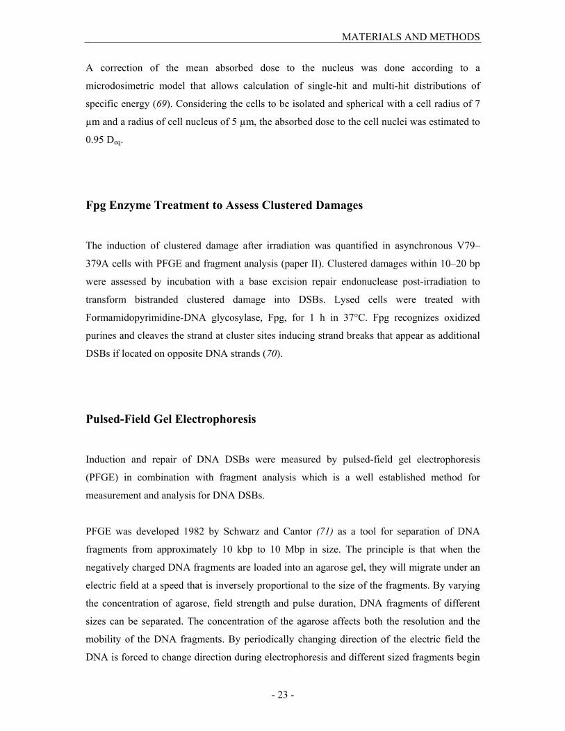
Pulsed-Field Gel Electrophoresis
Induction and repair of DNA DSBs were measured by pulsed-field gel electrophoresis
(PFGE) in combination with fragment analysis which is a well established method for
measurement and analysis for DNA DSBs.
PFGE was developed 1982 by Schwarz and Cantor (71) as a tool for separation of DNA
fragments from approximately 10 kbp to 10 Mbp in size. The principle is that when the
negatively charged DNA fragments are loaded into an agarose gel, they will migrate under an
electric field at a speed that is inversely proportional to the size of the fragments. By varying
the concentration of agarose, field strength and pulse duration, DNA fragments of different
sizes can be separated. The concentration of the agarose affects both the resolution and the
mobility of the DNA fragments. By periodically changing direction of the electric field the
DNA is forced to change direction during electrophoresis and different sized fragments begin
MATERIALS AND METHODS
- 24 -
to separate from each other. Longer pulse time and weaker electric fields lead to separation of
larger DNA fragments.
Figure 2. Illustration of PFGE and fragment analysis. DNA samples are loaded into a gel containing a matrix of
agarose. The negatively charged DNA fragments move toward the positve electrode in a rate proportional to
their length and will therefore be size separated. After staining with a fluorescent dye, DNA of different lengths
can be identified using specific size markers.
Conventionally, PFGE is used to calculate the number of DSBs from FAR values. The FAR
value, Fraction of Activity Released, describes the relative amount of 14C-labeled DNA that
migrates through the gel during electrophoresis and correlates in a non-linear way with
radiation dose. FAR values can be converted into DSB under the assumption of randomly
distributed DSBs along the DNA helix (72). This conversion is suitable after low-LET
irradiation but in the case of high-LET irradiation, however, this assumption is not correct.
Instead, another approach for determining the number of DSBs is needed, using further
separation of fragments. DNA markers, well characterized in size, are loaded together with
the samples in the gels and used to localize separated fragments of specific sizes. The total
amount of DSBs is then calculated by summarizing DSBs in all fragment size intervals. The
method is called fragment analysis and can also be used to determine the distribution of
fragments of different sizes along the DNA.
+
DNA fragments + Gel
particle
DNA
++
DNA fragments + Gel
particle
DNA
DNA fragmentsDNA fragments + Gel
particle
DNA
Gel
particle
Gel
particle
DNADNA
MATERIALS AND METHODS
- 25 -
In this work, two different electrophoresis protocols were used being optimized for separation
of DNA fragments <1.1 Mbp and 1.1–5.7 Mbp, respectively. The calculations of DSBs
induced were done by fragment analysis from the FAR values in each size segment. For this,
lanes of migrated DNA were sliced in segments using three different size standards: Lambda
DNA ladders, Saccharomyces cervisiae chromosomal DNA, and Schizosaccharomyces
pombe chromosomal DNA. The number of fragments was calculated by dividing the fraction
of 14C-incorporated DNA in a gel segment, measured by a liquid scintillation counter, with
the average fragment size in that segment.
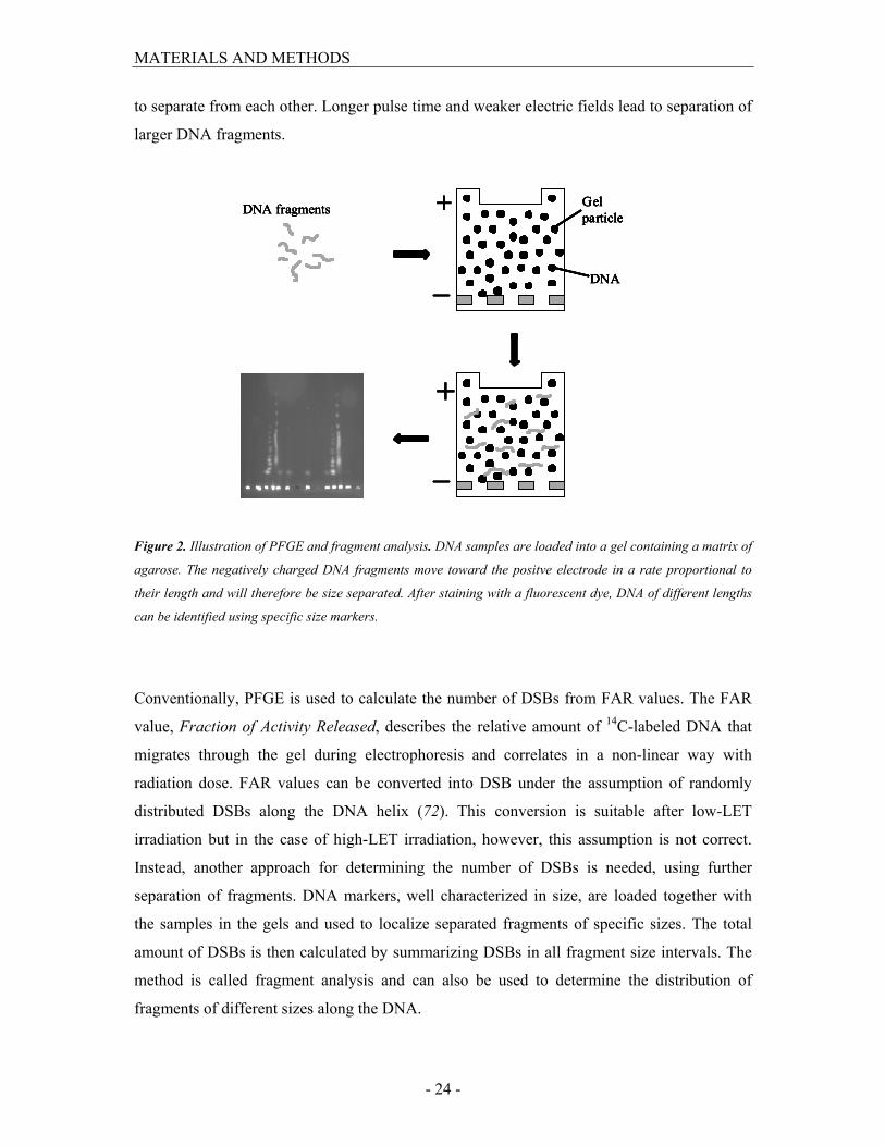
Today, the most widely used method to determine DSB induced by ionizing radiation is
detection of phosphorylation of the histone H2AX, called γ-H2AX in its activated form,
which can be visualized within individual cells as distinct foci using antibodies specific for its
phosphorylated form. The advantage with γ-H2AX over PFGE is the possibility to investigate
effects of clinically relevant doses. One problem with the detection of DSB with γ-H2AX is
the poor resolution of correlated DSB after high-LET radiation and therefore, the number of
DSB will be underestimated in high-LET-irradiated cells. One of the main aims of this work
was to estimate RBE for α-particles from 211At and therefore, detection of DSBs was made
using PFGE and fragment analysis instead of γ-H2AX foci quantification.
Figure 3. γ-H2AX-foci (green) in propidium iodide stained cell nuclei in cells receiving 1 Gy X-rays (left) or 1
Gy 211At (right).

MATERIALS AND METHODS
- 26 -
Micronuclei Assay
Radiation-induced chromosomal damage can be detected by investigating the formation of
micronuclei (MN) in living cells. MN are formed of acentric fragments or whole
chromosomes that have not been incorporated into daughter nuclei at mitosis and it is
assumed that a cell carrying MN has lost its clonogenic capacity (73). The most widely used
method for scoring MN is the cytokinesis-block micronucleus assay, which is a rapid and
simple method that can be used at clinically relevant doses. In this assay, the fraction of MN
is scored in binucleated cells when blocked from performing cytokines by cytochalasin B.
The advantage with the use of cytochalasin B is that MN are scored only in binucleated cells,
i.e. cells that have undergone one mitosis after irradiation, enabling reliable comparisons
between cell populations that differ in their cell division kinetics. However, in this work MN
formation was detected in cell cultures not treated with cytochalasin B due to its toxicity. HS
2429 cells, irradiated as confluent cultures had to be split directly after irradiation to prevent
contact inhibition of cell division, a process for which a cytoskeleton drug is very toxic.
Therefore, the number of MN formed after irradiation in the whole cell population was
scored. 48 and 72 h post-irradiation, cells were fixed and stained with DAPI. Nuclei were then
scored blind for MN using the criteria described by Fenech et al. (74). The mitotic index,
determined by detection of binucleated cells in the presence of the cytokinesis inhibitor
cytochalasin B, was in the same range, 33% and 22%, for cycling and stationary cells,
respectively. In these experiments cytochalasin B was added 6 hours after plating.
Measurements of Cell Proliferation
In this work, the cellular capacity to proliferate after irradiation was determined using two
different assays: clonogenic assay and growth assay. These two methods differ in that the
growth assay measures the total biomass and the proliferation of the entire cell population
whereas the clonogenic assay measure the capacity of an individual cell to divide infinite
times.
In paper II, cell survival of irradiated V79–379A cells was assessed by the clonogenic assay
in which the ability of a single cell to form a colony exceeding about 50 cells, representing 5–
MATERIALS AND METHODS
- 27 -
6 divisions, was determined. Asynchronous cells, cells in G1, early, mid and late S phases and
mitotic cells were irradiated as single cell suspensions with 211At or X-rays. Following
irradiation, cells at low density were seeded and incubated at 37°C for four days. The
resulting number of colonies, consisting of at least 50 cells, was scored and the surviving
fraction (SF) was calculated.
The growth kinetics in HS 2429 cultures after irradiation was determined by measuring the
total cell number after crystal violet staining (paper IV). Crystal violet is a dye that binds to
the cells resulting in optical density proportional to the cell number. Immediately after
irradiation the cells were seeded in 96-well plates and incubated 1–15 days. After being fixed
and stained with crystal violet, the optical density of dye extracts was measured using a micro
plate reader. This assay is a suitable method for determination of radiation effects when cells
do not form colonies and the clonogenic survival assay is not possible to use.
Measurements of Cell Cycle Arrests
Delays in cell cycle progression were examined by measuring cell cycle distributions with
flow cytometric analysis after propidium iodide staining 0–72 days (paper III) or 0–22 days
(paper IV) after irradiation. The percentage of cells in G0/G1, S phase and G2/M was
estimated by calculating the area under the DNA histogram assuming a Gaussian function.
In paper III, phosphorylation of the checkpoint kinase Chk2 at Threonine 68 was used as an
indicator for cell cycle arrest activation 0–120 h post-irradiation. Cells cultured on chamber
slides were fixed prior to incubation with a primary antibody binding to the phosphorylated
Chk2, followed by incubation with a FITC-conjugated secondary antibody. The number of
Chk2-foci in individual nuclei was scored blind using fluorescence microscopy.
MATERIALS AND METHODS
- 28 -
Modulation of Chromatin Structure
In paper I, intact cells were chemically modulated into a structure defined as nuclear
monolayer by permeabilizing the cell membrane with the ionic detergent Triton X-100. After
this treatment the nucleus was depleted of all soluble scavengers. Intact cells and nuclear
monolayers differ in that the DNA in nuclear monolayers lacks the protective effect of these
molecules surrounding the DNA, while the chromatin conformation remains mainly
unchanged.
In paper IV, the chromatin structure was modified by treating cells with the histone
deacytelase inhibitor trichostatin A (TSA) that prevents deacetylation of histones (50). The
molecular effects of TSA are rapid and reversible since increasing levels of acetylated histone
H3 on lysine 9 (Ac-H3K9) were seen already after 1 h exposure of 0.3 µM TSA while wash-
out after four hours decreased the levels of Ac-H3K9 (data not shown). TSA was added to the
cells four hours before irradiation and was removed prior to irradiation.
RESULTS AND DISCUSSION
- 29 -
RESULTS AND DISCUSSION
Effects on DNA Damage Induction
Double-Strand Break Induction (paper I-III)
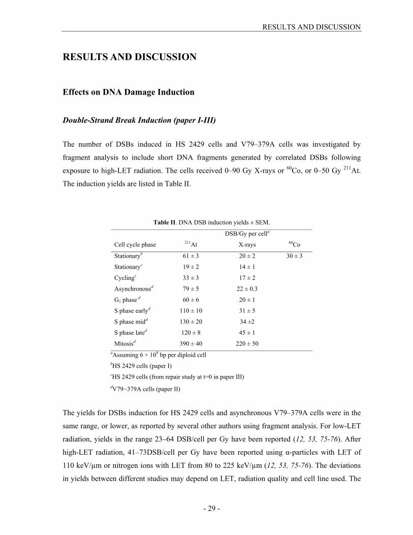
The number of DSBs induced in HS 2429 cells and V79–379A cells was investigated by
fragment analysis to include short DNA fragments generated by correlated DSBs following
exposure to high-LET radiation. The cells received 0–90 Gy X-rays or 60Co, or 0–50 Gy
211At.
The induction yields are listed in Table II.
Table II. DNA DSB induction yields ± SEM.
DSB/Gy per cella
Cell cycle phase 211At X-rays
60Co
Stationaryb 61 ± 3 20 ± 2 30 ± 3
Stationaryc 19 ± 2 14 ± 1
Cyclingc 33 ± 3 17 ± 2
Asynchronousd 79 ± 5 22 ± 0.3
G1 phase d 60 ± 6 20 ± 1
S phase earlyd 110 ± 10 31 ± 5
S phase midd 130 ± 20 34 ±2
S phase lated 120 ± 8 45 ± 1
Mitosisd 390 ± 40 220 ± 50
aAssuming 6 × 10⁹ bp per diploid cell
bHS 2429 cells (paper I)
cHS 2429 cells (from repair study at t=0 in paper III)
dV79–379A cells (paper II)
The yields for DSBs induction for HS 2429 cells and asynchronous V79–379A cells were in the
same range, or lower, as reported by several other authors using fragment analysis. For low-LET
radiation, yields in the range 23–64 DSB/cell per Gy have been reported (12, 53, 75-76). After
high-LET radiation, 41–73DSB/cell per Gy have been reported using α-particles with LET of
110 keV/µm or nitrogen ions with LET from 80 to 225 keV/µm (12, 53, 75-76). The deviations
in yields between different studies may depend on LET, radiation quality and cell line used. The
RESULTS AND DISCUSSION
- 30 -
yields for stationary HS 2429 cells after 211At clearly differed between paper I and III, for
unknown reasons.
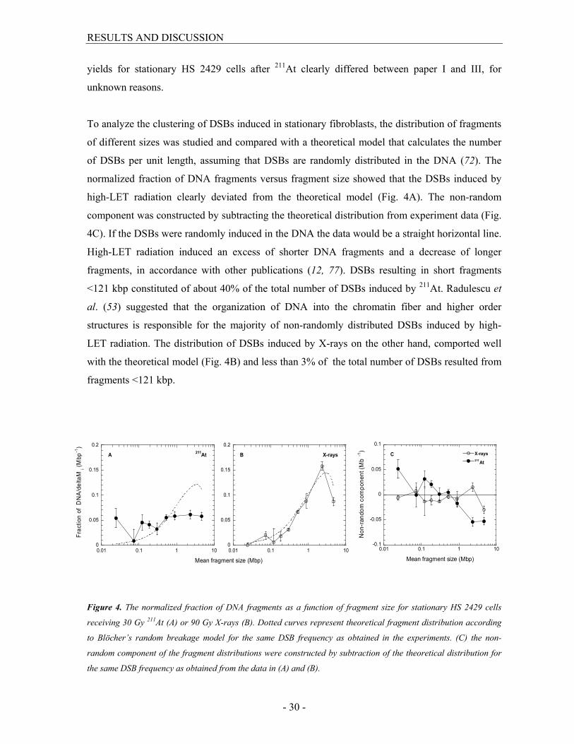
To analyze the clustering of DSBs induced in stationary fibroblasts, the distribution of fragments
of different sizes was studied and compared with a theoretical model that calculates the number
of DSBs per unit length, assuming that DSBs are randomly distributed in the DNA (72). The
normalized fraction of DNA fragments versus fragment size showed that the DSBs induced by
high-LET radiation clearly deviated from the theoretical model (Fig. 4A). The non-random
component was constructed by subtracting the theoretical distribution from experiment data (Fig.
4C). If the DSBs were randomly induced in the DNA the data would be a straight horizontal line.
High-LET radiation induced an excess of shorter DNA fragments and a decrease of longer
fragments, in accordance with other publications (12, 77). DSBs resulting in short fragments
<121 kbp constituted of about 40% of the total number of DSBs induced by 211At. Radulescu et
al. (53) suggested that the organization of DNA into the chromatin fiber and higher order
structures is responsible for the majority of non-randomly distributed DSBs induced by high-
LET radiation. The distribution of DSBs induced by X-rays on the other hand, comported well
with the theoretical model (Fig. 4B) and less than 3% of the total number of DSBs resulted from
fragments <121 kbp.
0
0.05
0.1
0.15
0.2
0.01 0.1 1 10
211At
Fraction of DNA/deltaM
i (Mbp-1)
A
0
0.05
0.1
0.15
0.2
0.01 0.1 1 10
X-rays
Mean fragment size (Mbp)
B
-0.1
-0.05
0
0.05
0.1
0.01 0.1 1 10
X-rays
211At
Non-random component (M
b-1)
Mean fragment size (Mbp)
C
Figure 4. The normalized fraction of DNA fragments as a function of fragment size for stationary HS 2429 cells
receiving 30 Gy 211At (A) or 90 Gy X-rays (B). Dotted curves represent theoretical fragment distribution according
to Blöcher’s random breakage model for the same DSB frequency as obtained in the experiments. (C) the non-
random component of the fragment distributions were constructed by subtraction of the theoretical distribution for
the same DSB frequency as obtained from the data in (A) and (B).

RESULTS AND DISCUSSION
- 31 -
In synchronized V79–379A cells, the number of DSBs increased when the cells progressed
through the cell cycle (Table II). The yields per cell and corresponding cell cycle differences
were similar to what has been demonstrated earlier for low-LET radiation. This cell cycle effect
was not the result of lagging fragments containing replication forks, since this decreased mobility
during electrophoresis was compensated for (78-79). One reason for the increased DSB yield
during cell cycle progression is the increase of DNA content during DNA replication, but could
also be a consequence of the chromatin structure. If the difference in DNA content was the only
reason for variations of DSB induction between cell cycle phases, there would be only a two-fold
increase of DSBs in mitotic cells. The induction yields showed that 6.6 and 11 times more DSBs
were induced in mitotic cells relative G1 cells irradiated with 211At and X-rays, respectively. A
similar finding has also been shown by Radford (47). He found a 3–4-fold increase of DSBs in
mitotic cells compared with cells in early S phase measured with the neutral filter elusion assay.
The large increase of DSBs in mitotic cells, compared to cells from other cell cycle phases could
in part be explained by the increase of short DNA fragments. Fragment analysis showed a
striking deviation from the random distribution in mitotic cells also for low-LET radiation. The
contributions of short DNA fragments <121 kbp were 13% in G1 cells and 80% in mitotic cells.
The remarkable increase of short DNA fragments in mitotic cells is probably a consequence of
the very compact chromatin organization. This indicates that fragment analysis, requisite for
quantification of non-randomly induced DSBs, may be a necessary tool for DSB measurements,
not only after high-LET radiation but also for low-LET under certain circumstances.
In summary, α-particles from 211At were more effective in inducing DSBs in a non-random
fashion, compared with X-rays. Induction yields varied depending on position of cell cycle phase
at the time of irradiation after both 211At and X-rays.
Induction of Clustered Damage (paper II)
The induction of clustered damage was investigated in asynchronous V79–379A cells and the
yield for cluster induction was 26 ± 1.3 and 15 ± 4.4 clusters/cell per Gy for X-rays (0–75 Gy)
and 211At (0–50 Gy), respectively. Somewhat more clusters than DSBs were induced in X-
irradiated cells (1.2 ± 0.05 clusters per DSB) in agreement with earlier reported findings (80-82).
In contradiction, five times more DSBs than clusters were induce after 211At. Only a few studies

RESULTS AND DISCUSSION
- 32 -
investigating Fpg-clusters after high-LET irradiation in intact cells have been performed. The
results from these studies show that none or less Fpg-clusters than DSB are induced after
exposure to high-LET radiation (16, 82-83). The ratio between clustered damage and DSB tends
to decline with LET and it appears as if non-DSB clustered damages are more dependent on the
radical mediated indirect effect of ionizing radiation.
Effects on the DNA Damage Response
Repair of DNA Double-Strand Breaks (paper III)
The repair kinetics of DNA DSBs was investigated at a single dose, expected from DSB yields in
paper I to induce the same amount of DSBs (30 Gy 211At and 90 Gy X-rays). From Fig. 5 it is
obvious that the repair kinetics differed between the two radiation qualities and three major
distinctions can be mentioned. First, overall the repair kinetics was much slower after 211At and
second, more residual damage was detected after a repair time of 24 h post-irradiation. In
stationary cells, 4% and 25% of the lesions remained unrepaired 24 h after X-rays and 211At,
respectively (Fig. 5A). Corresponding values in cycling cells were as high as 40% and 85%,
respectively (Fig. 5B). Numerous studies on DSB repair have shown a slower rejoining and a
larger amount of residual DSBs induced by high-LET radiation even after a long repair time (27-
30). For example, Jenner et al. (28) reported that only 30% of the DSBs were rejoined after α-
particles while >90% of those induced by X-rays were rejoined 3 h post-irradiation, at an
absorbed dose of 40 Gy. Also, the repair kinetics measured by γ-H2AX foci fluorescence was
dependent on LET with more persistent foci after long repair time (84). Groesser et al. (85) have
shown that the foci numbers decreased faster after γ irradiation and returned to control levels 22
h post-irradiation while approximately 20–40% foci persisted after irradiation with Fe ions. The
pronounced difference in the repair capacity between X-rays and 211At, despite the dose
adjustment, is probably due to the clustering of lesions along the α-particle track that may be
very difficult to repair. The insufficient repair after high-LET radiation enhances the risk of
failed or misrejoined DSBs, which in turn could lead to an increase of mutations, chromosomal
damage and cell death. It is also possible that low- and high-LET radiation induce different
amounts or ratios of DNA lesions in open versus compact chromatin, which may have
consequences for DNA repair efficiency.
RESULTS AND DISCUSSION
- 33 -
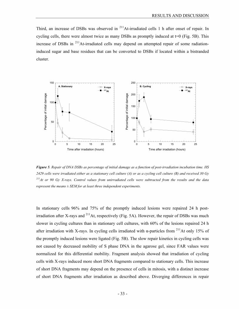
Third, an increase of DSBs was observed in 211At-irradiated cells 1 h after onset of repair. In
cycling cells, there were almost twice as many DSBs as promptly induced at t=0 (Fig. 5B). This
increase of DSBs in 211At-irradiated cells may depend on attempted repair of some radiation-
induced sugar and base residues that can be converted to DSBs if located within a bistranded
cluster.
0
50
100
150
0 5 10 15 20 25
A. Stationary X-rays211At
Percentage of initial damage
Time after irradiation (hours)
0
50
100
150
200
250
0 5 10 15 20 25
B. Cycling X-rays211At
Percentage of initial damage
Time after irradiation (hours)
Figure 5. Repair of DNA DSBs as percentage of initial damage as a function of post-irradiation incubation time. HS
2429 cells were irradiated either as a stationary cell culture (A) or as a cycling cell culture (B) and received 30 Gy 211At or 90 Gy X-rays. Control values from unirradiated cells were subtracted from the results and the data
represent the means ± SEM for at least three independent experiments.
In stationary cells 96% and 75% of the promptly induced lesions were repaired 24 h post-
irradiation after X-rays and 211At, respectively (Fig. 5A). However, the repair of DSBs was much
slower in cycling cultures than in stationary cell cultures, with 60% of the lesions repaired 24 h
after irradiation with X-rays. In cycling cells irradiated with α-particles from 211At only 15% of
the promptly induced lesions were ligated (Fig. 5B). The slow repair kinetics in cycling cells was
not caused by decreased mobility of S phase DNA in the agarose gel, since FAR values were
normalized for this differential mobility. Fragment analysis showed that irradiation of cycling
cells with X-rays induced more short DNA fragments compared to stationary cells. This increase
of short DNA fragments may depend on the presence of cells in mitosis, with a distinct increase
of short DNA fragments after irradiation as described above. Diverging differences in repair

RESULTS AND DISCUSSION
- 34 -
kinetics between proliferating and resting cells have also been reported by other authors (85-88).
For example, Hamasaki et al. (88) demonstrated a 1.5-fold increase of γ-H2AX foci in
phytohemagglutinin stimulated lymphocytes one hour after X irradiation, compared to resting
cells. Increased phosphorylation of H2AX in proliferating lymphocytes was also showed by
Vilasova et al. (86).
It seems as if the DSB repair machinery prefers to repair isolated DSBs over lesions located in
close proximity, since fragment analysis showed that the relative number of short fragments
increased with repair time. In stationary cells, the fraction of DNA fragments <185 kbp increased
from zero directly after X-irradiation to 50% after 3 h repair time. Corresponding numbers in
cycling cells were 18% and 51%. Indeed, Wang et al. (89) have shown that very short fragments
(<40 bp) prevent efficient Ku-binding, thereby decreasing DNA-PKCS recruitment to the two
ends of the DNA fragments, resulting in NHEJ retardation. This may explain the insufficient
repair after α-particles, primarily inducing correlated DSBs, and in cell irradiated as a cycling
culture.
In conclusion, repair of DSBs induced by 211At required longer time and resulted in higher
proportion of unligated breaks after 24 h. There was a large difference in repair kinetics between
cycling and stationary cells, with more insufficient repair in proliferating cells.
Effects of Radiation on Cell Cycle Progression (paper III and IV)
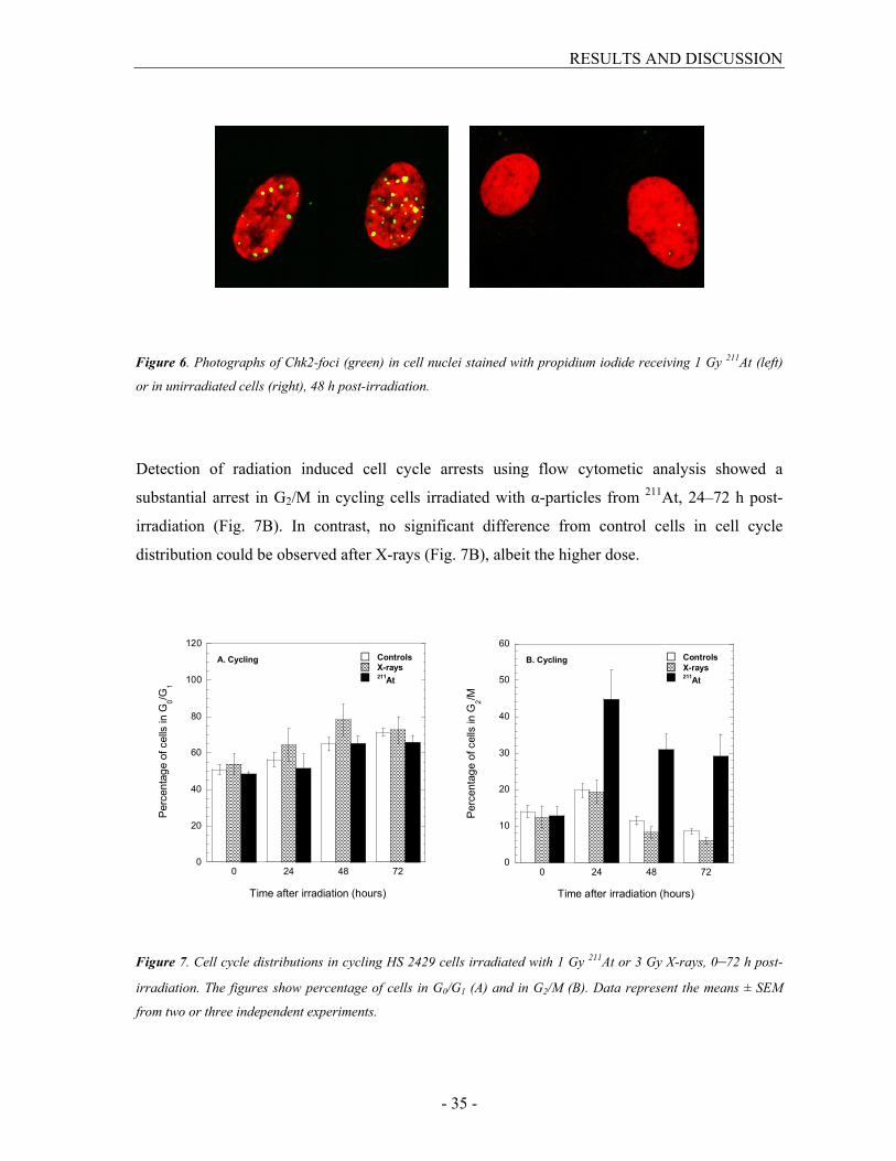
Detection of cell cycle arrests using immunofluorescent labeling of phosphorylated Chk2-foci
showed a massive activation of Chk2 one hour post-irradiation, in both cycling and stationary
cell cultures receiving 1 Gy 211At or 3 Gy X-rays. Re-incubation at 37°C resulted in a decreased
amount of foci by repair time, but even 120 h post-irradiation at least 50% more foci could be
found in irradiated cells than in unirradiated cells (Fig. 6). In cycling cells, twice as many Chk2-
foci remained after 211At compared with X-rays, despite the dose adjustment, probably reflecting
the inefficient repair in cells irradiated with 211At. In stationary cells, a difference between the
radiation qualities was first seen 48 h post-irradiation with about 1.5 times more foci after α-
particles. The difference between cycling and stationary cells was more pronounced after α-
particles with a two-fold increase of Chk2-foci 24 h and 48 h post-irradiation. After X-rays only
small differences were observed with no consistent trend.
RESULTS AND DISCUSSION
- 35 -
Figure 6. Photographs of Chk2-foci (green) in cell nuclei stained with propidium iodide receiving 1 Gy 211At (left)
or in unirradiated cells (right), 48 h post-irradiation.
Detection of radiation induced cell cycle arrests using flow cytometic analysis showed a
substantial arrest in G2/M in cycling cells irradiated with α-particles from 211At, 24–72 h post-
irradiation (Fig. 7B). In contrast, no significant difference from control cells in cell cycle
distribution could be observed after X-rays (Fig. 7B), albeit the higher dose.
0
20
40
60
80
100
120
0 24 48 72
A. Cycling Controls
X-rays211At
Percentage of cells in G
0/G
1
Time after irradiation (hours)
0
10
20
30
40
50
60
0 24 48 72
B. Cycling Controls
X-rays 211At
Percentage of cells in G
2/M
Time after irradiation (hours)
Figure 7. Cell cycle distributions in cycling HS 2429 cells irradiated with 1 Gy 211At or 3 Gy X-rays, 0–72 h post-
irradiation. The figures show percentage of cells in G0/G1 (A) and in G2/M (B). Data represent the means ± SEM
from two or three independent experiments.

RESULTS AND DISCUSSION
- 36 -
When cell cycle distributions were analyzed in a long time follow-up 0–22 days after irradiation,
both 211At and X-rays resulted in cell cycle progression disturbances corresponding to what was
found 24–72 h after irradiation. Irradiation of cycling cells with 211At resulted in a substantial and
persisted accumulation of cells with about twice as many cells in G2/M compared to unirradiated
cells (Fig. 8A). After exposure to X-rays (3 Gy), only a minor accumulation of cells in G2/M was
observed (Fig. 8B). No effect on cell progression was observed in cells receiving 1 Gy X-rays.
0
10
20
30
40
0 5 10 15 20 25
A. Cycling Controls
1 Gy 211At
Percentage of cells in G
2/M
Time after irradiation (days)
0
10
20
30
40
0 5 10 15 20 25
B. Cycling Controls
3 Gy X-rays
Percentage of cells in G
2/M
Time after irradiation (days)
Figure 8. Percentage of HS 2429 cells in G2/M in cycling cell cultures 0–22 days after irradiation with 211At (A) or
X-rays (B). Data are the means ± SEM from at least three independent experiments.
Such drastic G2/M arrest after α-particles is in accordance with presented data from others on
G2/M arrests induced by different high-LET radiations (90-92). Dose dependent G2/M delays
have been suggested earlier. For example, Raju et al. (93) reported that the delays of progression
through G2/M was dose dependent for both X-rays and α-particles and Lücke-Huhle et al. (90)
showed that the number of cells arrested in G2/M after exposure with heavy ions increased
linearly with dose up to at least 75% of the maximum fraction of cells in G2/M. Indeed, primary
fibroblasts requires doses >200 mGy to activate G2/M checkpoint. Further, Buscemi et al. (94)
suggested that the checkpoint kinase Chk2 needs >19 DSBs to be activated, which is in the same
range as the DSB induction yields presented here.

RESULTS AND DISCUSSION
- 37 -
Stationary cells were arrested in G0/G1 after release of the contact inhibition at plating, regardless
of radiation quality (Fig. 9A). Only 5–10% of the irradiated cells started to proliferate 24 h post-
irradiation, while this percentage was much higher in control cells.
0
20
40
60
80
100
120
0 24 48 72
A. Stationary Controls
X-rays 211At
Percentage of cells in G
0/G
1
Time after irradiation (hours)
0
10
20
30
40
50
60
0 24 48 72
B. Stationary Controls
X-rays 211At
Percentage of cells in G
2/M
Time after irradiation (hours)
Figure 9. Cell cycle distributions in stationary HS 2429 cells irradiated with 1 Gy 211At or 3 Gy X-rays, 0–72 h post-
irradiation. The figures show percentage of cells in G0/G1 (A) and in G2/M (B). Data represent the means ± SEM
from from two or three independent experiments.
In accordance, stationary cell cultures investigated for long times (0–22 days) underwent a
delayed cell cycle progression after release of the contact inhibition with a lower fraction of cells
entering S phase at later time points after irradiation (Fig. 10). This was found after both 211At
(Fig. 10A) and X-rays (Fig. 10B), but was more prominent after α-particle exposure.

RESULTS AND DISCUSSION
- 38 -
0
5
10
15
20
25
30
35
0 5 10 15 20 25
A. Stationary Controls
1 Gy 211At
Percentage of cells in S
Time after irradiation (days)
0
5
10
15
20
25
30
35
0 5 10 15 20 25
B. Stationary Controls
3 Gy X rays
Percentage of cells in S
Time after irradiation (days)
Figure 10. Percentage of HS 2429 cells in S phase in stationary cell cultures 0–22 days after irradiation with 211At
(A) or X-rays (B). Data are the means ± SEM from at least three independent experiments.
Delays of the progression into S phase in non-cycling cells that are allowed to re-enter the cell
cycle have been reported earlier (95-97). It is possible that a fraction of the cells irradiated as a
stationary cell culture in G0/G1 and re-seeded immediately after irradiation do not start to
proliferate but become permanently arrested. Indeed, Nasonova et al. (95) have shown that
approximately 20% of irradiated normal cells were irreversibly blocked in G1 following
subculture and Tenhumberg et al. (96) have shown that only 25% of normal fibroblasts reached
the first mitosis after exposure to low energy particles, supporting the data presented here. The
arrest in G1 provides extra time for the repair of DNA damage before the onset of replication and
thus protects the cells against the effects of radiation.
Several studies have been performed on disturbances in cell cycle progression after irradiation
with both low- and high-LET radiation. The predominant result from these studies was that an
accumulation of cells in G2 phase was observed after high-LET radiation (90-93, 98) , but also
delays in G1 and S phase have been demonstrated (91, 96-97). The duration of the block depends
on radiation quality and dose (92-93). It appears as if the G1/S checkpoint is somewhat weaker
than the G2/M checkpoint. Indeed, Deckbar et al. (99) have shown that the G1/S checkpoint fails
to prevent S phase entry of irradiated fibroblasts at early times after irradiation.

RESULTS AND DISCUSSION
- 39 -
In summary, results from paper III and IV showed that cell cycle arrests were dependent upon the
position of cells in the cell cycle at the time of irradiation. Cycling cells predominantly became
arrested in G2/M while stationary cells were arrested in G1. Also, α-particles from 211At resulted
in more pronounced arrests than X-rays.
Chromosomal Damage (paper IV)
Chomosomal damage was studied by investigating the formation of MN in stationary and cycling
HS 2429 cells, receiving 1 Gy 211At, or 1 and 3 Gy X-rays. The formation of MN was dependent
on radiation quality, dose and proliferation status. The amount of MN peaked at 72 post-
irradiation, but also 120 h post-irradiation a large fraction of cells exhibited MN in the culture
(data not shown). These remaining MN could be a consequence of both delayed cell cycle
progression and of some MN formed after the second post-irradiation mitosis (100). When
isodose was compared, 211At was more effective in inducing chromosome damage. The average
numbers of MN formed per cell after α-particles 72 h post-irradiation were 0.67 ± 0.04 and 0.47
± 0.03 for cycling and stationary cells, respectively. At the same dose of X-rays, only 0.25 ± 0.02
and 0.12 ± 0.01 MN were formed in cells irradiated as a cycling and stationary cell culture,
respectively. In cycling cells, increasing the dose of X-rays to 3 Gy resulted in the same amount
of MN produced as after α-particles (0.69 ± 0.03) but this was not the case in stationary cells
where the MN yield was much lower (0.28 ± 0.02). The complexity of chromosomal damage was
more pronounced in cells irradiated with 211At with more multiple MN per cell. Accordningly, in
cells receiving 1 Gy α-particles from 211At, the proportion of MN-positive cells with at least three
MN 72 h post-irradiation was 27% and 22% in cycling and stationary cells, respectively. The
corresponding numbers after 1 Gy of X-rays were only 9% and 2%. Increased complexity of
chromosomal aberrations with increasing LET has been reported earlier (101-102). The increased
complexity of chromosome damage after high-LET radiation is probably a consequence of
correlated DSB and the inefficient repair thereof.
RESULTS AND DISCUSSION
- 40 -
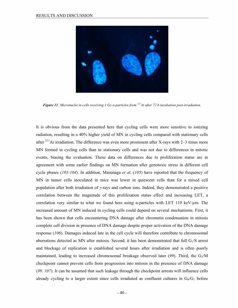
Figure 11. Micronuclei in cells receiving 1 Gy α-particles from 211At after 72 h incubation post-irradiation.
It is obvious from the data presented here that cycling cells were more sensitive to ionizing
radiation, resulting in a 40% higher yield of MN in cycling cells compared with stationary cells
after 211At irradiation. The difference was even more prominent after X-rays with 2–3 times more
MN formed in cycling cells than in stationary cells and was not due to differences in mitotic
events, biasing the evaluation. These data on differences due to proliferation status are in
agreement with some earlier findings on MN formation after genotoxic stress in different cell
cycle phases (103-104). In addition, Masunaga et al. (105) have reported that the frequency of
MN in tumor cells inoculated in mice was lower in quiescent cells than for a mixed cell
population after both irradiation of γ-rays and carbon ions. Indeed, they demonstrated a positive
correlation between the magnitude of this proliferation status effect and increasing LET, a
correlation very similar to what we found here using α-particles with LET 110 keV/µm. The
increased amount of MN induced in cycling cells could depend on several mechanisms. First, it
has been shown that cells encountering DNA damage after chromatin condensation in mitosis
complete cell division in presence of DNA damage despite proper activation of the DNA damage
response (106). Damages induced late in the cell cycle will therefore contribute to chromosomal
aberrations detected as MN after mitosis. Second, it has been demonstrated that full G1/S arrest
and blockage of replication is established several hours after irradiation and is often poorly
maintained, leading to increased chromosomal breakage observed later (99). Third, the G2/M
checkpoint cannot prevent cells from progression into mitosis in the presence of DNA damage
(99, 107). It can be assumed that such leakage through the checkpoint arrests will influence cells
already cycling to a larger extent since cells irradiated as confluent cultures in G0/G1 before

RESULTS AND DISCUSSION
- 41 -
plating may have sufficient time to complete repair before onset of replication many hours later.
Also, from DSB repair studies in paper III it is clear that the repair process was very inefficient in
cycling cells with a high proportion of unligated breaks remaining after 24 h. Such amount of
residual damage may lead to consequences in the form of chromosomal damage, as shown here.
In summary, cells irradiated with α-particles from 211At exhibited more chromosomal damage
than X-irradiated cells. Also, cycling cells had more MN compared with cells irradiated as
stationary cultures.
Clonogenic Cell Survival and Cell Growth (paper II and IV)
The radiosensitivity in asynchronous and synchronized V79–379A cells was measured with the
clonogenic survival assay. The experimental data from X-rays was fitted to the linear-quadratic
model (LQ), and data from 211At was fitted to an exponential curve. The surviving fractions (SF)
were much lower after 211At than X-rays in all cell cycle phases, as well as in asynchronous cells
(Fig. 12). In asynchronous cells, the dose required to reduce the SF to 37% was 5.5 Gy after X-
rays but only 0.67 Gy after exposure to 211At. The SF after 2 Gy was 81% and 5.2% for X-rays
and 211At, respectively.
The clonogenic survival after both X-rays and 211At was dependent on cell cycle position at the
time of irradiation. After X-rays, the lowest survival was found in mitotic cells followed by cells
in G1 phase. Cells in late S phase were the most radioresistant of all phases investigated. The
dose required to reduce the survival to 37% increased as cells progressed through the cell cycle
and was 1.6 times higher in late S phase compared with G1 cells. Corresponding value for mitotic
cells was 0.8. Since the classical findings by Terasiama and Tolmach (41), this pattern of
radiosensitivity after low-LET irradiation between cells in different position of the cell cycle has
been reported by a number of investigators (47, 108). The radioresistance in late S phase can be
explained by the fact that both non-homologous end joining and homologous repair may
contribute to a higher DSB repair fidelity and consequently also to the increased radioresistance
(109).
RESULTS AND DISCUSSION
- 42 -
In contrast to what is generally accepted for high-LET radiation, results from paper II showed
that irradiation with 211At also resulted in differences in radiosensitivity between different cell
cycle phases although variations were not as large as after low-LET radiation. Surprisingly, and
in sharp contrast to X-rays, mitotic cells were most radioresistant after 211At, where a 1.5-fold
higher dose was needed to reduce the survival to 37%. A possible explanation could be that DNA
may be unhit in some cells after α-particles, biasing the surviving fraction, as it is condensed in
its most compacted and sorted form, occupying less volume in the nucleus than chromatin in
other cell cycle phases. It should be remembered however, that is not a condition restricted to
cells in vitro. The cell cycle effect was more prominent when determined at the absorbed dose of
2 Gy. The SF in mitotic and mid S phase cells was only 25% of the SF in G1 cells. In late- and
early S phases, the SF was 50% and 75% of the SF in G1 cells, respectively.

RESULTS AND DISCUSSION
- 43 -
0.01
0.1
1
0 1 2 3 4 5 6 7
Asynchronous
X-ray
211At
Surviving fraction
Dose (Gy)
0.01
0.1
1
0 1 2 3 4 5 6 7
G1
X-ray
211At
Surviving fraction
Dose (Gy)
0.01
0.1
1
0 1 2 3 4 5 6 7
S early
X-ray
211At
Surviving fraction
Dose (Gy)
0.01
0.1
1
0 1 2 3 4 5 6 7
S mid
X-ray
211At
Surviving fraction
Dose (Gy)
0.01
0.1
1
0 1 2 3 4 5 6 7
S late
X-ray
211At
Surviving fraction
Dose (Gy)
0.01
0.1
1
0 1 2 3 4 5 6 7
Mitosis
X-ray
211At
Surviving fraction
Dose (Gy)
Figure 12. Clonogenic cell survival in V79–379A cells after 211At or X-rays, presented for asynchronous cells, G1
cells, early, mid and late S phase cells and cells in mitosis. The results derive from at least three independent
experiments ± SEM. Results from 211At were fitted to an exponential curve and the results from X-rays to the LQ-
model.

RESULTS AND DISCUSSION
- 44 -
In agreement with the result from the clonogenic survival assay, investigation of the proliferation
of irradiated cells using a growth assay (paper IV) showed that α-particles from 211At were more
retarding than X-rays. Cycling cells were somewhat more growth inhibited than stationary cells
with an almost absent regrowth regardless of radiation quality.
In summary, clonogenic survival was influenced by cell cycle position after both low- and high-
LET irradiation. Also, the number of clonogenic cells and culture proliferation were reduced
after 211At, demonstrating that the radiation quality differences observed at earlier endpoints
resulted in decreased survival of cells after irradiation with α-particle compared with X-rays.
Effects of Trichostatin A on Irradiated Cells (paper IV)
Effects of TSA-induced inhibition of chromatin deacetylation on the radiation-induced formation
of micronuclei and cell cycle progression were studied. Compared to non-treated irradiated cells,
TSA had an impact on cell cycle progression following irradiation, with an accumulation of cells
in G2/M in cells irradiated as cycling cultures after both 211At and X-rays,. In stationary cells, an
enhancement of the delay into S phase after 24 h post-irradiation in TSA treated cells was seen
after 211At but not after X-rays. In contradiction to this enhanced arrest activation, 10–27% less
MN were formed 48 h post-irradiation in TSA treated irradiated cells, but this protective effect
was reduced or abolished 72 h post-irradiation. In cycling cells, this could be explained by the
fact that cells treated with TSA were arrested in G2 after 24 h to a larger extent than non-treated
cells, and therefore took longer time to enter the first mitosis after irradiation. Consequently, a
higher proportion of TSA-treated cells could express chromosomal damage after 72 h compared
with earlier times. There are some studies investigating a combined effect of TSA and radiation
in cells showing that TSA treated cells were more radiosensitive than sham treated cells, with a
higher impact on proliferating than stationary cells in agreement with the data presented here (49,
110-111). In conclusion, there was only a minor effect of inhibition of histone deacetylation
which could be explained by the fact that there were no large heterochromatin regions in these
cultured fibroblasts, as determined by transmission electron microscopy, and therefore, TSA may
have only a limited influence on the radiation response.
RESULTS AND DISCUSSION
- 45 -
Effects of the Irradiation Temperature (paper I)
Irradiation of nuclear monolayers after removal of soluble radical scavengers at 37°C instead of
2°C resulted in an increased number of induced DSBs. The resulting dose modifying factors,
DMFtemp, were 1.6 and 1.7 for 60Co and
211At, respectively. For low-LET radiation, a similar
protective effect of low temperature on formation of DNA damage, correlated to the indirect
effects of ionizing radiation, has been reported earlier (112-113). The increase of DSBs at 37°C
found in this study also after high-LET irradiation is somewhat surprising. Possibly, irradiation
temperature may influence the induction of other LET-dependent labile lesions on opposite
strands, which could be converted into DSBs. No temperature effect was observed for intact cells
receiving a single dose in this study of 20 Gy α-particles from 211At, in accordance with earlier
findings (112-113).
Relative Radiation Quality Effects
Below follows the biological effects of α-particles from 211At, quantified as RBE values or
radiation quality effects, for the different endpoints classified either as inflicted DNA damage,
DNA damage response or biological consequences, investigated throughout this work.
Relative biological effectiveness for DSB induction was calculated from each corresponding
dose response curve fit and values are listed in Table III. The RBEs for DSB induction in HS
2429 cells and asynchronous V79–379A cells were in the same range or higher than others have
reported using the same method, with values between 1.2 and 1.8 for irradiations with LET of
40–225 keV/µm (53, 75, 114). Walicka et al. (115) have demonstrated almost a 10-fold higher
DSB yield after DNA-incorporated 211At compared with
125I, using the neutral filter elusion
assay. Interestingly, the RBE varied when the influence of cell cycle position was investigated
with the smallest difference between the two radiation qualities found in mitotic cells. This may
reflect the large increase of DSBs induced by X-rays, probably due to the chromatin organization
as described above, as well as the larger dose inhomogeneity after high-LET irradiation of nuclei
with condensed chromosomes.
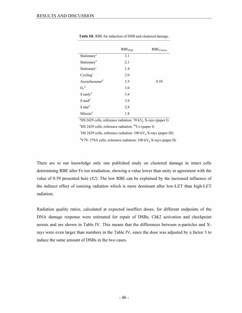
RESULTS AND DISCUSSION
- 46 -
Table III. RBE for induction of DSB and clustered damage.
RBEDSB
RBEClusters
Stationarya 3.1
Stationaryb 2.1
Stationaryc 1.4
Cyclingc 2.0
Asynchronousd 3.5 0.59
G1d 3.0
S earlyd 3.4
S midd 3.9
S lated 2.8
Mitosisd 1.8
aHS 2429 cells, reference radiation: 70 kVp X-rays (paper I)
bHS 2429 cells, reference radiation: 60Co (paper I)
cHS 2429 cells, reference radiation: 100 kVp X-rays (paper III)
dV79–379A cells, reference radiation: 100 kVp X-rays (paper II)
There are to our knowledge only one published study on clustered damage in intact cells
determining RBE after Fe ion irradiation, showing a value lower than unity in agreement with the
value of 0.59 presented here (82). The low RBE can be explained by the increased influence of
the indirect effect of ionizing radiation which is more dominant after low-LET than high-LET
radiation.
Radiation quality ratios, calculated at expected isoeffect doses, for different endpoints of the
DNA damage response were estimated for repair of DSBs, Chk2 activation and checkpoint
arrests and are shown in Table IV. This means that the differences between α-particles and X-
rays were even larger than numbers in the Table IV, since the dose was adjusted by a factor 3 to
induce the same amount of DSBs in the two cases.

RESULTS AND DISCUSSION
- 47 -
Table IV. Radiation quality ratios, after doses expected to induce the same amount of DSB, for different DNA
damage response endpoints.
Repair
3 ha
Residual
damage 24 ha
Chk2 24
hb
Chk2 48 hb G0/G1
arrestc
G2/M
arrestc
Stationary (HS 2429) 3.3 5.8 0.98 1.6 1.3
Cycling (HS 2429) 4.6 2.0 2.3 2.0 8.4
aPercentage of initial damage determined after 30 Gy 211At and 90 Gy X-rays.
bCalculated as the ratio of radiation effect between 1 Gy 211At and 3 Gy X-rays.
cCalculated as the ratio of radiation effect, expressed as the absolute area deviation from control values in cell cycle
distribution 0–72 h post-irradiation, between 1 Gy 211At and 3 Gy X-rays.
Radiation quality ratios varied with repair time. In stationary cells, the ratio increased with repair
time while the inverted relationship was found in cycling cells. This could in part be explained by
the very efficient repair after X-rays in stationary cells while cycling cells had insufficient repair
after both 211At and X-rays, resulting in a lower ratio between the two qualities.
Radiation quality ratios for cell cycle arrest (Chk2 activation and arrest in G0/G1) were lower in
stationary cells than in cycling cells (Chk2 activation and arrest in G2/M). RBE values for α-
particles between 4 and 9 for G2/M delay have been reported by others (90, 92, 116). In those
investigations these RBEs were determined either by comparing the doses required to produce
the maximum value of cells in G2/M at one specific time or by calculating the ratio of doses
needed to increase the fraction of cells in G2/M to a specific level. Only a few studies have
estimated RBE for G0/G1 arrests, ranging from 0.8 to 3 (96), lower than those reported for G2/M
arrests, in accordance with the data presented here.
Radiation quality ratios for biological consequences of 211At were determined for chromosomal
damage, long-term cell cycle disturbances and for clonogenic survival (Table V).

RESULTS AND DISCUSSION
- 48 -
Table V. Radiation quality ratios or RBE values for biological consequences.
Cell cycle phase
MN 72 h ratio
G0/G1
arresta
G2/M
arresta
RBE37%
SF2 ratio
Stationaryb 4.1 3.1
Cyclingb 2.7 13
Asynchronousc 8.6 18
G1 phasec 6.1 38
S phase earlyc 7.4 31
S phase midc 6.1 13
S phase latec 7.9 28
Mitosisc 3.1 6.9
aCalculated as the ratio of radiation effect, expressed as the absolute area deviation from control values
in cell cycle distribution 0–22 days post-irradiation, between 1 Gy 211At and 3 Gy X-rays.
Radiation quality ratios for chromosomal damage, assessed as MN formation, was 2.7 and 4.1,
72 h post-irradiation for cycling and stationary cells, respectively. Several studies have
determined RBE of α-particles for MN formation and values from 2.5 to 4.3 depending on LET,
cell line used and proliferation status have been reported (117-119), supporting the values shown
here.
The correlation between surviving fraction and radiation dose is not linear and therefore, RBE
will vary depending on the level of damage investigated. RBE for 37% survival varied between
3.1 and 8.6 and SF2 ratio between 6.9 and 38, depending on cell cycle phase. This demonstrates
that at higher doses within the clinically relevant range, differences between radiation qualities
will increase. Again, the RBE in cells irradiated in mitosis deviated notably from the other cell
cycle phases. A few studies have been published on RBE determination of α-particles from 211At
at 37% cell survival, and values ranging from 1.6 to 12 have been reported for different cell types
(66, 115, 120). Relative biological effectiveness of α-particles from 211At determined in vivo has
also been investigated. Bäck et al. (121) reported a RBE of 4.8 for growth inhibition of
subcutaneous xenografts of human ovarian cancer cells implanted in nude mice, and Elgqvist et
al. (122) reported RBEs of 3.4 and 5.9 for myelotoxicity, assessed as white blood cell counts, in
nude mice after intravenous or intraperitoneal administration of antibody-labeled 211At.
In summary, RBE values and radiation quality ratios of α-particles from 211At were dependent on
endpoint and dose level investigated, and varied depending on proliferation status.

SUMMARY AND CONCLUSIONS
- 49 -
SUMMARY AND CONCLUSIONS
To improve development of safe external and internal radiotherapy using high-LET radiation
it is important to increase our knowledge of early DNA interactions, cellular responses and
biological consequences of different high-LET radiation qualities. The aim of this thesis was
to investigate radiobiological effects in cells after exposure to α-particles from 211At, a
promising candidate nuclide for α-RIT of isolated tumor cells and microscopic clusters, and
X-rays in normal fibroblasts. In vitro, fibroblasts have been frequently used in radiobiology
studies, and represent a normal cell type with functioning checkpoint arrest, repair and
genomic surveillance signalling. In tissues, it is an abundant cell type in epithelium. In
radiotherapy, irradiated fibroblasts often give rise to late consequences that can be observed
months or years after treatment. In culture, fibroblasts can either be irradiated as a resting
(stationary) population or as a proliferating culture after plating.
From the data presented here it is obvious that 211At was more detrimental than X-rays. The
induction of DSBs was higher with correlated breaks along the α-particle track. In cooperation
with insufficient DSB repair, strong arrest activation was found with more Chk2-foci after
211At compared with X-rays. Formation of chromosomal damage and effects on proliferation
were also more prominent. Also, the clonogenic survival was reduced.
Interestingly, and in contrast to what is generally accepted, we found a cell cycle dependence
for DSB induction and clonogenic survival in synchronized fibroblasts after irradiation with
211At. For most endpoints studied here, a differential radioresponse between cells irradiated as
stationary or cycling cultures was found, with cycling cells being more sensitive to ionizing
radiation. Since irradiated tissue contains proliferating tumor cells as well as surrounding non-
proliferative normal tissue, such differential radioresponse could be of great importance and
should be further investigated.

ACKNOWLEDGEMENTS
- 50 -
ACKNOWLEDGEMENTS
This work has been performed at the Department of Oncology, University of Gothenburg,
Sweden and was supported by King Gustav V Jubilee Clinic Cancer Research Foundation, the
Swedish Cancer Society, the Swedish Radiation Authority and Assar Gabrielsson Foundation.
I would like to thank
My supervisor Kecke Elmroth, for introducing me to the interesting field of radiobiology.
Thanks for all your support on the way and for always being at hand. You have been an
excellent supervisor.
My co-supervisors Lars Jacobsson and Ragnar Hultborn, for all support on the way
Karin Magnander, my roommate and group-member, for excellent collaboration, valuable
discussions and your encouraging support during these years.
Madeleine Nordén Lyckesvärd, my roommate and group-member, for excellent help with
my work.
Helena Kahu and Ulla Delle, for excellent help with my work.
The TAT-group, especially Sture Lindegren for supplying Astatine-211.
All other staff and former PhD students at the Laboratory of Oncology for creating a friendly
atmosphere and a pleasent workplace.
My family

REFERENCES
- 51 -
REFERENCES
1. T. T. Puck and P. I. Marcus, A rapid method for viable cell titration and clone
production with HELA cells in tissue culture: the use of X-irradiated cells to supply
conditioning factors. Proc. Natl. Acad. Sci. U. S. A. 41, 432-437 (1955).
2. D. T. Goodhead, The initial physical damage produced by ionizing radiations. Int. J.
Radiat. Biol. 56, 623-634 (1989).
3. J. P. Pouget and S. J. Mather, General aspects of the cellular response to low- and
high-LET radiation. Eur. J. Nucl. Med. 28, 541-561 (2001).
4. C. J. Norbury and I. D. Hickson, Cellular responses to DNA damage. Annu. Rev.
Pharmacol. Toxicol. 41, 367-401 (2001).
5. L. Galluzzi, M. C. Maiuri, I. Vitale, H. Zischka, M. Castedo, L. Zitvogel and G.
Kroemer, Cell death modalities: classification and pathophysiological implications.
Cell Death Differ. 14, 1237-1243 (2007).
6. W. C. Dewey, C. C. Ling and R. E. Meyn, Radiation-induced apoptosis: relevance to
radiotherapy. Int. J. Radiat. Oncol. Biol. Phys. 33, 781-796 (1995).
7. R. Roots and S. Okada, Protection of DNA molecules of cultured mammalian cells
from radiation-induced single-strand scissions by various alcohols and SH
compounds. Int. J. Radiat. Biol. Relat. Stud. Phys. Chem. Med. 21, 329-342 (1972).
8. R. Hirayama, A. Ito, M. Tomita, T. Tsukada, F. Yatagai, M. Noguchi, Y. Matsumoto,
Y. Kase, K. Ando, et al., Contributions of direct and indirect actions in cell killing by
high-LET radiations. Radiat. Res. 171, 212-218 (2009).
9. D. T. Goodhead, Mechanisms for the biological effectiveness of high-LET radiations.
J. Radiat. Res. (Tokyo). 40 Suppl, 1-13 (1999).
10. P. E. Bryant, Enzymatic restriction of mammalian cell DNA: evidence for double-
strand breaks as potentially lethal lesions. Int. J. Radiat. Biol. Relat. Stud. Phys. Chem.
Med. 48, 55-60 (1985).
11. S. Vispe and M. S. Satoh, DNA repair patch-mediated double strand DNA break
formation in human cells. J. Biol. Chem. 275, 27386-27392 (2000).
12. M. Lobrich, P. K. Cooper and B. Rydberg, Non-random distribution of DNA double-
strand breaks induced by particle irradiation. Int. J. Radiat. Biol. 70, 493-503 (1996).

REFERENCES
- 52 -
13. B. Stenerlow, E. Hoglund, K. Elmroth, K. H. Karlsson and I. Radulescu, Radiation
quality dependence of DNA damage induction. Radiat. Prot. Dosimetry 99, 137-141
(2002).
14. S. S. Wallace, Enzymatic processing of radiation-induced free radical damage in
DNA. Radiat. Res. 150, S60-79 (1998).
15. K. Magnander, R. Hultborn, K. Claesson and K. Elmroth, Clustered DNA damage in
irradiated human diploid fibroblasts: influence of chromatin organization. Radiat. Res.
173, 272-282 (2010).
16. M. Hada and B. M. Sutherland, Spectrum of complex DNA damages depends on the
incident radiation. Radiat. Res. 165, 223-230 (2006).
17. J. Nygren, M. Ljungman and G. Ahnstrom, Chromatin structure and radiation-induced
DNA strand breaks in human cells: soluble scavengers and DNA-bound proteins offer
a better protection against single- than double-strand breaks. Int. J. Radiat. Biol. 68,
11-18 (1995).
18. C. Richardson and M. Jasin, Coupled homologous and nonhomologous repair of a
double-strand break preserves genomic integrity in mammalian cells. Mol. Cell Biol.
20, 9068-9075 (2000).
19. C. Allen, J. Halbrook and J. A. Nickoloff, Interactive competition between
homologous recombination and non-homologous end joining. Mol. Cancer Res. 1,
913-920 (2003).
20. K. Valerie and L. F. Povirk, Regulation and mechanisms of mammalian double-strand
break repair. Oncogene 22, 5792-5812 (2003).
21. B. K. Singleton, M. I. Torres-Arzayus, S. T. Rottinghaus, G. E. Taccioli and P. A.
Jeggo, The C terminus of Ku80 activates the DNA-dependent protein kinase catalytic
subunit. Mol. Cell Biol. 19, 3267-3277 (1999).
22. Y. Ma, U. Pannicke, K. Schwarz and M. R. Lieber, Hairpin opening and overhang
processing by an Artemis/DNA-dependent protein kinase complex in nonhomologous
end joining and V(D)J recombination. Cell 108, 781-794 (2002).
23. J. Huang and W. S. Dynan, Reconstitution of the mammalian DNA double-strand
break end-joining reaction reveals a requirement for an Mre11/Rad50/NBS1-
containing fraction. Nucleic Acids Res. 30, 667-674 (2002).
24. H. L. Hsu, S. M. Yannone and D. J. Chen, Defining interactions between DNA-PK
and ligase IV/XRCC4. DNA Repair (Amst) 1, 225-235 (2002).

REFERENCES
- 53 -
25. N. Saleh-Gohari and T. Helleday, Conservative homologous recombination
preferentially repairs DNA double-strand breaks in the S phase of the cell cycle in
human cells. Nucleic Acids Res. 32, 3683-3688 (2004).
26. C. Wyman, D. Ristic and R. Kanaar, Homologous recombination-mediated double-
strand break repair. DNA Repair (Amst) 3, 827-833 (2004).
27. B. Stenerlow, E. Blomquist, E. Grusell, T. Hartman and J. Carlsson, Rejoining of
DNA double-strand breaks induced by accelerated nitrogen ions. Int. J. Radiat. Biol.
70, 413-420 (1996).
28. T. J. Jenner, C. M. deLara, P. O'Neill and D. L. Stevens, Induction and rejoining of
DNA double-strand breaks in V79-4 mammalian cells following gamma- and alpha-
irradiation. Int. J. Radiat. Biol. 64, 265-273 (1993).
29. D. Blocher, DNA double-strand break repair determines the RBE of alpha-particles.
Int. J. Radiat. Biol. 54, 761-771 (1988).
30. M. A. Ritter, J. E. Cleaver and C. A. Tobias, High-LET radiations induce a large
proportion of non-rejoining DNA breaks. Nature 266, 653-655 (1977).
31. D. Klokov, S. M. MacPhail, J. P. Banath, J. P. Byrne and P. L. Olive, Phosphorylated
histone H2AX in relation to cell survival in tumor cells and xenografts exposed to
single and fractionated doses of X-rays. Radiother. Oncol. 80, 223-229 (2006).
32. N. Foray, C. F. Arlett and E. P. Malaise, Radiation-induced DNA double-strand
breaks and the radiosensitivity of human cells: a closer look. Biochimie 79, 567-575
(1997).
33. B. Elliott and M. Jasin, Double-strand breaks and translocations in cancer. Cell. Mol.
Life Sci. 59, 373-385 (2002).
34. B. B. Zhou and S. J. Elledge, The DNA damage response: putting checkpoints in
perspective. Nature 408, 433-439 (2000).
35. J. Lukas, C. Lukas and J. Bartek, Mammalian cell cycle checkpoints: signalling
pathways and their organization in space and time. DNA Repair (Amst) 3, 997-1007
(2004).
36. J. H. Lee and T. T. Paull, Activation and regulation of ATM kinase activity in
response to DNA double-strand breaks. Oncogene 26, 7741-7748 (2007).
37. Y. Andegeko, L. Moyal, L. Mittelman, I. Tsarfaty, Y. Shiloh and G. Rotman, Nuclear
retention of ATM at sites of DNA double strand breaks. J. Biol. Chem. 276, 38224-
38230 (2001).

REFERENCES
- 54 -
38. J. Falck, N. Mailand, R. G. Syljuasen, J. Bartek and J. Lukas, The ATM-Chk2-
Cdc25A checkpoint pathway guards against radioresistant DNA synthesis. Nature
410, 842-847 (2001).
39. A. Hirao, Y. Y. Kong, S. Matsuoka, A. Wakeham, J. Ruland, H. Yoshida, D. Liu, S. J.
Elledge and T. W. Mak, DNA damage-induced activation of p53 by the checkpoint
kinase Chk2. Science 287, 1824-1827 (2000).
40. S. Matsuoka, M. Huang and S. J. Elledge, Linkage of ATM to cell cycle regulation by
the Chk2 protein kinase. Science 282, 1893-1897 (1998).
41. T. Terasima and L. J. Tolmach, X-ray sensitivity and DNA synthesis in synchronous
populations of HeLa cells. Science 140, 490-492 (1963).
42. H. R. Withers, K. Mason, B. O. Reid, N. Dubravsky, H. T. Barkley, Jr., B. W. Brown
and J. B. Smathers, Response of mouse intestine to neutrons and gamma rays in
relation to dose fractionation and division cycle. Cancer 34, 39-47 (1974).
43. R. P. Bird and H. J. Burki, Survival of synchronized Chinese hamster cells exposed to
radiation of different linear-energy transfer. Int. J. Radiat. Biol. Relat. Stud. Phys.
Chem. Med. 27, 105-120 (1975).
44. F. Q. Ngo, E. A. Blakely, C. A. Tobias, P. Y. Chang and L. Lommel, Sequential
exposures of mammalian cells to low- and high-LET radiations. II. As a function of
cell-cycle stages. Radiat. Res. 115, 54-69 (1988).
45. H. Wang, S. Liu, P. Zhang, S. Zhang, M. Naidu and Y. Wang, S-phase cells are more
sensitive to high-linear energy transfer radiation. Int. J. Radiat. Oncol. Biol. Phys. 74,
1236-1241 (2009).
46. F. Zafar, S. B. Seidler, A. Kronenberg, D. Schild and C. Wiese, Homologous
recombination contributes to the repair of DNA double-strand breaks induced by high-
energy iron ions. Radiat. Res. 173, 27-39 (2010).
47. I. R. Radford, Effect of cell-cycle position and dose on the kinetics of DNA double-
strand breakage repair in X-irradiated Chinese hamster cells. Int. J. Radiat. Biol. Relat.
Stud. Phys. Chem. Med. 52, 555-564 (1987).
48. K. Luger, A. W. Mader, R. K. Richmond, D. F. Sargent and T. J. Richmond, Crystal
structure of the nucleosome core particle at 2.8 A resolution. Nature 389, 251-260
(1997).
49. M. Falk, E. Lukasova and S. Kozubek, Chromatin structure influences the sensitivity
of DNA to gamma-radiation. Biochim. Biophys. Acta 1783, 2398-2414 (2008).
REFERENCES
- 55 -
50. K. F. Toth, T. A. Knoch, M. Wachsmuth, M. Frank-Stohr, M. Stohr, C. P. Bacher, G.
Muller and K. Rippe, Trichostatin A-induced histone acetylation causes
decondensation of interphase chromatin. J. Cell Sci. 117, 4277-4287 (2004).
51. K. B. Glaser, HDAC inhibitors: clinical update and mechanism-based potential.
Biochem. Pharmacol. 74, 659-671 (2007).
52. R. R. Rosato and S. Grant, Histone deacetylase inhibitors in clinical development.
Expert Opin. Investig. Drugs 13, 21-38 (2004).
53. I. Radulescu, K. Elmroth and B. Stenerlow, Chromatin organization contributes to
non-randomly distributed double-strand breaks after exposure to high-LET radiation.
Radiat. Res. 161, 1-8 (2004).
54. C. P. Karger and O. Jakel, Current status and new developments in ion therapy.
Strahlenther. Onkol. 183, 295-300 (2007).
55. H. Song, R. F. Hobbs, R. Vajravelu, D. L. Huso, C. Esaias, C. Apostolidis, A.
Morgenstern and G. Sgouros, Radioimmunotherapy of breast cancer metastases with
alpha-particle emitter 225Ac: comparing efficacy with 213Bi and 90Y. Cancer Res.
69, 8941-8948 (2009).
56. D. E. Milenic, K. J. Wong, K. E. Baidoo, T. K. Nayak, C. A. Regino, K. Garmestani
and M. W. Brechbiel, Targeting HER2: a report on the in vitro and in vivo pre-clinical
data supporting trastuzumab as a radioimmunoconjugate for clinical trials. MAbs 2,
550-564 (2010).
57. G. W. Barendsen, C. J. Koot, G. R. Van Kersen, D. K. Bewley, S. B. Field and C. J.
Parnell, The effect of oxygen on impairment of the proliferative capacity of human
cells in culture by ionizing radiations of different LET. Int. J. Radiat. Biol. Relat. Stud.
Phys. Chem. Med. 10, 317-327 (1966).
58. H. Andersson, E. Cederkrantz, T. Back, C. Divgi, J. Elgqvist, J. Himmelman, G.
Horvath, L. Jacobsson, H. Jensen, et al., Intraperitoneal alpha-particle
radioimmunotherapy of ovarian cancer patients: pharmacokinetics and dosimetry of
(211)At-MX35 F(ab')2--a phase I study. J. Nucl. Med. 50, 1153-1160 (2009).
59. M. R. Zalutsky, D. A. Reardon, O. R. Pozzi, G. Vaidyanathan and D. D. Bigner,
Targeted alpha-particle radiotherapy with 211At-labeled monoclonal antibodies. Nucl.
Med. Biol. 34, 779-785 (2007).
60. J. M. Burke, P. C. Caron, E. B. Papadopoulos, C. R. Divgi, G. Sgouros, K. S.
Panageas, R. D. Finn, S. M. Larson, R. J. O'Reilly, et al., Cytoreduction with iodine-

REFERENCES
- 56 -
131-anti-CD33 antibodies before bone marrow transplantation for advanced myeloid
leukemias. Bone Marrow Transplant. 32, 549-556 (2003).
61. B. J. Allen, C. Raja, S. Rizvi, Y. Li, W. Tsui, P. Graham, J. F. Thompson, R. A.
Reisfeld and J. Kearsley, Intralesional targeted alpha therapy for metastatic melanoma.
Cancer Biol. Ther. 4, 1318-1324 (2005).
62. S. Nilsson, R. H. Larsen, S. D. Fossa, L. Balteskard, K. W. Borch, J. E. Westlin, G.
Salberg and O. S. Bruland, First clinical experience with alpha-emitting radium-223 in
the treatment of skeletal metastases. Clin. Cancer Res. 11, 4451-4459 (2005).
63. M. Lalande, A reversible arrest point in the late G1 phase of the mammalian cell
cycle. Exp. Cell Res. 186, 332-339 (1990).
64. Y. Saintigny, F. Delacote, G. Vares, F. Petitot, S. Lambert, D. Averbeck and B. S.
Lopez, Characterization of homologous recombination induced by replication
inhibition in mammalian cells. EMBO J. 20, 3861-3870 (2001).
65. S. Lindegren, T. Back and H. J. Jensen, Dry-distillation of astatine-211 from irradiated
bismuth targets: a time-saving procedure with high recovery yields. Appl. Radiat. Isot.
55, 157-160 (2001).
66. S. Palm, H. Andersson, T. Back, I. Claesson, U. Delle, R. Hultborn, L. Jacobsson, I.
Kopf and S. Lindegren, In vitro effects of free 211At,211At-albumin and 211At-
monoclonal antibody compared to external photon irradiation on two human cancer
cell lines. Anticancer Res. 20, 1005-1012 (2000).
67. S. Lindegren, H. Andersson, T. Back, L. Jacobsson, B. Karlsson and G. Skarnemark,
High-efficiency astatination of antibodies using N-iodosuccinimide as the oxidising
agent in labelling of N-succinimidyl 3-(trimethylstannyl)benzoate. Nucl. Med. Biol.
28, 33-39 (2001).
68. S. Lindegren, S. Frost, T. Back, E. Haglund, J. Elgqvist and H. Jensen, Direct
procedure for the production of 211At-labeled antibodies with an epsilon-lysyl-3-
(trimethylstannyl)benzamide immunoconjugate. J. Nucl. Med. 49, 1537-1545 (2008).
69. N. Chouin, K. Bernardeau, F. Davodeau, M. Cherel, A. Faivre-Chauvet, M.
Bourgeois, C. Apostolidis, A. Morgenstern, A. Lisbona and M. Bardies, Evidence of
extranuclear cell sensitivity to alpha-particle radiation using a microdosimetric model.
I. Presentation and validation of a microdosimetric model. Radiat. Res. 171, 657-663
(2009).
REFERENCES
- 57 -
70. B. M. Sutherland, P. V. Bennett, O. Sidorkina and J. Laval, Clustered damages and
total lesions induced in DNA by ionizing radiation: oxidized bases and strand breaks.
Biochemistry (Mosc). 39, 8026-8031 (2000).
71. D. C. Schwartz and C. R. Cantor, Separation of yeast chromosome-sized DNAs by
pulsed field gradient gel electrophoresis. Cell 37, 67-75 (1984).
72. D. Blocher, In CHEF electrophoresis a linear induction of dsb corresponds to a
nonlinear fraction of extracted DNA with dose. Int. J. Radiat. Biol. 57, 7-12 (1990).
73. L. G. Littlefield, A. M. Sayer and E. L. Frome, Comparisons of dose-response
parameters for radiation-induced acentric fragments and micronuclei observed in
cytokinesis-arrested lymphocytes. Mutagenesis 4, 265-270 (1989).
74. M. Fenech, W. P. Chang, M. Kirsch-Volders, N. Holland, S. Bonassi and E. Zeiger,
HUMN project: detailed description of the scoring criteria for the cytokinesis-block
micronucleus assay using isolated human lymphocyte cultures. Mutat. Res. 534, 65-75
(2003).
75. E. Hoglund, E. Blomquist, J. Carlsson and B. Stenerlow, DNA damage induced by
radiation of different linear energy transfer: initial fragmentation. Int. J. Radiat. Biol.
76, 539-547 (2000).
76. H. C. Newman, K. M. Prise and B. D. Michael, The role of higher-order chromatin
structure in the yield and distribution of DNA double-strand breaks in cells irradiated
with X-rays or alpha-particles. Int. J. Radiat. Biol. 76, 1085-1093 (2000).
77. B. Rydberg, Clusters of DNA damage induced by ionizing radiation: formation of
short DNA fragments. II. Experimental detection. Radiat. Res. 145, 200-209 (1996).
78. K. Elmroth and B. Stenerlow, Influence of chromatin structure on induction of double-
strand breaks in mammalian cells irradiated with DNA-incorporated 125I. Radiat. Res.
168, 175-182 (2007).
79. G. E. Iliakis, O. Cicilioni and L. Metzger, Measurement of DNA double-strand breaks
in CHO cells at various stages of the cell cycle using pulsed field gel electrophoresis:
calibration by means of 125I decay. Int. J. Radiat. Biol. 59, 343-357 (1991).
80. B. M. Sutherland, P. V. Bennett, J. C. Sutherland and J. Laval, Clustered DNA
damages induced by x rays in human cells. Radiat. Res. 157, 611-616 (2002).
81. M. Gulston, J. Fulford, T. Jenner, C. de Lara and P. O'Neill, Clustered DNA damage
induced by gamma radiation in human fibroblasts (HF19), hamster (V79-4) cells and
plasmid DNA is revealed as Fpg and Nth sensitive sites. Nucleic Acids Res. 30, 3464-
3472 (2002).

REFERENCES
- 58 -
82. D. Tsao, P. Kalogerinis, I. Tabrizi, M. Dingfelder, R. D. Stewart and A. G.
Georgakilas, Induction and processing of oxidative clustered DNA lesions in 56Fe-
ion-irradiated human monocytes. Radiat. Res. 168, 87-97 (2007).
83. K. M. Prise, C. H. Pullar and B. D. Michael, A study of endonuclease III-sensitive
sites in irradiated DNA: detection of alpha-particle-induced oxidative damage.
Carcinogenesis 20, 905-909 (1999).
84. A. Asaithamby, N. Uematsu, A. Chatterjee, M. D. Story, S. Burma and D. J. Chen,
Repair of HZE-particle-induced DNA double-strand breaks in normal human
fibroblasts. Radiat. Res. 169, 437-446 (2008).
85. T. Groesser, H. Chang, G. Fontenay, J. Chen, S. V. Costes, M. Helen Barcellos-Hoff,
B. Parvin and B. Rydberg, Persistence of gamma-H2AX and 53BP1 foci in
proliferating and non-proliferating human mammary epithelial cells after exposure to
gamma-rays or iron ions. Int. J. Radiat. Biol. (2011).
86. Z. Vilasova, M. Rezacova, J. Vavrova, A. Tichy, D. Vokurkova, F. Zoelzer, Z.
Rehakova, J. Osterreicher and E. Lukasova, Changes in phosphorylation of histone
H2A.X and p53 in response of peripheral blood lymphocytes to gamma irradiation.
Acta Biochim. Pol. 55, 381-390 (2008).
87. C. Mayer, O. Popanda, O. Zelezny, M. C. von Brevern, A. Bach, H. Bartsch and P.
Schmezer, DNA repair capacity after gamma-irradiation and expression profiles of
DNA repair genes in resting and proliferating human peripheral blood lymphocytes.
DNA Repair (Amst) 1, 237-250 (2002).
88. K. Hamasaki, K. Imai, K. Nakachi, N. Takahashi, Y. Kodama and Y. Kusunoki,
Short-term culture and gammaH2AX flow cytometry determine differences in
individual radiosensitivity in human peripheral T lymphocytes. Environ. Mol.
Mutagen. 48, 38-47 (2007).
89. H. Wang, X. Wang, P. Zhang and Y. Wang, The Ku-dependent non-homologous end-
joining but not other repair pathway is inhibited by high linear energy transfer ionizing
radiation. DNA Repair (Amst) 7, 725-733 (2008).
90. C. Lucke-Huhle, E. A. Blakely, P. Y. Chang and C. A. Tobias, Drastic G2 arrest in
mammalian cells after irradiation with heavy-ion beams. Radiat. Res. 79, 97-112
(1979).
91. M. Scholz, W. Kraft-Weyrather, S. Ritter and G. Kraft, Cell cycle delays induced by
heavy ion irradiation of synchronous mammalian cells. Int. J. Radiat. Biol. 66, 59-75
(1994).

REFERENCES
- 59 -
92. S. T. Palayoor, J. L. Humm, R. W. Atcher, J. J. Hines and R. M. Macklis, G2M arrest
and apoptosis in murine T lymphoma cells following exposure to 212Bi alpha particle
irradiation. Nucl. Med. Biol. 20, 795-805 (1993).
93. M. R. Raju, T. S. Johnson, N. Tokita, S. Carpenter and J. H. Jett, Differences in cell-
cycle progression delays after exposure to 238Pu alpha particles compared to X rays.
Radiat. Res. 84, 16-24 (1980).
94. G. Buscemi, P. Perego, N. Carenini, M. Nakanishi, L. Chessa, J. Chen, K. Khanna and
D. Delia, Activation of ATM and Chk2 kinases in relation to the amount of DNA
strand breaks. Oncogene 23, 7691-7700 (2004).
95. E. Nasonova, K. Fussel, S. Berger, E. Gudowska-Nowak and S. Ritter, Cell cycle
arrest and aberration yield in normal human fibroblasts. I. Effects of X-rays and 195
MeV u(-1) C ions. Int. J. Radiat. Biol. 80, 621-634 (2004).
96. S. Tenhumberg, E. Gudowska-Nowak, E. Nasonova and S. Ritter, Cell cycle arrest
and aberration yield in normal human fibroblasts. II: Effects of 11 MeV u-1 C ions
and 9.9 MeV u-1 Ni ions. Int. J. Radiat. Biol. 83, 501-513 (2007).
97. E. I. Azzam, S. M. de Toledo, A. J. Waker and J. B. Little, High and low fluences of
alpha-particles induce a G1 checkpoint in human diploid fibroblasts. Cancer Res. 60,
2623-2631 (2000).
98. C. Lucke-Huhle, Alpha-irradiation-induced G2 delay: a period of cell recovery.
Radiat. Res. 89, 298-308 (1982).
99. D. Deckbar, J. Birraux, A. Krempler, L. Tchouandong, A. Beucher, S. Walker, T.
Stiff, P. Jeggo and M. Lobrich, Chromosome breakage after G2 checkpoint release. J.
Cell Biol. 176, 749-755 (2007).
100. I. Diehl-Marshall and M. Bianchi, Induction of micronuclei by irradiation with
neutrons produced from 600 MeV protons. Br. J. Radiol. 54, 530-532 (1981).
101. R. M. Anderson, S. J. Marsden, E. G. Wright, M. A. Kadhim, D. T. Goodhead and C.
S. Griffin, Complex chromosome aberrations in peripheral blood lymphocytes as a
potential biomarker of exposure to high-LET alpha-particles. Int. J. Radiat. Biol. 76,
31-42 (2000).
102. P. Virsik-Kopp and H. Hofman-Huether, Chromosome aberrations induced by high-
LET carbon ions in radiosensitive and radioresistant tumour cells. Cytogenet. Genome
Res. 104, 221-226 (2004).
REFERENCES
- 60 -
103. S. Paglin, T. Delohery, R. Erlandson and J. Yahalom, Radiation-induced micronuclei
formation in human breast cancer cells: dependence on serum and cell cycle
distribution. Biochem. Biophys. Res. Commun. 237, 678-684 (1997).
104. A. Banasik, A. Lankoff, A. Piskulak, K. Adamowska, H. Lisowska and A. Wojcik,
Aluminum-induced micronuclei and apoptosis in human peripheral-blood
lymphocytes treated during different phases of the cell cycle. Environ. Toxicol. 20,
402-406 (2005).
105. S. Masunaga, K. Ando, A. Uzawa, R. Hirayama, Y. Furusawa, S. Koike and K. Ono,
The radiosensitivity of total and quiescent cell populations in solid tumors to 290
MeV/u carbon ion beam irradiation in vivo. Acta Oncol. 47, 1087-1093 (2008).
106. S. Giunta, R. Belotserkovskaya and S. P. Jackson, DNA damage signaling in response
to double-strand breaks during mitosis. J. Cell Biol. 190, 197-207 (2010).
107. A. Krempler, D. Deckbar, P. A. Jeggo and M. Lobrich, An imperfect G2M checkpoint
contributes to chromosome instability following irradiation of S and G2 phase cells.
Cell Cycle 6, 1682-1686 (2007).
108. J. Bussink, P. J. Tofilon and W. A. Brock, Repair of chromosome and DNA breaks
versus cell survival in Chinese hamster cells. Int. J. Radiat. Biol. 70, 23-32 (1996).
109. K. Rothkamm, I. Kruger, L. H. Thompson and M. Lobrich, Pathways of DNA double-
strand break repair during the mammalian cell cycle. Mol. Cell Biol. 23, 5706-5715
(2003).
110. I. A. Kim, J. H. Kim, J. H. Shin, I. H. Kim, J. S. Kim, H. G. Wu, E. K. Chie, Y. H.
Kim, B. K. Kim, et al., A histone deacetylase inhibitor, trichostatin A, enhances
radiosensitivity by abrogating G2/M arrest in human carcinoma cells. Cancer Res.
Treat. 37, 122-128 (2005).
111. F. Zhang, T. Zhang, Z. H. Teng, R. Zhang, J. B. Wang and Q. B. Mei, Sensitization to
gamma-irradiation-induced cell cycle arrest and apoptosis by the histone deacetylase
inhibitor trichostatin A in non-small cell lung cancer (NSCLC) cells. Cancer Biol.
Ther. 8, 823-831 (2009).
112. K. Elmroth, J. Nygren, L. J. Erkell and R. Hultborn, Radiation-induced double-strand
breaks in mammalian DNA: influence of temperature and DMSO. Int. J. Radiat. Biol.
76, 1501-1508 (2000).
113. K. Elmroth, J. Nygren, B. Stenerlow and R. Hultborn, Chromatin- and temperature-
dependent modulation of radiation-induced double-strand breaks. Int. J. Radiat. Biol.
79, 809-816 (2003).

REFERENCES
- 61 -
114. H. C. Newman, K. M. Prise, M. Folkard and B. D. Michael, DNA double-strand break
distributions in X-ray and alpha-particle irradiated V79 cells: evidence for non-
random breakage. Int. J. Radiat. Biol. 71, 347-363 (1997).
115. M. A. Walicka, G. Vaidyanathan, M. R. Zalutsky, S. J. Adelstein and A. I. Kassis,
Survival and DNA damage in Chinese hamster V79 cells exposed to alpha particles
emitted by DNA-incorporated astatine-211. Radiat. Res. 150, 263-268 (1998).
116. C. Lucke-Huhle, W. Comper, L. Hieber and M. Pech, Comparative study of G2 delay
and survival after 241 Americium-alpha and 60cobalt-gamma irradiation. Radiat.
Environ. Biophys. 20, 171-185 (1982).
117. Y. Yamada, Y. Oghiso, H. Enomoto and N. Ishigure, Induction of micronuclei in a rat
alveolar epithelial cell line by alpha particle irradiation. Radiat. Prot. Dosimetry 99,
219-222 (2002).
118. A. Bilbao, J. S. Prosser, A. A. Edwards, J. C. Moody and D. C. Lloyd, The induction
of micronuclei in human lymphocytes by in vitro irradiation with alpha particles from
plutonium-239. Int. J. Radiat. Biol. 56, 287-292 (1989).
119. A. L. Brooks, G. J. Newton, L. J. Shyr, F. A. Seiler and B. R. Scott, The combined
effects of alpha-particles and X-rays on cell killing and micronuclei induction in lung
epithelial cells. Int. J. Radiat. Biol. 58, 799-811 (1990).
120. E. Aurlien, R. H. Larsen, G. Akabani, D. R. Olsen, M. R. Zalutsky and O. S. Bruland,
Exposure of human osteosarcoma and bone marrow cells to tumour-targeted alpha-
particles and gamma-irradiation: analysis of cell survival and microdosimetry. Int. J.
Radiat. Biol. 76, 1129-1141 (2000).
121. T. Back, H. Andersson, C. R. Divgi, R. Hultborn, H. Jensen, S. Lindegren, S. Palm
and L. Jacobsson, 211At radioimmunotherapy of subcutaneous human ovarian cancer
xenografts: evaluation of relative biologic effectiveness of an alpha-emitter in vivo. J.
Nucl. Med. 46, 2061-2067 (2005).
122. J. Elgqvist, P. Bernhardt, R. Hultborn, H. Jensen, B. Karlsson, S. Lindegren, E.
Warnhammar and L. Jacobsson, Myelotoxicity and RBE of 211At-conjugated
monoclonal antibodies compared with 99mTc-conjugated monoclonal antibodies and
60Co irradiation in nude mice. J. Nucl. Med. 46, 464-471 (2005).

![Currently, thirty-nine astatine, thirty-nine radon, thirty …[26]. The name astatine was o cially accepted at the 15thIUPAC conference in Amsterdam in 1949 [27]. Thirty-nine astatine](https://static.fdocuments.us/doc/165x107/5e3842530588dd34cd732836/currently-thirty-nine-astatine-thirty-nine-radon-thirty-26-the-name-astatine.jpg)