7.3 Glycoconjugates: Proteoglycans, glycoproteins, and Glycolipids.

CHAPTER 17Preparation and Analysis ofGlycoconjugates
Ajit Varki,1 Hudson H. Freeze,2 and Adriana E. Manzi1
1University of California San Diego, La Jolla, California2Sanford Children’s Health Research Center and Burnham Institute for Medical Research,La Jolla, California
ABSTRACT
Whereas DNA, RNA, and proteins are linear polymers that can usually be directly sequenced,glycans show substantially more complexity, having branching and anomeric configurations(α and β linkages). The biosynthesis of glycans, termed glycosylation, is extremely complex,is not template-driven, varies among different cell types, and cannot be easily predicted fromsimple rules. This overview discusses the stereochemistry of mono- and oligosaccharides andprovides diagrammatic representations of monosaccharides (Fisher projections and Haworthrepresentations) and formulas for representation of glycan chains. A glossary of terms used inglycobiology is also provided. Curr. Protoc. Mol. Biol. 88:17.0.1-17.0.12. C© 2009 by John Wiley& Sons, Inc.
Keywords: glycan monosaccharide glycan analysis sugar symbols glycoconjugate
glycobiology
INTRODUCTIONThe modern revolution in molecular biol-
ogy was driven to a large extent by advancesin methods for analysis and manipulation ofDNA, RNA, and proteins. Although oligosac-charides (sugar chains or glycans) are also ma-jor macromolecules of the typical cell, they didnot initially share in this molecular revolution.The reasons for this were to a large extenttechnical. Whereas DNA, RNA, and proteinsare linear polymers that can usually be di-rectly sequenced, oligosaccharides show sub-stantially more complexity, having branchingand anomeric configurations (α and β link-ages). Thus, whereas three amino acids ornucleotides can be combined into six possi-ble sequences, three hexose monosaccharidescan theoretically generate 1056 possible gly-cans. In addition, the syntheses of DNA, RNA,and proteins are template-driven, and the se-quence of one can generally be predicted fromthat of another. In contrast, the biosynthe-sis of oligosaccharides, termed glycosylation,is extremely complex, is not template-driven,varies among different cell types, and cannotbe easily predicted from simple rules.
It is clear that glycans have important, al-beit varied, effects upon the biosynthesis, fold-
ing, solubility, stability, subcellular traffick-ing, turnover, and half-life of the moleculesto which they are attached. These are mattersof great importance to the cell biologist, pro-tein chemist, biotechnologist, and pharmacol-ogist. On the other hand, the successful growthof several glycosylation mutants as permanenttissue culture cell lines indicates that the pre-cise structure of many glycans is not criti-cal for the growth and viability of a singlecell in the protected environment of the cul-ture dish. Thus, until recently, it was possiblefor many researchers working with in vitrosingle-cell systems to ignore the existence ofglycans. However, with the increasing empha-sis on studying cell-cell interactions in normaldevelopment, tissue morphogenesis, immunereactions, and pathological conditions such ascancer and inflammation, the study of glycanstructure and biosynthesis has become veryimportant.
The term “glycobiology” has found accep-tance for denoting studies of the biology ofglycoconjugates in both simple and complexsystems. Many technical advances have oc-curred in the analysis of glycans, making itnow feasible to study them in detail. Theseadvances include the development of sensitive
Current Protocols in Molecular Biology 17.0.1-17.0.12, October 2009Published online October 2009 in Wiley Interscience (www.interscience.wiley.com).DOI: 10.1002/0471142727.mb1700s88Copyright C© 2009 John Wiley & Sons, Inc.
Preparation andAnalysis ofGlycoconjugates
17.0.1
Supplement 88
17.0.2
Supplement 88 Current Protocols in Molecular Biology
and specific assays and the availability of nu-merous purified enzymes (glycosidases) withhigh degrees of specificity.
In spite of all these advances, many analyti-cal techniques in glycobiology remained in thedomain of the few laboratories that specializedin the study of glycans. Likewise, publishedcompendia of carbohydrate methods were de-signed mainly for use by experts. This chap-ter attempts to place some of this technologywithin easy reach of any laboratory with basiccapabilities in biochemistry and molecular bi-ology. The techniques described here includemodern versions of time-honored methods andrecently developed methods, both of whichhave widespread applications. However, it isimportant to emphasize that the protocols pre-sented here are by no means comprehensive.Rather, they serve as a starting point for theuninitiated scientist who wishes to explore thestructure, biosynthesis, and biology of gly-can chains. In most cases, further analysis us-ing more sophisticated techniques will be re-
quired to obtain final and definitive results.Nonetheless, armed with results obtained us-ing the techniques described here, the typi-cal researcher can make intelligent decisionsabout the need for such further analyses.
The appearance of many commercial kitsfor analysis of glycoconjugates is another signthat the technology has arrived and that manylaboratories have developed an interest in gly-cobiology. It is worth noting that althoughsome of these kits are designed to simplifythe use of well-established techniques, othersemploy methodologies that have been newlydeveloped by the companies themselves. Ex-perience with the latter methodologies in aca-demic scientific laboratories may be limited,and the techniques in question may thereforenot be represented in this chapter. However,although the admonition caveat emptor is ap-propriate, some kits may well become usefuladjuncts to the methods presented here.
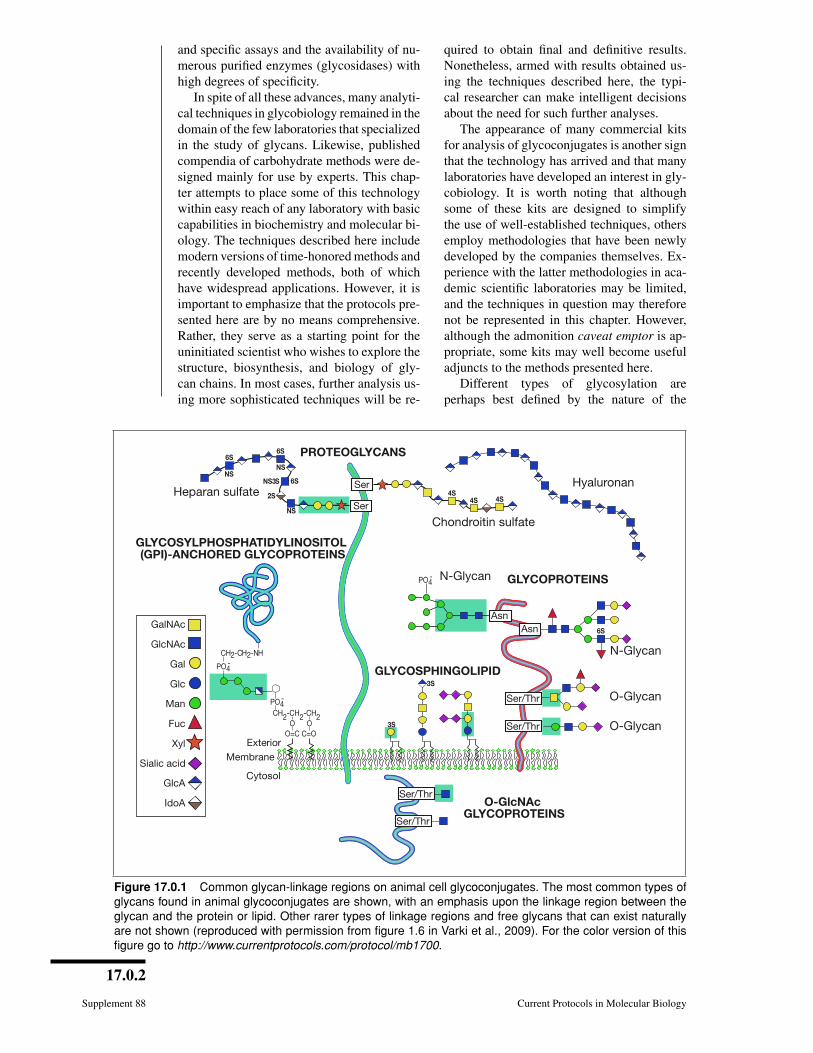
Different types of glycosylation areperhaps best defined by the nature of the
Figure 17.0.1 Common glycan-linkage regions on animal cell glycoconjugates. The most common types ofglycans found in animal glycoconjugates are shown, with an emphasis upon the linkage region between theglycan and the protein or lipid. Other rarer types of linkage regions and free glycans that can exist naturallyare not shown (reproduced with permission from figure 1.6 in Varki et al., 2009). For the color version of thisfigure go to http://www.currentprotocols.com/protocol/mb1700.

17.0.3
Current Protocols in Molecular Biology Supplement 88
linkage region of the oligosaccharide toa lipid or protein (Fig. 17.0.1). Althoughthe linkage regions of these molecules areunique, the sugar chains frequently tend toshare common types of outer sequences. Itis important to note that this chapter dealsonly with the major forms of glycosylationfound in “higher” animal glycoconjugates—N-acetylglucosamine (GlcNAc)-N-Asn-linked, N-acetylgalactosamine (GalNAc)-O-Ser/Thr-linked glycans on glycoproteins,xylose-O-Ser-linked glycosaminoglycanson proteoglycans, ceramide-linked gly-cosphingolipids, phosphatidylinositol-linkedglycophospholipid anchors, and O-linkedN-acetylglucosamine (GlcNAc-O-Ser). Thesestructures are depicted in Figure 17.0.1. Otherless common forms of glycosylation mayneed to be considered, especially if priorliterature suggests their existence in a givensituation. Examples of rarer sugar chainsinclude (1) O-linked glucose, mannose, andfucose; (2) N-linked glucose; (3) glucosyl-hydroxylysine; and (4) GlcNAc-P-Ser(phosphoglycosylation).
Likewise, this chapter deals only withthe most common forms of shared outersequences (e.g., sialylated and fucosy-
lated lactosamines, polylactosamines, O-glycosaminoglycan chains, and blood groupsequences), and does not deal with rarersequences (e.g., bisecting xylose residues,N-linked glycosaminoglycans, and β-linkedGalNAc residues on N-linked glycans). Forfurther information, the reader is directed tothe Key References section at the end of thischapter introduction.
CHOICE OF TECHNIQUESFor the novice experimenter in glycoconju-
gate analysis, the greatest difficulty is in de-ciding which protocols are applicable to thequestion at hand, are sensitive enough to yieldresults, and are most likely to give useful an-swers. The following tables are therefore pro-vided as a general guide to glycoconjugateanalysis. Suggestions are made for the pro-tocols that are most likely to be useful basedupon the questions being asked (Table 17.0.1),the amount of material that is available foranalysis (Table 17.0.2), and the type of glyco-conjugate that is being studied (Table 17.0.3).The user is advised to select protocols basedon the information in these tables and thento consult the commentary section of theselected protocols for further information
Table 17.0.1 Protocol Choice for Glycoconjugate Analysis Based on Question Being Asked
Question Suggested protocols (unit no.)
Is there anything special about purifying my glycoconjugate? 17.1, 17.2, 17.3
Is my protein glycosylated? 17.1, 17.2, 17.4, 17.5, 17. 6, 17.7, 17.8,
17.9, 17.10, 17.12, 17.13, 17.17, 17.18
How much of my glycoconjugate consists of sugar chains? 17.9, 17.10, 17.12, 17.13, 17.17, 17.18
What monosaccharides are in my glycoconjugate, and in what ratio? 17.4, 17.9, 17.12, 17.16, 17.18, 17.19
How many glycosylation sites are there on my protein? 17.10, 17.13, 17.14B
Can I specifically label the sugar chains on my glyconjugate? 17.4, 17.5, 17.6
Does my antibody recognize sugar chains on the glycoconjugate? 17.7, 17.8, 17.12, 17.13, 17.17
Can I selectively release the sugar chains frommy glycoconjugate?
17.8, 17.13, 17.15, 17.17
Does my protein have a glycophospholipid anchor? 17.4, 17.8
What type of glycosphingolipids does my cell have? 17.3, 17.4, 17.7
Are there glycosaminoglycan chains on my protein? 17.2, 17.13, 17.17, 17.22
Can I alter glycosylation during biosynthesis? 17.10, 17.11
Can I alter glycosylation on the surface of intact cells? 17.6, 17.10, 17.12, 17.13, 17.17
Can I release and isolate intact or fragmented oligosaccharides frommy glycoconjugate?
17.8, 17.12, 17.13, 17.14, 17.15, 17.17
What are the basic structural characteristics of the releasedoligosaccharides?
17.5, 17.6, 17.12, 17.13, 17.20, 17.21,
17.22, 17.23
Introduction
17.0.4
Supplement 88 Current Protocols in Molecular Biology
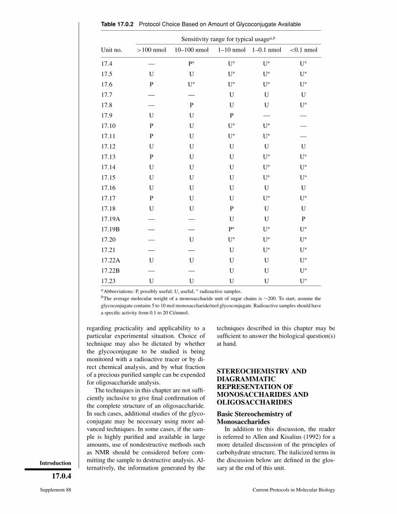
Table 17.0.2 Protocol Choice Based on Amount of Glycoconjugate Available
Sensitivity range for typical usagea,b
Unit no. >100 nmol 10–100 nmol 1–10 nmol 1–0.1 nmol <0.1 nmol
17.4 — P∗ U∗ U∗ U∗
17.5 U U U∗ U∗ U∗
17.6 P U∗ U∗ U∗ U∗
17.7 — — U U U
17.8 — P U U U∗
17.9 U U P — —
17.10 P U U∗ U∗ —
17.11 P U U∗ U∗ —
17.12 U U U U U
17.13 P U U U∗ U∗
17.14 U U U U∗ U∗
17.15 U U U U∗ U∗
17.16 U U U U U
17.17 P U U U∗ U∗
17.18 U U P U U
17.19A — — U U P
17.19B — — P∗ U∗ U∗
17.20 — U U∗ U∗ U∗
17.21 — — U U∗ U∗
17.22A U U U U U∗
17.22B — — U U U∗
17.23 U U U U U∗
aAbbreviations: P, possibly useful; U, useful; ∗ radioactive samples.bThe average molecular weight of a monosaccharide unit of sugar chains is ∼200. To start, assume theglycoconjugate contains 5 to 10 mol monosaccharide/mol glycoconjugate. Radioactive samples should havea specific activity from 0.1 to 20 Ci/mmol.
regarding practicality and applicability to aparticular experimental situation. Choice oftechnique may also be dictated by whetherthe glycoconjugate to be studied is beingmonitored with a radioactive tracer or by di-rect chemical analysis, and by what fractionof a precious purified sample can be expendedfor oligosaccharide analysis.
The techniques in this chapter are not suffi-ciently inclusive to give final confirmation ofthe complete structure of an oligosaccharide.In such cases, additional studies of the glyco-conjugate may be necessary using more ad-vanced techniques. In some cases, if the sam-ple is highly purified and available in largeamounts, use of nondestructive methods suchas NMR should be considered before com-mitting the sample to destructive analysis. Al-ternatively, the information generated by the
techniques described in this chapter may besufficient to answer the biological question(s)at hand.
STEREOCHEMISTRY ANDDIAGRAMMATICREPRESENTATION OFMONOSACCHARIDES ANDOLIGOSACCHARIDES
Basic Stereochemistry ofMonosaccharides
In addition to this discussion, the readeris referred to Allen and Kisalius (1992) for amore detailed discussion of the principles ofcarbohydrate structure. The italicized terms inthe discussion below are defined in the glos-sary at the end of this unit.

Preparation andAnalysis ofGlycoconjugates
17.0.5
Current Protocols in Molecular Biology Supplement 88
Table 17.0.3 Protocol Choice Based on Type of Glycoconjugate Studieda
Type of glyconjugateb
Unit no.N-GlcNAc-linked
glycoprotein
O-GalNAc-linked
glycoprotein
O-Xylose-linkedproteoglycan
Glyco-sphingo-lipid
Glyco-phospho-lipid
anchor
O-linkedGlcNAc
17.4 Ub U U U U P
17.5 U U — U P —
17.6 U U — — P U
17.7 U U — U P U
17.8 — — — — U —
17.9 U U — P P —
17.10 U — — — — —
17.11 — U U — — —
17.12 U U — U P —
17.13 U — — — — —
17.14A U P P — — —
17.14B U P — — P —
17.15 — U — — — —
17.16 U U — U P P
17.17 — — — U — —
17.18 U U P P P P
17.19A U U — U U P
17.19B — — U — — —
17.20 U P — P — —
17.21 U U — P — —
17.22A P P U — U —
17.22B — — U — — —
17.23 U U U P — —aThis chapter deals only with the most common forms of glycosylation on glycoconjugates.bAbbreviations: P, possibly useful; U, useful.
Glyceraldehyde, the simplest monosaccha-ride, has a single chiral (asymmetric) carbon(C-2). Therefore, it is a chiral molecule thatshows optical isomerism; it can exist in theform of two nonsuperimposable mirror imagescalled enantiomers (see Fig. 17.0.2). Theseenantiomers have identical physical proper-ties, except for the direction of rotation of theplane of polarized light (−, left hand; +, righthand). Historically, the (+)-glyceraldehydewas arbitrarily assigned the prefix D (for dex-trorotatory), and the (−)-glyceraldehyde, theprefix L (for levorotatory). The pair of enan-tiomers also have identical chemical proper-ties, except toward optically active reagents.This fact is particularly important in biolog-
ical systems, because most enzymes and thecompounds they work on are optically active.
The configuration of the highest-numberedasymmetric carbon atom in the carbon chainof a higher monosaccharide is determined bycomparison with the configuration of the chi-ral (or asymmetric) carbon of glyceraldehyde.Thus, a prefix D- is added to the name ofeach monosaccharide having the configurationof D-glyceraldehyde at the highest-numberedasymmetric carbon, and the prefix L- to thosehaving the configuration of L-glyceraldehydeat the highest-numbered asymmetric carbon.The direction in which the compound ro-tates the plane of polarized light needs to bedetermined experimentally, and is indicated
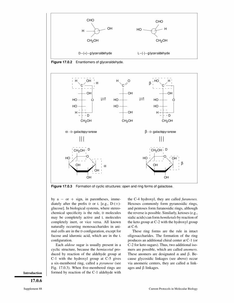
Introduction
17.0.6
Supplement 88 Current Protocols in Molecular Biology
H
CH2OH
OH HO
CH2OH
H
CHOCHO
D–(+)–glyceraldehyde L–(-)–glyceraldehyde
Figure 17.0.2 Enantiomers of glyceraldehyde.
CH2OH D
HO OH
OH
OH β
β - D- galactopyranose
HO
C
OH
H
HO
HO
CH2OH
β H
D
H OH
C
OH
H
HO
HO
CH2OH
α
D
H O
C
OH
HO
HO
CH2OH
OH
CH2OH D
HO
OH
OH
OH α
α - D- galactopyranose
O O
O O
Figure 17.0.3 Formation of cyclic structures: open and ring forms of galactose.
by a − or + sign, in parentheses, imme-diately after the prefix D or L [e.g., D-(+)-glucose]. In biological systems, where stereo-chemical specificity is the rule, D moleculesmay be completely active and L moleculescompletely inert, or vice versa. All knownnaturally occurring monosaccharides in ani-mal cells are in the D configuration, except forfucose and iduronic acid, which are in the L
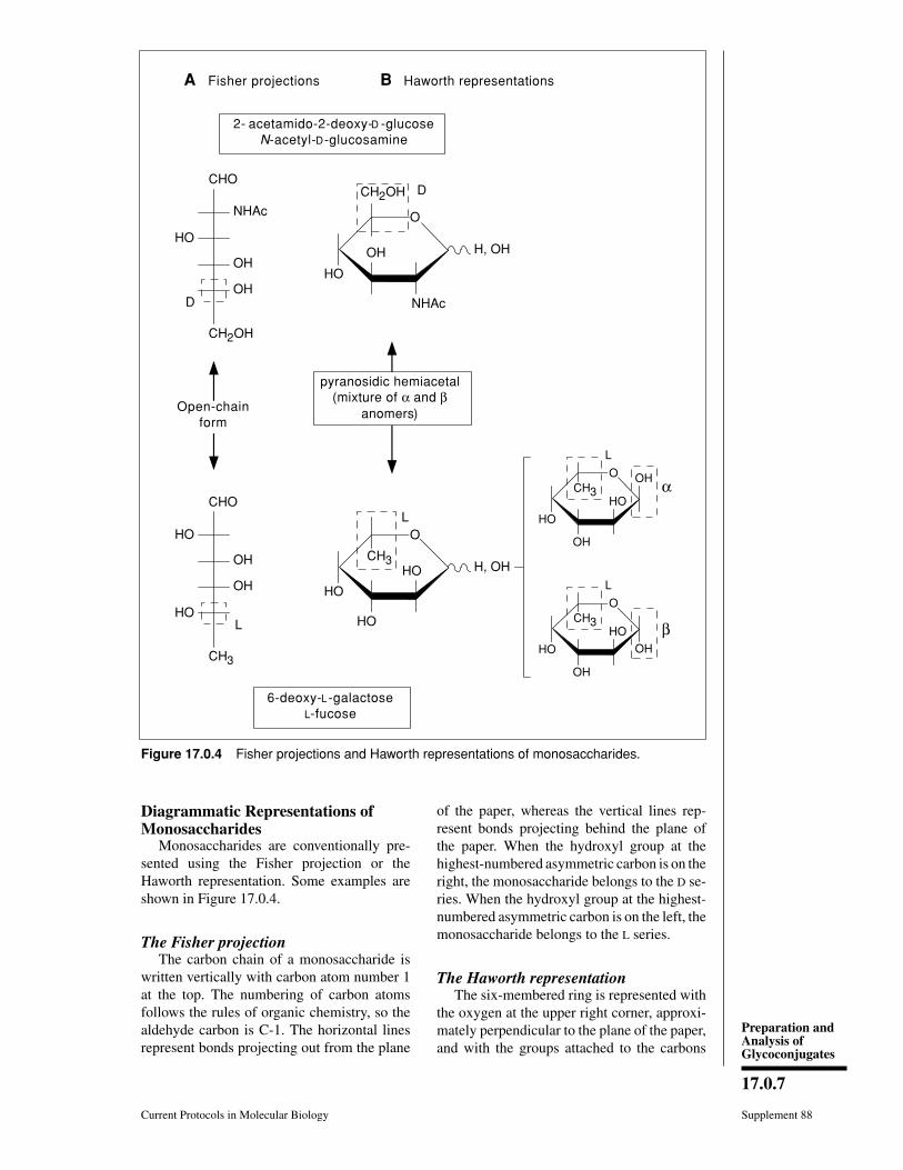
configuration.Each aldose sugar is usually present in a
cyclic structure, because the hemiacetal pro-duced by reaction of the aldehyde group atC-1 with the hydroxyl group at C-5 givesa six-membered ring, called a pyranose (seeFig. 17.0.3). When five-membered rings areformed by reaction of the C-1 aldehyde with
the C-4 hydroxyl, they are called furanoses.Hexoses commonly form pyranosidic rings,and pentoses form furanosidic rings, althoughthe reverse is possible. Similarly, ketoses (e.g.,sialic acids) can form hemiketals by reaction ofthe keto group at C-2 with the hydroxyl groupat C-6.
These ring forms are the rule in intactoligosaccharides. The formation of the ringproduces an additional chiral center at C-1 (orC-2 for keto sugars). Thus, two additional iso-mers are possible, which are called anomers.These anomers are designated α and β. Be-cause glycosidic linkages (see above) occurvia anomeric centers, they are called α link-ages and β linkages.
Preparation andAnalysis ofGlycoconjugates
17.0.7
Current Protocols in Molecular Biology Supplement 88
CHO
NHAc
HO
CH2OH
D
OH
OH
αCHO
HO
CH3
L
OH
OH
HO
CH2OH D
HO
NHAc
OH
O
H, OH
CH3
L
HO
HO
HO H, OH
CH3
L
HO
OH
HO
OH
βCH3
L
HO
OH
HO
OH
6-deoxy-L-galactoseL-fucose
2- acetamido-2-deoxy-D -glucoseN-acetyl-D-glucosamine
Open-chainform
O
O
O
A Fisher projections B Haworth representations
pyranosidic hemiacetal(mixture of α and β
anomers)
Figure 17.0.4 Fisher projections and Haworth representations of monosaccharides.
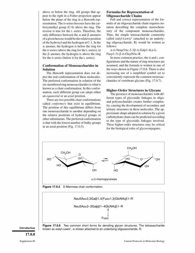
Diagrammatic Representations ofMonosaccharides
Monosaccharides are conventionally pre-sented using the Fisher projection or theHaworth representation. Some examples areshown in Figure 17.0.4.
The Fisher projectionThe carbon chain of a monosaccharide is
written vertically with carbon atom number 1at the top. The numbering of carbon atomsfollows the rules of organic chemistry, so thealdehyde carbon is C-1. The horizontal linesrepresent bonds projecting out from the plane
of the paper, whereas the vertical lines rep-resent bonds projecting behind the plane ofthe paper. When the hydroxyl group at thehighest-numbered asymmetric carbon is on theright, the monosaccharide belongs to the D se-ries. When the hydroxyl group at the highest-numbered asymmetric carbon is on the left, themonosaccharide belongs to the L series.
The Haworth representationThe six-membered ring is represented with
the oxygen at the upper right corner, approxi-mately perpendicular to the plane of the paper,and with the groups attached to the carbons
Introduction
17.0.8
Supplement 88 Current Protocols in Molecular Biology
above or below the ring. All groups that ap-pear to the right in a Fisher projection appearbelow the plane of the ring in a Haworth rep-resentation. The D-series hexoses have the car-boxymethyl group (C-6) above the ring. Thereverse is true for the L series. Therefore, theonly difference between the α and β anomersof a given hexose would be the relative positionof the hydroxyl and the hydrogen at C-1. In theα anomer, the hydrogen is below the ring forthe D series (above the ring for the L series); inthe β anomer, the hydrogen is above the ringfor the D series (below it for the L series).
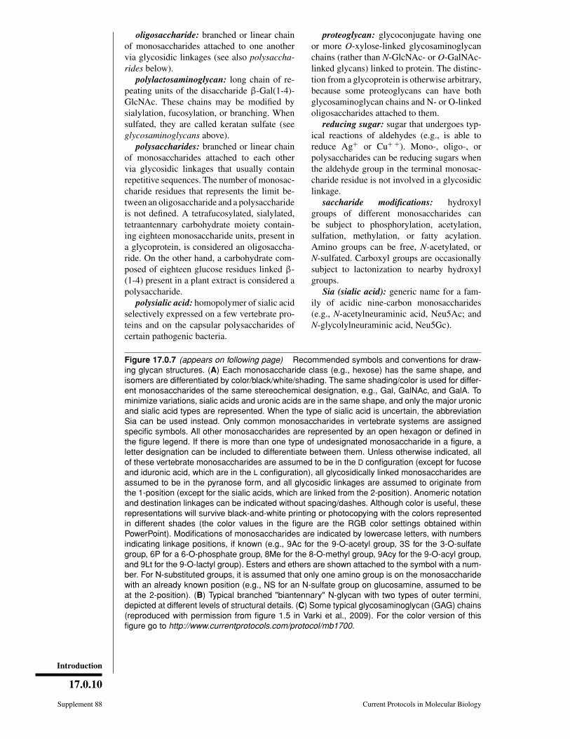
Conformation of Monosaccharides inSolution
The Haworth representation does not de-pict the real conformation of these molecules.The preferred conformation in solution of thesix-membered ring monosaccharides is what isknown as a chair conformation. In this confor-mation, each different group can adopt eitheran equatorial or an axial position.
There are two possible chair conformationscalled conformers that exist in equilibrium.The position of this equilibrium differs fromone monosaccharide to another depending onthe relative positions of hydroxyl groups orother substituents. The preferred conformationis that with the lowest number of bulky groupsin an axial position (Fig. 17.0.5).
Formulas for Representation ofOligosaccharide Chains
Full and correct representation of the for-mula of an oligosaccharide chain requires no-tation describing the complete stereochem-istry of the component monosaccharides.Thus, the simple tetrasaccharide commonlycalled sialyl-Lewisx (attached to an underly-ing oligosaccharide, R) would be written asfollows:
α-D-Neup5Ac-2-3β-D-Galp1-4(α-L-Fucp1-3)-β-D-GlcpNAc-R
In more common practice, the D and L con-figurations and the nature of ring structures areassumed, and the formula is written in one ofthe ways shown in Figure 17.0.6. There is alsoincreasing use of a simplified symbol set toconveniently represent the common monosac-charides of vertebrate glycans (Fig. 17.0.7).
Higher-Order Structures in GlycansThe presence of monosaccharides with dif-
ferent types of glycosidic linkages in oligo-and polysaccharides creates further complex-ity, causing the development of secondary andtertiary structures in these molecules. The ap-proximate shape adopted in solution by a givencarbohydrate chain can be predicted accordingto the type of glycosidic linkages involved.These higher-order structures may be criticalfor the biological roles of glycoconjugates.
CH2OH
HO
OH
HO
CH2OH
HO OH
HO
OHO
O
OH
α-D-mannopyranose
Figure 17.0.5 D-Mannose chair conformation.
Neu5Acα2,3Galβ1,4(Fucα1,3)GlcNAcβ1–R
Neu5Acα2–3Galpβ1–4GlcNAcβ1–R3
1Fucα
Figure 17.0.6 Two common short forms for denoting glycan structures. The tetrasaccharideknown as sialyl-Lewisx, is shown attached to an underlying oligosaccharide, R.

Preparation andAnalysis ofGlycoconjugates
17.0.9
Current Protocols in Molecular Biology Supplement 88
GLOSSARYThe following definitions are presented for
the terms most commonly used throughout thischapter.
aldose: monosaccharide with a carbonylgroup at the end of the carbon chain (alde-hyde group); the carbonyl group is assignedthe lowest possible number (i.e., carbon 1).
carbohydrates: polyhydroxyaldehydes orpolyhydroxyketones, or compounds that canbe hydrolyzed to them.
diastereoisomers: compounds with identi-cal formulas that have a different spatial distri-bution of atoms (e.g., galactose and mannose).
enantiomers: nonsuperimposable mirrorimages of any compound (e.g., D- and L-glucose).
epimers: two monosaccharides differingonly in the configuration of a single chiral car-bon.
Fuc (L-fucose): type of deoxyhexose (seealso types of monosaccharides below).
Gal (D-galactose): type of hexose (see alsotypes of monosaccharides below).
GalNAc (N-acetyl-D-galactosamine): typeof hexosamine (see also types of monosaccha-rides below).
ganglioside: anionic glycolipid containingone or more units of sialic acid.
Glc (D-glucose): type of hexose (see alsotypes of monosaccharides below).
GlcNAc (N-acetyl-D-glucosamine): typeof hexosamine (see also types of monosaccha-rides below).
glyceraldehyde: simplest monosaccharide(an aldotriose; i.e., containing three carbonatoms).
glycoconjugate: natural compound inwhich one or more monosaccharide oroligosaccharide units are covalently linked toa noncarbohydrate moiety.
GlUA or GlA (D-glucuronic acid): type ofuronic acid (see also types of monosaccharidesbelow).
glycolipid or glycosphingolipid: glycan at-tached via glucose or galactose to the terminalprimary hydroxyl group of ceramide, whichis composed of a long-chain base (i.e., sph-ingosine) and a fatty acid. Glycolipids can beneutral or anionic (negatively charged).
glycophospholipid anchor: glycan bridgebetween phosphatidylinositol and a phospho-ethanolamine in amide linkage to the C termi-nus of a protein; constitutes the sole membraneanchor for such proteins.
glycoprotein: glycoconjugate in which aprotein carries one or more glycan chains co-
valently attached to a polypeptide backbonevia N-GlcNAc- or O-GalNAc-linkages.
glycosaminoglycan: linear copolymers ofdisaccharide repeating units, each composedof a hexosamine and a hexose or hexuronicacid; these are the glycan chains that define theproteoglycans. The type of disaccharide unitcan define the glycosaminoglycans as chon-droitin or dermatan sulfate, heparan or heparinsulfate, hyaluronic acid, and keratan sulfate.The glycosaminoglycans (except hyaluronicacid) also contain sulfate esters substituting ei-ther hydroxyl or amino groups (N- or O-sulfategroups).
glycosidic linkage: linkage of a monosac-charide to another residue via the anomerichydroxyl group.
IdUA or IdA (L-iduronic acid): type ofuronic acid (see also types of monosaccharidesbelow).
ketose: monosaccharide with a carbonylgroup in an inner carbon; the carbonyl groupis assigned the lowest possible number (i.e.,carbon 2).
Man (D-mannose): type of hexose (see alsotypes of monosaccharides below).
monosaccharide: carbohydrate that cannotbe hydrolyzed into simpler units (see also typesof monosaccharides below).
mucin: large glycoproteins that containmany (up to several hundred) O-GalNAc-linked glycan chains that are often closelyspaced.
N-linked oligosaccharide: glycan cova-lently linked to an asparagine residue ofa polypeptide chain in the consensus se-quence -Asn-X-Ser/Thr. Although there aremany different kinds of N-linked glycans, theyhave certain common features: (1) a commoncore pentasaccharide containing two manno-syl residues α-linked to a third mannosyl unit,which is in turn β-linked to a chitobiosyl groupand (2) a chitobiosyl group that is β-linked tothe asparagine amide nitrogen. The N-linkedglycans can be divided into three main classes:high-mannose type, complex type, and hybridtype.
Neu5Ac (N-acetyl-D-neuraminic acid):type of sialic acid (Sia; see also types ofmonosaccharides below).
O-linked oligosaccharide: glycan linkedto the polypeptide via N-acetylgalactosamine(GalNAc) to serine or threonine. Note thatother types of O-linked glycans also exist (e.g.,O-GlcNAc) but that the O-GalNAc linkage,being the most well known, is often describedby this generic term.
Introduction
17.0.10
Supplement 88 Current Protocols in Molecular Biology
oligosaccharide: branched or linear chainof monosaccharides attached to one anothervia glycosidic linkages (see also polysaccha-rides below).
polylactosaminoglycan: long chain of re-peating units of the disaccharide β-Gal(1-4)-GlcNAc. These chains may be modified bysialylation, fucosylation, or branching. Whensulfated, they are called keratan sulfate (seeglycosaminoglycans above).
polysaccharides: branched or linear chainof monosaccharides attached to each othervia glycosidic linkages that usually containrepetitive sequences. The number of monosac-charide residues that represents the limit be-tween an oligosaccharide and a polysaccharideis not defined. A tetrafucosylated, sialylated,tetraantennary carbohydrate moiety contain-ing eighteen monosaccharide units, present ina glycoprotein, is considered an oligosaccha-ride. On the other hand, a carbohydrate com-posed of eighteen glucose residues linked β-(1-4) present in a plant extract is considered apolysaccharide.
polysialic acid: homopolymer of sialic acidselectively expressed on a few vertebrate pro-teins and on the capsular polysaccharides ofcertain pathogenic bacteria.
Figure 17.0.7 (appears on following page) Recommended symbols and conventions for draw-ing glycan structures. (A) Each monosaccharide class (e.g., hexose) has the same shape, andisomers are differentiated by color/black/white/shading. The same shading/color is used for differ-ent monosaccharides of the same stereochemical designation, e.g., Gal, GalNAc, and GalA. Tominimize variations, sialic acids and uronic acids are in the same shape, and only the major uronicand sialic acid types are represented. When the type of sialic acid is uncertain, the abbreviationSia can be used instead. Only common monosaccharides in vertebrate systems are assignedspecific symbols. All other monosaccharides are represented by an open hexagon or defined inthe figure legend. If there is more than one type of undesignated monosaccharide in a figure, aletter designation can be included to differentiate between them. Unless otherwise indicated, allof these vertebrate monosaccharides are assumed to be in the D configuration (except for fucoseand iduronic acid, which are in the L configuration), all glycosidically linked monosaccharides areassumed to be in the pyranose form, and all glycosidic linkages are assumed to originate fromthe 1-position (except for the sialic acids, which are linked from the 2-position). Anomeric notationand destination linkages can be indicated without spacing/dashes. Although color is useful, theserepresentations will survive black-and-white printing or photocopying with the colors representedin different shades (the color values in the figure are the RGB color settings obtained withinPowerPoint). Modifications of monosaccharides are indicated by lowercase letters, with numbersindicating linkage positions, if known (e.g., 9Ac for the 9-O-acetyl group, 3S for the 3-O-sulfategroup, 6P for a 6-O-phosphate group, 8Me for the 8-O-methyl group, 9Acy for the 9-O-acyl group,and 9Lt for the 9-O-lactyl group). Esters and ethers are shown attached to the symbol with a num-ber. For N-substituted groups, it is assumed that only one amino group is on the monosaccharidewith an already known position (e.g., NS for an N-sulfate group on glucosamine, assumed to beat the 2-position). (B) Typical branched "biantennary" N-glycan with two types of outer termini,depicted at different levels of structural details. (C) Some typical glycosaminoglycan (GAG) chains(reproduced with permission from figure 1.5 in Varki et al., 2009). For the color version of thisfigure go to http://www.currentprotocols.com/protocol/mb1700.
proteoglycan: glycoconjugate having oneor more O-xylose-linked glycosaminoglycanchains (rather than N-GlcNAc- or O-GalNAc-linked glycans) linked to protein. The distinc-tion from a glycoprotein is otherwise arbitrary,because some proteoglycans can have bothglycosaminoglycan chains and N- or O-linkedoligosaccharides attached to them.
reducing sugar: sugar that undergoes typ-ical reactions of aldehydes (e.g., is able toreduce Ag+ or Cu+ +). Mono-, oligo-, orpolysaccharides can be reducing sugars whenthe aldehyde group in the terminal monosac-charide residue is not involved in a glycosidiclinkage.
saccharide modifications: hydroxylgroups of different monosaccharides canbe subject to phosphorylation, acetylation,sulfation, methylation, or fatty acylation.Amino groups can be free, N-acetylated, orN-sulfated. Carboxyl groups are occasionallysubject to lactonization to nearby hydroxylgroups.
Sia (sialic acid): generic name for a fam-ily of acidic nine-carbon monosaccharides(e.g., N-acetylneuraminic acid, Neu5Ac; andN-glycolylneuraminic acid, Neu5Gc).

Preparation andAnalysis ofGlycoconjugates
17.0.11
Current Protocols in Molecular Biology Supplement 88
A
βα β
β β ββα β α
β β β
α
β β α
α
ββ
β β β
α
β β α
α β β α
β β α
β β β
αα β β α
β β β
β β α
α β β α α
α
α
B
β β β β β β β β β β β β
β β β β ββ β ββ β
α α
α β αα β α αα ββ
β β β β β α β α β β
β β ββ β β ββ
α β α β α β α β α α
β β
C
Figure 17.0.7 (legend appears on preceding page)

Introduction
17.0.12
Supplement 88 Current Protocols in Molecular Biology
types of monosaccharides: monosaccha-rides may have a carbonyl group at the end ofthe carbon chain (aldehyde group) or in an in-ner carbon (ketone group). The carbonyl groupis assigned the lowest possible number, e.g.,carbon 1 (C-1) for the aldehyde group; carbon2 (C-2) for the most common ketone groups.These two types are named aldoses and ke-toses, accordingly. The simplest monosaccha-ride is glyceraldehyde (see Fig. 17.0.2), an al-dotriose (i.e., containing three carbon atoms).Natural aldoses with different numbers of car-bon atoms in their chain are named accord-ingly (e.g., aldohexoses, containing six car-bon atoms). Two monosaccharides differingonly in the configuration of a single chiralcarbon are called epimers. For example, glu-cose and galactose are epimers of each otherat C-4. The common monosaccharides presentin animal glycoconjugates are (1) deoxyhex-oses (e.g., L-Fuc); (2) hexosamines, usuallyN-acetylated (e.g., D-GalNAc and D-GlcNAc);(3) hexoses (e.g., D-Glc, D-Gal, and D-Man);(4) pentoses (e.g., D-Xyl); (5) sialic acids (Sia;e.g., Neu5Ac); and (6) uronic acids (e.g., D-GlA and L-IdA).
Xyl (D-xylose): type of pentose (see alsotypes of monosaccharides above).
For further detailed information on theanalysis of glycans, the reader is referred to thespecific literature cited in the individual pro-tocol units. In addition, the following sourcescan be used for general information, for de-tails on specific methods, and for many ad-ditional methods that are not included in thischapter.
LITERATURE CITEDAllen, H.J. and Kisalius, E.C. (eds.). 1992. Glyco-
conjugates: Composition, Structure, and Func-tion. Marcel Dekker, New York.
Varki, A., Cummings, R.D., Esko, J.D., Freeze,H.H., Stanley, P., Bertozzi, C.R., Hart, G.W.,and Etzler, M.E. (eds.) 2009. Essentials of Gly-cobiology. Cold Spring Harbor Laboratory, ColdSpring Harbor, N.Y.
KEY REFERENCESAllen and Kisailus (eds.), 1992. See above.
Bertozzi, C. and Rabuka, D. 2009. Structural basisof glycan diversity. In Essentials of Glycobiol-ogy (A. Varki, R.D. Cummings, J.D. Esko, H.H.Freeze, P. Stanley, C.R. Bertozzi, G.W. Hart,and M.E. Etzler, eds.), pp. 601-616. Cold SpringHarbor Laboratory Press, Cold Spring Harbor,N.Y.
Bill, M.R., Revers, L., and Wilson, I.B.H. 1998.Protein Glycosylation. Kluwer Academic Pub-lishers, Boston.
Boons, G-J. (ed.) 1998. Carbohydrate Chemistry.Blackie Academic & Professional, London.
Chaplin, M.F. and Kennedy, J.F. (eds.) 1987. Car-bohydrate Analysis: A Practical Approach. IRLPress, Oxford.
Fleischer, S. and Fleischer, B. 1983. Biomem-branes, part L. Methods Enzymol. 98:1-688.
Ginsburg, V. (ed.). 1972. Complex Carbohydrates,Part B. Methods Enzymol. Vol. 28.
Ginsburg, V. (ed.). 1978. Complex Carbohydrates,Part C. Methods Enzymol. Vol. 50.
Ginsburg, V. (ed.). 1982. Complex Carbohydrates,Part D. Methods Enzymol. Vol. 83.
Ginsburg, V. (ed.). 1987. Complex Carbohydrates,Part E. Methods Enzymol. Vol. 138.
Ginsburg, V. (ed.). 1989. Complex Carbohydrates,Part F. Methods Enzymol. Vol. 179.
Hart, G. and Lennarz, W. 1993. Guide to techniquesin glycobiology. Methods Enzymol. 230.
Haslam, S.M., North, S.J., and Dell, A. 2006.Mass spectrometric analysis of N- and O-glycosylation of tissues and cells. Curr. Opin.Struct. Biol. 16:584-591.
Kamerling, J., Boons, G-J., Lee, Y., Suzuki, A.,Taniguchi, N., and Voragen, A.G.J. 2007. Com-prehensive glycoscience. 1-4. Elsevier Science,London.
Manzi, A.E. and van Halbeek, H. 1999. Glycanstructure and nomenclature. In Essentials ofGlycobiology. (A. Varki, R.D. Cummings, J.D.Esko, H.H. Freeze, P. Stanley, C.R. Bertozzi,G.W. Hart, and M.E. Etzler, eds.) pp. 17-29.Cold Spring Harbor Laboratory Press, ColdSpring Harbor, N.Y.
Manzi, A.E. and van Halbeek, H. 1999. Generalprinciples for the analysis and sequencing of gly-cans. In Essentials of Glycobiology. (A. Varki,R.D. Cummings, J.D. Esko, H.H. Freeze, P.Stanley, C.R. Bertozzi, G.W. Hart, and M.E.Etzler, eds.) pp. 581-598. Cold Spring HarborLaboratory Press, Cold Spring Harbor, N.Y.
McNaught, A.D. 1997. Nomenclature of carbohy-drates. Carbohydr. Res. 297:1-92.
McCloskey, J.A. (ed.). 1990. Mass Spectrometry.Methods Enzymol. 193.
Stick, R.V. 2001. Carbohydrates: The sweetmolecules of life. Academic Press, New York.
Varki, A. 1991. Radioactive tracer techniques in thesequencing of glycoprotein oligosaccharides.FASEB J. 5:226-235.
Vliegenthart, J.F.G., Dorland, L., and van Halbeek,H. 1983. NMR spectroscopy of carbohydrates.Adv. Carbohydr. Chem. Biochem. 41:209-378.
Whistler, R. and Wolfram, F. (eds.). 1968-1980.Methods in Carbohydrate Chemistry, Vols I-VIII. Academic Press, San Diego.
SECTION ISPECIAL CONSIDERATIONS OFGLYCOCONJUGATES AND THEIRPURIFICATIONBecause of their unique composition and structural characteristics, there are instances inwhich glycoconjugates can be studied in biological mixtures directly, without purifica-tion. However, as Arthur Kornberg has said, to answer a biological question the best courseof action is usually to “purify, purify, purify.” Indeed, the complete elucidation of anybiological system involving a glycoconjugate will require that it be purified for structuraland functional characterization.
This section describes general principles for purifying three major classes of glycocon-jugates—glycoproteins, glycolipids, and proteoglycans. These classes have been de-scribed in the chapter introduction and glossary. Each is a large and polydisperse familyof molecules with widely ranging structural and functional characteristics. Needless tosay, many of the classic principles used for purification of other macromolecules (e.g.,size fractionation and selective precipitation) apply to each of these types of glycoconju-gates; on the other hand, an individual situation may provide unique opportunities forselective purification of a particular molecule.
The units presented in this section take the middle road and outline the principlesparticularly applicable to the purification of each class of glycoconjugate, taking advan-tage of group-specific properties. Glycoproteins usually have sugar chains that can berecognized by specific plant lectins; thus, affinity chromatography using immobilizedlectins is a powerful technique, when properly applied (UNIT 17.1). Proteoglycans arecharacterized by a high density of negative charge and frequently by unusual buoyantdensities and sedimentation properties, which can be used to advantage in their purifica-tion (UNIT 17.2). Glycolipids as a group are more hydrophobic than the other glycoconju-gates, yet have amphipathic properties that allow them to be purified away from otherlipids (UNIT 17.3).
Selective purification of these groups of molecules facilitates their further fractionationand final characterization. However, it is important to realize that exceptions can beencountered (e.g., glycoproteins with unusually high negative charges or glycolipids withextremely hydrophilic properties). Also, the phenomenon of microheterogeneity in oli-gosaccharide structure can cause even a single glycoconjugate to manifest a range ofproperties. For example, if a single glycoprotein from a single cell type has heterogeneityin N-linked oligosaccharide processing, some (but not all) of the protein may be recoveredduring affinity chromatography on wheat germ agglutinin (WGA)-Sepharose (UNIT 17.1).
At the present time there is no generalized purification scheme that will provide goodyields of all of the major classes of glycoconjugates from a single starting sample. Forexample, the organic extractions required for glycolipids (UNIT 17.3) irreversibly denaturemost glycoproteins and proteoglycans. Conversely, the glycolipids are usually irretrivablefrom the detergent extractions used for membrane-bound glycoproteins (UNIT 17.1). Thus,if there is a need to examine all of the major glycoconjugates in a given cell or tissue, itis advisable to purify the major classes of molecules in parallel with one another, usingseparate aliquots of the same batch of starting material.
Even when studying a specific molecule, it is usually worthwhile to carry out a group-specific purification as a first step—e.g., to extract total cellular lipids before purifyingaway a specific disialoganglioside (UNIT 17.3). If the glycoconjugate in question has beenradiolabeled (see UNIT 17.4), one also has the advantage that purification to “radiometrichomogeneity” is sufficent for many types of analyses. Thus, if a glycoprotein has been
Supplement 22
Contributed by Hudson H. FreezeCurrent Protocols in Molecular Biology (1993) 17.1.1-17.1.10Copyright © 2000 by John Wiley & Sons, Inc.
17.1.1
Preparationand Analysis ofGlycoconjugates
metabolically labeled with [2-3H]mannose and the intent is to study the N-linked oligosac-charides, it may not matter that various unlabeled proteins or lipids are contaminating thefinal preparation. In the final analysis, the broad principles outlined in UNITS 17.1-17.3 mustbe adapted by the investigator to the situation at hand.
UNIT 17.1 Special Considerations for Glycoproteinsand Their Purification
Glycoproteins contain a variety of differentsugar chains. Some glycoproteins have manylarge and highly charged chains while othershave only a single neutral sugar residue that iseasily overlooked. This enormous diversitymakes it impossible to recommend any singleprocedure for purifying glycoproteins. Mostprocedures used to purify nonglycosylated pro-teins are equally useful for glycoproteins. How-ever, the presence of large amounts of carbohy-drate on glycoproteins often endows them withhigher charge, increased density, larger appar-ent size, and greater solubility than nonglyco-sylated proteins. Decades ago, these physicalproperties were cleverly exploited to purify andcharacterize many of the most abundant glyco-proteins (Gibbons, 1972). More recently,greater knowledge of carbohydrate structureand biosynthesis has led to development oftechniques that rely on more subtle and specificaspects of carbohydrate structure. Some ofthese approaches can also be used to detect asingle sugar on a few picomoles of protein (seeUNIT 17.7).
This unit begins by describing some prop-erties of glycoproteins—e.g., subcellular loca-tion and solubility—that may be useful in de-termining which purification techniques to try.This discussion is followed by two protocolsdescribing preparative glycoprotein purifica-tion using lectin-affinity chromatography, aswell as an outline for a small-scale pilot proce-dure designed to check lectin binding and elu-tion conditions. Lectins are often used for pu-rifying glycoproteins because, in contrast toconventional purification procedures (e.g., gelfiltration and ion-exchange chromatography;UNITS 10.9 & 10.10) that exploit general physicalproperties of glycoproteins, lectins recognizespecific three-dimensional structures createdby a cluster of sugar residues. Conventionalpurification procedures are generally tried be-fore applying lectin-affinity chromatography.
SPECIAL CONSIDERATIONS INTHE PURIFICATION OFGLYCOPROTEINS
Subcellular Location of GlycosylatedProteins
Most protein glycosylation occurs in thelumen of the endoplasmic reticulum and theGolgi apparatus, but certain types of glycosy-lation also occur in the cytoplasm (see UNIT
17.13). Secreted proteins, membrane proteins,and proteins that enter vesicles such aslysosomes are good candidates for having sometype of carbohydrate component. The mostcommon types of sugar chains are found in N-and O-linkages to proteins and as componentsof glycophospholipid anchors. Each of thesetypes of glycosylation can occur individuallyor in any combination. The physical propertiesthey impart can be effectively used for somepurifications. Knowledge of the type of glyco-sylation is thus necessary in order to determinethe optimal purification method. The methodsoutlined below may be helpful for determiningglycosylation type.
Secreted glycoproteins. It is relatively sim-ple to determine if the protein is secreted byanalyzing conditioned medium used to culturethe cells. This may require dialysis and concen-tration of the conditioned medium. If the cellsmust be grown in the presence of added serumcomponents during the time when the proteinis secreted, it may be very difficult to use anyspecific glycoprotein purification approach toenrich for your protein because competing se-rum glycoproteins will probably be in greatexcess. Thus it is important to determine if thetarget molecule can be secreted into serum-freemedium.
Cytoplasmic glycoproteins. If the protein isnot secreted, cells can be ruptured in an iso-os-motic medium (e.g., sucrose) and centrifugedto separate insoluble membranes and vesicles
Supplement 22 Current Protocols in Molecular Biology
17.1.2
SpecialConsiderations
for Glycoproteinsand Their
Purification
from soluble cytoplasmic components (UNIT
17.6). Glycoproteins are not commonly found inthe soluble fraction. If the protein of interest isdetected in the soluble fraction and is laterfound to be glycosylated, it may have beenproteolyzed from a membrane. This is the mostlikely explanation for N-linked glycoproteinsthat are found in the soluble fraction. It is alsopossible that the protein is one of the recentlydescribed cytoplasmic or nuclear glycoproteinsthat contain O-linked N-acetylglucosamine(GlcNAc), in which case it may bind to wheatgerm agglutinin (WGA, from Triticum vul-garis; alternate protocol).
Organelle glycoproteins. If the protein ofinterest is not present in the soluble cytoplasmicfraction, the membrane and vesicle pellet isthen resuspended in the presence of proteaseinhibitors and briefly exposed to a hypo-os-motic medium (or frozen and thawed) to lysesensitive organelles and release their solublecontents. Membranes are again sedimented bycentrifugation. Glycoprotein solubilized bythis procedure will be accompanied by lysoso-mal enzymes. These proteases are potentiallytroublesome, but protease inhibitors will lessenor eliminate their effect. Various exo- and en-doglycosidases may also be released by thisprocedure, but they are usually present at sucha low concentration that they do not degradethe sugar chains on other glycoproteins.
Membrane-bound and glycophospholipid-anchored proteins. If the protein of interest isnot solubilized by any of the above procedures,it is probably a peripheral or integral membraneprotein, or it is anchored to glycophospholipid.Different nonionic detergents or increasingconcentrations of a single detergent can be usedto solubilize these proteins. Detergents thathave high critical micelle concentrations(CMC) and can be easily dialyzed are preferred,e.g., hexyl-, heptyl-, and octyl-glucosides orglucamide detergents (MEGA-8, Calbiochem).However, a glucose-based detergent such asoctyl-glucoside may block protein binding onsome lectin columns such as Conconavalin A(Con A)–Sepharose, or may produce a veryhigh background in direct sugar analysis (UNIT
17.9) even after extensive dialysis. If the proteinis glycophospholipid-anchored, the lipid com-ponent will cause it to partition into the deter-gent phase of a Triton X-114/water two-phasesystem (UNIT 17.8) and to interact strongly withhydrophobic matrices such as phenyl-Sepharose. In some instances, the lipid portionof the anchor may be removed by digestion withphosphatidylinositol-specific phospholipase C
(UNIT 17.8). This cleavage leaves the carbohy-drate portion of the anchor associated with theprotein and converts the lipophilic protein intoa soluble, more hydrophilic protein. Phospholi-pase digestion reduces the protein’s affinity forhydrophobic matrices and results in its extrac-tion into the aqueous phase of Triton X-114/water mixture (UNIT 17.8).
Solubility PropertiesThe solubility properties of a glycoprotein
with a small amount of carbohydrate (∼5%)will not be influenced much by the sugar com-ponent. If the carbohydrate is part of a glyco-phospholipid anchor, detergent solubilizationmay be required. Standard ammonium sulfateor ethanol precipitation (UNIT 10.9) works wellfor many glycoproteins, but those with highsugar content may remain soluble, even in rela-tively high concentrations of ethanol. Very an-ionic proteins with abundant sialic acid or sul-fate esters may be resistant to precipitation withtrichloroacetic acid or perchloric acid (APPENDIX
2). This unusual solubility is sometimes ex-ploited as a purification step because most otherproteins will be precipitated under these condi-tions. However, a combination of 10% trichlo-roacetic acid and 2% phosphotungstic acid willprecipitate even highly charged proteins. Pro-longed exposure to low pH may cause partialloss of sialic acid and deamidation of Asn andGln, resulting in unwanted protein microhet-erogenity. Extremes of pH should be avoidedbecause of the possibility of hydrolysis of labilegroups such as phosphodiesters or acetyl esters.
Nonglycosylated proteins band in cesiumchloride gradients at ∼1.3 g/ml and polysaccha-rides at ∼1.6 to 2.0 g/ml. This substantial dif-ference in density can be used to separate veryhighly glycosylated proteins from those con-taining lesser amounts of carbohydrate. Thistreatment may permanently denature or inacti-vate some proteins, and is useful only if theprotein is stable, is active in the presence ofCsCl2, or renatures once the CsCl2 is removed.
Chromatographic and StainingProperties
Gel-filtration columns and SDS-PAGE.Proteins with high carbohydrate content (>20to 30%) elute much earlier on gel-filtrationcolumns (UNIT 10.9) than nonglycosylated pro-teins of similar size. If the amount of carbohy-drate is variable, it may broaden the peak aswell. Migration of proteins in SDS-PAGE (UNIT
10.2) is also affected by the extent of glycosyla-tion. A protein with multiple anionic sugar
Current Protocols in Molecular Biology Supplement 22
17.1.3
Preparationand Analysis ofGlycoconjugates
chains, each with a variable number of sialicacids or sulfate esters, can broaden a singleprotein band into a smear on gels. Closelyclustered, short, O-linked sugar chains can ap-pear to contribute as much as ten times theiractual mass to the apparent molecular weightof a protein.
Very anionic glycoproteins may not stainwith the normal Coomassie brilliant blue orsilver staining procedures (UNIT 10.6). Some gly-coproteins may even be seen as negative bandsagainst the brownish background of silver-stained gels. Other stains, such as the Coomas-sie-based ProBlue stain (Integrated SeparationSystems) or the cationic dyes toluidine blue andalcian blue, are often used for staining proteo-glycans and other highly anionic proteins.
Ion-exchange and isoelectric focusingchromatography. The presence of sialic acidsand sulfate esters makes some glycoproteinsbind well to ion-exchange columns (e.g.,DEAE-Sephadex, DEAE-Sephacel; UNIT 10.10).High-salt solutions elute these glycoproteinswell after most other proteins have been eluted.The variable number of negative charges canalso give multiple separate or very broad andpoorly resolved protein peaks (UNITS 10.3 & 10.10).Multiple peaks or broad bands are also encoun-tered in isoelectric focusing gels. Sialidase orendoglycosidase digestions can sometimessimplify these patterns by removing negativecharges.
In the case of “mucins” (highly chargedglycoproteins with closely clustered or ex-tended O-linked GalNAc chains) or proteogly-cans, ion-exchange columns may need to be runin 6 M urea to prevent protein aggregation.
Preparation of Protein Samples forCarbohydrate Analysis
Carbohydrate contamination. A highly pu-rified protein that gives only a single band bySDS-PAGE analysis may still be unsuitable forcarbohydrate analysis. Glucose is only veryrarely found on sugar chains of proteins. On theother hand, it is almost always a readily avail-able contaminant. DEAE- and CM-cellulose,agarose, Sephadex, Kimwipes, sucrose, glyc-erol, starch powder used on latex gloves, andglucoside-based detergents are all carbohy-drates and can make a relatively large contribu-tion to the sugar content of a sample, dependingupon exactly how the material is analyzed. Ifproteins are purified on sucrose gradients, ex-tracted with glucose-based detergents, or nor-mally stored in glycerol, these sugar contami-nants must first be completely removed by
dialysis. Acrylamide-based gels (e.g., Bio-Gelseries) are preferred over the dextran-type gels(Sephadex) for gel-filtration purification be-cause the latter can contribute fragments ofbeads as soluble but nondialyzable material(UNIT 10.9). As a final precaution, protein-con-taining solutions should be filtered through a0.2-µm filter to remove any particles that mayhave come from ion-exchange or affinity col-umns. If glucose is found by compositionalanalysis even after taking these precautions, itis still most likely to be a contaminant and nota component of the glycoprotein.
Lectin-affinity chromatography. The mostfrequently used specific purification procedurefor glycoproteins is lectin-affinity chromatog-raphy. Dozens of plant lectins have been iden-tified and in many cases their sugar specificityis known. This sugar specificity is the basis forthe separation and structural analysis of manyindividual oligosaccharides and glycopeptidesdescribed in this chapter, and for the identifica-tion of lectin-binding glycoproteins in UNIT 17.7.Glycoproteins often contain multiple chains ofa given sugar type and multiple interactionswith the immobilized lectin may make it diffi-cult to elute some of the proteins from the lectincolumn. The presence of multiple low-affinitysugar chains can still give substantial interac-tion with a lectin-containing column eventhough the individual sugar chains may them-selves have a low affinity. Thus, one should becareful not to interpret too much about sugarchain structure from a protein’s binding to alectin column. This by no means belittles theirusefulness for glycoprotein purification; lectin-affinity chromatography may be used to par-tially purify glycoproteins and to provide ratherlimited qualitative information about the natureof their carbohydrate components. Other prepa-rative purification steps, such as ammoniumsalt precipitation (UNIT 11.3) and gel-filtrationand ion-exchange chromatographies (UNITS 10.9
& 10.10), are usually done prior to using lectins,but this is not necessary.
Many different lectins are commerciallyavailable in an immobilized form suitable forglycoprotein purification, but some can berather expensive. Con A–Sepharose is the mostcommonly used lectin for glycoprotein purifi-cation. It is relatively inexpensive, it is stable,and it can bind to many different glycans.Bound proteins can be eluted with α-methyl-D-mannoside (αMM). WGA is the next mostpopular lectin for the same reasons although itis somewhat more expensive.
Supplement 22 Current Protocols in Molecular Biology
17.1.4
SpecialConsiderations
for Glycoproteinsand Their
Purification
Other notable lectins used to purify glyco-proteins include Ricinus communis lectin(RCA I) for proteins that carry Gal-terminatedsugar chains, and pea or lentil lectins for thoseN-linked oligosaccharides that have a fucoseresidue in the chitobiosyl core region. The util-ity of these lectins for characterizing glycopep-tides is described in UNIT 17.7. Limited availabil-ity, higher price, limited track records, and, insome cases, the requirement for exotic sugarsto elute bound proteins make some of theselectins less appropriate for routine use in gly-coprotein purification. However, the principlesof Con A–Sepharose and WGA-agarose lectin-affinity chromatography should apply to theseother lectins as well. A broad range of immo-bilized lectins is available from E-Y Laborato-ries, Pharmacia Biotech, Vector Labs, andSigma (APPENDIX 4).
Examples of lectin-affinity chromatogra-phy purifications. The basic and alternate pro-tocols presented below describe the use of lect-ins for preparative glycoprotein purification.Con A–Sepharose and WGA-agarose werechosen for convenience and availability. Thesupport protocol describes a small-scale pilotprocedure to test for lectin binding and to de-termine elution conditions. There are manyvariations on the basic procedure in the litera-ture, but all use the same principles: bind theprotein to immobilized lectin through its sugarchain, wash away unbound protein, and elutebound protein with a simple sugar that resem-bles the sugar ligand of the bound protein.Because many proteins have sugar chains thatcan bind to a specific lectin, this procedureseldom yields a pure protein.
BASICPROTOCOL
CON A–SEPHAROSE AFFINITY CHROMATOGRAPHY
Con A–Sepharose chromatography is used to partially purify glycoproteins that containterminal mannose or glucose residues; the steps presented below are typical conditionsfor this type of chromatography, although a variety of approaches have been utilized. Inthis protocol, bound glycoproteins are eluted with α-methyl-D-mannoside (αMM) afterthe column is first washed to remove unbound and weakly bound proteins. Beforeproceeding, it is advisable to conduct a pilot study to test the protein of interest for lectinbinding and elution conditions (see support protocol).
Materials
10 mg/ml Con A–Sepharose (Pharmacia Biotech or Sigma)Column buffer0.5 M αMM in column bufferProtein sample in column buffer
Glass wool1.5 × 30–cm glass or disposable chromatographic column
Additional reagents and equipment for degassing solutions (UNIT 10.12),phenol–sulfuric acid assay for sugars (UNIT 17.9) and specific assay fordetecting the protein of interest
NOTE: This procedure should be carried out at room temperature if the protein to beisolated will tolerate this condition. If not, carry it out in a cold room, and prechill allsolutions to maintain temperature.
1. Gently resuspend 50 ml settled Con A–Sepharose (10 mg lectin/ml packed resin) in50 ml column buffer to make a slurry. Degas the slurry.
This volume of lectin beads should be sufficient to bind ∼100 mg glycoproteins. If a 1.5 ×30–cm column is not available, either the dimensions of the column or the total amount ofresin can be modified. A 1.0 × 30–cm column will bind ∼50 mg total glycoprotein. If theamount of glycoprotein in the sample is considerably less than 50 to 100 mg, decrease thevolume of the column accordingly. The ratio of input protein to lectin does not seem tomatter as long as the column is not overloaded.
Current Protocols in Molecular Biology Supplement 22
17.1.5
Preparationand Analysis ofGlycoconjugates
2. Pack a glass wool plug over a scintered glass or polypropylene frit at the bottom ofthe column and pour the degassed slurry into the column.
The glass wool is important to prevent the beads from clogging the frit and slowing theflow rate.
3. Continue packing the column until the desired level is reached; for a 50-ml volumethis is ∼28 cm. Wash the gel with 2 to 3 column volumes of column buffer to removeany loosely bound or degraded Con A.
4. Wash with 2 to 3 column volumes of 0.5 M of αMM in column buffer or the highestconcentration of αMM that will be used.
5. Wash the column with >5 column volumes of column buffer without αMM toreequilibrate.
It is important to prewash the column with the eluting sugar and then to reequilibrate thecolumn to remove any materials which might have previously bound to the column. Checkthe completeness of washing by the phenol–sulfuric acid assay (UNIT 17.9). Estimate theamount of residual sugar using αMM as a standard. An acceptable level is <0.1 mM or∼20 g/ml.
6. Slowly load the protein sample on the column to permit binding without disturbingthe surface. A flow rate of ∼1 ml/min (∼0.5 ml/min per cm2 of area) is desirable.
7. Wash column with column buffer and monitor the flowthrough and subsequent washfractions by measuring the A280 until it approaches baseline value.
If the A280 does not rapidly return to baseline, it may indicate weak interaction of someproteins with the lectin, or overloading of the column.
8. Assay flowthrough and wash fractions for the presence of the protein of interest.
To check the lectin-binding properties, see support protocol below. To determine whetherthe column was overloaded, add a small amount of the flowthrough (1%) to a small volumeof beads as described in the support protocol. If the sample is bound to the beads asdetermined by the appropriate assay, overloading of the original column is indicated. Inthis case, reapply the flowthrough with excess glycoprotein to the larger column after thebound proteins of the first run are eluted with αMM and the column is reequilibrated.
9. Elute the column with 0.5 M αMM in column buffer and monitor fractions for A280
and activity. Pool peak-activity fractions.
Very broad peaks may result during elution with the sugar if the protein dissociates veryslowly. When this happens, sample recovery can be improved by filling the column with 0.5M αMM in column buffer and allowing the column to stand for a few hours. This allowsdissociation of the bound material; when the flow is started again, it should give a verysharp peak. Other possible remedies include warming a cold column to room temperaturein the presence of αMM, increasing the concentration of αMM to 1 M, or increasing theNaCl concentration to 1 M. A combination of these may be needed to elute a tightly boundprotein.
10. Regenerate the column by washing it with 10 vol column buffer or until the αMMconcentration is <20 µg/ml.
Column may now be used for another run or stored indefinitely at 4°C in column buffercontaining 0.02% (w/v) NaN3. Reequilibrate in column buffer before using again.
Supplement 22 Current Protocols in Molecular Biology
17.1.6
SpecialConsiderations
for Glycoproteinsand Their
Purification

SUPPORTPROTOCOL
PILOT STUDY TO DETERMINE LECTIN BINDING ANDELUTION CONDITIONS
If the target protein can be detected easily, it is worthwhile to test a small sample toestablish binding and elution conditions before applying the entire sample to a largecolumn. The easiest approach is to mix a small amount of sample with a measured amountof Con A–Sepharose beads in a series of microcentrifuge tubes. If the material in thesample binds, it can then be eluted from the washed beads with various amounts ofcompeting αMM to determine when the activity is eluted into the supernatant. This showswhether binding occurs and what concentration of competing sugar (or other conditions)will be required to elute it from the column. An abbreviated version of this procedure canbe done with a single sample to determine if the protein binds and if it can be eluted withonly one concentration (0.75 M) of αMM. It is important to keep the temperature constant,because changes can affect binding and dissociation of the ligands.
Additional Materials
Sepharose 4B (Pharmacia Biotech) or other beaded gel to fill space in the tubesαMM: 0, 0.1, 0.2, 0.4, 0.8, 1.0, and 1.5 M concentrations, in column buffer
1. Prepare a 50% slurry of Con A–Sepharose in column buffer. Cut ∼2 to 3 mm off thesmall end of a 1000-µl pipet tip. Resuspend the slurry immediately before eachpipetting, and use the truncated tip to dispense 200 µl of slurry into a series of sevenmicrocentrifuge tubes. To one of these tubes—the no-lectin control—add an equalvolume of 50% slurry of Sepharose 4B. Allow gel to settle to see if the dispensingwas reasonably accurate.
Gel material is needed in the controls to account for the volume changes caused by addingthe gel. Other nonionic beaded gels, such as Sephadex (Pharmacia Biotech) or Bio-Gel(Bio-Rad) may be substituted.
2. Add the protein sample in 50 µl column buffer and allow 15 min for binding (shakethe tube occasionally). Microcentrifuge the gel beads 1 min at 1000 × g to sediment.Remove the supernatant and save for analysis in step 6.
3. Use an appropriate assay for the protein of interest to determine if it binds to ConA–Sepharose and not to control beads.
4. Wash the gel beads with three 1.4-ml washes of column buffer; after each washmicrocentrifuge the beads using conditions in step 2.
5. Resuspend gel beads in 200 µl of buffer containing 0, 0.1, 0.2, 0.4, 0.8, 1.0, or 1.5 Mα-MM for each of the seven tubes containing Con A–Sepharose. Add the samevolume of column buffer or 1.5 M αMM in column buffer to the control.
6. Incubate 15 min and centrifuge the gel beads as in step 2. Analyze the supernatantsusing the appropriate detection assay to determine if the protein has been eluted andat what concentration of competing sugar.
Increasing the concentration of αMM should elute the target protein from the ConA–Sepharose. If it is not eluted, longer incubation times (10 hr) in the presence of αMMor higher temperature may be needed to elute the protein from the beads. Although theentire procedure can be done in the cold, binding is tighter at low temperature, and thismay make elution more difficult. Once the conditions are established for a particularsample or a lectin, scaling up the preparation for the basic protocol should proceedsmoothly.
Current Protocols in Molecular Biology Supplement 22
17.1.7
Preparationand Analysis ofGlycoconjugates
ALTERNATEPROTOCOL
WHEAT GERM AGGLUTININ (WGA)–AGAROSE AFFINITYCHROMATOGRAPHY
WGA-agarose chromatography is used to purify proteins that contain terminal N-acetyl-glucosamine (GlcNAc) or sialic acid residues. A protein sample is applied to the column,the column is washed to remove unbound and weakly bound proteins, and boundglycoproteins are eluted with GlcNAc. Most of the advice for purification of glycoproteinson Con A columns applies to similar columns containing immobilized WGA. It is best totest the binding of the target protein to the lectin using a pilot study like that for Con A(support protocol) before running a large column, substituting GlcNAc for the αMM.
Additional Materials
5 mg/ml wheat germ agglutinin (WGA)–agarose (E-Y Laboratories,Pharmacia Biotech, Sigma)
Phosphate-buffered saline (PBS; APPENDIX 2)0.1 M N-acetylglucosamine (GlcNAc) in PBSProtein sample in PBS1.0 × 10–cm glass or disposable column
1. Resuspend 1 vol WGA-agarose in 2 to 3 vol PBS to make a slurry.
One milliliter of gel should be enough to bind 1 to 2 mg glycoprotein. To be safe, assume∼5% to 10% of total cell lysate can bind. If target protein requires detergent to solubilizeit, WGA is still active in 25 mM Tris⋅Cl (pH 7.5)/1% Lubrol PX/0.1% sodium deoxycholate.Triton X-100 or NP-40 (∼2% to 3% v/v final) may also be used.
2. Place a glass wool plug over the frit at the bottom of a column whose length is 10times its diameter. Pour the slurry into the column. Wash the column with ∼2 columnvolumes of PBS to remove any unbound or degraded WGA, then wash with 1 columnvolume PBS containing 0.1 M GlcNAc. Finally, wash with 5 column volumes PBS.
The length of the column should be ∼10 times its diameter so that weakly bound proteinscan be eluted by prolonged PBS wash in the absence of any competing sugar. This willseparate them from proteins that do not interact at all, and differs from using Con A wherecontinued buffer wash in the absence of αMM does not usually elute weakly bound protein.The choice of these column dimensions for WGA and for most other lectins is based on thesuccessful fractionation of individual glycopeptides by serial lectin-affinity chromatogra-phy.
3. Add protein sample in ∼0.1 column volumes at a flow rate of ∼2 ml/cm2 per hour,and wash the column with PBS until the A280 returns to baseline.
With a small sample volume it is easier to detect proteins that do not bind to the column atall and elute promptly as a sharp peak in the flowthrough. Proteins that bind weakly to thecolumn are gradually washed off by continued elution with PBS alone; these proteins would“smear” as a trailing peak. If a preliminary test run of the target protein shows that it bindsstrongly to the lectin, and if the column is not overloaded, the sample can be added to thecolumn in a larger volume. The flow rate can be varied. Sometimes the protein of interestcan be absorbed to the lectin simply by gently mixing the two components for several hoursbefore pouring the loaded gel into the column. The protein can then be eluted after washingas determined by the pilot study.
4. Elute the column with 2 to 3 column volumes of 0.1 M GlcNAc in PBS, and assaythe fractions for the protein of interest.
Generally, 0.1 M GlcNAc is sufficient to elute the protein. If this does not work, try a higherGlcNAc concentration (e.g., up to 0.25 M), temporarily turn off the column, or raise thetemperature to elute the sample, as with Con A. Also, adding 0.5 M NaCl to the GlcNAc-containing elution buffer can sometimes improve recovery. A pilot study to determineelution conditions, similar to that described for Con A (support protocol), is highlyrecommended.
Supplement 22 Current Protocols in Molecular Biology
17.1.8
SpecialConsiderations
for Glycoproteinsand Their
Purification

5. Regenerate the column by washing with ≥10 column volumes of PBS or untilreducing sugar content is below 20 µg/ml. The column can be reused immediatelyor stored at 4°C in column buffer containing 0.02% (w/v) NaN3.
REAGENTS AND SOLUTIONS
Column buffer0.01 M Tris⋅Cl, pH 7.50.15 M NaCl1 mM CaCl2
1 mM MnCl2
Store indefinitely at room temperaturePrepare the MnCl2 within a day or two of use and add it to the buffer only after the pH hasbeen adjusted. The final concentration of metal ions can be decreased 10-fold or more ifneeded.
If detergents are required to solubilize the protein, Triton X-100 or Nonidet P-40 at ≤2%have a negligible effect on lectins. Do not use glucoside-based detergents because they mayinterfere with binding.
COMMENTARY
Background InformationSee the introductions to Chapter 17 and to
this unit for discussions concerning propertiesof glycoproteins that relate to their purification.
Critical ParametersLike most protein purification procedures,
successful lectin-affinity chromotography isempirical, so few parameters are “written instone.” However, in using lectins, it is importantto first determine the binding and elution con-ditions in a pilot study. Blindly loading a sam-ple onto a column and then trying differentelution conditions may give very low yields,either because the protein may become inacti-vated or because it cannot be eluted under thoseparticular conditions. It is usually easy to bindthe protein to the column, but elution may bemore difficult. There are a number of ways toelute tightly bound protein: increase the con-centration of sugar in the buffer used for elu-tion, increase temperature, increase the salt(NaCl) concentration, or stop the column flowfor an extended period during the elution withsugar. A combination of these may be needed.
These modifications might cause a proteinto elute differently from the same column.Thus, it may be useful to perform two consecu-tive runs with different elution conditions toseparate the protein of interest from differentcontaminants. Ketcham and Kornfeld (1992)describe the purification of a rare glycosyltransferase using WGA, whereby the lectin-af-finity chromatography step alone yielded a160-fold purification. Another important point
to remember is that environmental carbohy-drate contamination may contribute signifi-cantly to the total quantity of carbohydrate in apreparation. Follow the suggestions givenwithin the protocols to minimize this effect.
Anticipated ResultsLectins can give 2- to >100-fold purifica-
tions of glycoproteins and nearly quantitativeyields, depending upon the source of the sampleand elution conditions. If the protein of interestis reasonably stable at high salt concentrationsand room temperature, it is likely that specificelution conditions can be found that will pro-duce good yields.
Time ConsiderationsInitial determination of the binding and elu-
tion conditions can easily be done in a day,assuming that the detection assay for the targetprotein is simple and fast. The amount of timeneeded to conduct a single run once the elutionconditions are known can vary depending uponthe size of the sample, the column, and whetherelution requires long-term incubation in thepresence of the sugar. Even assuming the ex-tremes of sample size and elution conditions, asingle run should be completed in 2 to 3 days.
Literature CitedGibbons, R.A. 1972. Physico-chemical methods for
the determination of the purity, molecular sizeand shape of glycoproteins. In Glycoproteins:Their Composition, Structure and Function (A.Gottschalk, ed.) pp. 31-140. Elsevier SciencePublishing, New York.
Current Protocols in Molecular Biology Supplement 22
17.1.9
Preparationand Analysis ofGlycoconjugates
Ketcham, C.M. and Kornfeld, S. 1992. Purificationof UDP-N-acetylglucosamine: Glycoprotein N-acetylglucosamine-1-phosphotransferase fromAcanthamoeba castellanii and identification of asubunit of the enzyme. J. Biol. Chem.267:11645-11653.
Key ReferencesBeeley, J.G. 1984. Glycoprotein and Proteoglycan
Techniques. In Laboratory Techniques in Bio-chemistry and Molecular Biology, pp. 29-99.Elsevier Science Publishing, New York.
Good discussions about general properties of gly-coconjugates.
Dulaney, J.T. 1979. Binding interactions of glyco-proteins with lectins. Mol. Cell. Biochem. 21:43-62.
Lists many references and conditions used for lect-ins in protein purification.
Montreuil, J., Bouquelet, S., Debary, H., Fournat,B., Spik, G., and Strecker, G. 1986. Glycoprote-ins. In Carbohydrate Analysis: A Practical Ap-proach (M.F. Chaplin and J.F. Kennedy, eds.) pp.166-173. IRL Press, Washington, D.C.
Lists several conditions for selected lectin-affinitypurifications of proteins.
Contributed by Hudson H. FreezeLa Jolla Cancer Research FoundationLa Jolla, California
Supplement 22 Current Protocols in Molecular Biology
17.1.10
SpecialConsiderations
for Glycoproteinsand Their
Purification
UNIT 17.2Special Considerations for Proteoglycans andGlycosaminoglycans and Their PurificationProteoglycans (PG) contain long linear glycosaminoglycan (GAG) chains that consist ofrepeats of disaccharides and therefore differ from the short branched oligosaccharidesfound on glycoproteins and glycolipids. The principal GAGs of animal tissue PGs includechondroitin sulfate, dermatan sulfate, heparan sulfate, or keratan sulfate. Heparin, thecommonly used anticoagulant, is a highly modified form of heparan sulfate; it is madesolely by connective tissue mast cells. Repeating disaccharide units composed of glu-curonic acid (GlUA) or iduronic acid (IdUA) and an N-acetylhexosamine residue(GlcNAc or GalNAc) make up the backbone of most GAGs. Keratan sulfate, however,contains disaccharides of Gal and GlcNAc. All GAGs, except hyaluronan, contain a largenumber of sulfate residues that, together with the uronic acids, impart a large negative chargeto the chains. The high density of negative charges on GAGs distinguishes these polysaccha-rides from the oligosaccharides found on glycoproteins and glycolipids, and is the basis fortheir physical separation. This property facilitates GAG and PG purification by anion-ex-change chromatography and precipitation with cetylpyridinium chloride (CPC) and ethanol.
The synthesis of GAGs occurs while they are attached to core proteins; free chains canaccumulate because of proteolysis and endoglycolytic cleavage of the PG. For example,heparin chains are initiated and elongated on a core protein called serglycin. Soon afterassembly of the heparin proteoglycan, proteases degrade the serglycin core, and anendoglucuronidase generates short heparin oligosaccharides that sort to secretory gran-ules. Most cells constitutively secrete specific PGs into extracellular fluids and matrix.They also express several membrane-intercalated PGs that are shed from the cell surfaceby proteolysis. Fragmentation of matrix proteoglycans often occurs and gives rise to GAGchains attached to short peptides. Hyaluronan, a nonsulfated GAG, does not assemblewhile linked to protein, but associates noncovalently with PGs that contain specificbinding domains.
In general, the problems encountered in purifying PGs are those encountered in purifica-tion of other protein glycoconjugates. The purification protocol must ensure efficientextraction of tissue or cells, high recovery in separation and purification steps, andnegligible degradation. This unit describes two simple methods for extracting PGs andGAGs—one using high-salt/detergent extraction (first basic protocol) and one using alkalitreatment (first alternate protocol). These are complemented by two techniques forconcentrating samples that also yield a significant degree of purification—anion exchangechromatography (second basic protocol) and precipitation with CPC (second alternateprotocol).
BASICPROTOCOL
HIGH-SALT/DETERGENT EXTRACTION OF PROTEOGLYCANSAND GLYCOSAMINOGLYCANS
Efficient extraction of PGs requires the use of chaotropic salts in an appropriate buffer.Detergent is included to ensure extraction of membrane-intercalated PGs, to preventaggregation, and to decrease adsorption to glassware. In this protocol, samples (tissue,conditioned medium, biological fluid, or cultured cells) are treated with a high-salt/de-tergent (guanidine⋅HCl/Zwittergent 3-12) extraction buffer to extract PGs as well as freeGAG chains and GAGs with a short peptide chain. Extracted material can be concentratedand partially purified by anion-exchange chromatography or CPC and ethanol precipita-tion (second basic and alternate protocols, respectively). Studies involving proteoglycansare conducted at 4°C in the presence of protease inhibitors to minimize degradation ofcore proteins; work with glycosaminoglycans can be done at room temperature.
Supplement 22
Contributed by Jeffrey D. EskoCurrent Protocols in Molecular Biology (1993) 17.2.1-17.2.9Copyright © 2000 by John Wiley & Sons, Inc.
17.2.1
Preparationand Analysis ofGlycoconjugates

Materials
Tissue sample, conditioned medium, biological fluids, or cultured cellsGuanidine⋅HCl/Zwittergent 3-12 extraction buffer or Triton X-100 extraction
buffer, at 4°C200× protease inhibitor stock solutionsCentrifuge and rotor (e.g., Sorvall SS-34) or microcentrifuge, at 4°C
Additional reagents and equipment for measurement of uronic acids (UNIT 17.9) andmetabolic radiolabeling (UNIT 17.4)
1a. To extract from tissue samples: Chill, mince, and mix tissue with 5 to 10 vol of 4°Cguanidine⋅HCl/Zwittergent 3-12 extraction buffer per gram of tissue (wet weight).Stir the sample overnight at 4°C.
1b. To extract from conditioned medium or biological fluids: Chill samples to 4°C anddissolve solid guanidine⋅HCl to 4 M (final), Zwittergent 3-12 detergent to 0.2% w/v(final) and EDTA to 10 mM (final). Add protease inhibitors from stock solutions toachieve final concentrations of 10 mM NEM, 1 mM PMSF, 1 µg/ml pepstatin A, and0.5 µg/ml leupeptin.
The reagents are added in this way to minimize the increase in volume.
1c. To extract from cultured cells: Add ∼1 ml of guanidine⋅HCl/Zwittergent 3-12 orextraction buffer per 107 cells (in a pellet), or 3 to 5 ml per 75 cm2 of cell monolayer.Cell pellets and monolayers will dissolve within 1 hr at 4°C.
The PGs of cultured cells, conditioned medium, and biological fluids will also solubilizein Triton X-100 extraction buffer (see reagents and solutions), but the efficiency ofextraction should be tested (critical parameters). The use of Triton X-100 extraction buffereliminates the need to remove guanidine⋅HCl before further purification.
2. Centrifuge 20 min at 12,000 × g (10,000 rpm in a Sorvall SS-34 rotor), 4°C, ormicrocentrifuge 10 min at maximum speed, 4°C, to remove insoluble residue. Collectthe supernatant. If necessary, reextract residue with fresh extraction buffer, centrifugeas before, and combine the supernatants.
For conditioned medium, biological fluids, and cultured cells, the initial treatment withextraction buffer is usually sufficient. Tissue samples may need to be reextracted (seecritical parameters).
3. Purify the GAGs by anion-exchange chromatography or by treating with CPC andethanol (second basic and alternate protocols, respectively).
4. Quantitate the amount of GAG present by measuring the uronic acid content chemi-cally, or by measuring the amount of radioactive material present if the startingmaterial was radiolabeled with 35SO4 or [3H]GlcN.
The measurement of uronic acid is also described in Bitter and Muir (1962) and Filisetti-Cozzi and Carpita (1991).
ALTERNATEPROTOCOL
ALKALI EXTRACTION OF PROTEOGLYCANS ANDGLYCOSAMINOGLYCANS
If the primary objective is simply to determine whether a sample contains PGs or GAGs,an aliquot can be extracted with alkali. Alkali extraction results in some elimination ofchains from core proteins and possible cleavage of the core, so this technique is unaccept-able for isolating intact PGs. Alkali-extracted samples are usually subjected to exhaustiveproteolysis or complete β-elimination (UNIT 17.15) to liberate GAG chains. The GAGs arethen purified by anion-exchange chromatography or precipitation with CPC and ethanol
Supplement 22 Current Protocols in Molecular Biology
17.2.2
SpecialConsiderations
for Proteoglycansand Glycosamino-glycans and Their
Purification
(second basic and alternate protocols, respectively) and quantitated chemically or radio-chemically. This protocol is suitable for cultured cells, conditioned medium, or biologicalfluids; tissue samples should be extracted using the high-salt/detergent procedure (firstbasic protocol).
Additional Materials
0.1 N or 10 N sodium hydroxide (NaOH; APPENDIX 2)10 N acetic acid
Additional reagents and equipment for β-elimination (UNIT 17.15)
1. Solubilize samples in 0.1 N NaOH at room temperature. Incubate 10 min at 25°C.
Generally 0.5 ml of 0.1 N NaOH is sufficient for 107 cells as a pellet or 2 ml per 75 cm2 ofcell monolayer. Cells suspended in culture medium can be extracted by adding 10 l of 10N NaOH per milliliter medium.
2. Neutralize sample with 10 µl of 10 N acetic acid per milliliter sample. Remove anyprecipitate that forms by centrifuging 20 min at 12,000 × g (10,000 rpm in a SorvallSS-34 rotor), 4°C, or microcentrifuging 10 min at maximum speed, 4°C.
3. Treat neutralized samples with a nonspecific protease, such as Pronase or proteinaseK or subject samples to β-elimination conditions to liberate individual GAG chains.
4. Purify the GAGs by anion-exchange chromatography or by treating with CPC andethanol (second basic and alternate protocols, respectively).
5. Quantitate the amount of GAG present by measuring the uronic acid content chemi-cally or by measuring the amount of radioactive material present if the startingmaterial was radiolabeled with 35SO4 or [3H]GlcN.
BASICPROTOCOL
ANION-EXCHANGE CHROMATOGRAPHY OF PROTEOGLYCANS ANDGLYCOSAMINOGLYCANS
PGs and GAGs can be purified by anion-exchange chromatography on weak anion-ex-change resins (UNIT 10.10). This step allows collection of very small amounts of materialfrom a large volume of solution, thus concentrating the sample. Anion-exchange chroma-tography allows separation of different types of GAGs, but the low-salt elution conditionsdescribed here will yield a mixture of all GAGs and PGs regardless of composition.
Materials
PG-GAG extract (first basic or alternate protocol)Urea/Zwittergent 3-12 buffer20 mg/ml chondroitin sulfate or heparin in 20 mM Tris⋅Cl, pH 7.0DEAE-Sephacel (Pharmacia Biotech)0.2 M NaCl/50 mM sodium acetate, pH 6.0DEAE wash bufferDEAE elution bufferSephadex G-25 (Pharmacia Biotech)10% ethanol20 mM Tris⋅Cl, pH 7.4 (APPENDIX 2)Centrifuge and rotor (e.g., SS-34) or microcentrifuge, at 4°C
Additional reagents and equipment for dialysis (APPENDIX 3) and gel filtration andion-exchange chromatography (UNITS 10.9 & 10.10)
1. If the extract contains guanidine⋅HCl/Zwittergent 3-12, dialyze it against urea/Zwit-tergent 3-12 buffer. Remove any precipitate that forms during the exchange of urea
Current Protocols in Molecular Biology Supplement 22
17.2.3
Preparationand Analysis ofGlycoconjugates
for guanidine by centrifuging 20 min at 12,000 × g (10,000 rpm in a Sorvall SS-34rotor), 4°C, or microcentrifuging 10 min at maximum speed, 4°C.
Gel-filtration chromatography or ultrafiltration also can be used to change buffers.Extracts prepared in Triton X-100 extraction buffer (see critical parameters) can bechromatographed without further manipulation. Protease inhibitors can be included aslong as they do not contribute significantly to the ionic charge of the sample solution.
2. Add 1 to 2 mg chondroitin sulfate as carrier if the total GAG content of the sampleis <100 µg and if the addition will not interfere with subsequent analysis.
If the amount of PG or GAG is to be quantitated by measuring uronic acid, carrier GAGmust be omitted. Chondroitin sulfate is preferred because it is less expensive than heparinor heparan sulfate. Heparin can inhibit chondroitinases that are used to establish thepresence and composition of chondroitin sulfate chains.
3. Prepare the DEAE-Sephacel column. Wash the resin three times with 0.2 M NaCl/50mM sodium acetate, pH 6.0. Pour a small column by placing 0.5 to 1 ml resin in apolypropylene pipet tip (P-1000) plugged with a small piece of glass wool. Cut thetip slightly (1 to 2 mm) to widen the opening and improve flow rate. Remove thebottom one-third of a second tip with a razor blade and insert it into the first tip. Thisprovides ∼2.5 ml of additional head space above the resin bed.
The upper tip will accommodate a disposable 10-ml pipet for adding solutions. Largercolumn beds can be poured in commercial columns.
4. Equilibrate the column with ∼5 column volumes of DEAE wash buffer.
5. Apply the sample to the equilibrated column. Elute contaminants with 20 to 30column volumes of DEAE wash buffer. Elute the PGs and GAGs with 5 columnvolumes of DEAE elution buffer and collect as one fraction.
Avoid exceeding the binding capacity of the resin (∼5 mg of chondroitin sulfate, heparansulfate or heparin per milliliter of resin).
Buffers prepared without urea, detergent, and protease inhibitors can be substituted if thesereagents interfere with subsequent analysis (e.g., enzymatic digestions by polysaccharidelyases). Chromatography of free GAGs does not require urea or protease inhibitors, butdetergent should be included to improve recovery. Fast flow rates can be used.
6. Desalt sample using a Sephadex G-25 gel-filtration column run in 10% ethanol.
If the sample volume is ≤2.5 ml, disposable PD-10 columns (Pharmacia Biotech) can beused.
7. Lyophilize the sample and rehydrate it in 20 mM Tris⋅Cl, pH 7.4 or other suitablebuffer. Store at 4°C until further use.
Supplement 22 Current Protocols in Molecular Biology
17.2.4
SpecialConsiderations
for Proteoglycansand Glycosamino-glycans and Their
Purification

ALTERNATEPROTOCOL
CPC/ETHANOL PRECIPITATION OF PROTEOGLYCANS ANDGLYCOSAMINOGLYCANS
This protocol describes the concentration and partial purification of extracted PGs andGAGs with the lipophilic cation cetylpyridinium chloride (CPC). The CPC binds tocharged groups along the GAGs, rendering them insoluble. This method of precipitationshould be used only when the concentration of PGs and GAGs in the original sample islarge enough to avoid use of carrier or when the addition of carrier is of no consequence—e.g., when the sample is extracted from cells biosynthetically labeled with 35SO4 or[3H]GlcN (UNIT 17.4).
Additional Materials
20 mM Tris⋅Cl/0.2 M NaCl, pH 7.45% (w/v) cetylpyridinium chloride hydrate (CPC; Aldrich), in water0.5 M sodium acetate (APPENDIX 2)95% ethanol, 4°C0.5 M sodium acetate in 10% (v/v) ethanol (sodium acetate/ethanol)
Additional reagents and equipment for ethanol precipitation (UNIT 2.1)
1. If the extract contains guanidine⋅HCl/Zwittergent 3-12 or high salt (≥0.2 M), dialyzeit against 20 mM Tris⋅Cl/0.2 M NaCl (pH 7.4) or use gel-filtration chromatographyto exchange the buffers.
Electrolytes and detergents compete with CPC precipitation of GAGs. All GAGs willprecipitate if the salt concentration is ≤0.2 M. Protease inhibitors can be included if theydo not contribute significantly to the ionic charge of the sample.
2. Add chondroitin sulfate as carrier so that the final GAG content of the sample is ≥0.1mg/ml.
Precipitation with CPC requires that the GAG concentration be ≥0.1 mg/ml for highrecovery. If carrier is added, it is not possible to quantitate GAGs by measuring uronic acidcontent.
3. Add 1 ml of 5% CPC/4 ml sample. Incubate 1 hr at 37°C.
4. Centrifuge 20 min at 12,000 × g (10,000 rpm in a Sorvall SS-34 rotor), or microcen-trifuge 10 min at maximum speed, room temperature. Remove the supernatant andresuspend the pellet in a small volume of 0.5 M sodium acetate so that the final GAGconcentration is ∼10 mg/ml.
Centrifugation must be done at room temperature or the CPC will precipitate. If necessary,briefly incubate the sample in a boiling water bath to dissolve the pellet.
5. Add 4 vol of ice-cold 95% ethanol, mix, and incubate ≥2 hr at 4°C.
Precipitation with ethanol requires salt and adequate mass for high recovery. The samplesshould contain 0.1 M to 0.5 M salt and 0.1 mg/ml GAG. If adequate material is not present,add additional chondroitin sulfate as carrier.
6. Centrifuge 20 min at 12,000 × g (10,000 rpm in a Sorvall SS-34 rotor), or microcen-trifuge 10 min at maximum speed, to pellet the white precipitate that forms. Removeand discard supernatant.
7. Dissolve pellet in a small volume of 0.5 M sodium acetate/10% ethanol such that thefinal concentration is ∼1 mg/ml. Repeat the ethanol precipitation.
8. Dry precipitate under vacuum and resuspend in 20 mM Tris⋅Cl, pH 7.4 or othersuitable buffer. Store at 4°C until further use.
Current Protocols in Molecular Biology Supplement 22
17.2.5
Preparationand Analysis ofGlycoconjugates

REAGENTS AND SOLUTIONS
10 N acetic acidDilute 65.4 ml glacial acetic acid (15.3 M, ACS reagent grade) with water to a totalvolume of 100 ml. Store indefinitely at room temperature.
DEAE elution bufferAdd 58.5 g NaCl (1 M final) to 1 liter urea/Zwittergent 3-12 buffer. Store at 4°C forseveral months.
DEAE wash bufferAdd 11.7 g NaCl (0.2 M final) to 1 liter urea/Zwittergent 3-12 buffer. Store at 4°Cfor several months.
Guanidine⋅HCl/Zwittergent 3-12 extraction buffer382.1 g guanidine⋅HCl (4 M)2 g Zwittergent 3-12 (0.2% w/v)4.1 g sodium acetate (50 mM)3.8 g EDTA (10 mM)5 ml NEM stock solution (10 mM)5 ml PMSF stock solution (1 mM)5 ml pepstatin A stock solution (1 µg/ml)5 ml leupeptin stock solution (0.5 µg/ml)Dissolve guanidine⋅HCl Zwittergent 3-12, sodium acetate, and EDTA in 800 mlwater. Add protease inhibitors just before use. Adjust pH to 6.0 and bring volumeto 1 liter.
200× protease inhibitor stock solutionsLeupeptin (0.1 mg/ml). Dissolve 5 mg leupeptin (Ac-Leu-Leu-Arg-al hemisulfate,MW 475.6) in water and adjust volume to 50 ml. Store in aliquots at −20°C.
Leupeptin inhibits cathepsin B.
N-ethylmaleimide (NEM; 2 M). Dissolve 25 g NEM in ethanol and adjust volumeto 100 ml. Store in aliquots at −20°C.
NEM inhibits thiol proteases and prevents nonspecific disulfide exchange.
Pepstatin A (0.2 mg/ml). Dissolve 5 mg pepstatin A (Isovaleryl-Val-Val-Sta-Ala-Sta) in ethanol and adjust volume to 25 ml. Store in aliquots at −20°C.
Pepstatin A inhibits cathepsin D.
Phenylmethylsulfonyl fluoride (PMSF; 0.2 M). Dissolve 3.5 g PMSF in ethanoland adjust volume to 100 ml. Store in aliquots at −20°C.
PMSF inhibits serine proteases.
0.5 M sodium acetate/10% (v/v) ethanol.Dissolve 4.1 g sodium acetate in water and adjust volume to 90 ml. Add 10 mlethanol.
Triton X-100 extraction buffer5 g Triton X-100 (0.5% w/v)8.77 g NaCl (0.15 M)2.42 g Tris base (20 mM Tris⋅Cl)3.8 g EDTA (10 mM)5 ml NEM stock solution (10 mM)
continued
Supplement 22 Current Protocols in Molecular Biology
17.2.6
SpecialConsiderations
for Proteoglycansand Glycosamino-glycans and Their
Purification
5 ml PMSF stock solution (1 mM)5 ml pepstatin A stock solution (1 µg/ml)5 ml leupeptin stock solution (0.5 µg/ml)Dissolve Triton X-100, NaCl, Tris base, and EDTA in ∼800 ml water. Add proteaseinhibitors just before use. Adjust pH to 7.4 and bring volume to 1 liter.
Urea/Zwittergent 3-12 buffer240 g urea (ultrapure grade, 4 M)2 g Zwittergent 3-12 (0.2% w/v)4.1 g sodium acetate (50 mM)3.8 g EDTA (10 mM)5 ml NEM stock solution (10 mM)5 ml PMSF stock solution (1 mM)5 ml pepstatin A stock solution (1 µg/ml)5 ml leupeptin stock solution (0.5 µg/ml)Dissolve urea, Zwittergent 3-12, sodium acetate, and EDTA in ∼800 ml water. Addprotease inhibitors just before use. Adjust pH to 6.0 and bring volume to 1 liter.
COMMENTARY
Background InformationProteoglycan (PG) extraction methods were
first developed for studying PGs in cartilage.These methods use high salt and guanidine⋅HClto disrupt ionic interactions and they are con-sidered to be “dissociative” because their effec-tiveness depends on the dissociation of PGsfrom aggregates containing hyaluronan andmatrix proteins (Sajdera and Hascall, 1969).PGs and glycosaminoglycans (GAG) presentin other tissues, biological fluids, and culturedcell lines (Kjellén and Lindahl, 1991) differsignificantly in structure and composition fromthose found in cartilage, but the original extrac-tion methods remain effective. Many PGs as-sociate with membranes through hydrophobicdomains or glycosyl phosphatidylinositol an-chors (UNIT 17.8). Membrane PGs require deter-gent to disrupt hydrophobic interactions and toproduce a micellar dispersion. Current extrac-tion and purification protocols employ bothdenaturants and detergents (Yanagishita et al.,1987; Hascall and Kimura, 1982).
Critical Parameters andTroubleshooting
Extraction efficiency. The guanidine⋅HCl/Zwittergent 3-12 extraction buffer should ren-der the majority of macromolecules soluble.However, some tissues (e.g., aorta) may resistdispersion, leaving some PGs in the insolubleresidue. Mechanical homogenization of sam-ples in extraction buffer may improve recovery.To determine the overall efficiency of extrac-tion, measure the release of PGs over time.Incubate aliquots of sample in extraction buffer
and at certain intervals, separate the PGs fromother constituents by anion-exchange chroma-tography (second basic protocol) or precipita-tion with cetylpyridinium chloride (CPC) andethanol (second alternate protocol). If suffi-cient material is present (≥50 µg of uronic acid),monitor the uronic acid content of a sample tomeasure PG and GAG release. Alternatively, ifcultured cells are labeled biosynthetically with35SO4 or [6-3H]GlcN (UNIT 17.4), extraction ofPGs from cultured cells is easily followed ra-diochemically. When the yield of uronic acidor radioactivity in the extract reaches a plateau,no additional material can be obtained underthese conditions. Any insoluble residue shouldbe dissolved in 0.1 N NaOH and residual GAGsmeasured. If more than 10% of the materialremains in the residue, the extractions condi-tion may be varied by using a different deter-gent (e.g., 1% Triton X-100 or 1% deoxycho-late), by adding other salts (e.g., LiCl), bytreating samples with collagenase, by alteringthe pH, or by adding a reductant (e.g., 2-mer-captoethanol). Kimura et al. (1981) showed thatsequential addition of detergent and guanidi-ne⋅HCl extracts more proteoglycan from chon-drosarcoma than adding both agents simulta-neously.
Recovery. As in any purification scheme,adequate starting material is required to ensurehigh recovery. Samples should ideally containat least 0.1 mg of GAG as determined by uronicacid content. Because cell cultures tend to yieldsmall amounts of material (0.1 to 1 µg of GAGuronic acid per 106 cells), carrier GAGs shouldbe added. Commercial preparations of chon-
Current Protocols in Molecular Biology Supplement 37
17.2.7
Preparationand Analysis ofGlycoconjugates
droitin sulfate are inexpensive and provide asuitable carrier for purifying PGs as well asGAGs. However, addition of carrier makes itimpossible to monitor recovery by chemicallymeasuring uronic acid, so carrier should onlybe added to samples labeled biosyntheticallywith 35SO4 or [3H]GlcN. Sufficient mass (≥0.1mg/ml) is critical for efficient precipitation withCPC and ethanol.
Inclusion of protease inhibitors is critical tomaintain the integrity of PG core proteins. Thebuffers used in these protocols contain leupep-tin (to inhibit cathepsin B), NEM (to inhibitthiol proteases and prevent nonspecific disul-fide exchange), Pepstatin A (to inhibit cathep-sin D), PMSF (to inhibit serine proteases), andEDTA (to inhibit metalloproteases). In addi-tion, samples should be chilled rapidly andprocessed immediately, and the pH should bemaintained at pH 6.0 to diminish the activity ofacidic lysosomal proteases and neutral tissueproteases.
If degradation of core protein occurs underthe conditions described in the protocols (asdetected by variable recovery), other inhibitorsshould be used. For example, benzamidine⋅HClat 1 mM inhibits serine proteases; 6-aminohex-anoic acid at 0.1 M inhibits cathepsin D; phos-phoramidon at 0.5 mM inhibits collagenases,metalloendoproteinases, and stromelysin; soybean trypsin inhibitor at 0.1 mg/ml inhibitstrypsin-like proteases; and bestatin at 0.1 mMinhibits several aminopeptidases. Sodium do-decylsulfate (SDS) can also inhibit proteasesbecause of its ability to denature proteins. How-ever, SDS is difficult to remove; its incompati-bility with anion-exchange chromatographyand CPC precipitation complicates further pu-rification steps. Nevertheless, SDS helps main-tain solubility of aggregating proteins andshould be considered as an additive in purifica-tion steps after CPC precipitation or anion-ex-change chromatography.
Zwitterionic (e.g., Zwittergent 3-12) andnonionic detergents (e.g., Triton X-100) helpsolubilize membrane PGs and improve recov-ery by blocking nonspecific binding and aggre-gation. These detergents remain soluble both inthe presence of high concentrations of salts andat low temperature, and are compatible withmost purification methods.
Purity. Purification of PGs to chemical ho-mogeneity (a single species of PG with nocontaminants) presents a challenging problem.The procedures described in this unit shouldyield material enriched in PGs because fewmacromolecules exhibit the charge charac-
teristics imparted by the polyanionic GAGchains. Nucleic acids tend to copurify with PGsand GAGs, but nucleic acids and weaklycharged or neutral oligosaccharides and poly-saccharides are not precipitated efficiently byCPC. If necessary, samples can be treated withDNAse and RNAse (UNIT 3.12) to reduce thecontent of nucleic acids.
Some contaminants associate with PGsthrough ionic interactions with the GAGchains. Other purification methods should betried, including density-gradient centrifugationin CsCl (Hascall and Kimura, 1982), gel-filtra-tion chromatography in high salt or guanidi-ne⋅HCl (UNIT 10.9), reversed-phase chromatog-raphy on octyl-Sepharose (Yanagishita et al.,1987), and affinity purification using mono-clonal antibodies (UNIT 10.11A; Hascall andKimura, 1982; Yanagishita et al., 1987). Withthe exception of affinity purification protocolsthat use immobilized proteins or antibodies,most of these methods are compatible withdissociative conditions.
Some protein contaminants may aggregatewith core proteins through interchain disulfidebonds. Some matrix PGs may not extract effi-ciently if they are linked by disulfides to insol-uble matrix proteins (Parthasarathy and Spiro,1981). Reduction with 10 mM dithiothreitol atelevated temperature followed by alkylation(e.g., with iodoacetamide) should help dissoci-ate these proteins.
The purification protocol described abovedoes not separate free GAG chains from PGs.Free chains usually have different Stokes radiithan PGs, and gel-filtration chromatographyfrequently separates GAGs from PGs. Shift ofmaterial into more included fractions after β-elimination or proteolysis confirms that PGswere present in the original sample. If suffi-cient material is present, colorimetric quanti-tation of proteins (UNIT 10.1) can also help dis-tinguish PGs from GAGs, but the presence ofcontaminating proteins may complicate thisanalysis. Labeling PGs biosynthetically withradioactive amino acids and 35SO4 or[3H]GlcN provides another way to monitorelution of PGs and GAGs.
Several excellent reviews describe detailedprocedures for further characterization of intactPGs, their protein cores, and the GAG chains(Rodén et al., 1972; Hascall and Kimura, 1982;Yanagishita et al., 1987).
Anticipated ResultsThe high-salt/detergent and alkali extrac-
tion methods will reliably extract the majority
Supplement 37 Current Protocols in Molecular Biology
17.2.8
SpecialConsiderations
for Proteoglycansand Glycosamino-glycans and Their
Purification

of PGs from most tissues and cells. Ion-ex-change chromatography and precipitation withCPC and ethanol will yield a preparation of PGsor GAGs of adequate concentration and purityfor compositional analysis.
Time ConsiderationsTissues usually require overnight extrac-
tion, but extraction of cultured cells occurs veryrapidly, usually within an hour. Complete di-alysis requires several buffer changes over aperiod of 24 to 48 hr. Anion-exchange chroma-tography and CPC precipitation take ∼1 day tocomplete. Thus, it should take 3 to 4 days toextract, partially purify, and concentrate PGsand GAGs from biological samples.
Literature CitedBitter, T. and Muir, H.M. 1962. A modified uronic
acid carbazole reaction. Anal. Biochem. 4:330-334.
Filisetti-Cozzi, T.M. and Carpita, N.C. 1991. Meas-urement of uronic acids without interferencefrom neutral sugars. Anal. Biochem. 197:157-162.
Hascall, V.C. and Kimura, J.H. 1982. Proteoglycans:Isolation and characterization. Methods Enzy-mol. 82:769-800.
Kimura, J.H., Caputo, C.B., and Hascall, V.C. 1981.The effect of cycloheximide on synthesis of pro-teoglycans by cultured chondrocytes from theSwarm rat chondrosarcoma. J. Biol. Chem.1256:4368-4376.
Kjellén, L. and Lindahl, U. 1991. Proteoglycans:Structure and interactions. Ann. Rev. Biochem.60:443-475.
Parthasarathy, N. and Spiro, R.G. 1981. Charac-terization of the glycosaminoglycan componentof the renal glomerular basement membrane andits relationship to the peptide portion. J. Biol.Chem. 256:507-513.
Rodén, L., Baker, J.R., Cifonelli, J.A., and Mat-thews, M.B. 1972. Isolation and characterizationof connective tissue polysaccharides. MethodsEnzymol. 28:73-140.
Sajdera, S.W. and Hascall, V.C. 1969. Protein poly-saccharide complex from bovine nasal cartilage.A comparison of low and high shear extractionprocedures. J. Biol. Chem. 244:77-87.
Yanagishita, M., Midura, R., and Hascall, V.C. 1987.Proteoglycans: Isolation and characterizationfrom t issue cultures. Methods Enzymol.138:279-289.
Key ReferencesRodén et al., 1972. See above.
This review details conventional methods for isolat-ing and characterizing the composition, size, andfine structure of GAG chains.
Hascall et al., 1982. See above.
This review describes detailed methods for purifyingand characterizing tissue proteoglycans.
Yanagishita et al., 1987. See above
This review extends accepted methodology for puri-fying and characterizing tissue proteoglycans tothose found in cultured cells.
Contributed by Jeffrey D. EskoUniversity of AlabamaBirmingham, Alabama
Current Protocols in Molecular Biology Supplement 22
17.2.9
Preparationand Analysis ofGlycoconjugates

UNIT 17.3 Special Considerations for Glycolipids andTheir PurificationThe term “glycolipid” refers to a large class of heterogeneous and rather ill-definedconjugates composed of sugar and lipid. They range from the large oligosaccharideconjugates of polyprenols and dolichols (intermediates in the biosynthesis of bacterial-cell-wall polysaccharides and animal-cell glycoproteins, respectively) to membranecomponents of plants, yeast, mycobacteria, and animal cells. This unit presents methodsfor preparing glycosphingolipids, the most commonly studied glycolipids, for analysis.In general, these studies are used for comparison of glycosphingolipids from differentcells or cell lines or for comparison of experimentally treated and control cells or celllines.
The first basic protocol describes the extraction of all glycosphingolipids, including thosecarrying large oligosaccharides, from small amounts of tissue or cultured cells, and theirsubsequent separation based on their relative solubilities in polar and nonpolar solvents(Folch partitioning). A support protocol describes the preparation and use of Sep-Pak C18
cartridges for use in the first basic protocol. The second basic protocol describespreparation of gangliosides from the final product of the previous protocol (Folch upperphase) and their separation into mono-, di-, and polysialylated gangliosides by DEAE-Sephadex chromatography. The alternate protocol describes a more rapid method forseparation of gangliosides from the total lipid extract generated partway through the firstbasic protocol.
STRATEGIC PLANNING
Whether a class of glycolipids is involved in biosynthesis of other glycoconjugates or inthe anchoring of specific oligosaccharides or proteins to cell membranes, the uniquehydrophilic and hydrophobic properties that play a significant role in their function alsopresent unique challenges for their isolation and purification. Because of their diversity,no single protocol is convenient for purification of all classes of glycolipids; the methodof choice will depend on what glycolipids are of interest. Discussion of the purificationof all of the diverse classes of glycolipids is beyond the scope of a single unit. However,similarities in physical properties do permit the use of many similar or related techniquesfor the different classes.
Most research has focused on cell-surface glycolipids of animal cells, particularlyglycosphingolipids and glycosyl phosphatidylinositol (GPI) membrane anchors (UNIT
17.8). Glycosphingolipids are glycolipids composed of a ceramide (a fatty acyl derivativeof the long-chain base sphingosine) glycosidically linked to a monosaccharide, usuallyglucose or galactose, that may in turn be modified with additional sugar molecules (seeFig. 17.3.3). Glycosphingolipids are heterogeneous in their properties due to variation inthe fatty acid and/or sphingosine structure of the ceramide component as well as thediversity of the oligosaccharide component. For example, glycosphingolipids composedof ceramides with short fatty-acid side chains and large oligosaccharide chains will bemuch more soluble in polar solvents than glycosphingolipids composed of ceramides withlong fatty-acid side chains and short oligosaccharide chains. In addition, charged residueson oligosaccharides will decrease their hydrophobicity.
The two classes of glycosphingolipids to be considered in this unit are (1) the neutralglycosphingolipids—from monohexosylceramides up to very large neutral glycolipidsthat have been referred to as polyglycosyl ceramides; and (2) the negatively chargedglycosphingolipids—primarily gangliosides, which are glycosphingolipids containingnegatively charged sialic acid residues.
Contributed by David F. Smith and Pedro A. PrietoCurrent Protocols in Molecular Biology (1993) 17.3.1-17.3.13Copyright © 2000 by John Wiley & Sons, Inc.Supplement 22
17.3.1
SpecialConsiderationsfor Glycolipids
and TheirPurification
The first basic protocol separates the lipid extract into a nonpolar lower-phase fractioncontaining neutral glycolipids smaller than tetrahexosylceramides along with contami-nating neutral and zwitterionic lipids, and a polar upper-phase fraction containinggangliosides and neutral glycolipids generally larger than tetrahexosylceramides. Theupper-phase fraction is desalted by elution from a Sep-Pak C18 cartridge. The lower phasecan subsequently be fractionated using methods unaffected by contaminating neutrallipids and phospholipids. These lower-phase separations are not affected by neutral lipidand phospholipid contaminants if they are labeled with radioactive sugar precursors,because only the sugars will be detected by the separation technique. Contaminated lowerphases can also be analyzed with carbohydrate-specific reagents if the cells used asstarting material were metabolically radiolabeled (UNIT 17.4). Thin-layer chromatography(TLC; UNITS 10.12-10.14) combined with autoradiography (APPENDIX 3) can be used forseparation and detection. This permits detection and characterization of extremely smallamounts of glycolipids without the use of expensive instrumentation, such that singleculture dishes of radiolabeled cells can be used to obtain significant structural informationon purified, radiolabeled glycosphingolipids. Alternatively, high-performance liquidchromatography (HPLC; UNIT 9.6) or overlay analysis of thin-layer chromatograms using125I-labeled carbohydrate-binding proteins may be employed. The upper phase can betreated as described in the second basic protocol to purify the gangliosides. If only thegangliosides (and not the neutral glycosphingolipids) are of interest, the process can beshortened by carrying out just the first nine steps of the first basic protocol followed bythe rapid ganglioside extraction method described in the alternate protocol. Like the firstbasic protocol, both protocols for purifying gangliosides are suitable for analysis ofradiolabeled glycoconjugates (UNIT 17.4). All of these protocols are compatible withsubsequent extraction and analyses of total membrane glycopeptides or glycoproteins(UNITS 17.1, 17.4 & 17.5) from the same sample. Figure 17.3.1 diagrams the fractionationprocess and relates it to the individual protocols.
CAUTION: If cells being used are metabolically radiolabeled (UNIT 17.4), appropriateprocedures for manipulation and disposal of radioactive samples should be employed.
NOTE: Deionized, distilled water or its equivalent should be used throughout this unit.
BASICPROTOCOL
EXTRACTION AND PURIFICATION OF GLYCOLIPIDSFROM CELLS OR TISSUES
Because glycosphingolipids contain both oligosaccharide and ceramide components, theyare amphipatic—i.e., present the contradictory properties of being both hydrophilic andhydrophobic. Depending on the relative content of polar, nonpolar (neutral sugar), andcharged substituent groups, glycosphingolipids behave differently in aqueous andnonaqueous solvents. Thus, a wide variety of methods for extracting and separating thesecompounds based on solubility have been developed. This protocol details a method forextracting not only the nonpolar glycosphingolipids but also the charged and highlyglycosylated derivatives from cultured cells or small amounts of tissue (≤0.5 g). Total celllipids are first extracted with organic solvents, then separated into fractions by the Folchpartition method (Fig. 17.3.2).
Materials
Tissue sample or cell culture from one to several culture platesMethanol (HPLC grade)Chloroform (HPLC grade)4:8:3 (v/v/v) chloroform/methanol/water1:1 and 2:1 (v/v) chloroform/methanol100% ethanol
Current Protocols in Molecular Biology Supplement 22
17.3.2
Preparationand Analysis ofGlycoconjugates
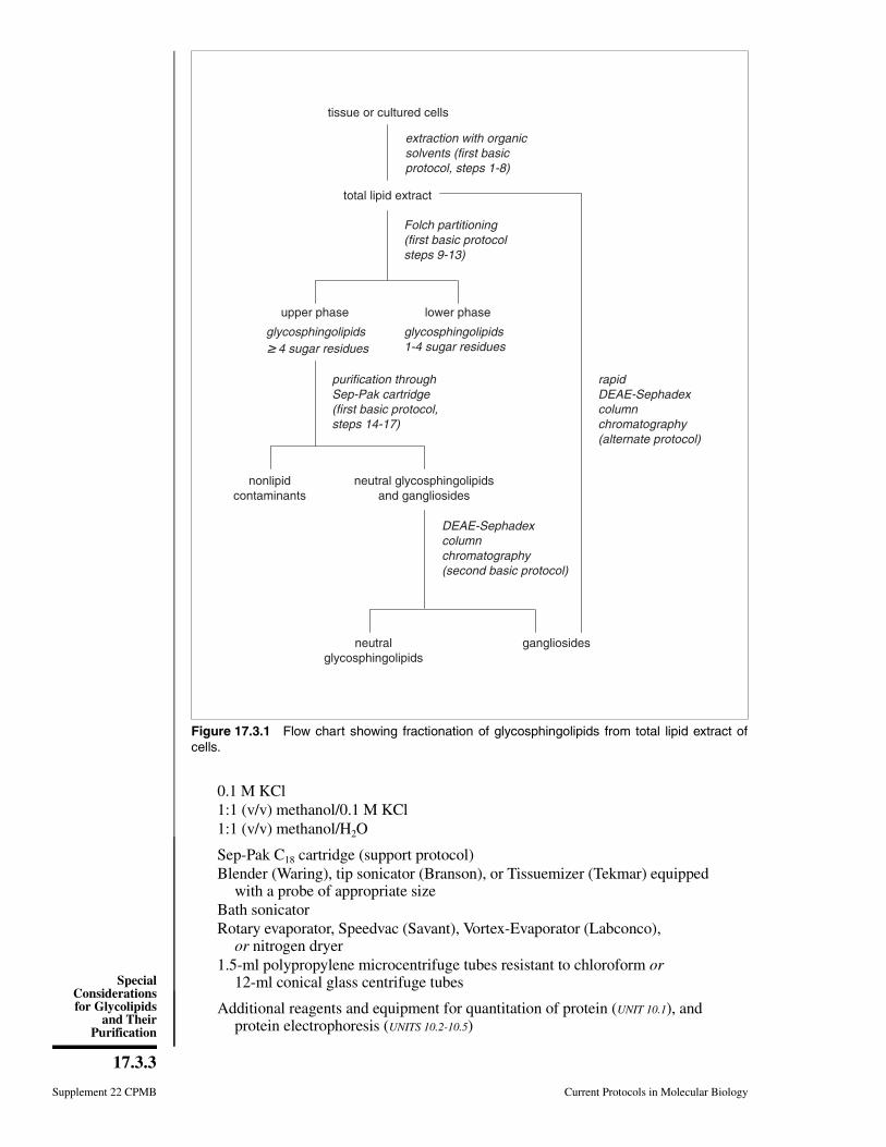
0.1 M KCl1:1 (v/v) methanol/0.1 M KCl1:1 (v/v) methanol/H2O
Sep-Pak C18 cartridge (support protocol)Blender (Waring), tip sonicator (Branson), or Tissuemizer (Tekmar) equipped
with a probe of appropriate sizeBath sonicatorRotary evaporator, Speedvac (Savant), Vortex-Evaporator (Labconco),
or nitrogen dryer1.5-ml polypropylene microcentrifuge tubes resistant to chloroform or
12-ml conical glass centrifuge tubes
Additional reagents and equipment for quantitation of protein (UNIT 10.1), andprotein electrophoresis (UNITS 10.2-10.5)
tissue or cultured cells
total lipid extract
upper phase lower phase
purification throughSep-Pak cartridge(first basic protocol,steps 14-17)
nonlipidcontaminants
neutral glycosphingolipidsand gangliosides
neutralglycosphingolipids
gangliosides
glycosphingolipids1-4 sugar residues
Folch partitioning(first basic protocolsteps 9-13)
rapidDEAE-Sephadexcolumnchromatography(alternate protocol)
extraction with organicsolvents (first basicprotocol, steps 1-8)
glycosphingolipids 4 sugar residues
DEAE-Sephadexcolumnchromatography(second basic protocol)
Figure 17.3.1 Flow chart showing fractionation of glycosphingolipids from total lipid extract ofcells.
Supplement 22 CPMB Current Protocols in Molecular Biology
17.3.3
SpecialConsiderationsfor Glycolipids
and TheirPurification
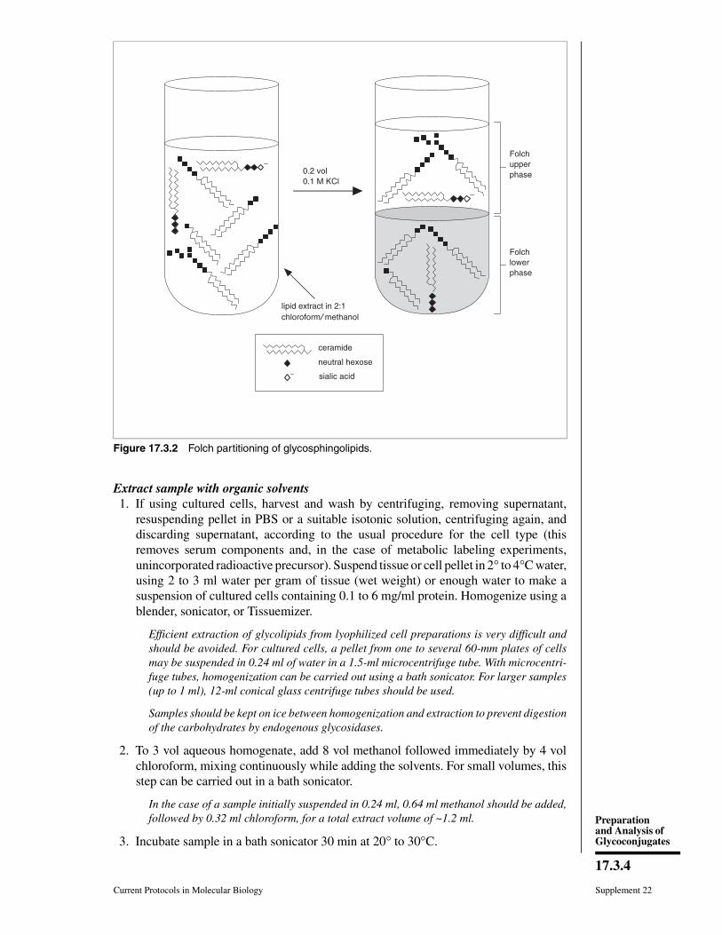
Extract sample with organic solvents1. If using cultured cells, harvest and wash by centrifuging, removing supernatant,
resuspending pellet in PBS or a suitable isotonic solution, centrifuging again, anddiscarding supernatant, according to the usual procedure for the cell type (thisremoves serum components and, in the case of metabolic labeling experiments,unincorporated radioactive precursor). Suspend tissue or cell pellet in 2° to 4°C water,using 2 to 3 ml water per gram of tissue (wet weight) or enough water to make asuspension of cultured cells containing 0.1 to 6 mg/ml protein. Homogenize using ablender, sonicator, or Tissuemizer.
Efficient extraction of glycolipids from lyophilized cell preparations is very difficult andshould be avoided. For cultured cells, a pellet from one to several 60-mm plates of cellsmay be suspended in 0.24 ml of water in a 1.5-ml microcentrifuge tube. With microcentri-fuge tubes, homogenization can be carried out using a bath sonicator. For larger samples(up to 1 ml), 12-ml conical glass centrifuge tubes should be used.
Samples should be kept on ice between homogenization and extraction to prevent digestionof the carbohydrates by endogenous glycosidases.
2. To 3 vol aqueous homogenate, add 8 vol methanol followed immediately by 4 volchloroform, mixing continuously while adding the solvents. For small volumes, thisstep can be carried out in a bath sonicator.
In the case of a sample initially suspended in 0.24 ml, 0.64 ml methanol should be added,followed by 0.32 ml chloroform, for a total extract volume of ~1.2 ml.
3. Incubate sample in a bath sonicator 30 min at 20° to 30°C.
Folchupperphase
Folchlowerphase
0.2 vol0.1 M KCl
ceramide
neutral hexose
sialic acid
lipid extract in 2:1chloroformmethanol
Figure 17.3.2 Folch partitioning of glycosphingolipids.
Current Protocols in Molecular Biology Supplement 22
17.3.4
Preparationand Analysis ofGlycoconjugates
Temperatures of 20° to 30°C may destroy labile O-acetyl groups and/or induce transacy-lation of O-acetylated derivatives of sialic acids. Lower temperatures may be required ifinformation on sialic acid acetylation is critical to subsequent analyses; in this casesolvents should be chilled to the appropriate temperature before use. Temperatures at orbelow room temperature can be maintained by adding ice to the bath sonicator.
4. Centrifuge the sample 1 min at 8800 × g (e.g., 11,000 rpm in a microcentrifuge) andcollect supernatant.
5. Extract the pellet from step 4 with a volume of 4:8:3 chloroform/methanol/waterroughly approximating the total extract volume in step 2 (e.g., ∼1 ml may be used fora sample initially suspended in 0.24 ml water). Centrifuge sample 1 min at 8800 × g.Collect supernatant and pool with that from step 4.
6. Extract the pellet from step 5 three more times as described in step 4, using 1:1chloroform/methanol, 2:1 chloroform/methanol, and 100% ethanol, respectively.Pool all the supernatants with that from steps 4 and 5.
7. If desired, analyze the ethanol-washed pellet (containing primarily lipid-free proteinsand glycoproteins) for total proteins by one- or two-dimensional gel electrophoresisor digestion with protease for isolation of total glycopeptides. Do not allow pellet todry; resuspend in a buffer containing SDS and store at −20° or −80°C prior to furthermanipulation if desired.
The two chloroform/methanol washes, using solvents with increasingly higher concentra-tions of nonpolar solvents compared to the sample, ensure the extraction of less polarglycolipids (i.e., mono- or disaccharide derivatives of ceramide that are more hydrophobicthan glycolipids with larger oligosaccharide components). The ethanol wash removes thechloroform and methanol to avoid any detrimental effects these solvents may have onsubsequent treatment of the cell pellet.
8. Evaporate the pooled supernatants from steps 4 to 6 to dryness using a rotaryevaporator, Vortex-Evaporator, Speedvac, or nitrogen dryer.
Evaporating the sample under a stream of nitrogen requires considerably more time toremove the final traces of H2O than does evaporation with a rotary evaporator, Vortex-Evaporator, or Speedvac. Dry crude lipid extract can be stored desiccated at −20°C priorto further manipulation.
Carry out Folch partitioning of glycosphingolipids9. Dissolve the crude lipid extract from step 8 in 1.0 ml of 2:1 chloroform/methanol (or
10% to 25% the volume of the initial suspended sample if this is larger than 1 ml).
10. Partition by adding 0.2 ml (or 0.2 vol) of 0.1 M KCl and mixing vigorously byvortexing. Centrifuge 1 min at 8800 × g (it will separate into two phases).
For small-scale preparations, this can be done in a 1.5-ml polypropylene microcentrifugetube. For larger volumes, phase separation may be accomplished by allowing the mixtureto stand at room temperature until separated (or in a separatory funnel overnight at 4°C).
11. Carefully remove the upper phase (∼50% of the total volume) with a Pasteur pipet.Wash lower phase by adding 1:1 methanol/0.1 M KCl in a volume equivalent to theremoved upper phase, mixing, and centrifuging 1 min at 8800 × g to separate thephases (this removes contaminating upper phase).
12. Collect the upper phase and pool with the first upper phase. Wash the lower phaseonce more with 1:1 methanol/0.1 M KCl and pool with upper phases from step 11.
Supplement 22 CPMB Current Protocols in Molecular Biology
17.3.5
SpecialConsiderationsfor Glycolipids
and TheirPurification
13. Wash the combined upper phases with 0.5 vol of 2:1 chloroform/methanol. Combinethe lower phases from the washes with the pooled lower phases from step 12.
The combined lower-phase fractions contain neutral glycolipids with oligosaccharidessmaller than tetrasaccharides, neutral lipids, and phospholipids. Tetrasaccharide-ceramides usually partition between the two phases, causing slight contamination. Thecombined upper-phase fractions contain all of the gangliosides and most of the largerneutral glycolipids (>tetrahexosylceramides) as well as all water-soluble materials fromthe original extract and inorganic salts introduced during washes of the lower phase. Whenmetabolically radiolabeled cells are used, the upper phase will also contain radioactivesugar precursors and their intermediates.
The combined lower-phase fractions can now be analyzed for glycosphingolipids usingmethods unaffected by contaminating neutral and phospholipids—e.g., TLC and autora-diography of glycolipids (if cells were radiolabeled with monosaccharide precursors),overlay analysis of thin-layer chromatograms using 125I-labeled carbohydrate-bindingproteins, or HPLC (Kannagi, 1987). The combined upper-phase fractions can be processedas described in the following steps and further purified as described in the last twoprotocols.
Prepare combined upper-phase fractions for analysis14. Dilute combined upper-phase fraction to 5 to 10 ml with 1:1 methanol/water and
apply it to a Sep-Pak C18 cartridge prepared as described in the support protocolbelow. Collect effluent in a test tube and reapply to the cartridge; repeat once.
The liquid should be applied to the Sep-Pak C18 cartridge as described in the supportprotocol. Passing the upper-phase fraction through the cartridge removes salts and othernonlipid contaminants. Collecting and reapplying the effluent to the cartridge ensurescomplete absorption of all lipid-containing compounds in the sample.
15. Wash the cartridge five times with 10-ml aliquots of water (50 ml total) to ensureremoval of all polar contaminants.
When radiolabeled cells are used, these washes can be pooled for further analysis ofradiolabeled precursors, but should contain no glycolipid. They should be discarded asradioactive waste.
16. Elute gangliosides and high-molecular-weight neutral glycolipids from the columnby washing five times with 1:1 chloroform/methanol and three times with 4:8:3chloroform/methanol/water.
The final wash with more polar solvent elutes glycolipids containing large oligosaccha-rides (when present in the sample) that were not eluted with 1:1 chloroform/methanol.
17. Combine solvent washes and evaporate to dryness. Store residue desiccated at −20°Cuntil needed.
SUPPORTPROTOCOL
PREPARATION OF SEP-PAK C18 CARTRIDGES
This protocol details preparation of Sep-Pak C18 cartridges for use in removing salts andother nonlipid contaminants from the upper-phase glycolipid fraction of Folch-partitionedmaterial. All lipid-containing compounds will be retained by the Sep-Pak C18 cartridge,water-soluble contaminants are eluted with polar solvents, and lipid-containing materialis eluted with nonpolar solvent.
Additional Materials
0.1 M ammonium acetate in 1:1 (v/v) methanol/waterSep-Pak C18 cartridges (Waters)10-ml glass syringe with locking hub (e.g., Becton Dickinson Luer-Lok)
Current Protocols in Molecular Biology Supplement 22
17.3.6
Preparationand Analysis ofGlycoconjugates
1. Remove plunger from the barrel of the syringe and attach the Luer-Lok hub of thesyringe to the long end of a Sep-Pak C18 cartridge. Pump 10 ml methanol through thecartridge by adding the liquid to the barrel of the syringe, inserting the plunger intothe syringe, and slowly pushing the liquid through the cartridge. Collect eluantmethanol in a beaker or other appropriate container. Remove the cartridge from thesyringe before removing the plunger. Remove the plunger and reattach the cartridge.
2. In the same fashion, carry out the following further washes (10 ml each):
3 washes with 1:1 chloroform/methanol1 wash with methanol2 washes with 0.1 M ammonium acetate in 1:1 methanol/water.
Sep-Pak cartridges may be reused at least five times. They should be prepared for storageby flushing the last of the methanol/water wash out with air and stored dry at roomtemperature. Preparation steps should be repeated after storage prior to use.
BASICPROTOCOL
PREPARATION OF GANGLIOSIDES
Fractionation of the glycolipid extract into neutral glycolipids and gangliosides can beeasily accomplished by separating charged glycolipids from neutral glycolipids of theupper-phase fraction obtained by Folch partitioning (the material from step 17 above). Asimple column chromatographic procedure is described here, but more sophisticatedmethods, including anion-exchange HPLC (UNIT 10.13), may be used. This protocol isadapted from a method originally developed to isolate the gangliosides from the upperphase of extracts from 50 g of tissue but also works well on a small scale for isolatinggangliosides from the upper phase of extracts of metabolically radiolabeled cultured cells.
Materials
MethanolDEAE-Sephadex equilibrated in 100% methanol,
50% slurry (reagents and solutions)Purified Folch upper-phase fraction (first basic protocol)0.01, 0.2, and 0.5 M ammonium acetate in methanolGlass wool
1. Set up a DEAE-Sephadex column in a 5-in. Pasteur pipet as follows: secure thePasteur pipet to a ring stand with a small clamp so that the column effluent can becollected into a beaker or into tubes in a test tube rack for manual processing. Use a9-in. pipet to position a small ball of glass wool in the narrow portion of the Pasteurpipet.
If the amount of glass wool is too large, or if it is packed too tightly into the tip of the pipet,the flow rate will be restricted. Alternatively, a column with appropriate solvent-resistantfittings can be used with a fraction collector for automatic sample collection.
2. Wash the glass wool with methanol to determine an appropriate flow rate in the emptycolumn (this also removes any contamination from the glass wool). If the flow rateis less than 1 drop/sec, repack the glass wool to ensure a reasonable flow rate whenthe column is packed with DEAE-Sephadex.
3. Add sufficient 50% slurry of DEAE-Sephadex in methanol to the pipet to obtain acolumn of resin 2 to 4 cm high.
A 4-cm column has a bed volume of ~1 ml; half that volume is normally sufficient capacityfor radiolabeled material.
Supplement 22 CPMB Current Protocols in Molecular Biology
17.3.7
SpecialConsiderationsfor Glycolipids
and TheirPurification
4. Wash the column with 2 to 3 column volumes of methanol.
5. Dissolve the upper-phase fraction in 0.1 to 1 ml methanol and apply to the columnof DEAE-Sephadex.
The upper-phase fraction is free of salts and other nonlipid contaminants. Because thecharged glycolipids (gangliosides) will be retained by the column under these conditions,the sample volume is not critical.
6. Elute neutral glycolipids in the upper phase by washing the column with 5 columnvolumes of methanol. Evaporate the eluant to dryness (e.g., using a nitrogen dryer).
This fraction contains desalted neutral glycolipids that are larger than trihexosyl-ceramides. These desalted neutral glycolipids can be combined with the lower-phasefraction that contains the remainder of the neutral glycolipids. This is a convenient fractionfor many analyses including thin-layer chromatography (TLC) and TLC overlay analyses.
7. Elute gangliosides that contain a single sialic acid (monosialylgangliosides) from thecolumn with 5 column volumes of 0.01 M ammonium acetate in methanol.
Eluant may be collected in equal-sized fractions if the elution profile is important, or itmay be collected in a single container. Concentrate monosialylgangliosides (e.g., byevaporation under nitrogen). If this fraction is to be used for TLC analysis, it must bedesalted. This can conveniently be done by adding water to 50% and applying the materialto a Sep-Pak C18 cartridge (support protocol).
8. Elute the gangliosides that contain two sialic acids per oligosaccharide with 5 columnvolumes of 0.2 M ammonium acetate in methanol.
9. Elute polysialylgangliosides (tri- and tetrasialylgangliosides) with 5 column volumesof 0.5 M ammonium acetate in methanol.
These fractions can be concentrated and/or desalted on Sep-Pak cartridges as describedfor monosialylgangliosides in step 7.
If the sample contains sulfated glycolipids, it may be necessary to elute them with 0.8 to1.0 M ammonium acetate in methanol.
ALTERNATEPROTOCOL
RAPID PURIFICATION OF GANGLIOSIDES
For projects that focus on the analysis of only gangliosides, this protocol describes amethod for rapid preparation of total gangliosides from total cellular lipid extract usingan ion-exchange column equilibrated in a more polar solvent.
Additional Materials
DEAE-Sephadex equilibrated in 60:30:8 methanol/chloroform/water (reagents andsolutions)
Total lipid extract (step 8 of first basic protocol)60:30:8 (v/v/v) methanol/chloroform/water60:30:8 (v/v/v) methanol/chloroform/0.8 M aqueous KCl
1. Prepare a DEAE-Sephadex column as described in step 1 of the second basic protocol,but using DEAE-Sephadex equilibrated in 60:30:8 methanol/chloroform/water.
2. Dissolve the total lipid extract from cells or tissues from step 8 of the first basicprotocol in 60:30:8 methanol/chloroform/water.
The wash volume is not critical; a volume equivalent to the volume of the original extractis generally appropriate.
Current Protocols in Molecular Biology Supplement 22
17.3.8
Preparationand Analysis ofGlycoconjugates
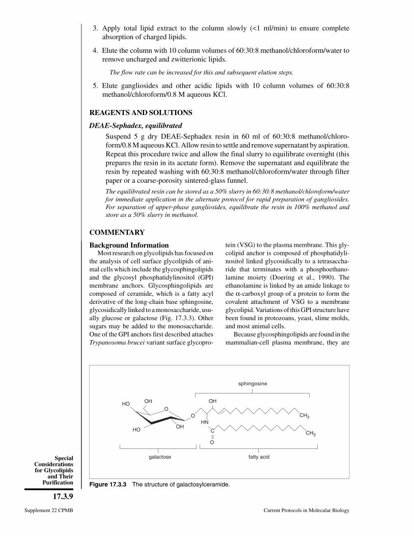
3. Apply total lipid extract to the column slowly (<1 ml/min) to ensure completeabsorption of charged lipids.
4. Elute the column with 10 column volumes of 60:30:8 methanol/chloroform/water toremove uncharged and zwitterionic lipids.
The flow rate can be increased for this and subsequent elution steps.
5. Elute gangliosides and other acidic lipids with 10 column volumes of 60:30:8methanol/chloroform/0.8 M aqueous KCl.
REAGENTS AND SOLUTIONS
DEAE-Sephadex, equilibrated
Suspend 5 g dry DEAE-Sephadex resin in 60 ml of 60:30:8 methanol/chloro-form/0.8 M aqueous KCl. Allow resin to settle and remove supernatant by aspiration.Repeat this procedure twice and allow the final slurry to equilibrate overnight (thisprepares the resin in its acetate form). Remove the supernatant and equilibrate theresin by repeated washing with 60:30:8 methanol/chloroform/water through filterpaper or a coarse-porosity sintered-glass funnel.
The equilibrated resin can be stored as a 50% slurry in 60:30:8 methanol/chloroform/waterfor immediate application in the alternate protocol for rapid preparation of gangliosides.For separation of upper-phase gangliosides, equilibrate the resin in 100% methanol andstore as a 50% slurry in methanol.
COMMENTARY
Background InformationMost research on glycolipids has focused on
the analysis of cell surface glycolipids of ani-mal cells which include the glycosphingolipidsand the glycosyl phosphatidylinositol (GPI)membrane anchors. Glycosphingolipids arecomposed of ceramide, which is a fatty acylderivative of the long-chain base sphingosine,glycosidically linked to a monosaccharide, usu-ally glucose or galactose (Fig. 17.3.3). Othersugars may be added to the monosaccharide.One of the GPI anchors first described attachesTrypanosoma brucei variant surface glycopro-
tein (VSG) to the plasma membrane. This gly-colipid anchor is composed of phosphatidyli-nositol linked glycosidically to a tetrasaccha-ride that terminates with a phosphoethano-lamine moiety (Doering et al., 1990). Theethanolamine is linked by an amide linkage tothe α-carboxyl group of a protein to form thecovalent attachment of VSG to a membraneglycolipid. Variations of this GPI structure havebeen found in protozoans, yeast, slime molds,and most animal cells.
Because glycosphingolipids are found in themammalian-cell plasma membrane, they are
O
OHHO
HO OH
O
OH
C
O
HNCH3
CH3
galactose fatty acid
sphingosine
Figure 17.3.3 The structure of galactosylceramide.
Supplement 22 CPMB Current Protocols in Molecular Biology
17.3.9
SpecialConsiderationsfor Glycolipids
and TheirPurification
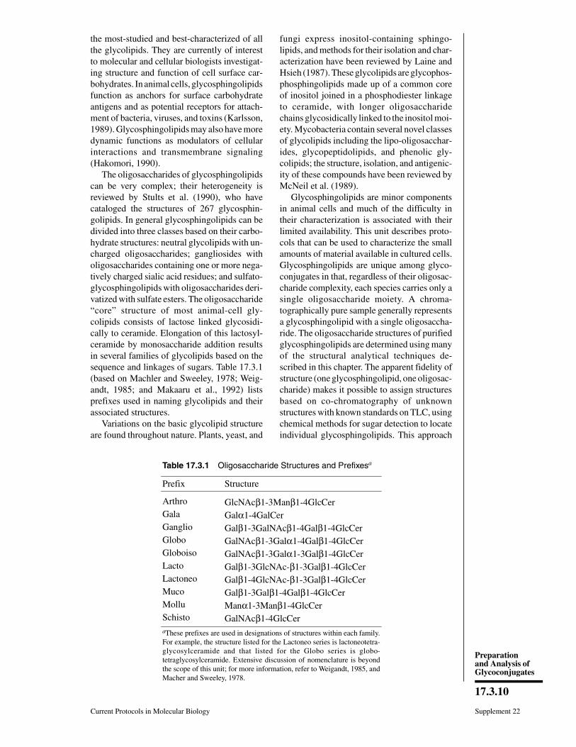
the most-studied and best-characterized of allthe glycolipids. They are currently of interestto molecular and cellular biologists investigat-ing structure and function of cell surface car-bohydrates. In animal cells, glycosphingolipidsfunction as anchors for surface carbohydrateantigens and as potential receptors for attach-ment of bacteria, viruses, and toxins (Karlsson,1989). Glycosphingolipids may also have moredynamic functions as modulators of cellularinteractions and transmembrane signaling(Hakomori, 1990).
The oligosaccharides of glycosphingolipidscan be very complex; their heterogeneity isreviewed by Stults et al. (1990), who havecataloged the structures of 267 glycosphin-golipids. In general glycosphingolipids can bedivided into three classes based on their carbo-hydrate structures: neutral glycolipids with un-charged oligosaccharides; gangliosides witholigosaccharides containing one or more nega-tively charged sialic acid residues; and sulfato-glycosphingolipids with oligosaccharides deri-vatized with sulfate esters. The oligosaccharide“core” structure of most animal-cell gly-colipids consists of lactose linked glycosidi-cally to ceramide. Elongation of this lactosyl-ceramide by monosaccharide addition resultsin several families of glycolipids based on thesequence and linkages of sugars. Table 17.3.1(based on Machler and Sweeley, 1978; Weig-andt, 1985; and Makaaru et al., 1992) listsprefixes used in naming glycolipids and theirassociated structures.
Variations on the basic glycolipid structureare found throughout nature. Plants, yeast, and
fungi express inositol-containing sphingo-lipids, and methods for their isolation and char-acterization have been reviewed by Laine andHsieh (1987). These glycolipids are glycophos-phosphingolipids made up of a common coreof inositol joined in a phosphodiester linkageto ceramide, with longer oligosaccharidechains glycosidically linked to the inositol moi-ety. Mycobacteria contain several novel classesof glycolipids including the lipo-oligosacchar-ides, glycopeptidolipids, and phenolic gly-colipids; the structure, isolation, and antigenic-ity of these compounds have been reviewed byMcNeil et al. (1989).
Glycosphingolipids are minor componentsin animal cells and much of the difficulty intheir characterization is associated with theirlimited availability. This unit describes proto-cols that can be used to characterize the smallamounts of material available in cultured cells.Glycosphingolipids are unique among glyco-conjugates in that, regardless of their oligosac-charide complexity, each species carries only asingle oligosaccharide moiety. A chroma-tographically pure sample generally representsa glycosphingolipid with a single oligosaccha-ride. The oligosaccharide structures of purifiedglycosphingolipids are determined using manyof the structural analytical techniques de-scribed in this chapter. The apparent fidelity ofstructure (one glycosphingolipid, one oligosac-charide) makes it possible to assign structuresbased on co-chromatography of unknownstructures with known standards on TLC, usingchemical methods for sugar detection to locateindividual glycosphingolipids. This approach
Table 17.3.1 Oligosaccharide Structures and Prefixesa
Prefix Structure
Arthro GlcNAcβ1-3Manβ1-4GlcCerGala Galα1-4GalCerGanglio Galβ1-3GalNAcβ1-4Galβ1-4GlcCerGlobo GalNAcβ1-3Galα1-4Galβ1-4GlcCerGloboiso GalNAcβ1-3Galα1-3Galβ1-4GlcCerLacto Galβ1-3GlcNAc-β1-3Galβ1-4GlcCerLactoneo Galβ1-4GlcNAc-β1-3Galβ1-4GlcCerMuco Galβ1-3Galβ1-4Galβ1-4GlcCerMollu Manα1-3Manβ1-4GlcCerSchisto GalNAcβ1-4GlcCeraThese prefixes are used in designations of structures within each family.For example, the structure listed for the Lactoneo series is lactoneotetra-glycosylceramide and that listed for the Globo series is globo-tetraglycosylceramide. Extensive discussion of nomenclature is beyondthe scope of this unit; for more information, refer to Weigandt, 1985, andMacher and Sweeley, 1978.
Current Protocols in Molecular Biology Supplement 22
17.3.10
Preparationand Analysis ofGlycoconjugates
has generated considerable information on thespecific tissue and species distribution of gly-cosphingolipids. As more sensitive techniqueshave become available, glycosphingolipids,like glycoproteins, have been found to possessfar greater structural variety than originallyassumed. Research in this field is shifting fromstructural to functional analysis. The develop-ment of methods that define receptor functionor immunological reactivity of glycolipidsprior to their structural analysis has been im-portant to functional studies.
The protocols in this unit are based on sev-eral procedures for analysis of glycosphingo-lipids extracted from mammalian tissues. InFolch partitioning, lipid extracts are partitionedinto upper and lower phases based on differen-tial solubility of glycolipids with different-sized oligosaccharide chains (Folch et al.,1957; Esselman et al., 1972; Ledeen and Yu,1982). The extraction procedure is fundamen-tally that described by Svennerholm and Fred-man (1980) for the quantitative extraction ofgangliosides from animal tissues—a procedureselected to ensure complete extraction of gly-colipids. The application of this procedure tothe extraction and analysis of glycolipids andglycoproteins from the same sample was sug-gested by Finne and Krusius (1982), who usedthe method to obtain lipid-free glycopeptides.Isolation and fractionation of gangliosides byDEAE-Sephadex chromatography is adaptedfrom Momoi et al. (1976). These protocols havebeen used successfully in the isolation, purifi-cation, and identification of immunologicallyreactive glycosphingolipids from both unla-beled (Magnani et al., 1982) and metabolicallyradiolabeled (Clark et al., 1991) cultured ani-mal cells.
Critical Parameters andTroubleshooting
The use of metabolically radiolabeled cellsas described in UNIT 17.4 permits analysis of asmall number of cells to obtain significant in-formation on the carbohydrate composition andstructure of glycosphingolipids.
The success of the separation and purifica-tion procedures will be obvious upon analysisof the glycosphingolipid fractions obtained.Poor resolution of glycolipids during mostchromatographic analyses is generally associ-ated with the presence of salts in the sample orwith poorly equilibrated columns. Use of theSep-Pak C18 cartridge is the most convenientmethod for removal of salt from the sample
(Williams and McClure, 1980).The combined lower-phase neutral gly-
colipid fraction will contain non-glycolipidcontaminants of the lower phase, includingneutral and zwitterionic lipids such as choles-terol, lecithin, ethanolamine, phospho-glycerides, etc. For many analyses this con-tamination does not contribute to, or interferewith, separation or detection methods. For ex-ample, in the case of material from cells meta-bolically labeled with monosaccharide precur-sors, essentially all of the detected label will bein the sugars. Label can, however, appear innon-glycolipid components of the lower phaseof a Folch extract. In extracts of Chinese ham-ster ovary cells, such material was tentativelyidentified as phosphatidylinositol (Smith et al.,1990). UNIT 17.16 describes how to determine thecomposition of radiolabeled monosaccharidesof isolated glycolipids. When specific reactionssuch as sugar-specific stains or carbohydrate-specific antibodies are used to identify gly-colipids on TLC plates, these contaminants willnot react. If removal of the contaminants isnecessary, it can be accomplished by saponifi-cation followed by chromatography on silicicacid (Lendeen and Yu, 1982). However, theseprocedures will destroy the base-labile substi-tutions of sialic acid on the gangliosides.
Contaminating phospholipids can also beremoved by saponification followed by dialy-sis. Conditions that affect sialic acids should beconsidered when planning extraction and puri-fication of gangliosides. To preserve any O-ace-tyl derivatives of the sialic acid residues ongangliosides, which may be very important infunctional analyses, it is important to avoid theuse of bases. The O-acetyl derivatives of sialicacid are extremely labile and migrate viatransacylation reactions in these cases.
The plastic microcentrifuge tubes used inthis unit must be resistant to chloroform-con-taining solvents. Most polypropylene tubesclaim resistance to chloroform, but it is worthtesting the tubes to be sure. Centrifugation stepsinvolving microcentrifuge tubes should be car-ried out in a cold room, because heat buildupduring centrifugation can increase pressure in-side capped tube (containing volatile solvents)and cause them to open during centrifugation,resulting in loss of solvent and possibly sample.For large-scale preparations, and where physi-cal methods such as mass spectroscopy are tobe used for structural analyses, use a solvent-resistant apparatus and substitute glass for plas-tic to avoid contamination with plasticizers.
Supplement 22 CPMB Current Protocols in Molecular Biology
17.3.11
SpecialConsiderationsfor Glycolipids
and TheirPurification

Anticipated ResultsThe protocols described in this section are
intended to provide the investigator with meth-ods for preparing neutral and/or acidic glycos-phingolipids from cultured animal cells orsmall samples of tissue. Because the glycos-phingolipids make up such a small amount oftotal cell mass, it is very difficult to obtaininformation related to the theoretical yield ofglycolipid; however, these protocols will pro-vide a representative sample of the neutral gly-colipids and gangliosides present in the startingmaterial. The results of each isolation step andhow each sample may be used for further analy-sis are described throughout the unit. The finalsamples of neutral glycolipid and/or gan-glioside are to be used for further analysis orcharacterization.
Time ConsiderationsThe total extract can be obtained and dried
in ∼3 hr, depending upon the method used forevaporation. Folch partitioning of the total ex-tract into upper and lower phases will require30 to 60 min. The preparation of the DEAE-Sephadex requires an overnight equilibrationstep; if large amounts of resin are equilibratedand stored, only a brief wash of the resin withthe starting solvent is required before each timethe procedure is carried out. Preparation of aSep-Pak C18 cartridge according to the supportprotocol will require ∼30 min. The desaltingstep using a Sep-Pak cartridge is somewhatcumbersome and time-consuming becausemultiple samples must be processed individu-ally and the procedure requires considerablemanipulation of syringe and cartridge. If mul-tiple samples are being processed, commercialvacuum manifolds that accommodate largenumbers of cartridges may be used. Severalhours are required to run the DEAE-Sephadexcolumn with a flow rate of 1 to 2 ml/min andmultiple solvent systems to fractionate gan-gliosides. From extraction to fractionation ofgangliosides can take up to 2 days. Rapid iso-lation of the ganglioside fraction (steps 1 to 12of the first basic protocol) for use in the alter-nate protocol can be accomplished within 1 to2 hr; the alternate protocol will produce neutraland acidic fractions within 1 day.
Literature CitedClark, G.F., Cummings, R.D., Mattox, S., Gorbea,
C., and Smith, D.F. 1991. Decreased biosynthe-sis of Forssman glycolipid after retinoic acid-in-duced differentiation of mouse F9 teratocarci-noma cells: Lectin affinity chromatography ofthe glycolipid-derived oligosaccharides. Carbo-hydr. Res. 213:155-168.
Doering, T.L., Masterson, W.J., Hart, G.W., andEnglund, P.T. 1990. Biosynthesis of glycosylphosphatidylinositol membrane anchors. J. Biol.Chem. 265:611-614.
Esselman, W.J., Laine, R.A., and Sweeley, C.C.1972. Isolation and characterization of glycos-phingolipids. Methods Enzymol. 28:140-159.
Finne, J. and Krusius, T. 1982. Preparation andfractionation of glycopeptides. Methods Enzy-mol. 83:269-277.
Folch, J., Lees, M.B., and Sloane Stanley, G.H.1957. A simple method for the isolation andpurification of total lipides from animal tissues.J. Biol. Chem. 226:497-503.
Hakomori, S. 1990. Bifunctional role of glycosphin-golipids. J. Biol. Chem. 265:18713-18716.
Kannagi, K., Watanabe, K., and Hakomori, S. 1987.Isolation and purification of glycosphingolipidsby high performance liquid chromatography.Methods Enzymol. 138:3-12.
Karlsson, K.-A. 1989. Animal glycosphingolipidsas membrane attachment sites for bacteria. Ann.Rev. Biochem. 58:309-350.
Laine, R.A. and Hsieh, T.C.-Y. 1987. Inositol-con-taining sphingolipids. Methods Enzymol.138:186-195.
Ledeen, R.W. and Yu, R.K. 1982. Gangliosides:Structure, isolation and analysis. Methods Enzy-mol. 83:139-191.
McNeil, M., Chatterjee, D., Hunter, S.W., and Bren-nan, P.J. 1989. Mycobacterial glycolipids: Isola-tion, structures, antigenicity, and synthesis.Methods Enzymol. 179:215-242.
Macher, B.A. and Sweeley, C.C. 1978. Glycosphin-golipids: Structure, biological source, and prop-erties. Methods Enzymol. 50:236-250.
Magnani, J.L., Nilsson, B., Brockhaus, M., Zopf, D.,Steplewski, Z., Koprowski, H., and Ginsburg, V.1982. A monoclonal antibody-defined antigenassociated with gastrointestinal cancer is a gan-glioside containing sialylated lacto-N-fucopen-taose II. J. Biol. Chem. 257:14365-14369.
Makaaru, C.K., Damian, R.T., Smith, D.F., andCummings, R.D. 1992. The human blood flukeSchistosoma mansoni synthesizes a novel type ofglycosphingolipid. J. Biol. Chem. 267:2251-2257.
Current Protocols in Molecular Biology Supplement 22
17.3.12
Preparationand Analysis ofGlycoconjugates
Momoi, T., Ando, S., and Nagai, Y. 1976. Highresolution preparative chromatography systemfor gangliosides using DEAE-Sephadex and thenew porous silica, iatrobeads. Biochim. Biophys.Acta 441:488-497.
Smith, D.F., Larsen, R.D., Mattox, S., Lowe, J.B.,and Cummings, R.D. 1990. Transfer and expres-sion of murine UDPGal:β-D-Galα1,3Galacto-syltransferase gene in transfected Chinese ham-ster ovary cells. J. Biol. Chem. 265:6225-6234.
Stults, C.L.M., Sweeley, C., and Macher, B.A. 1990.Glycosphingolipids: Structure, biologicalsource, and properties. Methods Enzymol.179:167-214.
Svennerholm, L. and Fredman, P. 1980. A procedurefor the quantitative isolation of brain gan-gliosides. Biochim. Biophys. Acta 617:97-109.
Weigandt, H. 1985. Glycolipids. In New Compre-hensive Biochemistry (A. Neuberger and L.L.M.van Deenen, eds.) Vol. 10. pp. 2-4. Elsevier Sci-ence Publishing, New York.
Williams, M.A. and McClure, R.H. 1980. The useof Sep-Pak C18 Cartridge during the isolation ofgangliosides. J. Neurochem. 35:266-269.
Contributed by David F. SmithThe University of GeorgiaAtlanta, Georgia
Pedro A. PrietoRoss LaboratoriesColumbus, Ohio
Supplement 22 CPMB Current Protocols in Molecular Biology
17.3.13
SpecialConsiderationsfor Glycolipids
and TheirPurification
UNIT 17.4Metabolic Radiolabeling of Animal CellGlycoconjugates
Sandra Diaz1 and Ajit Varki1
1University of California San Diego, La Jolla, California
ABSTRACT
Useful information about glycoconjugates can be obtained by labeling their aglycone(noncarbohydrate) portions—e.g., labeling proteins with radioactive amino acids—andthen using techniques described elsewhere in this chapter to infer the presence, type, andnature of glycan chains. This unit describes metabolic labeling techniques that providemore specific information about the structure, sequence, and distribution of the sugarchains of glycoconjugates. Following metabolic labeling, the radioactive glycoconjugateof interest is isolated, individual glycosylation sites are identified and separated if neces-sary, and the labeled glycans are subjected to structural analysis. Curr. Protoc. Mol. Biol.87:17.4.1-17.4.15. C© 2009 by John Wiley & Sons, Inc.
Keywords: monosaccharides radioactivity pulse-chase metabolic labeling
specific activity
INTRODUCTION
Useful information about glycoconjugates can be obtained by labeling their aglycone(noncarbohydrate) portions—e.g., labeling proteins with radioactive amino acids—andthen using techniques described elsewhere in this chapter to infer the presence, type,and nature of oligosaccharide (glycan) chains. This unit describes metabolic labelingtechniques that provide more specific information about the structure, sequence, anddistribution of the glycan chains of glycoconjugates. Following metabolic labeling, theradioactive glycoconjugate of interest is isolated, individual glycosylation sites are iden-tified and separated if necessary, and the labeled glycans are subjected to structuralanalysis. Although these techniques provide less information than would complete se-quencing of the glycan chains, the partial structural information derived is sufficient formany purposes.
Metabolic labeling is simple and easy to perform, and requires no sophisticated instru-mentation other than a scintillation counter. In addition, purification of the glycoconjugateto radiometric homogeneity is sufficient for further analysis (i.e., other contaminatingmolecules that are not labeled do not have to be removed). Important practical consider-ations for metabolic labeling experiments include selecting the type of experiment andthe labeled precursor, understanding the specificity of labeling, and maximizing uptakeand incorporation (see Critical Parameters). Before proceeding with experiments to labelglycoconjugates metabolically, labeling conditions should be evaluated to ensure thatcell viability and metabolism are not altered, and optimized for uptake and incorporationof the precursor (this should be done for each combination of precursor and cell line; seethe Basic Protocol and Alternate Protocol 1).
In the Basic Protocol, cells in culture are grown through several population doublings incomplete medium supplemented with radiolabeled glycan precursors to reach a steady-state level of incorporation. In the alternate protocols, cells are cultured for a short periodof time in a deficient medium that contains a high concentration of radiolabeled precursor.
Current Protocols in Molecular Biology 17.4.1-17.4.15, July 2009Published online July 2009 in Wiley Interscience (www.interscience.wiley.com).DOI: 10.1002/0471142727.mb1704s87Copyright C© 2009 John Wiley & Sons, Inc.
Preparation andAnalysis ofGlycoconjugates
17.4.1
Supplement 87
MetabolicRadiolabeling of
Animal CellGlycoconjugates
17.4.2
Supplement 87 Current Protocols in Molecular Biology
A pulse or pulse-chase labeling procedure (Alternate Protocol 1) can be used to analyzeprecursor-product relationships. With sequential pulse-labeling (Alternate Protocol 2),it is possible to obtain quantities of labeled glycoconjugates while using a minimumamount of labeled precursor by using the same batch of medium to pulse-label a seriesof cultures. A support protocol describes the preparation of multiply deficient medium(MDM) for use in making appropriate deficient media.
NOTE: Conventional protocols for handling, monitoring, shielding, and disposing ofradioactivity and radioactive waste (APPENDIX 1F) and for tissue culture of cells and sterilehandling of media should be followed throughout these protocols.
BASICPROTOCOL
STEADY-STATE LABELING WITH RADIOACTIVE PRECURSORS
When studying glycoconjugates in an established tissue culture cell line, molecules ofinterest can be labeled for structural characterization. In the following protocol, cells inculture are grown to a steady-state level of incorporation in complete medium supple-mented with radiolabeled glycoconjugate precursor.
Optimizing Conditions
An attempt should be made to label the molecules as close as possible to the metabolicsteady state—i.e., a constant level of radioactivity per mass unit of a given monosaccha-ride in all glycoconjugates in the cell—under conditions of normal growth. In practice,this is somewhat difficult to achieve with most cell types. The maximum possible numberof cells should be grown for the longest possible period of time in the minimum possiblevolume of complete medium supplemented with the maximum possible amount of ra-dioactive label. Before labeling is attempted, the following growth characteristics shouldbe determined for the cell type of interest: (1) population doubling time, (2) maximumdegree of dilution upon splitting that is compatible with proper regrowth, (3) maximumcell density compatible with healthy growth and metabolism (confluence), and (4) min-imum volume of medium that will sustain regrowth from a full split to confluence. Ifthese parameters are properly defined, growth from full split to confluence will allowat least three population doublings during the labeling period for most cell lines. Insome cases, however (e.g., with very slowly growing cell lines, lines requiring frequentmedium changes, or cells that cannot be diluted to low density when splitting), this maynot be feasible.
Materials
Radioactive precursor: 3H- or 14C-labeled monosaccharide, [35S]sulfate,[3H]acetate, or [32P]orthophosphate; at highest available specific activity
Complete tissue culture medium appropriate for long-term growth of tissue culturecell line, supplemented as necessary
Established tissue culture cell line, either suspension or monolayerPhosphate-buffered saline (PBS; APPENDIX 2), pH 7.2, ice cold
Disposable sterile 50-ml vacuum-suction filter device: filter flask fitted with0.22-μm filter
Disposable 0.22-μm sterile filter attached to sterile plastic syringe, both withLuer-Lok fittings
Sterile pipet tipsTissue culture plates or flasksScrew-cap centrifuge tubesTabletop centrifuges, at room temperature and 4CRubber policeman or disposable cell scraperScintillation counter
Preparation andAnalysis ofGlycoconjugates
17.4.3
Current Protocols in Molecular Biology Supplement 87
1. If necessary, dry radioactive precursor (in a ventilated hood) to completely removeany organic solvents. Dissolve in a small volume of complete tissue culture medium.
2. Filter sterilize radioactive medium, using a standard disposable 50-ml vacuum suc-tion filter device (filter flask fitted with a 0.22-μm filter) for volumes ≥10 ml or a0.22-μm filter attached directly to the tip of a disposable sterile syringe for smallervolumes. Wash out the original container with additional medium and use this fluidto wash through the filter (this maximizes recovery of label).
CAUTION: Dispose of the radioactive filter and/or syringe appropriately.
The radioactive precursor may be available in a sterile aqueous medium, ready for use.However, such preparations are more expensive, and after repeated opening of suchpackages, filtering is recommended to ensure continued sterility.
3. Add additional medium if necessary to bring the volume to the amount needed forthe experiment. Warm the medium in an incubator or water bath to the temperatureat which labeling will be done.
4. Using a sterile pipet tip, aliquot a small amount of radioactive medium and save forscintillation counting in step 8.
5. Split monolayer cells according to standard method (e.g., detach by trypsinization),resuspend in radioactive medium, and plate onto tissue culture plates. For suspensioncultures, dilute directly in radioactive medium or concentrate as follows: place culturein an appropriately sized screw-cap centrifuge tube and centrifuge in a tabletopcentrifuge 5 min at ∼500 × g, room temperature. Discard supernatant and resuspendcell pellet in radioactive medium. Incubate under standard conditions for the cell linebeing studied.
The number of cells needed, and therefore the culture vessels and volume of medium used,will depend on the growth characteristics of the particular cell line being studied andshould be optimized as described in the protocol introduction.
6. When the cells reach maximum growth (confluence or late-log phase), chill culture onice and harvest cells, scraping monolayer cells from plate using a rubber policemanor cell scraper. Pellet cells 5 min at ∼500 × g, 4C. Decant the labeled medium andsave an aliquot for scintillation counting in step 8.
If the glycoconjugates in the conditioned medium are to be studied, the medium should befiltered through a 0.22-μm filter to remove any cell debris remaining after the centrifu-gation.
7. Wash cell pellet twice in a >50-fold excess of ice-cold PBS, pH 7.2.
At this stage, the labeled cell pellet can be frozen at −20C to −80C for later analysisif desired.
8. Using a scintillation counter, determine the efficiency of incorporation by measuringthe radioactivity incorporated into an aliquot of the cells and the sample of condi-tioned medium from step 6. As a control, measure the radioactivity of the sample ofsterile radioactive medium from step 4.
It may be necessary to dilute the medium to get an accurate count.
ALTERNATEPROTOCOL 1
PULSE OR PULSE-CHASE LABELING WITH RADIOACTIVEPRECURSORS
If steady-state labeling (see Basic Protocol) does not yield sufficient incorporation oflabel into the glycoconjugate of interest, it may be desirable to provide labeled precursorin the absence of, or with decreased amounts of, the unlabeled form. The pulse proce-dure described in this protocol—in which cells are cultured briefly in deficient medium
MetabolicRadiolabeling of
Animal CellGlycoconjugates
17.4.4
Supplement 87 Current Protocols in Molecular Biology
supplemented with a radioactive precursor—may be used in these cases. Alternatively,the procedure can be expanded into a pulse-chase by adding a chase of nonradioac-tive medium; this permits brief labeling of the glycoconjugate of interest to establishprecursor-product relationships. These procedures are most useful for labeling monosac-charides that compete with glucose for uptake—i.e., Gal, Glc, Man, and GlcNH2 (seeCommentary). They are of limited value for monosaccharides that are not usually takenup efficiently by cells—i.e., GlcNAc, GalNAc, ManNac, Neu5Ac, Fuc, Xyl, and GlcA—unless very large quantities of such labeled molecules can be used for pulse labeling.Because pulse labelings are typically done for a short time (e.g., minutes or hours), cellsare usually used in a nearly confluent state, to maximize uptake and incorporation.
Optimizing Conditions
Before the experiment is performed, several pilot experiments should be carried out todetermine the optimal conditions for labeling.
Determining base incorporation level and the effect of glucose. MDM (multiply de-ficient medium) appropriate for growing cultures of the cell line of interest should bereconstituted (see Support Protocol) to 100% levels of all components except the onebeing presented as a radiolabeled precursor or competing molecule (e.g., glucose). Ifrequired by the cells, dialyzed serum should be added to the appropriate final concentra-tion. Finally, radiolabeled precursor should be added. A small-scale pilot labeling shouldbe carried out by culturing cells in the labeled medium and monitoring incorporationof the label into the macromolecule of interest or into whole-cell glycoproteins. If theradiolabeled precursor is a monosaccharide, the effect of glucose on incorporation ofradiolabeled precursor should be assessed by carrying out this pilot experiment in dupli-cate, using one lot of medium prepared as described and one prepared the same way butwithout glucose.
Determining optimal concentration of radiolabeled precursor. Enough MDM for sev-eral pilot experiments should be reconstituted to 100% levels of all components exceptthe one being presented as a radiolabeled precursor. The radiolabeled precursor shouldthen be added and the medium divided into aliquots. The aliquots should be supple-mented with an unlabeled 100× stock solution of the same precursor to yield a seriesof media containing different concentrations of the unlabeled precursor (e.g., 0%, 5%,10%, 20%, 50%, and 100% of the concentration in normal medium). These should beused for small-scale pilot labelings of the cell line of interest for defined periods oftime and incorporation of label should be monitored as described above. Incorporationshould be plotted against the total precursor concentration. The point at which the curvebreaks—i.e., where the percentage of label incorporated is markedly decreased by furtheraddition of unlabeled compound—is the point at which the precursor concentration isno longer limiting for biosynthetic reactions (Fig. 17.4.1). The optimal concentrationof unlabeled precursor will be a little higher than the break point for the curve, whereunlabeled precursor is not limiting, but incorporation is still good.
Checking cell growth at optimal precursor concentration. Reconstituted MDM contain-ing radioactive precursor plus the optimal concentration of unlabeled precursor shouldbe prepared and cells should be grown in this medium for the desired period of time.Incorporation of radiolabeled precursor should be monitored and linearity of uptake andincorporation of label over time should be assessed. Measures of cell growth, stability,and general biosynthetic capability—e.g., cell counts and viability assays and radioactiveamino acid incorporation—should also be assayed to determine if reducing the concen-tration of the precursor into this range has any detrimental effect upon the cells. It issometimes necessary to reach a compromise between the opposing factors of loweredconcentration, labeling time, and cell viability. An attempt should be made to determine
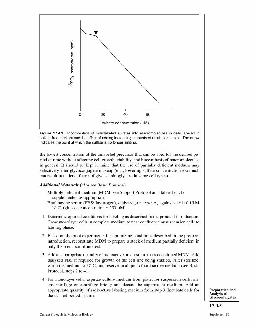
Preparation andAnalysis ofGlycoconjugates
17.4.5
Current Protocols in Molecular Biology Supplement 87
35 S
O4
inco
rpor
ated
(cp
m)
sulfate concentration (μM)
0 20 40 60
Figure 17.4.1 Incorporation of radiolabeled sulfates into macromolecules in cells labeled insulfate-free medium and the effect of adding increasing amounts of unlabeled sulfate. The arrowindicates the point at which the sulfate is no longer limiting.
the lowest concentration of the unlabeled precursor that can be used for the desired pe-riod of time without affecting cell growth, viability, and biosynthesis of macromoleculesin general. It should be kept in mind that the use of partially deficient medium mayselectively alter glycoconjugate makeup (e.g., lowering sulfate concentration too muchcan result in undersulfation of glycosaminoglycans in some cell types).
Additional Materials (also see Basic Protocol)
Multiply deficient medium (MDM; see Support Protocol and Table 17.4.1)supplemented as appropriate
Fetal bovine serum (FBS; Invitrogen), dialyzed (APPENDIX 3C) against sterile 0.15 MNaCl (glucose concentration ∼250 μM)
1. Determine optimal conditions for labeling as described in the protocol introduction.Grow monolayer cells in complete medium to near confluence or suspension cells tolate-log phase.
2. Based on the pilot experiments for optimizing conditions described in the protocolintroduction, reconstitute MDM to prepare a stock of medium partially deficient inonly the precursor of interest.
3. Add an appropriate quantity of radioactive precursor to the reconstituted MDM. Adddialyzed FBS if required for growth of the cell line being studied. Filter sterilize,warm the medium to 37C, and reserve an aliquot of radioactive medium (see BasicProtocol, steps 2 to 4).
4. For monolayer cells, aspirate culture medium from plate; for suspension cells, mi-crocentrifuge or centrifuge briefly and decant the supernatant medium. Add anappropriate quantity of radioactive labeling medium from step 3. Incubate cells forthe desired period of time.

MetabolicRadiolabeling of
Animal CellGlycoconjugates
17.4.6
Supplement 87 Current Protocols in Molecular Biology
Table 17.4.1 Composition of Multiply Deficient Medium (MDM)
ComponentFinal concentration incomplete medium
Stock
CaCl2 200 mg/liter Salt, reagent grade
KCl 400 mg/liter Salt, reagent grade
MgCl2a 75 mg/liter Salt, reagent grade
NaCl 6800 mg/liter Salt, reagent grade
NaH2PO4·H2O 140 mg/liter Salt, reagent grade
Phenol red 10 mg/liter 10×Sodium pyruvate 110 mg/liter 100×L-Alanine 25 mg/liter 100×L-Arginine 126 mg/liter 100×L-Asparagine 50 mg/liter 100×L-Aspartic acid 30 mg/liter 100×L-Cysteine NONEb —
L-Glutamic acid 75 mg/liter 100×L-Glutamine NONEb —
L-Glycine 50 mg/liter 100×L-Histidine 42 mg/liter 100×L-Isoleucine 52 mg/liter 100×L-Leucine 52 mg/liter 100×L-Lysine 72 mg/liter 100×L-Methionine NONEb —
L-Phenylalanine 32 mg/liter 100×L-Proline 40 mg/liter 100×L-Serine NONEb —
L-Threonine 48 mg/liter 100×L-Tryptophan 10 mg/liter 100×L-Tyrosine 36 mg/liter 100×L-Valine 46 mg/liter 100×MEM vitamins (mixture) 1× 100×aDo not use MgSO4 in place of MgCl2.bNot present in MDM; add as needed for specific experiments.
Labeling time should be based on the length of time the cells can survive in deficientmedium as determined in the optimizing pilot experiment.
Save radioactive medium for analysis, if necessary.
5. For pulse labeling, proceed to step 6. For pulse-chase labeling, remove medium asdescribed in step 4, add a chase of nonradioactive complete medium, and incubatefor the desired period of time.
For a pulse-chase experiment, several vessels of each cell culture should be set up andchased for varying periods of time.
6. Harvest and wash the cells. Determine efficiency of incorporation (see Basic Protocol,steps 6 to 8).

Preparation andAnalysis ofGlycoconjugates
17.4.7
Current Protocols in Molecular Biology Supplement 87
ALTERNATEPROTOCOL 2
SEQUENTIAL PULSE OR PULSE-CHASE LABELING WITH REUSE OFRADIOACTIVELY LABELED MEDIUM
In some cases, incorporation of label into the glycoconjugate of interest may be inad-equate, even under defined conditions with selectively deficient medium. In addition,prolonged exposure to deficient medium may result in alterations in synthesis of proteinsor other macromolecules. In these situations, it may be desirable to expose a series ofplates of cells sequentially to a small volume of medium containing a high concentrationof label. This also conserves expensive radiolabeled precursor. In this protocol, multi-ple sets of cell cultures are sequentially pulse-labeled. One set is incubated in labelingmedium for the pulse period, and then the labeling medium is removed and added tothe next set. For a pulse experiment (designed to obtain a sizable quantity of materialwith a high level of incorporation), the cells are harvested at this point; for a pulse-chase,a chase of nonradioactive complete medium is added to the first set of cultures. Thisprocess is repeated until all cells have been labeled (Fig. 17.4.2). Because the incubationperiod is relatively short (e.g., a few hours), it is assumed that only a very small fractionof the radiolabeled precursor is consumed from the medium during each labeling cycleand that the concentrations of other essential components are not substantially changed.To further conserve labeled medium, some investigators freeze it after limited usage andthaw it to reuse (the pH of thawed medium should be adjusted before use). When reusingthe medium, the experimental results should be interpreted carefully, especially withregard to the specific activity of labeled products and the possibility of metabolic effectsof secreted molecules.
Additional Materials (also see Basic Protocol)
Multiply deficient medium (MDM; see Support Protocol and Table 17.4.1),supplemented as appropriate
Fetal bovine serum (FBS; Invitrogen), dialyzed (APPENDIX 3C) against sterile 0.15 MNaCl (glucose concentration ∼250 μM)
1. Determine optimal conditions for labeling (see Alternate Protocol 1 introduction). Setup several identical sets of monolayer or suspension cells to be labeled sequentiallyand grow to confluence or late-log phase.
PULSE
PULSE
PULSE
TIME:
PULSE CHASE
HARVEST
CHASE
CHASE*
*
*
* Transfer labeling medium to next plate. Add complete medium to labeled cells.
Figure 17.4.2 Scheme for sequential pulse labeling of cells reutilizing radioactive medium.
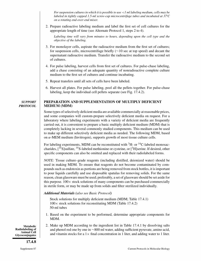
MetabolicRadiolabeling of
Animal CellGlycoconjugates
17.4.8
Supplement 87 Current Protocols in Molecular Biology
For suspension cultures in which it is possible to use <1 ml labeling medium, cells may belabeled in tightly capped 1.5-ml screw-cap microcentrifuge tubes and incubated at 37Con a rotating end-over-end mixer.
2. Prepare radioactive labeling medium and label the first set of cell cultures for theappropriate length of time (see Alternate Protocol 1, steps 2 to 4).
Labeling time will vary from minutes to hours, depending upon the cell type and theobjective of the labeling.
3. For monolayer cells, aspirate the radioactive medium from the first set of cultures;for suspension cells, microcentrifuge briefly (∼10 sec at top speed) and decant thesupernatant radioactive medium. Transfer the radioactive medium to the second setof cultures.
4. For pulse labeling, harvest cells from first set of cultures. For pulse-chase labeling,add a chase consisting of an adequate quantity of nonradioactive complete culturemedium to the first set of cultures and continue incubating.
5. Repeat transfers until all sets of cells have been labeled.
6. Harvest all plates. For pulse labeling, pool all the pellets together. For pulse-chaselabeling, keep the individual cell pellets separate (see Fig. 17.4.2).
SUPPORTPROTOCOL
PREPARATION AND SUPPLEMENTATION OF MULTIPLY DEFICIENTMEDIUM (MDM)
Some types of selectively deficient media are available commercially at reasonable prices,and some companies will custom-prepare selectively deficient media on request. For alaboratory where labeling experiments with a variety of deficient media are frequentlycarried out, it is convenient to prepare a basic multiply deficient medium (MDM) that iscompletely lacking in several commonly studied components. This medium can be usedto make up different selectively deficient media as needed. The following MDM, basedon α-MEM medium (Invitrogen), supports growth of most tissue culture cells.
For labeling experiments, MDM can be reconstituted with 3H- or 14C-labeled monosac-charides, [35S]sulfate, 35S-labeled methionine or cysteine, or [3H]serine. If desired, otherspecific components can also be omitted and replaced with their radiolabeled forms.
NOTE: Tissue culture–grade reagents (including distilled, deionized water) should beused in making MDM. To ensure that reagents do not become contaminated by com-pounds such as endotoxin as portions are being removed from stock bottles, it is importantto pour liquids carefully and use disposable spatulas for removing solids. For the samereason, clean glassware must be used; preferably, a set of glassware should be set aside forthis purpose. 100× stock solutions of many components can be purchased commerciallyin sterile form, or may be made up from solids and filter sterilized individually.
Additional Materials (also see Basic Protocol)
Stock solutions for multiply deficient medium (MDM; Table 17.4.1)100× stock solutions for reconstituting MDM (Table 17.4.2)50-ml tubes
1. Based on the experiment to be performed, determine appropriate components forMDM.
2. Make up MDM according to the ingredient list in Table 17.4.1 by dissolving saltsand phenol red one by one in ∼800 ml water, adding sufficient pyruvate, amino acid,and vitamin stocks for a 1× final concentration in 1 liter, and adding water to 1 liter.
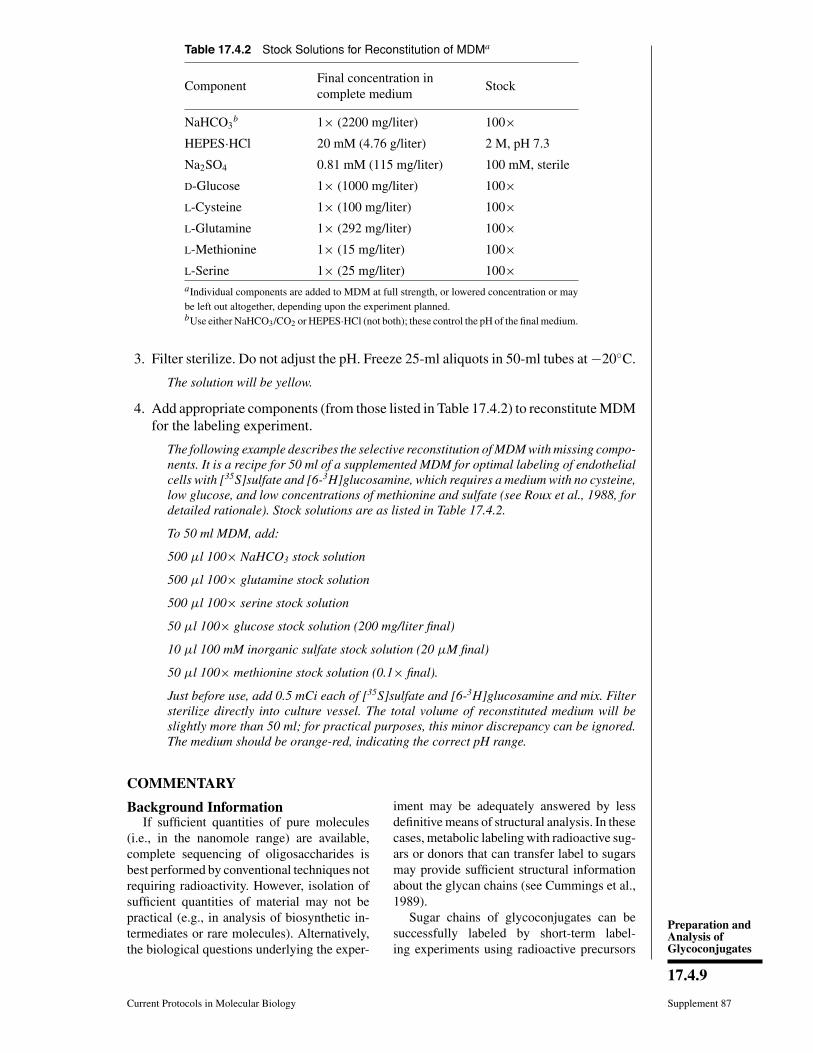
Preparation andAnalysis ofGlycoconjugates
17.4.9
Current Protocols in Molecular Biology Supplement 87
Table 17.4.2 Stock Solutions for Reconstitution of MDMa
ComponentFinal concentration incomplete medium
Stock
NaHCO3b 1× (2200 mg/liter) 100×
HEPES·HCl 20 mM (4.76 g/liter) 2 M, pH 7.3
Na2SO4 0.81 mM (115 mg/liter) 100 mM, sterile
D-Glucose 1× (1000 mg/liter) 100×L-Cysteine 1× (100 mg/liter) 100×L-Glutamine 1× (292 mg/liter) 100×L-Methionine 1× (15 mg/liter) 100×L-Serine 1× (25 mg/liter) 100×aIndividual components are added to MDM at full strength, or lowered concentration or maybe left out altogether, depending upon the experiment planned.bUse either NaHCO3/CO2 or HEPES·HCl (not both); these control the pH of the final medium.
3. Filter sterilize. Do not adjust the pH. Freeze 25-ml aliquots in 50-ml tubes at −20C.
The solution will be yellow.
4. Add appropriate components (from those listed in Table 17.4.2) to reconstitute MDMfor the labeling experiment.
The following example describes the selective reconstitution of MDM with missing compo-nents. It is a recipe for 50 ml of a supplemented MDM for optimal labeling of endothelialcells with [35S]sulfate and [6-3H]glucosamine, which requires a medium with no cysteine,low glucose, and low concentrations of methionine and sulfate (see Roux et al., 1988, fordetailed rationale). Stock solutions are as listed in Table 17.4.2.
To 50 ml MDM, add:
500 μl 100× NaHCO3 stock solution
500 μl 100× glutamine stock solution
500 μl 100× serine stock solution
50 μl 100× glucose stock solution (200 mg/liter final)
10 μl 100 mM inorganic sulfate stock solution (20 μM final)
50 μl 100× methionine stock solution (0.1× final).
Just before use, add 0.5 mCi each of [35S]sulfate and [6-3H]glucosamine and mix. Filtersterilize directly into culture vessel. The total volume of reconstituted medium will beslightly more than 50 ml; for practical purposes, this minor discrepancy can be ignored.The medium should be orange-red, indicating the correct pH range.
COMMENTARY
Background InformationIf sufficient quantities of pure molecules
(i.e., in the nanomole range) are available,complete sequencing of oligosaccharides isbest performed by conventional techniques notrequiring radioactivity. However, isolation ofsufficient quantities of material may not bepractical (e.g., in analysis of biosynthetic in-termediates or rare molecules). Alternatively,the biological questions underlying the exper-
iment may be adequately answered by lessdefinitive means of structural analysis. In thesecases, metabolic labeling with radioactive sug-ars or donors that can transfer label to sugarsmay provide sufficient structural informationabout the glycan chains (see Cummings et al.,1989).
Sugar chains of glycoconjugates can besuccessfully labeled by short-term label-ing experiments using radioactive precursors
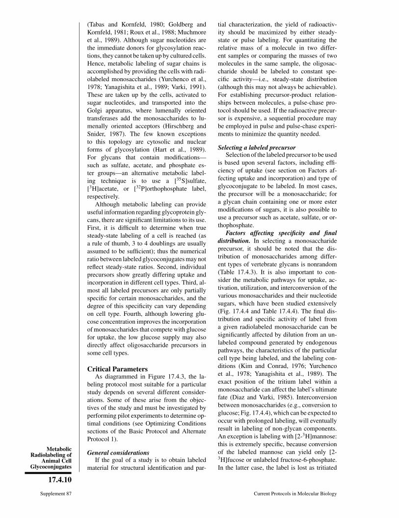
MetabolicRadiolabeling of
Animal CellGlycoconjugates
17.4.10
Supplement 87 Current Protocols in Molecular Biology
(Tabas and Kornfeld, 1980; Goldberg andKornfeld, 1981; Roux et al., 1988; Muchmoreet al., 1989). Although sugar nucleotides arethe immediate donors for glycosylation reac-tions, they cannot be taken up by cultured cells.Hence, metabolic labeling of sugar chains isaccomplished by providing the cells with radi-olabeled monosaccharides (Yurchenco et al.,1978; Yanagishita et al., 1989; Varki, 1991).These are taken up by the cells, activated tosugar nucleotides, and transported into theGolgi apparatus, where lumenally orientedtransferases add the monosaccharides to lu-menally oriented acceptors (Hirschberg andSnider, 1987). The few known exceptionsto this topology are cytosolic and nuclearforms of glycosylation (Hart et al., 1989).For glycans that contain modifications—such as sulfate, acetate, and phosphate es-ter groups—an alternative metabolic label-ing technique is to use a [35S]sulfate,[3H]acetate, or [32P]orthophosphate label,respectively.
Although metabolic labeling can provideuseful information regarding glycoprotein gly-cans, there are significant limitations to its use.First, it is difficult to determine when truesteady-state labeling of a cell is reached (asa rule of thumb, 3 to 4 doublings are usuallyassumed to be sufficient); thus the numericalratio between labeled glycoconjugates may notreflect steady-state ratios. Second, individualprecursors show greatly differing uptake andincorporation in different cell types. Third, al-most all labeled precursors are only partiallyspecific for certain monosaccharides, and thedegree of this specificity can vary dependingon cell type. Fourth, although lowering glu-cose concentration improves the incorporationof monosaccharides that compete with glucosefor uptake, the low glucose supply may alsodirectly affect oligosaccharide precursors insome cell types.
Critical ParametersAs diagrammed in Figure 17.4.3, the la-
beling protocol most suitable for a particularstudy depends on several different consider-ations. Some of these arise from the objec-tives of the study and must be investigated byperforming pilot experiments to determine op-timal conditions (see Optimizing Conditionssections of the Basic Protocol and AlternateProtocol 1).
General considerationsIf the goal of a study is to obtain labeled
material for structural identification and par-
tial characterization, the yield of radioactiv-ity should be maximized by either steady-state or pulse labeling. For quantitating therelative mass of a molecule in two differ-ent samples or comparing the masses of twomolecules in the same sample, the oligosac-charide should be labeled to constant spe-cific activity—i.e., steady-state distribution(although this may not always be achievable).For establishing precursor-product relation-ships between molecules, a pulse-chase pro-tocol should be used. If the radioactive precur-sor is expensive, a sequential procedure maybe employed in pulse and pulse-chase experi-ments to minimize the quantity needed.
Selecting a labeled precursorSelection of the labeled precursor to be used
is based upon several factors, including effi-ciency of uptake (see section on Factors af-fecting uptake and incorporation) and type ofglycoconjugate to be labeled. In most cases,the precursor will be a monosaccharide; fora glycan chain containing one or more estermodifications of sugars, it is also possible touse a precursor such as acetate, sulfate, or or-thophosphate.
Factors affecting specificity and finaldistribution. In selecting a monosaccharideprecursor, it should be noted that the dis-tribution of monosaccharides among differ-ent types of vertebrate glycans is nonrandom(Table 17.4.3). It is also important to con-sider the metabolic pathways for uptake, ac-tivation, utilization, and interconversion of thevarious monosaccharides and their nucleotidesugars, which have been studied extensively(Fig. 17.4.4 and Table 17.4.4). The final dis-tribution and specific activity of label froma given radiolabeled monosaccharide can besignificantly affected by dilution from an un-labeled compound generated by endogenouspathways, the characteristics of the particularcell type being labeled, and the labeling con-ditions (Kim and Conrad, 1976; Yurchencoet al., 1978; Yanagishita et al., 1989). Theexact position of the tritium label within amonosaccharide can affect the label’s ultimatefate (Diaz and Varki, 1985). Interconversionbetween monosaccharides (e.g., conversion toglucose; Fig. 17.4.4), which can be expected tooccur with prolonged labeling, will eventuallyresult in labeling of non-glycan components.An exception is labeling with [2-3H]mannose:this is extremely specific, because conversionof the labeled mannose can yield only [2-3H]fucose or unlabeled fructose-6-phosphate.In the latter case, the label is lost as tritiated

Preparation andAnalysis ofGlycoconjugates
17.4.11
Current Protocols in Molecular Biology Supplement 87
determine type ofglycoconjugate to be labeled
select radiolabeledprecursor(s)
choose labelingprotocol
check incorporation of radioactivity in complete medium
if sufficient for analysis if insufficient for analysis
try using more labeled precursor
decrease levelof unlabeled
precursor in medium
check effects on incorporation
of label
check effects oncells and molecule(s)
of interest
select optimal concentration of unlabeled precursor
proceed with steady-state, pulse, pulse-chase, or sequentialtransfer protocols for labeling
determine type of study(structural or biosynthetic)
Figure 17.4.3 Strategy for planning metabolic labeling of animal cell glycoconjugates.
water, which is diluted into the pool of cellu-lar water. Thus, in most instances, label from[2-3H]mannose remains confined to mannoseand fucose residues, regardless of how longthe labeling continues.
Factors affecting uptake and incorpora-tion. With radioactive amino acids, high-specific-activity labeling can be obtained byomitting the unlabeled molecule from themedium. Labeling with radioactive sugars orother glycan precursors, however, is usuallyless efficient. Thus, incorporation into the gly-coconjugate of interest must be optimized em-
pirically, taking into consideration the follow-ing factors:
Experimental parameters. Amount of la-bel, concentration of label in the medium, cellnumber, duration of labeling, and number ofcell doublings during labeling are obviouslyimportant. The goal is to expose the maximumnumber of cells to the maximum amount of la-bel in the minimum volume of medium for thelongest period of time. Some of these factorsare at odds with one another, and the correctbalance must be tailored to the particular celltype and the question being investigated.
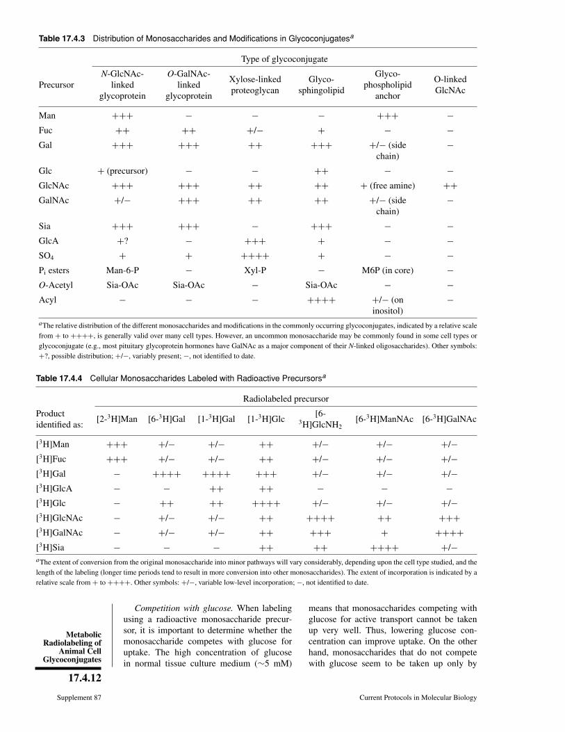
MetabolicRadiolabeling of
Animal CellGlycoconjugates
17.4.12
Supplement 87 Current Protocols in Molecular Biology
Table 17.4.3 Distribution of Monosaccharides and Modifications in Glycoconjugatesa
Type of glycoconjugate
PrecursorN-GlcNAc-
linkedglycoprotein
O-GalNAc-linked
glycoprotein
Xylose-linkedproteoglycan
Glyco-sphingolipid
Glyco-phospholipid
anchor
O-linkedGlcNAc
Man +++ − − − +++ −Fuc ++ ++ +/− + − −Gal +++ +++ ++ +++ +/− (side
chain)−
Glc + (precursor) − − ++ − −GlcNAc +++ +++ ++ ++ + (free amine) ++GalNAc +/− +++ ++ ++ +/− (side
chain)−
Sia +++ +++ − +++ − −GlcA +? − +++ + − −SO4 + + ++++ + − −Pi esters Man-6-P − Xyl-P − M6P (in core) −O-Acetyl Sia-OAc Sia-OAc − Sia-OAc − −Acyl − − − ++++ +/− (on
inositol)−
aThe relative distribution of the different monosaccharides and modifications in the commonly occurring glycoconjugates, indicated by a relative scalefrom + to ++++, is generally valid over many cell types. However, an uncommon monosaccharide may be commonly found in some cell types orglycoconjugate (e.g., most pituitary glycoprotein hormones have GalNAc as a major component of their N-linked oligosaccharides). Other symbols:+?, possible distribution; +/−, variably present; −, not identified to date.
Table 17.4.4 Cellular Monosaccharides Labeled with Radioactive Precursorsa
Radiolabeled precursor
Productidentified as:
[2-3H]Man [6-3H]Gal [1-3H]Gal [1-3H]Glc[6-
3H]GlcNH2[6-3H]ManNAc [6-3H]GalNAc
[3H]Man +++ +/− +/− ++ +/− +/− +/−[3H]Fuc +++ +/− +/− ++ +/− +/− +/−[3H]Gal − ++++ ++++ +++ +/− +/− +/−[3H]GlcA − − ++ ++ − − −[3H]Glc − ++ ++ ++++ +/− +/− +/−[3H]GlcNAc − +/− +/− ++ ++++ ++ +++[3H]GalNAc − +/− +/− ++ +++ + ++++[3H]Sia − − − ++ ++ ++++ +/−aThe extent of conversion from the original monosaccharide into minor pathways will vary considerably, depending upon the cell type studied, and thelength of the labeling (longer time periods tend to result in more conversion into other monosaccharides). The extent of incorporation is indicated by arelative scale from + to ++++. Other symbols: +/−, variable low-level incorporation; −, not identified to date.
Competition with glucose. When labelingusing a radioactive monosaccharide precur-sor, it is important to determine whether themonosaccharide competes with glucose foruptake. The high concentration of glucosein normal tissue culture medium (∼5 mM)
means that monosaccharides competing withglucose for active transport cannot be takenup very well. Thus, lowering glucose con-centration can improve uptake. On the otherhand, monosaccharides that do not competewith glucose seem to be taken up only by

Preparation andAnalysis ofGlycoconjugates
17.4.13
Current Protocols in Molecular Biology Supplement 87
Gal
Glc
Man
Gal-1-P UDP-Gal
Glc-6-P Glc-1-P UDP-Glc
UDP-GlcA
GlcA
GlcNH2
Man-6-P Man-1-P
UDP-Xyl Xyl
Fruc-6-P
GlcNH2-6-P
GDP-Man
GlcNAc-6-P
GlcNAc-1-P
UDP-GlcNAc
ManNAc
GDP-Fuc
ManNAc-6-P
Neu5Ac
CMP-Neu5Gc
Fuc
Neu5AcCMP-Neu5Ac
Neu5Ac-9-P
GlcNAc
GalNAc
UDP-GalNAc
active uptake, competing with glucose
passive uptake, not competing with glucose
Figure 17.4.4 Cytosolic pathways for the interconversion of monosaccharides and theirnucleotide sugar forms.
inefficient noncompetitive, passive mecha-nisms; thus, glucose concentration has lit-tle effect upon their already poor up-take. Experience indicates that glucosamine,galactosamine, galactose, and mannose cancompete with glucose for uptake intomost cells, whereas N-acetylglucosamine,N-acetylmannosamine, mannosamine, fucose,and xylose do not (Yurchenco et al., 1978).However, members of the glucose transporterfamily of gene products are tissue-specificin expression; a given cell type may expressmore than one, each with distinctive kineticand stereospecific uptake properties (Gouldand Bell, 1990). Thus, each monosaccharidemust be tested in the specific cell type understudy to see if it competes with glucose. A final
point that should be considered is that in somecell types, lipid-linked oligosaccharide precur-sors may become altered when glucose-freemedium is used (Rearick et al., 1981).
Other intracellular metabolic factors. Di-lution of label by endogenously synthe-sized monosaccharides, pool size of individ-ual monosaccharides and nucleotides, and fluxrates between interconverting pathways can allaffect the final specific activity and distribu-tion of the label. If cells are cultured in la-beling medium for prolonged periods of time,the medium may become depleted of glucose(Kim and Conrad, 1976; Varki and Kornfeld,1982). The specific activity of the labeledsugar nucleotides in the cells may then actu-ally rise as the medium glucose concentration
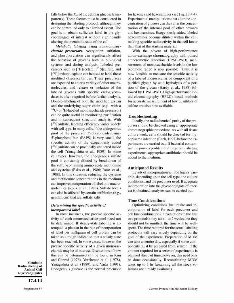
MetabolicRadiolabeling of
Animal CellGlycoconjugates
17.4.14
Supplement 87 Current Protocols in Molecular Biology
falls below the Km of the cellular glucose trans-porter(s). These factors must be considered indesigning the labeling protocol, although theycan be controlled only to a limited extent. Thegoal is to obtain sufficient label in the gly-coconjugate of interest without significantlyaltering the metabolic state of the cell.
Metabolic labeling using nonmonosac-charide precursors. Acetylation, sulfation,and phosphorylation can significantly affectthe behavior of glycans both in biologicalsystems and during analysis. Labeled pre-cursors such as [3H]acetate, [35S]sulfate, and[32P]orthophosphate can be used to label thesemodified oligosaccharides. These precursorsare expected to enter a variety of other macro-molecules, and release or isolation of thelabeled glycans with specific endoglycosi-dases is often required before further analysis.Double-labeling of both the modified glycanand the underlying sugar chain (e.g., with a14C- or 3H-labeled monosaccharide precursor)can be quite useful in monitoring purificationand in subsequent structural analysis. With[35S]sulfate, labeling efficiency varies widelywith cell type. In many cells, if the endogenouspool of the precursor 3′-phosphoadenosine-5′-phosphosulfate (PAPS) is very small, thespecific activity of the exogenously added[35S]sulfate can be practically unaltered insidethe cell (Yanagishita et al., 1989). In somecell types, however, the endogenous sulfatepool is constantly diluted by breakdown ofthe sulfur-containing amino acids methionineand cysteine (Esko et al., 1986; Roux et al.,1988). In this situation, reducing the cysteineand methionine concentrations in the mediumcan improve incorporation of label into macro-molecules (Roux et al., 1988). Sulfate levelscan also be affected by certain antibiotics (e.g.,gentamicin) that are sulfate salts.
Determining the specific activity ofincorporated label
In most instances, the precise specific ac-tivity of each monosaccharide pool need notbe determined. If steady-state labeling is at-tempted, a plateau in the rate of incorporationof label per milligram of cell protein can betaken as a rough indication that a steady statehas been reached. In some cases, however, theprecise specific activity of a given monosac-charide may be of interest. Discussions of howthis can be determined can be found in Kimand Conrad (1976), Yurchenco et al. (1978),Yanagishita et al. (1989), and Varki (1991).Endogenous glucose is the normal precursor
for hexoses and hexosamines (see Fig. 17.4.4).Experimental manipulations that alter the con-centration of glucose can thus alter the concen-tration of the internal pool of other hexosesand hexosamines. Exogenously added labeledhexosamines become diluted within the cell,making specific radioactivity in the cell lowerthan that of the starting material.
With the advent of high-performanceanion-exchange chromatography with pulsedamperometric detection (HPAE-PAD), mea-surement of monosaccharide levels in the lowpicomole range is now possible. Thus, it isnow feasible to measure the specific activityof a labeled monosaccharide component of apurified glycan by acid hydrolysis of a por-tion of the glycan (Hardy et al., 1988) fol-lowed by HPAE-PAD. High-performance liq-uid chromatography (HPLC)–based methodsfor accurate measurement of low quantities ofsulfate are also now available.
TroubleshootingIdeally, the radiochemical purity of the pre-
cursor should be checked using an appropriatechromatographic procedure. As with all tissueculture work, cells should be checked for my-coplasma infection (Fitch, 1997) before the ex-periments are carried out. If bacterial contam-ination poses a problem for long-term labelingexperiments, appropriate antibiotics should beadded to the medium.
Anticipated ResultsLevels of incorporation will be highly vari-
able, depending upon the cell type, the cultureconditions, and the precursor used. If adequateincorporation into the glycoconjugate of inter-est is obtained, analyses can be carried out.
Time ConsiderationsOptimizing conditions for uptake and in-
corporation of label for each precursor andcell line combination (introductions to the firsttwo protocols) may take 1 to 2 weeks, but theyshould not be omitted: the time will be well-spent. The time required for the actual labelingprotocols will vary widely depending on thegoal of the experiment. Preparation of MDMcan take an entire day, especially if some com-ponents must be prepared from scratch. If theamount required for a series of experiments isplanned ahead of time, however, this need onlybe done occasionally. Reconstituting MDMtakes up to 1 hr (assuming all the stock so-lutions are already available).
Preparation andAnalysis ofGlycoconjugates
17.4.15
Current Protocols in Molecular Biology Supplement 87
Literature CitedCummings, R.D., Merkle, R.K., and Stults, N.L.
1989. Separation and analysis of glycoproteinoligosaccharides. Methods Cell Biol. 32:141-183.
Diaz, S. and Varki, A. 1985. Metabolic labeling ofsialic acids in tissue culture cell lines: Methodsto identify substituted and modified radioactiveneuraminic acids. Anal. Biochem. 150:32-46.
Esko, J.D., Elgavish, A., Prasthofer, T., Taylor,W.H., and Weinke, J.L. 1986. Sulfate transport-deficient mutants of Chinese hamster ovarycells. Sulfation of glycosaminoglycans depen-dent on cysteine. J. Biol. Chem. 261:15725-15733.
Fitch, F.W., Gajewski, T.F., and Yokoyama, W.M.1997. Diagnosis and treatment of mycoplasma-contaminated cell cultures. Curr. Protoc. Im-munol. A.3E.1-A.3E.4.
Goldberg, D. and Kornfeld, S. 1981. The phos-phorylation of beta-glucuronidase oligosaccha-rides in mouse P388D1 cells. J. Biol. Chem.256:13060-13067.
Gould, G.W. and Bell, G.I. 1990. Facilitative glu-cose transporters: An expanding family. TrendsBiochem. Sci. 15:18-23.
Hardy, M.R., Townsend, R.R., and Lee, Y.C.1988. Monosaccharide analysis of glycoconju-gates by anion exchange chromatography withpulsed amperometric detection. Anal. Biochem.170:54-62.
Hart, G.W., Haltiwanger, R.S., Holt, G.D., andKelly, W.G. 1989. Glycosylation in the nucleusand cytoplasm. Annu. Rev. Biochem. 58:841-874.
Hirschberg, C.B. and Snider, M.D. 1987. Topog-raphy of glycosylation in the rough endoplas-mic reticulum and Golgi apparatus. Annu. Rev.Biochem. 56:63-87.
Kim, J.J. and Conrad, E.H. 1976. Kinetics of mu-copolysaccharide and glycoprotein biosynthe-sis by chick embryo chondrocytes. Effect ofD-glucose concentration in the culture medium.J. Biol. Chem. 251:6210-6217.
Muchmore, E.A., Milewski, M., Varki, A.,and Diaz, S. 1989. Biosynthesis of N-glycolylneuraminic acid: The primary site ofhydroxylation of N-acetylneuraminic acid is thecytosolic sugar nucleotide pool. J. Biol. Chem.264:20216-20223.
Rearick, J.I., Chapman, A., and Kornfeld, S. 1981.Glucose starvation alters lipid-linked oligosac-charide biosynthesis in Chinese hamster ovarycells. J. Biol. Chem. 256:6255-6261.
Roux, L., Holoyda, S., Sunblad, G., Freeze, H.H.,and Varki, A. 1988. Sulfated N-linked oligosac-charides in mammalian cells I: Complex-typechains with sialic acids and O-sulfate esters. J.Biol. Chem. 236:8879-8889.
Tabas, I. and Kornfeld, S. 1980. Biosynthetic in-termediates of beta-glucuronidase contain highmannose oligosaccharides with blocked phos-phate residues. J. Biol. Chem. 255:6633-6639.
Varki, A. 1991. Radioactive tracer techniques in thesequencing of glycoprotein oligosaccharides.FASEB J. 5:226-235.
Varki, A. and Kornfeld, S. 1982. The spectrum ofanionic oligosaccharides released by endo-β-N-acetylglucosaminidase H from glycoproteins.J. Biol. Chem. 258:2808-2818.
Yanagishita, M., Salustri, A., and Hascall, V.C.1989. Specific activity of radiolabeled hex-osamines in metabolic labeling experiments.Methods Enzymol. 179:435-445.
Yurchenco, P.D., Ceccarini, C., and Atkinson,P.H. 1978. Labeling complex carbohydrates ofanimal cells with monosaccharides. MethodsEnzymol. 50:175-204.

UNIT 17.5Chemical Labeling of Carbohydratesby Oxidation and Sodium BorohydrideReduction
This unit describes a collection of methods for chemical labeling of carbohydrates—freeoligosaccharides or oligosaccharides conjugated to proteins, peptides, or lipids—byoxidation followed by reduction or by direct reduction. Oligosaccharides can be labeledwith either radioisotopes or nonradioactive fluorescent molecules. These labelings allowone to follow the oligosaccharides during chromatography and in cells if labeled byfluorescent molecules.
Selective oxidation with mild periodate followed by reduction with tritiated sodiumborohydride (NaB[3H]4) results in selective radiolabeling of sialic acid residues onoligosaccharides or glycoproteins (first basic protocol). Alternatively, treatment of sam-ples with galactose oxidase (first alternate protocol) results in oxidation of galactose orN-acetylgalactosamine residues at nonreducing termini, rendering these residues sucep-tible to labeling with NaB[3H]4. Oxidized glycoconjugates can also be labeled using thefluorescent probe lucifer yellow CH (second alternate protocol). Free oligosaccharidescan be labeled by reduction with NaB[3H]4 (second basic protocol). The third alternateprotocol describes the release and simultaneous labeling of O-glycan oligosaccharidesby alkaline beta-elimination in the presence of NaB[3H]4.
CAUTION: This procedure should be performed only by personnel trained in the properuse of 3H isotope and in NRC-licensed sites. Standard precautions to prevent excessiveexposure and radioactive contamination of personnel and equipment should be followedat all times.
CAUTION: All NaB[3H]4 manipulations must be performed in a well-vented fume hood.
BASICPROTOCOL
RADIOLABELING OLIGOSACCHARIDES AFTER MILDPERIODATE OXIDATION
This protocol describes radiolabeling oligosaccharides by mild periodate oxidationfollowed by reduction with NaB[3H]4, selectively labeling sialic acid residues at nonre-ducing termini. The reaction is illustrated in Figure 17.5.1. Labeled oligosaccharides areprecipitated and separated from unincorporated label by gel-filtration chromatography.
MaterialsFor recipes, see Reagents and Solutions in this unit (or cross-referenced unit); for common stocksolutions, see APPENDIX 2; for suppliers, see APPENDIX 4.
Oligosaccharide or glycopeptide containing sialic acids, desalted0.2 mM sodium periodate (prepare fresh at room temperature and keep on ice)10 mM sodium acetate, pH 5.510 mM glycerol (store at 4°C)0.2 M sodium borate buffer, pH 9.5 (UNIT 11.16; adjust quantities of boric acid and
10 M NaOH appropriately)0.5 mCi/µl tritiated sodium borohydride (NaB[3H]4; 25 to 30 Ci/mmol) in 0.01 M
NaOH (see recipe)0.2 M sodium borohydride (NaBH4)/0.2 M sodium borate buffer, pH 9.51 M acetic acid in methanolNitrogen (N2) streamMethanol
Supplement 26
Contributed by Minoru FukudaCurrent Protocols in Molecular Biology (1994) 17.5.1-17.5.8Copyright © 2000 by John Wiley & Sons, Inc.
17.5.1
Preparation andAnalysis ofGlycoconjugates

7-ml conical glass (Pyrex) test tubes with Teflon-covered caps1.0 × 40–cm Sephadex G-15 or G-25 column in water (UNIT 10.9), calibrated to
identify void volume25° to 30°C water bath
CAUTION: Steps 3 to 8 must be performed in a well-vented fume hood to preventradioactive contamination. Excess NaB[3H]4 should be stored away in a radioactive wastebottle for appropriate disposal.
Label oligosaccharides1. Mix the following in a 7-ml conical glass test tube (200 µl total volume):
5 to 500 µg oligosaccharide or glycopeptide100 µl 0.2 mM sodium periodate100 µl 10 mM sodium acetate.
Loosely cover with Teflon-covered cap and incubate 20 min on ice.
2. Add 10 µl of 10 mM glycerol and incubate 20 min at 37°C.
The glycerol consumes any excess periodate that remains.
3. Add 100 µl of 0.2 M sodium borate buffer and 10 µl of 0.5 mCi/µl NaB[3H]4 (5 mCi).Incubate 1 hr at room temperature in a fume hood.
4. Add 100 µl of 0.2 M NaBH4/0.2 M borate buffer and incubate 30 min at roomtemperature.
Destroy excess NaB[3H]4 and remove borate as methyl borate5. Add 3 ml of 1 M acetic acid in methanol and dry under a nitrogen stream in a 25° to
30°C water bath, in a well-vented hood.
6. Repeat step 5.
7. Add 3 ml methanol (without acetic acid) and dry sample as in step 5.
O
OH
sodium periodate
OH
O R
O
OH
AcN1
2
34
5
HCOH
O
OH
[3H]sodium borohydride
H
H
3H
OHOH COOH
H 6
87 C
H
O
O R
COOH
O R
COOH
9
AcN
AcN
Figure 17.5.1 Labeling oligosaccharides by periodate oxidation followed by NaB[3H]4 reduction.
Supplement 50 Current Protocols in Molecular Biology
17.5.2
ChemicalLabeling of
Carbohydrates

Purify labeled oligosaccharides8. Dissolve sample in ∼200 µl water and apply to a calibrated 1.0 × 40–cm Sephadex
G-15 or G-25 column. Elute with water and collect 1-ml fractions with a fractioncollector.
This step separates the labeled oligosaccharides from the low-molecular-weight break-down products of NaB[3H]4.
9. Count 10-µl aliquots of each fraction in a scintillation counter.
10. Pool and dry fractions that contain radioactive oligosaccharides or glycopeptides.Store at −20°C until used (the label is stable for >12 months).
Oligosaccharides or glycopeptides should elute at or near the void volume, followed by alarger peak of breakdown products of NaB[3H]4. Be careful to avoid the second peak.
ALTERNATEPROTOCOL
RADIOLABELING OLIGOSACCHARIDES AFTER GALACTOSE OXIDASETREATMENT
Oxidation with galactose oxidase and reduction with NaB[3H]4 specifically labels galac-tose and N-acetylgalactosamine residues that are located at nonreducing termini ofoligosaccharides. Residues at internal positions are also oxidized and labeled, but muchless efficiently. This reaction is illustrated in Figure 17.5.2. Sialic acid attached togalactose hinders oxidation by galactose oxidase. If desired, sialic acid can be removedby treating the sample with sialidase (neuraminidase).
Additional MaterialsFor recipes, see Reagents and Solutions in this unit (or cross-referenced unit); for common stocksolutions, see APPENDIX 2; for suppliers, see APPENDIX 4.
1 U/µl galactose oxidase (partially purified, Sigma; store aliquots in PBS at −20°C)10 mM sodium phosphate buffer, pH 7.015-ml conical glass centrifuge tube
CAUTION: Steps 2 to 4 must be performed in a well-vented fume hood to preventradioactive contamination.
CH2OH
O
OH
galactose oxidase
OH
HO O R
HC=O
O
OH
OH
HO1
23
4
5
6
HC–OH
C
OH
OH
HO
[3H]sodium borohydride3H
O R
O R
Figure 17.5.2 Labeling oligosaccharides by galactose oxidase treatment followed by NaB[3H]4reduction.
Current Protocols in Molecular Biology Supplement 26
17.5.3
Preparation andAnalysis ofGlycoconjugates

1. Mix the following in a 15-ml conical centrifuge tube (110 µl total volume):
50 to 500 µg oligosaccharide or glycopeptide10 µl 1 U/µl galactose oxidase100 µl 10 mM sodium phosphate buffer.
Incubate 2 hr at 37°C.
Sialylated oligosaccharides or glycopeptides should be treated with sialidase (UNIT 17.12)before treatment with galactose oxidase because sialic acid hinders oxidation by galactoseoxidase. If the sample is treated with sialidase, either the sample should be desalted or thevolume of the galactose oxidase reaction should be increased to ensure that the pH is 7.0.
2. Add 100 µl of 0.2 M sodium borate buffer and 10 µl of 0.5 mCi/µl NaB[3H]4 (5 mCi).Incubate 1 hr at room temperature in a fume hood.
3. Add 100 µl of 0.2 M NaBH4/0.2 M sodium borate buffer and incubate 1 hr at roomtemperature.
4. Acidify, dry, and purify labeled oligosaccharides or glycopeptides as in steps 5 to 10of the first basic protocol.
ALTERNATEPROTOCOL
FLUORESCENCE LABELING OLIGOSACCHARIDES AFTER PERIODATEOR GALACTOSE OXIDASE TREATMENT
Following oxidation, oligosaccharide-containing samples may be labeled with the non-isotopic fluorescent probe lucifer yellow CH, instead of being radioactively labeled withNaB[3H]4. This technique can be used to label oligosaccharides on glycopeptides orglycolipids. Labeled oligosaccharides are separated from unincorporated dye by gel-fil-tration chromatography.
Additional MaterialsFor recipes, see Reagents and Solutions in this unit (or cross-referenced unit); for common stocksolutions, see APPENDIX 2; for suppliers, see APPENDIX 4.
Phosphate-buffered saline (PBS; APPENDIX 2)Lucifer yellow CH (Aldrich)0.1 M sodium cyanoborohydride (NaBH3CN)
0.5 × 15–cm Sephadex G-15 and 1.0 × 25–cm Sephadex G-25 columns inwater (UNIT 10.9), calibrated
25°C water bath
Oxidize oligosaccharides1. Oxidize oligosaccharide or glycopeptide sample (steps 1 to 2 of the first basic protocol
or step 1 of the first alternate protocol).
2. Apply sample to a 0.5 × 15–cm Sephadex G-15 column and elute with water. Collect0.5-ml fractions.
3. Pool and dry fractions that contain the oxidized oligosaccharides or glycopeptides.
The appropriate fractions should be identified based on column calibration with glycopep-tide standards (see UNIT 10.9). Oligosaccharides of glycopeptides elute at or near the voidvolume.
4. Dissolve sample in 100 µl PBS.
Supplement 26 Current Protocols in Molecular Biology
17.5.4
ChemicalLabeling of
Carbohydrates

Label oligosaccharides with fluorescent dye5. Add lucifer yellow CH to 5 mM final concentration and incubate 12 hr at room
temperature.
6. Apply reaction mixture to a 1.0 × 25–cm Sephadex G-25 column. Elute with waterand collect 1-ml fractions. Wash until the unincorporated dye elutes.
Purify labeled oligosaccharides7. Pool and dry fractions containing first fluorescent peak (labeled oligosaccharides).
It may be necessary to read UV absorbance or fluorescence of the eluted fractions, asfluorescence is not easily visible.
8. Dissolve sample in 0.1 ml of 0.1 M NaBH3CN and incubate 15 min at 25°C.Immediately apply to the same Sephadex G-25 column used in step 6 and elute withwater (avoid acidic conditions).
9. Collect, pool, and dry fluorescent oligosaccharides—i.e., the fractions at or near thevoid volume. Avoid light exposure as much as possible after this point.
Fractions that contain fluorescently labeled oligosaccharides or glycopeptides should elutein the void volume.
Remaining solutions should be discarded as toxic waste.
10. Dissolve sample in a minimal amount of water and store at −20°C until used.
BASICPROTOCOL
RADIOLABELING FREE OLIGOSACCHARIDES
Free oligosaccharides can be obtained from Asn-linked oligosaccharides by hydrazi-nolysis (UNIT 17.15) or PNGaseF (N-glycanase) treatment (UNIT 17.13). In the followingprocedure, free oligosaccharides are purified by a simple gel filtration and then directlyreduced with NaB[3H]4. After reduction, the aldehyde group at C-1 of the reducingterminus becomes an alcohol with one radioactive tritium (see Fig. 17.5.2). Introductionof 3H at the reducing terminal allows the oligosaccharides to be followed by monitoringradioactivity.
MaterialsFor recipes, see Reagents and Solutions in this unit (or cross-referenced unit); for common stocksolutions, see APPENDIX 2; for suppliers, see APPENDIX 4.
Free oligosaccharides (UNITS 17.15 & 17.12)0.3 M sodium borate buffer, pH 9.5 (UNIT 11.16; adjust quantities of boric acid
and 10 M NaOH appropriately)0.5 mCi/µl tritiated sodium borohydride (NaB[3H]4; 25 to 35 Ci/mmol) in
0.05 M NaOH (see recipe)1 M sodium borohydride (NaBH4)1 M acetic acid in methanolNitrogen (N2) streamMethanol
7-ml conical glass (Pyrex) tubes0.5 × 25–cm Sephadex G-25 or G-15 column (UNIT 10.9), calibrated to
determine void volume
Additional reagents and equipment for gel-filtration chromatography (UNIT 10.9)
CAUTION: Steps 4 to 8 must be performed in a well-vented fume hood to preventradioactive contamination.
Current Protocols in Molecular Biology Supplement 50
17.5.5
Preparation andAnalysis ofGlycoconjugates

1. Desalt 1 to 500 µg free oligosaccharides by gel-filtration chromatography on acalibrated 0.5 × 25–cm Sephadex G-25 or G-15 column. Elute in water. Collect 0.5-mlfractions.
Oligosaccharides must be free from peptides or amino acids that may incorporate labelduring reduction.
2. Pool and dry fractions that contain free oligosaccharides. Dissolve in 50 µl water.
3. Add 0.3 M sodium borate buffer and water to a 100-µl final volume and a 0.2M-sodium borate final concentration.
If oligosaccharides are dissolved in water, volume and concentration of buffer should beadjusted to give the desired final concentration.
4. Immediately add 10 to 20 µl of 0.5 mCi/µl NaB[3H]4 in 0.05 M NaOH (5 to 10 mCi)and incubate 2 hr at room temperature in a fume hood.
5. Add 100 µl of 1 M NaBH4 and incubate 2 hr at room temperature to complete thereduction.
6. Add 3 ml of 1 M acetic acid in methanol and dry under a nitrogen stream in a 30° to35°C water bath.
7. Repeat step 6.
8. Add 3 ml methanol and dry under a nitrogen stream in a 30° to 35°C water bath.
9. Dissolve sample in 50 to 200 µl water and store at −70°C until used.
ALTERNATEPROTOCOL
RADIOLABELING O-GLYCAN OLIGOSACCHARIDES
During release from glycoproteins or glycopeptides by beta-elimination, the reducingtermini of O-glycans can be labeled by alkaline borohydride treatment in the presence ofa large amount of NaB[3H]4. The reaction, however, must be carried out in the presenceof 1 M NaBH4 to prevent oligosaccharide degradation after release.
Additional MaterialsFor recipes, see Reagents and Solutions in this unit (or cross-referenced unit); for common stocksolutions, see APPENDIX 2; for suppliers, see APPENDIX 4.
100 to 1000 µg glycopeptides or ≤5 mg glycoproteins0.1 to 0.2 mCi/µl tritiated sodium borohydride (NaB[3H]4; 25 to 30 Ci/mmol) in
1 M NaBH4/0.05 M NaOH (see recipe)1 M acetic acid in methanolMethanol30° to 35°C and 45°C water baths
CAUTION: Steps 2 to 4 must be performed in a well-vented fume hood to avoidradioactive contamination.
1. Place 100 to 1000 µg glycopeptides or ≤5 mg glycoproteins in a 7-ml conical glasstube. Add 200 µl of 0.1 to 0.2 mCi/µl NaB[3H]4 in 1 M NaBH4/0.05 M NaOH (20 to40 mCi). Incubate 24 to 48 hr at 45°C in a fume hood.
For larger amounts of sample, the volume of solution should be increased to dissolve samplecompletely and the amount of Na[3H]4 should be increased to 100 mCi.
2. Add 3 ml of 1 M acetic acid in methanol and dry sample under a nitrogen stream ina 30° to 35°C water bath.
Supplement 50 Current Protocols in Molecular Biology
17.5.6
ChemicalLabeling of
Carbohydrates
3. Repeat step 2.
4. Add 3 ml methanol and dry under a nitrogen stream in a 30° to 35°C water bath.
5. Dissolve sample in 100 to 200 µl water and store at −20°C until used.
REAGENTS AND SOLUTIONS
Use deionized, distilled water in all recipes and protocol steps. For common stock solutions, seeAPPENDIX 2; for suppliers, see APPENDIX 4.
Tritiated sodium borohydride (NaB[3H]4), 0.5 mCi/lDissolve 100 mCi NaB[3H]4 (highest available specific activity, usually 25 to 35Ci/mmol) in 200 µl ice-cold 0.01 or 0.05 M NaOH. Store in 10-µl aliquots for severalmonths at −70°C until use. To make NaB[3H]4 in 1 M NaBH4/0.05M NaOH, dissolvesolid NaBH4 in tritiated solution in 0.05 M NaOH.
Alternatively, NaB[3H]4 can be dissolved in freshly distilled anhydrous dimethylformamideand stored at −20°C in well-sealed tubes. Some investigators report stability for longerperiods than with NaOH solutions.
COMMENTARY
Background InformationWhen sufficient quantities of purified gly-
coconjugates (e.g., glycoproteins or glycopep-tides) are available, their sugar chains can belabeled and used for structural or sequenceanalysis (Morell and Ashwell, 1972; Takasakiand Kobata, 1978). These sugar chains can beradioactively labeled by periodate or enzymaticoxidation followed by reduction with tritiatedsodium borohydride (NaB[3H]4). Sialic acidresidues can be preferentially oxidized by mildperiodate oxidation because they contain cis-glycols in exocylic carbons (Fig. 17.5.1). Suchglycols are much more reactive with periodatethan glycols in cylic structures because of freerotation at C-8 and C-9. The aldehyde formedat C-7 or C-8 can be reduced by NaB[3H]4 andradioactively labeled in the process (Fig.17.5.1). Galactose oxidase treatment oxidizesgalactose or N-acetylgalactosamine residues atthe nonreducing termini, at C-6, forming alde-hydes. The resultant aldehydes are reduced andlabeled with NaB[3H]4 (Fig. 17.5.2).
Oxidized samples can also be labeled withfluorescent molecules (Wilchek et al., 1980;Spiegel, 1987). After oxidation by periodate orgalactose oxidase treatment, the sample is re-acted with a fluorescent dye that contains ahydrazide group. After forming a Schiff base,the linkage can be stabilized by reduction. Oncea fluorescent label is attached to an oligosac-charide, it can be detected by a fluorescence orUV detector. Fluorescence-tagged oligosac-
charides can be incubated with cells and incor-porated oligosaccharides can be detected byfluorescence microscopy.
These methods have been used to examinethe biological significance of oligosaccharidestructures and their distribution on glycoconju-gates. In one study, the structures of O-linkedoligosaccharides were found to vary with thedifferent maturation stages along a cell lineage(Fukuda et al., 1986). In another study, glyco-sylation of erythropoietin, which is essential forits function, was examined and compared withthat of a recombinant erythropoietin (Sasaki etal., 1987).
Critical ParametersThere are two critical parameters for these
procedures. First, it is essential that each reac-tion take place at the designated pH. If the pHof a reaction mixture is not properly adjusted,the reaction will not occur. Second, the qualityof the NaB[3H]4 added is critical. WhenNaB[3H]4 is purchased, the reagent should bedark blue or slightly violet in color. WhenNaB[3H]4 absorbs water and becomes de-graded, it appears white and should not be usedfor labeling. Free oligosaccharides must bedesalted by gel-filtration chromatography toremove salts, free amino acids, and peptidesbefore chemical labeling.
An alternative method is to label the N-ace-tyl groups of O-glycans that have already beenreleased from glycoproteins by alkaline beta-
Current Protocols in Molecular Biology Supplement 26
17.5.7
Preparation andAnalysis ofGlycoconjugates
elimination. The reduced O-glycans can betreated with hydrazine to remove N-acetylgroups and reacetylated with radioactive aceticanhydride. This procedure is useful if anhy-drous hydrazine is readily available (see Amanoand Kobata, 1989).
Anticipated ResultsChemical labeling of carbohydrates by these
procedures generally depends on the amount ofsamples available. For example, NaB[3H]4 re-duction of oligosaccharides is one of the mostefficient methods to label oligosaccharides, butit still requires 0.05 to 0.1 µM for efficientlabeling. Since NaB[3H]4 reduction and fluo-rescence labeling are dependent on reactionswith aldehyde groups, it is also an advantage tohave a reasonable mass of sample, reducing thecontribution from contaminants. Once there isa sufficient amount of sample, however, totalradioactivity incorporated will allow many ma-nipulations on radioactively labeled samples.
Time ConsiderationsIn general, all procedures in this unit are
simple and rapid. Each of the protocols de-scribed should take 1 or 2 days to finish.
Literature CitedAmano, J. and Kobata, A. 1989. Quantitative con-
version of mucin-type sugar chains to radioac-tive oligosaccharides. Methods Enzymol.179:261-270.
Fukuda, M., Carlsson, S.R., Klock, J.C., and Dell,A. 1986. Structures of O-linked oligosaccharidesisolated from normal granulocytes, chronicmyelogenous leukemia cells, and acute myelo-genous leukemia cells. J. Biol . Chem.261:12796-12806.
Morell, A.G. and Ashwell, G. 1972. Tritium labelingof glycoproteins that contain terminal galactoseresidue. Methods Enzymol. 28:205-208.
Sasaki, H., Bothner, B., Dell, A., and Fukada, M.1987. Carbohydrate structure of erythropoietinexpressed in Chinese hamster ovary cells by ahuman erythropoietin cDNA. J. Biol. Chem.262:12059-12076.
Spiegel, S. 1987. Fluorescent gangliosides. MethodsEnzymol. 138:313-318.
Takasaki, S. and Kobata, A. 1978. Microdetermina-tion of sugar composition by radioisotope label-ing. Methods Enzymol. 50:50-54.
Wilchek, M., Spiegel, S., and Spiegel, Y. 1980.Fluorescent reagents for the labeling of glyco-conjugates in solution and on cell surfaces. Bio-chem. Biophys. Res. Commun. 92:1215-1222.
Contributed by Minoru FukudaLa Jolla Cancer Research FoundationLa Jolla, California
Supplement 26 Current Protocols in Molecular Biology
17.5.8
ChemicalLabeling of
Carbohydrates

UNIT 17.6Detection and Analysis of Proteins Modifiedby O-Linked N-Acetylglucosamine
The modification of Ser and Thr residues with O-linked β-N-acetylglucosamine (O-GlcNAc) is a common, dynamic, and essential modification of nuclear and cytoplasmicproteins. O-GlcNAc is ubiquitous, having been identified in all complex eukaryotesstudied, including filamentous fungi and plants. O-GlcNAc has been found on a diverserange of proteins including cytoskeletal proteins, nuclear pore proteins, RNA polymeraseII (RNA Pol II), transcription factors, proto-oncogene products, tumor suppressors,hormone receptors, phosphatases, and kinases (Comer and Hart, 2000).
The functional consequences of modifying proteins with O-GlcNAc is unclear, but it isrequired for survival at the single-cell level (Shafi et al., 2000). Three features suggestthat O-GlcNAc performs a regulatory role: (1) O-GlcNAc occurs at sites on the proteinbackbone that are similar to those modified by protein kinases; (2) O-GlcNAc is reciprocalwith phosphorylation on some well-studied proteins, such as RNA Pol II, estrogenreceptor-β, SV40 large T-antigen, and the c-Myc proto-oncogene product; and (3) likephosphorylation, O-GlcNAc is highly dynamic, with rapid cycling in response to cellularsignals or cellular stages. Perturbations in the metabolism of GlcNAc, which alter theregulation of many O-GlcNAc proteins, have been implicated in Alzheimer’s disease,diabetes, and cancer (Wells et al., 2001).
This unit concentrates on techniques for the detection and analysis of proteins modifiedby O-GlcNAc, as well as methods for the analysis of enzymes responsible for the additionand removal of O-GlcNAc. We have focused on methods that require standard laboratoryequipment. However, in some cases we also discuss more specialized technology.
The unit is set out in a stepwise manner. First, a protocol for increasing the stoichiometryof O-GlcNAc on proteins is given (see Basic Protocol 1). This is followed by simpletechniques for the detection/screening of O-GlcNAc modified proteins either by westernblotting or lectin affinity chromatography (see Basic Protocols 2 to 4). Separate protocolsverify that the glycan is O-linked GlcNAc (see Support Protocols 1 and 2). These methodsare followed by protocols for more comprehensive analysis of O-GlcNAc modifiedproteins, including labeling of O-GlcNAc residues with [3H]Gal, and subsequent productanalysis (see Alternate Protocol 1, see Basic Protocols 5 to 8, and see Support Protocols3 and 4). The final two protocols assay for O-GlcNAc transferase and O-GlcNAcaseactivity, respectively (see Basic Protocols 9 and 10 and Support Protocol 5).
BASICPROTOCOL 1
INCREASING THE STOICHIOMETRY OF O-GlcNAc ON PROTEINSBEFORE ANALYSIS
In many cultured mammalian cells, the number of O-GlcNAc moieties per proteinmolecule can be increased by treating cells with O-(2-acetamido-2-deoxy-D-glucopyra-nosylidene)-amino-N-phenyl-carbamate (PUGNAc), a potent and cell-permeable inhibi-tor of O-GlcNAcase (Haltiwanger et al., 1998). Alternatively, streptozotocin (STZ; Rooset al., 1998) and glucosamine (Han et al., 2000) have been used to increase thestoichiometry of O-GlcNAc on proteins. However, STZ has been shown to inducepoly-(ADP-ribose) polymerase–mediated apoptosis in Min6 cells (Gao et al., 2000) andshould be used with caution. In addition, STZ is only effective in cells that express theglucose transporter GLUT-2 (Schnedl et al., 1994).
Supplement 57
Contributed by Natasha E. Zachara, Robert N. Cole, Gerald W. Hart, and Yuan GaoCurrent Protocols in Molecular Biology (2002) 17.6.1-17.6.25Copyright © 2002 by John Wiley & Sons, Inc.
17.6.1
Preparation andAnalysis ofGlycoconjugates

Materials
Cells of interest growing in monolayer culture, and appropriate culture medium20 mM PUGNAc (CarboGen Laboratories) stock in Milli-Q water (filter sterilize
and store in aliquots up to 6 to 12 months at −80°C)500 mM glucosamine stock in 500 mM HEPES, pH 7.5 (make just prior to use;
filter sterilize)500 mM streptozotocin (STZ; Sigma) in 100 mM citrate buffer, pH 4.5 (make just
prior to use; filter sterilize)100-mm tissue culture dishes
1. Grow cells in monolayer culture in a sufficient number of 100-mm dishes.
2. Add (or replace growth medium with fresh medium containing) 40 to 100 µMPUGNAc (added from 20 mM stock), 5 mM glucosamine (added from 500 mMstock), or 2 to 5 mM STZ (added from 500 mM stock). Incubate cells in an incubatorfor 6 to 18 hr.
PUGNAc is taken up by both dividing and stationary cells.
If necessary, cells can be treated by PUGNAc for several days without any apparent celltoxicity. However, prolonged treatment does not appear to result in additional increase inO-GlcNAc compared to the 6 to 18 hr treatment.
When using glucosamine, mannitol is often added at the same concentration. This controlsfor changes in osmolarity due to the additional sugar in the medium (Heart et al., 2000).
3. At the end of treatment, take the dishes out of the incubator and place on ice. Extractas desired. Separate proteins by SDS-PAGE (UNIT 10.2A) and electroblot onto appro-priate membrane (UNIT 10.8).
Alternatively, extract proteins and proceed with protein purification (UNITS 10.9-10.14) orimmunoprecipitation (UNIT 10.16).
BASICPROTOCOL 2
DETECTION OF PROTEINS MODIFIED BY O-GlcNAc USING ANTIBODIES
Recently Comer et al. (2001) showed that an antibody (CTD 110.6) raised against theglycosylated C-terminal domain of the RNA polymerase II large subunit was a generalO-GlcNAc antibody. Unlike many lectins, CTD 110.6 shows little cross-reactivity towardterminal GlcNAc on complex glycans. Several other antibodies, HGAC 85 (Turner et al.,1990) and RL2 (Snow et al., 1987), have been reported as O-GlcNAc specific antibodies.However, these antibodies only recognize a subset of O-GlcNAc modified proteins, andRL2 in particular is dependent on the structure of the peptide backbone around theglycosylation site (Holt et al., 1987).
It is important to include an appropriate negative control (100 ng ovalbumin) forcross-reactivity toward N-linked glycans. Using the standard Amersham PharmaciaBiotech enhanced chemiluminescent (ECL) system, the authors have found that 10 µg ofcytoplasmic, nuclear, or total cell extract is sufficient. For purified proteins, Comer andco-workers found that 25 to 50 ng of a neoglycoconjugate was sufficient (Comer et al.,2001).
Materials
Purified or crude protein (e.g., Basic Protocol 1) separated by SDS-PAGE (UNIT
10.2A) and electroblotted to polyvinylidene difluoride (PVDF; UNIT 10.8) ornitrocellulose (duplicate blots are needed)
TBS-HT (see recipe)
Supplement 57 Current Protocols in Molecular Biology
17.6.2
Detection andAnalysis of
Proteins Modifiedby O-Linked N-
Acetylglucosamine
Antibody: CTD 110.6 ascites (Covance) diluted 1/2500 in TBS-HT (see recipe forTBS-HT)
N-acetylglucosamine (GlcNAc; Sigma)HRPO-conjugated anti–mouse IgM diluted 1/5000 in TBS-HTTBS-HD (see recipe)ECL kit (Amersham Pharmacia Biotech)
Additional reagents and equipment for visualization with chromogenic andluminescent substrates (UNIT 10.8)
1. Block blots by incubating with TBS-HT for 60 min at room temperature.
Batteiger et al. (1982) have shown that high concentrations of Tween 20 substitute forblocking with milk or bovine serum albumin (BSA).
2. Incubate blots with CTD 110.6 (1/2500 dilution in TBS-HT), in duplicate, with andwithout 10 mM GlcNAc, overnight at 4°C.
To control for specificity it is important to perform a control blot. Here, the antibody ispreincubated with 10 mM GlcNAc for ∼5 min on ice before being applied to the controlblot. Note that the concentration of antibody should be optimized with each new prepara-tion. We find that 1/2500 is a good place to start with ascites (containing antibody in themg/ml range).
3. Wash blots in TBS-HD twice, each time for 10 min at room temperature.
As the blots are washed under these conditions, prestained markers will fade. This doesnot appear to be the result of proteins being solubilized off the membranes, but of the dyeused to stain the markers. While the markers fade, they do not completely disappear. Theauthors usually double the amount of marker used to compensate for fading.
4. Wash blots in TBS-HT three times, each time for 10 min, room temperature.
5. Incubate blots with HRPO-conjugated anti–mouse IgM (1/5000 dilution in TBS-HT)for 50 min, room temperature.
The concentration of secondary antibody varies from lot to lot and should be optimizedeach time with each new preparation.
6. Wash blots in TBS-HD twice, each time for 10 min, room temperature.
7. Wash blots in TBS-HT three times, each time for 10 min, room temperature.
8. Develop the HRPO reaction using, e.g., the ECL system (UNIT 18.4).
Note that the antibody often cross-reacts with pre-stained markers.
BASICPROTOCOL 3
DETECTION OF PROTEINS MODIFIED BY O-GlcNAc USING THE LECTINsWGA
Many lectins are reportedly specific for β-GlcNAc residues. The authors have typicallyused succinylated wheat germ agglutinin (sWGA), which is widely available and isderivatized with a number of useful functional groups including horseradish peroxidase(HRPO). Before succinylation, WGA will recognize both silaic acid and GlcNAc (Mon-signy et al., 1980). For additional information concerning lectin chromatography, see UNIT
17.1.
The amount of “test” protein used is dependent on the technique(s) used to develop theHRPO reaction. Using the standard Amersham Pharmacia Biotech ECL system theauthors find that 7.5 µg of cytoplasmic or nuclear extract is sufficient.
Current Protocols in Molecular Biology Supplement 57
17.6.3
Preparation andAnalysis ofGlycoconjugates

It is important to include an appropriate positive (100 ng ovalbumin) and negative (100ng of BSA) control. As a control, a portion of the sample should also be treated withhexosaminidase (see Support Protocol 2), to show that reactivity is toward GlcNAc. Inaddition, the sample should be subjected to reductive β-elimination to verify that lectin/an-tibody reactivity is towards O-linked glycans (see Support Protocol 1).
Materials
Purified or crude protein separated by SDS-PAGE (UNIT 10.2A) and electroblotted topolyvinylidene difluoride (PVDF; UNIT 10.8) or nitrocellulose (duplicate blots areneeded)
5% (w/v) BSA in TBST (see recipe for TBST)TBST (see recipe)0.1 µg/ml HRPO-conjugated sWGA (EY Labs) in TBST (see recipe for TBST):
the lectin can be stored at 1 mg/ml in 0.01 M PBS, pH 7.4 (APPENDIX 2), at−20°C for at least 1 year
N-acetylglucosamine (GlcNAc; Sigma)High-salt TBST (HS-TBST): TBST (see recipe) containing 1 M NaClTris-buffered saline (TBS; see recipe)ECL kit (Amersham Pharmacia Biotech)
Additional reagents and equipment for visualization with chromogenic andluminescent substrates (UNIT 10.8)
1. Wash duplicate blots for 10 min in 5% BSA/TBST, room temperature.
2. Block by incubating blots in 5% (w/v) BSA/TBST for at least 60 min at roomtemperature.
IMPORTANT NOTE: Milk cannot be used as the blocking agent, since many of the proteinsin milk are modified by glycans that react with sWGA.
3. Wash blots three times, each time for 10 min in TBST, room temperature.
4. Incubate blots in 0.1 µg/ml sWGA-HRPO in TBST, in duplicate, with and without 1M GlcNAc, overnight at 4°C.
To control for lectin specificity it is important to perform a control blot. Here, the lectin ispreincubated with 1 M GlcNAc for ∼5 min on ice before being applied to the control blot.
5. Wash blots six times, each time for 10 min, in HS-TBST.
6. Wash blots once in TBS for 10 min.
7. Develop the HRPO-reaction using, e.g., the the ECL system (UNIT 10.8).
Using the ECL system described, 100 ng of ovalbumin should be visualized in 5 to 15 sec.
SUPPORTPROTOCOL 1
CONTROL FOR O-LINKED GLYCOSYLATION
Traditionally, mild alkaline reduction (reductive β-elimination) has been used to releaseO-linked carbohydrates from proteins (Amano and Kobata, 1990). This method has beenadapted for blots to show that lectin/antibody reactivity is toward O-linked rather thanN-linked glycans (Duk et al., 1997). Proteins blotted to PVDF are treated with 55 mMNaOH overnight (releasing O-linked sugars) and then probed using lectins or antibodies.
There are a number of reasons why lectin/antibody reactivity could be lost after NaOHtreatment, e.g., the sugars were destroyed instead of being released, or the protein wasdegraded. To control for these, it is important to have control proteins with N- and O-linkedsugars, and to stain one blot for protein after treatment preferably with an antibody. The
Supplement 57 Current Protocols in Molecular Biology
17.6.4
Detection andAnalysis of
Proteins Modifiedby O-Linked N-
Acetylglucosamine

authors suggest a control blot of bovine asialofetuin (Sigma) which contains both N- andO-linked sugars terminating in GlcNAc, treated and not treated with PNGase F (UNIT
17.13A).
Materials
Protein samples and controls blotted to PVDF (triplicate blots are needed;nitrocellulose is not suitable as it dissolves in 55 mM NaOH)
Tris-buffered saline (TBS; see recipe)55 mM NaOH3% (w/v) BSA in TBST (see recipe for TBST)40°C water bath
Additional reagents and equipment for probing protein blots with protein-specificantibodies (see Basic Protocol 2) or lectins (see Basic Protocol 3)
1. Wash blots once in TBS for 10 min.
2. Incubate two blots in 55 mM NaOH at 40°C overnight; incubate the control blot inMilli-Q water at 40°C overnight.
The blots treated with NaOH will yellow slightly.
3. Wash blots three times, each time for 10 min at room temperature, in TBST.
4. Block by incubating blots in 3% w/v BSA/TBST for 60 min at room temperature.
5. Probe blots (one treated and one untreated) with carbohydrate-specific lectins (seeBasic Protocol 3) or antibodies (see Basic Protocol 2). Probe the second NaOH-treated blot with a protein-specific antibody (see Basic Protocol 2).
On the untreated blot, asialofetuin ± PNGase F should react with sWGA, as both the N-and O-linked sugars contain terminal GlcNAc residues. On the treated blot only theasialofetuin − PNGase F should react with sWGA.
BASICPROTOCOL 4
DETECTION AND ENRICHMENT OF PROTEINS USING sWGA-AGAROSE
sWGA lectin affinity chromatography provides a convenient method for enriching anddetecting O-GlcNAc modified proteins. This procedure has been adapted for detectingproteins that are difficult to purify or are present in low copy number, such as transcriptionfactors. In this protocol, the protein of interest is synthesized in a rabbit reticulocyte lysate(RRL) in vitro transcription translation (ITT) system (Promega) and labeled with either[35S]Met, [35S]Cys, or [14C]Leu. After desalting, the proteins are tested for their ability tobind sWGA agarose in a GlcNAc-specific manner (Roquemore et al., 1994).
Alternatively, the lectin Ricinus communis agglutinin 1 (RCA1) has been used to selectfor O-GlcNAc proteins that have previously been labeled by galactosyltransferase (seeAlternate Protocol 1). Proteins modified by terminal Gal are specifically retained on aRCA1 affinity column. Labeled O-GlcNAc proteins are released under mild conditions,while those containing N-linked structures require lactose addition to the buffer beforeelution results (Hayes et al., 1995; Greis and Hart, 1998). The method described in thisprotocol can be adapted for RCA1 affinity chromatography by substituting RCA1-agarose(EY Labs) for sWGA-agarose and changing the order of the Gal and GlcNAc elutionbuffer.
Materials
cDNA subcloned into an expression vector with an SP6 or T7 promoter (∼0.5 to 1µg/µl)
Kit for RRL ITT system (Promega)
Current Protocols in Molecular Biology Supplement 57
17.6.5
Preparation andAnalysis ofGlycoconjugates
Label: [35S]Met, or [35S]Cys, or [14C]LeusWGA-agarose (Vector Laboratories)sWGA wash buffer: PBS (APPENDIX 2) containing 0.2% (v/v) NP-40sWGA Gal elution buffer (see recipe)sWGA GlcNAc elution buffer (see recipe)1-ml tuberculin syringe with glass wool plug at bottom to support chromatography
matrix or Bio-Rad Bio-Spin disposable chromatography column
Additional reagents and equipment for digesting proteins with hexosaminidase(see Support Protocol 2), desalting (see Support Protocol 5), SDS-PAGE (UNIT
10.2A), and autoradiography (APPENDIX 3A)
Prepare proteins1. Synthesize proteins to incorporate the desired label ([35S]Met, [35S]Cys, or [14C]Leu)
using the RRL ITT system according to the manufacturer’s instructions. Include theprotein of interest, a positive control for sWGA binding (for example, the nuclearpore protein p62), a negative control (luciferase, supplied with kit), and a no-DNAcontrol.
2. Treat half of each sample with hexosaminidase (see Support Protocol 2).
3. Desalt samples using spin filtration (e.g., Amersham Pharmacia Biotech MicrospinG-50 columns) or a 1-ml G-50 desalting column (as for desalting O-GlcNActransferase; see Support Protocol 5).
The following procedure is carried out at 4°C.
Apply protein samples to chromatography columns4. Equilibrate sWGA-agarose and pack column as follows:
a. If resin is supplied as 50% slurry (i.e., 50% resin/50% storage solution) remove300 µl (double the volume required) and pipet into a 1-ml tuberculin syringe ordisposable chromatography column.
b. Let storage solution drain from resin.
c. Equilibrate resin by washing column four times, each time with 1 ml of sWGAwash buffer. Cap column.The volumes given are appropriate for a sample derived from an ITT. For enrichment ofother protein samples, the volume of sWGA should be optimized for the protein sampleapplied. The authors find that 50 mg of cell extract requires 1 ml of sWGA-agarose,assuming that 1% to 2% of the total cell extract is modified by O-GlcNAc.
5. Apply sample (∼30 µl of an ITT reaction) to the column and let stand at 4°C for 30min, or cap and incubate at 4°C for 30 min with rotating or rocking.
Wash column and elute GlcNAc6. At the end of the 30-min incubation, uncap the column and allow the sample to “run
through” the resin. Collect this as the “run through” fraction. Wash column with 15ml of sWGA wash buffer at 10 ml/hr, collecting 0.5-ml fractions.
7. Load the column with 300 µl of sWGA Gal elution buffer and let stand at 4°C for 20min.
8. Wash column with 5 ml of sWGA Gal elution buffer, collecting 0.5 ml fractions.
9. Repeat steps 6 to 8 using GlcNAc elution buffer.
10. Count 25 µl of each fraction using a liquid scintillation counter.
Supplement 57 Current Protocols in Molecular Biology
17.6.6
Detection andAnalysis of
Proteins Modifiedby O-Linked N-
Acetylglucosamine

11. Pool positive fractions that elute in the presence of GlcNAc and precipitate usingTCA or methanol.
To precipitate proteins with methanol, mix 1 vol of sample with 10 vol of ice-cold methanol.Incubate overnight at −20°C. Recover protein by microcentrifuging 10 min at 16,000 × g,4°C, in a microcentrifuge tube (which is the most efficient procedure) or in 15-ml conicalcentrifuge tubes for 10 min at 3000 × g, 4°C. Resuspend samples in SDS-PAGE samplebuffer (UNIT 10.2A).
As many proteins in rabbit reticulocyte lysate contain O-GlcNAc and bind WGA, theauthors do not recommend the addition of carrier proteins at this point. Typically, a fractionof the GlcNAc elution containing a total of 1000 to 2000 dpm [35S]Met is precipitated andanalyzed by SDS-PAGE (UNIT 10.2A) and autoradiography (APPENDIX 3A).
The use of acetone to precipitate proteins is not recommended, as free GlcNAc will alsoprecipitate.
12. Analyze pellet by SDS-PAGE (UNIT 10.2A) and autoradiography (APPENDIX 3A).
SUPPORTPROTOCOL 2
DIGESTION OF PROTEINS WITH HEXOSAMINIDASE
Terminal GlcNAc and O-GlcNAc can be removed from proteins using commerciallyavailable hexosaminidases; these enzymes will also cleave terminal GalNAc residues.Unlike O-GlcNAcase, commercial hexosaminidases have low pH optima, typically pH4.0 to 5.0.
Materials
Protein sample for digestion (include a positive control, e.g., ovalbumin)2% (w/v) SDS2× hexosaminidase reaction mixture (see recipe)
Additional reagents and equipment for SDS-PAGE (UNIT 10.2A) and electroblotting(UNIT 10.8)
1. Mix sample 1:1 with 2% SDS and boil for 5 min.
2. Mix sample 1:1 with 2× hexosaminidase reaction mixture and incubate at 37°C for4 to 24 hr.
3. To assess completeness of the digestion, separate an aliquot of the reaction bySDS-PAGE (UNIT 10.2A) and electroblot (UNIT 10.8) onto an appropriate membrane.Probe blots with carbohydrate-specific lectins or antibodies.
ALTERNATEPROTOCOL 1
DETECTION OF PROTEINS MODIFIED BY O-GlcNAc USINGGALACTOSYLTRANSFERASE
The enzyme β-1,4-galactosyltransferase (from bovine milk) will label any terminalGlcNAc residue with Gal, using uridine diphospho-D-Gal (UDP-Gal) as a donor substrate(Brew et al., 1968). Hart and colleagues have exploited this property, using the enzymeto label terminal GlcNAc residues on proteins with [6-3H]Gal, forming a [3H]-βGal1-4βGlcNAc (Torres and Hart, 1984; Roquemore et al., 1994; Greis and Hart, 1998). Thelabeled sugar can be chemically released (via β-elimination) and analyzed by size-exclu-sion chromatography on a BioGel-P4 column, using the 3H radiolabel to detect the fractionof interest (Roquemore et al., 1994). Labeling the O-GlcNAc allows for the subsequentdetection of the proteins and peptides of interest during SDS-PAGE, HPLC, proteasedigestion, and Edman degradation steps. Researchers have been able to identify glycosy-lation sites on as little as 10 pmol using these methods (Greis et al., 1996).
Current Protocols in Molecular Biology Supplement 57
17.6.7
Preparation andAnalysis ofGlycoconjugates

To achieve efficient labeling of some proteins, it is necessary to denature samples, forexample by boiling in the presence of 10 mM DTT and 0.5% (w/v) SDS. Galactosyltrans-ferase has been shown to be active in solutions containing 5 mM DTT, 0.5 M NaCl, upto 2% (v/v) Triton X-100, up to 2% (v/v) NP-40, and 1 M urea. Up to 0.5% (w/v) SDScan be used, if it is titrated with a 10-fold molar excess of either Triton X-100 or NP-40in the final reaction mixture. Digitonin, which is commonly used to solubilize cells, shouldbe used with caution, as it is a substrate for galactosyltransferase. The total ionic strengthshould be less than 0.2 M.
Galactosyltransferase requires 1 to 5 mM Mn2+ for activity, but is inhibited by Mg2+ andconcentrations of Mn2+ >20 mM. EDTA (or analogs) should be avoided unless titratedwith appropriate levels of Mn2+. Note that 1 mol of EDTA binds 2 mol of Mn2+.
Free UDP is also an inhibitor of galactosyltransferase. For studies where completelabeling of the GlcNAc is preferable, such as site mapping, calf intestinal alkalinephosphatase is included in the reaction, as it degrades UDP (Unverzagt et al., 1990). Whilethis increases the efficiency of the reaction, it is important to add this to the control assome preparations of alkaline phosphatase contain proteins what will label with galacto-syltransferase (R.N. Cole, pers. commun.).
NOTE: Protease inhibitors, such as PIC 1, PIC 2, and PMSF (see recipe for 1000× proteaseinhibitors in Reagents and Solutions), can be included (final concentrations, 1×), butGlcNAc and 1-amino GlcNAc should be removed prior to labeling by spin filtration oranother method of desalting.
Materials
Protein sample(s)Dithiothreitol (DTT)Sodium dodecyl sulfate (SDS)Label: 1.0 mCi/ml UDP-[3H]Gal, (17.6 Ci/mM; Amersham Pharmacia Biotech) in
70% v/v ethanolNitrogen source25 mM 5′-adenosine monophosphate (5′-AMP), in Milli-Q water, pH 7.0Buffer H (see recipe)10× galactosyltransferase labeling buffer (see recipe)Galactosyltransferase, autogalactosylated (see Support Protocol 3)Calf intestinal alkaline phosphataseUnlabeled UDP-GalStop solution: 10% (w/v) SDS/0.1 M EDTA
100°C water bath30 × 1–cm Sephadex G-50 column equilibrated in 50 mM ammonium
formate/0.1% (w/v) SDS
Additional reagents and equipment for acetone precipitation of protein (UNIT
17.10A), PNGase F digestion of proteins (UNIT 17.13A), SDS-PAGE (UNIT 10.2A), andproduct analysis (see Basic Protocol 6)
Prepare the reaction1. Denature protein sample by adding DTT to 10 mM and SDS to 0.5% (w/v), then
boiling the sample for 10 min.
2. Decide how many reactions are going to be carried out and thus how much label willbe needed (∼1 to 2 µCi/reaction).
Supplement 57 Current Protocols in Molecular Biology
17.6.8
Detection andAnalysis of
Proteins Modifiedby O-Linked N-
Acetylglucosamine
A positive control (ovalbumin, 2 g), a negative control (because galactosyltransferase canlabel itself), and a sample-minus-enzyme control will be needed.
3. Remove solvent from label in a Speed-Vac evaporator or under a stream of nitrogen.
Ethanol can inhibit the galactosyltransferase reaction, but if <4 l is required the labelcan be added directly to the reaction (final reaction volume, 500 l).
4. Resuspend appropriate amount of label for each reaction, respectively, in 50 µl of 25mM 5′-AMP.
The AMP is included to inhibit possible phosphodiesterase reactions, which might competefor label during the labeling experiment.
5. Set up reactions as follows:
Up to 50 µl protein sample (final concentration 0.5 to 5 mg/ml)350 µl buffer H50 µl 10× galactosyltransferase labeling buffer50 µl UDP-[3H]Gal/5′-AMP mixture from step 430 to 50 µl autogalactosylated galactosyltransferase1 to 4 U calf intestinal alkaline phosphataseMilli-Q water to final volume of 500 µl
Reaction volumes can be scaled down to 50 l.
6. Labeling at 37°C for 2 hr or at 4°C overnight.
These are the typical conditions. Galactosyltransferase is active over a range of tempera-tures.
7. Add unlabeled UDP-Gal to a final concentration of 0.5 to 1.0 mM and another 2 to5 µl of galactosyltransferase.
For studies where complete labeling of the GlcNAc is required, such as site mapping, thereactions are chased with unlabeled UDP-Gal and fresh galactosyltransferase.
8. Add 50 µl of stop solution to each sample and heat to 100°C for 5 min in a waterbath.
Isolate the product9. Resolve the protein from unincorporated label using a Sephadex G-50 column
equilibrated in 50 mM ammonium formate/0.1% w/v SDS. Collect 1-ml fractions.
Size-exclusion chromatography using Sephadex G-50 is traditionally used to desalt sam-ples. However, TCA precipitation, spin filtration/buffer exchange, or other forms ofsize-exclusion chromatography (e.g., Pharmacia PD-10 desalting column) can be used.The addition of carrier proteins such as BSA (∼67 kDa) and cytochrome c (∼12.5 kDa) tosamples and buffers will reduce the amount of protein lost due to nonspecific proteinadsorption.
10. Count a 50-µl aliquot of each fraction using a liquid scintillation counter.
Approximately 2 × 10⁶ dpm of [3H]Gal should be incorporated into 2 g of ovalbumin.
11. Combine the void volume and lyophilize to dryness.
12. Resuspend samples in Milli-Q water and precipitate with acetone (UNIT 17.10A).
13. Treat samples with PNGase F (UNIT 17.13A), separate by SDS-PAGE (UNIT 10.2A) anddetect by autoradiography (APPENDIX 3A). Alternatively, subject samples to “productanalysis” to confirm that the label was incorporated onto O-GlcNAc (see BasicProtocol 6).
Current Protocols in Molecular Biology Supplement 57
17.6.9
Preparation andAnalysis ofGlycoconjugates
SUPPORTPROTOCOL 3
AUTOGALACTOSYLATION OF GALACTOSYLTRANSFERASE
As galactosyltransferase contains N-linked glycosylation sites, it is necessary to blockthese before using this enzyme to probe other proteins for terminal GlcNAc.
Materials
10× galactosyltransferase labeling buffer (see recipe)10,000 U/ml aprotinin2-mercaptoethanolUDP-GalSaturated ammonium sulfate: >17.4 g (NH4)2SO4 in 25 ml Milli-Q water85% ammonium sulfate: 14 g (NH4)2SO4 in 25 ml Milli-Q waterGalactosyltransferase storage buffer (see recipe)
30- to 50-ml centrifuge tubesRefrigerated centrifuge
1. Resuspend 25 U of galactosyltransferase in 1 ml of 1× galactosyltransferase labelingbuffer.
2. Transfer sample to 30- to 50-ml centrifuge tube.
The centrifuge tubes should be able to withstand a centrifugal force of 15,000 × g.
3. Remove a 5-µl aliquot for an activity assay.
This is the “Pre-Gal” sample to be used in Support Protocol 4.
4. Add 10 µl of 10,000 U/ml aprotinin, 3.5 µl of 2-mercaptoethanol, and 1.5 to 3.0 mgof UDP-Gal.
5. Incubate the sample on ice for 30 to 60 min.
6. Add 5.66 ml of prechilled saturated ammonium sulfate in a dropwise manner.Incubate on ice for 30 min.
7. Centrifuge 15 min at >10,000 × g, 4°C. Pour off supernatant.
8. Resuspend pellet in 5 ml cold 85% ammonium sulfate and incubate on ice for 30 min.
9. Centrifuge 15 min at >10,000 × g, 4°C, and pour off supernatant.
10. Resuspend pellet in 1 ml of galactosyltransferase storage buffer and divide into 50-µlaliquots, saving 5 µl for an activity assay as the “Auto-Gal” sample. Assay that aliquotfor activity (see Support Protocol 4).
11. Store remaining aliquots up to 1 year at −20°C pending use in Alternate Protocol 1.
SUPPORTPROTOCOL 4
ASSAY OF GALACTOSYLTRANSFERASE ACTIVITY
As sample and activity may be lost during the autogalactosylation procedure, it isimportant to assess the activity of the enzyme.
Materials
1.0 mCi/ml UDP-[3H]Gal, (17.6 Ci/mM; Amersham Pharmacia) in 70% v/vethanol
Nitrogen source1× galactosyltransferase dilution buffer: galactosyltransferase storage buffer (see
recipe) supplemented with 5 mg/ml BSA10× galactosyltransferase labeling buffer (see recipe)
Supplement 57 Current Protocols in Molecular Biology
17.6.10
Detection andAnalysis of
Proteins Modifiedby O-Linked N-
Acetylglucosamine
25 mM 5′-adenosine monophosphate (5′-AMP) in Milli-Q water, pH 7.0“Pre-Gal” sample aliquot (see Support Protocol 3, step 3) and “Auto-Gal” sample
aliquot (see Support Protocol 3, step 10)200 mM GlcNAcDowex AG1-X8 resin (PO4 form) slurry in 20% (v/v) ethanolGlass wool
1. Dry 40 µl of 0.1 µCi/µl of UDP-[3H]Gal in a Speed-Vac evaporator or under a streamof nitrogen.
2. Resuspend in 90 µl of 25 mM 5′-AMP.
3. Make 1/1000, 1/10,000, and 1/100,000 serial dilutions of the “Pre-Gal” and “Auto-Gal” sample aliquots, in 1× galactosyltransferase dilution buffer. Using these dilu-tions, 200 mM GlcNAc, and 10× galactosyltransferase labeling buffer, preparereaction mixtures as described in Table 17.6.1.
4. Start the reaction by adding 10 µl of 0.05 µCi/µl UDP-[3H]Gal (see step 2) to eachtube.
5. Incubate samples at 37°C for 30 min.
While the samples are incubating, prepare the columns.
6. Pour 1 ml of Dowex AG1-X8 slurry (PO4 form) into 13 Pasteur pipets, each pluggedwith a small amount of glass wool.
The glass wool prevents the resin from flowing out of the column. If too much glass woolis used, it will reduce the flow rate of the column. The glass wool plug should be 0.3 to 0.5mm long and should not be over-compressed.
7. Wash with at least 3 ml of Milli-Q water. Do not let the columns run dry.
8. When almost all the Milli-Q water has eluted, place each column over a separate15-ml scintillation vial.
9. Stop the reaction (still incubating from step 5) by adding 500 µl of Milli-Q water.
10. Load each sample onto the corresponding column and add a 500 µl water wash ofthe tube. Collect eluate as fraction A.
11. Elute with two 1-ml additions of Milli-Q water. Collect eluates as fractions B and C,respectively.
Table 17.6.1 Reaction Mixtures for Assay of Galactosyltransferase Activity
Sample DilutionVol. (µl) ofdilutedsample
Vol. (µl) of200 mMGlcNAc
Vol. (µl)10× Gallabelingbuffer
Vol. (µl)Milli-Qwater
Blank 0 10 10 70Pre-Gal 1/1000 10 10 10 60
1/10,000 10 10 10 601/100,000 10 10 10 60
Auto-Gal 1/1000 10 10 10 601/10,000 10 10 10 601/100,000 10 10 10 60
Current Protocols in Molecular Biology Supplement 57
17.6.11
Preparation andAnalysis ofGlycoconjugates

12. Count 100 µl of the sample and 10 µl of the UDP-[3H]Gal using a liquid scintillationcounter.
13. Calculate activity.
a. Calculate the total moles of UDP-Gal transferred to GlcNAc as follows:
cpm(with GlcNAc) − cpm(no GlcNAc)/total cpm × 1.76 nmolNote, cpm(with GlcNAc) represents counts in the sample, cpm(no GlcNAc) represents countsin the no-enzyme control, and total cpm represents the total counts available to transfer.cpm(with GlcNAc) − cpm(-GlcNAc)/total cpm represents the portion of [3H]Gal transferred inthe reaction. This proportion is multiplied by the total number of moles of UDP-Gal in thereaction. If the specific activity of the UDP-[3H]Gal is 17.6 Ci/mM and 10 l of label wasused, then there are 1.76 nmol of UDP-Gal in 10 l.
b. Calculate the activity; one unit of activity (U) is defined as 1 µM of Gal transferredto GlcNAc per minute at 37°C.
BASICPROTOCOL 5
DETECTION OF PROTEINS MODIFIED BY O-GlcNAc USING METABOLICLABELING
Metabolic labeling of O-GlcNAc-bearing proteins provides a useful means to test if aprotein of interest is modified by O-GlcNAc, to observe the gross dynamic changes inO-GlcNAc levels and to study the subcellular localization in response to stimulation orduring cell cycle. In the protocol described below, cells are labeled with [3H]glucosamine,which is metabolized to UDP-[3H]GlcNAc in the hexosamine synthetic pathway. Forlabeling, it is critical that the labeled sugar compete with glucose import; this ensuresefficient uptake of the label. While glucosamine is a good competitor, N-acetylglu-cosamine is not. For further discussions on metabolic labeling of glycoconjugates, readersare encouraged to consult Varki (1994) for details.
Materials
Cells of interest, growing in cultureBiosynthetic labeling medium (see recipe)Phosphate-buffered saline (PBS; APPENDIX 2)
Additional reagents and equipment for PNGase F digestion of proteins (UNIT
17.13A), SDS-PAGE (UNIT 10.2A), and autoradiography (APPENDIX 3A)
1. Replace growth medium with biosynthetic labeling medium. Label cells for 5 to 24hr in an incubator.
The labeling is done in a glucose-free medium to maximize labeling efficiency. The additionof nonessential amino acids to the medium reduces the influx of glucosamine into the aminoacid biosynthetic pathways (Medina et al., 1998).
2. Wash labeled cells twice, each time by resuspending in 5 ml of PBS and centrifuging10 min at 500 × g, 4°C.
3. Extract as desired.
See Critical Parameters for a discussion on reducing O-GlcNAcase activity.
There are many types of cell fractionation and extraction techniques available. Those thatminimize plasma membrane and organelle contamination are preferable. If a particularstudy concerns one or a few proteins, and specific antibodies are available, the protein(s)can be isolated by immunoprecipitation after metabolic labeling.
4. Treat sample with PNGase F (UNIT 17.13A).
Supplement 57 Current Protocols in Molecular Biology
17.6.12
Detection andAnalysis of
Proteins Modifiedby O-Linked N-
Acetylglucosamine
5. Separate proteins by SDS-PAGE (UNIT 10.2A) and visualize labeled proteins byautoradiography (APPENDIX 3A).
BASICPROTOCOL 6
CHARACTERIZATION OF LABELED GLYCANS BY -ELIMINATION ANDCHROMATOGRAPHY
This protocol has three steps: (1) the release of carbohydrates as sugar alditols by reductiveβ-elimination; (2) desalting the sample, while confirming the size of the labeled sugaralditol(s); and (3) confirmation that the product is [3H] βGal1-4βGlcNAcol (from galac-tosyltransferase labeling) or [3H]GlcNAcol (from metabolic labeling).
Materials
Labeled proteinsβ-elimination reagent: 1 M NaBH4/0.1 M NaOH (prepare fresh)4 M acetic acidScrew-cap microcentrifuge tubes
Additional reagents and equipment for acetone or methanol precipitation ofproteins (UNIT 17.10A), size-exclusion (gel-filtration) chromatography (UNIT 10.9),and Dionex chromatography (Townsend et al., 1990; Hardy and Townsend,1994)
1. Acetone or methanol precipitate labeled proteins (UNIT 17.10A) in screw-cap microcen-trifuge tubes.
The β-elimination reaction is performed in screw-cap microcentrifuge tubes to prevent thelids from popping open during the lengthy incubation, which would result in evaporationof the sample. The tubes should be tightly sealed. Take care in opening the tubes at the endof the reaction, as gas generated during the reaction will escape.
2. Resuspend sample in 500 µl of β-elimination reagent and incubate at 37°C for 18 hr.
After several hours check that the pH is >13. Add more β-elimination reagent if needed.
3. Cool the sample on ice.
4. Neutralize the reaction by adding 5 µl cold 4 M acetic acid in a stepwise manner.Check that the pH is between pH 6 and 7.
Samples can be desalted either by chromatography on a Sephadex G-50 column (1 × 30cm, equilibrated in 50 mM ammonium formate, 0.1% SDS) or by anion-exchange chroma-tography on a 1-ml Bio-Rad Dowex AG 50W-X2 200-400 mesh (H+ form) columnequilibrated in water. Fractions containing [3H]GlcNAc or [3H]Gal are pooled andlyophilized. Residual NaBH4 is removed by washing the sample with methanol; NaBH4 isvolatile in the presence of methanol and is removed in a Speed-Vac evaporator or under astream on nitrogen (Fukuda, 1990).
5. Resuspend the sugar alditols in Milli-Q water. Analyze by size-exclusion chromatog-raphy or by Dionex chromatography.
To determine the size of the oligosaccharide, labeled glycans released by β-elimination aresubjected to size exclusion chromatography. Readers are referred to several standardmethods using BioGel P4 (Kobata, 1994) or TSK Fractogel (Fukuda, 1990) chromatogra-phy. Alternatively, the Amersham Pharmacia Biotech Superdex Peptide column, equili-brated in 30% v/v CH3CN, 0.1% v/v TFA, has been used to size oligosaccharides (R.N.Cole, pers. commun.).
To determine the nature of the monosaccharide alditol or disaccharide alditol generatedfrom either metabolic labeling or galactosyltransferase labeling, samples released byβ-elimination can be analyzed by high-voltage paper electrophoresis or high-pH anionexchange chromatography (HAPEC) with pulsed amperometric detection on a DionexCarboPac PA100 column (Townsend et al., 1990; Hardy and Townsend, 1994).
Current Protocols in Molecular Biology Supplement 57
17.6.13
Preparation andAnalysis ofGlycoconjugates
BASICPROTOCOL 7
SITE MAPPING BY MANUAL EDMAN DEGRADATION
This protocol describes methods for mapping sites on peptides that have been labeledwith [3H]Gal (see Alternate Protocol 1) or [3H]glucosamine (see Basic Protocol 5).Proteins of interest can be digested in solution or in gel (Riviere and Tempst, 1995; Stoneand Williams, 1995) and the peptides separated by conventional reversed-phase HPLC(UNIT 10.14).
Glycopeptides are covalently attached to arylamine-derivatized PVDF disks via theactivation of peptide carboxyl groups using water-soluble N-ethyl-N′-dimethylamine-no-propylcarbodiimide (EDC; Kelly et al., 1993). Note that both the C-terminus and anyacidic residue will couple to the membrane and as such, cycles with acidic amino acidswill yield blanks.
Some researchers have shown that glycosylated amino acids can be visualized duringautomated Edman degradation. However, this technique requires at least 20 pmol ofstarting material, where at least 20% of the sample is glycosylated. For more details,readers are referred to Zachara and Gooley (2000).
Materials
Acetonitrile (CH3CN; HPLC-grade)Trifluoroacetic acid (TFA; sequencing grade)Sequelon-AA Reagent Kit (Millipore) containing: Mylar sheets Carbodiimide Coupling buffer N-ethyl-N′-dimethylamine-nopropylcarbodiimide (EDC)Labeled sample and control peptides (must contain ≥1000 dpm and not be in
amine-containing buffers such as Tris)Methanol (HPLC-grade)Sequencing reagent (see recipe)100 mM Tris⋅Cl, pH 7.4 (APPENDIX 2)
Heating blockScrew-capped polypropylene microcentrifuge tubes
Couple the sample to a PVDF disk1. Resuspend peptide in 10% to 30% (v/v) CH3CN and up to 0.1% (v/v) TFA.
2. Place disk on a Mylar sheet (from Sequelon kit) on a heating block at 55°C.
3. Wet disk with 10 µl of methanol and allow excess methanol to evaporate.
4. Apply sample in 10-µl aliquots, allowing the membrane to come to near-drynessbetween aliquots.
5. Dry the membrane after all of the sample has been applied. Place the disc in the lidof a screw-cap microcentrifuge tube for subsequent reactions.
6. Prepare coupling reagent by combining 1 mg carbodiimide per 100 µl of couplingbuffer (both provided with Sequelon kit). Add ∼1 mg of EDC per 100 µl of couplingreagent and carefully pipet ∼50 µl onto each disk.
7. Incubate samples at 4°C for 30 min. At the end of the reaction, remove the couplingreagent to a microcentrifuge tube.
Incubation of the sample at 4°C increases the yield. The membrane should not be incubatedfor >30 min.
Supplement 57 Current Protocols in Molecular Biology
17.6.14
Detection andAnalysis of
Proteins Modifiedby O-Linked N-
Acetylglucosamine
8. Place the membrane in a microcentrifuge tube. Wash the membrane alternately threetimes with 1 ml methanol and 1 ml Milli-Q water, vortexing briefly after eachaddition. Combine each wash with the coupling reagent from step 7.
9. Dry the membrane. Samples are stable at −20°C on disks for at least 6 months.
10. Count an aliquot of the pooled coupling reagent and washes (step 8) and determinethe coupling efficiency.
Sequencing peptidesThe times given in this procedure are critical. Steps 12 to 17 represent a “cycle.” Thismethod is only efficient for 10 to 20 cycles.
11. Place disk in a screw-capped polypropylene microcentrifuge tube.
12. Add 0.5 ml of the sequencing reagent to the disk and incubate 10 min at 50°C in aheating block.
This step derivatizes the N-terminus of the peptide, forming the phenylthiocarbamylderivative.
13. Wash disk five times in microcentrifuge tube, each time with 1 ml of methanol,vortexing after each addition. Dry disk in a Speed-Vac evaporator for 5 min.
14. Add 0.5 ml TFA and incubate at 50°C for 6 min. Remove and save the supernatant.
In this step, the derivatized amino acid is released from the peptide.
15. Wash the disk with 1 ml of methanol. Save the supernatant and combine thesupernatant from step 14.
16. Wash disk five times, each time with 1 ml methanol.
17. Return to step 12 and repeat the cycle.
Steps 12 to 17 represent a “cycle,” with one amino acid being released per cycle from theN-terminus of the protein or peptide. This method is only efficient for 10 to 20 cycles. Aseach cycle represents the release of an amino acid, it is not necessary to perform 20 cyclesif the peptide in question is 10 amino acids long. The authors usually perform 15 cycles,as the data become hard to interpret after this.
18. Dry the combined supernatants from steps 14 and 15 on a 50°C heating block, or onthe bench overnight.
19. Add 0.5 ml of 100 mM Tris⋅Cl, pH 7.4 and 15 ml liquid scintillation fluid. Count ona liquid scintillation counter.
Edman degradation is a sequential process, with one amino acid being released from theN-terminus of the protein or peptide per cycle. In the method described here, glycosylatedamino acids are being detected by following the [3H]Gal label.
To interpret the data, plot the dpm per cycle as a bar graph. A cycle with a glycosylatedamino acid should have much higher counts than either the preceding or following cycle,unless there are two sites in a row.
Manual Edman degradation is often performed in concert with other techniques that willidentify the peptide being sequenced, e.g., conventional Edman degradation or MALDI-TOF MS (UNITS 10.21 & 10.22). When looking at data sets from several techniques, the cyclewith the counts should line up with a Ser or Thr residue.
Current Protocols in Molecular Biology Supplement 57
17.6.15
Preparation andAnalysis ofGlycoconjugates

BASICPROTOCOL 8
SITE MAPPING BY MASS SPECTROMETRY AFTER β-ELIMINATION
As mass spectrometry (MS; UNITS 10.21 & 10.22) has become more readily available, severalMS techniques have been applied to the characterization of O-GlcNAc proteins andpeptides (Reason et al., 1992; Greis et al., 1996; Haynes and Aebersold, 2000). Whenanalyzing proteins or peptides modified by O-GlcNAc via mass spectrometry, severalchallenges need to be overcome. Some researchers have shown that when using matrix-assisted laser desorption ionization-time of flight mass spectrometry (MALDI-TOF; UNIT
10.22) with a conventional matrix (such as α-cyano-4-hydroxycinnaminic acid) the addi-tion of a single GlcNAc to a peptide may reduce the signal by approximately five-fold.In addition, the presence of the unglycosylated peptide further suppresses the signal (Hartet al., 2000). Secondly, the β-O-GlcNAc bond is more labile in the mass spectrometerthan other bonds formed by protein glycosylation. Thus, in conventional electrosprayionization-MS, O-GlcNAc is released at lower orifice potentials and collision energiesthan is required to sequence peptides.
Greis et al. (1996) developed a technique where peptides are analyzed before and afterβ-elimination. During β-elimination Ser (89 atomic mass units) and Thr (101 atomic massunits) residues are converted to 2-aminopropenoic acid (69 atomic mass units) andα-aminobutyric acid (83 atomic mass units), respectively. These amino acids havedifferent masses than their parent amino acids and can be used to map the site ofattachment. It should be noted that this method will also release phosphate linked to Serand Thr residues. In the protocol described here, glycans are released from peptides byβ-elimination, and then analyzed using conventional MS-MS strategies.
Materials
Samples from β-elimination (see Basic Protocol 7)200 mM NaOH300 mM acetic acid
Screw-cap polypropylene microcentrifuge tubes45°C heating block
Additional reagents and equipment for ESI-MS (UNIT 10.21), reversed-phase HPLCof peptides (UNIT 10.14), and MALDI-TOF MS (UNIT 10.22)
1. Combine the following in a screw-cap polypropylene microcentrifuge tube:
≤25 µl sample from β-elimination25 µl 200 mM NaOHMilli-Q water to a final volume of 50 µl
2. Incubate at 45°C for 4 hr in a heating block.
Time is critical, as increased incubation times will result in the peptide backbone hydro-lyzing.
3. Place sample on ice and add 25 µl of 300 mM acetic acid in 5-µl increments.
The reaction mixture will boil over unless the acetic acid is added in a stepwise manner.
4. Dry sample in a Speed-Vac evaporator and store at −80°C until analyzed by ESI-MS(UNIT 10.21). Alternatively, analyze peptides by MALDI-TOF MS (UNIT 10.22) afterdesalting using reversed-phase chromatography (UNIT 10.14).
Zip-Tips (Millipore) are ideal for desalting small quantities of peptide before MALDI-TOFMS.
Supplement 57 Current Protocols in Molecular Biology
17.6.16
Detection andAnalysis of
Proteins Modifiedby O-Linked N-
Acetylglucosamine

BASICPROTOCOL 9
ASSAY FOR OGT ACTIVITY
The detection and analysis of O-GlcNAc on proteins is only the first step in the analysisof O-GlcNAc and the protein(s) of interest. More important is determining the functionof the modification. Protocols for the analysis of the enzymes that add and removeO-GlcNAc have been included, as they may aid in understanding the role of O-GlcNAc.Recent examples where studies such as this have been critical include those which haveshown the reciprocity between O-GlcNAc and O-phosphate on the C-terminal domain ofRNA Pol II; studies showing elevated activity of enzymes in certain tissue/cell lines andtissue fractions; and, finally, studies which have indicated that the enzymes responsiblefor the addition and removal of O-GlcNAc copurify with kinases and phosphatases.
O-GlcNAc transferase (OGT), or uridine diphospho-N-acetylglucosamine:polypeptideβ-N-acetylglucosaminyltransferase, transfers GlcNAc to the hydroxyl groups of Ser andThr residues of proteins and peptides using UDP-GlcNAc as a donor substrate (Halti-wanger et al., 1992; Kreppel et al., 1997). OGT activity is assayed by determining therate at which [3H]GlcNAc is transferred to an acceptor peptide. A number of peptideshave been ident ified as substrates for OGT in vi tro, but a pept ide(340PGGSTPVSSANMM352) from the α-subunit of casein kinase II (CKII) is the mostefficient in vitro substrate known to date (Kreppel and Hart, 1999).
OGT activity can be assayed in crude preparations (Haltiwanger et al., 1992) or usingrecombinant protein (Kreppel and Hart, 1999). OGT activity is sensitive to salt inhibition,so it is important to desalt the preparation before assaying if high salt concentrations arepresent (see Support Protocol 5). For pure preparations 0.2 to 1 µg is typically used perassay, in crude preparations 20 to 50 µg, though the latter is precipitated using ammoniumsulfate (40% to 60%).
Materials
0.1 mCi/ml UDP-[3H]GlcNAc (20 to 45 Ci/mmol; NEN Life Science Products) in70% ethanol
25 mM 5′-adenosine monophosphate (5′-AMP), in Milli-Q water, pH 7.0Nitrogen sourceCrude or purified OGT sample, desalted (see Support Protocol 5)10× OGT assay buffer (see recipe)CKII peptide substrate (+H2N-PGGSTPVSSANMM-COO−): dissolve in H2O to 10
mM and adjust to pH 7 if necessary50 mM formic acidMethanol (HPLC-grade)Waters Sep-Pak C18 cartridges
1. Dry down an aliquot of UDP-[3H]GlcNAc in a Speed-Vac evaporator or under astream of nitrogen just prior to use. Resuspend in an appropriate volume of 25 mM5′-AMP, so that the concentration is 0.02 µCi to 0.1 µCi/µl.
AMP is included in the assay to competitively inhibit any pyrophosphatase in the samplethat will hydrolyze the UDP-GlcNAc.
2. Set up assay reactions as follows:
5 µl of 10× OGT assay buffer10 µl of 10 mM CKII peptide substrate5 µl of 0.02 to 0.1 µCi/µl UDP-[3H]GlcNAc (from 0.1 to 0.5 µCi total)≤25 µl of desalted OGT to be analyzedH2O to 50 µl
Current Protocols in Molecular Biology Supplement 57
17.6.17
Preparation andAnalysis ofGlycoconjugates
It is critical to include a negative control. A mimic of the CKII peptide where the Ser andThr residues are replaced with Ala is appropriate, or, simply, a “no-enzyme” control canbe included.
The results generated are variable and the reactions should be set up in duplicate.
3. Incubate at room temperature for 30 min.
An incubation time of 15 to 30 min at room temperature is usually sufficient.
4. Stop reaction by adding 450 µl of 50 mM formic acid.
5. Wet a Waters Sep-Pak C18 cartridge with methanol, then wash the cartridge with 5 mlH2O.
6. Load the reaction (500 µl total) onto the cartridge with a syringe. Wash with 5 mlH2O.
The CKII peptide binds to the matrix of the C18 cartridge. Unincorporated UDP-[3H]GlcNAc is eliminated by the wash.
7. Elute the peptide with 2 to 4 ml methanol, directly into a 15-ml liquid scintillationcounter tube.
8. Add 10 ml scintillation fluid and count 3H. Calculate OGT activity according to thefollowing equations.
µCi of GlcNAc incorporated = (dpm in sample − dpm in blank)/(2.22 × 106 dpm ⋅µCi−1)
mmol of GlcNAc incorporated = (µCi of GlcNAc incorporated)/(specific activity inµCi/mmol)This number should be expressed in terms of mg of OGT or cell extract. If the assay is doneat several time points, it can be expressed as mmol/min.
The activity can be expressed either as dpm 3H incorporation into the peptide, or as mol3H incorporation (1 Ci = 2.22 × 10⁶ dpm).
SUPPORTPROTOCOL 5
DESALTING THE O-GlcNAc TRANSFERASE
OGT activity is sensitive to salt inhibition (IC50 = 40 to 50 mM NaCl). It is important todesalt the enzyme preparation before assay if high concentration of salt is present.
Materials
Sephadex G-50 slurry (Pharmacia Biotech)OGT desalting buffer (see recipe)Protein sample for OGT assay in volume ≤200 µl
1-ml tuberculin syringe1.5-ml tubes, prechilled
1. Pack a column containing exactly 1 ml of Sephadex G-50 slurry in a 1-ml tuberculinsyringe. Wash the column with 5 ml of OGT desalting buffer.
Sephadex G-50 is usually supplied in 20% (v/v) ethanol.
2. Load protein sample onto column.
The volume of sample can be up to 200 l.
3. Wash column with desalting buffer so that the total volume of this wash and theprotein sample is 350 µl. For example, if sample volume is 150 µl, add 200 µldesalting buffer to the column at this step.
Supplement 57 Current Protocols in Molecular Biology
17.6.18
Detection andAnalysis of
Proteins Modifiedby O-Linked N-
Acetylglucosamine
4. Transfer the syringe column to a clean, prechilled 1.5-ml tube. Elute protein with 200µl desalting buffer. Keep on ice.
This is the desalted sample.
Alternatively, PD-10 desalting columns (Amersham Pharmacia Biotech) can be used ifO-GlcNAc transferase is in larger volume (1 to 2.5 ml).
BASICPROTOCOL 10
ASSAY FOR O-GlcNAcase ACTIVITY
O-GlcNAcase, also known as N-acetylglucosaminidase or hexosaminidase C (EC3.2.1.52), is a cytosolic glycosidase specific for O-linked β-GlcNAc. The activity ofO-GlcNAcase can be conveniently assayed in vitro with a synthetic substrate, p-nitrophe-nol N-acetylglucosaminide (pNP-β-GlcNAc). The cleavage product, pNP, has an absor-bance peak at 400 nm.
Materials
Partially purified O-GlcNAcase (0.2 to 1 µg) or cell extract sample (20 to 50 µg,precipitated with 30% to 50% ammonium chloride)
10× O-GlcNAcase assay buffer (see recipe)100 mM (50×) p-nitrophenol N-acetylglucosaminide (pNP-GlcNAc) in DMSO500 mM Na2CO3
96-well flat bottom plates or 1.5 ml microcentrifuge tubesPlate reader or spectrophotometer
1. Prepare O-GlcNAcase.
Native or recombinant O-GlcNAcase can be partially purified from animal tissues orcultured cells by several chromatographic steps (Dong and Hart, 1994; Gao et al., 2001).Alternatively, a crude enzyme preparation can be generated by passing cell extract over a1-ml Con-A column. Most of the interfering acidic hexosaminidases are modified byN-linked sugars and bind to Con-A, while neutral O-GlcNAcase is in the flow-through(Izumi and Suzuki, 1983).
2. Precool 96-well plate or microcentrifuge tubes on ice.
3. Set up reactions in the precooled plate wells or tubes as follows:
1 to 50 µl partially purified O-GlcNAcase enzyme or cell extract10 µl 10× O-GlcNAcase assay buffer2 µl 100 mM pNP-GlcNAcH2O to 100 µl
The total reaction volume can be scaled up to 500 l in microcentrifuge tubes.
pNP-GlcNAc breaks down chemically. A blank reaction without enzyme should be includedto determine the background.
50 mM GalNAc is included in the reaction to inhibit lysosomal hexosaminidases A and Bwhich may be present in the enzyme preparation. O-GlcNAcase is not inhibited by 50 mMGalNAc.
4. Mix well and cover.
5. Incubate at 37°C for 30 min to 4 hr.
Yellow color will develop as pNP-GlcNAc is hydrolyzed by O-GlcNAcase. Reactions shouldbe optimized to keep the absorbance within the linear range of the spectrophotometer. Theauthors find that 20 to 50 g of cell extract used in a reaction of 100 l, with a 1 to 2 hrincubation time is appropriate.
Current Protocols in Molecular Biology Supplement 57
17.6.19
Preparation andAnalysis ofGlycoconjugates
6. At the end of incubation, add an equal volume of 500 mM Na2CO3 to each well (100µl or 500 µl).
Na2CO3 raises the pH to >pH 9.0, intensifying the yellow color and stopping the reaction,as O-GlcNAcase has little activity at pH 9 to 10.
7. Read the absorbance at 400 nm on a plate reader or spectrophotometer.
8. Calculate O-GlcNAcase activity according to the following equation:
mM of GlcNAc released = A400/(17.4 × 10 mM−1 ⋅ cm−1 × pathlength)One unit is the amount of enzyme catalyzing the release of 1 mol/min of pNP frompNP-GlcNAc.
The molar extinction coefficient for pNP is 17.4 × 10 mM−1⋅cm−1 at pH 10. The path lengthfor 200 l on a 96-well plate is 0.71 cm.
REAGENTS AND SOLUTIONS
Use Milli-Q-purified water or equivalent for the preparation of all buffers. For common stock solutions,see APPENDIX 2; for suppliers, see APPENDIX 4.
Biosynthetic labeling mediumGlucose-free culture medium containing:50 µCi/ml D-[6-3H]glucosamine (22 Ci/mmol; Amersham Pharmacia Biotech)10% (v/v) FBSPrepare fresh
Buffer H50 mM HEPES, pH 6.850 mM NaCl2% (v/v) Triton X-100Store up to 1 month at room temperature
Citrate-phosphate buffer, pH 4.0, 2×Dissolve 12.9 g citric acid monohydrate (mol. wt. 210) and 20.6 g disodiumhydrogen phosphate heptahydrate (Na2HPO4⋅6H2O) in 300 ml Milli-Q water. Bringvolume to 500 ml.
Galactosyltransferase labeling buffer, 10×100 mM HEPES, pH 7.5100 mM galactose50 mM MnCl2
Store up to 1 month at 4°C
Galactosyltransferase storage buffer2.5 mM HEPES, pH 7.42.5 mM MnCl2
50% (v/v) glycerolStore up to 1 month at room temperature
Supplement 57 Current Protocols in Molecular Biology
17.6.20
Detection andAnalysis of
Proteins Modifiedby O-Linked N-
Acetylglucosamine

Hexosaminidase reaction mixture, 2×Per reaction:25 µl 2× citrate-phosphate buffer (see recipe)1 U N-acetyl-β-D-glucosaminidase (V-Labs)0.01 U aprotinin1 µg leupeptin1 µg α2-macroglobulin
O-GlcNAcase assay buffer, 10×500 mM sodium cacodylate, pH 6.4500 mM N-acetylgalactosamine (GlcNAc)3% (w/v) bovine serum albumin (BSA)Prepare fresh
OGT assay buffer, 10×500 mM sodium cacodylate, pH 6.010 mg/ml bovine serum albumin (BSA)10 mM 1-amino-GlcNAc (2-acetamido-1-amino-1,2-dideoxy-β-D-glucopyranose;
Sigma)Prepare fresh
OGT desalting buffer20 mM Tris⋅Cl, pH 7.8 (APPENDIX 2)1 mg/ml bovine serum albumin (BSA)20% (v/v) glycerol0.02% (w/v) NaN3
Store up to 1 week at 4°C
Protease inhibitors, 1000×PIC 1, 1000×:Dissolve the following in 10,000 U/ml aprotinin solution (Sigma)1 mg/ml leupeptin2 mg/ml antipain10 mg/ml benzamidePIC 2, 1000×:Prepare in DMSO1 mg/ml chemostatin2 mg/ml pepstatinPMSF, 1000×:0.1 M phenylmethylsulfonyl fluoride in 95% ethanol
Sequencing reagent7 ml methanol (HPLC-grade)1 ml triethylamine (TEA; sequencing grade)1 ml phenylisothiocyanate (PITC; sequencing grade)1 ml Milli-Q waterStable 24 hr at 4°C
sWGA Gal elution bufferPhosphate-buffered saline (PBS; APPENDIX 2) containing:0.2% (v/v) NP-401 M D-(+)-galactose (Gal)Store up to 1 week at 4°C
Current Protocols in Molecular Biology Supplement 57
17.6.21
Preparation andAnalysis ofGlycoconjugates

sWGA GlcNAc elution bufferPhosphate-buffered saline (PBS; APPENDIX 2) containing:0.2% (v/v) NP-401 M N-acetylglucosamine (GlcNAc)Store up to 1 week at 4°C
TBS-HD10 mM Tris⋅Cl, pH 7.5 (APPENDIX 2)150 mM NaCl1% (v/v) Triton X-1000.1% (w/v) SDS0.25% (w/v) deoxycholic acidStore up to 1 month at 4°C
TBS-HT10 mM Tris⋅Cl, pH 7.5 (APPENDIX 2)150 mM NaCl0.3% (v/v) Tween 20Store up to 1 month at room temperature
TBST10 mM Tris⋅Cl, pH 7.5 (APPENDIX 2)150 mM NaCl0.05% (v/v) Tween 20Store up to 1 month at room temperature
Tris-buffered saline (TBS)10 mM Tris⋅Cl, pH 7.5 (APPENDIX 2)150 mM NaClStore up to 1 month at room temperature
COMMENTARY
Background Information(β)-D-1-4-galactosylaminyltransferase from
bovine milk recognizes terminal N-acetylglu-cosamine (GlcNAc) residues and modifies themby the addition of a single Gal residue. Torresand Hart (1984) first used this enzyme in com-bination with UDP-[3H]Gal to demonstrate thatbovine lymphocytes contain proteins modifiedby O-linked GlcNAc. Further refinements ofthis experiment led them to propose that theproduct, βGal1-4βGlcNAc, was the result of thegalactosyltransferase recognizing and modify-ing a single GlcNAc residue O-linked to Ser/Thrresidues of nuclear and cytoplasmic proteins(Holt and Hart, 1986). Since this report, manycytosolic and nuclear proteins from mammaliancells were shown to be modified by O-GlcNAc.This method has remained the “gold standard”technique for the detection of O-GlcNAc-modi-fied proteins, as the label provides a “tag” forsubsequent analyses, such as those describedunder Product Characterization, below (seeCritical Parameters and Troubleshooting).
In subsequent years, methods such as WGAaffinity and western blotting with GlcNAc-spe-cific lectins and antibodies have become popu-lar as simple techniques for the initial charac-terization of target proteins.
Critical Parameters andTroubleshooting
Extraction of proteins from cellsThe O-GlcNAc modification can be re-
moved from proteins either by cytosolic O-GlcNAcase or lysosomal hexosaminidases.The inclusion of inhibitors during the extrac-tion and purification process will preserve thelevels of O-GlcNAc on proteins. Commonlyused inhibitors (Dong and Hart, 1994) include1-amino-GlcNAc (1 mM), GlcNAc (100 mM),and PUGNAc (5 µM). Note that these may haveto be removed, as they will act as inhibitors inother methods.
Supplement 57 Current Protocols in Molecular Biology
17.6.22
Detection andAnalysis of
Proteins Modifiedby O-Linked N-
Acetylglucosamine
Product characterizationProduct characterization is a critical step
showing that a protein is modified by O-GlcNAc, and not other glycans. While manyproteins modified by O-GlcNAc have beenidentified, there is evidence based on metaboliclabeling (Medina et al., 1998) and lectin label-ing studies (Hart et al., 1989) that indicate thatO-GlcNAc is not the only intracellular carbo-hydrate post-translational modification. In ad-dition, at least one peptide mimic of O-GlcNAchas been identified in cytokeratins (Shikhmanet al., 1994).
Moreover, many techniques used for break-ing open cells also release proteins that aremodified by complex N- and O-linked sugars,which may contain terminal GlcNAc. Many ofthe techniques described in this unit will rec-ognize any GlcNAc residue, and it is importantto perform the described controls such asPNGase F digestion to show specificity.
Product analysis is critical for metaboliclabeling with glucosamine. While UDP-GlcNAc is the major product, glucosamine canenter other biosynthetic pathways, such asthose used for amino acid synthesis. This issuewas highlighted by studies of the SV40 largeT-antigen. Some researchers have found thatthe SV40 large T-antigen labels with a numberof different tritiated carbohydrates. However,O-GlcNAc is the only carbohydrate post-trans-lational modification of the SV40 large T-anti-gen. The incorporation of glucosamine intoamino acid biosynthetic pathways could bereduced by growing cells in the presence ofexcess nonessential amino acids (Medina et al.,1998).
Lastly, while galactosyltransferase is spe-cific for terminal GlcNAc residues, researchers(Elling et al., 1999) have shown that galacto-syltransferase will modify GlcNAc linked ineither the α- or β-anomeric conformation. Theauthors of this unit have shown that proteinsmodified by α-O-GlcNAc will be labeled usingthe procedure described (N. Zachara, unpub.observ.). While α-O-GlcNAc has not beenidentified in complex eukaryotes, it is a com-mon modification of cell surface proteins ofsimple eukaryotes such as trypanosomes andDictyostelium. Product analysis, such asHPAEC of the sugar alditols, will resolve manyof the issues discussed.
Time ConsiderationsDetection of O-GlcNAc proteins using an-
tibodies (see Basic Protocol 2) and lectins (see
Basic Protocol 3) will take approximately 2 to3 days after the extraction of the proteins fromcells. Samples and controls must be treated withPNGase F and/or hexosaminidase (1 hr to over-night), before SDS-PAGE and blotting. Ineither case, overnight incubation at 4°C pro-vides the best signal-to-noise ratio.
WGA affinity chromatography of low-copy-number proteins will take 2 to 3 days. TheITT and WGA affinity chromatography can becompleted in 1 day; subsequent analysis of theproduct by SDS-PAGE will take 1 to 2 daysdepending on the label used and the amount oflabel incorporated into proteins eluting fromthe WGA-agarose.
Autogalactosylation of the galactosyltrans-ferase and subsequent analysis of the activitywill take 1 to 2 days. As the enzyme is stablefor 6 to 12 months at −20°C, autogalactosyla-tion does not need to be repeated for eachanalysis. Labeling of the proteins can take sev-eral hours to overnight, though optimization ofthe conditions may take a few days. The sub-sequent analysis, as well as desalting (depend-ent on the technique used), PNGase F digestion(1 hr to overnight), precipitation of protein (3hr to overnight), SDS-PAGE, and autoradiog-raphy (1 to 10 days), can take up to 2 weeks.The length of time allotted to product analysisis dependent on the methods chosen, but willalmost certainly require 7 to 10 days. Furtheranalysis, including digestion of labeled pro-teins and subsequent purification of peptides,will take at least 3 days.
Literature CitedAmano, J. and Kobata, A. 1990. Quantitative con-
version of mucin-type sugar chains to radioac-tive oligosaccharides. Methods Enzymol.179:261-270.
Batteiger, B., Newhall, W.J., and Jones, R.B. 1982.The use of Tween 20 as a blocking agent in theimmunological detection of proteins transferredto nitrocellulose membranes. J. Immunol. Meth-ods 55:297-307.
Brew, K., Vanaman, T.C., and Hill, R.L. 1968. Therole of alpha-lactalbumin and the A protein inlactose synthetase: A unique mechanism for thecontrol of a biological reaction. Proc. Natl. Acad.Sci. U.S.A. 59:491-497.
Comer, F.I. and Hart, G.W. 2000. O-Glycosylationof nuclear and cytosolic proteins: Dynamic in-terplay between O-GlcNAc and O-phosphate. J.Biol. Chem. 275:29179-29182.
Comer, F.I., Vosseller, K., Wells, L., Accavitti, M.A.,and Hart, G.W. 2001. Characterization of amouse monoclonal antibody specific for O-linked N-acetylglucosamine. Anal. Biochem.293:169-177.
Current Protocols in Molecular Biology Supplement 57
17.6.23
Preparation andAnalysis ofGlycoconjugates
Dong, D.L. and Hart, G.W. 1994. Purification andcharacterization of an O-GlcNAc selective N-acetyl-β-D-glucosaminidase from rat spleen cy-tosol. J. Biol. Chem. 269:19321-19330.
Duk, M., Ugorski, M., and Lisowska, E. 1997. Beta-elimination of O-glycans from glycoproteinstransferred to immobilon P membranes: Methodand some applications. Anal. Biochem. 253:98-102.
Elling, L., Zervosen, A., Gallego, R.G., Nieder, V.,Malissard, M., Berger, E.G., Vliegenthart, J.F.,and Kamerling, J.P. 1999. UDP-N-Acetyl-alpha-D-glucosamine as acceptor substrate of beta-1,4-galactosyltransferase: Enzymatic synthesis ofUDP-N-acetyllactosamine. Glycocon. J.16:327-336.
Fukuda, M. 1990. Characterization of O-linked sac-charide structures from cell surface glycoprote-ins. Methods Enzymol. 179:17-29.
Gao, Y., Parker, G.J., and Hart, G.W. 2000. Strepto-zotocin-induced β-cell death is independent ofits inhibition of O-GlcNAcase in pancreaticMin6 cells. Arch. Biochem. Biophys. 383:296-302.
Gao, Y., Wells, L., Comer, F.I., Parker, G.J., and Hart,G.W. 2001. Dynamic O-glycosylation of nuclearand cytosolic proteins: Cloning and charac-terization of a neutral, cytosolic β-N-acetylglu-cosaminidase from human brain. J. Biol. Chem.276:9838-9845.
Greis, K.D. and Hart, G.W. 1998. Analytical meth-ods for the study of O-GlcNAc glycoproteins andglycopeptides. Methods Mol. Biol. 76:19-33.
Greis, K.D., Hayes, B.K., Comer, F.I., Kirk, M.,Barnes, S., Lowary, T.L., and Hart, G.W. 1996.Selective detection and site-analysis of O-GlcNAc-modified glycopeptides by beta-elimi-nation and tandem electrospray mass spectrome-try. Anal. Biochem. 234:38-49.
Haltiwanger, R.S., Blomberg, M.A., and Hart, G.W.1992. Glycosylation of nuclear and cytoplasmicproteins: Purification and characterization of auridine diphospho-N-acetylglucosamine:polypep-tide β-N-acetylglucosaminyltransferase. J. Biol.Chem. 267:9005-9013.
Haltiwanger, R.S., Grove, K., and Philipsberg, G.A.1998. Modulation of O-linked N-acetylglu-cosamine levels on nuclear and cytoplasmic pro-teins in vivo using the peptide O-GlcNAc-β-N-acetylglucosaminidase inhibi tor O-(2-acetamido-2-deoxy-D-glucopyranosylidene)amino-N-phenylcarbamate. J. Biol. Chem.273:3611-3617.
Han, I., Oh, E.-S., and Kudlow, J.E. 2000. Respon-siveness of the state of O-linked N-acetylglu-cosamine modification of nuclear pore proteinp62 to the extracellular glucose concentration.Biochem. J. 350:109-114.
Hardy, M.R. and Townsend, R.R. 1994. High-pHanion exchange chromatography of glycopro-tein-derived carbohydrates. Methods Enzymol.230:208-225.
Hart, G.W., Holt, G.D., Haltiwanger, R.S., andKelly, W.G. 1989. Cytoplasmic and nuclear gly-cosylation. Annu. Rev. Biochem. 58:841-874.
Hart, G.W., Cole, R.N., Kreppel, L.K., Arnold, C.S.,Comer, F.I., Iyer, S., Cheng, X., Carroll, J., andParker, G.J. 2000. Glycosylation of proteins: Amajor challenge in mass spectrometry and pro-teomics. In Proceedings of the 4th InternationalSymposium on Mass Spectrometry in the Healthand Life Sciences (A. Burlingame, S. Carr, andM. Baldwin, eds.) pp. 365-382. Humana Press,Totowa, N.J.
Hayes, B.K., Greis, K.D., and Hart, G.W. 1995.Specific isolation of O-linked N-acetylglu-cosamine glycopeptides from complex mixtures.Anal. Biochem. 228:115-122.
Haynes, P.A. and Aebersold, R. 2000. Simultaneousdetection and identification of O-GlcNAc-modi-fied glycoproteins using liquid chromatography-mass spectrometry. Anal. Chem. 72:5402-5410.
Heart, E., Choi, W.S., and Sung, C.K. 2000. Glu-cosamine-induced insulin resistance in 3T3-L1adipocytes. Am. J. Physiol. Endocrinol. Metab.278:E103-E112.
Holt, G.D. and Hart, G.W. 1986. The subcellulardistribution of terminal N-acetylglucosaminemoieties: Localization of a novel protein-saccha-ride linkage, O-linked GlcNAc. J. Biol. Chem.261:8049-8057.
Holt, G.D., Snow, C.M., Senior, A., Haltiwanger,R.S., Gerace, L., and Hart, G.W. 1987. Nuclearpore complex glycoproteins contain cytoplasmi-cally disposed O-linked N-acetylglucosamine. J.Cell Biol. 104:1157-1164.
Izumi, T. and Suzuki, K. 1983. Neutral β-N-Acetyl-hexosaminidases of rat brain: Purification andenzymatic and immunological characterization.J. Biol. Chem. 258:6991-6999.
Kelly, W.G., Dahmus, M.E., and Hart, G.W. 1993.RNA polymerase II is a glycoprotein: Modifica-tion of the COOH-terminal domain by O-GlcNAc. J. Biol. Chem. 268:10416-10424.
Kobata, A. 1994. Size fractionation of oligosaccha-rides. Methods Enzymol. 230:200-208.
Kreppel, L.K. and Hart, G.W. 1999. Regulation of acytosolic and nuclear O-GlcNAc transferase:Role of the tetratricopeptide repeats. J. Biol.Chem. 274:32015-32022.
Kreppel, L.K., Blomberg, M.A., and Hart, G.W.1997. Dynamic glycosylation of nuclear and cy-tosolic proteins: Cloning and characterization ofa unique O-GlcNAc transferase with multipletetratricopeptide repeats. J. Biol . Chem.272:9308-9315.
Medina, L., Grove, K., and Haltiwanger, R.S. 1998.SV40 large T antigen is modified with O-linkedN-acetylglucosamine but not with other forms ofglycosylation. Glycobiology 8:383-391.
Reason, A.J., Morris, H.R., Panico, M., Marais, R.,Treisman, R.H., Haltiwanger, R.S., Hart, G.W.,Kelly, W.G., and Dell, A. 1992. Localization ofO-GlcNAc modification on the serum response
Supplement 57 Current Protocols in Molecular Biology
17.6.24
Detection andAnalysis of
Proteins Modifiedby O-Linked N-
Acetylglucosamine
transcription factor. J. Biol. Chem. 267:16911-16921.
Riviere L.R., and Tempst, P. 1995. Enzymatic diges-tion of proteins in solution. In Current Protocolsin Protein Science (J.E. Coligan, B.M. Dunn,H.L. Ploegh, D.W. Speicher, and P.T. Wingfield,eds.) pp. 11.1.1-11.1.19. John Wiley & Sons,New York.
Roos, M.D., Xie, W., Su, K., Clark, J.A., Yang, X.,Chin, E., Paterson, A.J., and Kudlow, J.E. 1998.Streptozotocin, an analog of N-acetylglu-cosamine, blocks the removal of O-GlcNAcfrom intracellular proteins. Proc. Assoc. Am.Physicians 110:422-432.
Roquemore, E.P., Chou, T.Y., and Hart, G.W. 1994.Detection of O-linked N-acetylglucosamine (O-GlcNAc) on cytoplasmic and nuclear proteins.Methods Enzymol. 230:443-460.
Schnedl, W.J., Ferber, S., Johnson, J.H., and New-gard, C.B. 1994. STZ transport and cytotoxicity:Specific enhancement in GLUT-2 expressingcells. Diabetes 43:1326-1333.
Shafi, R., Iyer, S.P., Ellies, L.G., O’Donnell, N.,Marek, K.W., Chui, D., Hart, G.W., and Marth,J.D. 2000. The O-GlcNAc transferase gene re-sides on the X chromosome and is essential forembryonic stem cell viability and mouse onto-geny. Proc. Natl. Acad. Sci. U.S.A. 97:5735-5739.
Shikhman, A.R., Greenspan, N.S., and Cunning-ham, M.W. 1994. Cytokerat in peptideSFGSGFGGGY mimics N-acetyl-beta-D-glu-cosamine in reaction with antibodies and lectins,and induces in vivo anti-carbohydrate antibodyresponse. J. Immunol. 153:5593-5606.
Stone, K. L., and Williams, K.R. 1995. Digestion ofproteins in gels for sequence analysis. In CurrentProtocols in Protein Science (J.E. Coligan, B.M.Dunn, H.L. Ploegh, D.W. Speicher, and P.T.Wingfield, eds.) pp. 11.3.1-11.3.13. John Wiley& Sons, New York.
Snow, C.M., Senior, A., and Gerace, L. 1987. Mono-clonal antibodies identify a group of nuclear pore
complex glycoproteins. J. Cell Biol. 104:1143-1156.
Torres, C.R. and Hart, G.W. 1984. Topography andpolypeptide distribution of terminal N-acetyl-glucosamine residues on the surfaces of intactlymphocytes. Evidence for O-linked GlcNAc. J.Biol. Chem. 259:3308-3317.
Townsend, R.R., Hardy, M.R., and Lee, Y.C. 1990.Separation of oligosaccharides using high-per-formance anion-exchange chromatography withpulsed amperometric detection. Methods Enzy-mol. 179:65-76.
Turner, J.R., Tartakoff, A.M., and Greenspan, N.S.1990. Cytologic assessment of nuclear and cyto-plasmic O-linked N-acetylglucosamine distribu-tion by using anti-streptococcal monoclonal an-tibodies. Proc. Natl. Acad. Sci. U.S.A. 87:5608-5612.
Unverzagt, C., Kunz, H., and Paulson, J.C. 1990.High-efficiency synthesis of sialyloligosaccha-rides and sialoglycopeptides. J. Am. Chem. Soc.112:9308-9309.
Varki, A. 1994. Metabolic radiolabeling of glyco-conjugates. Methods Enzymol. 230:16-32.
Wells, L., Vosseller, K., and Hart, G.W. 2001. Gly-cosylation of nucleocytoplasmic proteins: Signaltransduction and O-GlcNAc. Science 291:2376-2378.
Zachara, N.E. and Gooley, A.A. 2000. Identificationof glycosylation sites in mucin peptides by Ed-man degradation. Methods Mol. Biol. 125:121-128.
Contributed by Natasha E. Zachara, Robert N. Cole, and Gerald W. HartThe Johns Hopkins University Medical SchoolBaltimore, Maryland
Yuan GaoDeakin UniversityVictoria, Australia
Current Protocols in Molecular Biology Supplement 57
17.6.25
Preparation andAnalysis ofGlycoconjugates

UNIT 17.7Lectin Analysis of Proteins Blotted onto FiltersLectins are proteins that bind with great specificity to certain carbohydrate structures.Plant lectins are widely used for investigations of carbohydrate structure (UNIT 17.3) andfor fractionation and purification of individual oligosaccharides and glycopeptides (UNIT
17.18). This unit describes the use of lectins as sensitive indicators for the presence ofcertain carbohydrate structures linked to proteins blotted onto filters. A tagged lectin isincubated with a blot containing the target protein and binding of the lectin is detectedby one of several different procedures. Direct approaches include using lectins labeledwith 125I or conjugated to horseradish peroxidase or alkaline phosphatase, which can bedetected by chromogenic or luminescent visualization systems. Indirect approachesinvolve using lectins conjugated to biotin or digoxigenin followed by a second incubationwith alkaline phosphatase–conjugated avidin or antibodies specific for the haptenicdigoxigenin group and then by visualization. Several commercial kits are available thatprovide labeled lectins, control proteins, and developing reagents needed for visualiza-tion. These systems can also be adapted for use with lectins other than those supplied withkits. The following protocol is easy to perform with or without a kit. However, the results,while suggestive of carbohydrate structure, are not definitive.
BASICPROTOCOL
Materials
Purified protein sample containing 1 to 10 µg of target proteinLectin blot kit (e.g., E-Y Laboratories Lectin Staining Kit; Genzyme Lectin Link
Kit; Boehringer Mannheim Glycan Differentiation Kit) or equivalent materials: Glycoprotein known to bind with the chosen lectins (positive control;
Table 17.7.1) Blocking solution: e.g., 1% to 2% (w/v) gelatin in TTBS or 3% periodate-
oxidized bovine serum albumin (BSA) Incubation buffer: e.g., 0.1% (v/v) Tween 20 in Tris-buffered saline
(TTBS; UNIT 10.8) Labeled or conjugated lectin (Tables 17.7.2 & 17.7.3) in TTBS or other
appropriate incubation buffer Enzyme- or antibody-linked antibody (secondary reagent), if needed Chromogenic or luminescent visualization reagent for detecting tagged
lectin or antibody (Table 10.8.1)
Additional reagents and equipment for SDS-PAGE (UNIT 10.2), immunoblottingand immunodetection (UNIT 10.8), and autoradiography (APPENDIX 3)
Table 17.7.1 Lectin-Binding Glycoproteinsa
Glycoprotein Molecular weight(kDa) Lectin boundb
RNase B 17 Con AOvalbumin 45 Con ATransferrin 80 SNAFetuin 68 SNA, MAAAsialofetuin 61 RCA Iα1-acid glycoprotein 45 DSAaGlycoproteins known to bind with the chosen lectins are used as positive controls andas standards for quantitative analysis. They are supplied with some lectin blot kits (e.g.,Boehringer Mannheim’s) or available separately from Sigma.bAbbreviations: Con A, concanavalin A; SNA, Sambucus nigra agglutinin; MAA,Maackia amurensis agglutinin; RCA I, Ricinus communis agglutinin I; DSA, Daturastramonium agglutinin.
Supplement 23
Contributed by Hudson H. FreezeCurrent Protocols in Molecular Biology (1993) 17.7.1-17.7.8Copyright © 2000 by John Wiley & Sons, Inc.
17.7.1
Preparationand Analysis ofGlycoconjugates

1. Separate the proteins in the sample by one-dimensional SDS-PAGE and transfer ontonitrocellulose by electroblotting. Include positive control (a glycoprotein known tobind to the lectins) and extra sample lanes for incubation controls (see step 5annotation).
Inclusion of a positive control is particularly important in early experiments when incuba-tion conditions are being optimized. These procedures are useful for relatively pureproteins. Crude mixtures of proteins are not suitable for these tests.
If some samples are to be digested with exo- or endoglycosidases to facilitate interpretationof results (see critical parameters), digestions should be carried out prior to running thesample on the gel. In this case a gel with separate wells must be used and a glycosidase-onlycontrol lane included for each digestion. Many of the glycosidases are themselves glyco-proteins and will bind lectins.
2. Incubate nitrocellulose blot in blocking solution with agitation ≥30 min at roomtemperature.
Table 17.7.2 Lectins Suitable for Analysis of Proteins Blotted onto Filters
Lectin Specifitya Notes Kitb
Con-A(Concanavalin A)
αMan, αGlc Strongly indicates N-linkedoligosaccharide chains. Binds highmannose, hybrid, or bianntennerycomplex chains
EY-LGN-LL
GNA(Galanthus nivalisagglutinin)
ManαMan Binds terminal mannose-linked , α1-3,α1-6 or α1-2 linked to mannose. It willidentify “high mannose” N-glycanchains or O-glycosidically linkedmannose chains in yeast glycoproteins
BM-GD
WGA(Wheat germagglutinin)
β-GlcNAc orsialic acid
Binds terminal βGlcNAc or sialic acidson various glycans
GN-LL,EY-L
RCA I(Ricinus communisagglutinin)
βGal Binds Gal-terminated N-linkedoligosaccharides, but not exclusive
GN-LL
DSA(Datura stramoniumagglutinin)
Galβ1-4GlcNAc Binds Galβ-1-4GlcNAc in complex andhybrid N-glycans and GlcNAc inO-glycans, also polylactosamines
BM-GDGN-LL
MAA(Maackia amurensisagglutinin)
SAα 2-3Gal-β-GlcNAc
Binds sialic acid linked α2-3 togalactose in N- and O-linked glycans
BM-GDGN-LL
SNA(Sambucus nigraagglutinin)
SAα 2-6 Gal orGalNAc
Binds sialic acid linked α2-6 to Gal orGalNAc; identifies sialylated O-glycan,N-glycan chains
BM-GDGN-LL
PNA(Peanut agglutinin)
Galβ1-3 GalNAc Recognizes the core disaccharidegalactose β1-3 N-acetylgalactosamineO-glycosidically linked chains (exceptof yeast glycoproteins)
BM-GD
aAbbreviations: Man, mannose; Glc, glucose; GlcNAc, N-acetylglucosamine; Gal, galactose; SA, sialic acid; GalNAc,N-acetylgalactosamine.bEY-L, E-Y Laboratories Lectin Staining Kit; BM-GD, Boehringer Mannheim Glycan Differentiation Kit; GN-LL,Genzyme Lectin Link Kit.
Supplement 23 Current Protocols in Molecular Biology
17.7.2
Lectin Analysis ofProteins Blotted
onto Filters
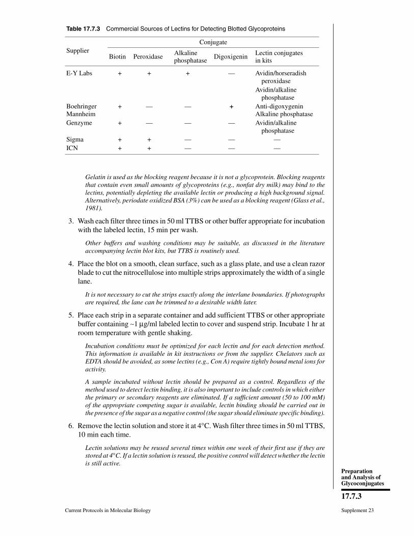
Gelatin is used as the blocking reagent because it is not a glycoprotein. Blocking reagentsthat contain even small amounts of glycoproteins (e.g., nonfat dry milk) may bind to thelectins, potentially depleting the available lectin or producing a high background signal.Alternatively, periodate oxidized BSA (3%) can be used as a blocking reagent (Glass et al.,1981).
3. Wash each filter three times in 50 ml TTBS or other buffer appropriate for incubationwith the labeled lectin, 15 min per wash.
Other buffers and washing conditions may be suitable, as discussed in the literatureaccompanying lectin blot kits, but TTBS is routinely used.
4. Place the blot on a smooth, clean surface, such as a glass plate, and use a clean razorblade to cut the nitrocellulose into multiple strips approximately the width of a singlelane.
It is not necessary to cut the strips exactly along the interlane boundaries. If photographsare required, the lane can be trimmed to a desirable width later.
5. Place each strip in a separate container and add sufficient TTBS or other appropriatebuffer containing ∼1 µg/ml labeled lectin to cover and suspend strip. Incubate 1 hr atroom temperature with gentle shaking.
Incubation conditions must be optimized for each lectin and for each detection method.This information is available in kit instructions or from the supplier. Chelators such asEDTA should be avoided, as some lectins (e.g., Con A) require tightly bound metal ions foractivity.
A sample incubated without lectin should be prepared as a control. Regardless of themethod used to detect lectin binding, it is also important to include controls in which eitherthe primary or secondary reagents are eliminated. If a sufficient amount (50 to 100 mM)of the appropriate competing sugar is available, lectin binding should be carried out inthe presence of the sugar as a negative control (the sugar should eliminate specific binding).
6. Remove the lectin solution and store it at 4°C. Wash filter three times in 50 ml TTBS,10 min each time.
Lectin solutions may be reused several times within one week of their first use if they arestored at 4°C. If a lectin solution is reused, the positive control will detect whether the lectinis still active.
Table 17.7.3 Commercial Sources of Lectins for Detecting Blotted Glycoproteins
SupplierConjugate
Biotin Peroxidase Alkaline phosphatase
Digoxigenin Lectin conjugatesin kits
E-Y Labs + + + — Avidin/horseradish peroxidaseAvidin/alkaline phosphatase
BoehringerMannheim
+ — — + Anti-digoxygeninAlkaline phosphatase
Genzyme + — — — Avidin/alkaline phosphatase
Sigma + + — — — ICN + + — — —
Current Protocols in Molecular Biology Supplement 23
17.7.3
Preparationand Analysis ofGlycoconjugates

7a. For lectins directly tagged with 125I, biotin, horseradish peroxidase, or alkalinephosphatase: Visualize by autoradiography or using the appropriate chromogenic orluminescent visualization solution.
7b. For lectins conjugated to biotin or digoxigenin: Incubate filter in the appropriatesecondary reagent 1 hr at room temperature. Wash filter 3 times with 50 ml TTBS,10 min each time. Detect bound lectin using the appropriate chromogenic or lumi-nescent visualization solution.
Lectins conjugated to these groups are detected by incubation with avidin–alkalinephosphatase conjugates or anti-digoxigenin–alkaline phosphatase.
COMMENTARY
Background InformationLectins have been used for many years to
detect the presence of various types of sugarchains. Lectin blots provide quick glimpses ofthe types of sugars attached to purified proteins.Even though the blots provide useful informa-tion, they cannot provide proof of a sugar struc-ture and hence cannot substitute for the morespecific and quantitative analytical approachespresented in this chapter. This is because lectinbinding and its quantitation depend upon manyfactors that vary with different individual pro-teins. The advantage of using lectins is thatmany different ones are available and analysisrequires only modest amounts of protein androutine laboratory equipment. Also, many de-tection methods are available. In order to haveconfidence in the results of lectin-binding stud-ies, it is necessary to show that their bindingspecificity is abolished by coincubation with acompeting sugar (usually a monosaccharide) orby prior enzymatic digestion to remove someor all of the protein-linked sugar chains. Lectinslabeled with fluorescent indicators can be usedto visualize glycoconjugates directly in hy-drated gels (West and McMahon, 1977). How-ever, this approach requires several days. Thisunit describes a less time-consuming method(based on Tanner and Anstee, 1976) in whichthe proteins are blotted onto filters prior todetection by one of several methods.
Critical Parameters andTroubleshooting
Information concerning critical parametersand troubleshooting for protein blotting, immu-noprobing, and chromogenic and luminescentdetection is provided in UNIT 10.8.
Experimental designTwo points are especially important in de-
signing lectin-binding experiments: the choiceof lectin and the choice of method for detecting
lectin bound to glycoprotein. If the goal of theexperiment is to obtain preliminary evidencethat a protein contains carbohydrate groups, abattery of lectins should be used along withappropriate controls (e.g., competition by ap-propriate sugars). If the experiment is meant toserve as a guide to further structural analysis,the effects of various chemical or enzymatictreatments must be combined with analysisusing one or more lectins known to bind to theglycoconjugate. Table 17.7.2 shows the speci-ficity of the most popular lectins, some of whichare supplied in the various kits.
The choice of detection method is affectedby the amount of protein available and theextent of glycosylation. If the sample contains1 to 10 µg of target protein or if the material isextensively glycosylated, lectins labeled di-rectly with 125I, alkaline phosphatase, or horse-radish peroxidase can be used. If either theprotein quantity or extent of glycosylation isminimal, an indirect system using lectins thatare biotinylated or coupled to digoxigenin anda secondary enzyme- or antibody-linked re-agent has the advantage of signal amplification.
KitsThe various reagents can be purchased sepa-
rately or as prepackaged kits from the supplierslisted in Table 17.7.3. E-Y Laboratories offersthe largest selection of purified lectins andlectins tagged with various conjugates.
The E-Y Laboratories kit uses lectins cou-pled to horseradish peroxidase. The Genzymeand Boehringer Mannheim kits use alkalinephosphatase–coupled molecules to detect thetagged lectins. Alternatively, tagged lectins caneasily be prepared in the laboratory (UNIT 11.1).The major advantages of using kits are that theyare self-sufficient, containing all of the reagentsand positive controls needed, and that the lect-ins they employ are among those with thebest characterized specificities. When coupled
Supplement 23 Current Protocols in Molecular Biology
17.7.4
Lectin Analysis ofProteins Blotted
onto Filters
with glycosidase digestions, the kits probablyprovide the most information about the sugarchains in the least amount of time for the leastamount of money. Because there are limitationsto the information that can be obtained, thedecision about which individual lectins or kitsto buy will depend upon the precision of infor-mation required.
The Glycan Differentiation Kit (BoehringerMannheim) supplies lectins that are coupled tothe steroid digoxigenin using an N-hydroxy-succinimide ester. An alkaline phosphatase–conjugated antiserum against this steroid isused to visualize bound lectin on target glyco-proteins (Haselbeck et al., 1990). Digoxigeninis a naturally occurring steroid found only inplants of the Digitalis family and not in ani-mals, which minimizes background. The sheepFab fragment to the hapten is affinity purifiedand coupled to alkaline phosphatase. The com-pany has used the digoxigenin system exten-sively in different types of kits to detect pro-teins, glycoproteins, and nucleic acids (UNIT
3.18). Lectin binding is simply an extension ofthis technology. The system has also beenadapted to visualize glycoconjugates by lightand electron microscopy (Sata et al., 1990).
The Lectin Link Kit (Genzyme) uses a biot-inylated lectin and an alkaline phosphatase–avidin conjugate to visualize glycoproteins onthe blot. The basic ABC (avidin–biotin conju-gate) technology is well established in the lit-erature for many assays. This kit as presentedis primarily intended to identify and differenti-ate between various types of N-linked oligosac-charides. The conclusion that binding of anylectin is exclusively due to the presence of aparticular structure on an N-linked oligosac-charide chain is valid only if all lectin bindingis destroyed by PNGase F digestion (UNIT 17.13),because, except for Con A, no lectin is specificfor N-linked oligosaccharides. It is importantto note that not all N-linked oligosaccharidesare sensitive to PNGase F (UNIT 17.13); resistanceto PNGase F does not prove that a glycan is, byelimination, O-linked.
Anticipated ResultsEven though lectins are quite specific, re-
sults are more qualitative than quantitative. Theresults obtained using lectins should be re-garded as guides and preliminary indicationsof likely structures. Lectin-binding studies, es-pecially when combined with a battery of en-zymatic digestions, are useful experiments forpreliminary characterization of glycoproteinstructure because they are quick, reasonably
priced, and use only a few micrograms (or less)of purified sample material. However, they can-not substitute for the more precise structuraland chemical analyses presented in other unitsin Chapter 17.
Inclusion of a positive control provides aqualitative and rough quantitative estimate forthe amount of reactive material in the samplerelative to the standards. This may be especiallyimportant in assessing the effects and com-pleteness of exo- or endoglycosidase diges-tions. The problem with attempting accuratequantitation is that the intensity of staining willdepend upon the binding affinity, the amountof protein, and the number of accessible gly-cans on the protein. All of these factors areimportant, and a high number of low-affinitybinding sites present on a large quantity ofprotein can give an intensity on a blot similarto that of a single high-affinity binding site ona smaller amount of protein.
Two examples illustrate useful approachesfor maximizing the information from lectinbinding. In both, lectin-binding results firstsuggest the presence of certain types of carbo-hydrate chains on the protein. This informationis used to design glycosidase digestions (UNIT
17.13) that should alter the binding of some orall of the lectins. Digested glycoproteins areprobed with the lectins to confirm the predic-tions. All of the data are combined into a com-posite picture of the sugar chains present. To beconclusive, results of lectin binding and gly-cosidase digestions should be internally consis-tent. Because some of the glycosidases arethemselves glycoproteins, controls should in-clude a lane with glycosidase alone to identifybands due to the presence of glycosidase in thedigested sample.
Example 1A hypothetical purified glycoprotein reacts
with wheat germ agglutinin (WGA) and Sam-bucus nigra agglutinin (SNA; see Table 17.7.2).WGA binds to terminal GlcNAc residues orsialic acids. Terminal GlcNAc may occur incomplex N-linked oligosaccharides, such asthose in ovomucoid, or as multiple residues ofO-β-GlcNAc or GlcNAc-1-P directly bound toprotein. Sialic acid could be present in α2-3 orα2-6 linkages to underlying βGal residues onN- or O-linked chains. SNA binds to sialic acidlinked α2-6 to βGal. One way to distinguishamong the possibilities is to treat the samplewith sialidase. Loss of WGA binding followingsialidase treatment indicates that WGA bindingis due to sialic acid and not terminal GlcNAc.
Current Protocols in Molecular Biology Supplement 23
17.7.5
Preparationand Analysis ofGlycoconjugates
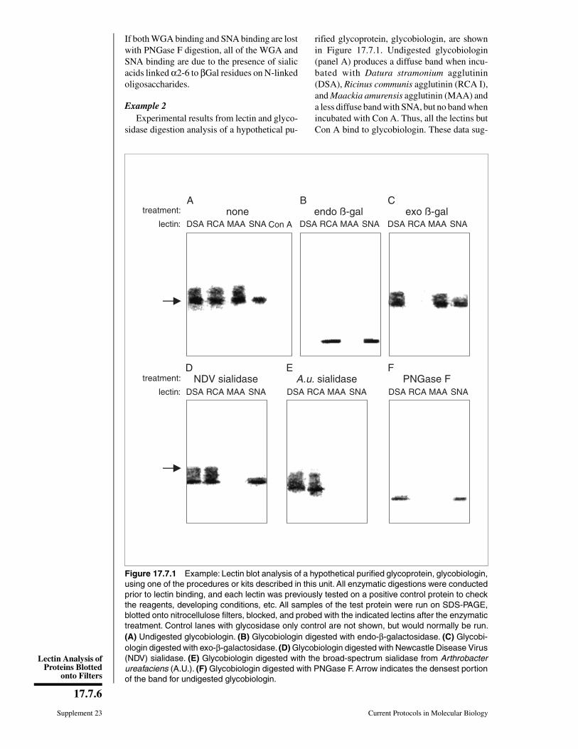
If both WGA binding and SNA binding are lostwith PNGase F digestion, all of the WGA andSNA binding are due to the presence of sialicacids linked α2-6 to βGal residues on N-linkedoligosaccharides.
Example 2Experimental results from lectin and glyco-
sidase digestion analysis of a hypothetical pu-
rified glycoprotein, glycobiologin, are shownin Figure 17.7.1. Undigested glycobiologin(panel A) produces a diffuse band when incu-bated with Datura stramonium agglutinin(DSA), Ricinus communis agglutinin (RCA I),and Maackia amurensis agglutinin (MAA) anda less diffuse band with SNA, but no band whenincubated with Con A. Thus, all the lectins butCon A bind to glycobiologin. These data sug-
A
DSA RCA MAA SNA Con A
treatment:
lectin:
B
DSA RCA MAA SNA
C
DSA RCA MAA SNA
D
DSA RCA MAA SNA
treatment:
lectin:
E
DSA RCA MAA SNA
F
DSA RCA MAA SNA
none endo ß-gal exo ß-gal
NDV sialidase A.u. sialidase PNGase F
Figure 17.7.1 Example: Lectin blot analysis of a hypothetical purified glycoprotein, glycobiologin,using one of the procedures or kits described in this unit. All enzymatic digestions were conductedprior to lectin binding, and each lectin was previously tested on a positive control protein to checkthe reagents, developing conditions, etc. All samples of the test protein were run on SDS-PAGE,blotted onto nitrocellulose filters, blocked, and probed with the indicated lectins after the enzymatictreatment. Control lanes with glycosidase only control are not shown, but would normally be run.(A) Undigested glycobiologin. (B) Glycobiologin digested with endo-β-galactosidase. (C) Glycobi-ologin digested with exo-β-galactosidase. (D) Glycobiologin digested with Newcastle Disease Virus(NDV) sialidase. (E) Glycobiologin digested with the broad-spectrum sialidase from Arthrobacterureafaciens (A.U.). (F) Glycobiologin digested with PNGase F. Arrow indicates the densest portionof the band for undigested glycobiologin.
Supplement 23 Current Protocols in Molecular Biology
17.7.6
Lectin Analysis ofProteins Blotted
onto Filters
gest that the glycoprotein may contain polylac-tosamine chains (DSA), terminal β-Gal resi-dues (RCA I), and both α2-3- and α2-6-linkedsialic acids (MAA and SNA, respectively).High-mannose type chains are probably absent(Con A).
This is useful information, but performing afew enzyme digestions and then probing withall of the lectins will provide much more. First,specific enzyme digestions should be done toshow that each of the lectins is, in fact, bindingto the expected sugar structure. Digestion withendo-β-galactosidase (panel B), which re-moves polylactosamine chains (UNIT 17.13), abol-ishes all bands in the DSA lane, showing thatbinding to DSA was due entirely to polylac-tosamine chains. The apparent Mr of the bandhas dropped and its boundaries have sharpened,showing that the polylactosamines contributedto the diffuse appearance of the original band.Glycoproteins often produce diffuse bands ingels and on blots because of microhetero-geneity in the carbohydrate chains (UNIT 17.1).This digestion also destroys all of the MAAbinding, suggesting that the polylactosaminechains, and only those chains, were terminatedby α2-3-linked sialic acids (UNIT 17.12). Bandsare still observed in the RCA I lane, showingthat terminal β-Gal residues were not digested,and in the SNA lane, showing that the polylac-tosamine chains do not terminate in α2-6 sialicacids. If the digestion is done with exo-β-galac-tosidase (panel C), which removes terminalβ-Gal residues (UNIT 17.18), it destroys the bandin the RCA I lane but has minimal effects onthe position and appearance of the other bands.This shows that the lectin was bound to thepredicted terminal sugar residues. There areprobably only a small number of these and theydid not contribute much to the heterogeneity ofthe protein. Digestion with Newcastle DiseaseVirus (NDV) sialidase (panel D), which spe-cifically removes α2-3-linked sialic acids,abolished the binding of MAA, showing thatMAA bound the α2-3 sialic acid, as predicted.The significant reduction in the apparent sizeof the protein size shows that the sialic acidsmake a large contribution to the apparent size.Digestion with Arthrobacter ureafaciens siali-dase (panel E), a broad-spectrum sialidase (UNIT
17.12), abolishes the bands in both the MAA andSNA lanes. Diffuse bands are still seen in theDSA and RCA I lanes; the apparent protein sizeis slightly smaller than in panel D, showing thatthe α2-6-linked sialic acids make a relativelysmall contribution to the apparent size com-pared to those linked α2-3. For each digestion
the bands are reduced in Mr and/or becomesharper compared to those in the undigestedglycobiologin.
The final digestion (panel F) is with peptideN:glycosidase F (PNGase F), which removesessentially all N-linked chains; however, someare resistant (UNIT 17.13). Any lectin binding thatis completely destroyed by this digestion is dueonly to components found on N-linked chains.PNGase F digestion of glycobiologin com-pletely abolishes the bands in the RCA I andMAA lanes but only partially reduces the in-tensity of the bands in the DSA and SNA lanes.The lack of binding to RCA I and MAA indi-cates that all of the β-Gal-terminated chains andthose with α2-3 sialic acids are exclusivelyN-linked (RCA I and MAA). Reduced bindingto SNA indicates that some of the N-linkedchains also contain α2-6 sialic acid, which mayalso be found on O-GalNAc-linked chains. Re-duced binding to DSA suggests either that theprotein also contains polylactosamine units onboth N- and O-linked chains or possibly thatthe PNGase F digestion was incomplete. Theband is still diffuse because of residual poly-lactosamine chains. The apparent protein sizeis considerably smaller, showing that most ofthe heterogeneity was due to polylactosaminechains on N-linked oligosaccharides.
It is important to stress again that lectin blotsgive only preliminary evidence for the presenceof the suggested structures. The results shouldbe viewed as useful guides.
Time ConsiderationsMost enzymatic digestions are carried out
overnight. If the proteins are separated onminigels, the entire analysis including blottingand development can be done in a single day.The procedure can be interrupted followingprotein transfer to nitrocellulose. Incubation inblocking buffer can be extended to overnight at4°C.
Literature CitedDetailed references regarding detection of
the various types of conjugates are given in UNIT
10.8. Kit manufacturers will supply relevant ref-erences upon request. Much of their informa-tion may have been obtained exclusively fromdata produced in company laboratories, al-though some has also appeared in the peer-re-viewed literature.
Glass, W.F., Briggs, R.C., and Hnilica, L.S. 1981.Use of lectins for detection of electrophoreticallyseparated glycoproteins transferred onto nitro-cellulose. Anal. Biochem. 115:219-224.
Current Protocols in Molecular Biology Supplement 23
17.7.7
Preparationand Analysis ofGlycoconjugates
Sata, T., Zuber, C., and Roth, J. 1990. Lectin-digoxi-genin conjugates: A new hapten system for gly-coconjugate cytochemistry. Histochem. 94:1-11.
West, C.M. and McMahon, D. 1977. Identificationof concanavalin A receptors and galactose-bind-ing proteins in purified plasma membranes ofDictyostelium discoideum. J. Cell Biol. 74:264-273.
Key ReferencesHaselbeck, A., Schickaneder, E., von der Eltz, H.,
and Hösel, W. 1990. Structural characterizationof glycoprotein carbohydrate chains by usingdigoxigenin-labeled lectins on blots. Anal. Bio-chem. 191:25-30.
Description of techniques for digoxigenin-labeledlectins, showing uses for highly purified glycoprote-ins on blots.
Tanner, M.J. and Anstee, D.J. 1976. A method forthe direct demonstration of the lectin-bindingcomponents of the human erythrocyte mem-brane. Biochem. J. 153:265-270.
Good overview of procedure.
Contributed by Hudson H. FreezeLa Jolla Cancer Research FoundationLa Jolla, California
Supplement 23 Current Protocols in Molecular Biology
17.7.8
Lectin Analysis ofProteins Blotted
onto Filters
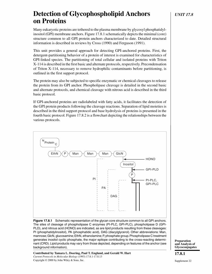
UNIT 17.8Detection of Glycophospholipid Anchorson ProteinsMany eukaryotic proteins are tethered to the plasma membrane by glycosyl phosphatidyl-inositol (GPI) membrane anchors. Figure 17.8.1 schematically depicts the minimal (core)structure common to all GPI protein anchors characterized to date. Detailed structuralinformation is described in reviews by Cross (1990) and Ferguson (1991).
This unit provides a general approach for detecting GPI-anchored proteins. First, thedetergent-partitioning behavior of a protein of interest is examined for characteristics ofGPI-linked species. The partitioning of total cellular and isolated proteins with TritonX-114 is described in the first basic and alternate protocols, respectively. Precondensationof Triton X-114, necessary to remove hydrophilic contaminants before partitioning, isoutlined in the first support protocol.
The protein may also be subjected to specific enzymatic or chemical cleavages to releasethe protein from its GPI anchor. Phospholipase cleavage is detailed in the second basicand alternate protocols, and chemical cleavage with nitrous acid is described in the thirdbasic protocol.
If GPI-anchored proteins are radiolabeled with fatty acids, it facilitates the detection ofthe GPI protein products following the cleavage reactions. Separation of lipid moieties isdescribed in the third support protocol and base hydrolysis of proteins is presented in thefourth basic protocol. Figure 17.8.2 is a flowchart depicting the relationships between thevarious protocols.
ProteinN
C
P Man Man Man GlcN
Inositol
PI
PA
DAG
EthN
P
HONO
GPI-PLD
PI-PLC,GPI-PLC
Figure 17.8.1 Schematic representation of the glycan core structure common to all GPI anchors.The sites of cleavage of phospholipase C enzymes (PI-PLC, GPI-PLC), phospholipase D (GPI-PLD), and nitrous acid (HONO) are indicated, as are lipid products resulting from these cleavages:PI (phosphatidylinositol), PA (phosphatidic acid), DAG (diacylglycerol). Other abbreviations: Man,mannose; GlcN, glucosamine; EthN, ethanolamine; P, phosphate group. Phospholipase C treatmentgenerates inositol cyclic phosphate, the major epitope contributing to the cross-reacting determi-nant (CRD). Lipid products may vary from those depicted, depending on features of the anchor (seebackground information).
Supplement 22
Contributed by Tamara L. Doering, Paul T. Englund, and Gerald W. HartCurrent Protocols in Molecular Biology (1993) 17.8.1-17.8.13Copyright © 2000 by John Wiley & Sons, Inc.
17.8.1
Preparationand Analysis ofGlycoconjugates

Strategic PlanningThe techniques described below require that a method for identifying the protein ofinterest be available. Proteins may be visualized after one-dimensional gel electrophoresis(UNIT 10.2) and Coomassie blue or silver staining (UNIT 10.6), or they may be identified byimmunoprecipitation (UNIT 10.16), or immunoblotting (UNIT 10.8) using available specificantibodies. Enzymes may also be detected by their known activities (e.g., see Kodukulaet al., 1992).
It is extremely convenient, particularly for low-abundance species, to use proteins thatare labeled with radioactive amino acids (UNIT 10.18) or GPI components (see Fig. 17.8.1).The ability to radiolabel proteins with GPI precursors such as fatty acids, ethanolamine,myo-inositol, and certain monosaccharide precursors (UNIT 17.4) is in itself suggestive ofGPI anchorage. Because GPI-anchored proteins commonly represent a small fraction ofeukaryotic glycoconjugates, metabolic radiolabeling may be inefficient. Tunicamycin(UNIT 17.10) may be used to increase relative incorporation of monosaccharide precursorsinto GPIs.
Rigorous identification of specific GPI anchor structures requires detailed analysis. Thesemethods, which are beyond the scope of this unit, are described in Ferguson (1992).
TritonX-114
partitioning
one-dimensionalgel electrophoresis
(UNIT 10.2)immunoblotting
(UNIT 10.8)immunoprecipitation
(UNIT 10.16)
Triton X-114partitioning
immunofluorescence(UNIT 14.6)
immunoblotting(UNIT 10.8)
immunoprecipitation(UNIT 10.16)
TLC(UNITS 10.12-
10.15)
auto-radiography
(APPENDIX 3)
cells, membranes
proteinsTriton X-114partitioning
(first basic andalternate protocols)
nitrous acidcleavage
(third basicprotocol)
basehydrolysis
(fourth basicprotocol)
phospholipasedigestion
(second basic andalternate protocols)
anti-CRDAb detection
(second supportprotocol)
separation oflipid moiety
(third supportprotocol)
radiolabeling (UNITS 10.8 & 17.4)
Figure 17.8.2 Flowchart for the detection of GPI-anchored proteins.
Supplement 22 Current Protocols in Molecular Biology
17.8.2
Detection ofGlycophospho-
lipid Anchors onProteins

BASICPROTOCOL
EXTRACTION AND PARTITIONING OF TOTAL PROTEINS FROM CELLSOR MEMBRANES WITH TRITON X-114
Extraction of cells or tissues with the detergent Triton X-114 provides a profile (andpotentially an initial purification) of amphiphilic proteins, including integral membraneproteins and species bearing GPI anchors. Once a GPI-containing protein is released fromthe lipid component of its anchor, it will no longer partition into the detergent-enrichedphase. This alteration of partitioning behavior provides a rapid assay for presence orabsence of anchors. It may be used to monitor the changes in proteins produced by theother procedures.
In this protocol, cells or membrane fractions are first resuspended in TBS, then extractedwith a Triton X-114 incubation on ice. The mixture is then warmed and separates intotwo protein-containing phases due to aggregation of detergent micelles (see backgroundinformation). The upper (detergent-depleted) phase contains hydrophilic proteins; am-phiphilic proteins (including those with GPI anchors) partition to the lower (detergent-enriched) phase. Both phases are then analyzed to clarify the group to which each proteinbelongs.
Materials
Cells, membrane fraction, or other source of proteinTris-buffered saline (TBS), ice-coldPrecondensed Triton X-114 stock solution in TBS (support protocol), ice-cold
15-ml polypropylene centrifuge tubesCentrifuges: low-speed (tabletop) and equipped with appropriate rotor (e.g.,
SS-34), at 4°C and room temperature
1. Resuspend cells (or membranes) in ice-cold TBS to a final protein concentration of≤4 mg/ml in a 15-ml tube.
The amount of protein required is that which will give detectable levels at the end of theprocedure and will depend on the system the individual researcher is using. As a guideline,use several times the amount of protein that is believed to be needed, to account forpartitioning and losses (see critical parameters).
2. Add 1⁄5 vol precondensed Triton X-114 stock solution (∼ 2% final concentration).
3. Extract cells by incubating 15 min on ice with occasional mixing.
At this point in the procedure there will only be one phase.
4. Centrifuge cell mixture 10 min at 10,000 × g (9000 rpm in an SS-34 rotor), 4°C, andtransfer supernatant to a fresh tube. Resuspend pellet in ice-cold TBS and save onice for analysis in step 7.
The cold supernatant fraction contains both soluble and detergent-extracted proteins; thelatter group includes proteins anchored by GPI structures or by transmembrane polypep-tide domains. The pellet contains additional GPI species, as well as cell elements insolublein nonionic detergents.
5. Warm supernatant fraction to 37°C in a water bath until the solution becomes cloudy.
6. Centrifuge the solution in a tabletop centrifuge 10 min at 1000 × g, room temperature.Collect upper and lower phases in separate tubes.
The upper phase (detergent-depleted) contains soluble proteins; the lower (detergent-en-riched) phase contains proteins anchored by GPI structures or by transmembrane domains.
7. Analyze the resuspended pellet from step 4 and each phase from step 6 for the proteinsof interest.
Current Protocols in Molecular Biology Supplement 22
17.8.3
Preparationand Analysis ofGlycoconjugates
As discussed in strategic planning, analysis may be by an activity assay, one-dimensionalgel electrophoresis and staining of proteins (UNITS 10.2 & 10.6), or immunoprecipitation (UNIT
10.16) or immunoblotting (UNIT 10.8) using specific antibodies.
ALTERNATEPROTOCOL
PARTITIONING OF ISOLATED PROTEINS WITH TRITON X-114
A modification to the basic protocol for Triton X-114 extraction of proteins from wholecells or membranes is used to partition isolated proteins.
1. Dilute or dissolve the protein in 1 ml ice-cold TBS in a 1.5-ml microcentrifuge tube.Add 0.2 ml Triton X-114 and mix.
See critical parameters for guidelines on the total amount of protein to use.
2. Warm protein mixture 15 min (or until it becomes cloudy) in a 37°C water bath.Microcentrifuge 5 min at maximum speed.
3. Collect the upper and lower phases and analyze each phase for the protein(s) ofinterest.
SUPPORTPROTOCOL
PRECONDENSATION OF TRITON X-114 DETERGENT
Triton X-114 often contains hydrophilic contaminants, and must be precondensed byseveral rounds of phase separation before use.
Materials
Triton X-114 detergentTris-buffered saline (TBS)
50-ml centrifuge tubesTabletop centrifuge
1. Dissolve 1.5 g Triton X-114 in 50 ml TBS, place in a 50-ml centrifuge tube, then chillon ice.
At this point, the solution will be clear.
2. Warm the solution to 37°C in a water bath.
Following this warming, the solution will be turbid.
3. Centrifuge the solution in a tabletop centrifuge 10 min at 1000 × g, room temperature.
4. Remove and discard the upper (detergent-depleted) phase and redissolve the lower(detergent-enriched) phase in an equal volume of ice-cold TBS.
Hydrophilic contaminants partition into the upper phase.
5. Repeat partitioning (steps 2 to 4) three times.
The final detergent-enriched phase contains ∼12% detergent. It may be stored at 4°C forroutine use or frozen for long-term storage.
Supplement 22 Current Protocols in Molecular Biology
17.8.4
Detection ofGlycophospho-
lipid Anchors onProteins
BASICPROTOCOL
IDENTIFICATION OF GPI-ANCHORED PROTEINS BY PI-PLC DIGESTIONOF INTACT CELLS
Phospholipase C (PLC) cleaves within the GPI membrane anchors (see Fig. 17.8.1) ofglycoproteins, causing the release of protein from cell membranes. In this protocol, intactcells are digested with phosphatidylinositol-specific phospholipase C (PI-PLC). Follow-ing incubation with the enzyme, the cell mixture is centrifuged and both the resultingpellet and supernatant are analyzed for the presence of the protein of interest. Proteinsreleased from GPI anchors are found in the supernatant; lack of release may indicateabsence of a GPI anchor, modification of the GPI protein, or other factors (see backgroundinformation).
Materials
Cells (or membrane preparation)Bacterial phosphatidylinositol-specific phospholipase C (PI-PLC;
e.g., from Bacillus thuringiensis, Oxford Glycosystems)Hanks balanced salt solution (HBSS; GIBCO/BRL), buffered saline,
or culture medium
Tabletop centrifuge and appropriate centrifuge tubes
1. Obtain cells (or membranes) of interest in dispersed suspension. Wash duplicatealiquots twice in HBSS, buffered saline, or culture medium, then resuspend in anyof these solutions.
The final protein concentration of the samples (typically 0.1 to 0.5 mg/ml) should be basedon supplier specifications for the enzyme preparation used. See critical parameters forguidelines on the total amount of protein to use.
2. Add bacterial PI-PLC to one aliquot. Add no enzyme to the second aliquot (control).Incubate tubes 1 hr at 37°C.
The amount of enzyme should be determined by supplier recommendations.
3. Centrifuge cells in a tabletop centrifuge 5 min at 1000 × g. Remove each supernatantto a fresh tube and save for analysis in step 4. Resuspend pellet in a volume of bufferedsaline equal to the volume of supernatant removed.
4. Assay supernatant and pellet fractions for protein(s) of interest.
The proteins may be assayed by Triton X-114 detergent partitioning (first basic protocol),one-dimensional gel electrophoresis (UNIT 10.2) followed by Coomassie blue or silverstaining (UNIT 10.6), immunoprecipitation (UNIT 10.16) or immunoblotting (UNIT 10.8)
ALTERNATEPROTOCOL
IDENTIFICATION OF GPI ANCHORAGE BY PHOSPHOLIPASETREATMENT OF ISOLATED PROTEINS
Several specific phospholipases that effectively degrade only GPI species are useful foranalyzing GPI anchorage of isolated proteins (see background information). In thisprotocol, aliquots of the protein are digested in parallel with the following phospholipases:bacterial phosphatidylinositol-specific phospholipase C (PI-PLC), a GPI-specific phos-pholipase C (GPI-PLC) from Trypanosoma brucei, and a GPI-specific phospholipase D(GPI-PLD) from mammalian serum (rat, rabbit, or human). Proteins are partitioned byTriton X-114 extraction (first alternate protocol), then analyzed. Specific cleavage ofproteins by phospholipase treatment strongly indicates GPI anchorage.
Current Protocols in Molecular Biology Supplement 22
17.8.5
Preparationand Analysis ofGlycoconjugates
Materials
Isolated proteinAcetone, −20°CAppropriate enzyme buffer: GPI-PLD buffer, GPI-PLC buffer, and PI-PLC bufferPhospholipase enzyme: GPI-PLD from rat, rabbit or human whole serum,
GPI-PLC from Trypanosoma brucei (Oxford GlycoSystems), and PI-PLCfrom Bacillus thuringiensis (Oxford GlycoSystems) or B. cereus(Boehringer Mannheim or Sigma)
Tris-buffered saline (TBS)Precondensed Triton X-114 solution (first support protocol)Centrifuge and rotor (e.g., SS-34)
1. If the protein sample is lyophilized or concentrated in a buffer similar to thephospholipase buffers, begin at step 3. If it is not, acetone-precipitate the protein byadding 8 vol cold (−20°C) acetone to the protein solution and incubating it ≥3 hr at−20°C.
See critical parameters for guidelines on the total amount of protein to use. For low-abun-dance material, efficiency of precipitation may be increased by including 15 g of a carrierprotein (e.g., cytochrome c or bovine serum albumin).
2. Centrifuge the sample 20 min at 15,000 × g (∼11,000 rpm in an SS-34 rotor), 4°C.Aspirate supernatant and allow pellet to air dry.
3. For each enzyme to be tested, dissolve two aliquots of the protein in 100 µl of theappropriate enzyme buffer in microcentrifuge tubes.
4. To one aliquot from each pair add the appropriate enzyme. Add an equal volume ofthe appropriate enzyme buffer to the second aliquot (control). Mix tubes and incubate1 hr at 37°C.
The amount of enzyme added should be based on the activity of the lipase preparation (seesupplier specifications) and the estimated amount of protein present. It may be desirableto titrate the enzyme, using various amounts in parallel reactions, to be sure that allsensitive proteins are completely digested.
GPI-PLD is not generally available, but whole serum (rat, rabbit or human) may serve asa source of this activity. Aliquots of serum may be stored frozen and thawed before use. Forthe assay, use 2 l whole serum in a 100-l reaction.
5. Dilute reaction with 200 µl ice-cold TBS, then add 60 µl Triton X-114. Chill 15 minon ice.
6. Partition the proteins as in steps 2 and 3 of the first alternate protocol.
GPI-anchored proteins partition into the detergent-enriched phase of Triton X-114 in thecontrol reaction (uncleaved) and into the detergent-depleted phase after cleavage of theGPI anchor by phospholipase.
7. Analyze each phase for the presence of protein(s) of interest as in step 7 of the firstbasic protocol.
Alternatively, cleavage products can be detected by reactivity with α-CRD antibody(second support protocol) and, if the anchor is radiolabeled with fatty acids (UNIT 17.4), byseparation of radiolabeled lipid products.
Migration of phospholipase-digested proteins on gel electrophoresis is not predictable andshould not be used alone to assess cleavage. No shift, or a shift in either direction (relativeto uncleaved protein migration) may be observed upon GPI anchor removal, depending onthe properties of the protein.
Supplement 22 Current Protocols in Molecular Biology
17.8.6
Detection ofGlycophospho-
lipid Anchors onProteins

SUPPORTPROTOCOL
DETECTION OF PRODUCTS AFTER PHOSPHOLIPASE TREATMENT BYREACTIVITY WITH ANTI-CRD ANTIBODY
Cleavage of GPI structures with phospholipase C reveals a cryptic antigen termed thecross-reacting determinant (CRD). The major epitope contributing to the CRD is aninositol 1,2-(cyclic) monophosphate formed during phospholipase C cleavage; otherstructural features may also contribute to the antigenicity. Reactivity of a protein withanti-CRD antibody after solubilization by phospholipase C strongly indicates the pres-ence of a GPI anchor. Anti-CRD antibody (Oxford GlycoSystems) may be used afterphospholipase C treatment to immunoprecipitate (UNIT 10.16) or immunoblot (UNIT 10.8)proteins of interest. It should be noted that phospholipase D cleavage does not create theepitope required for αCRD activity (see Fig. 17.8.1).
BASICPROTOCOL
NITROUS ACID CLEAVAGE OF GPI-ANCHORED PROTEINS
An unusual property common to all GPI structures is the presence of a nonacetylatedglucosamine that is glycosidically linked to inositol. The glycosidic bond is specificallycleaved when the glucosamine undergoes nitrous acid deamination, thereby cleaving theGPI anchor (see Fig. 17.8.1). Because nonacetylated glucosamine rarely occurs outsideof GPIs, nitrous acid deamination may be used to identify GPI linkage to protein. Due toside reactions, deamination is not always quantitative or precisely reproducible, but itdoes serve a useful diagnostic purpose.
In this protocol, the protein is incubated in nitrous acid to cleave the GPI anchors. Cleavedand uncleaved proteins are then separated by Triton X-114 partitioning.
Materials
Protein(s)0.1 M acetate buffer, pH 3.50.5 M NaNO2, made fresh0.5 M NaCl
1. Dry two aliquots of the protein of interest in microcentrifuge tubes and dissolve eachin 0.1 ml of 0.1 M acetate buffer, pH 3.5.
See critical parameters for guidelines on the total amount of protein to use.
Nonidet P-40 (NP-40) detergent (0.1% v/v final) may be added to aid solubilization.
2. Add 0.1 ml of 0.5 M NaNO2 to one tube. To the second tube (control), add 0.1 ml of0.5 M NaCl. Incubate 3 hr at room temperature.
3. Detect the release of protein from GPI anchor by partitioning and analyzing thereaction products as in the first alternate protocol (Triton X-114 partitioning ofisolated proteins).
If the anchor is radiolabeled with fatty acids (UNIT 17.4), the support protocol for separationof lipid moiety may be used.
Current Protocols in Molecular Biology Supplement 22
17.8.7
Preparationand Analysis ofGlycoconjugates

SUPPORTPROTOCOL
SEPARATION OF LIPID MOIETY TO DETECT CLEAVAGE OFGPI-ANCHORED PROTEINS
If GPI-anchored proteins are radiolabeled with fatty acids, the efficacy of GPI anchorcleavage may be conveniently assessed by examining the released radiolabeled lipidproducts (see Fig. 17.8.1). In this protocol, radiolabeled proteins that have been cleavedby phospholipase or nitrous acid treatment are extracted with butanol. Released lipidmoieties are extracted into the butanol phase and are analyzed and quantitated.
Materials
Radiolabeled protein, cleaved by phospholipase or nitrous acid treatment (secondalternate and third basic protocols)
Water-saturated n-butanol
1. Add 100 µl water to each of two 1.5-ml microcentrifuge tubes. To one tube, add 100µl cleaved radiolabeled protein. To the second, add 100 µl control (uncleaved) protein.
2. Add 200 µl water-saturated n-butanol to each tube and vortex vigorously.
3. Microcentrifuge 30 sec at maximum speed, room temperature, to separate the twophases.
4. Remove the upper (butanol) phase to a fresh tube, and set aside. Add another 200 µlof water-saturated butanol to the original tubes.
5. Vortex and centrifuge as in steps 2 and 3 above.
6. Remove the second butanol phase and pool it with the first, retaining the lower(aqueous) phase for analysis.
7. Quantitate the percentage of lipid moieties released by the cleavage reactions byliquid scintillation counting of both butanol and aqueous phases. Compare countsrecovered in butanol phases of the cleaved material with those recovered from controlreactions to assess specific release.
To characterize released products fully, examine the butanol-extracted products by thinlayer chromatography; compare to standards (Doering et al., 1990b). Products of phos-pholipase C digestion include diacylglycerol, alkyl-acyl glycerol, or ceramide, dependingupon the original structure. Phosphatidylinositol (from nitrous acid treatment) or phos-phatidic acid (released by phospholipase D) may also be analyzed and quantitated. Thisassay has been used for purification of phospholipase activity (Hereld et al., 1986).
BASICPROTOCOL
BASE HYDROLYSIS OF RADIOLABELED PROTEINS
For proteins radiolabeled with fatty acids, it is useful to ascertain whether incorporatedfatty acids are actually part of a GPI structure or are present in amide linkage (N-myris-toylation) or thioester linkage (as in palmitoylation of cysteine residues). This can becharacterized by assessing the lability of the fatty acid linkage to various chemicaltreatments.
In this protocol, aliquots of radiolabeled proteins are resolved by electrophoresis and thevarious lanes are treated with KOH in methanol or hydroxylamine. Methanol and Trisbuffer are used to treat the control lanes. The lanes are then processed for fluorography.If the chemical bonds in the protein are susceptible to these treatments, a radiolabeledsignal will no longer be produced at the protein band of interest.
Supplement 22 Current Protocols in Molecular Biology
17.8.8
Detection ofGlycophospho-
lipid Anchors onProteins

Materials
Protein radiolabeled with fatty acid0.2 M KOH in methanolMethanol1 M hydroxylamine⋅HCl, pH 7.5, made fresh1 M Tris⋅Cl, pH 7.5 (APPENDIX 2)
Additional reagents and equipment for one-dimensional gel electrophoresis(UNIT 10.2), staining of gels (UNIT 10.6), and autoradiography (APPENDIX 3)
1. Resolve five identical samples of protein radiolabeled with fatty acid by SDS-PAGEelectrophoresis. Include appropriate markers.
2. Cut apart the five gel lanes, and stain one to visualize protein and markers.
3. Soak the other four lanes in one of the following solutions for 1 hr at roomtemperature:
Lane 1: 0.2 M KOH in methanolLane 2: methanol aloneLane 3: 1 M hydroxylamine⋅HCl, pH 7.5Lane 4: 1 M Tris⋅Cl, pH 7.5
4. Soak the treated gel slices in three washes (10 min each) of water.
5. Process gel pieces for autoradiography.
If bonds are susceptible to the treatments above, radiolabel from fatty acids will no longerproduce a signal at the position of the protein band of interest. KOH in methanol cleavesthio- and oxyesters but not amide linkages. Hydroxylamine treatment cleaves thioestersonly. Fatty acids that are part of a GPI moiety will be released by KOH in methanol, butnot by hydroxylamine treatment. The methanol and Tris⋅Cl treatments serve as controls.Ceramide-containing anchors are not sensitive to mild base treatment (see anticipatedresults and Conzelmann et al., 1992).
REAGENTS AND SOLUTIONS
0.1 M acetate buffer, pH 3.5Dissolve 0.82 g sodium acetate in 95 ml distilled water. Adjust pH to 3.5 with aceticacid, then bring volume to 100 ml with distilled water. Store indefinitely at roomtemperature.
GPI-PLC buffer50 mM Tris⋅Cl, pH 8.05 mM EDTA1% NP-40Store indefinitely at 4°C
GPI-PLD buffer50 mM Tris⋅Cl, pH 7.410 mM NaCl2.5 mM CaCl2
0.1 % NP-40Store indefinitely at 4°C
1 M hydroxylamine⋅HCl, pH 7.5Dissolve 6.95 g hydroxylamine⋅HCl in 95 ml distilled water. Adjust pH to 7.5
with NaOH, then increase volume to 100 ml with water. Make fresh for eachuse.
Current Protocols in Molecular Biology Supplement 22
17.8.9
Preparationand Analysis ofGlycoconjugates
PI-PLC buffer (for B. thuringiensis enzyme)25 mM Tris acetate, pH 7.40.1% (w/v) sodium deoxycholate (Na–DOC)Store indefinitely at 4°C
Tris-buffered saline (TBS)10 mM Tris⋅Cl, pH 7.5150 mM NaClStore indefinitely at room temperature
Water-saturated n-butanolMix equal volumes of distilled water and n-butanol. Cap tightly and shake vigor-ously. Let stand to allow phases to separate; the upper phase is the butanol. Storeindefinitely at room temperature.
COMMENTARY
Background InformationGlycosyl phosphatidylinositol (GPI) mem-
brane anchors are one mode of anchoring pro-teins to cell surfaces. These structures consistof a glycan bridge between phosphatidylinosi-tol and phosphoethanolamine; the phospho-ethanolamine is in amide linkage to the C-ter-minus of the protein (see Fig. 17.8.1). Theglycan core of GPIs (shown schematically inFig. 17.8.1) is remarkably conserved through-out evolution, although it is modified in manycell types by the addition of side chains. Thelipid moieties of GPIs, however, are quite het-erogeneous. This portion may consist of diacylglycerol, alkyl acyl glycerol, lyso-acyl com-pounds, or ceramide. The hydrocarbon chainsalso vary in length and degree of saturation, andmay be present as mixtures of species. GPIstructures are reviewed in Ferguson (1991).
Much of our understanding of GPI anchorsis derived from studies of the variant surfaceglycoprotein (VSG) of trypanosomes. Earlywork showed that VSG is associated with themembrane via a lipid moiety that contains di-myristoyl phosphatidylinositol (Cardoso de Al-meida and Turner, 1983; Ferguson and Cross,1984; Ferguson et al., 1985, 1986). Other in-vestigations had shown that bacterial phos-pholipase C releases certain proteins from cellmembranes (reviewed by Low, 1989). To-gether, these studies led to the definition of aclass of proteins bearing a C-terminal gly-colipid anchor.
Further investigations in this field have ledto the elucidation of the pathway of GPI bio-synthesis (reviewed by Doering et al., 1990a;Field and Menon, 1991), the purification of aGPI-specific phospholipase C (GPI-PLC), andthe development of anti-cross-reacting deter-minant (anti-CRD) antibodies as probes for
GPIs. Phosphatidylinositol-specific phosphol-ipase C (PI-PLC) and GPI-specific phospholi-pase D (GPI-PLD) are also useful in identifica-tion of GPI-anchored proteins.
The protocols included here are designed toallow identification of GPI-anchored proteinswithout detailed structural analysis. Becausethese techniques are rather diverse, further ex-planatory information is outlined below.
Triton X-114 partitioning. Triton X-114forms a clear micellar solution at low tempera-tures; the solution separates into two phaseswhen warmed above 20°C, due to aggregationof detergent micelles (Bordier, 1981). Whencellular material is extracted in Triton X-114 atlow temperatures, the solution contains solubleproteins, integral membrane proteins, and GPI-anchored proteins. Centrifugation results in apellet containing some of the GPI-anchoredspecies as well as cellular components that areinsoluble in nonionic detergents (Hooper andBashir, 1991). When the detergent solution iswarmed, amphiphilic proteins (including thosewith GPI anchors) associate with the detergent-enriched phase, while hydrophilic ones parti-tion to the detergent-depleted phase. SomeGPI-anchored proteins exhibit detergent in-solubility that develops shortly after synthesis(Brown and Rose, 1992); this is thought to bedue to aggregation of the anchored proteinswith glycosphingolipids, possibly as a step indirected protein transport.
Once a GPI-containing protein is releasedfrom the lipid component of its anchor, it willno longer partition into the detergent-enrichedphase. This alteration of partitioning behaviorprovides a rapid assay for the presence or ab-sence of anchors. It may be used to monitor thechanges in proteins produced by the other pro-cedures.
Supplement 22 Current Protocols in Molecular Biology
17.8.10
Detection ofGlycophospho-
lipid Anchors onProteins
Phospholipase digestion. Bacterial phos-phatidylinositol-specific phospholipase C (PI-PLC) efficiently cleaves GPI anchors, releasingproteins in aqueous soluble forms. This enzymealso cleaves phosphatidylinositol (PI), lyso-PI,and PI-linked glycans. Proteins specifically re-leased from membranes by PI-PLC have allbeen found to be GPI-anchored, making this anextremely reliable mode of analysis (for reviewsee Ikezawa, 1991).
Several other specific phospholipases thateffectively degrade only GPI species are alsouseful for analysis. These enzymes are gener-ally more expensive than PI-PLC. One of these,initially purified from Trypanosoma brucei, isa GPI-specific phospholipase C (GPI-PLC).Another, present in mammalian serum (rat,rabbit, or human), is a GPI-specific phospholi-pase D (GPI-PLD). These enzymes are conven-ient tools for identifying the presence of a GPIanchor.
Release of a protein from its GPI anchor isfollowed by using appropriate protein detectionmethods—e.g., enzyme activity assays, elec-trophoretic separation (UNIT 10.2) followed byprotein staining (UNIT 10.6), and, if specific anti-bodies are available, immunoprecipitation(UNIT 10.16) or immunoblotting (UNIT 10.8). Alter-natively, treated cells may be studied for lossof a specific protein using immunofluorescencedetection methods (UNIT 14.6), including fluores-cence-activated cell sorting. Also, because re-lease by PLC reveals a specific epitope (termedthe cross-reacting determinant, or CRD), anti-bodies directed against this epitope may beemployed to identify cleavage products result-ing from PLC treatment (second support pro-tocol).
Lack of release may indicate absence of aGPI anchor, although some caution is requiredin interpreting negative results. Certain GPImolecules, bearing an additional fatty acid oninositol, are not susceptible to cleavage byeither bacterial PI-PLC or trypanosomal GPI-PLC. Some proteins are partly sensitive, sug-gesting that only a fraction of these polypep-tides bear anchors with acyl-inositol. Serumphospholipase D (GPI-PLD) cleaves GPI struc-tures that contain acylated inositols, and isuseful in their identification. However, it is noteffective on membrane-bound proteins in theabsence of detergent.
When intact cells are treated with phospholi-pase, partial or total resistance to cleavage canalso occur due to inaccessibility of the proteinto the enzyme, expression of a protein on thecell surface in both GPI-anchored and trans-
membrane-anchored forms, or tight associationof a protein with a nonsusceptible protein onthe cell surface (reviewed by Rosenberry,1991).
Critical ParametersSome details of these procedures will be
affected by the method used to detect eachprotein under study. For example, specificquantities of starting materials are not providedbecause detection methods vary widely interms of sensitivity. Also, for activity assays,the conditions of each procedure and whetherthe activity would survive such treatment mustbe considered. In general, plan to use two to tentimes the amount of protein that would bereasonable for the mode of detection employed,depending upon the availibility of material.This should allow for potential losses sustainedduring the procedure, as well as for the fact thatproteins may be recovered in several fractions.
Triton X-114 partitioning provides a firsthint of GPI anchorage, although transmem-brane domains are also concentrated in thedetergent phase of this partition. For example,if a protein is known from sequence data tohave no membrane-spanning domain, its de-tection in the detergent phase of a Triton X-114partition would be suggestive of lipid modifi-cation. In general, however, this method ismost useful as a means of assessing release ofa protein from its anchor, as shown by analteration of partitioning behavior. Many of theother methods described specifically releaseproteins from their anchors to demonstrateGPI-linkage (e.g., nitrous acid deamination orphospholipase digestion); Triton X-114 is aconvenient tool for assessing such release. Tri-ton X-114 often contains hydrophilic contami-nants, and should be precondensed by severalrounds of phase separation before use (Bor-dier, 1981).
Phospholipase digestion is useful for deter-mining GPI linkage, and bacterial PI-PLC isperhaps the best enzyme to try initially forreasons of cost and efficacy. Although GPI-PLC is more specific, it is generally more ex-pensive.
Some GPI anchors are not susceptible tocleavage by phospholipase C. These structureshave an additional fatty acid covalently at-tached to the inositol of the anchor (Rosenberry,1991). For this reason, failure of cleavage byPLC does not necessarily imply that a proteinis not GPI-anchored. In such cases GPI-PLDmay be useful, although this enzyme does notexpose the epitope for anti-CRD detection. Ex-
Current Protocols in Molecular Biology Supplement 22
17.8.11
Preparationand Analysis ofGlycoconjugates

traction and analysis of radiolabeled lipid prod-ucts from phospholipase digestions providesrapid assessment of cleavage. As mentionedearlier, radiolabeling the protein of interest isoften helpful and permits less material andreagents to be used. In particular, if a proteinmay be radiolabeled with fatty acids, base hy-drolysis is a helpful analytical tool, especiallyif lipid modification is suspected but othermethods have yielded negative results.
Anticipated ResultsProteins anchored by GPI structures without
acylated inositols should require detergent forsolubilization. They may be extracted by TritonX-114 or remain in the cell pellet after suchextraction (depending on solubility propertiesof the protein). In a warmed Triton X-114solution, anchored protein(s) partition into thedetergent-enriched phase. These proteins canbe chemically cleaved by nitrous acid deami-nation, and enzymatically hydrolyzed by treat-ment with GPI-PLC, PI-PLC, or GPI-PLD. Theprotein cleaved with PLC will react with α-CRD antibody, and appropriate lipid productswill be generated from all cleavage reactions(Fig. 17.8.1). Radiolabel incorporated into theanchor as fatty acid is released by treatmentwith KOH in methanol, but not by hydroxy-lamine treatment, if the fatty acid is hydroxy-esterified to glycerol (as in diacyl or alkyl-acylanchors). Some PI-PLC and nitrous acid-sen-sitive anchors have been found to contain cera-mide instead of diacyl or alkyl-acyl glycerol(e.g., in yeast). These anchors are resistant tomild base treatment, but are sensitive to condi-tions that hydrolyze amides (Conzelmann et al.,1992).
If the GPI anchor includes an acylated inosi-tol, the above characteristics will hold trueexcept that PLC treatment will not cleave theanchor. In these cases, nitrous acid deamina-tion, GPI-PLD digestion, and radiolabel studiesare used to characterize the linkage.
Time ConsiderationsIn general, the methods described above are
fairly rapid, with enzyme incubations or chemi-cal reactions and subsequent partitioning re-quiring 1 to 2 hr. The time required for comple-tion of these analyses depends primarily on themethod used to detect the protein(s) of interest.For example, if a protein is radiolabeled in thefatty acid portion of the anchor, cleavage may
be assessed by lipid extraction and scintillationcounting in ≤1 hr. If immunoprecipitation orimmunoblotting is used for product detection,analysis will be more lengthy.
Literature CitedBordier, C. 1981. Phase separation of integral mem-
brane proteins in Triton X-114 solution. J. Biol.Chem. 256:1604-1607.
Brown, D.A. and Rose, J.K. 1992. Sorting of GPI-anchored proteins to glycolipid-enriched mem-brane subdomains during transport to the apicalsurface. Cell 68:533-544.
Cardoso de Almeida, M.L. and Turner, M.J. 1983.The membrane form of variant surface glycopro-teins of Trypanosoma brucei. Nature 302:349-352.
Conzelmann, A., Puoti, A., Lester, R.L., and Des-ponds, C. 1992. Two different types of lipidmoieties are present in glycophosphoinositol-an-chored membrane proteins of Saccharomycescerevisiae. EMBO J. 11:457-466.
Cross, G.A.M. 1990. Glycolipid anchoring ofplasma membrane proteins. Annu. Rev. Cell Biol.6:1-39.
Doering, T.L., Masterson, W.J., Hart, G.W., andEnglund, P.T. 1990a. Biosynthesis of glycosylphosphatidylinositol membrane anchors. J. Biol.Chem. 265:611-614.
Doering, T.L., Raper, J., Buxbaum, L.U., Hart,G.W., and Englund, P.T. 1990b. Biosynthesis ofglycosyl phosphatidylinositol protein anchors.Methods 1:288-296.
Ferguson, M.A.J. and Cross, G.A.M. 1984. Myristy-lation of the membrane form of Trypanosomabrucei variant surface glycoprotein. J. Biol.Chem. 259:3011-3015.
Ferguson, M.A.J., Haldar, K., and Cross, G.A.M.1985. Trypanosoma brucei variant surface gly-coprotein has a sn-1,2-dimyristoyl glycerolmembrane anchor at its COOH-terminus. J. Biol.Chem. 260:4963-4968.
Ferguson, M.A.J., Duszenko, M., Lamont, G.S.,Overath, P., and Cross, G.A.M. 1986. Biosynthe-sis of Trypanosoma brucei variant surface gly-coprotein: N-glycosylation and addition of aphosphatidylinositol membrane anchor. J. Biol.Chem. 261:356-362.
Ferguson, M.A.J. 1991. Lipid anchors on membraneproteins. Current Opinion in Structural Biology.1:522-529.
Ferguson, M.A.J. 1992. The chemical and enzymicanalysis of GPI fine structure. In Lipid Modifi-cations of Proteins: A Practical Approach. (A.J.Turner and N. Hooper, eds.) pp. 191-230, IRLPress, Oxford.
Field, M.C. and Menon, A.K. 1991. Biosynthesis ofglycolipid anchors in Trypanosoma brucei.Trends Genet. 3:107-115.
Supplement 22 Current Protocols in Molecular Biology
17.8.12
Detection ofGlycophospho-
lipid Anchors onProteins

Hereld, D., Krakow, J.L., Bangs, J.D., Hart, G.W.,and Englund, P.T. 1986. A phospholipase C fromTrypanosoma brucei which selectively cleavesthe glycolipid on the variant surface glycopro-tein. J. Biol. Chem. 261:13813-13819.
Hooper, N.M. and Bashir, A. 1991. Glycosyl-phos-phatidylinositol-anchored membrane proteinscan be distinguished from transmembranepolypeptide-anchored proteins by differentialsolubilization and temperature-induced phaseseparation in Triton X-114. Biochem. J. 280:745-751.
Ikezawa, H. 1991. Bacterial PIPLcs—unique prop-erties and usefulness in studies on GPI anchors.Cell Biol. Int. Rep. 15:1115-1131.
Kodukula, K., Gerber, L.D., Amthauer, R., Brink,L., and Udenfriend, S. 1992. Biosynthesis ofglycosylphosphatidylinositol (GPI)-anchoredmembrane proteins in intact cells: Specificamino acid requirements adjacent to the site ofcleavage and GPI attachment. J. Cell Biol.120:657-664.
Low, M.G. 1989. The glycosyl-phosphatidylinositolanchor of membrane proteins. Biochim. Biophys.Acta 988:427-454.
Rosenberry, T.L. 1991. A chemical modification thatmakes glycoinositol phospholipids resistant tophospholipase C cleavage: Fatty acid acylationof inositol. Cell Biol. Int. Rep. 15:1133-1149.
Key ReferencesFerguson, M.A.J., Homans, S.W., Dwek, R.A., and
Rademacher, T.W. 1988. Glycosyl-phospha-tidylinositol moiety that anchors Trypanosomabrucei variant surface glycoprotein to the mem-brane. Science 239:753-759.
This landmark paper first described the core glycanof GPI anchors, providing a scheme for their struc-tural analysis.
Post-translational Modifications of Proteins by Lip-ids: A Laboratory Manual. 1988. (U. Brodbeckand C. Bordier, eds.) Springer-Verlag, N.Y.
A useful laboratory manual of methods dealing withvarious post-translational modifications of proteinsby lipids; many methods relate to GPI-linked pro-teins.
Methods: A Companion to Methods in Enzymology,Vol. 1, Number 3. (P. Casey, ed.) Academic Press,San Diego, Calif.
This issue of Methods, titled ‘Covalent modificationof proteins by lipids,’ contains two chapters specifi-cally about GPIs—Doering et al., 1990b (seeabove); Mayor and Menon (pp. 297-305)—as wellas related topics.
Contributed by Tamara L. DoeringUniversity of CaliforniaBerkeley, California
Paul T. Englund and Gerald W. HartJohns Hopkins Medical SchoolBaltimore, Maryland
Current Protocols in Molecular Biology Supplement 32
17.8.13
Preparationand Analysis ofGlycoconjugates
UNIT 17.9 Direct Chemical Analysis of Glycoconjugatesfor CarbohydratesThe following unit presents protocols for detection of different sugars bound to glyco-conjugates. The solution containing the carbohydrate material is treated with a specificreagent, generating a colored reaction product that can be detected spectrophotometri-cally. For each assay, the absorbance of the colored solution is proportional to the amountof sugar present in the glycoconjugate.
BASICPROTOCOL 1
PHENOL–SULFURIC ACID ASSAY FOR HEXOSES AND PENTOSESThis protocol describes the use of the phenol–sulfuric acid colorimetry to quantitativelydetermine the total level of hexoses or pentoses and their derivatives in an experimentalsample. Monosaccharide samples of known concentration are used to construct a standardcurve for comparison. The assay is optimized for use with small samples.
NOTE: Distilled or deionized water and high-quality sulfuric acid must be used for thisprocedure, as contamination of these reagents may lead to a false-positive result. It isadvisable to check each new bottle of sulfuric acid and the water being used as describedin steps 2 to 8 before using them for the assay. Both reagents should show an absorbance<0.05 (measured against water that has not been submitted to the assay).
Materials
Sample to be assayedMonosaccharide standard: e.g., D-(+)-galactose (Sigma, purified-grade anhydrous
crystals; Critical Parameters)5% (w/v) phenol (see recipe)Concentrated sulfuric acid (ACS reagent-grade)
Thick-walled, 16 × 125–mm Pyrex test tubes, either brand new and washed withdistilled water, or acid washed
Automatic glass dispenser or glass pipet with a portion of the tip removed forrapid flow, suitable for use with concentrated sulfuric acid
1.0-ml glass cuvettes (1-cm path length)Visible-light spectrophotometer
1. Dissolve sample in water or buffer at an appropriate concentration to 300 or 450 µlfinal volume (for duplicate or triplicate samples, respectively).
The sample is dissolved in a new or acid-washed tube.
The quantity of sample dissolved must be such that the sugar concentration is ≥10 ng/ml.If insufficient data are available about the sample to estimate its sugar concentration, it isadvisable to assay two solutions having at least a ten-fold difference in concentration.Depending on the results, it may be necessary to increase or reduce the concentration toobtain absorbance in the linear range of the assay (0.1 to 0.8). Volumes can be variedaccording to the quantity of material available as long as the proportions of reagentsdescribed are maintained.
2. Transfer two 150-µl aliquots of the dissolved sample (or three if possible) into newor acid-washed 16 × 125–mm Pyrex test tubes.
If the sample volume is scaled up or down, the test tube used must have a volumeconsiderably higher than the final volume of reaction mixture to reduce the chance ofspilling concentrated sulfuric acid. The use of wide-mouth tubes slows dissipation of theheat (produced by the exothermic reaction between sulfuric acid and water) that is requiredfor color development.
Contributed by Adriana Manzi and Jeffrey EskoCurrent Protocols in Molecular Biology (1995) 17.9.1-17.9.11Copyright © 2000 by John Wiley & Sons, Inc.Supplement 32
17.9.1
Direct ChemicalAnalysis of
Glycoconjugatesfor Carbohydrates
3. Set up three blank tubes using 150 µl sample solvent (water or buffer; see step 1) ineach.
4. In triplicate, prepare a series of tubes containing 10, 20, 30, 40, 50, 60, 70, and 80ng/µl of the monosaccharide standard in sample solvent. Adjust all volumes to 150µl with water.
To minimize errors, prepare a stock solution of the standard and make sequential dilutions.Keep stock solutions at −20°C.
5. Add 150 µl of 5% (w/v) phenol solution to each tube and vortex briefly to mix.
CAUTION: Rubber gloves and eye protection must be worn while performing steps 6 to8, which involve handling of concentrated sulfuric acid and generation of heat.
6. Rapidly add 750 µl concentrated sulfuric acid to each tube, using an automatic glassdispenser or a glass pipet with a portion of the tip removed. Ensure that the acid streamhits the liquid surface directly to produce rapid mixing and even heat distribution.Let tubes stand 10 min at room temperature.
7. Vortex tubes again briefly to mix. Let stand 30 min at room temperature while thecolor develops.
The color is stable for an additional several hours.
8. Transfer the solution from each tube to a 1.0-ml (1-cm path length) glass cuvette.Using the spectrophotometer, read the absorbance of each tube at 480 nm (to detectpentoses, uronic acids, and their methylated derivatives) and/or 490 nm (to detecthexoses and their methylated derivatives).
Care must be taken when transferring the solutions to the cuvettes. Any spill inside thespectrophotometer should be cleaned up immediately, as the corrosive sulfuric acid willdamage the instrument.
9. Average the absorbances of the triplicate tubes for each standard. Subtract theabsorbance of the blank from each average. Prepare a standard curve of absorbanceversus concentration in ng/µl for each absorbance (A480 or A490) that was monitored.
10. Average the absorbance of the duplicate or triplicate sample tubes and subtract theabsorbance of the blank. Determine the amount of sugar in the sample by referenceto the standard curve.
Reagents and SolutionsUse deionized, distilled water in all recipes and protocol steps. For common stock solutions, seeAPPENDIX 2; for suppliers, see APPENDIX 4.
Phenol, 5% (w/v)
Dissolve 5 g of 99% phenol crystals in water and bring the volume to 100 ml withwater. Store solution in a dark bottle at room temperature (stable indefinitely).
CAUTION: Use gloves and eye protection when weighing phenol, as it is very corrosive.Store the crystals in a dark bottle at 4°C to prevent oxidation.
Commentary
Background InformationThe phenol–sulfuric acid assay for detecting
sugars has been widely used since it was firstdeveloped by Dubois et al. (1956). The methodis simple, rapid, sensitive, accurate, specific forcarbohydrates, and widely applicable. Most
sugars (except hexosamines; see UNIT 17.11),sugar derivatives, oligosaccharides, and poly-saccharides react with these reagents. The assayis based on the fact that when carbohydrates areheated in the presence of strong acids, they areconverted into furfural and its homologs by
Current Protocols in Molecular Biology Supplement 32
17.9.2
Preparation andAnalysis ofGlycoconjugates

oxidation, reduction, and condensation proc-esses. These products can react with organicsubstances such as phenol to form colored com-pounds. The type and ratio of the reactionproducts varies from carbohydrate to carbohy-drate and also depends on reagent concentra-tions, temperature, and time of heating. Al-though the exact composition of the coloredproducts is indeterminate, the assay has beenstandardized and the results obtained for eachtype of carbohydrate are highly reproducible.The reagents are inexpensive, readily available,and stable.
Critical Parameters and TroubleshootingThe monosaccharide used as a standard for
constructing the standard curve should be cho-sen to match the expected composition of thesample, as different individual monosaccha-rides will give different results. For glycopro-teins of animal origin, galactose can be used asthe standard. The assay will certainly indicatewhether carbohydrates are present in a sample,and will give an idea of its percentage (quanti-tation). However, when assaying unknowncomplex carbohydrates, it is not possible toselect the exact matching standard, and thevalues obtained will only be approximate.
Triplicate measurement minimizes errors.For accurate and reproducible results, it is im-portant to use brand-new test tubes that havebeen rinsed with distilled water to eliminate anylint or paper fiber, or tubes that have beenacid-cleaned and rinsed. Accidental contami-nation with lint may still occur, producing highreadings, but the use of triplicate tubes makesit possible to identify and discard such false-positive tubes.
If the assay is used to check the elutionprofile of a carbohydrate-containing sample
eluted from a column, the buffers and columneluant must first be tested for positive reactions.With columns packed with carbohydrate-basedpolymers (e.g., Sephadex, Pharmacia), anybreakdown of the packing material will causean extremely intense color in the assay. Buffersused for elution may also contain sugar-bearingcontaminants such as bacteria, in which casefresh lots should be prepared before attemptingthis kind of monitoring. The blank should con-tain buffer eluted from the column prior to thesample. If organic solvents are present, theymust be removed by evaporation prior to per-forming the assay.
Anticipated ResultsThe phenol–sulfuric acid assay provides a
quantitative measure of the total content ofpentoses or hexoses and their derivatives in thesample analyzed, expressed as equivalents ofthe monosaccharide used as a standard (e.g.,galactose). When carried out in triplicate asdescribed, the assay yields results accurate to±2%.
Time ConsiderationsIn general, the complete assay can be per-
formed in <1 hr. However, if a considerablenumber of experimental samples are to be as-sayed (e.g., when monitoring effluent from acolumn), the time required may be longer.
Literature CitedDubois, M., Gilles, K.A., Hamilton, J.K., Rebers,
P.A., and Smith, F. 1956. Colorimetric methodfor determination of sugars and related sub-stances. Anal. Chem. 28:350-356.
Original description of this technique, containingthe most complete study to date of the reactivity ofdifferent types of carbohydrates.
BASICPROTOCOL 2
FERRIC ORCINOL ASSAY FOR SIALIC ACIDS
When sialic acids are oxidized in concentrated acid in the presence of orcinol, ablue-purple chromophore is formed. The reaction provides an estimation of the sialic acidcontent.
Materials
Sample to be assayedSialic acid standard: 1 mM N-acetylneuraminic acid (Neu5Ac; Boehringer
Mannheim; store frozen at −20°C)Bial reagent (see recipe)Isoamyl alcohol
Thick-walled 16 × 125–mm Pyrex test tubesHeating block or boiling water bath
Supplement 32 Current Protocols in Molecular Biology
17.9.3
Direct ChemicalAnalysis of
Glycoconjugatesfor Carbohydrates
Glass marbles1.0-ml glass cuvettes (1-cm path length)Visible-light spectrophotometer
1. Dissolve sample in water to an appropriate concentration.
When no information regarding the sialic acid content of a sample is available, it is a goodpractice to assay two aliquots differing in concentration ∼5- to 10-fold.
2. Transfer three 150-µl aliquots to 16 × 125–mm Pyrex test tubes. Adjust volume to200 µl with water.
3. In triplicate, prepare a series of tubes containing 5, 10, 20, 30, and 40 nmol Neu5Acstandard (5 to 40 µl of 1 mM Neu5Ac). Adjust to 200 µl water. Prepare a blank using200 µl of water.
4. Add 200 µl Bial reagent, vortex, and cover tubes with glass marbles. Heat tubes 15min at 100°C.
5. Cool tubes by immersing in room temperature tap water.
6. Add 1 ml isoamyl alcohol to each tube. Vortex tubes and let sit 5 min in an ice bath.
7. Centrifuge tubes 3 min in a tabletop centrifuge to separate the phases.
8. Transfer upper phase to 1.0-ml cuvettes with a Pasteur pipet. Measure the absorbanceat 570 nm and subtract the absorbance of the blank.
9. Prepare standard curve of absorbance versus nmol Neu5Ac using the average of thetriplicate standard solutions.
10. Determine the amount of sialic acid in each unknown sample by comparing to thestandard curve. Quantity is expressed as nmol Neu5Ac.
Reagents and SolutionsUse deionized, distilled water in all recipes and protocol steps. For common stock solutions, seeAPPENDIX 2; for suppliers, see APPENDIX 4.
Bial reagent0.2 g orcinol (5-methylresorcinol)81.4 ml concentrated HCl2 ml 1% (w/v) ferric chloride (analytical grade)H2O to 100 mlStore ≤1 week at 4°C
Commentary
Background InformationGlycosidically bound and free sialic acids
form a blue-purple chromophore when heatedin the presence of orcinol/Fe3+ (Bial reagent)in concentrated HCl. The chromophore is sol-uble in organic solvents. The assay is accurateand reproducible, and the minimum detectionlimit is ∼5 nmol (1.5 µg). Determination of freesialic acids is best achieved by the more sensi-tive 2′-thiobarbituric acid (TBA) assay (see UNIT
17.18). A more accurate determination can becarried out later, after release of the sugar withsialidase or mild acid and purification as de-
scribed in UNIT 17.18. N-acetylneuraminic acid(Neu5Ac) is used as a standard. However, itshould be noted that the extinction coefficientis different for different sialic acid derivatives,such as N-glycolylneuraminic acid. Any O-acy-lated sialic acids will have the same extinctioncoefficients as their parent molecules because,under the strong acidic conditions, all estergroups are rapidly hydrolyzed. Even crude bio-logical materials can be tested, and thereforethe assay is adequate for monitoring purifica-tion and fractionation protocols.
The ferric orcinol assay was introduced by
Current Protocols in Molecular Biology Supplement 32
17.9.4
Preparation andAnalysis ofGlycoconjugates
Klenk and Langerbeins (1941) and has beenused since then with some adaptations to reducethe amount of sample required (Schauer, 1978).Interference caused by other saccharides hasbeen observed (Svennerholm, 1963; Spiro,1966; Veh et al., 1977).
Critical ParametersFree or glycosidically bound pentoses,
hexoses, and uronic acids can interfere with theassay, limiting its quantitative value. Therefore,some error can be expected when analyzingbiological materials that, purified or not, havea high content of these sugars. Nevertheless,this assay provides a simple way to detect sialicacids.
Anticipated ResultsWhen working with a crude biological mix-
ture, a clearly positive ferric orcinol assay willindicate the presence of sialic acids in the prepa-ration; however, an accurate estimation of thetotal content of sialic acids cannot be obtainedby this method. A negative assay, on the con-trary, will not rule out the presence of sialicacid, because small amounts of these may bemasked by other substances (Critical Parame-ters).
Time ConsiderationsThe content of sialic acids in a set of up to
ten samples can be obtained in 1 hr, providedstandards and reagents are already prepared.
Literature CitedKlenk, E. and Langerbeins, H. 1941. Uber die
Verteilung der Neuraminsaure im Gehirn (Miteiner Mikromethode zur quantitativen Bestim-mung der Substanz im Nervengewebe). Hoppe-Seyler’s Z. Physiol. Chem. 270:185-193.
Schauer, R. 1978. Characterization of sialic acids.Methods Enzymol. 50:64-89.
Spiro, R.G. 1966. Analysis of sugars found in gly-coproteins. Methods Enzymol. 8:3-26.
Svennerholm, L. 1963. Sialic acids and derivatives:Estimation by the ion-exchange method. Meth-ods Enzymol. 6:459-462.
Veh, R.W., Corfield, A.P., Sander, M., and Schauer,R. 1977. Neuraminic acid–specific modificationand tritium labelling of gangliosides. Biochim.Biophys. Acta 486:145-160.
Key ReferenceSchauer, R. 1978. See above.
Describes the method and comments on its specific-ity and sensitivity.
BASICPROTOCOL 3
MBTH ASSAY FOR HEXOSAMINES AND ACETYLHEXOSAMINES
Most methods reported for determining the concentration of hexosamines or N-acetyl-hexosamines are based on the procedures proposed by Elson and Morgan (see UNIT 17.18).However, the Elson-Morgan reaction is not specific for hexosamines and requires strictattention to reaction conditions. Although several modifications have been developed, ineach case the formation of chromophore requires free amino sugars (not bound within anoligosaccharide), N-acetylated or not. Quantitative liberation of amino sugars fromoligosaccharides is difficult because of their resistance to acid hydrolysis (see UNIT 17.16).In this protocol, the concentration of free hexosamines as well as those bound within anoligosaccharide (including N-acetylated species) can be determined after deamination ofthe sample with 3-methyl-2-benzothiazolinone hydrazone hydrochloride (MBTH) undermild acidic conditions.
Materials
Sample to be analyzed1 M HCl (ACS reagent-grade)Hexosamine standard: 1 mM N-acetyl-D-glucosamine or N-acetyl-D-galactosamine
(Sigma)2.5% (w/v) sodium nitrite (Sigma; store in dark bottle at room temperature;
prepare fresh weekly)12.5% (w/v) ammonium sulfamate (ACS reagent-grade; store ≥2 months at room
temperature)0.25% (w/v) 3-methyl-2-benzothiazolinone hydrazone hydrochloride (MBTH;
Kodak; store in dark bottle at 4°C and replace if yellowish)
Supplement 32 Current Protocols in Molecular Biology
17.9.5
Direct ChemicalAnalysis of
Glycoconjugatesfor Carbohydrates
0.5% (w/v) ferric chloride (certified ACS reagent-grade; store indefinitely at roomtemperature)
13 × 100–mm Pyrex test tubes with Teflon-lined screw capsHeating block, 110°CWater baths, room temperature and 37°C1.0-ml glass cuvettes (1-cm path length)Visible-wavelength spectrophotometer
1. In triplicate, add 100 µl of sample to be analyzed to 13 × 100–mm Pyrex test tubes.Add 100 µl of 1 M HCl. Mix well by vortexing.
2. Set up a blank tube containing 100 µl water and 100 µl of 1 M HCl.
3. Prepare a series of tubes containing 5, 10, 15, 20, 25, and 30 nmol N-acetyl-D-glu-cosamine or N-acetyl--D-galactosamine standard in 100 µl water. Add 100 µl of 1 MHCl. Prepare each determination in triplicate.
4. Cap tubes and place in heating block 2 hr at 110°C.
5. Transfer tubes to a rack and place in a water bath. Allow samples to cool to roomtemperature.
6. In a fume hood, add 400 µl of 2.5% sodium nitrite and vortex. Let stand 15 min atroom temperature.
CAUTION: Steps 6 to 8 should be done in a fume hood.
7. Add 200 µl of 12.5% ammonium sulfamate and vortex. Let stand 5 min at roomtemperature.
Allow the nitrogen oxides (brownish fumes) to dissipate.
8. Remove tubes from fume hood. Add 200 µl of 0.25% MBTH and vortex. Cap tubesand incubate 30 min at 37°C.
9. Add 200 µl of 0.5% ferric chloride, cap tubes, and incubate 5 min at 37°C.
10. Allow tubes to cool to room temperature and carefully transfer solutions to 1.0-mlcuvette with a Pasteur pipet.
11. Measure absorbance of samples at 650 nm, using reagent blank to zero spectro-photometer.
Absorbances between 0.1 and 1.0 U should be expected for the standards.
12. Prepare standard curve of absorbance versus nmol hexosamine using the averagevalue for each hexosamine concentration.
13. Determine the amount of hexosamine in the sample by comparing to standard curve.
Commentary
Background InformationThe MBTH assay is highly specific for
amino sugars. This method is based on theformation of an intense blue-colored complexbetween the 2,5-anhydrohexoses producedupon deamination of hexosamines and 3-methyl-2-benzothiazolinone hydrazone hydro-chloride (MBTH) under mild acidic conditions.Strong acidic conditions are not required be-cause complete cleavage of the glycosidic link-age is not necessary for the MBTH color reac-
tion to occur. Heating in mild acid is sufficientfor the complete de-N-acetylation of N-acetyl-hexosamines (i.e., N-acetylglucosamine and N-acetylgalactosamine) that is required for thereaction to proceed. Sodium nitrite is added tothe acidic solution and the liberated nitrous acidproduces the deamination. Sodium sulfamateis used to destroy the excess of nitrous acid.Final color is developed upon addition of ferricchloride to the samples. The proposed reactionmechanism is shown in Figure 17.9.1. The
Current Protocols in Molecular Biology Supplement 32
17.9.6
Preparation andAnalysis ofGlycoconjugates

method is useful for determination of free orglycosidically bound hexosamines, includingN-acetylated species.
The MBTH method was introduced by Tsujiet al. (1969a), who proposed a mechanism for theformation of the blue chromophore. They alsofound that methyl glucosaminide gives an extinc-tion coefficient almost identical to that of freehexosamine, showing that complete hydrolysis isnot required (Tsuji et al., 1969b). Several modi-fications were introduced (Smith and Gilkerson,1979) to reduce the difficulty of the originalprotocol developed by Tsuji et al. (1969a).
Critical ParametersThe color reaction is highly specific for
amino sugars because N-acetylated hexosam-ines, neutral monosaccharides, glucuronic
acid, ascorbic acid, and most amino acids yieldno color. Some amino acids (e.g., tryptophan,threonine, and methionine) exhibit color, butthe intensities are far lower than those obtainedwith hexosamines. The complete de-N-acety-lation of N-acetylhexosamines necessary forthe reaction is achieved by heating the sugar-containing samples in 0.5 M HCl for 2 hr at110°C. Glucosamine and galactosamine givepractically identical color yields (εmax = 3.86and 3.62 × 10⁻⁴, respectively). Both monosac-charides show linear absorbances in the rangeof 1 to 30 µg/ml. The response of mannosamineis very low (εmax = 0.80 × 10⁻⁴), although it isalso linear. This color reaction also allows theestimation of free hexosamines in the presenceof N-acetylhexosamines, if the de-N-acetyla-tion step is omitted.
CH3
NH2
NC
SN
CH3
NN
CS
N CH
HOH2C
HO
HO
O
NH2
CH2OH
HO
H,OHOHO O
CH2OH
OH
HO
CHO
CH3
NH+N
CS
N
Fe3+
CH3
NN
CS
N C
HOH2C
HO
HO
O
CH3
N N CS
N
+
2,5-anhydrohexose
3-methyl-2-benzothiazolinone hydrazone
blue-colored complex
hexosamine
Figure 17.9.1 Probable mechanism of MBTH color reaction.
Supplement 32 Current Protocols in Molecular Biology
17.9.7
Direct ChemicalAnalysis of
Glycoconjugatesfor Carbohydrates
Anticipated ResultsThe application of this protocol will indicate
the total content of glucosamine or galac-tosamine (N-acetylated or free) in the sampleunder analysis.
Time ConsiderationsThe complete assay requires 4 hr, provided
all reagents and solutions are prepared in ad-vance.
Literature CitedSmith, R.L. and Gilkerson, E. 1979. Quantitation of
glycosaminoglycan hexosamine using 3-methyl-
2-benzothiazolone hydrazone hydrochloride.Anal. Biochem. 98:478-480.
Tsuji, A., Kinoshita, T., and Hoshino, M. 1969a.Microdetermination of hexosamines. Chem.Pharm. Bull. 17:217-218.
Tsuji, A., Kinoshita, T., and Hoshino, M. 1969b.Analytical chemical studies on amino sugars. II.Determination of hexosamines using 3-methyl-2-benzothiazolone hydrazone hydrochloride.Chem. Pharm. Bull. 17:1505-1510.
Key ReferenceSmith, R.L. and Gilkerson, E. 1979. See above.
Describes the simplified protocol and its applicationto glycosaminoglycans.
ASSAYS FOR URONIC ACIDS
Hexuronic acids occur in polysaccharides of plants, fungal cell walls, bacterial capsules,animal tissues, and cultured cells. In animal tissues and cells, most uronic acid is foundin glycosaminoglycans and proteoglycans. Considerable effort has been expended todevelop specific, rapid methods for detecting and quantifying free and polymeric uronicacids. The simplest procedures involve hydrolysis of polysaccharides with mineral acid,dehydration of the uronic acid, and conjugation to a chromogen such as carbazole (BasicProtocol 4) or meta-hydroxybiphenyl (Alternate Protocol) to form a chromophore thatcan be quantitated by spectrophotometry. The reactions are generally specific forhexuronic acids, sufficiently sensitive for most laboratory situations (≥10 nmol), andrequire only routine laboratory equipment.
BASICPROTOCOL 4
Carbazole Assay for Uronic Acids
Uronic acid is detected by monitoring the formation of a chromophore following reactionwith the chromogen carbazole. The reaction is carried out in two steps: samples are firsttreated with strong acid to hydrolyze polysaccharides and dehydrate the sugars, and theproducts are then treated with carbazole or meta-hydroxybiphenyl to generate the chro-mophore.
Materials
Sample to be assayedUronic acid standard: 1 mM glucuronolactone (see recipe)4 M ammonium sulfamate (Sigma)25 mM sodium tetraborate in sulfuric acid (see recipe)0.1% (w/v) carbazole (Kodak) in 95% ethanol (store at 4°C)
16 × 125–mm Pyrex tubes (open, glass-stoppered, or screw-cap culture tubes fittedwith Teflon-lined caps)
Water bath, 100°C, or heating blockDisposable glass or plastic 1.0-ml cuvettesUV/VIS spectrophotometer
1. Prepare duplicates of unknown sample in 16 × 125–mm Pyrex tubes. Adjust volumeto 200 µl with water. Set up a blank reaction tube containing 200 µl water.
2. Prepare a series of tubes containing 10, 20, 30, 40, 50, 75, and 100 nmol glucurono-lactone standard in 200 µl water. Include a blank containing only 0.2 ml water.
3. Add 20 µl of 4 M ammonium sulfamate and vortex.
Current Protocols in Molecular Biology Supplement 32
17.9.8
Preparation andAnalysis ofGlycoconjugates
4. Add 1 ml of 25 mM sodium tetraborate in H2SO4 and mix carefully. Cover tubes withcaps or marbles and heat 5 min at 100°C. Cool samples to room temperature.
CAUTION: Mixing sulfuric acid with the sample liberates considerable heat. Someinvestigators chill the samples on ice and use cold ammonium sulfamate. Make sure thetubes are vented during the heating procedure and wear eye protection.
5. Add 40 µl of 0.1% carbazole and heat 15 min at 100°C. Cool samples to roomtemperature.
Color develops during this step.
6. Transfer samples to 1.0-ml disposable cuvettes using a Pasteur pipet and readabsorbance at 520 nm.
Absorbance of blank sample should be ≤0.025 when read against water.
NOTE: Plastic disposable cuvettes can be used. Do not spill the sulfuric acid–containingsamples on the outside of the cuvette or into the spectrophotometer.
7. Prepare a standard curve of A520 versus nmol glucuronic acid. Determine the amountof uronic acid in unknown sample by reference to the standard curve.
Amount of uronic acid will be expressed as glucuronic acid equivalents.
ALTERNATEPROTOCOL
Meta-Hydroxybiphenyl Assay for Uronic Acids
Substitution of meta-hydroxybiphenyl for the carbazole used in Basic Protocol 4 allowscolor development at room temperature.
Additional Materials (also see Basic Protocol 4)
0.15% (w/v) meta-hydroxybiphenyl (Kodak) in 0.5% (w/v) NaOH
1. Prepare samples and treat with ammonium sulfamate and sodium tetraborate insulfuric acid (see Basic Protocol 4, steps 1 to 4).
2. Add 40 µl of 15% meta-hydroxybiphenyl and incubate 15 min at room temperature.
3. Transfer samples to disposable cuvettes, read absorbance, and determine the amountof uronic acid by reference to the standard curve (see Basic Protocol 4, steps 6 and 7).
Reagents and SolutionsUse deionized, distilled water in all recipes and protocol steps. For common stock solutions, seeAPPENDIX 2; for suppliers, see APPENDIX 4.
Glucuronolactone, 1 mM
Dissolve 88 mg of glucuronolactone in 1 ml water and freeze in 0.2-ml aliquots.Before assay, dilute one 0.2-ml aliquot to 100 ml with water to obtain 1 mM solution.Store 3 to 6 months at 4°C.
Sodium tetraborate, 25 mM in H2SO4
Working in a fume hood, dissolve 0.95 g sodium tetraborate (Na2B4O7⋅10H2O;Sigma) in 100 ml concentrated sulfuric acid. Stir at room temperature with magneticstir bar.
A large volume of solution can be prepared in advance because the reagent is quite stable.Store at room temperature.
CAUTION: Sulfuric acid will cause severe burns.
Supplement 32 Current Protocols in Molecular Biology
17.9.9
Direct ChemicalAnalysis of
Glycoconjugatesfor Carbohydrates
Commentary
Background InformationCarbazole reacts with hexuronic acids
treated with strong acid. The reaction was firstdescribed by Dische (1947) and subsequentmodifications have improved sensitivity, speci-ficity, and rapidity. The original reaction con-ditions gave varying color yields with differenturonic acids, but the inclusion of borate (Bitterand Muir, 1962) lessened this problem. Never-theless, it should be noted that different uronicacids give somewhat different responses in theassay. When uronic acids constitute a smallproportion of the total sugar, the accuracy ofthe assay is reduced due to the Browning reac-tion of other sugars in hot sulfuric acid. Inclu-sion of sulfamate (Galambos, 1967) reduces thecolor yield from neutral sugars to ≤5% that ofuronic acids. Substitution of meta-hydroxybi-phenyl for carbazole further reduces interfer-ence from neutral sugars (Blumenkrantz andAsboe-Hansen, 1973; Filisetti-Cozzi and Car-pita, 1991), because color development can becarried out at room temperature. Thus, the Al-ternate Protocol should be used if uronic acidrepresents <20% of the total hexose in thesample.
Critical ParametersThe three most substantial problems en-
countered in quantitating uronic acids are thevarying color yields of different uronic acids,presence of colored reaction products fromother components in mixtures, and interferenceby certain salts. The inclusion of borate sub-stantially improves the reactivity of recalcitranturonic acids such as mannuronic acid. Uronicacids in glycosaminoglycan chains can reactdifferently than free sugars, e.g., heparin givesan anomalous high color yield (Kosakai andYosizawa, 1978). The differential color yieldsof uronic acids require use of appropriate stand-ards for accuracy. When the identity of theglycosaminoglycan (GAG) chains is known,preparation of a standard curve using the sameGAG rather than glucuronolactone will givemore accurate results.
The substitution of meta-hydroxybiphenylfor carbazole lowers the production of nonspe-cific color due to hexoses beyond that obtainedby inclusion of 0.1 M sulfamate. Hexosaminesand pentoses do not produce color in eitherreaction. The amount of sulfamate includedassures maximum reduction of interference.Larger amounts will result in decreased produc-
tion of the uronic acid chromophore. Carbazoleproduces somewhat more chromophore thanmeta-hydroxybiphenyl, but the difference issmall. Addition of sulfamate reduces the coloryield by only ∼30%.
Interference by salts and other cellular com-ponents has been surveyed in greater detail inthe carbazole reaction than in the meta-hy-droxybiphenyl reaction. Samples containingsodium chloride (<2 M), urea (<6 M), guanid-ine⋅HCl (<2 M), and protein (<0.2 mg/ml) yieldthe expected color. Sodium nitrite (>5 µM) andhydrogen peroxide (>10 nM) give a green colorthat overwhelms the signal from uronic acids.Sulfhydryl reagents accelerate color formation.Because the meta-hydroxybiphenyl assayworks under milder conditions, it is preferableto the carbazole assay. However, interferenceby contaminants should be checked by addingthem to a standard solution.
Anticipated ResultsThe assay will provide an estimate of total
glycosyluronic acids in a sample. The extinc-tion coefficient at 520 nm for a 1% solution is∼1000. Thus, the assay is sufficiently sensitiveto detect ∼10 to 25 nmol uronic acid.
Time ConsiderationsThe assay can be completed in ∼20 to 30 min
if the reagents and solution are prepared inadvance.
Literature CitedBitter, T. and Muir, H.M. 1962. A modified uronic
acid carbazole reaction. Anal. Biochem. 4:330-334.
Blumenkrantz, N. and Asboe-Hansen, G. 1973. Newmethod for quantitative determination of uronicacids. Anal. Biochem. 54:484-489.
Dische, Z. 1947. A new specific color reaction ofhexuronic acids. J. Biol. Chem. 167:189-198.
Filisetti-Cozzi, T.M. and Carpita, N.C. 1991. Meas-urement of uronic acids without interferencefrom neutral sugars. Anal. Biochem. 197:157-162.
Galambos, J.T. 1967. The reaction of carbazole withcarbohydrates. I. Effect of borate and sulfamateon the carbazole color of sugars. Anal. Biochem.19:119-132.
Kosakai, M. and Yosizawa, Z. 1978. Study on thefactors yielding high color in the carbazole reac-tion with hexuronic acid–containing substances.J. Biochem. 84:779-785.
Current Protocols in Molecular Biology Supplement 32
17.9.10
Preparation andAnalysis ofGlycoconjugates

Key ReferencesBitter and Muir, 1962. See above.
Filisetti-Cozzi and Carpita, 1991. See above.
These two references describe the most recent modi-fications of the methods.
Contributed by Adriana ManziUniversity of California San DiegoLa Jolla, California
Jeffrey EskoUniversity of Alabama at BirminghamBirmingham, Alabama
Supplement 32 Current Protocols in Molecular Biology
17.9.11
Direct ChemicalAnalysis of
Glycoconjugatesfor Carbohydrates

UNIT 17.10AInhibition of N-Linked GlycosylationBASICPROTOCOL
Treatment of cells with inhibitors of the enzymes that synthesize N-linked oligosaccharidechains results in production of glycoproteins (this unit) or glycolipids (UNIT 17.10B)containing missing or altered chains. This approach is useful for examining potentialfunctional roles of oligosaccharides on specific proteins or intact cells. Table 17.10.1lists the enzymes blocked by each inhibitor and the consequences; Figure 17.10.1diagrams the early processing steps in the assembly of N-linked oligosaccharides,showing the enzymes blocked by each inhibitor except tunicamycin (which affects anearlier stage). These inhibitors can be used to prevent N-linked glycosylation in culturedcells. First, the optimal concentration of inhibitor (i.e., highest nontoxic concentration) isdetermined by monitoring [35S]methionine incorporation as a measure of protein biosyn-thesis (UNIT 10.18). The inhibitor’s ability to inhibit oligosaccharide processing is thendetermined by analyzing cells labeled with [3H]mannose using TCA precipitation (UNIT
Supplement 32
Contributed by Leland D. PowellCurrent Protocols in Molecular Biology (1995) 17.10.1-17.10.9Copyright © 2000 by John Wiley & Sons, Inc.
Table 17.10.1 Inhibitors of Enzymes that Synthesize N-linked Oligosaccharides
Type of inhibitor Target enzyme Effect on oligosaccharide structure Effectiveconcentration range
Glycosylation inhibitorTunicamycin GlcNAc transferase Prevents assembly of GlcNAc-PP-dolichol and
thus assembly of G3M9GlcNAc2-PP-dolichola;glycosylation of Asn residues does not occur;proteins migrate faster on SDS-PAGE andgenerally show less size heterogeneity; proteinswill not shift to a faster mobility on SDS-PAGEafter PNGase F treatment
0.5-10 µg/ml
Processing inhibitorsDeoxynojirimycinCastanospermine
Glucosidase Iand/or II
Prevents removal of first glucose residue, therebyinhibiting any further processing of theoligosaccharide chain; proteins may migrate witha larger or smaller size on SDS-PAGE, dependingon extent of processing that normally occurs;sensitive to endo Hb
0.5-200 mM1-50 µg/ml
Deoxymanno-jirimycin
α-mannosidase I Prevents removal of mannose residues on theα1-3 arm of the high mannose structure, therebyblocking the activity of GlcNAc T I and thusα-mannosidase II; proteins generally run as asmaller size on SDS-PAGE, depending on size ofthe normal oligosaccharide; structures remainsensitive to endo Hc
1-5 mM
Swainsonine α-mannosidase II Prevents removal of mannose residues on theα1-6 arm of the high mannose structure,preventing the activities of GlcNAc T II and V(which add GlcNAc to the α1-6 mannoseresidue); addition of GlcNAc, galactose, andsialic acid to the α1-3 mannose can occurnormally, although the structures remain sensitiveto endo H digestion
1-10 µg/ml
aAbbreviations: G3M9GlcNAc2-PP-dolichol, Glc3Man9GlcNAc-PP-dolichol; GlcNAc T I, II, V, N-acetylglucosaminyltransferase I, II, V; PNGase F,peptide N:glycosidase F.bMature processed oligosaccharide structures cannot be cleaved from the protein by endoglycosidase H; structures retaining mannose residues on theα1-6 arm can be removed, generally resulting in a difference in size easily detected by SDS-PAGE.cEndomannosidases can bypass this block.
17.10.1

Manβ1–4GlcNAcβ1–4GlcNAc–Asn Manα1
63
Manα1–2Manα1
63
Glcα1–2Glcα1–3Glcα1–3Manα1–2Manα1–2Manα1
Manα1–2Manα1
A
glucosidase Iglucosidase II
blocked bycastanospermine,deoxynojirimycin
mannosidase I blocked bydeoxymannojirimycin
GlcNAc transferase I + UDP-GlcNAc
mannosidase II blocked byswainsonine
GlcNAc transferase II + UDP-GlcNAcGalβ1-4 transferase + UDP-Gal
Manα1 63
Manα1–2Manα1
63
Manβ1–4GlcNAcβ1–4GlcNAc–Asn Manα1
Manα163
Manα1
63
Manβ1–4GlcNAcβ1–4GlcNAc–Asn GlcNAcβ1–2Manα1
Manα1 63
Manα1
63
Manβ1–4GlcNAcβ1–4GlcNAc–Asn GlcNAcβ1–2Manα1
Manα1 63
Manβ1–4GlcNAcβ1–4GlcNAc–Asn Galβ1–4GlcNAcβ1–2Manα1
Galβ1–4GlcNAcβ1–2Manα1 63
Manβ1–4GlcNAcβ1–4GlcNAc–Asn Manα1–2Manα1–2Manα1
Manα1–2Manα1
Manα1
Manα1
B
C
D
E
F
Figure 17.10.1 Structures of early processing intermediates in the assembly of N-linked oligosaccharides.Structure A is initially assembled on the lipid carrier dolichol phosphate, a process blocked by tunicamycin, andthen transferred to the Asn residue. The initial trimming glucosidases I and II convert A into B; these glucosidasesare inhibited by castanospermine and deoxynojirimycin. Deglucosidation is followed by removal of the outer fourmannose residues by mannosidase I (inhibited by deoxymannojirimycin), to form C. Removal of these mannoseresidues is the signal for the first N-acetylglucosaminyltransferase (GlcNAc Transferase I) to form D. Structure D,but not C, is a substrate for mannosidase II, resulting in the formation of E, which can be further extended into bi-,tri- and tetraantennary structures (sialylated and neutral) by a series of N-acetylglucosaminyltransferases,galactosyltransferases, and sialyltransferases plus the appropriate sugar-nucleotide donors.
Supplement 32 Current Protocols in Molecular Biology
17.10.2
10.18) or endo H digestion (UNIT 17.13). Further suggestions are given on how to use methodsfor identifying a specific glycoprotein (if available) to measure the effect of the inhibitoron its N-linked oligosaccharide chains. A support protocol details a method for concen-trating proteins by acetone precipitation.
NOTE: All media and solutions should be made with distilled, deionized water. All mediaand equipment coming into contact with cells should be sterile. Incubations of cells shouldbe performed in a humidified 5% CO2, 37°C incubator.
Materials
Cultured cell line, either adherent or suspensionComplete culture mediumInhibitor of N-linked glycosylation (one or more of the following: tunicamycin,
deoxynojirimycin, castanospermine, deoxymannojirimycin, or swainsonine;see reagents and solutions and Table 17.10.2)
Solvent used for making inhibitor solution (see reagents and solutions)Multiply deficient medium (MDM; UNIT 17.4) without glucose[3H]mannose (5 to 20 Ci/mmol)Phosphate-buffered saline (PBS; APPENDIX 2), ice-coldLysis buffer (UNIT 10.16) without 1% bovine hemoglobin0.5 U/ml endoglycosidase H (endo H) and endo H digestion buffer
(reagents and solutions)20% (w/v) SDSSephacryl S-200 column (Table 10.9.2)25 mM ammonium formate/0.1% (w/v) SDS
24-well tissue culture plate100-mm tissue culture platesDisposable plastic scraper or rubber policeman1.5-ml, 15-ml, or 50-ml conical polypropylene centrifuge tube
Additional reagents and equipment for tissue culture (Chapter 9 introduction),counting viable cells (UNIT 11.5), short-term labeling of suspension or adherentcells with [35S]methionine (UNIT 10.18), metabolic radiolabeling ofglycoconjugates (UNIT 17.4), lysis of cells (as for immunoprecipitation; UNIT 10.16),TCA precipitation (UNIT 10.18), endo H digestion (UNIT 17.13), gel-filtrationchromatography (UNIT 10.9), and one-dimensional gel electrophoresis of proteins(UNIT 10.2)
Determine optimal inhibitor concentration1. For each inhibitor tested, set up 15 wells of cells in a 24-well tissue culture plate by
splitting the cells (by trypsinization for adherent cells or dilution for suspension cells)into 1.8 ml final per well of complete medium. Incubate 24 hr.
A ratio of splitting should be used to yield confluent (but not overgrown) cultures by thethird day (i.e., 48 hr after splitting).
2. While cells are incubating, make a series of 1:1 (v:v) dilutions of inhibitor(s) asfollows. Place the volume of inhibitor stock solution indicated in Table 17.10.2 in asterile microcentrifuge tube and dilute to 800 µl final volume with complete culturemedium. Transfer 400 µl to a second tube and dilute with 400 µl complete culturemedium. Repeat for a total of 7 tubes. As a control, prepare an identical series ofdilutions of the solvent used for making the inhibitor solution.
Current Protocols in Molecular Biology Supplement 22
17.10.3
Preparationand Analysis ofGlycoconjugates
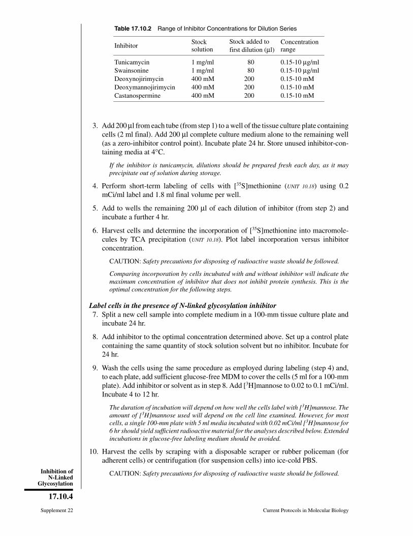
3. Add 200 µl from each tube (from step 1) to a well of the tissue culture plate containingcells (2 ml final). Add 200 µl complete culture medium alone to the remaining well(as a zero-inhibitor control point). Incubate plate 24 hr. Store unused inhibitor-con-taining media at 4°C.
If the inhibitor is tunicamycin, dilutions should be prepared fresh each day, as it mayprecipitate out of solution during storage.
4. Perform short-term labeling of cells with [35S]methionine (UNIT 10.18) using 0.2mCi/ml label and 1.8 ml final volume per well.
5. Add to wells the remaining 200 µl of each dilution of inhibitor (from step 2) andincubate a further 4 hr.
6. Harvest cells and determine the incorporation of [35S]methionine into macromole-cules by TCA precipitation (UNIT 10.18). Plot label incorporation versus inhibitorconcentration.
CAUTION: Safety precautions for disposing of radioactive waste should be followed.
Comparing incorporation by cells incubated with and without inhibitor will indicate themaximum concentration of inhibitor that does not inhibit protein synthesis. This is theoptimal concentration for the following steps.
Label cells in the presence of N-linked glycosylation inhibitor7. Split a new cell sample into complete medium in a 100-mm tissue culture plate and
incubate 24 hr.
8. Add inhibitor to the optimal concentration determined above. Set up a control platecontaining the same quantity of stock solution solvent but no inhibitor. Incubate for24 hr.
9. Wash the cells using the same procedure as employed during labeling (step 4) and,to each plate, add sufficient glucose-free MDM to cover the cells (5 ml for a 100-mmplate). Add inhibitor or solvent as in step 8. Add [3H]mannose to 0.02 to 0.1 mCi/ml.Incubate 4 to 12 hr.
The duration of incubation will depend on how well the cells label with [3H]mannose. Theamount of [3H]mannose used will depend on the cell line examined. However, for mostcells, a single 100-mm plate with 5 ml media incubated with 0.02 mCi/ml [3H]mannose for6 hr should yield sufficient radioactive material for the analyses described below. Extendedincubations in glucose-free labeling medium should be avoided.
10. Harvest the cells by scraping with a disposable scraper or rubber policeman (foradherent cells) or centrifugation (for suspension cells) into ice-cold PBS.
CAUTION: Safety precautions for disposing of radioactive waste should be followed.
Table 17.10.2 Range of Inhibitor Concentrations for Dilution Series
Inhibitor Stocksolution
Stock added to first dilution (µl)
Concentrationrange
Tunicamycin 1 mg/ml 80 0.15-10 µg/mlSwainsonine 1 mg/ml 80 0.15-10 µg/mlDeoxynojirimycin 400 mM 200 0.15-10 mMDeoxymannojirimycin 400 mM 200 0.15-10 mMCastanospermine 400 mM 200 0.15-10 mM
Supplement 22 Current Protocols in Molecular Biology
17.10.4
Inhibition ofN-Linked
Glycosylation

Measure incorporated macromolecular radioactivity11. If tunicamycin was used as the inhibitor, determine the amount of incorporated
macromolecular radioactivity by TCA precipitation and proceed to step 18. If anotherinhibitor was used, carry out steps 12 to 17.
The amount of radioactivity incorporated into macromolecules reflects the effect of theinhibitor(s) on oligosaccharide biosynthesis: the presence of inhibitor should block incor-poration of [3H]mannose into macromolecules.
12. Wash the cells twice by resuspending the pellet in ice-cold PBS, centrifuging, andremoving the supernatant. Resuspend the pellet in lysis buffer without bovinehemoglobin and lyse as described in UNIT 10.16 (steps 1 to 4, basic protocol).
13. Divide each sample into two equal aliquots in conical polypropylene centrifuge tubesand precipitate protein with acetone (support protocol).
Use 1.5-ml, 15-ml, or 50-ml centrifuge tubes depending on the quantity of sample (8 volacetone must be added).
14. Resuspend pellets in 20 to 40 µl endo H digestion buffer per 107 cells and boil 10min to inactivate endogenous hydrolases.
15. Add 5 µl endo H per 100 µl sample to one tube of each sample pair and incubate bothovernight at 37°C.
16. Add 1⁄10 vol of 20% (w/v) SDS to each sample and boil 3 to 5 min to terminate thedigestion.
17. Apply one sample at a time to a calibrated Sephacryl S-200 column equilibrated in25 mM ammonium formate/0.1% SDS, in a sample volume that is <5% of the bedvolume. Collect the eluate and use a scintillation counter to measure the radioactivityassociated with glycoproteins (eluting in the void volume of the column, Vo) and withfree oligosaccharides (eluting after Vo) for each of the four samples. Wash columnwith 2 to 4 vol 25 mM ammonium formate/0.1% SDS between sample applications.
In the absence of endo H digestion, all of the radioactivity from both the inhibitor-treatedand untreated samples should elute in Vo. Elution of any radioactive material after Vo mayindicate that the wash steps following acetone precipitation did not completely removelow-molecular-weight contaminants present in the cell lysate.
For samples digested with endo H, elution profiles should demonstrate a marked increasein the amount of endo H-releasable radioactivity in samples from inhibitor-treated cellsrelative to treated cells. For samples not treated with inhibitor, the oligosaccharides cleavedfrom the peptide backbone by endo H represent immature oligosaccharides still beingprocessed in the Golgi apparatus and a small population of endo H-sensitive structuresfound on some mature glycoproteins. For samples treated with inhibitor, a much higherpercentage of the total radioactivity should be released, as the inhibitors prevent theprocessing of the oligosaccharides into mature oligosaccharide structures (Fig. 17.10.1)and the underprocessed oligosaccharides contain more mannose residues than the proc-essed ones. Theoretically, 100% of the radioactivity should be releasable by endo Hdigestion of the inhibitor-treated samples (see critical parameters), although in reality noinhibition is ever complete.
To save time, analyze only the first and last tubes of the control cells in this fashion, i.e.,those not treated at all and those treated with the highest concentration of the inhibitorsolvent (from step 2 above). If there is no difference in the results from these two groups ofcells, it is reasonable to assume that those cells treated with lower concentrations of theinhibitor solvent will yield the same results. However, if the solvent is found to be toxic,then the cells exposed to lower concentrations will need to be examined to find themaximally tolerated concentration of inhibitor solvent.
Current Protocols in Molecular Biology Supplement 22
17.10.5
Preparationand Analysis ofGlycoconjugates
18. If desired, and if methods are available for purifying and identifying a specificglycoprotein from cells radiolabeled with [35S]methionine, examine the effect of agiven inhibitor by SDS-PAGE and autoradiography.
Each N-linked carbohydrate chain contributes ∼2000 to 4000 Da to a protein’s mass, sosynthesis of a protein in the presence of tunicamycin will result in faster mobility onSDS-PAGE. The effect of other inhibitors can be demonstrated by an increased sensitivityto digestion by endo H, which is also detectable as an increase in migration rate inSDS-PAGE.
SUPPORTPROTOCOL
ACETONE PRECIPITATION
Acetone precipitation is a useful step for concentrating proteins and for exchanging themfrom one buffer to another. As little as 10 ng of protein can be precipitated successfully.
Additional Materials
100% acetone (HPLC or ACS grade), −20°C1.5-ml, 15-ml, or 50-ml conical polypropylene or other centrifuge tubes
1. Place the protein solution on ice, add 8 vol acetone (−20°C), mix gently, andprecipitate at −20°C overnight.
If the sample volume is too large to be conveniently diluted with 8 vol, concentrate it bylyophilizing and resuspending in a smaller volume. If the sample contains NP-40 or TritonX-100 detergent, however, there is a limit to how much it can be concentrated, as highconcentrations of detergent (e.g., >1%) will partition out of acetone solutions, appearingas an oily pellet after centrifugation (step 2).
If a large amount of protein is present (i.e., a precipitate is readily visible after the acetoneis added), it can be precipitated after standing at −20°C for only a few hours instead ofovernight. Leaving the material in acetone for longer than 1 day should be avoided, as itmay become increasingly difficult to redissolve the protein.
2. Collect the precipitate by centrifuging 15 min at 3000 × g, 4°C (a small pellet shouldbe visible). Carefully invert the tube and pour out the acetone into a clean tube.
If the pellet is oily due to the presence of detergent, it should be extracted by adding 85%(v/v) acetone (−20°C), mixing, incubating at −20°C for >1 hr, and recentrifuging.
3. Centrifuge the tube containing the pellet briefly to concentrate the remaining acetoneand remove it with a micropipettor. Allow pellet to dry until moist but not powder-dry,and resuspend in an appropriate quantity of the desired solvent.
Care must be taken in drying the pellet, as great difficulty may be encountered inresuspending some proteins if the pellet dries completely. This insolubility depends on boththe individual protein and the total amount present.
REAGENTS AND SOLUTIONS
NOTE: Deionized, distilled water should be used for buffer and inhibitor solutions.
Endo H digestion buffer15 µl 0.5 M sodium citrate, pH 5.575 µl water5 µl 10% PMSF in isopropanolThis is sufficient for 5 µl of 0.5 U/ml endo H (100 µl digestion volume). The PMSFprevents proteolysis. The presence of nonionic detergent is not required. Preparejust before use; PMSF is unstable in water.
Supplement 22 Current Protocols in Molecular Biology
17.10.6
Inhibition ofN-Linked
Glycosylation
Inhibitor stock solutionsDissolve castanospermine (mol. wt. 189.2), 1-deoxymannojirimycin (mol. wt.199.6), or 1-deoxynojirimycin (mol. wt. 163.2) in water to 400 mM; dissolveswainsonine (mol. wt. 173.2) in water to 1 mg/ml. Sterilize by filtration through a0.2-µm filter. Store frozen at −20°C (stable indefinitely).Dissolve tunicamycin (mol. wt. 840) in dimethylsulfoxide (DMSO), dimethylfor-mamide (DMF), 95% ethanol, or 25 mM NaOH to 1 mg/ml. Store frozen at −20°C(stable ∼1 yr).
If NaOH is used, store stock solution in a small tube that is tightly capped to preventadsorption of carbon dioxide, which will lower the pH. Tunicamycin’s solubility in neutralaqueous solution is <1 mg/ml (attempting to make such a solution will produce a visibleprecipitate).
COMMENTARY
Background InformationAssembly of N-linked oligosaccharide
chains involves the sequential action of severalglycosidases and glycosyltransferases (re-viewed by Hubbard and Ivatt, 1981, and Kobataand Takasaki, 1992). Growth of cells in thepresence of inhibitors of these enzymes pro-duces glycoproteins with underprocessed ormissing oligosaccharide chains. Depending onthe protein, these changes may alter biologicalfunctions and/or rates of transport, secretion,or turnover (Elbein, 1987; McDowell andSchwarz, 1988).
N-linked glycosylation begins with the as-sembly of the lipid-linked precursor glu-cose3mannose9N-acetylglucosamine2-pyro-phosphate-dolichol (Glc3Man9GlcNAc2-PP-dolichol; similar to Structure A, Fig. 17.10.1).Assembly of this precursor requires N-acetyl-g lucosamine-pyrophosphate-dol ichol(GlcNAc-PP-dolichol), whose formation isblocked by tunicamycin (Tkacz and Lampen,1975). Thus, the presence of tunicamycin re-sults in proteins missing some or all of theirN-linked side chains; however, those that areadded due to incomplete inhibition are proc-essed normally. Tunicamycin may also inhibitprotein synthesis; not all proteins will be af-fected to the same degree. Tunicamycin alsoblocks the assembly of type II keratan sulfatechains, because these glycans also utilizeGlcNAc-PP-dolichol (Hart and Lennarz,1978). Glycolipid biosynthesis may also beinhibited, although the mechanism underlyingthis has not been established (Yusuf et al., 1983;Guarnaccia et al., 1987).
The glycosidase inhibitors castanosper-mine, deoxynojirimycin, deoxymannojirimy-cin, and swainsonine are considerably lesstoxic than tunicamycin. Castanospermine in-hibits glucosidase I, and deoxynojirimycin in-
hibits both glucosidases I and II (Fig. 17.10.1;Pan et al., 1983; Saunier et al., 1982). In somestudies, the metabolic block produced by thesetwo drugs is not complete, yielding a mixtureof normally processed and underprocessedstructures. More recently, an endo-α-D-man-nosidase has been described that cleaves thetetrasaccharide Glc3-Man1 from Glc3Man9-
GlcNAc2, effectively bypassing the block pro-duced by the glucosidase inhibitors (Lubas andSpiro, 1987). Although the question has notbeen examined fully, the level of this endo-α-D-mannosidase may vary between different celllines.
Deoxymannojirimycin and swainsonine areinhibitors of mannosidases I and II, respec-tively (Fig. 17.10.1; Fuhrmann et al., 1984;Tulsiani et al., 1982). By inhibiting mannosi-dase I, deoxymannojirimycin prevents additionof the α1-2 N-acetylglucosamine (GlcNAc)residue required to produce Structure D, Figure17.10.1, which is a necessary step for the actionof mannosidase II. Thus, deoxymannojirimy-cin prevents addition of any GlcNAc residues,blocking galactosylation and sialylation aswell. Swainsonine blocks mannosidase II.However, it does not prevent the single GlcNAcresidue (Structure D, Fig. 17.10.1) from beinggalactosylated and sialylated, resulting in hy-brid structures (Tulsiani and Touster, 1983).
Some of the glycosidase inhibitors are alsoactive against lysosomal enzymes, although thesignificance of this effect in tissue culture cellshas been little explored. Deoxynojirimycin caninhibit synthesis of the lipid-linked precursorin cell-free extracts (Romero et al., 1985); thisalso has not been thoroughly investigated withintact cells. No information is available on themetabolism or half-lives of these inhibitors incultured cells.
Current Protocols in Molecular Biology Supplement 22
17.10.7
Preparationand Analysis ofGlycoconjugates
Critical Parameters andTroubleshooting
The two most critical variables are durationof exposure and inhibitor concentration. Thetreatment times suggested in this unit (24 hr)will permit a cell to replace most of its endo-genous glycoproteins with glycoproteins syn-thesized in the presence of inhibitor. For pro-teins that turn over slower or faster than aver-age, longer or shorter incubation times may beappropriate. If pulse-chase studies with radio-labeled sugar or amino acid precursors (UNITS
17.4 & 10.18) are being carried out, it is necessaryto incubate cells in inhibitor for only 1 to 2 hrbefore adding label.
N-linked glycosylation inhibitors may betoxic to the cells, as indicated by decreased cellviability and/or reduced protein synthesis. Theconcentration of tunicamycin that can be usedis generally limited by its toxicity, which maybe considerable in some cell lines (more so withtumor cells than nontransformed cells). Otherglycosidase inhibitors are usually not toxic inthe concentration ranges suggested in Table17.10.1 (the highest concentrations listed rep-resent those used in most published studies).
It is important to realize that in a given cellline, not all glycoproteins will be affected to thesame extent by a given inhibitor. Moreover, itis possible that not all sites on the same glyco-protein will be affected equally. These caveatsshould be kept in mind when interpreting theresults from studies employing these inhibitors.
Failure to observe an effect from an inhibitormay indicate that concentration of inhibitor wastoo low and/or length of exposure was too short.Cell lines differ in their susceptibility to thesecompounds.
Safety precautions and experimental para-meters for labeling—including labeling time,label concentration and specific activity, celldensity, and culture conditions—are discussedin the critical parameters of UNIT 10.18.
Anticipated ResultsThese experiments will demonstrate the
chosen inhibitor’s toxicity, as assessed by pro-tein synthesis, and its ability to alter oligosac-charide biosynthesis, as assessed by total[3H]mannose incorporation (for tunicamycin)or endo H release of the oligosaccharides (forthe other inhibitors listed in Table 17.10.1).
Time ConsiderationsSetting up the initial round of cell cultures
for determining the optimal inhibitor concen-tration takes 2 hr, after which they are incubated24 hr. On the second day, the dilution series ofmedium containing inhibitor is set up, the cellsare transferred to these media (<1 hr), and theplates incubated 24 hr. On the third day, theincubation of the cells with the radiolabeledprecursor is set up (∼4 hr); the actual incubationtakes 4 to 16 hours. Following incubation, thecells are assayed for radiolabel incorporationby TCA precipitation (2 to 4 hr).
The main part of the experiment follows asimilar time course if the inhibitor used istunicamycin. If a different inhibitor is used,analysis of endo H susceptibility by SephacrylS-200 chromatography will take ∼1 day, in-cluding the overnight incubation.
Literature CitedElbein, A.D. 1987. Inhibitors of the biosynthesis and
processing of N-linked oligosaccharide chains.Annu. Rev. Biochem. 56:497-534.
Fuhrmann, U., Bause, E., Legler, G., and Ploegh, H.1984. Novel mannosidase inhibitor blockingconversion of high mannose to complex oli-gosaccharides. Nature (Lond.) 307:755-758.
Guarnaccia, S.P., Shaper, J.H., and Schnaar, R.L.1987. Tunicamycin inhibits ganglioside biosyn-thesis in neuronal cells. Proc. Natl. Acad. Sci.U.S.A. 80:1551-1555.
Hart, G.W. and Lennarz, W.J. 1978. Effects of tuni-camycin on the biosynthesis of glycosaminogly-cans by embryonic chick cornea. J. Biol. Chem.253:5795-5801.
Hubbard, S.C. and Ivatt, R.J. 1981. Synthesis andprocessing of asparagine-linked oligosaccha-rides. Annu. Rev. Biochem. 50:555-583.
Kobata, A. and Takasaki, S. 1992. Structure andbiosynthesis of cell surface carbohydrates. InCell Surface Carbohydrates and Cell Develop-ment (M. Fukuda, ed.) pp. 1-24. CRC Press,Boca Raton, Fla.
Lubas, W.A. and Spiro, R.G. 1987. Golgi endo-α-D-mannosidase from rat liver, a novel N-linkedcarbohydrate unit processing enzyme. J. Biol.Chem. 262:3775-3781.
McDowell, W. and Schwarz, R.T. 1988. Dissectingglycoprotein biosynthesis by the use of specificinhibitors. Biochimie (Paris) 70:1535-1549.
Pan, Y.T., Hori, H., Saul, R., Sanford, B.A., Moly-neux, R.J., and Elbein, A.D., 1983. Castanosper-mine inhibits the processing of the oligosaccha-ride protion of the influenza viral hemagglutinin.Biochemistry 22:3975-3984.
Supplement 22 Current Protocols in Molecular Biology
17.10.8
Inhibition ofN-Linked
Glycosylation

Romero, P., Friedlander, P., and Herscovics, A.1985. Deoxynojirimycin inhibits the formationof Glc3Man9GlcNAc2-PP-dolichol in intestinalepithelial cells in cultures. FEBS Lett. 183:29-32.
Saunier, B., Kilker, R.D., Tkacz, J.S., Quaroni, A.,and Herscovics, A. 1982. Inhibition of N-linkedcomplex oligosaccharide formation by 1-deoxy-nojirimycin, an inhibitor of processing glucosi-dases. J. Biol. Chem. 257:14155-14161.
Tkacz, J.S. and Lampen, J.O. 1975. Tunicamycininhibition of polyisoprenyl N-acetylglucos-aminyl pyrophosphate formation in calf livermicrosomes. Biochem. Biophys. Res. Commun.65:248-55.
Tulsiani, D.R.P., Harris, T.M., and Touster, O. 1982.Swainsonine inhibits the biosynthesis of com-plex glycoproteins by inhibition of Golgi man-nosidase II. J. Biol. Chem. 257:7936-7939.
Tulsiani, D.R.P. and Touster, O. 1983. Swainsoninecauses the production of hybrid glycoproteins byhuman skin fibroblasts and rat liver Golgi prepa-rations. J. Biol. Chem. 258:7578-7585.
Yusuf, H.K.M., Pohlentz, G., Schwarzmann, G., andSandhoff, K. 1983. Ganglioside biosynthesis inGolgi apparatus of rat liver. Eur. J. Biochem.134:47-54.
Key ReferencesElbein, A.D. 1987. See above.
Useful general review.
McDowell, W. and Schwarz, R.T. 1988. See above.
Useful general review.
Hubbard, S.C. and Ivatt, R.J. 1981. See above.
Good review on early work on the processing path-way.
Kobata, A. and Takasaki, S. 1992. See above.
Good review of recent work on some of the complexi-ties in oligosaccharide processing.
Contributed by Leland D. PowellUniversity of California San DiegoLa Jolla, California
Current Protocols in Molecular Biology Supplement 32
17.10.9
Preparationand Analysis ofGlycoconjugates
UNIT 17.10B Inhibition of Glycolipid BiosynthesisBASIC
PROTOCOLAdequate inhibition of glycolipid biosynthesis allows the study of their biologicalfunctions. The method presented in this unit employs a synthetic analog of ceramide,PDMP (1-phenyl-2-decanoylamino-3-morpholino-1-propanol), that inhibits glycolipidbiosynthesis in cultured cells. Optimum conditions for inhibition of glycolipid biosyn-thesis are determined, glycolipids extracted from cultures grown with and withoutinhibitor, and the patterns of glycolipids analyzed by HPTLC (UNIT 9.7A). Detection isachieved using colorimetric reactions, or by monitoring radioactivity when cells havebeen metabolically radiolabeled. The effects of these changes in biological functions canthen be studied later.
Materials
Adherent cell culture10 mM PDMP in ethanol (see recipe)Culture medium appropriate for cell line, with or without serumEthanolRadiolabeled precursors (UNIT 17.4; for metabolic labeling)PBS (APPENDIX 2)2:1, 1:1, and 1:2 (v/v) chloroform/methanol10:10:1 (v/v/v) chloroform/methanol/waterHPTLC standards: mixture of gangliosides or neutral lipids from bovine brain
(Sigma or Accurate Chemical) or mixture of neutral glycolipids (AccurateChemical)
Orcinol/sulfuric acid reagent (see recipe)Resorcinol/hydrochloric acid reagent (see recipe)
Screw-cap glass tube, sterileConical glass tubes (10- or 50-ml, depending on volume of pellet to be extracted)Sonicator bathNitrogen streamRotary or shaker evaporatorSilica-gel HPTLC platesEnhance spray (DuPont NEN; for metabolic labeling)Glass spray unitOven adjustable up to 140°C
Additional reagents and equipment for HPTLC (UNIT 9.7A) and for metabolicradiolabeling (UNIT 17.4) and autoradiography (APPENDIX 3A; both optional)
NOTE: All incubations should be carried out under the conditions normally used for thecell line to be analyzed.
Incubate cells with inhibitor1. Prepare cell cultures and maintain as usual until subconfluent. Set up sufficient flasks
or plates for the set of experiments using different POMP concentrations andincubation times (see steps 4 and 5).
This procedure can be performed in either the presence or absence of fetal bovine serum,and with adherent cells or (with appropriate modifications) suspension cultures.
2. Allow 10 mM PDMP in ethanol to warm up to room temperature. Transfer to a sterilescrew-cap glass tube a sufficient amount of this solution to set up a series ofincubations with 5 to 25 µM final concentration of PDMP and different time points(see steps 4 and 5). Evaporate to dryness with a nitrogen stream.
Contributed by Adriana ManziCurrent Protocols in Molecular Biology (1995) 17.10.10-17.10.14Copyright © 2000 by John Wiley & Sons, Inc.Supplement 32
17.10.10
Inhibition ofGlycolipid
Biosynthesis
3. Add culture medium and sonicate 10 min in a sonicator bath to suspend the drug.
4. Add PDMP solution to aliquots of the cell culture to obtain final concentrations of5, 10, 15, 20, and 25 µM. Add ethanol as needed to achieve the same ethanolconcentration in all samples, and add the same total volume of ethanol without PDMPto a control culture.
5. Incubate the cells for selected durations.
Periods ranging from 10 hr to 4 days have been employed. Optimum inhibition times maydiffer from one particular cell line to another. It is necessary to check the cells for viabilityas well as for morphological changes, particularly after prolonged incubations with theinhibitor.
6. Remove medium and add fresh medium containing PDMP as before. If the cells arebeing metabolically labeled, add the radiolabeled precursor at this point (see UNIT
17.4).
Prepare total lipid extract7. Harvest cells into a 10- or 50-ml conical glass tube, pellet the cells, and wash three
times with PBS.
8. Add 10 vol of 2:1 (v/v) chloroform/methanol to the cell pellet. Extract the lipids byhomogenizing or sonicating 5 min. Centrifuge 10 min at ∼350 × g, 4°C. Transfer thesupernatant (extract) to a clean glass tube using a Pasteur pipet.
9. Repeat the extraction as in step 8 with, successively, 1:1 (v/v) chloroform/methanol,1:2 (v/v) chloroform/methanol, and 10:10:1 (v/v/v) chloroform/methanol/water.
10. Pool all extracts and dry them under reduced pressure, using a rotary or shakerevaporator or a nitrogen stream depending on the volume of extract to be dried.
Analyze extract by HPTLC and (optionally) autoradiography11. Dissolve the pooled dry extract (total lipid extract) in a small volume of 2:1 (v/v)
chloroform/methanol and spot one aliquot on silica-gel HPTLC plates (UNIT 9.7A).Spot a mixture of ganglioside and/or neutral glycolipid standards on one side of theplate. Perform HPTLC analysis. Allow the plates to dry completely after developing.
The amount of total lipid required for positive resulting in observable bands (∼1nmol/band) will vary between cell lines because of different compositions and ratios ofcomponents. It may be necessary to do several trials until adequate results are obtained.When analyzing radiolabeled extracts, it is advisable to spot the same number of totalcounts in each lane.
When analyzing nonradiolabeled glycolipids, and if sufficient material is available, twodifferent HPTLC plates can be spotted in parallel with the same total lipid extracts. Stainingone with orcinol/sulfuric acid and the other with resorcinol/hydrochloric acid will allowdetermination of which bands correspond to neutral glycolipids and which to gangliosides.
12. When analyzing nonradiolabeled glycolipids, proceed directly to step 13. Whenanalyzing metabolically radiolabeled glycolipids, visualize them by autoradiography(APPENDIX 3A) using Enhance scintillant spray, then proceed to step 13 to visualize thenonradiolabeled standards.
13a. Using a glass spray unit, spray plates with orcinol/sulfuric acid reagent. Heat in a100°C oven until bands are observed (∼10 min, but check the plate often to avoidoverstaining).
This step detects all glycolipids, which give brown bands.
Current Protocols in Molecular Biology Supplement 32
17.10.11
Preparation andAnalysis ofGlycoconjugates
In steps 13a and 13b, it is possible to stain only the side of the plate containing the standardsby covering the rest of the plate with a piece of glass held in position with binder clips.
13b. Alternatively, spray the plate with resorcinol/hydrochloric acid reagent and heat 15min in a 140°C oven.
This step detects only gangliosides, which give blue bands.
14. Compare the pattern of glycolipids obtained for the control cells with those obtainedwith different concentrations of inhibitor after different incubation periods.
15. If cells were metabolically radiolabeled, quantitate the individual components byscraping off the corresponding areas of silica gel and measuring radioactivity byliquid scintillation counting. If a scanning densitometer is available, quantitationcan be achieved by scanning the autoradiograph or the stained glass plate.
REAGENTS AND SOLUTIONS
Use deionized, distilled water in all recipes and protocol steps. For common stock solutions, seeAPPENDIX 2; for suppliers, see APPENDIX 4.
PDMP stock solution, 10 mM in ethanol
Dissolve 3.86 mg d,l-threo-1-phenyl-2-decanoylamino-3-morpholino-1-propan-ol⋅HCl (PDMP; FW 390.6; available from Matreya) in 1.0 ml ethanol. Store at −20°Cuntil used.
PDMP is quite stable in aqueous solvents. No breakdown was observed after incubating a1 mM solution in PBS (pH 7.4) for 10 days at 41°C. Stability in ethanol is even higher.
Orcinol/sulfuric acid reagent
Dissolve 200 mg orcinol in 33 ml water. Dilute to 100 ml with concentrated sulfuricacid. Store in dark bottle at 4°C. Discard when the solution turns brownish (8 to 10days).
Orcinol from Sigma (FW 142.2) is suitable for making this reagent.
Resorcinol/hydrochloric acid reagent80 ml concentrated HCl10 ml 2% resorcinol (1,3-benzenediol) in H2O250 µl 0.1 M copper sulfate in H2O (2.5 mM final concentration)Store in a dark bottle at 4°CDiscard if the solution turns brown
Resorcinol from Sigma (crystalline; ∼98% pure; FW 110.1) is suitable for making thisreagent.
COMMENTARY
Background InformationMany studies have shown that glycosphin-
golipids and gangliosides are involved in a greatvariety of biological phenomena. As constitu-ents of mammalian cell membranes, gan-gliosides significantly affect cell-surface andtransmembrane signalling events such as cellu-lar immune responses, cell growth, and differ-entiation. Other observed effects of gan-gliosides include those of “shed” gangliosidesin malignancy; the effects of gangliosides andtheir products on protein kinase C; calcium
binding; and their interaction with variousbioeffectors, including bacterial toxins, bacte-rial binding proteins, interferons, serotonin, fi-bronectin, plasma low-density lipoproteins,lectins, and glycoprotein hormones.
Many studies of the role of these moleculeshave been carried out by adding specific exoge-nous glycosphingolipids or gangliosides, ormixtures thereof, to cultured cells. Interpreta-tion of these studies is difficult for variousreasons. True incorporation of the added mole-cules into the cell membrane is hard to prove,
Supplement 32 Current Protocols in Molecular Biology
17.10.12
Inhibition ofGlycolipid
Biosynthesis
and the possibility of abnormal effects causedby micelles associated with the cell is hard torule out. Also, exogenous material may dis-place the controlling factor from its normallocation in the cell membrane or influence itsmetabolism.
Another approach to clarifying the biologi-cal roles of glycosphingolipids and gan-gliosides is the use of enzyme inhibitors tostudy the effects of their depletion in culturedcells and animals. This inhibition can beachieved using a synthetic analog of ceramide,PDMP. This method was developed by Radinand Inokuchi, whose idea that cancer resultsfrom microsphingolipidosis led them to ex-plore ways to change the biological processesin which glycolipids are involved. One suchprocess is the synthesis of glycolipids fromnonglycolipid precursors and lower gly-colipids. Most neutral glycolipids and gan-gliosides have a common precursor, GlcCer,which is formed enzymatically from ceramideand UDP-Glc. The synthetase responsible forthis transfer is UDP-glucose:N-acylsphingos-ine glycosyltransferase (EC 2.4.1.80). Becauserapid liver growth was observed in mice whenglucosylceramide accumulated (Datta andRadin, 1986), the authors explored the possi-bility that inhibiting the synthesis of the com-mon precursor GlcCer would decrease theamount of glycolipids due to normal turnover(Hospattankar and Radin, 1982). After a fewattempts, they obtained an active inhibitor,PDMP (D-threo-1-phenyl-2-decanoylamino-3-morpholino-1-propanol; Inokuchi and Radin,1987). This compound was shown to inhibitganglioside biosynthesis effectively in intactcells, without any major toxic effects (Inokuchiand Radin, 1987; Inokuchi et al., 1987). Theeffects of the enzyme inhibitor were counter-acted by including GlcCer in the culture me-dium (Inokuchi et al., 1989). For a review of theavailable data, see Radin and Inokuchi (1991).
Critical ParametersIt is critical to understand that very little is
known about the regulation of glycosphing-olipid biosynthesis, turnover, or even whichsteps are regulation points. On the other hand,the composition of glycosphingolipids variestremendously between different types of cells.The time required to obtain optimum inhibitionof any glycolipid or collection of glycolipidsdepends on how completely the enzyme isinhibited, the activity of the specific hydrolasesinvolved, the susceptibility of each glycolipidto attack by its hydrolase, and the rates of the
synthases that recycle smaller glycolipids intolarger ones. Although the effects on simplegangliosides such as GM3 are readily observed,complex molecules such as GD1b require moretime to become affected. Complete inhibition,as well as complete recovery from the inhibitor,should be expected to take at least 24 hr.
In consequence, the conditions required forinhibition and recovery of each particular sys-tem need to be carefully established. Variousconcentrations of inhibitor should be tried toestablish the minimum amount required forinhibition. It is also important to understandthat an inhibitor may have pleiotropic effects,including the accumulation of precursors (e.g.,ceramide), expansion of alternate pathways(e.g., sphingomyelin synthesis), and other non-specific effects (e.g., on protein synthesis). De-pending on the experiment, such alternate ex-planations for the observed effects may need tobe explored.
Anticipated ResultsThe protocol allows optimization of the con-
ditions for maximum inhibition of glycolipidbiosynthesis with minimum effects on the mor-phology and viability of cells. Once these con-ditions are established, different studies of thebiological roles of glycosphingolipids and gan-gliosides can be performed.
Time ConsiderationsComplete inhibition of glycolipid biosyn-
thesis may require up to 4 days of cell culture.Further extraction of glycolipids and analysisof the resulting data are expected to take amaximum of 2 days, including quantitation.When metabolic labeling and autoradiographyare included, extra time is required for detec-tion, and will depend on the total counts avail-able and their distribution among components.
Literature CitedDatta, S.C. and Radin, N.S. 1986. Glucosylceramide
and level of glucosidase-stimulating proteins.Lipids 21:702-709.
Hospattankar, A.V. and Radin, N.S. 1982. Changesin liver lipids after administration of 2-decanoyl-amino-3-morpholinopropiophenone and chlor-promazine. Lipids 17:538-543.
Inokuchi, J.I. and Radin, N.S. 1987. Preparation ofthe active isomer of 1-phenyl-2-decanoylamino-3-morpholino-1-propanol, inhibitor of murineglucocerebroside synthetase. J. Lipid Res.28:565-571.
Inokuchi, J.I., Mason, I., and Radin, N.S. 1987.Antitumor activity via inhibition of glycosphing-olipid biosynthesis. Cancer Ltrs. 38:23-30.
Current Protocols in Molecular Biology Supplement 32
17.10.13
Preparation andAnalysis ofGlycoconjugates

Inokuchi, J., Momosaki, K., Shimeno, H., Naga-matsu, A., and Radin N.S. 1989. Effects of D-threo-PDMP, an inhibitor of glucosylceramidesynthetase, on expression of cell surface gly-colipid antigen and binding to adhesive proteinsby B16 melanoma cells. J. Cell. Physiol.141:573-583.
Radin, N.S. and Inokuchi, J. 1991. Use of PDMP forthe study of glycosphingolipid functions. TrendsGlycos. Glycotech. 3:200-213.
Key ReferenceRadin and Inokuchi, 1991. See above.
Excellent review, providing very useful referencesfor comparing conditions used for different systemsalong with specific protocols for studying the differ-ent biological roles affected by inhibition of gly-colipid biosynthesis.
Contributed by Adriana E. ManziUniversity of California San DiegoLa Jolla, California
Supplement 32 Current Protocols in Molecular Biology
17.10.14
Inhibition ofGlycolipid
Biosynthesis
UNIT 17.11Synthetic Glycosides as Primers ofOligosaccharide Biosynthesis and Inhibitorsof Glycoprotein and Proteoglycan Assembly
With the exception of hyaluronic acid, all mammalian saccharides assemble whileattached to a lipid or protein primer. Several cases are now known in which oligosaccha-ride synthesis will occur on synthetic glycoside primers added to cells. β-D-xylosidesinitiate glycosaminoglycan (GAG) synthesis (Okayama et al., 1973) by substituting forendogenous xylosylated core proteins (Basic Protocol 1). At high concentration xylosideswill also prime oligosaccharides that resemble glycolipids (Freeze et al., 1993). N-ace-tyl-α-D-galactosaminides initiate the synthesis of O-linked oligosaccharides found onmucins and other glycoproteins (Basic Protocol 2) in an analogous manner. Evendisaccharides, such as peracetylated N-acetyllactosaminide, can act as primers (Sarkar etal., 1995). Because these primers compete with endogenous substrates, they also act asinhibitors of proteoglycan (PG) and glycoprotein synthesis. Thus, primers have utility forstudying the biological activity of glycoconjugates in cells, tissues, and animals. This unitdescribes procedures for using glycoside primers in cell culture.
BASICPROTOCOL 1
XYLOSIDE INITIATION OF GAG SYNTHESIS AND INHIBITION OFPG ASSEMBLY
Materials
0.2 M p-nitrophenyl-β-D-xyloside in DMSO (see recipe)Tissue culture growth medium appropriate for cell lineCultured cells, in suspension or adherentDimethylsulfoxide (DMSO, C2H6SO; mol. wt. 78.13)Radioactive precursors: H2
35SO4, [6-3H]GlcNH2, or [1-3H]Gal
Additional reagents and equipment for metabolic radiolabeling (UNIT 17.4), isolatingproteoglycans and glycosaminoglycans (UNIT 17.3), and gel filtration (UNIT 17.20)and reversed-phase chromatography (UNIT 17.21)
NOTE: All incubations should be carried out in a humidified 37°C, 5% CO2 incubatorunless otherwise specified.
1. Add 0.2 M p-nitrophenyl-β-D-xyloside in DMSO to aliquots of growth medium toachieve final concentrations of 0, 0.01, 0.03, 0.1, 0.3, and 1 mM. Add DMSO asneeded to achieve the same concentration of the solvent in all samples.
Alternatively, dissolve enough inhibitor directly in growth medium to achieve a finalconcentration of 10 mM, then prepare the indicated serial dilution series with fresh growthmedium.
DMSO can cause some cells to differentiate, which can change the pattern of glycosylation.
2. Add each supplemented growth medium to a previously established cell culture byreplacing the spent growth medium with the supplemented medium. Incubate 1 hr.
Do not add concentrated stocks of glycosides directly to cell cultures, as high localconcentrations of solvent and glycoside can kill cells.
3. Add radioactive precursors to each culture (generally 10 µCi/ml 35SO42−, 20 µCi/ml
[6-3H]GlcNH2, or 50 µCi/ml [1-3H]Gal should be sufficient; see UNIT 17.4). Continueto incubate cells ≥1 hr.
Supplement 32
Contributed by Jeffrey D. Esko and Rebecca I. MontgomeryCurrent Protocols in Molecular Biology (1995) 17.11.1-17.11.6Copyright © 2000 by John Wiley & Sons, Inc.
17.11.1
Preparation andAnalysis ofGlycoconjugates

4. Isolate the PGs and GAGs made by the cells and the oligosaccharides primed on theglycoside from the growth medium (UNIT 17.3).
5. Analyze the material by gel filtration and reversed-phase chromatography (see UNITS
17.20 & 17.21).
Oligosaccharides generated on primers are generally smaller than glycoproteins andproteoglycans.
BASICPROTOCOL 2
N-ACETYL-α-D-GALACTOSAMINIDE INITIATION OFOLIGOSACCHARIDE SYNTHESIS AND INHIBITION OFO-LINKED GLYCOPROTEIN ASSEMBLY
Additional Materials (also see Basic Protocol 1)
0.2 M p-nitrophenyl-N-acetyl-α-D-galactosaminide in DMSO (see recipe)
NOTE: All incubations should be carried out in a humidified 37°C, 5% CO2 incubatorunless otherwise specified.
1. Add 0.2 M p-nitrophenyl-N-acetyl-α-D-galactosaminide in DMSO to aliquots ofgrowth medium to achieve final concentrations of 0, 1, 2, 5, and 10 mM. Add DMSOas needed to achieve the same concentration of the solvent in all samples.
Alternatively, dissolve enough inhibitor directly in growth medium to achieve a finalconcentration of 10 mM, then prepare the indicated serial dilution series with fresh growthmedium.
2. Add each supplemented growth medium to a previously established cell culture.Incubate 1 hr.
Do not add concentrated stocks of glycosides directly to cell cultures, as high localconcentrations of solvent and glycoside can kill the cells.
3. Add radioactive precursors to cultures (generally 20 µCi/ml [6-3H]GlcNH2 or 50µCi/ml [1-3H]Gal should be sufficient; see UNIT 17.4). Incubate cells a further 3 hr.
4. Harvest the glycoproteins from the cells and growth medium (UNIT 17.3).
5. Analyze the material by gel filtration and reversed-phase chromatography (UNITS 17.20
& 17.21). Small oligosaccharides linked to hydrophobic aglycones will bind to C18reversed-phase resins, aiding the separation of primed material from endogenousglycoproteins.
REAGENTS AND SOLUTIONS
Use deionized, distilled water in all recipes and protocol steps. For common stock solutions, seeAPPENDIX 2; for suppliers, see APPENDIX 4.
p-nitrophenyl-N-acetyl-α-D-galactosaminide, 0.2 M in DMSODissolve 68.5 mg p-nitrophenyl-N-acetyl-α-D-galactosaminide (C14H18N2O8; mol.wt. 342.3) in 1 ml dimethylsulfoxide. Warm if necessary to dissolve.
p-nitrophenyl-β-D-xyloside, 0.2 M in DMSODissolve 54.2 mg p-nitrophenyl-β-D-xyloside (C11H13NO7; mol. wt. 271.2) in 1 mldimethylsulfoxide. Warm if necessary to dissolve.
Other β-D-xylosides are also commercially available, including the fluorescent compoundsnaphthol-β-D-xyloside and 4-methylumbelliferyl-β-D-xyloside. Using these latter com-pounds, the fluorescence of the aglycone can be used to monitor the recovery of oligosac-charides primed on the glycoside.
Supplement 32 Current Protocols in Molecular Biology
17.11.2
SyntheticGlycosides as
Primers ofOligosaccharide
Biosynthesis
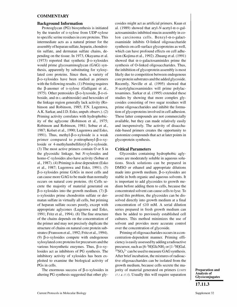
COMMENTARY
Background InformationProteoglycan (PG) biosynthesis is initiated
by the transfer of D-xylose from UDP-xyloseto specific serine residues in core proteins. Thisintermediate acts as a natural primer for theassembly of heparan sulfate, heparin, chondroi-tin sulfate, and dermatan sulfate chains, de-pending on the tissue. In 1973, Okayama et al.(1973) reported that synthetic β-D-xylosideswould prime glycosaminoglycan (GAG) syn-thesis, apparently by substituting for xylosy-lated core proteins. Since then, a variety ofβ-D-xylosides have been studied as primerswith the following results. (1) Priming requiresthe β-anomer of D-xylose (Galligani et al.,1975). Other pentosides (β-D-lyxoside, β-D-ri-boside, and α-L-arabinoside) and hexosides ofthe linkage region generally lack activity (Ro-binson and Robinson, 1985; F.N. Lugemwa,A.K. Sarkar, and J.D. Esko, unpub. observ.). (2)Priming activity correlates with hydrophobic-ity of the aglycone (Robinson et al., 1975;Robinson and Robinson, 1981; Sobue et al.,1987; Kolset et al., 1990; Lugemwa and Esko,1991). Thus, methyl-β-D-xyloside is a weakprimer compared to p-nitrophenyl-β-D-xy-loside or 4-methylumbelliferyl-β-D-xyloside.(3) The most active primers contain O or S inthe glycosidic linkage, but N-xylosides andhomo-C-xylosides also have activity (Sobue etal., 1987). (4) Priming is dose dependent (Eskoet al., 1987; Lugemwa and Esko, 1991). (5)β-D-xylosides prime GAGs in most cells andcan cause more GAG to be made than normallyoccurs on natural core proteins. (6) Cells se-crete the majority of material generated onβ-D-xylosides into the growth medium. (7) β-D-xylosides prime chondroitin sulfate or der-matan sulfate in virtually all cells, but primingof heparan sulfate occurs poorly, except withappropriate aglycones (Lugemwa and Esko,1991; Fritz et al., 1994). (8) The fine structureof the chains depends on the concentration ofthe primer and may not precisely duplicate thestructure of chains on natural core protein sub-strates (Fransson et al., 1992; Fritz et al., 1994).(9) β-D-xylosides compete with endogenousxylosylated core proteins for precursors and thevarious biosynthetic enzymes. Thus, β-D-xy-losides act as inhibitors of PG synthesis. Theinhibitory activity of xylosides has been ex-ploited to examine the biological activity ofPGs in cells.
The enormous success of β-D-xylosides inaltering PG synthesis suggested that other gly-
cosides might act as artificial primers. Kuan etal. (1989) showed that aryl-N-acetyl-α-D-gal-actosaminides inhibited mucin assembly in co-lon carcinoma cells. Benzyl-α-D-galact-osaminide inhibits O-linked oligosaccharidesynthesis on cell-surface glycoproteins as well,which can have profound effects on cell adhe-sion (Kojima et al., 1992). Zhuang et al. (1991)showed that α-D-galactosaminides prime thesynthesis of O-linked oligosaccharides. Thus,the inhibition of glycoprotein assembly is mostlikely due to competition between endogenouscore protein substrates and the added glycoside.Recently, Neville et al. (1995) showed thatN-acetylglucosaminides will prime polylac-tosamines. Sarkar et al. (1995) extended thesestudies by showing that more complex gly-cosides consisting of two sugar residues willprime oligosaccharides and inhibit the forma-tion of glycoproteins involved in cell adhesion.These latter compounds are not commerciallyavailable, but they can made relatively easilyand inexpensively. The activity of disaccha-ride-based primers creates the opportunity tocustomize compounds that act at later points inglycoprotein synthesis.
Critical ParametersGlycosides containing hydrophobic agly-
cones are moderately soluble in aqueous solu-tions. Stock solutions can be prepared inDMSO or ethanol and appropriate dilutionsmade into growth medium. β-D-xylosides arestable in both organic and aqueous solvents. Itis important to add glycosides to growth me-dium before adding them to cells, because theconcentrated solvent can cause cells to lyse. Toavoid this problem, the glycosides can be dis-solved directly into growth medium at a finalconcentration of ≤10 mM. A serial dilutionseries prepared in fresh growth medium canthen be added to previously established cellcultures. This method minimizes the use ofsolvent and provides more accurate controlover the concentration of glycoside.
Priming of oligosaccharides occurs in a con-centration-dependent manner. Priming effi-ciency is easily assessed by adding a radioactiveprecursor, such as [6-3H]GlcNH2 or [1-3H]Gal.35SO4
2− can be used to measure GAG synthesis.After brief incubation, the mixtures of radioac-tive oligosaccharides can be isolated from thegrowth medium, because cells secrete the ma-jority of material generated on primers (UNITS
17.1 & 17.3). Usually this will require separation
Current Protocols in Molecular Biology Supplement 32
17.11.3
Preparation andAnalysis ofGlycoconjugates
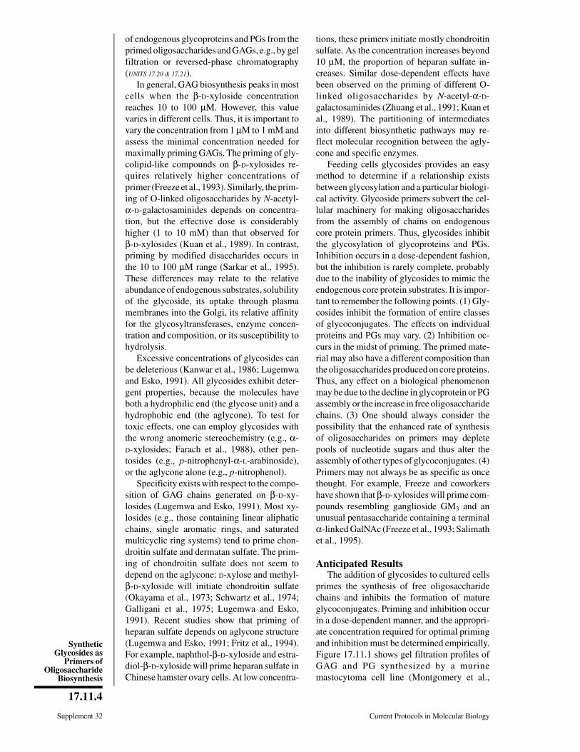
of endogenous glycoproteins and PGs from theprimed oligosaccharides and GAGs, e.g., by gelfiltration or reversed-phase chromatography(UNITS 17.20 & 17.21).
In general, GAG biosynthesis peaks in mostcells when the β-D-xyloside concentrationreaches 10 to 100 µM. However, this valuevaries in different cells. Thus, it is important tovary the concentration from 1 µM to 1 mM andassess the minimal concentration needed formaximally priming GAGs. The priming of gly-colipid-like compounds on β-D-xylosides re-quires relatively higher concentrations ofprimer (Freeze et al., 1993). Similarly, the prim-ing of O-linked oligosaccharides by N-acetyl-α-D-galactosaminides depends on concentra-tion, but the effective dose is considerablyhigher (1 to 10 mM) than that observed forβ-D-xylosides (Kuan et al., 1989). In contrast,priming by modified disaccharides occurs inthe 10 to 100 µM range (Sarkar et al., 1995).These differences may relate to the relativeabundance of endogenous substrates, solubilityof the glycoside, its uptake through plasmamembranes into the Golgi, its relative affinityfor the glycosyltransferases, enzyme concen-tration and composition, or its susceptibility tohydrolysis.
Excessive concentrations of glycosides canbe deleterious (Kanwar et al., 1986; Lugemwaand Esko, 1991). All glycosides exhibit deter-gent properties, because the molecules haveboth a hydrophilic end (the glycose unit) and ahydrophobic end (the aglycone). To test fortoxic effects, one can employ glycosides withthe wrong anomeric stereochemistry (e.g., α-D-xylosides; Farach et al., 1988), other pen-tosides (e.g., p-nitrophenyl-α-L-arabinoside),or the aglycone alone (e.g., p-nitrophenol).
Specificity exists with respect to the compo-sition of GAG chains generated on β-D-xy-losides (Lugemwa and Esko, 1991). Most xy-losides (e.g., those containing linear aliphaticchains, single aromatic rings, and saturatedmulticyclic ring systems) tend to prime chon-droitin sulfate and dermatan sulfate. The prim-ing of chondroitin sulfate does not seem todepend on the aglycone: D-xylose and methyl-β-D-xyloside will initiate chondroitin sulfate(Okayama et al., 1973; Schwartz et al., 1974;Galligani et al., 1975; Lugemwa and Esko,1991). Recent studies show that priming ofheparan sulfate depends on aglycone structure(Lugemwa and Esko, 1991; Fritz et al., 1994).For example, naphthol-β-D-xyloside and estra-diol-β-D-xyloside will prime heparan sulfate inChinese hamster ovary cells. At low concentra-
tions, these primers initiate mostly chondroitinsulfate. As the concentration increases beyond10 µM, the proportion of heparan sulfate in-creases. Similar dose-dependent effects havebeen observed on the priming of different O-linked oligosaccharides by N-acetyl-α-D-galactosaminides (Zhuang et al., 1991; Kuan etal., 1989). The partitioning of intermediatesinto different biosynthetic pathways may re-flect molecular recognition between the agly-cone and specific enzymes.
Feeding cells glycosides provides an easymethod to determine if a relationship existsbetween glycosylation and a particular biologi-cal activity. Glycoside primers subvert the cel-lular machinery for making oligosaccharidesfrom the assembly of chains on endogenouscore protein primers. Thus, glycosides inhibitthe glycosylation of glycoproteins and PGs.Inhibition occurs in a dose-dependent fashion,but the inhibition is rarely complete, probablydue to the inability of glycosides to mimic theendogenous core protein substrates. It is impor-tant to remember the following points. (1) Gly-cosides inhibit the formation of entire classesof glycoconjugates. The effects on individualproteins and PGs may vary. (2) Inhibition oc-curs in the midst of priming. The primed mate-rial may also have a different composition thanthe oligosaccharides produced on core proteins.Thus, any effect on a biological phenomenonmay be due to the decline in glycoprotein or PGassembly or the increase in free oligosaccharidechains. (3) One should always consider thepossibility that the enhanced rate of synthesisof oligosaccharides on primers may depletepools of nucleotide sugars and thus alter theassembly of other types of glycoconjugates. (4)Primers may not always be as specific as oncethought. For example, Freeze and coworkershave shown that β-D-xylosides will prime com-pounds resembling ganglioside GM3 and anunusual pentasaccharide containing a terminalα-linked GalNAc (Freeze et al., 1993; Salimathet al., 1995).
Anticipated ResultsThe addition of glycosides to cultured cells
primes the synthesis of free oligosaccharidechains and inhibits the formation of matureglycoconjugates. Priming and inhibition occurin a dose-dependent manner, and the appropri-ate concentration required for optimal primingand inhibition must be determined empirically.Figure 17.11.1 shows gel filtration profiles ofGAG and PG synthesized by a murinemastocytoma cell line (Montgomery et al.,
Supplement 32 Current Protocols in Molecular Biology
17.11.4
SyntheticGlycosides as
Primers ofOligosaccharide
Biosynthesis
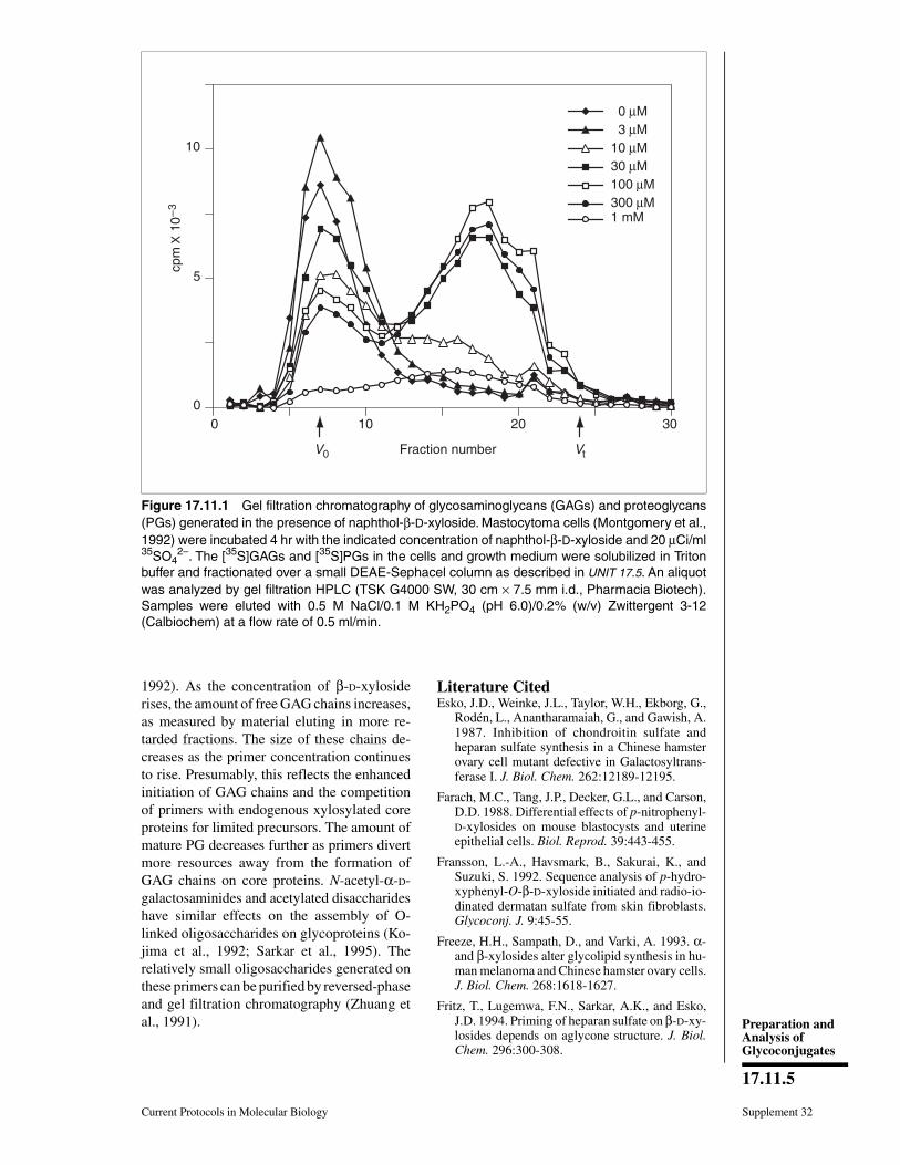
1992). As the concentration of β-D-xylosiderises, the amount of free GAG chains increases,as measured by material eluting in more re-tarded fractions. The size of these chains de-creases as the primer concentration continuesto rise. Presumably, this reflects the enhancedinitiation of GAG chains and the competitionof primers with endogenous xylosylated coreproteins for limited precursors. The amount ofmature PG decreases further as primers divertmore resources away from the formation ofGAG chains on core proteins. N-acetyl-α-D-galactosaminides and acetylated disaccharideshave similar effects on the assembly of O-linked oligosaccharides on glycoproteins (Ko-jima et al., 1992; Sarkar et al., 1995). Therelatively small oligosaccharides generated onthese primers can be purified by reversed-phaseand gel filtration chromatography (Zhuang etal., 1991).
Literature CitedEsko, J.D., Weinke, J.L., Taylor, W.H., Ekborg, G.,
Rodén, L., Anantharamaiah, G., and Gawish, A.1987. Inhibition of chondroitin sulfate andheparan sulfate synthesis in a Chinese hamsterovary cell mutant defective in Galactosyltrans-ferase I. J. Biol. Chem. 262:12189-12195.
Farach, M.C., Tang, J.P., Decker, G.L., and Carson,D.D. 1988. Differential effects of p-nitrophenyl-D-xylosides on mouse blastocysts and uterineepithelial cells. Biol. Reprod. 39:443-455.
Fransson, L.-A., Havsmark, B., Sakurai, K., andSuzuki, S. 1992. Sequence analysis of p-hydro-xyphenyl-O-β-D-xyloside initiated and radio-io-dinated dermatan sulfate from skin fibroblasts.Glycoconj. J. 9:45-55.
Freeze, H.H., Sampath, D., and Varki, A. 1993. α-and β-xylosides alter glycolipid synthesis in hu-man melanoma and Chinese hamster ovary cells.J. Biol. Chem. 268:1618-1627.
Fritz, T., Lugemwa, F.N., Sarkar, A.K., and Esko,J.D. 1994. Priming of heparan sulfate on β-D-xy-losides depends on aglycone structure. J. Biol.Chem. 296:300-308.
10
5
00 10 20 30
0 µM 3 µM10 µM30 µM100 µM300 µM1 mM
Fraction number
cpm
X 1
0–
3
V0 Vt
Figure 17.11.1 Gel filtration chromatography of glycosaminoglycans (GAGs) and proteoglycans(PGs) generated in the presence of naphthol-β-D-xyloside. Mastocytoma cells (Montgomery et al.,1992) were incubated 4 hr with the indicated concentration of naphthol-β-D-xyloside and 20 µCi/ml35SO4
2−. The [35S]GAGs and [35S]PGs in the cells and growth medium were solubilized in Tritonbuffer and fractionated over a small DEAE-Sephacel column as described in UNIT 17.5. An aliquotwas analyzed by gel filtration HPLC (TSK G4000 SW, 30 cm × 7.5 mm i.d., Pharmacia Biotech).Samples were eluted with 0.5 M NaCl/0.1 M KH2PO4 (pH 6.0)/0.2% (w/v) Zwittergent 3-12(Calbiochem) at a flow rate of 0.5 ml/min.
Current Protocols in Molecular Biology Supplement 32
17.11.5
Preparation andAnalysis ofGlycoconjugates
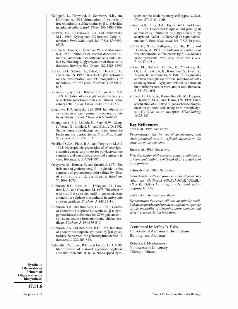
Galligani, L., Hopwood, J., Schwartz, N.B., andDorfman, A. 1975. Stimulation of synthesis offree chondroitin sulfate chains by β-D-xylosidesin cultured cells. J. Biol. Chem. 250:5400-5406.
Kanwar, Y.S., Rosenzweig, L.J., and Jakubowski,M.L. 1986. Xylosylated-PG-induced Golgi al-terations. Proc. Natl. Acad. Sci. U.S.A. 83:6499-6503.
Kojima, N., Handa, K., Newman, W., and Hakomori,S.-I. 1992. Inhibition of selectin dependent tu-mor cell adhesion to endothelial cells and plate-lets by blocking O-glycosylation of these cells.Biochem. Biophys. Res. Comm. 182:1288-1295.
Kolset, S.O., Sakurai, K., Ivhed, I., Overvatn, A.,and Suzuki, S. 1990. The effect of β-D-xylosideson the proliferation and PG biosynthesis ofmonoblastic U-937 cells. Biochem. J. 265:637-645.
Kuan, S.-F., Byrd, J.C., Busbaum, C., and Kim, Y.S.1989. Inhibition of mucin glycosylation by aryl-N-acetyl-α-galactosaminides in human coloncancer cells. J. Biol. Chem. 264:19271-19277.
Lugemwa, F.N. and Esko, J.D. 1991. Estradiol β-D-xyloside, an efficient primer for heparan sulfatebiosynthesis. J. Biol. Chem. 266:6674-6677.
Montgomery, R.I., Lidholt, K., Flay, N.W., Liang,J., Vertel, B., Lindahl, U., and Esko, J.D. 1992.Stable heparin-producing cell lines from theFurth murine mastocytoma. Proc. Natl. Acad.Sci. U.S.A. 89:11327-11331.
Neville, D.C.A., Field, R.A., and Ferguson, M.A.J.1995. Hydrophobic glycosides of N-acetylglu-cosamine can act as primers for polylactosaminesynthesis and can affect glycolipid synthesis invivo. Biochem. J. 307:791-797.
Okayama, M., Kimata, K., and Suzuki, S. 1973. Theinfluence of p-nitrophenyl-β-D-xyloside on thesynthesis of proteochondroitin sulfate by slicesof embryonic chick cartilage. J. Biochem.74:1069-1073.
Robinson, H.C., Brett, M.J., Tralaggan, P.J., Low-ther, D.A., and Okayama, M. 1975. The effect ofD-xylose, β-D-xylosides and β-D-galactosides onchondroitin sulphate biosynthesis in embryonicchicken cartilage. Biochem. J. 148:25-34.
Robinson, J.A. and Robinson, H.C. 1981. Controlof chondroitin sulphate biosynthesis. β-D-xylo-pyranosides as substrates for UDP-galactose: D-xylose transferase from embryonic-chicken car-tilage. Biochem. J. 194:839-846.
Robinson, J.A. and Robinson, H.C. 1985. Initiationof chondroitin sulphate synthesis by β-D-galac-tosides. Substrates for galactosyltransferase II.Biochem. J. 227:805-814.
Salimath, P.V., Spiro, R.C., and Freeze, H.H. 1995.Identification of a novel glycosaminoglycancore-like molecule II. α-GalNAc-capped xylo-
sides can be made by many cell types. J. Biol.Chem. 270:9164-9168.
Sarkar, A.K., Fritz, T.A., Taylor, W.H., and Esko,J.D. 1995. Disaccharide uptake and priming inanimal cells: Inhibition of sialyl Lewis X byacetylated Galβ1→4GlcNAcβ-O-naphthalene-methanol. Proc. Natl. Acad. Sci. U.S.A. In press.
Schwartz, N.B., Galligani, L., Ho, P.L., andDorfman, A. 1974. Stimulation of synthesis offree chondroitin sulfate chains by β-D-xylosidesin cultured cells. Proc. Natl. Acad. Sci. U.S.A.71:4047-4051.
Sobue, M., Habuchi, H., Ito, K., Yonekura, H.,Oguri, K., Sakurai, K., Kamohara, S., Ueno, Y.,Noyori, R., and Suzuki, S. 1987. β-D-xylosidesand their analogues as artificial initiators of GAGchain synthesis. Aglycone-related variation intheir effectiveness in vitro and in ovo. Biochem.J. 241:591-601.
Zhuang, D., Grey, A., Harris-Brandts, M., Higgins,E., Kashem, M.A., and Dennis, J.W. 1991. Char-acterization of O-linked oligosaccharide biosyn-thesis in cultured cells using para-nitrophenyl-α-D-GalNAc as an acceptor. Glycobiology1:425-433.
Key ReferencesFritz et al., 1994. See above.
Demonstrates that the type of glycoaminoglycanchain produced on a β-D-xyloside depends on thestructure of the aglycone.
Kuan et al., 1989. See above.
First description of N-acetyl-α-galactosaminides asprimers and inhibitors of O-linked glycosylation ofglycoproteins.
Salimath et al., 1995. See above.
β-D-xylosides will also prime unusual oligosaccha-rides—e.g., GalNAcα1-4GlcAβ1-3Galβ1-3Galβ1-4Xyl-R, GM3-l ike compounds, and otheroligosaccharides.
Sarkar et al., in press. See above.
Demonstrates that cells will take up suitably modi-fied disaccharides and use them as primers, openingup the possibility of designing more complex andselective glycosylation inhibitors.
Contributed by Jeffrey D. EskoUniversity of Alabama at BirminghamBirmingham, Alabama
Rebecca I. MontgomeryNorthwestern UniversityChicago, Illinois
Supplement 32 Current Protocols in Molecular Biology
17.11.6
SyntheticGlycosides as
Primers ofOligosaccharide
Biosynthesis
SECTION IIIRELEASE OF SACCHARIDES FROMGLYCOCONJUGATESIn many instances, it is useful to release a part or all of the oligosaccharide chains froma glycoconjugate before analyzing their structure, and the units presented in this sectiondescribe a variety of ways in which this can be achieved. Some of the methods describedhere (particularly the enzymatic ones) can also be applied to the study of free oligosac-charides or to oligosaccharides that are still attached to macromolecules.
Enzymes that degrade sugar chains fall into two general classes. Endoglycosidases cleaveat defined sites within a sugar chain, with a specificity often based on certain features ofthe adjacent monosaccharide units. Thus, they are analogous to the restriction endonu-cleases that work on nucleic acids (UNIT 3.1). On the other hand, exoglycosidases specifi-cally release single monosaccharides only when they are present as terminal units on sugarchains. Unlike the cases with exonucleases, the types of possible terminal units onoligosaccharide chains are numerous. Correspondingly, the number of available exogly-cosidases with different specificities is quite large. In this section, one of the mostcommonly used group of exoglycosidases, the sialidases (also known as neuraminidases),is discussed in UNIT 17.12. Some commonly used endoglycosidases and glycoamidases forN-linked oligosaccharides (UNIT 17.13A) and polysaccharide lyases (UNIT 17.13B) are thenconsidered.
Additional methods will be provided in future supplements (see Chapter 17 table ofcontents). One of the simplest and most direct ways to free an oligosaccharide chain froma glycoprotein or proteoglycan is to digest away the peptide with a broad-spectrumprotease such as pronase or proteinase K (UNIT 17.14). In these cases, most of the peptideis removed except for the amino acids immediately surrounding the glycosylation site(s).If needed, more specific proteases (e.g., trypsin, chymotrypsin) can be used to generateglycopeptides with large stretches of amino acids still attached to the glycosylation sites.This approach is particularly useful for the detection of individual glycosylation sites onglycoproteins (UNIT 17.14).
There are also many classic and well-established methods for the chemical release ofsaccharides from glycoconjugates (UNIT 17.15). Some are designed to release intact sugarschains and others release the individual monosaccharide units. Although these methodstend to be somewhat less specific and potentially more destructive than the enzymaticapproaches, they are cheap, convenient, and generally easy to use. These methods will bepresented in future supplements.
Of course, there are many other techniques for the release of saccharides from glycocon-jugates that could have been presented in this section. The current and upcomingselections were based on the criterion of broad general utility to the average molecularbiologist. As methods in “glycotechnology” improve and simplify, further additions tothis section may become appropriate.
Supplement 27
Contributed by Leland D. Powell and Ajit P. VarkiCurrent Protocols in Molecular Biology (1994) 17.12.1-17.12.8Copyright © 2000 by John Wiley & Sons, Inc.
17.12.1
Preparation andAnalysis ofGlycoconjugates
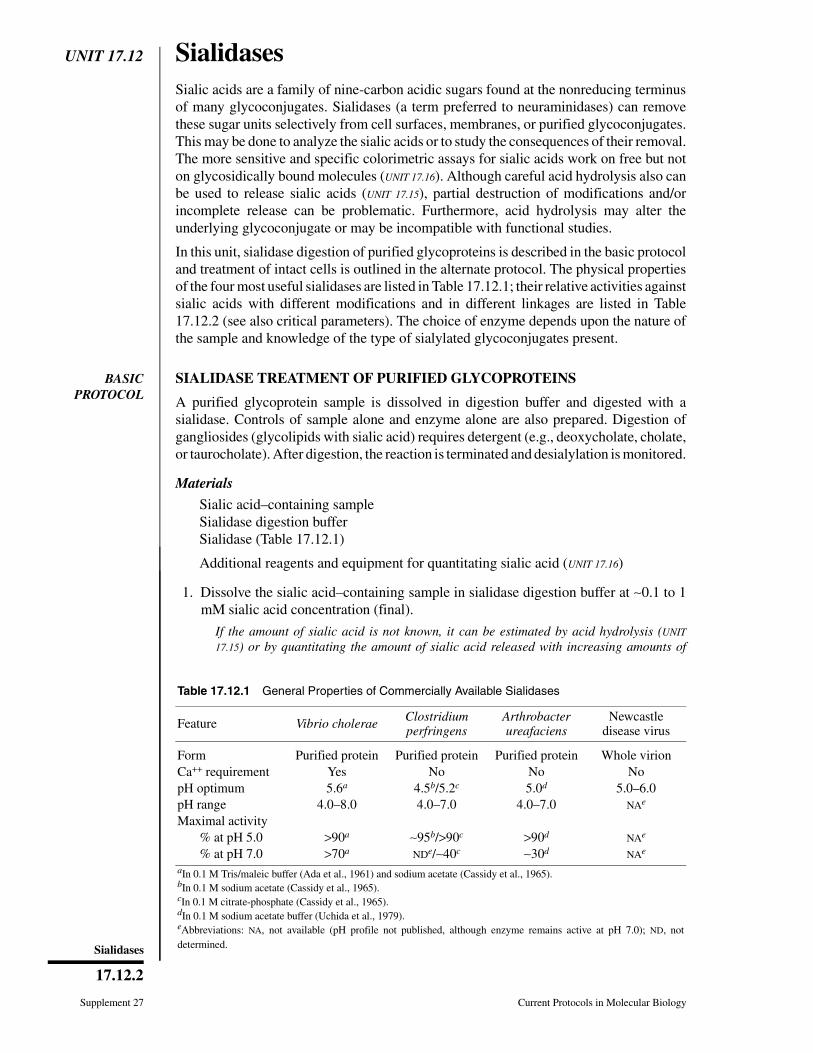
UNIT 17.12 SialidasesSialic acids are a family of nine-carbon acidic sugars found at the nonreducing terminusof many glycoconjugates. Sialidases (a term preferred to neuraminidases) can removethese sugar units selectively from cell surfaces, membranes, or purified glycoconjugates.This may be done to analyze the sialic acids or to study the consequences of their removal.The more sensitive and specific colorimetric assays for sialic acids work on free but noton glycosidically bound molecules (UNIT 17.16). Although careful acid hydrolysis also canbe used to release sialic acids (UNIT 17.15), partial destruction of modifications and/orincomplete release can be problematic. Furthermore, acid hydrolysis may alter theunderlying glycoconjugate or may be incompatible with functional studies.
In this unit, sialidase digestion of purified glycoproteins is described in the basic protocoland treatment of intact cells is outlined in the alternate protocol. The physical propertiesof the four most useful sialidases are listed in Table 17.12.1; their relative activities againstsialic acids with different modifications and in different linkages are listed in Table17.12.2 (see also critical parameters). The choice of enzyme depends upon the nature ofthe sample and knowledge of the type of sialylated glycoconjugates present.
BASICPROTOCOL
SIALIDASE TREATMENT OF PURIFIED GLYCOPROTEINS
A purified glycoprotein sample is dissolved in digestion buffer and digested with asialidase. Controls of sample alone and enzyme alone are also prepared. Digestion ofgangliosides (glycolipids with sialic acid) requires detergent (e.g., deoxycholate, cholate,or taurocholate). After digestion, the reaction is terminated and desialylation is monitored.
Materials
Sialic acid–containing sampleSialidase digestion bufferSialidase (Table 17.12.1)
Additional reagents and equipment for quantitating sialic acid (UNIT 17.16)
1. Dissolve the sialic acid–containing sample in sialidase digestion buffer at ∼0.1 to 1mM sialic acid concentration (final).
If the amount of sialic acid is not known, it can be estimated by acid hydrolysis (UNIT
17.15) or by quantitating the amount of sialic acid released with increasing amounts of
Table 17.12.1 General Properties of Commercially Available Sialidases
Feature Vibrio cholerae Clostridiumperfringens
Arthrobacterureafaciens
Newcastledisease virus
Form Purified protein Purified protein Purified protein Whole virionCa++ requirement Yes No No NopH optimum 5.6a 4.5b/5.2c 5.0d 5.0–6.0pH range 4.0–8.0 4.0–7.0 4.0–7.0 NAe
Maximal activity% at pH 5.0 >90a ∼95b/>90c >90d NAe
% at pH 7.0 >70a NDe/∼40c ∼30d NAe
aIn 0.1 M Tris/maleic buffer (Ada et al., 1961) and sodium acetate (Cassidy et al., 1965).bIn 0.1 M sodium acetate (Cassidy et al., 1965).cIn 0.1 M citrate-phosphate (Cassidy et al., 1965).dIn 0.1 M sodium acetate buffer (Uchida et al., 1979).eAbbreviations: NA, not available (pH profile not published, although enzyme remains active at pH 7.0); ND, notdetermined.
Supplement 27 Current Protocols in Molecular Biology
17.12.2
Sialidases
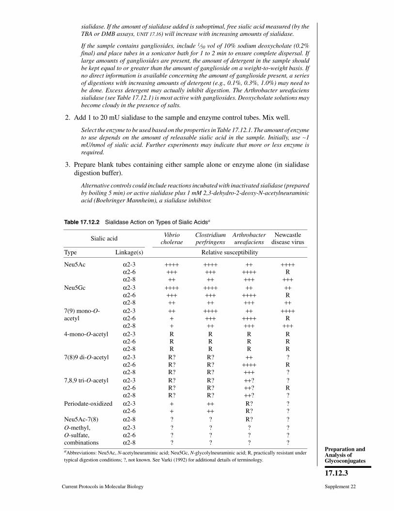
sialidase. If the amount of sialidase added is suboptimal, free sialic acid measured (by theTBA or DMB assays, UNIT 17.16) will increase with increasing amounts of sialidase.
If the sample contains gangliosides, include 1⁄50 vol of 10% sodium deoxycholate (0.2%final) and place tubes in a sonicator bath for 1 to 2 min to ensure complete dispersal. Iflarge amounts of gangliosides are present, the amount of detergent in the sample shouldbe kept equal to or greater than the amount of ganglioside on a weight-to-weight basis. Ifno direct information is available concerning the amount of ganglioside present, a seriesof digestions with increasing amounts of detergent (e.g., 0.1%, 0.3%, 1.0%) may need tobe done. Excess detergent may actually inhibit digestion. The Arthrobacter ureafacienssialidase (see Table 17.12.1) is most active with gangliosides. Deoxycholate solutions maybecome cloudy in the presence of salts.
2. Add 1 to 20 mU sialidase to the sample and enzyme control tubes. Mix well.
Select the enzyme to be used based on the properties in Table 17.12.1. The amount of enzymeto use depends on the amount of releasable sialic acid in the sample. Initially, use ∼1mU/nmol of sialic acid. Further experiments may indicate that more or less enzyme isrequired.
3. Prepare blank tubes containing either sample alone or enzyme alone (in sialidasedigestion buffer).
Alternative controls could include reactions incubated with inactivated sialidase (preparedby boiling 5 min) or active sialidase plus 1 mM 2,3-dehydro-2-deoxy-N-acetylneuraminicacid (Boehringer Mannheim), a sialidase inhibitor.
Table 17.12.2 Sialidase Action on Types of Sialic Acidsa
Sialic acid Vibriocholerae
Clostridiumperfringens
Arthrobacterureafaciens
Newcastledisease virus
Type Linkage(s) Relative susceptibility
Neu5Ac α2-3α2-6α2-8
+++++++++
+++++++++
+++++++++
++++R
+++Neu5Gc α2-3
α2-6α2-8
+++++++++
+++++++++
+++++++++
++R++
7(9) mono-O-acetyl
α2-3α2-6α2-8
++++
+++++++++
+++++++++
++++R
+++4-mono-O-acetyl α2-3
α2-6α2-8
RRR
RRR
RRR
RRR
7(8)9 di-O-acetyl α2-3α2-6α2-8
R?R?R?
R?R?R?
+++++++++
?R?
7,8,9 tri-O-acetyl α2-3α2-6α2-8
R?R?R?
R?R?R?
++?++?++?
?R?
Periodate-oxidized α2-3α2-6
++
++++
R?R?
??
Neu5Ac-7(8) α2-8 ? ? R? ?O-methyl,O-sulfate,combinations
α2-3α2-6α2-8
???
???
???
???
aAbbreviations: Neu5Ac, N-acetylneuraminic acid; Neu5Gc, N-glycolylneuraminic acid; R, practically resistant undertypical digestion conditions; ?, not known. See Varki (1992) for additional details of terminology.
Current Protocols in Molecular Biology Supplement 22
17.12.3
Preparation andAnalysis ofGlycoconjugates

4. Incubate 3 to 4 hr at 37°C.
Digestion of gangliosides may require 24 to 48 hr.
For selective digestion of α(2-3) and α(2-8) linkages by the Newcastle Disease virussialidase, use 1 to 5 mU in a 25 to 50 l reaction and incubate 15 to 30 min at 37°C. Theseconditions are appropriate when the enzyme is present in excess of substrate, as is oftenthe case when radiochemical amounts of substrate are present. The enzyme has a low levelof activity against α(2-6) linkages; this becomes evident with prolonged incubations. Iflarge amounts of sialic acids are present (e.g., >1 nmol), it will be necessary to titrate theenzyme. This can be done by setting up digests of standards containing α(2-3) or α(2-6)-linked sialic acids (Table 17.12.3) at the same concentration as the unknown sample anddetermining the amount of Newcastle disease virus sialidase that releases >90% of theα(2-3)-linked and <5% of the α(2-6)-linked sugar.
5. Terminate the reaction by boiling for 5 min. Check for release of sialic acid bymonitoring:
a. Sialic acid by direct measurement (UNIT 17.16)
b. Shift in pI of a glycoprotein by isoelectric focusing (UNIT 10.4)
c. Shift in TLC pattern for glycolipids (UNIT 17.3)
d. Shift in apparent molecular weight on SDS-PAGE (UNIT 10.2; generally a loss ofseveral sialic acid residues will be visible by small shifts on SDS-PAGE to eitherlower or higher apparent molecular weights)
e. Alteration in lectin-binding patterns for some lectins (UNIT 17.7)
f. Shift in negative charge of isolated oligosaccharides or glycolipids (UNIT 17.17).
Table 17.12.3 Sialic Acid Standards
Glycoconjugate Molecule Linkages Sialic acidsa Sialic acid concentration (nmol/mg)
Stock solution preparationb
Sialylactose(mixed isomersc)
Oligosaccharide α2-3 α2-6
Neu5Ac 1600 31 mg/ml in water (100×)
Fetuind Glycoprotein(N- and O-linked)
α2-3 α2-6
Neu5AcNeu5Gc(?)
280 20 mg/ml in water (10×)
Bovine submaxillarymucin
Mucin(O-linked)
α2-6 mono- anddi-O-acetylatedNeu5Ac andNeu5Gc
150-400 20 mg/ml in water (10×)
Colominic acid Polysaccharide α2-8 Neu5Ac 3400 15 mg/ml in water (100×)Mixed braingangliosides
Glycolipid α2-3 α2-8
Neu5Acf 950e 5 mg/ml in 2:1:0.1 (v/v/v)chloroform/methanol/water(10×)
aAbbreviations: Neu5Ac, N-acetylneuraminic acid; Neu5Gc, N-glycolylneuraminic acid.bStore all stock solutions at −20°C. Dilution to 1× will yield ∼0.5 mM sialic acid.cRatio of α(2-3) to α(2-6) ∼4:1, although lot-to-lot variation probably occurs.dRatio of α(2-3) to α(2-6) ∼1.6:1.4 in commercial preparations, including Sigma (#F-3004) or GIBCO/BRL (Townsend et al., 1989).eDepending on the manufacturer.fCommercial preparations of bovine brain gangliosides have been treated with base during purification, resulting in loss of O-acetyl esters.
Supplement 22 Current Protocols in Molecular Biology
17.12.4
Sialidases
ALTERNATEPROTOCOL
SIALIDASE TREATMENT OF INTACT CELLS
Although the pH optima of bacterial and viral sialidases range from 4.5 to 5.5, theseenzymes can be used to treat intact, viable cells at pH 7.0. Cells are washed andresuspended in isotonic serum-free buffer, then treated with the sialidase. The reaction isterminated by centrifugation and washing.
Additional Materials
Cells, prepared as a single-cell suspensionHEPES-buffered saline (HeBS)Culture medium appropriate for cells, with serum
1. Wash cell suspension free from serum-containing medium by centrifuging 10 min at500 × g and resuspending twice in HeBS. After the last wash, resuspend in HeBS at0.2–1 × 10⁷ cells/ml. Divide the cells between two tubes.
Use HeBS containing 1 mM CaCl2 if Vibrio cholerae sialidase is to be used.
Cell number can be determined by trypan blue exclusion as described in UNIT 11.5.
2. To one tube, add sialidase. To the second tube, add no enzyme. Incubate 30 min at37°C.
Select the enzyme to be used based on the properties in Table 17.12.1. Initially use thesialidase in vast excess, e.g., 100 mU/ml for 107 cells/ml (presuming 107 cells will yield ∼1nmol), then titrate down for the optimum amount.
Alternative controls could include reactions incubated with inactivated sialidase (preparedby boiling 5 min) or active sialidase plus 1 mM 2,3-dehydro-2-deoxy-N-acetylneuraminicacid (Boehringer Mannheim), a sialidase inhibitor.
3a. To characterize the sialic acid molecules released from the cells: Chill reactions to4°C and centrifuge 10 min at 2000 × g, 4°C. Collect supernatant, boil 5 min toinactivate sialidase, and proceed with characterization of the released sialic acid (UNIT
17.16).
It is important to inactivate the sialidase as otherwise it will continue to work on anyglycoproteins shed or secreted into the medium during the 30-min incubation.
3b. To characterize the glycoconjugates on the cell surface: Wash cells three times inculture medium containing serum (to remove free sialidase) before proceeding withbiochemical or functional assays.
REAGENTS AND SOLUTIONS
HEPES-buffered saline (HeBS)476 mg HEPES (20 mM final)812 mg NaCl (140 mM final)H2O to 100 mlAdjust pH to 7.0 with HClStable <1 year at 4°C.
Sialidase digestion buffersClostridium perfringens and Arthrobacter ureafaciens sialidases: Dissolve 8.6 gsodium acetate in 100 ml water to obtain a 0.1 M sodium acetate stock solution.Adjust pH to 5.5 with 0.1 M acetic acid. Store <1 year at 4°C.Vibrio cholerae sialidase: Add 14.7 mg CaCl2 dihydrate and 580 mg NaCl to 100ml of 0.1 M sodium acetate, pH 5.5. Store <1 year at 4°C.Newcastle disease virus sialidase: Add 2 mg of fatty acid–free BSA (Sigma A7511)
Current Protocols in Molecular Biology Supplement 22
17.12.5
Preparation andAnalysis ofGlycoconjugates

per milliliter of 0.1 M sodium acetate, pH 5.5. Any high-purity BSA can be used(contaminating glycosidases may be present in low levels in some BSA prepara-tions). Store indefinitely at −20°C.
SialidasesCommercial sources for sialidases include Sigma, Calbiochem, Boehringer Mann-heim, and Oxford GlycoSystems (see Table 17.12.1 and APPENDIX 4).Clostridium perfringens sialidase: Supplied as a lyophilized powder. Reconstituteto 10 mU/µl in digestion buffer (see above) and store for several months at 4°C.Arthrobacter ureafaciens sialidase: Supplied as a lyophilized powder. Reconstituteto 1 to 10 mU/µl per manufacturer’s instructions. Store for ∼6 months at 4°C.Vibrio cholerae sialidase: Supplied in solution. Store per manufacturer’s instruc-tions.Newcastle disease virus sialidase: Available commercially. Alternatively, prepareas per Paulson et al. (1982). Store stocks in 5- to 10-µl aliquots at −70°C. Thaw onlyonce and store on wet ice at 4°C until use.
COMMENTARY
Background InformationSialidases from many viral and bacterial
strains have been described (Drzeniek, 1973).In general, bacterial enzymes have the broadestspectrum of activity, although the limited speci-ficity of the viral enzymes can be an advantagefor certain analyses.
Sialic acid is found in α(2-3) or α(2-6)linkages to neutral sugars (e.g., galactose, N-acetylgalactosamine, N-acetylglucosamine) orin α(2-8) linkage to another sialic acid. Theα(2-3)- and α(2-6)-linked sialic acid residuesare found in glycoproteins (both N- and O-linked) and in glycolipids. α(2-8)-linked resi-dues are found in glycolipids, colominic acid(from E. coli), and a few mammalian glycopro-teins (e.g., neural cell adhesion molecule). Asoutlined in Table 17.12.2, not all sialidases areactive towards all linkages, and these differ-ences can be exploited in structural analyses.Sialidases are only active against α-linked sialicacids. The only natural β-linked sialic acid is inCMP (cytidine monophosphate)-sialic acid.
The common sialic acid N-acetylneuraminicacid (Neu5Ac) can carry modifications (pri-marily O-acetylation but also glycolylation,sulfation, and methylation; see Varki, 1992, andTable 17.12.2). Sialidases differ in their activi-ties towards these modified sialic acids. Unfor-tunately, the distribution of sialic acids contain-ing these modifications has not been well-char-acterized in most mammalian systems.
Critical ParametersSeveral caveats must be kept in mind in
using sialidases.
Cleavage.The relative catalytic rates of thedifferent enzymes for the various linkagetypes—i.e., α(2-3), α(2-6), and α(2-8)—arenot of much consequence in most instances,because the enzymes are usually used in excess.Important exceptions to this include: (1) TheNewcastle disease virus sialidase preferentiallycleaves α(2-3) and α(2-8) linkages. However,α(2-6)-linked sialic acid will be hydrolyzedslowly if an excessive amount of enzyme isemployed or if incubations are continued >30min. (2) The Vibrio cholerae enzyme does notcleave the α(2-3)-linked “internal” sialic acidof extended gangliosides (e.g., GM1), althoughit is active against the oligosaccharide ifceramide is removed. (3) The α(2-8) linkagesof colominic acid and b series gangliosides arerelatively resistant to release. Combined treat-ments with sialidases and endosialidases (Troy,1992) may be used for polysialic acids withmore than five sialic acid units. (4) Gan-glioside-bound sialic acids may require deter-gents for complete release. Deoxycholate andcholate work the best.
Effect of substitution. Substitutions havevariable effects upon release depending on theenzyme used, as summarized in Table 17.12.2.The decrease in rate with the N-glycolyl modi-fication is not relevant in using any of theenzymes. The decrease in rates for the 9- or7-mono-O-acetyl substitutions are also notpractically relevant, except for the Vibrio en-zyme, and particularly with gangliosides. Onthe other hand, the 4-mono-O-acetyl substitu-tion causes complete resistance to all knownsialidases. The effects of di- and tri-O-acetyl
Supplement 22 Current Protocols in Molecular Biology
17.12.6
Sialidases
substitutions have not yet been carefully stud-ied, and it is not safe to extrapolate from dataon mono-O-acetyl molecules. Likewise, com-binations of substitutions (e.g., N-glycolyl andO-acetyl) have not been studied carefully. Fi-nally, there is no information on the rarer typesof sialic acids (e.g., O-methyl and O-sulfatesubstituted). If O-acetylation is suspected, priortreatment of the glycoconjugates with base(UNIT 17.15) will cause de-O-acetylation and im-prove release.
Controls. Whenever possible, controls suchas the sample without enzyme and the enzymealone must be studied in parallel to be sure thatthe sialic acids detected are actually releasedfrom the sample. This is particularly importantwhen ultrasensitive methods (such as TBA withHPLC detection or DMB derivatization; UNIT
17.16) are employed. These methods are so sen-sitive that “environmental contamination” withsialic acids can become a problem.
Analysis of results. It is useful to comparethe amount of sialic acid released (UNIT 17.16) bysialidase with the amount released by mild acidhydrolysis (UNIT 17.15). If the amount releasedby the sialidases is significantly less than thatreleased by mild acid, then the sialic acid resi-dues may be resistant due to (1) incorrect choiceof sialidase, (2) modification of the sialic acidresidues, (3) steric hindrance, or (4) suboptimalconditions for digestion.
It may be important to demonstrate that theobserved effect produced by sialidase treatment(e.g., shift in isoelectric point on a focusing gelor a functional response) is in fact due to thesialidase activity. Treatment with heat-inacti-vated enzyme (prepared by boiling for 5 min)or inhibition with 1 mM 2,3-dehydro-2-deoxy-N-acetylneuraminic acid, a specific inhibitor ofsialidases, can be used. The reaction should betitrated so that just enough sialidase is beingused for the observed result, and then the effectof the inhibitor assessed.
Sialidase inhibitors. Inhibitors of sialidasesdo exist. Dextran sulfate inhibits the Vibriocholerae sialidase. Other polyanions (e.g.,DNA, RNA, and proteoglycans) may also in-hibit this or other sialidases. Free sialic acidalso can inhibit the Vibrio cholerae enzyme,with a Ki of ∼5 mM.
Agglutination. Treating intact cells withNewcastle disease virus can cause agglutina-tion of cells. This effect should not be confusedwith clumping due to cell death.
Buffer systems. The sialidases are compat-ible with several different buffer systems, withsome differences in pH optima noted (Table
17.12.1). Vibrio cholerae sialidase works wellin sodium acetate and Tris/maleate at pH 5.0 to6.0 but is considerably more active inTris/maleate at pH >5.0. Clostridium perfrin-gens sialidase has pH optima of 4.5 in sodiumacetate but 5.5 in citrate phosphate. Arthrobac-ter ureafaciens sialidase digestion proceedsmaximally at pH 5.5, yet retains >50% of ac-tivity at pH 4.5 and 7.0. For all sialidases,alternative choices include sodium acetate, am-monium acetate, Tris, HEPES, phosphate, andcacodylate. Acetate interferes with HPLC onAX-5 resin (UNIT 17.12) and β-hexosaminidasedigestion (UNIT 17.13).
TroubleshootingFailure to digest a known sialoglycoconju-
gate to completion could be due to failure toadd sufficient sialidase, use of an old or improp-erly reconstituted and/or stored enzyme ali-quot, or inactivation of the enzyme after addi-tion. If the sample is an unknown but potentiallycontains >1 nmol of releasable sialic acid, atitration comparing increasing amounts of siali-dase versus sialic acid released is useful. Siali-dases can lose activity rapidly (e.g., <1 hr) afterdilution into an assay mix; inclusion of BSAwill help prevent this inactivation. Some manu-factures recommend that the Arthrobacter ure-afaciens and Clostridium perfringens siali-dases be reconstituted with BSA for stabilityon storage.
Anticipated ResultsIf the type of sialidase used is tailored to the
type of sialic acid and linkage, it should bepossible to obtain nearly complete release ofthese residues.
Time ConsiderationsOnce the appropriate buffers and standards
are made, the actual digestions will take 30 minto 4 hr or longer (especially for gangliosides).Time required for analysis of the digested prod-uct(s) will depend on the specific analyticalmethod employed (as listed in step 6 of the basicprotocol).
Literature CitedAda, G.L., French, E.L., and Lind, P.E. 1961. Puri-
fication and properties of neuraminidase fromVibrio cholerae. J. Gen. Microbiol. 24:409-421.
Cassidy, J.T., Jourdian, G.W., and Roseman, S.1965. The sialic acids IV: Purification and prop-erties of sialidase from Clostridium perfringens.J. Biol. Chem. 240:3501-3506.
Drzeniek, R. 1973. Substrate specificity of neu-raminidases. Histochem. J. 5:271-290.
Current Protocols in Molecular Biology Supplement 22
17.12.7
Preparation andAnalysis ofGlycoconjugates

Townsend, R.R., Hardy, M.R., Cumming, D.A.,Carver, J.P., and Bendiak, B. 1989. Separation ofbranched sialylated oligosaccharides usingHPAE-PAD. Anal. Biochem. 182:1-8.
Troy, F.A. 1992. Polysialylation: From bacteria tobrains. Glycobiology 2:5-24.
Uchida, Y., Tsukada, Y., and Sugimori, T. 1979.Enzymatic properties of neuraminidases fromArthrobacter ureafaciens. J. Biochem. 86:1573-1585.
Varki, A. 1992. Diversity in the sialic acids. InGlycobiology Vol. 2 (1): 25-40.
Key ReferencesCorfield, A.P., Wember, M., Schauer, R., and Rott,
R. 1982. The specificity of viral sialidases. Eur.J. Biochem. 124:521-525.
Describes biochemical properties of the Newcastledisease virus sialidase.
Corfield, A.P., Higa, H.H., Paulson, J.C., andSchauer, R. 1983. The specificity of viral andbacterial sialidases for alpha (2-3) and alpha(2-6) linked sialic acids in glycoproteins. Bio-chim. Biophys. Acta 744:121-126.
Provides characterization of sialidases on α(2-3)and α(2-6) linked sialic acids.
Corfield, A.P., Sander Wewer, M., Veh, R.W., Wem-ber, M., and Schauer, R. 1986. The action ofsialidases on substrates containing O-acetylsialic acids. Biol. Chem. Hoppe-Seyler 367:433-439.
Describes the activity of different sialidases on O-acetylated sialic acids.
Varki, A., and Diaz, S. 1984. The release and purifi-cation of sialic acids from glycoconjugates:Methods to minimize the loss and migration ofO-acetyl groups. Anal. Biochem. 137:236-247.
Describes essential steps necessary to preserve O-acetyl groups during release and characterizationof sialic acids.
Contributed by Leland D. Powell and Ajit P. VarkiUniversity of California San DiegoLa Jolla, California
Supplement 22 Current Protocols in Molecular Biology
17.12.8
Sialidases

UNIT 17.13AEndoglycosidase and GlycoamidaseRelease of N-Linked GlycansHudson H. Freeze1 and Christian Kranz1
1Burnham Institute for Medical Research, La Jolla, California
ABSTRACT
Nearly all proteins entering the lumen of the endoplasmic reticulum (ER) become glyco-sylated en route to a cellular organelle, the plasma membrane, or the extracellular space.Many glycans can be attached to proteins, but the most common are the N-linked gly-cans (oligosaccharides). These chains are added very soon after a protein enters the ER,but they undergo extensive remodeling (processing), especially in the Golgi. Processingchanges the sensitivity of the N-glycan to enzymes that cleave entire sugar chains or in-dividual monosaccharides, which also changes the migration of the protein on SDS gels.These changes can be used to indicate when a protein has passed a particular subcellularlocation. This unit details some of the methods used to track a protein as it trafÞcksfrom the ER to the Golgi toward its Þnal location. Curr. Protoc. Mol. Biol. 89:17.13A.1-17.13A.25. C© 2010 by John Wiley & Sons, Inc.
Keywords: ER/Golgi oligosaccharide glycan N-glycosylation glycosidase
intracellular trafÞcking
INTRODUCTION
Carbohydrate chain modiÞcations are often used to monitor glycoprotein movementthrough the secretory pathway. This is because stepwise sugar-chain processing is unidi-rectional and generally corresponds to the forward or anterograde movement of proteins.This unit offers a group of techniques that will help analyze the general structure ofcarbohydrate chains on a protein and, therefore, glycan-processing mileposts. The min-imum requirements are that the protein can be labeled metabolically (UNIT 10.18) andimmunoprecipitated (UNIT 10.16) and clearly seen on a gel or blot (UNIT 10.8). The sugarchains themselves are not analyzed, but their presence and structure are inferred from gelmobility differences after one or more enzymatic digestions. This approach is most oftenused in combination with [35S]Met pulse-chase metabolic labeling protocols, but can beapplied to any suitably labeled protein (e.g., biotinylated or 125I-labeled). As the glycansmature, they become either sensitive or resistant to highly speciÞc glycosidases. Someof these enzymes cleave intact glycans from the proteine.g., endo H, endo F2, endoF3, peptide:N-glycosidase F (PNGase F), endo D, and O-glycosidase. Others strip onlyterminal sugars (e.g., sialidase) or degrade a selected portion of the chain (e.g., endo-β-galactosidase). The techniques can be adapted to count the number ofN-linked glycans ona protein. One unusual protease, O-sialoglycoprotease, degrades only proteins containingtight clusters of O-linked sialylated sugar chains. These techniques work best on averagesize proteins (<100 kDa) that contain a few percent carbohydrate by weight, where a gelshift of 1 kDa can be seen. A summary of the enzymes and their applications is shown inTable 17.13A.1.
All enzymes except O-sialoglycoprotease (Accurate Chemical and ScientiÞc), endo D(Seikagaku), and endo-β-galactosidase (MP Biomedicals or Seikagaku) are availablefromCalbiochemor Sigma-Aldrich.Other providers are ProZyme,NewEnglandBiolabs,Roche Applied Science, and Takara Biochemical.
Current Protocols in Molecular Biology 17.13A.1-17.13A.25, January 2010Published online January 2010 in Wiley Interscience (www.interscience.wiley.com).DOI: 10.1002/0471142727.mb1713as89Copyright C© 2010 John Wiley & Sons, Inc.
Preparation andAnalysis ofGlycoconjugates
17.13A.1
Supplement 89

Release ofN-Linked Glycans
17.13A.2
Supplement 89 Current Protocols in Molecular Biology
Table 17.13A.1 Enzymes Described in This Unit
Enzyme Indications and uses Monitorsa
Endo D Transient appearance of highly processed,sensitive forms prior to addition of GlcNAcby GlcNAc transferase I
Cis to medial Golgi
Endo F2 Presence of biantennary chains ± corefucose
Medial Golgi
Endo F3 Presence of core fucosylated biantennary α
chains and/or triantennary chains ± corefucosylation
Medial Golgi
Endo-β-galactosidase Presence of poly-N-acetyllactosamines Trans-Golgi and TGN
Endo H Conversion of high mannose to complextype N-linked chains
Cis to medial Golgi
O-Glycosidase Presence of Galβ1,3GalNAc-α-Thr/SerO-linked chains
Cis to medial Golgi
PNGase F Presence of N-linked chains; cleaves nearlyall N-linked chains; only enzyme thatcleaves tetrantennary chains
Medial Golgi
Sialidase Acquisition of sialic acids Trans-Golgi and TGN
O-Sialoglycoprotease Presence of mucin-like proteins with clusterof sialylated glycans
Trans-Golgi and TGN
aAbbreviation: TGN, trans-Golgi network.
This unit provides information on how to measure changes in carbohydrate struc-ture and how these changes relate to protein trafÞcking. Fortunately, the techniquesare independent of mechanistic views, although it should be borne in mind thatthe organization and distribution of many of these indicator enzymes are cell-typedependent.
The starting material for these protocols is assumed to be [35S]Met-labeled, immuno-precipitated protein bound to ∼20 μl of protein ASepharose beads (as described inUNIT10.16). The trace amount of protein is eluted by heating in a small volume of 0.1%SDS,diluted in the appropriate buffer, and then digested with one or more enzymes in a smallvolume. The digest is analyzed on an appropriate SDS-PAGE system that can detect a 1-to 2-kDa size change. A change in the mobility of the protein after digestion is evidencethat the carbohydrate chain was sensitive to the enzyme, and therefore that the protein hadencountered a certain enzyme in the processing pathway. Alternatively, the analysis canbe done by two-dimensional isoelectric focusing (IEF)/SDS-PAGE or two-dimensionalnonequilibrium pH gradient electrophoresis (NEPHGE)/SDS-PAGE (UNIT 10.4) to seethe loss of charged sugar residues or of anionic glycans. The same digestions andSDS-PAGE analysis also apply to proteins that are radioiodinated or biotinylated, or toimmunoprecipitates derived from subcellular fractions separated on sucrose or Percollgradients.
It is important to present the glycosylation pathways, as a detailed description of thepathways is needed to appreciate how they will be used in this unit. A single protein canhavemore than one kind of glycan (N-linked and O-linked), and each individual N-linkedchain can mature into a different Þnal form. The same is true for O-linked chains. Eachis described below.
Preparation andAnalysis ofGlycoconjugates
17.13A.3
Current Protocols in Molecular Biology Supplement 89
THE N-LINKED PATHWAY
The N-linked glycan maturation pathway is most frequently used for tracking proteinmovement through the Golgi complex. A common feature of all N-linked chains is thecore region pentasaccharide shown in Figure 17.13A.1, which consists of three mannoseunits and two N-acetylglucosamine units. The mannose units comprise the trimanno-syl core, and two of these residues are α-linked to the only β-linked mannose in themolecule. The β-linked mannose is bound to one of the two N-acetylglucosamines.Because they are β1-4 linked to each other, resembling the polysaccharide chitin,this is called a chitobiose disaccharide. Initially, all N-glycosylated proteins beginlife when a preformed, lipid-associated glycan is transferred within the lumen of theendoplasmic reticulum (ER) to Asn of proteins having an Asn-X-Thr/Ser sequence.This precursor glycan contains three glucose (Glc), nine mannose (Man), and twoN-acetylglucosamine (GlcNAc) sugar residues, and has the structure shown inFigure 17.13A.1. There are several ways to depict this structure. The short-hand symbolmethod is the most convenient, but be sure to note the linkages of the individual sugars,as they are important. The α and β symbols denote the anomeric conÞguration of thesugar, and the number indicates which hydroxyl group of the next sugar is involvedin the glycosidic linkage. In all cases, the anomeric position is 1, except in sialic acidwhere it is 2. The symbols chosen for the sugar residues are those used by Essentialsof Glycobiology, Second Edition, Cold Spring Harbor Laboratory Press (Varki et al.,2008; http://www.ncbi.nlm.nih.gov/bookshelf/br.fcgi?book=glyco2) and the Consortiumfor Functional Glycomics (http://www.functionalglycomics.org).
The details of the pathway are presented in Figures 17.13A.2 and 17.13A.3, alongwith thesensitivity to each endoglycosidase or glycoamidase. The Þgures show the steps between
Asn
α α3 6
α
α
4β4β
α
α
α
Asn Asn
α α6
4β4β
α αα α 22
22
3
2
3
Asncore structure precursor oligosaccharide
acetylglucosamine (GlcNAc) mannose (Man) glucose (Glc)
3
A B
Figure 17.13A.1 Symbol structures for the core region and precursor of N-linked sugar chains.Each sugar is given a symbol and abbreviation at the bottom of the figure. Each one exceptsialic acid uses its anomeric carbon (C-1) for linking to other sugars. Sialic acid uses C-2 forglycosidic linkage to other sugars. Glycosidases and glycosyltransferases are anomeric specificand distinguish α or β configurations of each sugar. The core structure (A) is common to allN-linked chains and is composed of three Man and two GlcNAc residues. The α or β configurationof each sugar is indicated, and the OH group to which that sugar is linked is shown on the barlinking the two symbols. Thus, GlcNAcβ1-4GlcNAcβ is represented by two filled squares with β
and 4 between them. When a structure is first presented, it will have full display such as thaton the left side; if it is repeated, only the symbols will be used, as shown immediately to theright. The precursor glycan (B) for all N-linked chains is synthesized in the ER and transferredcotranslationally to the peptide containing an available Asn-X-Thr/Ser sequon. For the color versionof the figure go to http://www.currentprotocols.com/protocol/mb1713a.
Release ofN-Linked Glycans
17.13A.4
Supplement 89 Current Protocols in Molecular Biology
β
α
α
β
β
Figure 17.13A.2 N-linked glycan maturation pathway for high-mannose and hybrid types, andsensitivities to various enzymes. Brackets (top) show the structures designated as high-mannoseand hybrid chains. The boxes indicate ER or Golgi localization. The pathway begins with theprecursor glycan (see Fig. 17.13A.1). Each successive numbered step in circles representsa glycosidase or glycosyl transferase that generates a new sugar chain with different sen-sitivities to the various endoglycosidases or PNGase F. (1) precursor glycan is trimmed byα-glucosidases I and II, removing three Glc. (2) ER mannosidase removes one Man. (3) α-Mannosidase I in Golgi complex removes two Man to make Man6GlcNAc2, with a single re-maining α1-2Man. (4) The final α1-2Man is removed by a Golgi complex α-mannosidase I.(5) GlcNAc transferase I adds GlcNAc to Man5GlcNAc2. (6) α-Mannosidase II or α-mannosidaseIIx (MX) removes the α1-3 and α1-6Man units to make GlcNAc1Man3GlcNAc2. Sensitivity to vari-ous enzymes (bottom) changes when moving from left to right, but remains the same within verticalcolumns. NOTE: This continued maturation to form complex chains is shown in Figure 17.13A.3.Additionally, these figures are not comprehensive; many glycosylation steps have not been in-cluded, but they do not affect the sensitivities to the enzymes listed. For the color version of thefigure go to http://www.currentprotocols.com/protocol/mb1713a.
Preparation andAnalysis ofGlycoconjugates
17.13A.5
Current Protocols in Molecular Biology Supplement 89
4β
α 3/6
+++++PNGase F-++--F3--++-F2-----D-----endoH
mannose (Man)
12
13
8
9
10
11
α
β 2 6
6
4β
(Gal) fucose (Fuc)
β
13 13 13
12 12 12
glucose (Glc)
Figure 17.13A.3 N-linked glycan maturation pathway for complex types, and sensitivities tovarious enzymes (see Fig. 17.13A.2 for additional details). (8) GlcNAc transferase II adds asecond GlcNAc to initiate a biantennary chain. (9) GlcNAc transferase IV adds a third GlcNActo initiate a triantennary chain. (10) GlcNAc transferase V adds a fourth GlcNAc to initiate atetraantennary chain. (11) Fucosyltransferase adds α1-6Fuc to the core region of complex chains.(12) β1-4Gal is added to available GlcNAc residues of hybrid and complex chains. (13) α2-3 orα2-6Sia is added to Gal residues of hybrid and complex chains. For the color version of the figurego to http://www.currentprotocols.com/protocol/mb1713a.
high-mannose and hybrid types (Fig. 17.13A.2) and complex types (Fig. 17.13A.3).The three Glc residues [Þlled (blue) circles] are removed from properly folded proteinswithin the ER by two different glycan-processing α-glucosidases. The Þrst α1-2Glcis cleaved by α-glucosidase I, and the next two α1-3Glc residues by α-glucosidase II(Fig. 17.13A.2, step 1). An ER-associated α-mannosidase removes one Man residue[shaded (green) circle; Fig. 17.13A.2, step 2]. The protein then moves on to the Þrststep in Golgi-localized processingthe removal of the three remaining α1-2 Man unitsby Golgi α-mannosidase I to produce Man5GlcNAc2 (Fig. 17.13A.2, steps 3 and 4).Many proteins have only high-mannose-type glycans with Þve to nine Man residues, andno further processing occurs. Alternatively, one to Þve GlcNAc residues [Þlled (blue)squares] can be added to the trimannosyl core, and these are usually extended with
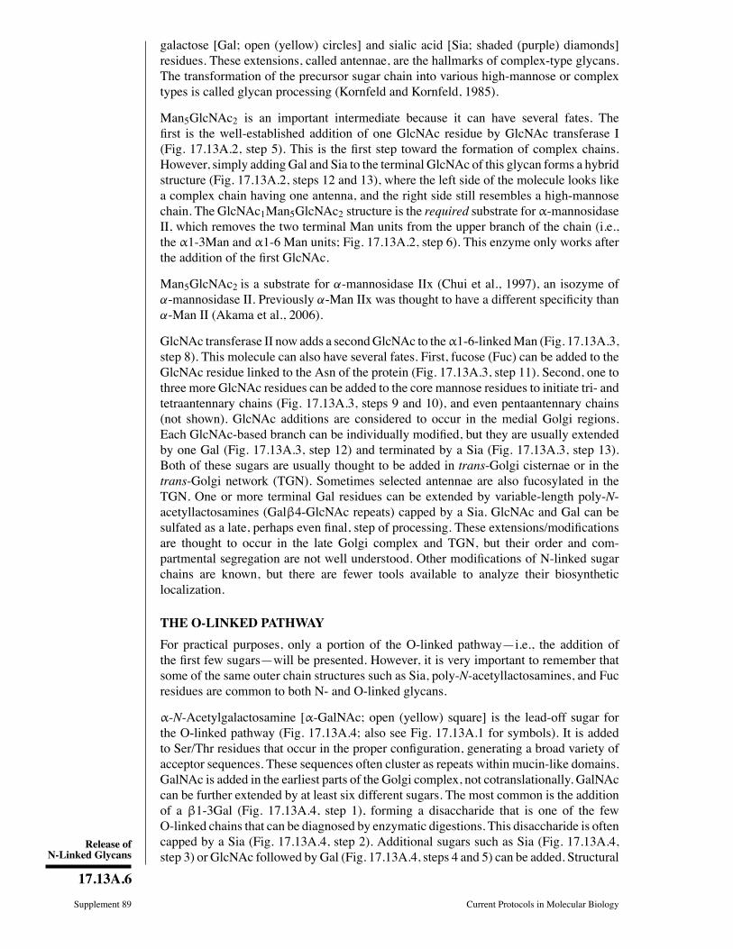
Release ofN-Linked Glycans
17.13A.6
Supplement 89 Current Protocols in Molecular Biology
galactose [Gal; open (yellow) circles] and sialic acid [Sia; shaded (purple) diamonds]residues. These extensions, called antennae, are the hallmarks of complex-type glycans.The transformation of the precursor sugar chain into various high-mannose or complextypes is called glycan processing (Kornfeld and Kornfeld, 1985).
Man5GlcNAc2 is an important intermediate because it can have several fates. TheÞrst is the well-established addition of one GlcNAc residue by GlcNAc transferase I(Fig. 17.13A.2, step 5). This is the Þrst step toward the formation of complex chains.However, simply adding Gal and Sia to the terminal GlcNAc of this glycan forms a hybridstructure (Fig. 17.13A.2, steps 12 and 13), where the left side of the molecule looks likea complex chain having one antenna, and the right side still resembles a high-mannosechain. The GlcNAc1Man5GlcNAc2 structure is the required substrate for α-mannosidaseII, which removes the two terminal Man units from the upper branch of the chain (i.e.,the α1-3Man and α1-6 Man units; Fig. 17.13A.2, step 6). This enzyme only works afterthe addition of the Þrst GlcNAc.
Man5GlcNAc2 is a substrate for α-mannosidase IIx (Chui et al., 1997), an isozyme ofα-mannosidase II. Previously α-Man IIx was thought to have a different speciÞcity thanα-Man II (Akama et al., 2006).
GlcNAc transferase II now adds a secondGlcNAc to theα1-6-linkedMan (Fig. 17.13A.3,step 8). This molecule can also have several fates. First, fucose (Fuc) can be added to theGlcNAc residue linked to the Asn of the protein (Fig. 17.13A.3, step 11). Second, one tothree more GlcNAc residues can be added to the core mannose residues to initiate tri- andtetraantennary chains (Fig. 17.13A.3, steps 9 and 10), and even pentaantennary chains(not shown). GlcNAc additions are considered to occur in the medial Golgi regions.Each GlcNAc-based branch can be individually modiÞed, but they are usually extendedby one Gal (Fig. 17.13A.3, step 12) and terminated by a Sia (Fig. 17.13A.3, step 13).Both of these sugars are usually thought to be added in trans-Golgi cisternae or in thetrans-Golgi network (TGN). Sometimes selected antennae are also fucosylated in theTGN. One or more terminal Gal residues can be extended by variable-length poly-N-acetyllactosamines (Galβ4-GlcNAc repeats) capped by a Sia. GlcNAc and Gal can besulfated as a late, perhaps even Þnal, step of processing. These extensions/modiÞcationsare thought to occur in the late Golgi complex and TGN, but their order and com-partmental segregation are not well understood. Other modiÞcations of N-linked sugarchains are known, but there are fewer tools available to analyze their biosyntheticlocalization.
THE O-LINKED PATHWAY
For practical purposes, only a portion of the O-linked pathwayi.e., the addition ofthe Þrst few sugarswill be presented. However, it is very important to remember thatsome of the same outer chain structures such as Sia, poly-N-acetyllactosamines, and Fucresidues are common to both N- and O-linked glycans.
α-N-Acetylgalactosamine [α-GalNAc; open (yellow) square] is the lead-off sugar forthe O-linked pathway (Fig. 17.13A.4; also see Fig. 17.13A.1 for symbols). It is addedto Ser/Thr residues that occur in the proper conÞguration, generating a broad variety ofacceptor sequences. These sequences often cluster as repeats within mucin-like domains.GalNAc is added in the earliest parts of the Golgi complex, not cotranslationally. GalNAccan be further extended by at least six different sugars. The most common is the additionof a β1-3Gal (Fig. 17.13A.4, step 1), forming a disaccharide that is one of the fewO-linked chains that can be diagnosed by enzymatic digestions. This disaccharide is oftencapped by a Sia (Fig. 17.13A.4, step 2). Additional sugars such as Sia (Fig. 17.13A.4,step 3) or GlcNAc followed by Gal (Fig. 17.13A.4, steps 4 and 5) can be added. Structural
Preparation andAnalysis ofGlycoconjugates
17.13A.7
Current Protocols in Molecular Biology Supplement 89
-Ser/Thr- -Ser/Thr- -Ser/Thr-1 2
3
4
5 6
α 33β
6
β6
43
α
sialic acids (Sia)
N acetylgalactosamine (GalNAc) galactose (Gal)
N acetylglucosamine (GlcNAc)
-Ser/Thr-
β
α
α
Figure 17.13A.4 A small portion of the O-GalNAc pathway. The first step of the O-linked pathwayoccurs in the early Golgi complex with the addition of α-GalNAc. There are at least six othersugars that can be added at this point in this complex pathway. Often β1-3Gal is added (1), quicklyfollowed by α2-3Sia (2). The presence of these structures can be detected with a combinationof O-glycosidase and sialidase. Additional sugars can be added as shown. α2-6Sia (3) or β1-6GlcNAc (4) followed by β1-4Gal (5) and α2-3Sia (6) on Gal. Each of these sugars must beremoved before O-glycosidase can cleave the disaccharide. For the color version of the figure goto http://www.currentprotocols.com/protocol/mb1713a.
analysis can be done by sequential exoglycosidase digestion, but given the complexity andheterogeneity of the sugar chains, such analysis is not a very useful indicator for trackingprotein movement through the Golgi complex. Many O-linked chains have terminal Siaresidues and, when tightly clustered on Ser/Thr residues, these chains promote proteolysisby O-sialoglycoprotease regardless of the structure of the underlying sugar chain.
Another type of O-linked glycosylation is the addition of glycosaminoglycan (GAG)chains to form proteoglycans. This occurs by a different pathway than the α-GalNAclinkage. Instead, the chains begin by addition of a β-xylose (Xyl) residue to Ser andare then elongated by two Gal residues and a glucuronic acid (GlcA) residue. This corestructure can be further elongated by the addition of GlcAβ1-3GalNAcβ disaccharidesto form the backbone of chondroitin/dermatan sulfate chains, or by GlcAβ1-3GlcNAcαto form the backbone of heparan sulfate chains. Biosynthesis and movement of theseproteins have also been followed through the Golgi complex. Initiation begins in lateER/early Golgi complex, and the core tetrasaccharide is probably Þnished within themedial Golgi, but the addition of chondroitin chains appears to be conÞned to the TGN.In addition to the well-known O-linked GAG chains, there is clear evidence for theexistence of a class of N-linked GAG chains.
BASICPROTOCOL 1
ENDOGLYCOSIDASE H DIGESTION
Endoglycosidase H (endo H) cleaves N-linked glycans between the two N-acetylglucosamine (GlcNAc) residues (Fig. 17.13A.5) in the core region of the glycanchain (Fig. 17.13A.1) on high-mannose and hybrid, but not complex, glycans. In thisprotocol, a fully denatured protein is digested with endo H to obtain complete release ofsensitive glycans.
Materials
Immunoprecipitated protein of interest (UNIT 10.16)0.1 M 2-mercaptoethanol (2-ME)/0.1% (w/v) SDS (ultrapure electrophoresisgrade; prepare fresh)
0.5 M sodium citrate, pH 5.51% (w/v) phenylmethylsulfonyl ßuoride (PMSF) in isopropanol

Release ofN-Linked Glycans
17.13A.8
Supplement 89 Current Protocols in Molecular Biology
α α3 6
4β4β
PNGase F β
endo HDF1F2F3
X Y
Figure 17.13A.5 PNGase F and endoglycosidase-sensitive bonds in the core of N-linked gly-cans. PNGase F is a glycoamidase that severs the bond between GlcNAc and Asn, liberatingthe entire sugar chain and converting Asn into Asp. The endoglycosidases (H, D, and Fs) cleavethe bond between the two GlcNAc residues in the core region, leaving one GlcNAc still boundto the protein. The differential specificity of the endoglyosidases is based on the structure ofthe sugar chain in a fully denatured protein. Incomplete denaturation may not expose all sensi-tive linkages. X and Y are unspecified sugar residues. For the color version of the figure go tohttp://www.currentprotocols.com/protocol/mb1713a.
0.5 U/ml endoglycosidase H (endo H; natural or recombinant)10 × SDS sample buffer (UNIT 10.2A)Water baths, 30 to 37C and 90C
Additional reagents and equipment for SDS-PAGE (UNIT 10.2A) and autoradiography(APPENDIX 3A)
1. Add 20 to 30 μl of 0.1 M 2-ME/0.1% SDS to immunoprecipitate in a microcentrifugetube, mix well, and heat denature 3 to 5 min at 90C.Use the larger amount of reagent (≥30 μl) for more complete recovery. This treatmentmay also release some (unlabeled) antibody molecules.
Protein solubilization in nonionic detergents such as Triton X-100 or Nonidet P-40 isnot always sufÞcient to completely expose all susceptible cleavage sites. Only strongdenaturation with SDS exposes all sites for maximum cleavage.
2. Cool and microcentrifuge for 1 sec at 1000 × g to collect condensed droplets in thebottom of the tube.
3. Place 10-μl aliquots of solubilized, denatured protein (supernatant) in each of twoclean microcentrifuge tubes, one for a control (no enzyme) and the other to digest(plus enzyme).
4. Add in the following order, mixing after each addition:
6 μl 0.5 M sodium citrate, pH 5.520 μl H2O2 μl 1% PMSF (in isopropanol)1 μl 0.5 U/ml endo H (enzyme digest only; substitute with water in control).
The PMSF prevents proteolysis. Nonionic detergent is not required to prevent inactivationof endo H as long as high-purity SDS is used.
The amount of water can be varied. Other solutions (or an increased amount of sam-ple) may be substituted for water if needed, but potassium buffers should be avoidedbecause they precipitate SDS as a potassium salt. The reaction volume can be scaled upproportionally if required, but in nearly all cases the enzyme will be in vast excess.
5. Incubate overnight at 30C to 37C.

Preparation andAnalysis ofGlycoconjugates
17.13A.9
Current Protocols in Molecular Biology Supplement 89
6. Immediately prior to electrophoresis, inactivate endo H by adding 4 μl of 10× SDSsample buffer and heating 5 min at 90C.
7. Analyze protein by one-dimensional SDS-PAGE (UNIT 10.2) and autoradiography(APPENDIX 3A).
The presence of high mannose and/or hybrid N-linked oligsaccharide chains will beevidenced by increased mobility of the digested proteins on SDS-PAGE.
BASICPROTOCOL 2
ENDOGLYCOSIDASE D DIGESTION
Like endo H, endo D also cleaves between the two GlcNAc residues in the core ofthe N-linked sugar chains (Fig. 17.13A.5). However, its narrow substrate speciÞcitymakes it useful for detecting the transient appearance of just a few early processingintermediates. It requires that the 2 position of theα1-3-linked coreMan be unsubstituted.This intermediate sometimes arises after more extensive processing, but prior to additionof the Þrst GlcNAc or action of α-mannosidase II. Cells with a defect in GlcNAc Itransferase (e.g., Lec 1 CHO cells) do not add the Þrst GlcNAc residue (Fig. 17.13A.2,step 5), and N-linked glycans will remain sensitive to endo D because they cannot modifythe α1-3Man residue.
Materials
Immunoprecipitated protein of interest (UNIT 10.16)0.1 M 2-mercaptoethanol (2-ME)/0.1% (w/v) SDS (ultrapure electrophoresisgrade; prepare fresh)
0.5 M NaH2PO4, pH 6.510% (w/v) Triton X-100 or Nonidet P-40 (NP-40)0.5 U/ml endoglycosidase D (endo D)10 × SDS sample buffer (UNIT 10.2A)Water baths, 37 and 90C
Additional reagents and equipment for SDS-PAGE (UNIT 10.2A) and autoradiography(APPENDIX 3A)
1. Denature immunoprecipitated protein in 20 to 30 μl of 0.1 M 2-ME/0.1% SDS byheating for 3 to 5 min at 90C.Use the larger amount of reagent (≥30 μl) for more complete recovery. This treatmentmay also release some (unlabeled) antibody molecules.
2. Cool and microcentrifuge at 1000 × g for 1 sec to collect condensed droplets in thebottom of the tube.
3. Transfer 10-μl aliquots of supernatant into two microcentrifuge tubes, one for acontrol (no enzyme) and the other to digest (plus enzyme).
4. Add in the following order, mixing after each addition:
2 μl 10% Triton X-100 or NP-40 (20-fold excess over SDS)2 μl 0.5 M NaH2PO4, pH 6.55 μl H2O1 μl 1 IU/ml endo D (enzyme digest only; substitute with water in control).
The 20-fold excess of nonionic detergent is essential to prevent inactivation of endo D bySDS.
The amount of water can be varied. Other solutions (or an increased amount of sam-ple) may be substituted for water if needed, but potassium buffers should be avoidedbecause they precipitate SDS as a potassium salt. The reaction volume can be scaled upproportionally if required, but in nearly all cases the enzyme will be in vast excess.
Release ofN-Linked Glycans
17.13A.10
Supplement 89 Current Protocols in Molecular Biology
5. Incubate overnight at 37C.
6. Immediately prior to electrophoresis, inactivate by adding 2 μl of 10× SDS samplebuffer and heating for 5 min at 90C to 95C.
7. Analyze protein by one-dimensional SDS-PAGE (UNIT 10.2A) and autoradiography(APPENDIX 3A).
EndoD sensitivity is detected by increased electrophoretic mobility of the digested proteinson SDS-PAGE.
BASICPROTOCOL 3
ENDOGLYCOSIDASE F2 DIGESTION
Endo F2, like endo H and endo D, cleaves between the two GlcNAc residues in the chito-biose core (Fig. 17.13A.5). It preferentially releases biantennary complex-type glycansfrom glycoproteins, but does not cleave tri- or tetraantennary chains.
Materials
Immunoprecipitated protein of interest (UNIT 10.16)0.1 M 2-mercaptoethanol (2-ME)/0.1% (w/v) SDS (ultrapure electrophoresisgrade; prepare fresh)
0.5 M sodium acetate, pH 4.5 (APPENDIX 2)10% (w/v) Triton X-100 or Nonidet P-40 (NP-40)0.1 M 1,10-phenanthroline in methanol200 mU/ml endoglycosidase F2 (endo F2)4 × SDS sample buffer (UNIT 10.2A)Water baths, 30 to 37C and 90C
Additional reagents and equipment for SDS-PAGE (UNIT 10.2A) and autoradiography(APPENDIX 3A)
1. Denature immunoprecipitated protein in 20 to 30 μl of 0.1 M 2-ME/0.1% SDS byheating for 3 to 5 min at 90C.Use the larger amount of reagent (≥30 μl) for more complete recovery. This treatmentmay also release some (unlabeled) antibody molecules.
2. Cool and microcentrifuge at 1000 × g for 1 sec to collect condensed droplets in thebottom of the tube.
3. Transfer 10-μl aliquots of supernatant into two microcentrifuge tubes, one for acontrol (no enzyme) and the other to digest (plus enzyme).
4. Add in the following order, mixing after each addition:
15 μl 0.5 M sodium acetate, pH 4.53 μl 0.1 M 1,10-phenanthroline in methanol2 μl 10% Triton X-100 or NP-40 (20-fold excess over SDS)1 μl 200 mU/ml endo F2 (enzyme digest only; substitute 0.5 M sodium acetatein control).
A 10- to 20-fold excess of nonionic detergent is required to stabilize the enzyme.
The amount of water can be varied. Other solutions (or an increased amount of sam-ple) may be substituted for water if needed, but potassium buffers should be avoidedbecause they precipitate SDS as a potassium salt. The reaction volume can be scaled upproportionally if required, but in nearly all cases the enzyme will be in vast excess.
5. Incubate the mixture overnight at 30 to 37C.Some inactivation of the enzyme occurs at 37C, even with nonionic detergent present;however, if the enzyme is present in sufÞcient excess, incubation can generally be carriedout successfully at 37C.

Preparation andAnalysis ofGlycoconjugates
17.13A.11
Current Protocols in Molecular Biology Supplement 89
6. Immediately before electrophoresis, inactivate by adding 8 μl of 4× SDS samplebuffer and heating for 5 min at 90C to 95C.
7. Analyze the protein by one-dimensional SDS-PAGE (UNIT 10.2A) and autoradiography(APPENDIX 3A).
Sensitivity to endo F2 is detected by increased electrophoretic mobility on SDS-PAGE.
BASICPROTOCOL 4
ENDOGLYCOSIDASE F3 DIGESTION
Endoglycosidase F3 (endo F3) is another endoglycosidase with a narrow substrate rangeand, therefore, high speciÞcity: it cleaves triantennary chains, but not high-mannose, hy-brid, nonfucosylated biantennary or tetraantennary chains. A core-fucosylated bianten-nary chain is the only other demonstrated substrate. When both endo F3 and endo F2digestions are done in parallel on a sample, it can provide evidence for chain branching andcore fucosylation. The approach is essentially the same as for the other endoglycosidases.
Materials
Immunoprecipitated protein of interest (UNIT 10.16)0.1 M 2-mercaptoethanol (2-ME)/0.1% (w/v) SDS (ultrapure electrophoresisgrade; prepare fresh)
0.5 M sodium acetate, pH 4.5 (APPENDIX 2)10% (w/v) Triton X-100 or NP-400.1 U/ml endoglycosidase F3 (endo F3)10× SDS sample buffer (UNIT 10.2A)Water baths, 37 and 90C
Additional reagents and equipment for SDS-PAGE (UNIT 10.2A) and autoradiography(APPENDIX 3A)
1. Denature immunoprecipitated protein in 20 to 30 μl of 0.1 M 2-ME/0.1% SDS andheat denature 3 to 5 min at 90C.Use the larger amount of reagent (≥30 μl) for more complete recovery. This treatmentmay also release some (unlabeled) antibody molecules.
2. Cool and microcentrifuge for 1 sec at 1000 × g to collect condensed droplets at thebottom of the tube.
3. Transfer 10-μl aliquots of supernatant into two microcentrifuge tubes, one for acontrol (no enzyme) and the other to digest (plus enzyme).
4. Add in the following order, mixing after each addition:
2 μl 10% Triton X-100 or NP-40 (20-fold excess over SDS)4 μl 0.5 M sodium acetate, pH 4.55 μl H2O1 μl 0.1 U/ml endo F3 (enzyme digest only; substitute with water in control).
The amount of water can be varied. Other solutions (or an increased amount of sam-ple) may be substituted for water if needed, but potassium buffers should be avoidedbecause they precipitate SDS as a potassium salt. The reaction volume can be scaled upproportionally if required, but in nearly all cases the enzyme will be in vast excess.
5. Incubate overnight at 37C.
6. Immediately prior to electrophoresis, inactivate by adding 2 μl of 10× SDS samplebuffer and heating for 5 min at 90C to 95C.

Release ofN-Linked Glycans
17.13A.12
Supplement 89 Current Protocols in Molecular Biology
7. Analyze by one-dimensional SDS-PAGE (UNIT 10.2A) and autoradiography (AP-PENDIX 3A).
Sensitivity to endo F3 is detected by increased mobility on SDS-PAGE.
BASICPROTOCOL 5
PEPTIDE: N-GLYCOSIDASE F DIGESTION
PNGase F is a glycoamidase that cleaves the bond between the Asn residue of the proteinand the GlcNAc residue that joins the carbohydrate to the protein (Fig. 17.13A.5).Because it liberates nearly all known N-linked glycans from glycoproteins, it is thepreferred enzyme for complete removal of N-linked chains. It is the only enzyme thatreleases tetra- and pentaantennary chains. The glycoprotein sample must be denaturedand digested with PNGase F to remove N-linked glycans completely.
Materials
Immunoprecipitated protein of interest (UNIT 10.16)0.1 M 2-mercaptoethanol (2-ME)/0.1% (w/v) SDS (ultrapure electrophoresisgrade; prepare fresh)
0.5 M Tris·Cl, pH 8.6 as determined at 37C (APPENDIX 2)10% (w/v) Triton X-100 or Nonidet P-40 (NP-40)200 to 250 mU/ml peptide:N-glycosidase F (PNGase F)10× SDS sample buffer (UNIT 10.2A)Water baths, 30 to 37C and 90C
Additional reagents and equipment for SDS-PAGE (UNIT 10.2A) and autoradiography(APPENDIX 3A)
1. Denature immunoprecipitated protein in 20 to 30 μl of 0.1 M 2-ME/0.1% SDS byheating 3 to 5 min at 90C.Use the larger amount of reagent≥30 μl) for more complete recovery. This treatment mayalso release some (unlabeled) antibody molecules.
2. Cool and microcentrifuge for 1 sec at 1000 × g to collect condensed droplets in thebottom of the tube.
3. Transfer 10-μl aliquots of supernatant into two microcentrifuge tubes, one for acontrol (no enzyme) and the other to digest (plus enzyme).
4. Add in the following order, mixing after each addition:
3 μl 0.5 M Tris·Cl, pH 8.65 μl H2O2 μl 10% NP-40 or Triton X-1005 μl 200 to 250 mU/ml PNGase F (enzyme digest only; substitute with 0.5 MTris·Cl in control).
Sodium phosphate or HEPES buffer, pH 7.0, can be used instead of Tris·Cl. Avoid potas-sium buffers because these may cause precipitation of a potassium SDS salt. Use of a non-ionic detergent is essential, because SDS inactivates PNGase F. A 10-fold weight excessof any of the above nonionic detergents over the amount of SDS will stabilize the enzyme.
The amount of water can be varied. Other solutions (or an increased amount of sam-ple) may be substituted for water if needed, but potassium buffers should be avoidedbecause they precipitate SDS as a potassium salt. The reaction volume can be scaled upproportionally if required, but in nearly all cases the enzyme will be in vast excess.
5. Incubate overnight at 30 to 37C.
6. Immediately prior to electrophoresis, inactivate the enzyme by adding 2.5 μl of 10×SDS sample buffer and heating 3 to 5 min at 90C.
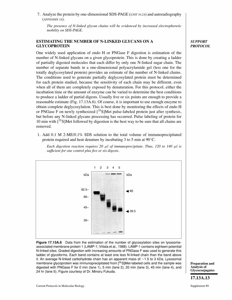
Preparation andAnalysis ofGlycoconjugates
17.13A.13
Current Protocols in Molecular Biology Supplement 89
7. Analyze the protein by one-dimensional SDS-PAGE (UNIT 10.2A) and autoradiography(APPENDIX 3A).
The presence of N-linked glycan chains will be evidenced by increased electrophoreticmobility on SDS-PAGE.
SUPPORTPROTOCOL
ESTIMATING THE NUMBER OF N-LINKED GLYCANS ON AGLYCOPROTEIN
One widely used application of endo H or PNGase F digestion is estimation of thenumber of N-linked glycans on a given glycoprotein. This is done by creating a ladderof partially digested molecules that each differ by only one N-linked sugar chain. Thenumber of separate bands in a one-dimensional polyacrylamide gel (less one for thetotally deglycosylated protein) provides an estimate of the number of N-linked chains.The conditions used to generate partially deglycosylated protein must be determinedfor each protein studied, because the sensitivity of each chain may be different, evenwhen all of them are completely exposed by denaturation. For this protocol, either theincubation time or the amount of enzyme can be varied to determine the best conditionsto produce a ladder of partial digests. Usually Þve or six points are enough to provide areasonable estimate (Fig. 17.13A.6). Of course, it is important to use enough enzyme toobtain complete deglycosylation. This is best done by monitoring the effects of endo Hor PNGase F on newly synthesized [35S]Met pulse-labeled protein just after synthesis,but before any N-linked glycans processing has occurred. Pulse labeling of protein for10 min with [35S]Met followed by digestion is the best way to be sure that all chains areremoved.
1. Add 0.1 M 2-ME/0.1% SDS solution to the total volume of immunoprecipitatedprotein required and heat denature by incubating 3 to 5 min at 90C.Each digestion reaction requires 20 μl of immunoprecipitate. Thus, 120 to 140 μl issufÞcient for one control plus Þve or six digests.
kDa
92.5–
66–
30–
45–
1 2 3 4 5
kDa
85
39.5
Figure 17.13A.6 Data from the estimation of the number of glycosylation sites on lysosome-associated membrane protein 1 (LAMP-1; Viitala et al., 1988). LAMP-1 contains eighteen potentialN-linked sites. Graded digestion with increasing amounts of PNGase F was used to generate thisladder of glycoforms. Each band contains at least one less N-linked chain than the band aboveit. An average N-linked carbohydrate chain has an apparent mass of ∼1.5 to 3 kDa. Lysosomalmembrane glycoprotein was immunoprecipitated from [35S]Met-labeled cells and the sample wasdigested with PNGase F for 0 min (lane 1), 5 min (lane 2), 20 min (lane 3), 45 min (lane 4), and24 hr (lane 5). Figure courtesy of Dr. Minoru Fukuda.
Release ofN-Linked Glycans
17.13A.14
Supplement 89 Current Protocols in Molecular Biology
2. Cool and centrifuge for 1 sec at 1000× g to collect condensed droplets at the bottomof the tube.
3. Aliquot 10 μl supernatant to the number of microcentrifuge tubes required to coverthe concentration range (e.g., 0.01 to 1 mU/ml PNGase F) or incubation times (e.g.,5 to 60 min) plus one for an undigested control.
4. Add remaining reagents as speciÞed for endo H (see Basic Protocol 1, step 4) orPNGase F (see Basic Protocol 5, step 4), adjusting the enzyme concentration asdesired.
5. Incubate at 30C for the desired length of time.High enzyme concentration (10 mU/ml) and prolonged incubation (16 hr) must be amongthe conditions included, in order to ensure that there is a data point for maximum degly-cosylation.
For varying enzyme concentrations, incubate for the same amount of time, but the durationof incubation should be shorter than what would give complete digestion because the goalis to obtain increasing extent of incomplete cleavage.
6. After the desired incubation time, inactivate enzyme by adding 0.1 volume of 10×SDS sample buffer and heating 5 min at 90 to 95C.
7. Analyze the sample from each concentration/time point, including undigested sample,by one-dimensional SDS-PAGE (UNIT 10.2A) and autoradiography (APPENDIX 3A).
Most newly formed N-linked chains will have a molecular weight in the range of 1500to 2200, and loss of one chain is sufÞcient to change the migration of a protein. Thisprocedure has been used to count up to eighteen N-linked sites on one molecule. A sampleresult is shown in Figure 17.13A.6.
BASICPROTOCOL 6
SIALIDASE (NEURAMINIDASE) DIGESTION
Sialic acids are the terminal sugars on many N- and O-linked glycans. The great ma-jority are released with broad-speciÞcity sialidases (neuraminidases) such as that fromArthrobacter ureafaciens. Because sialic acids are charged, their loss usually changesthe mobility on one-dimensional SDS polyacrylamide gels, but it will always change themobility on a two-dimensional gel. Since one-dimensional analysis is easier, it can betried Þrst.
Materials
Immunoprecipitated protein of interest (UNIT 10.16)0.1 M 2-mercaptoethanol (2-ME)/0.1% (w/v) SDS (ultrapure electrophoresisgrade; prepare fresh)
10% (w/v) Triton X-100 or Nonidet P-40 (NP-40)0.5 M sodium acetate, pH 5.0 (APPENDIX 2)1 IU/ml neuraminidase from Arthrobacter ureafaciens10× SDS sample buffer (UNIT 10.2A)Water baths, 37 and 90C
Additional reagents and equipment for SDS-PAGE (UNIT 10.2A), for IEF/SDS-PAGEor NEPHGE/SDS-PAGE (UNIT 10.3), and for autoradiography (APPENDIX 3A)
1. Denature immunoprecipitated protein in 20 to 30 μl of 0.1 M 2-ME/0.1% SDS byheating 3 to 5 min at 90C.Denaturation is less important here, because the sialic acids are exposed at the ends ofthe sugar chains. In most instances, the denaturation step can probably be omitted andthe digestion done while the protein is still bound to the beads.
Preparation andAnalysis ofGlycoconjugates
17.13A.15
Current Protocols in Molecular Biology Supplement 89
2. Cool and microcentrifuge for 1 sec at 1000 × g to collect condensed droplets in thebottom of the tube.
3. Transfer 10-μl aliquots of supernatant to two clean microcentrifuge tubes, one for acontrol (no enzyme) and the other to digest (plus enzyme).
4. Add in the following order, mixing after each addition:
2 μl 10% Triton X-100 or NP-40 (20-fold excess over SDS)4 μl 0.5 M sodium acetate, pH 5.05 μl H2O1 μl 1 IU/ml neuraminidase (enzyme digest only; substitute with water forcontrol).
This amount of neuraminidase should be in great excess. Addition of nonionic detergentis not needed if the digestion is done while the protein was still bound to the beads.
The amount of water can be varied. Other solutions (or an increased amount of sam-ple) may be substituted for water if needed, but potassium buffers should be avoidedbecause they precipitate SDS as a potassium salt. The reaction volume can be scaled upproportionally if required, but in nearly all cases the enzyme will be in vast excess.
5. Incubate overnight at 37C.This time can be shortened to 2 hr, if necessary, but longer incubations are better.
6. Immediately prior to electrophoresis, inactivate the enzyme by adding 2 μl of 10×SDS sample buffer and heating 3 to 5 min at 90C.
If the protein will be analyzed by IEF or NEPHGE, addition of sample buffer is replacedby lysis buffer used for these techniques.
7. Analyze the protein using either the appropriate one-dimentional SDS-PAGE system(UNIT 10.2A) or a two-dimentional IEF/SDS-PAGE or NEPHGE/SDS-PAGE system(UNITS 10.3 & 10.4), and detect by autoradiography (APPENDIX 3A).
Removal of sialic acids usually results in a decrease in apparent molecular weight on one-dimensional gel analysis, or an increase in the isoelectric point of the protein analyzed bytwo-dimentional gel analysis.
BASICPROTOCOL 7
ENDO-ββ-GALACTOSIDASE DIGESTION
The endo-β-galactosidase from Escherichia freundii degrades poly-N-acetyllactosaminechains (Galβ1-4GlcNAcβ1-3)n found on both N- and O-linked glycans. The variablelength of these repeating units usually causes the protein to run as a broad band or asmear on the gel. Although not all linkages are equally cleaved by this enzyme (seeFig. 17.13A.7), sensitive proteins that often run as broad bands or smears on gelse.g.,lysosome-associated membrane protein 1 (LAMP-1)produce both sharper bands andlower molecular weight species after digestion.
Materials
Immunoprecipitated protein of interest (UNIT 10.16)0.1 M 2-mercaptoethanol (2-ME)/0.1% (w/v) SDS (ultrapure electrophoresisgrade; prepare fresh)
0.5 M sodium acetate buffer, pH 5.8 (APPENDIX 2)10% (w/v) Triton X-100 or Nonidet P-40 (NP-40)100 mU/ml endo-β-galactosidase10× SDS sample buffer (UNIT 10.2A)Water baths, 37 and 95C
Additional reagents and equipment for SDS-PAGE (UNIT 10.2A) and autoradiography(APPENDIX 3A)

Release ofN-Linked Glycans
17.13A.16
Supplement 89 Current Protocols in Molecular Biology
sialic acids (Sia) acetylglucosamine (GlcNAc)galactose (Gal) fucose (Fuc)
α3 4
β3
β4
β3
β4
β3
β4
β3
β4
α 3β
Figure 17.13A.7 Endo-β-galactosidase-sensitive linkages in poly-N-acetyllactosamines. Linear,unsubstituted poly-N-acetyllactosamine units (GlcNAcβ1-3Galβ1-4) are sensitive to digestion withendo-β-galactosidase, while substitutions—such as sulfate esters (S) or branches starting withGlcNAc (not shown)—completely block digestion. Substitution of neighboring GlcNAc with Fuc orsulfate esters slows the rate, but does not block cleavage. Sensitive sites are shown with boldarrows, slowly hydrolyzed sites with a lighter arrow, and resistant bonds are struck out. Varioussubstitutions are possible, leading to broad bands on gels. This will create variable sensitivities,but even partial sensitivity should give a sharper, more defined band. For the color version of thefigure go to http://www.currentprotocols.com/protocol/mb1713a.
1. Denature immunoprecipitated protein in 20 to 30 μl of 0.1 M 2-ME/0.1% SDS byheating 3 to 5 min at 95C.Use the larger amount of reagent (≥30 μl) for more complete recovery. This treatmentmay also release some (unlabeled) antibody molecules.
2. Cool and microcentrifuge 1 sec at 1000 × g to collect condensed droplets in thebottom of the tube.
3. Transfer 10-μl aliquots of supernatant into two microcentrifuge tubes, one for acontrol (no enzyme) and the other to digest (plus enzyme).
4. Add in the following order, mixing after each addition:
2 μl 10% Triton X-100 or NP-40 (20-fold excess over SDS)4 μl 0.5 M sodium acetate, pH 5.85 μl H2O1 μl 100 mU/ml endo-β-galactosidase (enzyme digest only; substitute withwater in control).
The amount of water can be varied. Other solutions (or an increased amount of sam-ple) may be substituted for water if needed, but potassium buffers should be avoidedbecause they precipitate SDS as a potassium salt. The reaction volume can be scaled upproportionally if required, but in nearly all cases the enzyme will be in vast excess.
5. Incubate overnight at 37C.
6. Immediately prior to electrophoresis, inactivate by adding 3 μl of 10× SDS samplebuffer and heating for 5 min at 90C.
7. Analyze protein by one-dimensional SDS-PAGE (UNIT 10.2A) and autoradiography(APPENDIX 3A).
If the protein has poly-N-acetyllactosamine chains, its mobility should increase afterdigestion.
BASICPROTOCOL 8
ENDO-α-N-ACETYLGALACTOSAMINIDASE DIGESTION
This enzyme (also known as O-glycosidase or O-glycanase) has limited utility becauseit is highly speciÞc for cleaving only one O-linked disaccharide, Galβ1-3GalNAcα-Ser/Thr. Adding any more sugars, including sialic acid, renders the molecule resistantto cleavage and requires removal of each residue before the enzyme will work. Prior
Preparation andAnalysis ofGlycoconjugates
17.13A.17
Current Protocols in Molecular Biology Supplement 89
sialidase digestion is sometimes used (see Basic Protocol 6), and this can be done whilethe protein is still bound to the immunoprecipitation beads.
Materials
Immunoprecipitated protein of interest (UNIT 10.16)0.1 M 2-mercaptoethanol (2-ME)/0.1% (w/v) SDS (ultrapure electrophoresisgrade; prepare fresh)
0.5 M sodium citrate phosphate buffer, pH 6.0, containing 500 μg/ml BSA(complete buffer supplied with enzyme)
10% (w/v) Triton X-100 or Nonidet P-40 (NP-40)300 mU/ml endo-α-N-acetylgalactosaminidase (use according to manufacturersdirections)
10× SDS sample buffer (UNIT 10.2A)Water bath, 37 and 95C
Additional reagents and equipment for SDS-PAGE (UNIT 10.2A) and autoradiography(APPENDIX 3A)
1. Denature immunoprecipitated protein in 20 to 30 μl of 0.1 M 2-ME/0.1% SDS byheating 3 to 5 min at 95C.Use the larger amount of reagent (≥30 μl) for more complete recovery. This treatmentmay also release some (unlabeled) antibody molecules.
2. Cool and microcentrifuge 1 sec at 1000 × g to collect condensed droplets in thebottom of the tube.
3. Transfer 10-μl aliquots of supernatant into two microcentrifuge tubes, one for acontrol (no enzyme) and the other to digest (plus enzyme).
4. Add in the following order, mixing after each addition:
2 μl 10% Triton X-100 or NP-40 (20-fold excess over SDS)4 μl 0.5 M sodium citrate phosphate buffer, pH 6.0, with 500 μg/ml BSA3 μl H2O1 μl 300 mU/ml endo-α-N-acetylgalactosidase (enzyme digest only; substitutewith water in control).
The amount of water can be varied. Other solutions (or an increased amount of sam-ple) may be substituted for water if needed, but potassium buffers should be avoidedbecause they precipitate SDS as a potassium salt. The reaction volume can be scaled upproportionally if required, but in nearly all cases the enzyme will be in vast excess.
5. Incubate overnight at 37C.
6. Immediately prior to electrophoresis, inactivate by adding 2 μl of 10× SDS samplebuffer and heating for 5 min at 90C.
7. Analyze protein by one-dimensional SDS-PAGE (UNIT 10.2A) and autoradiography(APPENDIX 3A).
If the protein contains the disaccharide unit, the mobility of the protein should increase.Presence of only a single unit (mol. wt. ∼400 Da) may be difÞcult to detect unless ahigh-resolution gel is used.
BASICPROTOCOL 9
O-SIALOGLYCOPROTEASE DIGESTION
Digestion with O-sialoglycoprotease requires that the substrate have a tight cluster ofsialylated O-linked glycans. Proteins with a single O-linked chain or a few widely spacedchains will not be cleaved. This property makes the enzyme a valuable diagnostic tool.

Release ofN-Linked Glycans
17.13A.18
Supplement 89 Current Protocols in Molecular Biology
Materials
Immunoprecipitated protein of interest (UNIT 10.16)0.1 M 2-mercaptoethanol (2-ME)/0.1% (w/v) SDS (ultrapure electrophoresisgrade; prepare fresh)
0.4 M HEPES buffer, pH 7.410% (w/v) Triton X-100 or Nonidet P-40 (NP-40)2.4 mg/ml O-sialoglycoprotease (O-sialoglycoprotein endoglycoprotease;reconstituted according to manufacturers directions)
10× SDS sample buffer (UNIT 10.2A)Water baths, 37 and 95C
Additional reagents and equipment for SDS-PAGE (UNIT 10.2A) and autoradiography(APPENDIX 3A)
1. Denature immunoprecipitated protein in 20 to 30 μl of 0.1 M 2-ME/0.1% SDS byheating 3 to 5 min at 95C.Use the larger amount of reagent (≥30 μl) for more complete recovery. This treatmentmay also release some (unlabeled) antibody molecules.
2. Cool and microcentrifuge 1 sec at 1000 × g to collect condensed droplets in thebottom of the tube.
3. Transfer 10-μl aliquots of supernatant into two microcentrifuge tubes, one for acontrol (no enzyme) and the other to digest (plus enzyme).
4. Add in the following order, mixing after each addition:
2 μl 10% Triton X-100 or NP-40 (20-fold excess over SDS)4 μl 0.4 M HEPES buffer, pH 7.45 μl H2O2 μl 2.4 mg/ml O-sialoglycoprotease (enzyme digest only; substitute with waterin control).
O-Sialoglycoprotein endopeptidase is a partially puriÞed enzyme, and the speciÞc activityis relatively low. A quantity of 1.0μg of this enzyme preparationwill cleave 5μg of sensitivesubstrate per hour at 37C. Human glycophorin A can serve as a positive control.
The amount of water can be varied. Other solutions (or an increased amount of sam-ple) may be substituted for water if needed, but potassium buffers should be avoidedbecause they precipitate SDS as a potassium salt. The reaction volume can be scaled upproportionally if required, but in nearly all cases the enzyme will be in vast excess.
5. Incubate overnight at 37C.
6. Immediately prior to electrophoresis, inactivate by adding 2.5 μl of 10× SDS samplebuffer and heating for 5 min at 90C.
7. Analyze protein by one-dimensional SDS-PAGE (UNIT 10.2A) and autoradiography(APPENDIX 3A).
If the digestion was successful, the target protein will be undetectable or may be cleavedinto small fragments.
COMMENTARY
Background InformationThe results of digestion of a hypotheti-
cal protein with two N-linked carbohydratechains as it moves through the ER and Golgicomplex with various enzymes are shown in
Figure 17.13A.8. At 0 min, both N-linkedchains are high-mannose type. They have losttheir Glc residues and one Man residue in theER. Both are sensitive to endo H and PNGaseF digestion, yielding a protein with only two
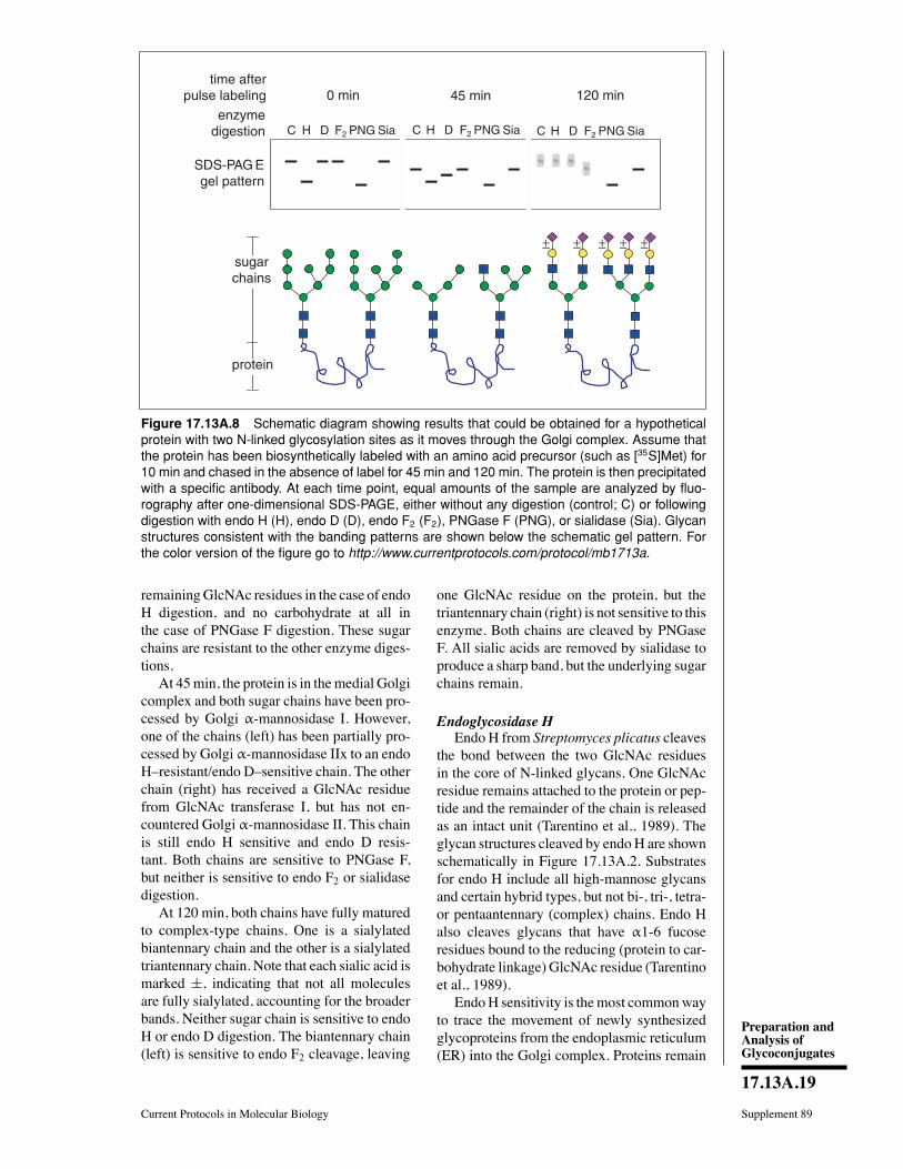
Preparation andAnalysis ofGlycoconjugates
17.13A.19
Current Protocols in Molecular Biology Supplement 89
45 min 120 min0 mintime after
pulse labeling
enzymedigestion
sugarchains
SDS-PAG Egel pattern
C H D F2
protein
PNG Sia C H D F2 PNG Sia C H D F2 PNG Sia
± ± ± ± ±
Figure 17.13A.8 Schematic diagram showing results that could be obtained for a hypotheticalprotein with two N-linked glycosylation sites as it moves through the Golgi complex. Assume thatthe protein has been biosynthetically labeled with an amino acid precursor (such as [35S]Met) for10 min and chased in the absence of label for 45 min and 120 min. The protein is then precipitatedwith a specific antibody. At each time point, equal amounts of the sample are analyzed by fluo-rography after one-dimensional SDS-PAGE, either without any digestion (control; C) or followingdigestion with endo H (H), endo D (D), endo F2 (F2), PNGase F (PNG), or sialidase (Sia). Glycanstructures consistent with the banding patterns are shown below the schematic gel pattern. Forthe color version of the figure go to http://www.currentprotocols.com/protocol/mb1713a.
remainingGlcNAc residues in the case of endoH digestion, and no carbohydrate at all inthe case of PNGase F digestion. These sugarchains are resistant to the other enzyme diges-tions.At 45min, the protein is in themedial Golgi
complex and both sugar chains have been pro-cessed by Golgi α-mannosidase I. However,one of the chains (left) has been partially pro-cessed by Golgi α-mannosidase IIx to an endoHresistant/endo Dsensitive chain. The otherchain (right) has received a GlcNAc residuefrom GlcNAc transferase I, but has not en-countered Golgi α-mannosidase II. This chainis still endo H sensitive and endo D resis-tant. Both chains are sensitive to PNGase F,but neither is sensitive to endo F2 or sialidasedigestion.At 120 min, both chains have fully matured
to complex-type chains. One is a sialylatedbiantennary chain and the other is a sialylatedtriantennary chain. Note that each sialic acid ismarked ±, indicating that not all moleculesare fully sialylated, accounting for the broaderbands. Neither sugar chain is sensitive to endoH or endo D digestion. The biantennary chain(left) is sensitive to endo F2 cleavage, leaving
one GlcNAc residue on the protein, but thetriantennary chain (right) is not sensitive to thisenzyme. Both chains are cleaved by PNGaseF. All sialic acids are removed by sialidase toproduce a sharp band, but the underlying sugarchains remain.
Endoglycosidase HEndo H from Streptomyces plicatus cleaves
the bond between the two GlcNAc residuesin the core of N-linked glycans. One GlcNAcresidue remains attached to the protein or pep-tide and the remainder of the chain is releasedas an intact unit (Tarentino et al., 1989). Theglycan structures cleaved by endoH are shownschematically in Figure 17.13A.2. Substratesfor endo H include all high-mannose glycansand certain hybrid types, but not bi-, tri-, tetra-or pentaantennary (complex) chains. Endo Halso cleaves glycans that have α1-6 fucoseresidues bound to the reducing (protein to car-bohydrate linkage) GlcNAc residue (Tarentinoet al., 1989).EndoH sensitivity is themost commonway
to trace the movement of newly synthesizedglycoproteins from the endoplasmic reticulum(ER) into the Golgi complex. Proteins remain
Release ofN-Linked Glycans
17.13A.20
Supplement 89 Current Protocols in Molecular Biology
sensitive to endo H while they are in the ERand in early regions of the Golgi complex; theybecome endo Hresistant after they are pro-cessed by enzymes located in the medial Golgicomplex. EndoH cleaves the N-linked glycansfrom proteins as long as they have not lost theα1-3Man residue cleaved by Golgi mannosi-dase II or mannosidase IIx (Fig. 17.13A.2).After removal of that mannose, the glycan be-comes endo H resistant. Endo H is the bestenzyme for identifying high-mannose and hy-brid chains and their general transition towardcomplex chains.
Endoglycosidase DThe narrow substrate speciÞcity of endo D
makes it useful for detecting a very restrictedset of processing intermediates, particularlyfor distinguishing α-mannosidase II from α-mannosidase IIx processing. When the resultsof endo D digestion are combined with the re-sults of an endo H digestion of the same sam-ple, it can help determine which of the twoalternate processing pathways is being used.The key requirement for endo D cleavage is anunsubstituted 2 position on the α1-3Man thatforms the trimannosyl core (Fig. 17.13A.1).This window of endo D sensitivity occurs onlyafter removal of the α1-2Man residue by α-mannosidase I and before the addition of aGlc-NAc residue to this same position by GlcNActransferase I (Beckers et al., 1987; Davidsonand Balch, 1993). The immediate precursorof both pathways is Man6GlcNAc2, which isendo D resistant/endo H sensitive. The better-knownα-mannosidase II pathway involves theremoval of the last α1-2Man residue by α-mannosidase I (making the glycan endo Dsensitive/endo H sensitive), and then the ad-dition of GlcNAc via GlcNAc transferase I(making it endo D resistant/endo H sensi-tive). α-Mannosidase II removes two of theremaining Þve Man residues, speciÞcally theα1-3Man and α1-6Man that form the upperbranch.Once this occurs, this endoDresistantmolecule also becomes endo H resistant. Thisstructure is also a substrate for GlcNAc trans-ferase II on the way to forming a biantennarychain.Mutant cell line CHO Lec1 lacks GlcNAc
transferase I activity and cannot synthesizeeither complex or hybrid chains. These cellscan be used to measure the kinetics of acquir-ing endo D sensitivity (Beckers et al., 1987).This cell line can be obtained through ATCC(CRL-1735). The chains become permanentlyendo D sensitive, because they lack GlcNAc
transferase I and cannot be converted backto an endo Dresistant form. These chainswill also remain endo H sensitive, because α-mannosidase II requires GlcNAc transferaseI. However, rarely, chains are more exten-sively processed, and become endo H resis-tant. Again, the combination of endo D andendo H digestions can reveal which pathwaywas used.CHO Lec1 cells are also useful for track-
ing the movement of a protein from the ERinto the earliest Golgi compartment where α-mannosidase I is located. Acquisition of endoD sensitivity requires the action of this en-zyme. The advantage of using Lec1 cells isthat the proteins remain permanently endo Dsensitive and there is no risk of kineticallymissing that small window of sensitivity be-fore the sugar chain might become endo Dresistant once again.
Endoglycosidase F and peptideN-glycosidase FElder and Alexander (Elder and Alexander,
1982; Alexander and Elder, 1989) madea landmark discovery when they identiÞedan enzyme in culture Þltrates of the bac-terium Flavobacterium meningosepticum thatcleaved N-linked glycans from glycoproteins.This preparation had a broad substrate speci-Þcity. The endoglycosidase activity in thispreparation (endo F) was actually due to aset of enzymes, each with a more restrictedsubstrate range. Like endo H, the endo F en-zymes cleave the sugar chains between the twocore GlcNAc residues (Fig. 17.13A.5). Endo Fwas originally thought to be a single enzyme,but it is now known that each of the threeenzymes has a distinct speciÞcity (Plummerand Tarentino, 1991). The speciÞcity of endoF1 is very similar to that of endo H, whileendo F2 prefers biantennary chains, and endoF3 will cleave core fucosylated biantennaryand triantennary, but not tetraantennary, chains(Fig. 17.13A.2 and Fig. 17.13A.3).Plummer et al. (1984) carefully analyzed
Flavobacterium Þltrates and found that thevery broad substrate range was actually dueto a glycoamidase activity rather than an en-doglycosidase activity. The glycoamidase re-leases the entire carbohydrate chain from theprotein by cleaving the Asp-GlcNAc bond(Fig. 17.13A.5). The enzyme is called by var-ious names, including peptide:N-glycosidaseF (PNGase F), glycopeptidase F, and N-glycanase, but the proper name is pep-tide N-4(N-acetyl-β-glucosaminyl)asparagine
Preparation andAnalysis ofGlycoconjugates
17.13A.21
Current Protocols in Molecular Biology Supplement 89
amidase F. PNGase F has the broadest speci-Þcity, and it releases most of the N-linked gly-cans from proteins.
Endo F2 and endo F3Endo F2 prefers biantennary chains over
high-mannose chains by∼20 fold. Thus, endoH and PNGase F are better choices for broadlydistinguishing high-mannose from complexchains as described above in the endo H pro-tocol (Tarentino and Plummer, 1994).Many proteins have core-fucosylated N-
linked glycans, and the addition of fucose canbe used as an additional trafÞcking marker.Endo F3 will hydrolyze triantennary chains,but endo F2 will not. Endo F2 hydrolyzesbiantennary chains; however, endo F3 willalso hydrolyze core-fucosylated biantennarychains only a bit more slowly than it does tri-antennary chains. Thus, if all the chains on aprotein are sensitive to endo F2 (biantennary)and to endo F3, this is evidence for the pres-ence of a core fucose on those chains.The enhancement of endo F3 activity on
biantennary chains with a coreα1-6Fuc pointsout that some speciÞcities are really a matterof relative rates of cleavage. If both endo F2(biantennary) and endo F3 (triantennary andbiantennary with core fucose) cleave the pro-tein, they may be acting on different chainson the same molecule. If there is only a singlechain, repeat the experiment under the sameconditions using 10- to 20-fold dilutions ofeach enzyme. If both still cleave the chainabout equally, it is evidence for core fucosyla-tion of a biantennary chain.
PNGase FPNGase F has the broadest speciÞcity of
all the enzymes that cleave N-linked glycans.It is indifferent to all extended structures onthe chains, such as sulfate, phosphate, poly-N-acetyllactosamines, polysialic acids, and eventhe occasional glycosaminoglycan chain.Mostof the modiÞcations in theMan3GlcNAc2 coreregion also make no difference in chain cleav-age. The only glycan structural feature thatconfers PNGase F resistance is the presenceof an α1-3Fuc on the GlcNAc bound to Asn(Tretter et al., 1991). ThismodiÞcation is com-monly found in plants and in some insect gly-coproteins, but it is rare in most mammaliancell lines. However, caution is warranted, asthere is evidence that some mammalian cellsdo have the critical α1-3Fuc transferase, andsome studies show that a majority of N-linkedchains of bovine lung are actually PNGase Fresistant! It is not known how common this
resistance may be. It is thus important to docu-ment N-glycosylation with proteins still in theER (see Support Protocol) before they mightbe processed to a PNGase Fresistant form.
SialidaseSialidases are also called neuraminidases
because the most common form of sialic acidis N-acetyl neuraminic acid. The sialic acidsare a family with over forty different members,but fortunately the very great majority of themcan be removed from the glycans by the broad-spectrum sialidase from Arthrobacter ureafa-ciens (AUS). It can even digest polysialicacids, a rare modiÞcation found on only afew proteins such as neuronal cell adhesionmolecule (NCAM). Sialidases with selectedspeciÞcities from other sources are availablebut would not usually be needed. AUS has anoptimum pH of 5.0, with ∼30% of maximumactivity at pH 7.Because sialic acids are charged, they af-
fect gel mobility of proteins more than wouldbe expected from their nominal molecularweight. The magnitude of the gel shift de-pends on the number of residues. It is difÞcultto estimate their number by sialidase diges-tion, but the mobility change is usually sufÞ-cient if there are several sialic acid residues.On the other hand, if a protein has only onesialic acid, its presence could be missed usingstandard one-dimensional SDS-PAGE. To becertain of the effects of sialidase, the samplecan be analyzed by a two-dimensional system,using IEF or NEPHGE in the Þrst dimension(UNITS 10.3 & 10.4). The loss of even a singlesialic acid will be evident because it changesthe isoelectric point.AUS will remove sialic acids from both N-
and O-linked chains, so the type of chain car-rying them must be determined independentlyusing PNGase F or possibly O-glycosidase incombinationwith sialidase.A proteinwill gen-erally be partially or completely resistant toO-glycosidase because the required disaccha-ride, Galβ1-3GalNAc, is usually extended andoften sialylated. Until the sialic acid isremoved, it will be resistant.The presence of sialic acid (sialidase sen-
sitivity) is often used as an indication of thetransport of a protein into the trans-Golgi net-work (TGN). This may be true in general,but it is important to remember that the dis-tribution of Golgi enzymes is cell type de-pendent. For instance, α-mannosidases I andII, which are typically considered cis/medialGolgi enzymes, are strongly expressed on thebrush border of enterocyteshardly a Golgi
Release ofN-Linked Glycans
17.13A.22
Supplement 89 Current Protocols in Molecular Biology
compartment. There are other similar exam-ples of various distributions of sialyl trans-ferase. Moreover, there are different sialyltransferases and each may have its own uniquedistribution. Although one should be cautious,it is probably safe to place sialyl transferasein the late Golgi compartment rather than anearly one.
Endo-α-N-acetylgalactosaminidaseThis enzyme from Diplococcus pneumo-
niae also goes by various names, includingO-glycosidase and O-glycanase. This enzymehas a narrow substrate range and cleaves onlyGalβ1-3GalNAcα-Ser/Thr. These are only theÞrst two sugars added in the diverse O-linkedpathway that can produce glycans with adozen or more sugar units. A portion of thepathway is shown in Figure 17.13A.4. For-tunately, many, but far from all, O-linkedchains have the simple trisaccharide struc-ture and would be sensitive to cleavage af-ter removing the Sia. Thus, sequential indi-vidual digestions or mixed digestions can beused. As both Gal and GalNAc (and proba-bly Sia) are added in the early Golgi com-plex, sensitivity to the enzyme shows that theprotein carries O-linked chains, but matchingenzyme sensitivity and a Golgi compartmentto further chain extension is difÞcult. Com-bining a battery of exoglycosidases (sialidase,α-fucosidase, α-N-acetylgalactosaminidase,and β-hexosaminidase) with endo-α-N-acetylgalactosaminidasewill probably removemost O-linked sugar chains, except sulfatedones. The bottom line is that it is easy to use theenzyme in combination with sialidase to showthat a protein has simple O-linked chains, butit is difÞcult to conclude much more concern-ing either the structure of the sugar chain orintracellular trafÞcking.
Endo-β-galactosidaseEscherichia freundii endo-β-galactosidase
is one of several enzymes that speciÞcallydegrade poly-N-acetyllactosamines by cleav-ing linear chains of GlcNAcβ1-3Galβ1-4 re-peats at the Galβ1-4 linkage. Any substitutionon the galactose itself blocks cleavage; how-ever, modiÞcations of the neighboring sugarscan slow hydrolysis (Fig. 17.13A.7). For in-stance, fucosylation and/or sulfation of nearbyGlcNAc slows cleavage, but chain branchingor sulfation at Gal block it. Even with thesepotential complexities, digestion with endo-β-galactosidase will sharpen a broad bandeven if it does not cleave every linkage. Therepeating GlcNAcβ1-3Galβ1-4 units can be
found on both N- and O-linked chains, sosensitivity to PNGase F digestion can po-tentially distinguish the location. Lysosome-associated membrane protein 1 (LAMP-1) haspoly-N-acetyllactosamine repeats on N-linkedchains. Remember that glycosylation is nottemplate driven, so glycans often exist as acontinuum of different structures on individualproteins. For example, heavily sulfated poly-N-acetyllactosamine repeats are also knownas keratan sulfate and are degraded by ker-atanases.
O-SialoglycoproteaseO-Sialoglycoprotease (also called O-
glycoprotease or O-sialoglycoprotein en-dopeptidase; Mellors and Lo, 1995) is a neu-tral metalloprotease produced by Pasteurellahaemolytica. This enzyme requires clusters ofsialylated glycans on Ser or Thr residues (Nor-gard et al., 1993). Having a single sialylatedO-linked sugar chain on a protein will not leadto degradation, nor will having a nonsialy-lated sugar chain. Therefore, this enzyme canbe used in a typical pulse-chase experimentto indicate whether there are tightly groupedO-linked chains and when they are sialylated.Adding the initialα-GalNAc in the earlyGolgicomplex is insufÞcient to cause proteolysis;sialylation is speciÞcally required, and thismay occur in a later Golgi compartment. Pro-teolysis can generate smaller fragments of atarget protein that are still visible on gels, orthe fragments may be so small that they are noteven seen on the gels. It depends on the protein.Many leukocyte antigens such as the P-selectinligand, or others such as CD43, CD44, CD45,and CD34 found on hematopoietic stem cells,are all substrates. As sialic acids are found onboth N- and O-linked chains (in clusters andnot), sequential digestions using PNGase F,sialidase, and O-sialoglycoprotease endopep-tidase in different orders can reveal differentkinds of sialylated glycans. If used as part of apulse-chase protocol, they can reveal differentkinetic subcompartments. Not all sialyl trans-ferases are necessarily within the same Golgicompartment.
Critical ParametersFor nearly all of the digestions, complete
denaturation of the protein can be important, asmaximum deglycosylation occurs only whenthe sugar chains of glycoproteins are com-pletely exposed. This is not important forsialidase digestions since sialic acids are ex-posed. Usually endo H digestions will workwithout full denaturation, but the unprocessed
Preparation andAnalysis ofGlycoconjugates
17.13A.23
Current Protocols in Molecular Biology Supplement 89
high-mannose chains remain unprocessed be-cause they are often less exposed to the pro-cessing enzymes. This may or may not be trueof the target protein, but it is better to be safeand denature it completely before digestion.Heating the immunoprecipitate with 0.1 M 2-mercaptoethanol/0.1% SDS is the best way torelease the protein in a denatured state. As-suming that the immunoprecipitate contains<10 μg of protein, 30 μl of 0.1% SDS stillprovides a three-fold excess over the protein.Adding SDS presents another problem: freeSDS may denature the digesting enzyme be-fore it has a chance to Þnish its job. For mostenzymes, be sure that the free SDS concentra-tion is <0.01%. The best way to do this is toadd a 10- to 20-fold weight excess of nonionicdetergent with a low critical micellar concen-tration (e.g., Triton X-100 or NP-40). Thesedetergents will form mixed micelles with thefree SDSandkeep it fromdenaturing the addedenzymes.The amount of enzyme and the incubation
time recommended in the protocols are in ex-cess and should be sufÞcient to cleave any ofthe sensitive linkages. The incubation timescan be shortened, if necessary, but it is betterto keep the enzyme concentration as indicated.Many of the digestions (e.g., sialidase, O-
sialoglycoprotease) can be adapted for use onmembrane preparations or on live cell surfacesby simply omitting the ionic and nonionicdetergents and decreasing the incubation time.The problem is that some linkages may notbe exposed and/or sensitive to the digestion.Thus, the usefulness of this approach needs tobe determined on a case-by-case basis.
Enzyme sources and availabilityAll enzymes except O-sialoglycoprotease
(Accurate Chemical and ScientiÞc), endoD (Calbiochem), and endo-β-galactosidase(MP Biomedicals or Seikagaku) are availablefrom Calbiochem or Sigma-Aldrich. Otherproviders are ProZyme, NewEnglandBiolabs,Roche Applied Science, and Takara Biochem-ical.
Endo HEndo H has a broad pH optimum between
5.5 and 6.5, and phosphate or citrate/phosphatebuffers can be used in place of citrate. Endo His very stable to proteases, freezing and thaw-ing, and prolonged incubations. No additivesare required for storage of the enzyme. Atconcentrations below 5 to 10 μg/ml (200 to400 mU/ml), endo H will bind to glass, so it
should be stored in plastic vials (e.g., screw-cap microcentrifuge tubes).
Endo DEndo D has a broad pH optimum of 4 to
6.5. One unit of enzyme activity will cleave1 μmole of a Man5GlcNAc3 glycopeptide permin at 37C. After reconstitution, the enzymeis stable for 6 months at −20C. It should befrozen in small aliquots to avoid the need forfreezing and thawing.
Endo FCommercial endo F preparations are mix-
tures of endo F1, F2, and F3. Endo F prepa-rations should not be used for routine degly-cosylation or to draw conclusions about thestructure of the released glycans unless thespeciÞcity is clearly deÞned.
Endo F2Endo F2 has a broad pH optimum of 4 to
6 and retains >50% of its activity at pH 7.The enzyme is sensitive to SDS, but addingnonionic detergents prevents denaturation ofthe enzyme by SDS. Although the enzymeis stable at 4C for months, it can be frozenin aliquots at −70C as long as repeatedfreeze/thaw cycles are avoided. The 1,10-phenanthroline can be used to inhibit a traceof a zinc metalloprotease that may be present.
PNGase FThe pH optimum for PNGase F is 8.6, but
80% of full activity occurs between 7.5 to9.5 with a range of buffers including phos-phate, ammonium bicarbonate, Tris·Cl, andHEPES. Borate buffers inhibit the enzyme.Commercial PNGase F is available from nat-ural or recombinant sources (two main sup-pliers, see Enzyme sources and availabil-ity, above) and is stable for 6 months at4C, or indeÞnitely at −70C. However, itshould be stored in small aliquots and re-peated freeze/thaw cycles should be avoided.PNGase F will bind to glass and plastic sur-faces and should not be stored in dilute so-lutions (<0.1 mU/ml). All of the unit activi-ties of commercial preparations are based oncleavage of dansylated glycopeptides; they areexpressed in nmoles/min, which are actuallymU, not true International Units (1 Interna-tional Unit= 1 μmole/min). SDS inactivatesPNGase F, but adding a ten-fold weight ex-cess of nonionic detergents protects the en-zyme (Tarentino et al., 1989; Tarentino andPlummer, 1994).

Release ofN-Linked Glycans
17.13A.24
Supplement 89 Current Protocols in Molecular Biology
SialidaseRecombinant AUS is available as a liquid
(see Enzyme sources and availability). The na-tive enzyme can be purchased as a lyophilizedpowder and should be reconstituted in water at1 to 10mU/μl, according to themanufacturersdirections. The native enzyme is stable for 1year at 4C if unopened. After reconstitution,it should be used within 7 days and stored at4C. The recombinant enzyme is stable for 1year at 4C. Treatment with sialidase is alsoused in assays of protein transport to the cellsurface.
Endo-α-N-acetylgalactosaminidase(O-glycosidase)Endo-α-N-acetylgalactosaminidase has a
pH optimum of 6.0 and has 50% activity at5.5 to 7.0. The thiol inhibitor parachloromer-curic benzoate (PCMB; 1 mM) inactivates theenzyme, and 1 mM EDTA inhibits it (63%),as do Mn2+ and Zn2+ (50%). Chloride also in-hibits the enzyme, so HCl-containing buffersshould be avoided. The enzyme will have fullaccess to the sugar chains only after denaturat-ing the protein with SDS, but the excess SDSneeds to be removed by forming mixed mi-celles with nonionic detergents. The enzymeis stable at 4C and at−20C, but freeze/thawcycles should be avoided.
Endo-ββ-galactosidaseThis enzyme is free of contaminating endo-
and exoglycosidases. It has a pH optimum of5.8 and can be stored at −20C, for up to2 years. Dissolved enzyme is only stable for1 month at −20C. EDTA, Ca2+, Mn2+, andMg2+ do not affect stability or activity, butPCMB inactivates it.
O-SialoglycoproteaseThe partially puriÞed enzyme is supplied by
Accurate Chemical and ScientiÞc as a lyophi-lyzed powder containing nonsubstrate bovineserum proteins and HEPES buffer. The en-zyme should be reconstituted according tothe manufacturers instructions, divided intoaliquots appropriate for a single use, and storedat −20C. Freeze/thaw cycles inactivate theenzyme, and it is inhibited by EDTA or 1,10-phenanthroline. It is possible to check the ac-tivity with a positive control of glycophorin A,which is available through Sigma-Aldrich.
TroubleshootingMost of the procedures should work as de-
scribed, but there is a chance that the enzyme is
inactive because of a variety of factors such asage, poor storage, or excess SDS. To check ac-tivity, it is worthwhile to run a positive controldigestion using the same solutions includingSDS and nonionic detergents as for the sam-ples. Since the positive controls are simply gly-coproteins that are visualized by Coomassie orsilver staining, this requires running a separategel for staining. This should not be requiredon a routine basis if the enzymes are used andstored as directed. The most likely culprit infailed digestions is using SDS solutions thatare too old or too impure.
Anticipated ResultsIf the digestions are effective, the labeled
band will usually show increased mobility onthe gel. In rare instances, digestions can actu-ally decrease mobility. The amount of changewill depend on the contribution of that com-ponent to the overall mass of the protein. Asmentioned before, a gel system that allows vi-sualization of a 1-kDa change should be used.Proteins that are >100 kDa may cause prob-lems for Þne resolution. Here are a few num-bers to keep in mind.1. The smallest N-linked chain (Man3Glc-
NAc2) will have a mass of ∼0.9 kDa.2. Two sialic acids on a single N-linked
biantennary chain will have a mass of∼0.6 kDa, but their loss may appear larger. Ifthey occur on clustered O-linked chains (sialo-glycoprotease sensitive), the apparent size dif-ference will be even larger.3. Most poly-N-acetyllactosamines are
three or more repeats, and therefore their masswould be −1 kDa. The protein will probablyrun as a heterogeneous smear or broad bandbefore digestion.4. A single O-glycosidase-sensitive disac-
charide (0.4 kDa) may be below detectionlimits.
Time ConsiderationsAll digestions can be done overnight for
convenience, but the amount of enzymesshould be sufÞcient for complete digestion inless time. The gels are run the next day, but thetime needed for the developsent of autoradio-grams will depend on the strength of the sig-nal. A low-abundance protein labeled for 10 to30 min with [35S]Met may give a weak signaland require long exposures (e.g., 2 weeks).TrafÞcking of abundant glycoproteins such asviral coat proteins require only short exposuretimes (e.g., a few hours).
Preparation andAnalysis ofGlycoconjugates
17.13A.25
Current Protocols in Molecular Biology Supplement 89
Literature CitedAkama, T.O., Nakagawa, H., Wong, N.K., Sutton-Smith,M., Dell, A.,Morris, H.R., Nakayama, J.,Nishimura, S., Pai, A., Moremen, K.W., Marth,J.D., and Fukuda, M.N. 2006. Essential and mu-tually compensatory roles of α-mannosidase IIand α-mannosidase IIx in N-glycan processingin vivo in mice. Proc. Natl. Acad. Sci. U.S.A.13;103:8983-8988.
Alexander, S. and Elder, J.H. 1989. Endoglycosi-dases from Flavobacterium meningosepticum:Application to biological problems. MethodsEnzymol. 179:505-518.
Beckers, C.J.M., Keller, D.S., and Balch, W.E.1987. Semi-intact cells permeable to macro-molecules: Use in reconstitution of proteintransport from the endoplasmic reticulum to theGolgi complex. Cell 50:523-534.
Chui, D., Oh-Eda, M., Liao, Y.-F., Panneerselvam,K., Lal, A., Marek, K.W., Freeze, H.H.,Moremen, K.W., Fukuda, M.N., andMarth, J.D.1997. Alpha-mannosidase-II deÞciency resultsin dyserythropoiesis and unveils an alternatepathway in oligosaccharide biosynthesis. Cell90:157-167.
Davidson, H.W. and Balch, W.E. 1993. Differentialinhibition of multiple vesicular transport stepsbetween the endoplasmic reticulum and transGolgi network. J. Biol. Chem. 268:4216-4226.
Elder, J.H. and Alexander, S. 1982. Endo-β-N-acetylglucosaminidase F: Endoglycosidasefrom Flavobacterium meningosepticum thatcleaves both high mannose and complex glyco-proteins. Proc. Natl. Acad. Sci. U.S.A. 79:4540-4544.
Kornfeld, R. and Kornfeld, S. 1985. Assembly ofasparagine-linked oligosaccharides. Annu. Rev.Biochem. 54:631-664.
Mellors, A. and Lo, R.Y. 1995. O-Sialoglycopro-tease from Pasteurella haemolytica. MethodsEnzymol. 248:728-740.
Norgard, K.E., Moore, K.L., Diaz, S., Stults, N.L.,Ushiyama, S., McEver, R.P., Cummings, R.D.,and Varki, A. 1993. Characterization of a spe-ciÞc ligand for P-selectin on myeloid cells.A minor glycoprotein with sialylated O-linkedoligosaccharides. J. Biol. Chem. 268:12764-12774.
Plummer, T.H. Jr. and Tarentino, A.L. 1991. PuriÞ-cation of the oligosaccharide-cleaving enzymes
of Flavobacterium meningosepticum. Glycobi-ology 1:257-263.
Plummer, T.H. Jr., Elder, J.H., Alexander, S.,Phelan, A.W., and Tarentino, A.L. 1984.Demonstration of peptide: N-glycosidase Factivity in endo-β-N-acetylglucosaminidase Fpreparations. J. Biol. Chem. 259:10700-10704.
Tarentino, A.L. and Plummer, T.H. Jr. 1994. En-zymatic deglycosylation of asparagine-linkedglycans: PuriÞcation, properties, and speci-Þcity of oligosaccharide-cleaving enzymes fromFlavobacterium meningosepticum. MethodsEnzymol. 230:44-57.
Tarentino, A.L., Trimble, R.B., and Plummer, T.H.Jr. 1989. Enzymatic approaches for studying thestructure, synthesis, and processing of glycopro-teins.Methods Cell Biol. 32:111-139.
Tretter, V., Altmann, F., andMarz, L. 1991. Peptide-N4-(N-acetyl-β-glucosaminyl) asparagine ami-dase F cannot release glycans with fucoseattached α1→3 to the asparagine-linked N-acetylglucosamine residue. Eur. J. Biochem.199:647-652.
Varki, A., Cummings, R.D., Esko, J.D., Freeze,H.H., Stanley, P., Bertozzi, C.R., Hart, G.W.,and Etzler, M.E. 2008. Essentials of Gly-cobiology. Cold Spring Harbor LaboratoryPress, Cold Spring Harbor, New York (http://www.ncbi.nlm.nih.gov/bookshelf/br.fcgi?book=glyco2).
Viitala, J., Carlsson, S.R., Siebert, P.D., and Fukuda,M. 1988.Molecular cloning of cDNAs encodinglamp A, a human lysosomal membrane glyco-proteinwith apparentMr approximately equal to120,000. Proc. Natl. Acad. Sci. U.S.A. 85:3743-3747.
Key ReferencesBeckers et al., 1987. See above.Describes the use of Lec 1 CHO cells and endo Dto study processing.
Chui et al., 1997. See above.Demonstrates the importance of α-mannosidaseIIx.
Kornfeld and Kornfeld, 1985. See above.Landmark review of processing.
Tarentino and Plummer, 1994. See above.Best review of the use of these enzymes.
UNIT 17.13BAnalysis of Glycosaminoglycans withPolysaccharide Lyases
Polysaccharide lyases are a class of enzymes useful for analysis of glycosaminoglycans(GAGs) and the glycosaminoglycan component of proteoglycans (PGs). These enzymescleave specific glycosidic linkages present in acidic polysaccharides and result in depo-lymerization (Linhardt et al., 1986). These enzymes act through an eliminase mechanismresulting in unsaturated oligosaccharide products that have UV absorbance at 232 nm.The lyases are derived from a wide variety of pathogenic and nonpathogenic bacteria andfungi (Linhardt et al., 1986). This class of enzymes includes heparin lyases (heparinases),heparan sulfate lyases (heparanases or heparitinases), chondroitin lyases (chondroiti-nases), and hyaluronate lyases (hyaluronidases), all of which are described in this unit.
Polysaccharide lyases can be used, alone or in combinations, to confirm the presence ofGAGs in a sample as well as to distinguish between different GAGs (see Table 17.13B.1and Commentary). The protocols given for heparin lyase I are general and, with minormodifications (described for each lyase and summarized in Table 17.13B.2), can be usedfor any of the polysaccharide lyases.
The basic protocol describes depolymerization of GAGs in samples containing 1 µg to 1mg of GAGs. The alternate protocol describes depolymerization of GAGs in samplescontaining <1 µg of radiolabeled GAG. Two support protocols describe assays to confirmand quantitate the activity of heparin and chondroitin ABC lyases. It is recommended thatenzyme activity be assayed before the enzyme is used in an experiment to be sure it isactive and has been stored properly.
The standard definition of a unit (U), 1 µmol product formed/min, is used throughout thisarticle. Some lyases are sold in nonstandard units (e.g., 0.1 µmol/hr, ∆A232/min), and theseshould either be converted to standard units or the activity should be determined usingthe appropriate support protocol.
Supplement 48
Contributed by Robert J. LinhardtCurrent Protocols in Molecular Biology (1999) 17.13B.1-17.13B.16Copyright © 1999 by John Wiley & Sons, Inc.
Table 17.13B.1 Polysaccharide Lyases Used to Identify Glycosaminoglycans
Glycosaminoglycan Enzyme
Heparin, heparan sulfate, chondroitin sulfate,dermatan sulfate, hyaluronate
Heparin lyases I, II, III, and chondroitinABC, AC lyases
Heparin, heparan sulfate, chondroitin sulfate,dermatan sulfate
Heparin lyases I, II, III, and chondroitinABC lyase
Heparin, heparan sulfate Heparin lyases I, II, III
Chondroitin sulfate, dermatan sulfate,hyaluronate
Chondroitin ABC, AC lyases
Chondroitin sulfate, dermatan sulfate Chondroitin ABC lyase
Heparin (Heparin lyase I–heparin lyase III)a
Heparan sulfate Heparin lyase III
Chondroitin sulfate Chondroitin AC lyase (or chondroitin ABClyase–chondroitin B lyase)a
Dermatan sulfate Chondroitin B lyase (or chondroitin ABClyase–chondroitin AC lyase)a
Hyaluronate Hyaluronate lyaseaSample is divided into two portions and each is treated with a different lyase. The amount of depolymerization (i.e.,counts moving from Vo to Vt of a gel-filtration column) is determined for each portion, and the difference gives a measureof the amount of glycosaminoglycan.
17.13B.1
Preparation andAnalysis ofGlycoconjugates
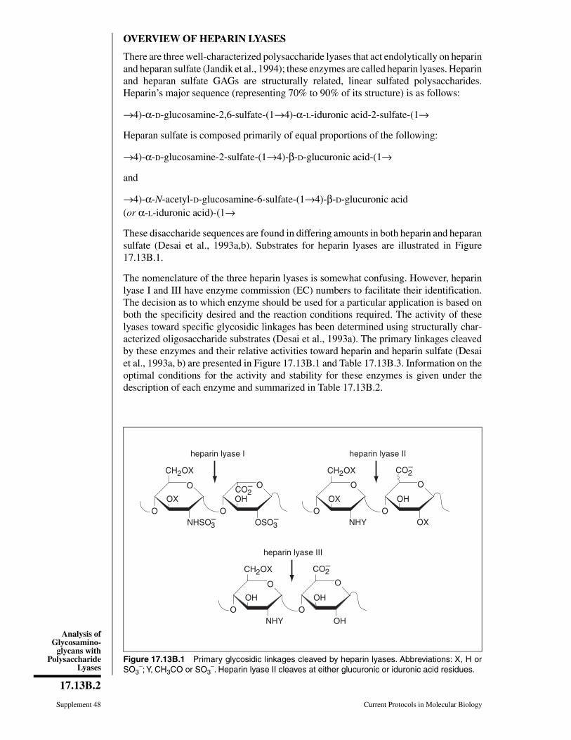
OVERVIEW OF HEPARIN LYASES
There are three well-characterized polysaccharide lyases that act endolytically on heparinand heparan sulfate (Jandik et al., 1994); these enzymes are called heparin lyases. Heparinand heparan sulfate GAGs are structurally related, linear sulfated polysaccharides.Heparin’s major sequence (representing 70% to 90% of its structure) is as follows:
→4)-α-D-glucosamine-2,6-sulfate-(1→4)-α-L-iduronic acid-2-sulfate-(1→
Heparan sulfate is composed primarily of equal proportions of the following:
→4)-α-D-glucosamine-2-sulfate-(1→4)-β-D-glucuronic acid-(1→
and
→4)-α-N-acetyl-D-glucosamine-6-sulfate-(1→4)-β-D-glucuronic acid(or α-L-iduronic acid)-(1→
These disaccharide sequences are found in differing amounts in both heparin and heparansulfate (Desai et al., 1993a,b). Substrates for heparin lyases are illustrated in Figure17.13B.1.
The nomenclature of the three heparin lyases is somewhat confusing. However, heparinlyase I and III have enzyme commission (EC) numbers to facilitate their identification.The decision as to which enzyme should be used for a particular application is based onboth the specificity desired and the reaction conditions required. The activity of theselyases toward specific glycosidic linkages has been determined using structurally char-acterized oligosaccharide substrates (Desai et al., 1993a). The primary linkages cleavedby these enzymes and their relative activities toward heparin and heparin sulfate (Desaiet al., 1993a, b) are presented in Figure 17.13B.1 and Table 17.13B.3. Information on theoptimal conditions for the activity and stability for these enzymes is given under thedescription of each enzyme and summarized in Table 17.13B.2.
CH2OX
O
OX
heparin lyase I
NHSO3–
O
CO2– O
OH
OSO3–
O
CH2OX
O
OX
NHYO
CO2–
O
OH
OXO
heparin lyase II
CH2OX
O
OH
heparin lyase III
NHYO
CO2–
O
OH
OHO
Figure 17.13B.1 Primary glycosidic linkages cleaved by heparin lyases. Abbreviations: X, H orSO3
−; Y, CH3CO or SO3−. Heparin lyase II cleaves at either glucuronic or iduronic acid residues.
Supplement 48 Current Protocols in Molecular Biology
17.13B.2
Analysis ofGlycosamino-
glycans withPolysaccharide
Lyases

ENZYMEHEPARIN LYASE I (Lohse and Linhardt, 1992)
Heparin lyase I (EC 4.2.2.7), from Flavobacterium heparinum (Cytophagia heparinia),is commonly referred to as heparinase. The enzyme has a molecular weight of 42,800 Daand a pI of 9.1 to 9.2. Heparin lyase I has a random endolytic action pattern—i.e., itrandomly acts on any site with the appropriate primary structure within the polymericsubstrate (Fig. 17.13B.1; Jandik et al., 1994).
BASICPROTOCOL
Complete Heparin Lyase–Catalyzed Depolymerization of anUnlabeled Sample
Samples consisting of tissues, biological fluids, PGs, and GAGs (UNIT 17.2) that containmicrogram quantities of heparin and are not metabolically labeled can be analyzed usingheparin lyase (see Critical Parameters for method to distinguish between heparin andheparan sulfate).
Table 17.13B.2 Reaction Conditions for Polysaccharide Lyases withOptimum Buffers and Reaction Temperatures
Lyase Buffera Optimumtemperature
Heparin I Sodium phosphate/NaCl, pH 7.1 30°C
Heparin II Sodium phosphate, pH 7.1 35°C
Heparin III Sodium phosphate, pH 7.6 35°C
Chondroitin ABC Tris⋅Cl/sodium acetate, pH 8 37°C
Chondroitin AC Tris⋅Cl/sodium acetate, pH 8 37°C
Chondroitin B Ethylenediamine/acetic acid/NaCl, pH 8
25°C
Hyaluronate Sodium acetate/NaCl, pH 5.2 >30°C
a See Reagents and Solutions for buffer recipes.
Table 17.13B.3 Activity of Heparin Lyases
Activity andsubstrate conversion
Heparin lyase I
Heparin lyase II
Heparin lyase III
Heparina
% Activityb 100 58 <1% Conversionc 58 (76)d 85 6
Heparan sulfatee
% Activity 13 100 100% Conversion 19 39 94aPorcine mucosal heparin. b Percent activity = [initial rate on the substrate examined/initial rate onsubstrate giving the highest activity] × (100).cPercent conversion = [moles of linkages cleaved/total moles of hexos-amine→uronic acid linkages] × (100).dBovine lung heparin.eBovine kidney heparan sulfate.
Current Protocols in Molecular Biology Supplement 48
17.13B.3
Preparation andAnalysis ofGlycoconjugates

Materials
Heparin- or heparan sulfate–containing sampleSodium phosphate/NaCl buffer (see recipe)Heparin lyase I solution (see recipe)
Spectropor dialysis membrane, MWCO 1000 (Spectrum)500-µl polypropylene microcentrifuge tubes30° and 100°C water baths
Additional reagents and equipment for polysaccharide dialysis (APPENDIX 3D),HPLC (UNIT 17.18), and gel-filtration chromatography (UNITS 10.9 & 17.17)
1. Dissolve sample, containing 1 µg to 1 mg heparin, in 50 µl sodium phosphate/NaClbuffer. Dialyze sample against sodium phosphate/NaCl buffer using 1000 MWCOdialysis membrane.
2. Thaw and assay activity of a frozen aliquot of enzyme.
3. Thaw 10 µl heparin lyase I solution at room temperature and add 40 µl sodiumphosphate/NaCl buffer to the 500-µl tube containing enzyme. Add 50 µl sodiumphosphate/NaCl buffer to another 500-µl tube to serve as a blank control.
Additional enzyme (10- to 100-fold) may be required to break down small, resistantoligosaccharides (Rice and Linhardt, 1989; Desai et al., 1993a).
For heparin lyase II and III, the chondroitin lyases, and hyaluronate lyase, see alternativeconditions listed in Table 17.13B.2 and in descriptions of individual enzymes below.
4. Add 50 µl sample to each tube and incubate 8 to 12 hr at 30°C.
5. Heat tube 2 min at 100°C to terminate the reaction. Analyze the product by a methodappropriate for its purity and concentration.
A pure sample containing >10 g of heparin can be analyzed by measuring the differencein absorbance at 232 nm (∆A232) in 30 mM HCl between enzyme-treated sample and blank(ε = 5500 M−1 for oligosaccharide products in 30 mM HCl). Enzyme-treated sample isdiluted with measured amounts of 30 mM HCl until the A232 is between 1 and 2. The blankis diluted with the same quantity of 30 mM HCl and its A232 is measured. The differencebetween the two measured A232 values, ∆A232, is used to calculate the moles of oligosac-charide product formed. Ten moles of oligosaccharide product is obtained for each moleof heparin. If the treated sample contains substantial amounts of protein or other sub-stances that absorb at or near 232 nm, disappearance of polysaccharide substrate can bemeasured using a dye-binding assay (Grant et al., 1984). Smaller quantities of samples orsamples of lower purity can be analyzed by HPLC or gel-filtration chromatography usingUV or conductivity detection methods.
Supplement 48 Current Protocols in Molecular Biology
17.13B.4
Analysis ofGlycosamino-
glycans withPolysaccharide
Lyases

ALTERNATEPROTOCOL
Complete Heparin Lyase–Catalyzed Depolymerization of Very Small Amounts ofRadiolabeled Glycosaminoglycans
When attempting to use heparin lyase to depolymerize radiolabeled samples that containvery small quantities of heparin, it is often useful to add cold substrate as a carrier so theactivity of heparin lyase can be distinguished from that of trace amounts of chondroitinlyases that are often present in heparin lyase preparations. Chondroitin lyase may pose aproblem when using a heparin lyase to distinguish between heparin/heparan sulfate andchondroitin/dermatan sulfate in radiolabeled samples (see Critical Parameters and Table17.13B.3). Therefore, it is recommended that cold carrier chondroitin/dermatan sulfatebe added to block the action of minor chondroitin lyase contaminants. Alternatively,chondroitin ABC lyase from Proteus vulgaris (an organism free of enzymes acting onheparin and heparan sulfate) can be used to detect chondroitin sulfate chains specifically(see discussion of chondroitin lyases).
Additional Materials (also see Basic Protocol)
Radiolabeled heparin-containing sample (UNIT 17.4)20 mg/ml chondroitin sulfate A solution (see recipe)20 mg/ml chondroitin sulfate C solution (see recipe)20 mg/ml dermatan sulfate solution (see recipe)
1. Dissolve GAG sample containing radiolabeled heparin in 50 µl sodium phos-phate/NaCl buffer. Dialyze sample against sodium phosphate/NaCl buffer using 1000MWCO dialysis membrane.
2. Thaw 10 µl of heparin lyase I solution at room temperature, immediately prior to use.
For heparin lyase II and III, the chondroitin lyases, and hyaluronate lyase, see alternativeconditions listed in Table 17.13B.2 and in descriptions of individual enzymes below.
3. Add 30 µl sodium phosphate/NaCl buffer to the 500-µl tube containing enzyme.
4. Optional: Add 1.7 µl each of 20 mg/ml chondroitin sulfate A, chondroitin sulfate C,and dermatan sulfate substrate solutions (34 µg each) to the enzyme in buffer.
This step should be performed if there is any concern that the enzyme contains chondroitinlyase impurities.
5. Add 50 µl radiolabeled heparin solution and incubate 8 to 12 hr at 30°C.
6. Heat 2 min at 100°C to terminate the reaction.
7. Analyze depolymerized radioactive sample by HPLC or gel-filtration chromatogra-phy using radioisotope detection methods.
In gel-filtration chromatography following treatment with heparin lyase, counts in frac-tions corresponding to an apparent molecular weight <1500 Da confirm the presence ofheparin/heparan sulfate. Similar results are obtained with heparin lyase II and III exceptheparin lyase III does not act on heparin (Table 17.13B.3).
Current Protocols in Molecular Biology Supplement 48
17.13B.5
Preparation andAnalysis ofGlycoconjugates
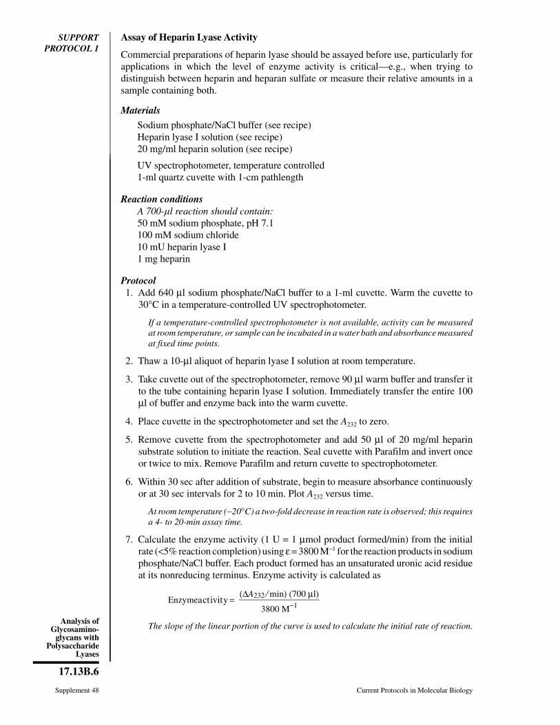
SUPPORTPROTOCOL 1
Assay of Heparin Lyase Activity
Commercial preparations of heparin lyase should be assayed before use, particularly forapplications in which the level of enzyme activity is critical—e.g., when trying todistinguish between heparin and heparan sulfate or measure their relative amounts in asample containing both.
Materials
Sodium phosphate/NaCl buffer (see recipe)Heparin lyase I solution (see recipe)20 mg/ml heparin solution (see recipe)
UV spectrophotometer, temperature controlled1-ml quartz cuvette with 1-cm pathlength
Reaction conditionsA 700-l reaction should contain:50 mM sodium phosphate, pH 7.1100 mM sodium chloride10 mU heparin lyase I1 mg heparin
Protocol1. Add 640 µl sodium phosphate/NaCl buffer to a 1-ml cuvette. Warm the cuvette to
30°C in a temperature-controlled UV spectrophotometer.
If a temperature-controlled spectrophotometer is not available, activity can be measuredat room temperature, or sample can be incubated in a water bath and absorbance measuredat fixed time points.
2. Thaw a 10-µl aliquot of heparin lyase I solution at room temperature.
3. Take cuvette out of the spectrophotometer, remove 90 µl warm buffer and transfer itto the tube containing heparin lyase I solution. Immediately transfer the entire 100µl of buffer and enzyme back into the warm cuvette.
4. Place cuvette in the spectrophotometer and set the A232 to zero.
5. Remove cuvette from the spectrophotometer and add 50 µl of 20 mg/ml heparinsubstrate solution to initiate the reaction. Seal cuvette with Parafilm and invert onceor twice to mix. Remove Parafilm and return cuvette to spectrophotometer.
6. Within 30 sec after addition of substrate, begin to measure absorbance continuouslyor at 30 sec intervals for 2 to 10 min. Plot A232 versus time.
At room temperature (∼20°C) a two-fold decrease in reaction rate is observed; this requiresa 4- to 20-min assay time.
7. Calculate the enzyme activity (1 U = 1 µmol product formed/min) from the initialrate (<5% reaction completion) using ε = 3800 M−1 for the reaction products in sodiumphosphate/NaCl buffer. Each product formed has an unsaturated uronic acid residueat its nonreducing terminus. Enzyme activity is calculated as
Enzymeactivity = (∆A232 ⁄ min) (700 µl)
3800 M−1
The slope of the linear portion of the curve is used to calculate the initial rate of reaction.
Supplement 48 Current Protocols in Molecular Biology
17.13B.6
Analysis ofGlycosamino-
glycans withPolysaccharide
Lyases

ENZYMEHEPARIN LYASE II (Lohse and Linhardt, 1992)
Heparin lyase II (no EC number), an endolytic enzyme from Flavobacterium heparinum,has a molecular weight of 84,100 Da and a pI of 8.9 to 9.1. The substrate for heparin lyaseII is indicated in Table 17.13B.3 and Figure 17.13B.1.
Protocols for the assay and use of heparin lyase II are identical to those for heparin lyaseI, with the following modifications (see Table 17.13B.2):
1. Prepare enzyme and substrate solutions in sodium phosphate buffer, pH 7.1 (seerecipes).
2. Carry out the reaction at 35°C.
3. Use 20 mg/ml heparan sulfate solution (see recipe) in Support Protocol 1 to assay forenzyme activity.
ENZYMEHEPARIN LYASE III (Lohse and Linhardt, 1992)
Heparin lyase III (EC 4.2.2.8), an endolytic enzyme from Flavobacterium heparinum, iscommonly referred to as heparitinase and has a molecular weight of 70,800 Da and a pIof 9.9 to 10.1. Heparin lyase III can be used to confirm the presence of heparan sulfate ina sample (Fig. 17.13B.1 and Tables 17.13B.1 and 17.13B.3).
Protocols for the assay and use of heparin lyase III are identical to those of heparin lyaseI, with the following modifications (see Table 17.13B.2):
1. Prepare enzyme and substrate solutions in sodium phosphate buffer, pH 7.6 (seerecipes).
2. Carry out the reaction at 35°C.
3. Use 20 mg/ml heparan sulfate solution (see recipe), instead of heparin, as the substrateto assay for enzyme activity in Support Protocol 1.
OVERVIEW OF CHONDROITIN SULFATE LYASES
There are several polysaccharide lyases that act on chondroitin sulfates, dermatan sulfate,and hyaluronate. Chondroitin sulfate galactosaminoglycans are structurally related, sul-fated, alternating 1→3, 1→4 linked, linear polysaccharides. The structure of the majordisaccharide linkage found in each chondroitin sulfate and the enzyme that acts at eachlinkage are shown in Figure 17.13B.2. Hyaluronate has the same backbone structure,except that it is not sulfated and contains N-acetylglucosamine in place of N-acetylgalac-tosamine (Fig. 17.13B.3).
The decision of which chondroitin lyase to use should be based on both the specificitydesired and the reaction conditions required (Tables 17.13B.1 & 17.13B.2).
ENZYMECHONDROITIN ABC LYASE (Yamagata et al., 1968)
Chondroitin ABC lyase (chondroitinase ABC, EC 4.2.2.4), from Proteus vulgaris, has amolecular weight of 150,000 Da. This enzyme acts endolytically on chondroitin sulfatesA-E (Fig. 17.13B.2; Jandik et al., 1994), slowly on hyaluronic acid, and not at all onheparin, heparan sulfate, or keratan sulfate.
Current Protocols in Molecular Biology Supplement 48
17.13B.7
Preparation andAnalysis ofGlycoconjugates
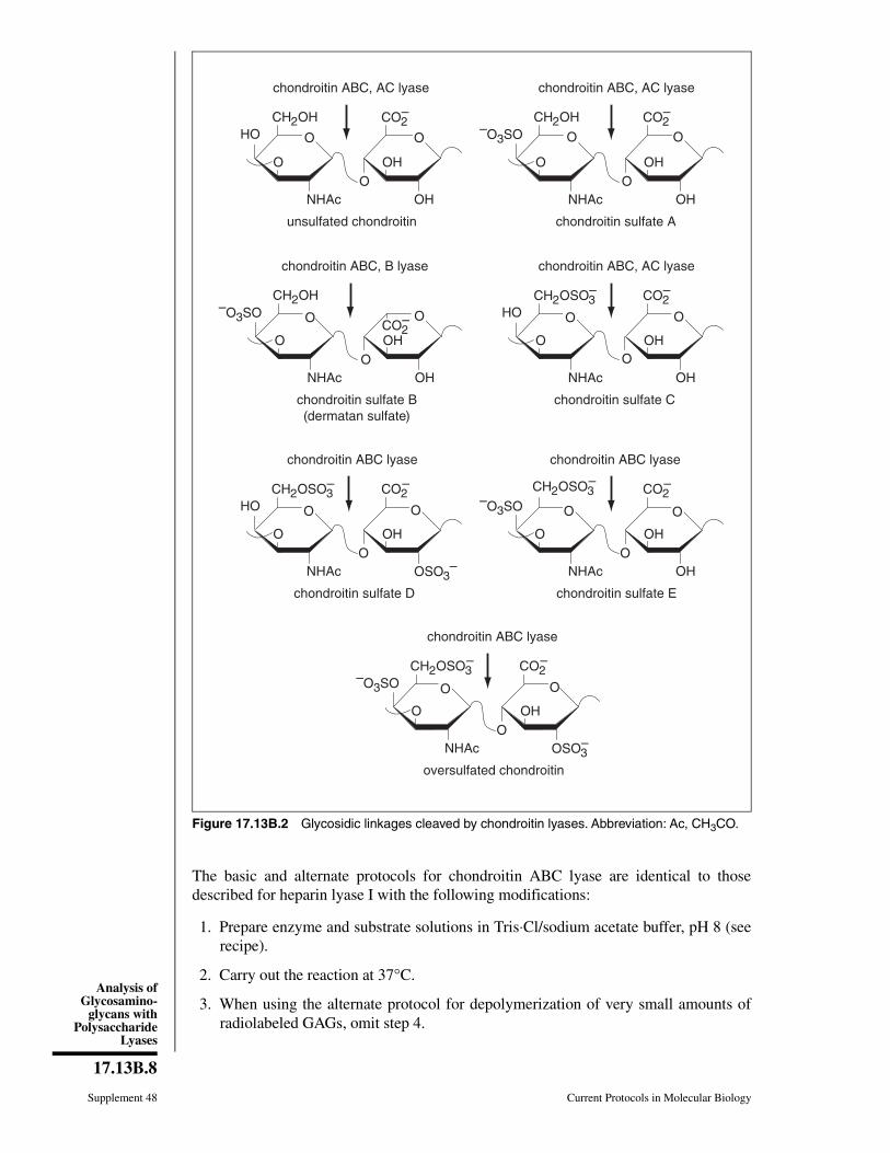
The basic and alternate protocols for chondroitin ABC lyase are identical to thosedescribed for heparin lyase I with the following modifications:
1. Prepare enzyme and substrate solutions in Tris⋅Cl/sodium acetate buffer, pH 8 (seerecipe).
2. Carry out the reaction at 37°C.
3. When using the alternate protocol for depolymerization of very small amounts ofradiolabeled GAGs, omit step 4.
CH2OSO3–
CH2OSO3–
CH2OSO3–
CH2OH
O
chondroitin ABC, AC lyase
NHAc
HOCO2
–
OH
OHO
unsulfated chondroitin
CH2OH
O
chondroitin ABC, AC lyase
NHAc
CO2–
OH
OHO
chondroitin sulfate A
–O3SO
CH2OSO3–
O
NHAc
HOCO2
–
OH
OHO
chondroitin ABC, B lyase
chondroitin sulfate B(dermatan sulfate)
CH2OH
O
NHAc
CO2–
OH
OHO
–O3SO
chondroitin ABC, AC lyase
chondroitin sulfate C
O
chondroitin ABC lyase
NHAc
HOCO2
–
OH
OSO3–
OO
chondroitin ABC lyase
NHAc
CO2–
OH
OHO
–O3SO
chondroitin ABC lyase
O
NHAc
CO2–
OHO
–O3SO
chondroitin sulfate D
oversulfated chondroitin
chondroitin sulfate E
OSO3–
O O
O O
O O
O O
O O O O
O O
Figure 17.13B.2 Glycosidic linkages cleaved by chondroitin lyases. Abbreviation: Ac, CH3CO.
Supplement 48 Current Protocols in Molecular Biology
17.13B.8
Analysis ofGlycosamino-
glycans withPolysaccharide
Lyases

A radioactive sample containing chondroitin sulfate or dermatan sulfate can be analyzed,following chondroitin ABC lyase treatment, by HPLC (UNIT 17.18) or gel-filtration chro-matography (UNITS 10.9 & 17.17) using radioisotope detection methods. In gel-filtrationchromatography following treatment with chondroitin ABC lyase, counts in fractionscorresponding to a molecular weight <1000 Da confirm the presence of chondroitinsulfate or dermatan sulfate.
SUPPORTPROTOCOL 2
Assay of Chondroitin ABC Lyase Activity
Commercial preparations of chondroitin lyase should be assayed before use, particularlyfor applications in which the level of enzyme activity is critical.
Materials
Tris⋅Cl/sodium acetate buffer, pH 8.0 (see recipe)Chondroitin ABC lyase solution (see recipe)20 mg/ml chondroitin sulfate A (see recipe), chondroitin sulfate C solution (see
recipe), or dermatan sulfate solution (see recipe)
UV spectrophotometer, temperature controlled1-ml quartz cuvette with 1-cm path length
Reaction conditionsA 700-l reaction should contain:50 mM Tris⋅Cl, pH 860 mM sodium acetate10 mU chondroitin lyase1 mg chondroitin sulfate A or C, or dermatan sulfate
Protocol1. Add 640 µl Tris⋅Cl/sodium acetate buffer to a 1-ml quartz cuvette. Warm the cuvette
to 37°C in a temperature-controlled spectrophotometer.
If a temperature-controlled spectrophotometer is not available, activity can be measuredat room temperature or the sample can be incubated in a water bath and the absorbancemeasured at fixed time points.
2. Thaw a 10-µl aliquot of chondroitin ABC lyase solution at room temperature.
3. Take cuvette out of the spectrophotometer, remove 90 µl warm buffer, and transfer itto enzyme solution. Immediately transfer entire 100 µl buffer plus enzyme back tothe warm cuvette.
4. Place cuvette in spectrophotometer and set the absorbance at 232 nm (A232) to zero.
CH2OH
O
hyaluronate lyase, AC, ABC
NHAcHO
CO2–
OH
OHO
hyaluronate
O O
Figure 17.13B.3 Glycosidic linkagein hyaluronate cleaved by hyaluronatelyase and chondroitin AC and ABClyases. Abbreviation: Ac, CH3CO.
Current Protocols in Molecular Biology Supplement 48
17.13B.9
Preparation andAnalysis ofGlycoconjugates
5. Remove cuvette from spectrophotometer and add 50 µl of 20 mg/ml chondroitin Aor C or dermatan sulfate solution to initiate reaction. Seal cuvette with Parafilm andinvert once or twice to mix. Remove Parafilm and return cuvette to spectrophotome-ter.
To assay for chondroitin AC lyase activity, use chondroitin A or C as substrate. To assayfor chondroitin B lyase activity, use dermatan sulfate as substrate (see Table 17.13B.1 anddescription of individual enzymes below).
6. Within 30 sec after adding substrate begin to measure the absorbance continuouslyor at 30-sec intervals for 2 to 10 min. Plot A232 versus time.
7. Calculate the enzyme activity (1 U = 1 µmol product formed/min) from the initialrate (<5% reaction completion) using ε = 3800 M−1 for reaction products at pH 8.Enzyme activity is calculated as
Enzymeactivity = (∆A232 ⁄ min) (700 µl)
3800 M−1
Calculate the number of product molecules formed per substrate molecule from theA232 measured at reaction completion.
ENZYME CHONDROITIN AC LYASE (Yamagata et al., 1968; Hiyama and Okada, 1975;Michelacci and Dietrich, 1975)
Two chondroitin AC lyases from Arthrobacter aurescens and Flavobacterium heparinumshare the same enzyme commission number (EC 4.2.2.5). Chondroitin AC lyase from A.aurescens has a molecular weight of 76,000 Da (Hiyama and Okada, 1975) and a pI of5.46. It acts exolytically on chondroitin sulfate A and C (Jandik et al., 1994) and exhibitsa three-fold higher activity on hyaluronate. It can act at the glucuronic acid residues indermatan sulfate (Fig. 17.13B.2) but does not act on heparin or heparan sulfate (Fig.17.13B.1). Chondroitin AC lyase from F. heparinum acts endolytically on chondroitinsulfates A and C (Jandik et al., 1994), on hyaluronate, and at the glucuronate residues ofdermatan sulfate (Fig. 17.13B.2; Gu et al., 1993). It does not act on heparin, heparansulfate, or keratan sulfate (Yamagata et al., 1968).
The protocols for chondroitin AC lyase are identical to those described for chondroitinABC lyase except chondroitin A or C should be used as substrate in Support Protocol 2for assaying chondroitin lyase activity.
ENZYME CHONDROITIN B LYASE (Michelacci and Dietrich, 1975)
Chondroitin B lyase (no EC number), from Flavobacterium heparinum, has a molecularweight of 55,000 Da. Chondroitin B lyase acts only on dermatan sulfate and not on theglucuronate residues of chondroitin sulfates A, C, and hyaluronate, and not on heparin orheparan sulfate (Fig. 17.13B.2).
This enzyme can be assayed as described for chondroitin ABC lyase using dermatansulfate as substrate. The heparin lyase I protocols are used for sample analysis with thisenzyme, with the following modifications (see Table 17.13B.2):
1. Prepare enzyme and substrate solutions in ethylenediamine/acetic acid/NaCl buffer,pH 8 (see recipe).
2. Carry out the reaction at 25°C.
3. Use dermatan sulfate solution (see recipe) as substrate in Support Protocol 2.
Supplement 48 Current Protocols in Molecular Biology
17.13B.10
Analysis ofGlycosamino-
glycans withPolysaccharide
Lyases

ENZYMEHYALURONATE LYASE (Vesterberg, 1968; Rautela and Abramson, 1973;Sasaki et al., 1982)
Hyaluronate lyases (EC 4.2.2.1 and EC 4.2.99.1) act only on hyaluronate (Hiyama andOkada, 1975) allowing hyaluronate (Fig. 17.13B.3) to be distinguished from chondroitinsulfate (Fig. 17.13B.2 and Table 17.13B.1). The enzyme from Streptomyces hyaluro-lyticus has been purified and has a pI <7 (Sasaki et al., 1982). Homogeneous Staphylo-coccus aureus hyaluronate lyase has a molecular weight of 84,000 Da (Rautela andAbramson, 1973) and a pI of 7.4 to 7.9 (Vesterberg, 1968).
Reaction conditionsA 700-l reaction should contain:50 mM sodium acetate, pH 5.2125 mM sodium chloride10 mU hyaluronate lyase1 mg hyaluronate
The basic, alternate, and support protocols for hyaluronate lyase are identical to thosedescribed for heparin lyase with the following modifications (see Table 17.13B.2):
Protocol1. Use sodium acetate/NaCl buffer, pH 5.2 (see recipe), to prepare enzyme (hyaluronate
lyase; see recipe) and substrate solutions.
2. Perform the reaction at 30° to 60°C.
Higher temperatures can be useful to reduce substrate viscosity—hyaluronic acid is veryviscous. Higher temperatures will also increase the reaction rate, and they can be used toinhibit the activity of other lyases that may be present as contaminants of the hyaluronatelyase preparation.
3. Use hyaluronate solution (see recipe), instead of heparin solution, as substrate toassay enzyme activity (see Support Protocol 1).
4. When using the Alternate Protocol for depolymerization of very small amounts ofradiolabeled GAGs, omit step 4.
Following treatment with hyaluronate lyase, a radioactive sample containing hyaluronatecan be analyzed by HPLC (UNIT 17.18) or gel-filtration chromatography (UNIT 17.17) usingradioisotope detection methods. In gel-filtration following treatment with hyaluronatelyase, counts in fractions corresponding to a molecular weight <500 Da confirm thepresence of hyaluronate.
Testicular hyaluronidase is a hydrolase (EC 3.2.1.35) that also acts on hyaluronate (alsoon chondroitin sulfates A and C). Because it is not a polysaccharide lyase, testicularhyaluronidase cannot be assayed with the support protocol, but it can be used in the basicand alternate protocols with the modifications described above.
Current Protocols in Molecular Biology Supplement 48
17.13B.11
Preparation andAnalysis ofGlycoconjugates
REAGENTS AND SOLUTIONS
Use deionized, distilled water in all recipes and protocol steps. For common stock solutions, seeAPPENDIX 2; for suppliers, see APPENDIX 4.
Chondroitin lyase solutions, 1 mU/lDissolve 0.1 U lyophilized enzyme in 100 µl Tris⋅Cl/sodium acetate buffer (seerecipe) for chondroitin ABC lyase (chondroitinase ABC; Sigma or Seikagaku) andchondroitin AC lyase (chondroitinase AC from Arthrobacter aurescens and Flavo-bacterium heparinum; Sigma or Seikagaku) or ethylenediamine/acetic acid/NaClbuffer (see recipe) for chondroitin B lyase (chondroitinase B; Sigma or Seikagaku).Store in 10-mU aliquots in 500-µl polypropylene tubes <1 year at −70°C.
Different suppliers use different definitions for units of enzyme activity. Enzyme activity levelsshould be assayed and standardized, as described in the support protocols, in the laboratorybefore the enzymes are used in analytical studies.
Chondroitin sulfate A or C solution, 20 mg/mlDissolve 20 mg chondroitin sulfate A or C, sodium salt (Sigma or Seikagaku) in1 ml Tris⋅Cl/sodium acetate buffer, pH 8 (see recipe) or other buffer appropriate toenzyme/substrate pair (see Table 17.13B.2). Store 1 year at <0°C.
Dermatan sulfate solution, 20 mg/mlDissolve 20 mg dermatan sulfate, sodium salt (Sigma or Seikagaku) in 1 mlethylenediamine/acetic acid/NaCl buffer, pH 8 (see recipe) or other buffer appro-priate to enzyme/substrate pair (see Table 17.13B.2). Store 1 year at <0°C.
Ethylenediamine/acetic acid/NaCl bufferDissolve 3.0 g ethylenediamine (50 mM final) and 1.7 g NaCl (30 mM final) in900 ml H2O. Adjust pH with glacial acetic acid to pH 8, and bring volume to 1 literwith H2O. Store <1 month at 4°C.
Heparan sulfate solution, 20 mg/mlDissolve 20 mg heparan sulfate, sodium salt from bovine kidney (Seikagaku) in 1 mlsodium phosphate buffer, pH 7.6 (see recipe). Store 1 year at 0°C.
Heparin lyase solutions, 1 mU/lDissolve 0.1 U lyophilized enzyme in 100 µl sodium phosphate/NaCl buffer (seerecipe) for heparin lyase I or sodium phosphate buffer (see recipe) for heparin lyaseII and III adjusted to the appropriate pH. Store enzyme in 10 mU aliquots in 500-µlpolypropylene tubes <1 year at −70°C.
Heparin lyase I from Flavobacterium heparinum is sold as heparinase I (Sigma) andheparinase (Seikagaku). Heparin lyase II is sold as heparinase II (Sigma) and heparitinaseII (Seikagaku). Heparin lyase III from Flavobacterium heparinum is sold as heparinase III(Sigma) and heparatinase or heparatinase I (Seikagaku).
Different suppliers use different definitions for units of enzyme activity. Enzyme activity levelsshould be assayed and standardized, as described in the support protocols, in the laboratorybefore the enzymes are used in analytical studies.
Heparin solution, 20 mg/mlDissolve 20 mg heparin, sodium salt (140 to 180 USP U/mg), from porcine intestinalmucosa or bovine lung (Sigma) in sodium phosphate/NaCl buffer, pH 7.1 (seerecipe). Store 1 year at 0°C.
Different suppliers use different definitions for units of enzyme activity. Enzyme activity levelsshould be assayed and standardized, as described in the support protocols, in the laboratorybefore the enzymes are used in analytical studies.
Supplement 48 Current Protocols in Molecular Biology
17.13B.12
Analysis ofGlycosamino-
glycans withPolysaccharide
Lyases
Hyaluronate lyase solution, 1 mU/lDissolve 0.1 U lyophilized enzyme (hyaluronidase from Streptomyces hyaluro-lyticus; Sigma or Seikagaku) in 100 µl sodium acetate/NaCl buffer, pH 5.2 (seerecipe). Aliquot 10 mU to 500-µl polypropylene tubes and store <1 year at −70°C.
Different suppliers use different definitions for units of enzyme activity. Enzyme activity levelsshould be assayed and standardized, as described in the support protocols, in the laboratorybefore the enzymes are used in analytical studies.
Hyaluronate solution, 1.4 mg/mlDissolve 1.4 mg hyaluronate in 1 ml sodium acetate/NaCl buffer, pH 5.2 (see recipe).Store 1 year at <0°C.
Hyaluronate of high molecular weight is very viscous. It is best to use a hyaluronate of lowto medium molecular weight to measure activity.
Sodium acetate/NaCl bufferDissolve 4.1 g sodium acetate (30 mM final) and 7.3 g NaCl (125 mM final) in 900ml H2O. Adjust pH with glacial acetic acid to pH 5.2 and bring volume to 1 literwith H2O. Store <1 month at 4°C.
Sodium phosphate bufferDissolve 7.1 g dibasic sodium phosphate (50 mM final) in 900 ml H2O. Adjust pHwith concentrated phosphoric acid to pH 7.1 for heparin lyase II and pH 7.6 forheparin lyase III. Bring volume to 1 liter with H2O. Store <1 month at 25°C.
Sodium phosphate/NaCl bufferDissolve 7.1 g dibasic sodium phosphate (50 mM final) and 5.8 g sodium chloride(100 mM final) in 900 ml H2O. Adjust pH with phosphoric acid to pH 7.1 and bringvolume to 1 liter with H2O. Store <1 month at 25°C.
Tris⋅Cl/sodium acetate bufferDissolve 6.05 g Tris base (50 mM final) and 8.17 g sodium acetate (60 mM final)in 900 ml H2O. Adjust pH to 8.0 with concentrated hydrochloric acid, and bringvolume to 1 liter with H2O. Store <1 month at 4°C.
COMMENTARY
Background InformationProteoglycans (PGs) are primarily found in
the extracellular matrix and are important incell-cell interaction. Glycosaminoglycans(GAGs) are the dominant physical, chemical,and biological features of PGs. Lyases cleavespecific glycosidic linkages at C4 of uronic acidresidues present in GAGs (Figs. 17.13B.1,17.13B.2, and 17.13B.3) through an eliminasemechanism, resulting in unsaturated oligosac-charide products that have UV absorbancespectra maxima at 232 nm (Linhardt et al.,1986). An eliminase catalyzes an eliminationreaction that results in the formation of a doublebond. In contrast, a hydrolase (the most com-mon class of enzymes acting on polysaccha-rides) breaks down polysaccharides throughthe addition of water and does not result in theformation of a double bond. Lyases are usefulfor identifying and distinguishing the GAGs
present in unlabeled or radiolabeled samples(see Table 17.13B.1).
Critical Parameters andTroubleshooting
Lyases are primarily of microbial origin andoften a single organism produces multiplelyases acting on a variety of PGs and GAGs.Thus, it must be recognized that despite thehigh level of purity of the commercially avail-able enzymes, they may contain small amountsof enzymatic impurities. These impurities cancause misleading results, particularly whenlarge quantities of enzyme are used to treat verysmall quantities of sample—e.g., when theseenzymes are used to analyze radiolabeled sam-ples.
Protease contamination can also be presentin the enzyme preparation. Commercial en-zymes often contain bovine serum albumin
Current Protocols in Molecular Biology Supplement 48
17.13B.13
Preparation andAnalysis ofGlycoconjugates
(BSA) for stabilization during lyophilization,and this greatly reduces potential problemsassociated with proteolytic contamination.
Complete degradation of a GAG. Table17.13B.1 shows how the polysaccharide lyasescan be used, alone or in combination, to deter-mine the content of a GAG. All three heparinlyases used in combination (at equal-unit con-centrations in 50 mM sodium phosphate buffer,pH 7, at 30°C) will degrade heparin or heparansulfate to disaccharides, although a small quan-tity of lyase-resistant tetrasaccharide may re-main. Chondroitin ABC lyase can be used tocompletely digest a mixture of chondroitin sul-fates. If hyaluronate and chondroitin sulfate areboth present, it is advisable to use an equal-unitmixture of chondroitin ABC and AC lyases. Forcomplete degradation of GAGs, except keratansulfate (see UNIT 17.2), it is advisable to treat thesample with an equal-unit mixture of heparinlyases I, II, and III and chondroitin ABC andAC lyases in sodium phosphate/NaCl buffer,pH 7, 30°C.
Heparin lyase III can be used to distinguishbetween samples containing heparan sulfateand those containing heparin. Although heparinlyase I (heparinase) has been used by investi-gators to demonstrate the presence of heparinin a sample, it also acts at some linkages presentin heparan sulfate (Table 17.13B.3). Gel-filtra-tion analysis can help distinguish betweenheparin and heparan sulfate because the largeoligosaccharides formed from heparan sulfateelute in the void volume. In addition, oligosac-charide product compositional analysis (UNIT
17.19) is useful in distinguishing heparin fromheparan sulfate when using heparin lyase I.
Caution is required when attempting to dis-tinguish heparin/heparan sulfate from chon-droitin/dermatan sulfate using heparin lyases.Flavobacterium heparinum produces bothheparin and chondroitin/dermatan sulfatelyases; thus, minor contaminating activities canresult in false positives. Step 4 of the AlternateProtocol for the use of heparin lyase on radio-labeled samples—incubation in the presence ofnonspecific substrate—is included to eliminatefalse positives. The use of chondroitin ABClyase, which contains no heparin lyase activity,is complicated by heparin’s inhibition of chon-droitin ABC lyase (Nakada and Wolfe, 1961).This inhibition is overcome by using excessenzyme (Linhardt et al., 1991).
Chondroitin sulfate is routinely distin-guished from dermatan sulfate using chondroi-tin AC and ABC lyases (Saito et al., 1968). Thiscan give slightly different results from those
obtained using chondroitin AC and B lyases(Linhardt et al., 1991). Chondroitin AC lyasesfrom F. heparinum and Arthrobacter aurescensboth act at glucuronate-containing linkages indermatan sulfates (Gu et al., 1993) and areuseful for analysis of the glucuronic acid con-tent of dermatan sulfate (Linhardt et al., 1991).
Hyaluronate content can be determined us-ing hyaluronate lyase, which is specific forhyaluronate. Although hyaluronate lyases areinhibited by chondroitin sulfates, the additionof salt (150 mM NaCl) overcomes this inhibi-tion (Nakada and Wolfe, 1961; Yamagata et al.,1968).
Contaminants in samples. The presence ofcertain metals (particularly divalent metals),proteases, polyanions, detergents (SDS andTriton X-100), and denaturants (urea andguanidine) can interfere with the activity oflyases. Before using these enzymes, detergentsshould be removed by precipitation with potas-sium chloride or by using a detergent-removalcolumn such as Biobeads (Bio-Rad). Urea andguanidine should be removed by exhaustivedialysis using controlled-pore dialysis mem-brane (MWCO 1000).
Reaction conditions. Lyases are compatiblewith a wide range of buffers including succi-nate, acetate, ethylenediamine acetate, Tris⋅Cl,bis-Trispropane⋅HCl, sodium phosphate,MOPS, TES, and HEPES (Lohse and Linhardt,1992). The presence of calcium may eitherenhance or reduce lyase activity. Its effect isprobably due to changes calcium causes in theGAG substrate conformation and not throughdirect interaction with the enzyme. Becausecalcium is incompatible with certain buffers,e.g., phosphate, and can lead to variable results,its use is not recommended. The pH optima forlyases are broad—between pH 5 and 9.Hyaluronate lyase works best at pH <7, andchondroitin and heparin lyases work best at pH≥7. Lyases can be used at temperatures between20° and 40°C. Optimum temperatures are al-ways a compromise between activity and sta-bility considerations. If enzyme instability is aconcern due to elevated temperature or pro-longed incubation time, heparin lyase II shouldbe used to degrade heparin or heparan sulfate,chondroitin ABC lyase should be used to de-grade chondroitin sulfates and dermatan sul-fates, and hyaluronate lyase should be used forhyaluronate. Optimal reaction conditions forthe polysaccharide lyases are summarized inTable 17.13B.2.
Removal of lyase and lyase activity. Follow-ing the use of a lyase, residual lyase activity can
Supplement 48 Current Protocols in Molecular Biology
17.13B.14
Analysis ofGlycosamino-
glycans withPolysaccharide
Lyases
be destroyed by heating the reaction mixture to100°C or by adding denaturants or detergents.Most lyases are cationic proteins and can beremoved from anionic oligosaccharide prod-ucts by passing the reaction mixture through asmall cation-exchange column, such as SP-Sephadex (Sigma), adjusted to an acidic pH.The oligosaccharide products (void volume)are then readjusted to neutral pH and analyzed(UNITS 17.17 & 17.19). This method can also be usedto remove BSA, an excipient found in many ofthe commercial enzymes, from the oligosac-charide products.
Enzyme stability during storage. These en-zymes can be stored in their lyophilized orreconstituted states at −20° or −70°C for ≤1year. Once an enzyme is reconstituted, it shouldbe aliquoted and frozen immediately. Singlealiquots can be thawed to assay the enzyme orfor use in an experiment. Heparin lyase II isvery stable but heparin lyase III is unstable andshould be used immediately after removingfrom frozen storage. The heparin lyases, par-ticularly heparin lyase III, are sensitive tofreeze-thawing and lyophilization. Among thechondroitin lyases, chondroitin AC lyase ismost susceptible to thermal inactivation(Michelacci and Dietrich, 1975). Lyase storagestability is enhanced by high (>2 mg/ml) pro-tein concentrations. This is often accomplishedby addition of BSA.
Anticipated ResultsWhen used correctly, an active lyase should
specifically catalyze the breakdown of its GAGsubstrates. The types of GAGs present in asample can be easily identified by using multi-ple lyases. The amount of product formed, andthus the amount of GAG in the sample, can bedetermined. Accuracy of analysis depends on anumber of factors including the complexity ofthe sample and the types and concentrations ofGAGs present. For a sample containing oneGAG in high concentration with no contami-nating proteins, salts, detergents, etc., GAGconcentration can be determined to ±5%. For acomplex sample containing many differentGAGs at low concentrations in the presence ofhigh levels of contaminants, it may only bepossible to estimate GAG concentration to±100%.
Time ConsiderationsIt takes about half a day to prepare buffers
and solutions and to reconstitute, aliquot, andfreeze the enzyme. One aliquot of frozen en-zyme can be thawed and assayed to ensure that
the enzyme is active and has been stored prop-erly. Application of the enzyme to determinethe presence or type of GAG in a sample re-quires ∼3 days for sample preparation, over-night treatment with enzyme, and analysis.
Literature CitedDesai, U.R., Wang, H.H., and Linhardt, R.J. 1993a.
Substrate specificity of the heparin lyase fromFlavobacterium heparinum Arch. Biochem. Bio-phys. 306:461-468.
Desai, U.R., Wang, H.M., and Linhardt, R.J. 1993b.Specificity studies on the heparin lyases fromFlavobacterium heparinum. Biochemistry32:8140-8145.
Grant, A.C., Linhardt, R.J., Fitzgerald, G., Park, J.J.,and Langer, R. 1984. Metachromatic activity ofheparin and heparin fragments. Anal. Biochem.137:25-32.
Gu, K., Liu, J., Pervin, A., and Linhardt, R.J. 1993.Comparison of the activity of two chondroitinAC lyases on dermatan sulfate. Carbohydr. Res.244:369-377.
Hiyama, K. and Okada, S. 1975. Crystallization andsome properties of chondroitinase from Arthro-bacter aurescens. J. Biol. Chem. 250:1824-1828.
Jandik, K.A., Gu, K., and Linhardt, R.J. 1994. Ac-tion pattern of polysaccharide lyases on glycos-aminoglycans. Glycobiology 4:000-000.
Linhardt, R.J., Cooney, C.L., and Galliher, P.M.1986. Polysaccharide lyases. Appl. Biochem.Biotechnol. 12:135-177.
Linhardt, R.J., Al-Hakim, A., Liu, J., Hoppensteadt,D., Mascellani, G., Bianchini, P., and Fareed, J.1991. Structural features of dermatan sulfatesand their relationship to anticoagulant and an-tithrombotic activities. Biochem. Pharmacol.42:1609-1619.
Lohse, D.L. and Linhardt, R.J. 1992. Purificationand characterization of heparin lyases from Fla-vobacterium heparinum. J. Biol . Chem.267:24347-24355.
Michelacci, Y.M. and Dietrich, C.P. 1975. A com-parative study between a chondroitinase B and achondroitinase AC from Flavobacterium hepar-inum. Biochem. J. 151:121-129.
Nakada, H.I. and Wolfe, J.B. 1961. Studies on theenzyme chondroitinase: Product structure andion effects. Arch. Biochem. Biophys. 94:244-251.
Rautela, G.S. and Abramson, C. 1973. Crystal-lization and partial characterization of Staphylo-coccus aureus hyaluronate lyase. Arch. Biochem.Biophys. 158:687-693.
Rice, K.G. and Linhardt, R.J. 1989. Study of definedoligosaccharide substrates of heparin andheparan monosulfate lyases. Carbohydr. Res.190:219-233.
Saito, H., Yamagata, T., Suzuki, S. 1968. Enzymaticmethods for the determination of small quanti-ties of isomeric chondroitin sulfates. J. Biol.Chem. 243:1536-1542.
Current Protocols in Molecular Biology Supplement 48
17.13B.15
Preparation andAnalysis ofGlycoconjugates
Sasaki, I., Gotoh, H., Yamamoto, R., Tanaka, H.,Takami, K., Yamashita, K., Yamashita, J., andHorio, T. 1982. Hydrophobic-ionic chromatog-raphy: Its application to microbial glucose oxi-dase, hyaluronidase, cholesterol oxidase, andcholesterol esterase. J. Biol. Chem. 91:1555-1561.
Vesterberg, O. 1968. Studies on extracellular pro-teins from Staphylococcus aureus III. Investiga-tions on the heterogeneity of hyaluronate lyaseusing the method of isoelectric focusing. Bio-chim. Biophys. Acta 168:218-226.
Yamagata, T., Saito, H., Habuchi, O., and Suzuki, S.1968. Purification and properties of bacterialchondroitinases and chondrosulfatases. J. Biol.Chem. 243:1523-1535.
Key ReferencesDesai et al., 1993a, b. See above.
Describes the specificity of the heparin lyases indetail.
Lohse and Linhardt, 1992. See above.
Describes physical and catalytic properties of theheparin lyases.
Michelacci and Dietrich, 1975. See above.
Describes kinetic properties of chondroitin B lyaseand chondroitin AC lyase.
Seikagaku Company Product Literature, 1991.
Describes general properties and assay conditionsfor all of the polysaccharide lyases.
Saito et al., 1968. See above.
Good example of how different chondroitin sulfatescan be distinguished using chondroitin ABC lyaseand chondroitin AC lyase.
Contributed by Robert J. LinhardtUniversity of IowaIowa City, Iowa
Supplement 48 Current Protocols in Molecular Biology
17.13B.16
Analysis ofGlycosamino-
glycans withPolysaccharide
Lyases

UNIT 17.14APreparation of Glycopeptides
Generation of glycopeptides from glycoproteins is frequently useful when analyzing aprotein’s oligosaccharide side chains. Freed from the bulk of the polypeptide backboneby proteolysis, glycopeptides can be characterized by a variety of techniques. Thisapproach is especially useful for proteins with multiple glycosylation sites, but it is alsoapplicable when concern exists that the protein’s structure and solubility will interferewith particular techniques such as gel-filtration and lectin-affinity chromatography (UNIT
17.1), glycosyltransferase assays (UNIT 17.6), and colorimetric assays (UNIT 17.9).
Extensive proteolysis with pronase or proteinase K results in oligosaccharides with oneor a few amino acid residues attached. These glycopeptides are suitable for compositionalanalyses (UNIT 17.16), exo- and endoglycosidase digestions (UNITS 17.12 & 17.13), and lectin-affinity chromatography (UNIT 17.1). This technique, detailed in the first basic protocol, isoften employed as a first step in characterizing oligosaccharides on very large glycopro-teins such as proteoglycans and mucins.
Limited proteolysis with a specific endoproteinase (e.g., trypsin, α-chymotrypsin, and V8protease), as described in the second basic protocol, leaves a larger peptide attached tothe oligosaccharide. The resulting glycopeptides are generally suitable substrates forPeptide:N-glycosidase F (PNGase F; UNIT 17.13), an enzyme useful in defining oligosac-charide-peptide linkages. Additionally, they can be separated by C18 reversed-phasechromatography, resulting in a glycopeptide map that is analogous to a peptide map (UNIT
10.12), and used for detection of glycosylation sites (UNIT 17.14B).
Alternative approaches for removing oligosaccharide side chains from the peptide includeβ-elimination (UNIT 17.15), endoglycosidases (UNIT 17.13), phospholipases (for GPI-an-chored proteins; UNIT 17.8), or hydrazinolysis (Takasaki et al., 1982).
BASICPROTOCOL
EXTENSIVE PROTEOLYTIC DIGESTION OF GLYCOPROTEINS
In this protocol, glycoproteins are applied to a Sephacryl S-200 gel-filtration column toseparate them from any lower-molecular-weight material that may be present (e.g.,degradation products or other low-molecular-weight contaminants). Denatured glycopro-teins in SDS will generally elute in the void volume (Vo); the contaminants elute after theVo. The glycoproteins pooled from the Vo are then digested with pronase or proteinase Kand rechromatographed on the same column. Glycopeptides are recovered in the materialthat now elutes after the Vo and are desalted before further use.
If the sample has already been purified by techniques such as immunoprecipitation (UNIT
10.16), dialysis (APPENDIX 3), or gel filtration (UNIT 10.9) which reliably remove low-molecu-lar weight contaminants, it can be digested directly with pronase or proteinase K. Afterboiling to terminate the protease reaction, the glycopeptides can be desalted. Thus, theuse of SDS and passage over Sephacryl S-200 can be completely avoided.
Generation of glycopeptides from a sample metabolically labeled with a radioactive sugarprecursor (UNIT 17.4) is described and radioactivity is followed for each gel-filtration run.However, the same approach can be employed with unlabeled glycoproteins, followingthe column profiles with a carbohydrate-specific colorimetric assay (UNITS 17.9 & 17.16) ifsufficient mass is present (see specific units for detection limits). Suitable samples rangefrom total cellular lysates (containing multiple different glycoproteins together withdegradation products and precursors) to highly purified protein samples.
Supplement 32
Contributed by Leland D. PowellCurrent Protocols in Molecular Biology (1995) 17.14.1-17.14.9Copyright © 2000 by John Wiley & Sons, Inc.
17.14.1
Preparationand Analysis ofGlycoconjugates

Materials
Radiolabeled glycoprotein sample (UNIT 17.4)20% (w/v) sodium dodecyl sulfate (SDS)1 M 2-mercaptoethanol (2-ME)Sephacryl S-200 gel-filtration column (50- to 75-ml bed volume; Sigma or
Pharmacia Biotech), equilibrated (UNIT 10.9) in ammonium formate/azide solutioncontaining 0.1% (w/v) SDS (see recipe)
100% and 85% (v/v) acetone (HPLC or ACS grade), ice-cold0.1 M Tris⋅Cl, pH 7.5 (APPENDIX 2), without or with 10 mM CaCl2 (Tris/CaCl2)Pronase or proteinase K stock solutionPhenylmethylsulfonylfluoride (PMSF), in 100% ethanolSephadex G-15 (Sigma or Pharmacia Biotech) or Bio-Gel P-2 (Bio-Rad) columns
(30- to 60-ml bed volume), equilibrated in ammonium formate/azide solution
15- or 50-ml conical polypropylene tubesBeckman TJ-6 centrifuge or equivalentWater bath, 50°C
Additional reagents and equipment for preparation and standardization ofgel-filtration columns and sample desalting (UNIT 10.9), quantitation of proteins(UNIT 10.1), and acetone precipitation (UNIT 17.10)
Purify the protein sample1. Place radiolabeled glycoprotein sample in a 15-ml conical polypropylene tube. Add
1⁄10 vol of 20% SDS and 1⁄50 vol of 1 M 2-ME and boil 5 min. Cool to room temperatureand centrifuge 10 min at 1000 × g (2000 rpm in a Beckman TJ-6), room temperatureto remove any precipitate. Save the supernatant.
The protein sample should contain <1 g to several mg, depending on the source. Thesample volume should be ≤1 ml. If the volume of the initial sample is >1 ml, lyophilize andresuspend it in 1 ml water (or less, if solubility permits). Large amounts of protein mayrequire a larger volume, depending on the protein’s solubility. If a larger volume is used,the volume of the Sephacryl S-200 column should be increased so that the sample volumeis no more than 5% of the bed volume of the column.
Boiling ensures that any endogenous proteases or glycosidases are inactivated. Forproteins known to be soluble without SDS, the addition of SDS can be eliminated. Afterboiling, the sample can be freed of low-molecular-weight contaminants, if necessary, bychromatography directly on Sephacryl S-200 (step 2) using a buffer in which the sample isknown to be soluble.
2. Apply supernatant to Sephacryl S-200 column (50- to 75-ml bed-volume) equili-brated in ammonium formate/azide solution containing 0.1% (w/v) SDS. Collect 1.0-to 1.5-ml fractions and count 2% to 5% of each eluted fraction in a scintillationcounter to determine the elution profile.
Sephacryl S-200 is utilized because almost all glycoproteins will elute in the Vo in thepresence of SDS. The profile should contain a peak eluting at Vo. Additional peak(s) elutingafter the Vo, resulting from degradation products or unincorporated isotope, may or maynot be present, depending on the source of the sample and its preparation (see criticalparameters).
3. Pool the Vo into a 15- or 50-ml polypropylene tube and determine the amount ofprotein present. Lyophilize the sample, resuspend in 1⁄10 the original volume withwater, and add 8 vol of ice-cold 100% acetone. Precipitate overnight at −20°C.
It is only necessary to determine the amount of protein in the sample if >20 g may bepresent, so that sufficient protease may be added.
Supplement 32 Current Protocols in Molecular Biology
17.14.2
Preparation ofGlycopeptides
4. Centrifuge 15 min at 3000 × g, 4°C. Wash pellet gently with ice-cold 85% acetoneand centrifuge as before. Remove supernatant with a Pasteur pipet and save pellet.
The second wash step is necessary to remove residual SDS which may interfere with thedigestion. If desired, transfer the sample to a smaller polypropylene or a 5- or 10-mlscrew-cap glass tube prior to the addition of acetone.
Digest with protease5a. For pronase digestion:Add 200 µl of Tris/CaCl2 buffer to the pellet. If the total protein
is <20 µg, add 20 µl pronase stock solution. Tightly cap the tube and incubate 2 to 4hr at 50°C.
5b. For proteinase K digestion: Add 200 µl of 0.1 M Tris⋅Cl, pH 7.5, to the pellet. If thetotal protein is <20 µg, add 20 µl proteinase K stock solution. Tightly cap the tubeand incubate 2 to 4 hr at 50°C.
See commentary for a discussion on the choice and uses of the two proteases. If the samplecontains >20 g glycoprotein, add sufficient protease stock to give a protease:sample ratioof 1:20 (w/w). If larger amounts of protein are present, add larger amounts of digestionbuffer. Depending on the protein sample, concentrations ≤100 mg/ml are acceptable.
6. Add a second aliquot of protease (equal to that added in steps 5a or 5b) and continuethe 50°C digestion for 12 to 16 hr.
The pellet should be completely solubilized during the digestion. If not, continue digestionfor 48 or 72 hr. Inclusion of 2-ME to 20 mM (final) may increase solubility.
If a 50°C water bath is unavailable, the digestion may proceed equally well at 37°C.However, because microbial growth may be a problem at 37°C, 0.02% sodium azide shouldbe added to the reaction if the digestion is done at this temperature.
7. Add 1⁄10 vol of 20% SDS to completely solubilize any residual aggregated material.Boil 10 min, cool, and centrifuge 10 min at 1000 × g, room temperature.
Any protease not destroyed by boiling may contaminate the Sephacryl S-200 column andact to digest samples applied subsequently, resulting in obvious confusion if nondigestedsamples are applied. Inactivation of proteinase K can be augmented by phenylmethylsul-fonyl fluoride (PMSF). Add 1⁄100 vol of 0.1 M solution in 100% ethanol to the sample andincubate 20 to 30 min at room temperature prior to boiling.
8. Apply the supernatant to the Sephacryl S-200 column, collect fractions, and deter-mine the elution profile as in step 2.
Recover glycopeptides9. Compare the elution profiles before and after protease digestion. Material that elutes
after the Vo (often a broad peak) represents glycopeptides released from the glyco-proteins that eluted in the Vo of the first column. Pool the glycopeptides and add1⁄50 vol saturated KCl. Incubate 4 to 12 hr at 4°C.
The KCl treatment removes the bulk of the SDS.
10. Remove the insoluble K+-SDS precipitate by centrifuging 10 min at 1000 × g, 4°C.Collect the supernatant, concentrate by lyophilizing, and resuspend in 0.5 to 1.0 mlwater.
Some residual K+-SDS precipitate may be apparent after resuspension and can be removedby repeating the centrifugation step as above.
11. Desalt glycopeptides on a Sephadex G-15 or Bio-Gel P-2 column (30- to 60-ml bedvolume) equilibrated in ammonium formate/azide solution. Collect fractions that are
Current Protocols in Molecular Biology Supplement 23
17.14.3
Preparationand Analysis ofGlycoconjugates
∼5% of the total bed volume and determine the elution profile as in step 2. Pool theglycopeptides eluting in the Vo and lyophilize. Store at −20°C.
The samples may be stored frozen indefinitely.
BASICPROTOCOL
PROTEOLYTIC DIGESTION OF PURIFIED GLYCOPROTEINS WITHENDOPEPTIDASES
Glycoproteins labeled metabolically with a radioactive sugar (UNIT 17.4) are first reducedand alkylated to increase solubility and prevent the cross-linking of peptides by disulfidebonds, and then digested with a site-specific endoproteinase. Unlike digestion withpronase or proteinase K, the resulting glycopeptides will generally be attached to a largerpeptide of a defined length.
Materials
Radiolabeled protein sample (UNIT 17.4)Tris/SDS solutionDithiothreitol (DTT)Iodoacetamide1N sodium hydroxide (NaOH), prepared fresh every 4 weeks2-mercaptoethanol (2-ME), undilutedSephadex G-50 column (30- to 60-ml bed volume; Sigma or Pharmacia
Biotech) column, equilibrated in ammonium formate/azide solutioncontaining 0.1% (w/v) SDS (UNIT 10.9)
100% and 85% (v/v) acetone, ice-coldNitrogen (N2) streamProtease digestion bufferProtease stock solution
5- to 15-ml Pyrex or polypropylene tubesAluminum foilpH paper, range pH 7 to 1210-ml round-bottomed polypropylene tubesBeckman TJ-6 centrifuge or equivalent1.5-ml polypropylene microcentrifuge tubes
Additional reagents and equipment for preparation and standardization ofgel-filtration columns and sample desalting (UNIT 10.9) and acetoneprecipitation (UNIT 17.10)
1. Place the purified radiolabeled protein sample in a 5- to 15-ml Pyrex or polypropylenetube and dissolve in 700 µl Tris/SDS solution.
Recovery of radiolabeled glycopeptides proteins can be aided by the addition of a carrierprotein (e.g., 50 g of ovalbumin, bovine immunoglobulin, or fetuin), which should beadded before the reduction step.
In principle, any denaturing and strongly buffered solution similar to the reducing buffercan be employed. For instance, if the protein is in a PBS/NP-40 buffer, 1⁄10 vol of 3 M Tris⋅Cl,pH 8.6/20% SDS stock solution could be added to approximate the reduction buffer.Alternatively, the protein sample can be exchanged into reducing buffer by gel-filtrationchromatography (UNIT 10.9), dialysis (APPENDIX 3), or acetone precipitation (UNIT 17.10).
2. Add 11.9 mg DTT and boil 3 min. Incubate 1 hr at 37°C.
3. Remove the sample from the water bath and wrap tube in aluminum foil to protectfrom light. Add 32.3 mg of iodoacetamide. Immediately check the pH with pH paper
Supplement 23 Current Protocols in Molecular Biology
17.14.4
Preparation ofGlycopeptides
and adjust to pH 8 to 9 by adding 50-µl aliquots of 1 N NaOH (5 to 10 aliquots maybe needed). Continue the incubation for 1 hr at room temperature, shielded from light.
Alkylation of the free sulfhydryl groups prevents the reformation of disulfide bonds.Improper pH during this procedure will inhibit the alkylation. Exposure to light canproduce unwanted side reactions, but dim light is acceptable to use.
4. Terminate the reaction with 50 µl of undiluted 2-ME and apply to a Sephadex G-50column (30- to 60-ml bed volume) equilibrated in ammonium formate/azide solutioncontaining 0.1% (w/v) SDS. Collect 1.0- to 1.5-ml fractions and count 2% to 5% ofeach eluted fraction in a scintillation counter to locate the glycoprotein eluting in theVo. Pool the appropriate fractions into a 10-ml round-bottomed polypropylene tubeand lyophilize.
5. Redissolve the reduced and alkylated protein in 0.3 to 0.4 ml water. Add 8 vol ofice-cold 100% acetone and precipitate overnight at −20°C.
6. Centrifuge 15 min at 3000 × g, 4°C, and discard the acetone. Rinse the walls of thetube and the pellet with 1 ml of 85% ice-cold acetone, and centrifuge as before. Usea Pasteur pipet to carefully aspirate most of the acetone, leaving all precipitatedmaterial behind, and dry with a gentle stream of N2.
The pellet must be dry because any remaining acetone will interfere with the endopeptidasereaction.
7. Add 200 µl of protease digestion buffer and a small stirbar. Add 10 µl protease stock,rotate the tube gently to coat the liquid over the walls where the sample has been incontact, and stir gently at room temperature. Add another 10 µl protease after 8 to 12hr and digest for a total of 24 hr.
See commentary for choice of protease. For radiolabeled proteins, generally present inmicrogram quantities or less, the amount of protease (2 mg) used here should represent aconsiderable excess over the sample. If carrier protein as been added, or the mass of thesample is known, sufficient protease should be added to ensure at least a 1:1 protease:sam-ple (w/w) ratio. For nonradiolabeled proteins, protease:sample ratios of 1:10 to 1:30 areemployed.
8. Terminate the reaction by boiling 5 min. Use a large magnetic stirbar on the outsideof the tube to pull the small stirbar halfway up the side. Rinse the small stirbar with0.2 to 0.4 ml water, then remove it. Centrifuge the sample 10 min at 2000 × g, roomtemperature, and transfer the supernatant to a 1.5-ml microcentrifuge tube. Wash thelarge tube and pellet once with water, centrifuge as before, and combine supernatants.
9. Resuspend the pellet in 0.5 ml digestion buffer and count an aliquot (∼5%) in ascintillation counter. Likewise, count an aliquot (∼5%) of the supernatant. Calculatethe percent release of radioactivity as soluble peptides by:
100 × cpm in supernatant/(cpm in supernatant + cpm in pellet)
If a significant amount of material remains insoluble, redigest with more of the sameor another protease (e.g., α-chymotrypsin or V8 protease). Store samples frozen.
Soluble material will represent both digested sample (i.e., peptides and glycopeptides) andpossibly undigested sample (e.g., if no appropriate sites of proteolysis are present oraccessible). Likewise, insoluble material may represent undigested sample (insolublebecause of denaturation and acetone precipitation) and perhaps glycopeptides and pep-tides as well. Some peptides and glycopeptides are insoluble in aqueous solutions at neutralpH and are only soluble in dilute acid (e.g., 25 mM sodium phosphate, pH 2.5), alkali (e.g.,ammonium hydroxide), organic solvents (e.g., acetonitrile), or denaturants (e.g., SDS orguanidine). The choice of buffer used to resuspend the digested material in this step may
Current Protocols in Molecular Biology Supplement 23
17.14.5
Preparationand Analysis ofGlycoconjugates
be altered depending on the next analytical step planned. For example, if the sample is tobe analyzed by SDS-PAGE (UNIT 10.2), the digested material can be resuspended directlyin sample buffer containing SDS. If reverse-phase liquid chromatography is planned, thesample can be resuspended in TFA or TFA containing 6M guanidine buffer. Thus, thedemonstration that the endopeptidase added in step 7 has cleaved the sample depends noton the solubility of the sample but rather on direct biochemical evidence of proteolysis,such as a change in size as detected by SDS-PAGE, gel-filtration chromatography, orreverse-phase peptide mapping. The calculation of percent release of soluble radioactivityis essential to determine if insolubility and thus sample loss at this point is a problem.
REAGENTS AND SOLUTIONS
Ammonium formate/azide solution 1.58 g ammonium formate (25 mM)0.2 g sodium azide (NaN3; 0.2% w/v)H2O to 1 literStore indefinitely at room temperature
Prepare the Sephacryl S-200 and Sephadex G-50 column equilibration buffers by adding 1.0g sodium dodecyl sulfate (SDS; 0.1% w/v) to this solution.
Protease digestion buffers Trypsin or α-chymotrypsin digestion buffers 0.79 g ammonium bicarbonate H2O to 100 ml Titrate with NH4OH to pH 8.0. Filter through a 0.45-µm membrane and
store at −20°C.
V8 protease digestion buffer 74.4 mg Na2EDTA (2 mM) 0.39 g ammonium bicarbonate (50 mM) or 0.71 g sodium phosphate
(50 mM) H2O to 100 ml Titrate with NH4OH to pH 7.8 Store at −20°C
In the presence of ammonium acetate or bicarbonate, V8 protease cleaves on the carboxylside of Asp residues; in the presence of phosphate, it cleaves after both Asp and Glu residues(Drapeau, 1977).
Protease stock solutions For α-chymotrypsin: Dissolve 10 mg in 100 µl of 1 mM HCl. Store unused portionat 4°C for the second addition (step 8, alternate protocol). Do not use >1 day old.For pronase: Dissolve 10 mg/ml pronase in Tris/CaCl2 and incubate 2 hr at 50°C todestroy contaminating glycosidases. Store at 4°C; use within 48 hr.For proteinase K: Resuspend 2 mg/ml proteinase K in 0.1 ml of 0.1 M Tris⋅Cl, pH7.5, containing 2 mM calcium chloride (22 mg anhydrous CaCl2 in 100 ml buffer).Store stock solution ≤2 weeks at 4°C or ≤1 month at −20°C without loss of activity.
Calcium reduces autolysis of the protease. Proteinase K is also available in solution.
For trypsin: Dissolve 10 mg TPCK-treated trypsin (Worthington) or DPCC-trypsin(Sigma) in 100 µl of 1 mM HCl. Store unused portion at 4°C for the second addition(step 8, alternate protocol). Do not use TPCK-treated trypsin if the second digestionis planned to be done with α-chymotrypsin. Do not use >1 day old.For V8 protease (endoproteinase Glu-C or staphylococcal V8 protease): Make a 10mg/ml stock in V8 protease digestion buffer. Store at 4°C. Do not use >1 day old.
Supplement 23 Current Protocols in Molecular Biology
17.14.6
Preparation ofGlycopeptides

Saturated KCl 45 g KCl100 ml H20Mix to saturation by stirring overnight, or by boiling for 5 min and allowing solutionto cool to room temperature. Store indefinitely at room temperature.
Tris/CaCl2 solution 0.11 g anhydrous calcium chloride (CaCl2; 10 mM)100 ml 0.1 M Tris⋅Cl, pH 7.5Store indefinitely at room temperature
Tris/SDS solution To 90 ml H2O add:6.05 g Tris⋅Cl (0.5 M)2.0 g sodium dodecyl sulfate (SDS; 2% w/v)Titrate with HCl to pH 8.6Add H2O to 100 mlStore indefinitely at room temperature
COMMENTARY
Background InformationPronase is widely utilized to generate gly-
copeptides with minimal peptide content, irre-spective of the nature of the oligosaccharide-peptide linkage (Spiro, 1966). This is of obvi-ous advantage when the nature of the glycosidiclinkage is not known. Thus, the total oligosac-charide population from a sample can be re-leased and characterized either functionally orstructurally by serial lectin-affinity chromatog-raphy (UNIT 17.18) or endoglycosidase digestion(UNIT 17.13). Gel-filtration chromatography islimited by potential heterogeneity of the pep-tides remaining attached to the oligosaccharide.For the same reason, ion-exchange chromatog-raphy can not be used to determine the numberof charged groups, such as sialic acid or sulfate,although similarly prepared samples can becompared with one another.
Pronase is a mixture of several proteases ofdiffering specificities that are capable (in prin-ciple) of removing all amino acid residues ex-cept the linkage residue from a glycopeptide.However, in some applications, more than oneamino acid residue is left attached. Althoughthere are few published applications of protei-nase K in the generation of glycopeptides, itoffers several advantages over pronase. Theseinclude equally broad activity, wider pH range(pH 4 to 12), greater tolerance of SDS and urea,lack of divalent cation requirement, and inacti-vation by PMSF (Ebeling et al., 1974). Also,contaminating glycosidases and phosphatasesdo not appear to be a problem, eliminating theneed for the predigestion step required for pro-
nase. Like pronase, proteinase K will digest aprotein into small fragments, showing a pref-erence for cutting after hydrophobic amino acidresidues. The generation of oligosaccharideslinked to single amino acids probably does notoccur with great efficiency because proteinaseK is not active against dipeptide substrates withunblocked amino groups.
Studies specifically directed at proteoglycananalyses often start with pronase digestion of[3H]glucosamine and 35SO4
−2-labeled material.A useful overview of this approach, includingsteps on sample preparation, can be found inBritz and Hart (1983).
Glycopeptides prepared with pronase orproteinase K are not reliable substrates forPNGase F (UNIT 17.13), which requires a peptidelinkage on both sides of the glycosylated Asnresidue for activity. Glycopeptides derivedfrom a site-specific endopeptidase are gener-ally suitable substrates for PNGase F. Occa-sionally glycopeptides resistant to PNGase Fmay be encountered, if the endoproteinase hap-pens to cleave adjacent to the glycosylated Asnresidue. Digestion with a different endoprotei-nase should circumvent this problem.
Endopeptidases (Judd, 1990; Swiedler et al.,1983) that require a slightly alkaline pH aresuitable for work with glycoproteins. Digestionbuffers requiring an acidic pH and chemicalcleavage techniques may degrade some oli-gosaccharide structures. Trypsin, α-chy-motrypsin, and V8 protease are active betweenpH 7.5 to 8.5 and in 0.1% SDS, and representa reasonable first choice. O-acetyl groups on
Current Protocols in Molecular Biology Supplement 23
17.14.7
Preparationand Analysis ofGlycoconjugates
sialic acid, however, are stable under acidic butnot alkaline conditions (UNIT 17.12).
Critical Parameters andTroubleshooting
The details of sample preparation willlargely depend on the source of material, itspurity, and the detection method. Total cellularextracts, containing both secreted and integralmembrane proteins, require detergents for solu-bility, as may denatured proteins. In contrast,secreted and plasma glycoproteins do not re-quire detergents. [3H]Mannose will label onlyN-linked oligosaccharides and GPI anchors(UNITS 17.4 & 17.8), and therefore proteoglycans,glycolipids, and O-linked chains will be mostlyinvisible. [3H]Glucosamine will label all ofthese structures, and they are all detected (al-though with variable sensitivities) by hexose-specific colorimetric assays (UNIT 17.9).
The steps required in sample preparationwill depend on the sample source and the spe-cific questions being asked. The followingpoints should be considered:
1. Samples need to be purified away fromlow-molecular-weight glycopeptides, degrada-tion products, and sugar precursors prior toanalysis.
2. Glycolipids can cause problems. Aloneor with nondenaturing detergents, they migrateat a high molecular weight by gel filtration (asmicelles), but at a low molecular weight in thepresence of excess SDS or denaturing solvents.On SDS-PAGE, they can migrate as small pro-teins (∼10,000 MW). They may precipitatewith acid, acetone, or ethanol (depending onthe amount of salts and protein present), but canbe difficult to extract from tissues with acetone.If glycolipids will be a problem, specific stepsfor their removal should be employed (Finneand Krusius, 1982; Britz and Hart, 1983).
3. The solubility of denatured glycoprote-ins can be a limiting factor in any of the steps.Occasionally, prolonged incubations at ele-vated temperatures or the addition of 2-mercap-toethanol to 20 mM (final) will solve problem.
Careful bookkeeping of recoveries at eachstep is important to detect unexpected losses.The use of polypropylene tubes and a carrierprotein with radioactive samples may help de-crease these losses.
Both protocols assume that digestion willyield glycopeptides containing single glycosy-lation sites. This is generally the case for N-linked oligosaccharides, as their sites arespaced far apart on the peptide backbone. O-linked glycosylation sites can be clustered in
regions of high serine (Ser) and/or threonine(Thr) content and be resistant to proteolysis.Heterogeneity in glycosylation (both in termsof the size of the oligosaccharide and whethera particular Ser/Thr residue is glycosylated)may cause a partial block in protease accessi-bility to certain regions of the peptide, resultingin incomplete and variable proteolysis and het-erogeneity in the resulting glycopeptides.
Anticipated ResultsDigestion of radiolabeled samples with pro-
nase or proteinase K will yield molecules con-sisting of the oligosaccharide structure foundat a given glycosylation site, plus one or a fewamino acids. Such structures can be studiedfunctionally (e.g., in blocking, binding, or ad-hesion assays) or structurally (UNITS 17.6, 17.13,
17.16, 17.18, & 17.19).Digestion of purified glycoproteins with
site-specific proteases will generate glycopep-tides with a larger peptide portion attached.These glycopeptides can be fractionated byreversed-phase chromatography because thepeptides are of a defined length (UNIT 10.12), usedas substrates for endoglycosidases, or subjectedto other biochemical analyses.
Time ConsiderationsEach individual step (e.g., enzyme diges-
tion, gel filtration, and desalting) will requireless than half a day. Lyophilization and acetoneprecipitation are overnight procedures. Theprocedure from starting sample to purified gly-copeptides can easily be done in five calendardays.
Literature CitedBritz, J.S. and Hart, G.W. 1983. Biosynthesis of
glycosaminoglycans by epithelial and lympho-cytic components of murine thymus. J. Immunol.130:1848-1855.
Drapeau, G.R. 1977. Cleavage at glutamic acid withStaphylococcal protease. Methods Enzymol.47:189-191.
Ebeling, W., Hennrich, N., Clockow, M., Metz, H.,Orth, H.D., and Lang, H. 1974. Proteinase Kfrom Tritirachium album Limber. Eur. J. Bio-chem. 47:91-97.
Finne, J. and T. Krusius. 1982. Preparation andfractionation of glycopeptides. Methods Enzy-mol. 83:269-277.
Judd, R.C. 1990. Peptide mapping. Methods Enzy-mol. 182:613-626.
Spiro, R.G. 1966. Characterization of carbohydrateunits of glycoproteins. Methods Enzymol. 8:26-52.
Supplement 23 Current Protocols in Molecular Biology
17.14.8
Preparation ofGlycopeptides
Swiedler, S.J., Hart, G.W., Tarentino, A.L., Plum-mer, T.H., and Freed, J.H. 1983. Stable oligosac-charide microheterogeneity at individual glyco-sylation sites of a murine major histocompatibil-ity antigen derived from a B-cell lymphoma. J.Biol. Chem. 258:11515-11523.
Takasaki, S., Mizuochi, T., and Kobata, A. 1982.Hydrazinolysis of asparagine-linked sugarchains to produce free oligosaccharides. Meth-ods Enzymol. 83:263-268.
Key ReferencesJudd, 1990. See above.
Useful review on specific endopeptidases.
Spiro, 1966. See above.
Describes use of pronase and techniques for han-dling glycopeptides.
Swiedler et al., 1983. See above.
Technique from which the basic protocol on endo-proteinase digestion was adapted.
Contributed by Leland D. PowellUniversity of California San DiegoLa Jolla, California
Current Protocols in Molecular Biology Supplement 32
17.14.9
Preparationand Analysis ofGlycoconjugates
UNIT 17.14B Detection of Individual Glycosylation Siteson GlycoproteinsMany glycoproteins contain multiple sites of glycosylation. They may all have the sameoligosaccharide–amino acid linkage (GlcNAc-Asn, GalNAc-Ser/Thr, GlcNAc-Ser/Thr,Xyl-Ser/Thr, or GPI anchor), or different types may be present. Although many of themethods in this unit are directed towards characterizing the total oligosaccharide popu-lation on a glycoprotein, for some investigations it may be necessary to determine (1) howmany different sites exist, (2) the sugar–amino acid linkage of each site, and (3) thestructural characteristics of the oligosaccharides found at the different sites.
Although such an in-depth study is not feasible for all sites on all glycoproteins, a generalapproach (described in the Basic Protocol) based on peptide mapping techniques has beenemployed successfully. Glycopeptides generated by endopeptidase digestion (UNIT 17.14A)are separated by reversed-phase chromatography (UNIT 10.12) using C18 resin. Elution timesare determined primarily by hydrophobic interactions between the peptide and the resin.However, the presence of hydrophilic, negatively charged oligosaccharides shortensretention times, causing glycopeptides to elute in considerably broader peaks than dopeptides. By following the elution profile either radiochemically (for material preparedwith radioactive sugar precursors or sulfate; UNIT 17.4) or colorimetrically (using hexose-specific assays; UNITS 17.9 & 17.16), the peaks corresponding to unique glycopeptides can beidentified. With proper controls, the number of peaks will correspond to the number ofdifferent glycosylation sites. The eluted fractions are suitable for analysis by lectinchromatography (for N-linked oligosaccharides; UNIT 17.1), and the peptide sugar linkagecan be defined either by endoglycosidase digestion (see Support Protocol and UNIT 17.13)or chemical cleavage (β-elimination; UNIT 17.15). Oligosaccharides freed from the peptideas described in the Support Protocol can be characterized by size or charge (UNIT 17.17),techniques not generally applicable with glycopeptides.
BASICPROTOCOL
FRACTIONATION OF GLYCOPEPTIDES BY REVERSED-PHASE HPLC
A highly purified glycoprotein metabolically labeled with a radioactive sugar precursor(UNIT 17.4) is digested with a site-specific protease (UNIT 17.14A). The resulting glycopep-tides, each of which contains a single or just a few glycosylation site(s), are fractionatedby reversed-phase HPLC. The individual glycopeptide fractions are pooled separately anddesalted in preparation for further analytical studies (see Support Protocol).
Materials
Highly purified glycoprotein metabolically labeled with a radioactive sugarprecursor (UNIT 17.4)
HPLC buffers A and B (see recipe)1 M Tris⋅Cl, pH 8.4 (APPENDIX 2)Organic solvents: chloroform, DMSO, acetonitrile, and methanol10 mM ammonium formate in water and in 50% acetonitrile (see recipe)
Ventilated oven, 40° to 45°CNitrogen tankC18 cartridge (e.g., Sep-Pak cartridge, Waters)1-cc and glass 10-cc syringes50-ml polypropylene tube
Additional reagents and equipment for peptide isolation by reversed-phase HPLC(UNIT 10.12)
Contributed by Leland D. PowellCurrent Protocols in Molecular Biology (1995) 17.14.10-17.14.15Copyright © 2000 by John Wiley & Sons, Inc.Supplement 32
17.14.10
Detection ofIndividual
GlycosylationSites on
Glycoproteins

1. Prepare glycopeptides from purified, labeled glycoprotein sample by reduction,alkylation, and specific endopeptidase digestion (UNIT 17.14A). Lyophilize the digestedsamples.
If the primary sequence of the protein is known, choose a protease or combination ofproteases that will result in peptides having a unique glycosylation site. Even if one is onlyinterested in N-linked sites and is working with material labeled with [2-3H]mannose, thepotential of other glycosylation sites to interfere with proteolysis must be kept in mind.
If sequence data are not available, several proteases or combinations thereof may need tobe compared.
2. Immediately prior to HPLC analysis, resuspend the lyophilized sample in 100 to 200µl HPLC buffer A, wetting the walls of the tube exposed to sample to ensure maximalrecovery.
3. Microcentrifuge the sample 10 min at 10,000 × g, 4°C. Using a pipet tip with 2 to 4mm of the tip cut off, transfer solution and as much of the particulate material aspossible to a 1.5-ml microcentrifuge tube on ice. Rinse out the digestion tube againand combine with the first wash. Keep the total volume to less than that of the injectorsample loop.
As the HPLC buffers are acidic and sialic acid residues potentially acid labile, minimizethe time of exposure of the sample to the buffer and keep it on ice until just before injectingonto the HPLC.
4. Microcentrifuge the sample material 10 min at 10,000 × g, 4°C. Carefully removethe supernatant fluid, measure the radioactivity of an aliquot by scintillation counting,and inject the rest on the HPLC column. If minimal amounts of the sample areavailable, wash the insoluble material once in HPLC buffer A, spin as before, andpool the supernatant fluids.
Do not inject particulate material onto the HPLC column.
Depending on the complexity of the pattern and purity of the sample, anywhere from 5,000to 50,000 cpm of sample are necessary for an adequate analysis.
5. Determine the amount of glycopeptide insoluble in HPLC buffer A (the “core”) bywashing the insoluble particulate material once in water, resuspending it in water,and measuring the radioactivity of an aliquot by scintillation counting. If a significantpercentage of the total sample remained insoluble after this first proteolytic digestion,redigest the material with the same protease (using a larger amount) or anotherprotease.
If TPCK-treated trypsin was used, chymotrypsin digestion cannot follow. If digestion withchymotrypsin is anticipated, use the highest-grade trypsin available (not TPCK-treated).
6. Perform HPLC exactly as described in UNIT 10.12, except starting with the columnequilibrated in HPLC buffer A and eluting the peptides with a linear gradient to 100%HPLC buffer B over 90 min, using a flow rate of 1 ml/min, and collecting 1-minfractions.
With experience, adjustments may need to be made in the shape of the gradient used tooptimize the fractionation of individual glycopeptides.
7. Collect the effluent into tubes containing 0.2 to 0.5 ml of 1 M Tris⋅Cl, pH 8.4 (ensurefinal pH >5). As the run proceeds, remove tubes and mix to assure timely neutraliza-tion. Determine the elution profile by measuring the radioactivity of an aliquot ofeach fraction by liquid scintillation counting.
Current Protocols in Molecular Biology Supplement 32
17.14.11
Preparation andAnalysis ofGlycoconjugates
Analytical runs do not require neutralization. Instead, collect the fractions directly intoscintillation vials and dry by placing them (in a cardboard or metal rack) overnight in aventilated oven at 40° to 45°C, resuspending the salt pellet in 0.2 ml water followed byscintillation cocktail, and counting.
8. Pool the appropriate peaks and dry under a nitrogen stream.
Once most of the organic solvent is removed, the sample can be frozen, dried under vacuum,and stored at −20°C. Samples should be stable at least 1 year.
9. Prepare a C18 cartridge by washing sequentially with chloroform, DMSO, acetoni-trile, methanol, and water (10 ml per wash), and finally with 20 ml of 10 mMammonium formate in water.
10. Resuspend the sample in 1 to 2 ml water and apply to the cartridge using a 1-ccsyringe. Rinse the sample tube with 1 ml of 10 mM ammonium formate and applyto the cartridge. Repeat once more, then wash the cartridge with an additional 20 mlof 10 mM ammonium formate, saving all the aqueous washes.
11. Using a glass syringe, elute the cartridge with 6 ml of 10 mM ammonium for-mate/50% acetonitrile into a 50-ml polypropylene tube and dry under nitrogen asbefore. Store frozen.
The fractionated glycopeptides can now be analyzed by further digestion (see SupportProtocol).
12. Follow the recovery of glycopeptide throughout by measuring the radioactivity of afixed percentage (1% to 5%) of the initial sample, the aqueous washes, and theacetonitrile-eluted sample by scintillation counting, being careful to mix each samplebefore removing the aliquots for counting.
Most glycopeptides will be soluble in aqueous solutions in the absence of detergents ororganic solvents. However, poor recoveries or unexpected losses when pipetting suggestthat solubility may be limiting. To check for “sticky” glycoprotein, rinse tubes and pipettips that have been exposed to samples with 0.1% SDS and measure the radioactivity ofthe wash by scintillation counting. Resuspension of the sample, after removal of theacetonitrile, in 0.1% NP-40, 10 mM β-octylglucoside, or another nondenaturing detergentmay be necessary.
SUPPORTPROTOCOL
ENDOGLYCOSIDASE DIGESTION OF PURIFIED GLYCOPEPTIDES
Glycopeptides pooled and desalted from a reversed-phase chromatogram (see BasicProtocol) are digested with a specific endoglycosidase (UNIT 17.13A). Sensitivity to diges-tion is determined by rechromatographing over the same C18 column.
Additional Materials (also see Basic Protocol)
Glycopeptide sample (see Basic Protocol, step 11)HPLC apparatus with 500-µl injector sample loop
Additional reagents and equipment for endoglycosidase digestion (UNIT 17.13)
1. Resuspend glycopeptide sample in 10 mM ammonium formate in water and transferto a microcentrifuge tube. Dry in a vacuum concentrator.
2. Add 10 to 50 ml of an appropriate buffer for digestion with PNGase F, endo F, orendo H (UNIT 17.13A) or endo-O-α-N-acetylgalactosaminidase (UNIT 17.17). Add theappropriate enzyme and allow digestion to proceed as specified in the respective unit.
Supplement 32 Current Protocols in Molecular Biology
17.14.12
Detection ofIndividual
GlycosylationSites on
Glycoproteins

Detergents can be omitted from the enzyme buffer formulations, as most glycopeptides aresoluble in aqueous solutions.
3. Terminate the reaction by boiling 3 min.
4. Just prior to HPLC chromatography, dilute with 5 to 10 vol HPLC buffer A (but notexceeding the capacity of the injector loop), microcentrifuge 10 min at 10,000 × g,4°C, and perform HPLC (see Basic Protocol, steps 6 and 7).
A 500-l injector sample loop is required. Whenever possible, do not load over 400 l inthe loop, as the presence of a stationary phase along the walls of microbore tubing limitstheir usable volume.
5. Determine the elution time of the sample.
A decrease in elution time relative to that of the undigested glycopeptide indicates that itsoligosaccharide was a substrate for the chosen endoglycosidase.
As in Basic Protocol step 7, glycopeptides are retained by the C18 column. After removalof the peptide, the free oligosaccharide will elute in the first few minutes of the column run(with or just after the breakthrough volume). Nondigested material will elute at the sametime as seen initially in Basic Protocol step 7. The percentage release of the oligosaccharidecan be calculated directly. These runs can be performed either analytically or prepara-tively.
An alternative to HPLC is to fractionate the digested oligosaccharide on a Sep-Pak C18cartridge; glycopeptides will be retained and released oligosaccharides will elute in thewater wash (unless the original glycopeptide elutes with water or very early in the HPLCgradient).
Nondigested glycopeptides may be recovered as in Basic Protocol steps 7 to 12. Releasedoligosaccharides may be desalted on a 1 × 20–cm column of Sephadex G25 or Bio-GelP-2, eluted in 10 mM ammonium formate for addition analyses.
REAGENTS AND SOLUTIONS
Use deionized, distilled water in all recipes and protocol steps. For common stock solutions, seeAPPENDIX 2; for suppliers, see APPENDIX 4.
HPLC buffers A and B
2× HPLC buffer stock28.1 g NaClO4 (HPLC grade)2.35 ml phosphoric acidH2O to 1 liter
The pH will be 1.7 to 1.9 (do not adjust).
Buffer A: Mix 500 ml of 2× stock and 500 ml water.
Buffer B: Mix 500 ml of 2× stock and 500 ml HPLC-grade acetonitrile.
Use deionized, distilled water throughout. Filter and degas solvents.
Ammonium formate, 10 mM, in water and in 50% acetonitrile
Prepare 20 mM ammonium formate in water; do not adjust the pH, which will be∼6.7 to 6.9. Dilute 1:1 (v/v) with water (to make 10 mM ammonium formate) orwith acetonitrile (to make 10 mM ammonium formate/50% acetonitrile).
Current Protocols in Molecular Biology Supplement 32
17.14.13
Preparation andAnalysis ofGlycoconjugates

COMMENTARY
Background InformationAnalysis of protein glycosylation on a site-
by-site basis is not widely done, as it requiresmore time and material than analysis of totalprotein oligosaccharide. However, as more re-fined questions in glycobiology are addressed,such detailed structural information will in-creasingly be needed. Most published reportson this topic have focused on N-linked oli-gosaccharides, as these are in general morestructurally complex than O-linked (mucin-like) oligosaccharides. Additionally, owing totheir spacing on the peptide backbone, N-linked oligosaccharides are relatively easilyseparated into unique peptides by endopepti-dase digestion and thus lend themselves tostudy by this technique. In principle, this ap-proach could be used to identify a C-terminalpeptide with a GPI anchor (released by PI-PLCdigestion; UNIT 17.8). In contrast, O-linkedmucin-type structures are frequently, althoughnot always, clustered on the peptide backboneand cannot be separated by this approach. As[2-3H]mannose labels predominantly N-linkedand GPI structures, it is often the radiolabel ofchoice for these analyses.
Two major questions are addressed in asite-by-site analysis.
1. How many sites are glycosylated? Theconsensus sequence for glycosylation of O-linked sites and proteoglycans is not clearlyestablished. The sequence for N-linked glyco-sylation (Asn-X-Ser/Thr) is easily identified,yet not all possible acceptor sequences are gly-cosylated. Thus, direct biochemical methodsmust be employed to determine the number ofglycosylation sites.
2. How do the structures found at the dif-ferent sites compare? It is widely accepted thatfor a glycoprotein with a single glycosylationsite, multiple different oligosaccharide struc-tures will be found. The various factors respon-sible for this heterogeneity have been recentlyreviewed (Kobata and Takasaki, 1992). More-over, site-specific microheterogeneity is alsopresent: at the different glycosylation sites ona given glycoprotein, different populations ofoligosaccharides will be found. These differ-ences can only be assessed if the different sitesare first separated and then analyzed inde-pendently.
Site-specific glycosylation patterns havebeen determined for several glycoproteins, in-cluding α1-acid glycoprotein (Treuheit et al.,
1992), murine histocompatibility antigens(Swiedler et al., 1983), and macrophage adhe-sion molecules (Dahms et al., 1985).
Critical ParametersDetailed studies such as those outlined in
this unit require very clean glycoprotein prepa-rations. Even if, based on SDS-PAGE andfluorographic analysis, a sample appears to beclean, with just low levels of radioactivity mi-grating diffusely outside of the band of interest,it is still possible for this low-level backgroundto be concentrated by the C18 column and thusturn into a significant contaminant. Thus, it maybe useful to purify the radiolabeled sample bySDS-PAGE before proceeding with proteolysisand HPLC. Proteolysis of gel-purified samplescan be performed directly in the acrylamide gelslices, on material blotted onto nitrocellulose,or after eluting from the gel slice.
The choice of radioactive precursor for la-beling will depend on a variety of factors; seeUNIT 17.4 Commentary for discussion. As indi-cated in Background Information, [2-3H]man-nose is often used for the analysis of N-linkedoligosaccharides.
The potential presence of multiple glycosyla-tion sites on each glycopeptide, either from clus-tered sites or from incomplete proteolysis, is asignificant concern. This is most worrisome forO-linked sites, which frequently (but not always)appear in clusters on the peptide backbone.
Large proteins with multiple sites (e.g., >6)can be difficult to analyze owing to the com-plexity of the resulting chromatogram. An al-ternative strategy entails separating the proteininto large glycopeptides by cleaving with CNBror by limited proteolysis of nondenatured pro-teins, resulting in domains that can be fraction-ated by size, reversed-phase HPLC (UNIT 10.12),or ion-exchange chromatography (UNIT 10.10)and subsequently subjected to more extensiveproteolysis.
TroubleshootingThe possibility of obtaining overly complex
reversed-phase chromatograms is one of themost significant limitations of this methodol-ogy. Heterogeneously glycosylated peptideswill migrate as broad clusters of peaks ratherthan the discrete peaks seen with peptides;incomplete proteolytic digestion can also resultin increased peak complexity. If confrontedwith a complex or uninterpretable profile, ini-
Supplement 32 Current Protocols in Molecular Biology
17.14.14
Detection ofIndividual
GlycosylationSites on
Glycoproteins

tially rule out incomplete digestion by repeat-ing the process with more protease, longerdigestion times, and/or combinations of pro-teases (trypsin and chymotrypsin). Alterna-tively, reduce the oligosaccharide heterogene-ity (as with sialidase digestion) and repeat theC18 analysis.
Recovery of glycopeptides at each stepshould be monitored closely to detect unex-pected losses and/or solubility problems. As arule, glycopeptides are very soluble in aqueousbuffers, but large proteolytic fragments fromdenatured proteins may exhibit problematicsolubility behavior.
Some large N-linked glycopeptides are re-sistant to cleavage by PNGase F. The basis ofthis inhibition has not yet been determined.
Anticipated ResultsThe C18 chromatogram should contain dis-
crete clusters of peaks representing single pep-tides with heterogeneous oligosaccharide sidechains. Comparison of glycopeptides radiola-beled with different radioactive sugar precur-sors should indicate the the type of oligosac-charide present (N-, O-, or GPI-linked struc-tures). Comparison of the amount of materialin each peak will indicate if some sites areunderglycosylated. If the procedure is done ona preparative scale, the different glycopeptidefragments are suitable for more detailed struc-tural analysis.
Time ConsiderationsPreparation of glycopeptides is described
(including time considerations) in UNIT 17.14A.
Performing the HPLC analysis will require aday’s effort. Endoglycosidase digestions re-quire ∼1 hr to set up, an overnight digestion,and 2 to 4 hr to analyze. Multiple sites willrequire proportionally more time.
Literature CitedDahms, N.M. and Hart, G.W. 1985. Lymphocyte
function-associated antigen 1 contains sulfatedN-linked oligosaccharides. J. Immunol .134:3978-3986.
Kobata, A. and Takasaki, S. 1992. Structure andbiosynthesis of cell surface carbohydrates. InCell Surface Carbohydrates and Development(M. Fukuda, ed.) pp. 1-24.
Swiedler, S.J., Hart, G.W., Tarentino, A.L., Plum-mer, T.H., and Freed, J.H. 1983. Stable oligosac-charide microheterogeneity at individual glyco-sylation sites of a murine major histocompatibil-ity antigen derived from a B-cell lymphoma. J.Biol. Chem. 258:11515-11523.
Treuheit, M.J., Costello, C.E., and Halsall, H.B.1992. Analysis of the five glycosylation sites ofhuman α1-acid glycoprotein. Biochem. J.283:105-112.
Key ReferenceSwiedler et al., 1983.
The reference upon which most of these methods arebased.
Contributed by Leland D. PowellUniversity of California San Diego School of MedicineLa Jolla, California
Current Protocols in Molecular Biology Supplement 32
17.14.15
Preparation andAnalysis ofGlycoconjugates

UNIT 17.15A-Elimination for Release of O-LinkedGlycosaminoglycans from Proteoglycans
BASICPROTOCOL
O-linked glycosaminoglycan (GAG) chains in proteoglycans are readily released fromtheir core proteins by treatment with alkali at room temperature. This β-elimination is thesame type of reaction as that for releasing O-linked oligosaccharides from their coreproteins. Under the reaction conditions described here, N-linked oligosaccharides remainattached to the core protein, but any O-linked oligosaccharides will be released along withthe GAG chains. The procedure can be used to isolate the free GAG chains, the freeO-linked oligosaccharides, and the core protein (which will still have any N-linkedoligosaccharides that were originally present).
Materials
Proteoglycan solution to be analyzedAlkaline borohydride reagent (see recipe)2 M HCl1 M NaOH
Additional reagents and equipment for dialysis (APPENDIX 3)
1. Dialyze proteoglycan solution exhaustively against water to remove all buffer salts.Transfer to small test tube or centrifuge tube and dry in gentle stream of air or bylyophilization.
2. Dissolve dried proteoglycan sample in minimal volume of alkaline borohydridereagent and incubate 24 hr at room temperature.
The reaction tube should not be capped because evolution of hydrogen gas may result inpressure buildup.
3. Add 2 M HCl at room temperature, monitoring acidity with pH paper until the pH ofthe solution is 0 (to convert excess borohydride to hydrogen gas).
4. When evolution of hydrogen has ceased, add 1 M NaOH until pH is 6 to 8 as measuredwith pH paper.
For detailed discussion of further treatment of the products, see Commentary.
REAGENTS AND SOLUTIONS
Use deionized, distilled water in all recipes and protocol steps. For common stock solutions, seeAPPENDIX 2; for suppliers, see APPENDIX 4.
Alkaline borohydride reagentDissolve 22.4 mg sodium borohydride (NaBH4) in 2 ml of 0.4 M NaOH just beforeuse.
The resulting solution contains 0.3 M NaBH4.
Supplement 31
Contributed by H. Edward ConradCurrent Protocols in Molecular Biology (1995) 17.15.1-17.15.3Copyright © 2000 by John Wiley & Sons, Inc.
17.15.1
Preparation andAnalysis ofGlycoconjugates
COMMENTARY
Background InformationChondroitin sulfate, dermatan sulfate, and
heparan sulfate are glycosaminoglycans(GAGs) occurring in cell-surface proteogly-cans, in which they are O-linked to their coreproteins through a xylosyl→serine linkage.Cartilage keratan sulfate is also O-linked toproteins in proteoglycans (through N-acetyl-galactosamine, or GalNAc), whereas cornealkeratan sulfate occurs as an N-linked polysac-charide. Hyaluronic acid, however, does notoccur linked to a protein. Although heparin issynthesized in the mast cell as a proteoglycan,it is cleaved from its core protein in the finalstages of synthesis and occurs, for the most part,as a mixture of free polysaccharide chains thatare released from the cells only when the cellsdegranulate.
The basic protocol gives essentially com-plete release of O-linked GAGs, along with anyO-linked oligosaccharides attached to the coreprotein. This is accomplished in 24 hr withoutremoval of N-linked oligosaccharides (Mayoand Carlson, 1970; Seno and Sekizuka, 1978).The serine residues to which the GAGs areO-linked are converted to dehydroalanine resi-dues, but the core protein is not cleaved. Thus,the molecular weight of both the core proteinand the released GAG chains can be estimatedby gel electrophoresis or gel filtration (Bienk-owski and Conrad, 1985; Fedarko and Conrad,1986). Sodium borohydride (NaBH4) is in-cluded in the reaction mixture to prevent alka-line cleavage of the newly formed xylosyl orGalNAc reducing terminals from the rest of theGAG chain (known as the peeling reaction). Ifdesired, t r it iated sodium borohydride(NaB3H4) can be used in the reaction to labelthe reducing-terminal xylose of the GAGchains (as well as the reducing-terminal resi-dues of the O-linked oligosaccharides, if pre-sent). If NaB3H4 is used, all reactions must becarried out in a designated radioactive fumehood because oxidation of NaB3H4 yields 3H2
gas. N-linked oligosaccharides remain attachedto the core protein under the mild reactionconditions described here.
This protocol is useful for demonstrating theproteoglycan nature of an unknown samplecontaining GAGs, as the molecular weight ofproteoglycans will be reduced, but that of freeGAG chains will not (Bienkowski and Conrad,1984; Fedarko and Conrad, 1986). It is also anecessary step for characterizing the proteogly-can and its constituent core protein and GAG
chain(s) after the GAGs are separated from thesmaller O-linked oligosaccharides that are alsoreleased in the reaction. If NaB3H4 is used inthe reaction to label the reducing-terminal xyli-tol, the molar ratio of total disaccharides in theGAG chains to [3H]xylitol can be used to esti-mate the size of the GAG chains. In addition,the linkage region can be recovered from an3H-labeled GAG chain by trimming away themain chain with lyases (UNIT 17.14B) or nitrousacid (for heparan sulfate), leaving the labeledlinkage region. If the starting proteoglycancontains both O-linked GAGs and O-linkedoligosaccharides, the products will consist ofcore protein, polysaccharides, and oligosac-charides. After the reaction the products canbe separated from each other by gel filtrationto resolve the oligosaccharides from the coreprotein and GAG chains. The polymericfraction can then be resolved by DEAE-cel-lulose chromatography in a salt gradient toseparate the core protein from the morehighly charged GAG chains (Bienkowski andConrad, 1984).
Critical ParametersAlthough the reaction can be run at higher
temperatures (35° to 50°C) to increase the re-action rate, loss of sulfate residues, cleavage ofN-linked oligosaccharides (Ogata and Lloyd,1982), or cleavage of the polypeptide chain mayresult. Under the milder conditions describedhere, these side-reactions appear to be minimal,and both the carbohydrate chains and the coreproteins can be recovered intact. These condi-tions release both xylose-linked glycosamino-glycans (GAGs), GalNAc-linked keratan sul-fate, and O-linked oligosaccharides, and thereseems to be little difference in the rates ofβ-elimination of these different carbohydrates.No comparisons of these rates, however, havebeen reported. Sodium borohydride must bepresent to reduce the new reducing terminals ofthe carbohydrate chains as they are releasedfrom the core protein, so that alkaline degrada-tion of the carbohydrate chains cannot occur. Itis advisable to free the proteoglycan from oxy-anions such as phosphate prior to reaction be-cause these will catalytically destroy sodiumborohydride (Conrad et al., 1973).
The procedure described in this unit doesnot include palladium chloride (PdCl2), whichhas been used by other workers (Seno andSekizuka, 1978) to reduce dehydroalanine toalanine as it is formed during the elimination.
Supplement 31 Current Protocols in Molecular Biology
17.15.2
β-Eliminationfor Release of
O-Linked Glycos-aminoglycans
fromProteoglycans
PdCl2 may be useful to stabilize the core proteinwhen more harsh conditions are used for theβ-elimination, and to allow amino acid analysisof the resulting core protein by observing theconversion of serine to alanine. It is not, how-ever, necessary for the reaction, and the colloi-dal Pd formed in the reaction may interfere withrecovery of the reaction products. It should benoted that the number of moles of serine con-verted to alanine is a reflection of the numberof moles of carbohydrate chains originally O-linked to the core protein.
Anticipated ResultsThe β-elimination reaction is virtually
stoichiometric. Assuming that the proteoglycanis pure before the reaction is run, the productsof this reaction will be the intact core protein,the intact glycosaminoglycan chains, and anyO-linked oligosaccharides that were also at-tached to the core protein.
Time ConsiderationsThis reaction can be run ≥24 hr without any
serious side reactions. Normally, a 24-hr incu-bation is sufficient to obtain complete cleavageof the proteoglycan.
Literature CitedBienkowski, M.J. and Conrad, H.E. 1984. Kinetics
of proteoheparan sulfate synthesis, secretion, en-docytosis, and catabolism by a hepatocyte cellline. J. Biol. Chem. 259:12989-12996.
Conrad, H.E., James, M.E., and Varboncoeur, E.1973. Qualitative and quantitative analysis ofreducing carbohydrates by radiochromatogra-phy on ion exchange papers. Anal. Biochem.51:486-500.
Fedarko, N.F. and Conrad, H.E. 1986. A uniqueheparan sulfate in the nuclei of hepatocytes.Structural changes with the growth state of thecells. J. Cell Biol. 102:587-599.
Mayo, J.W. and Carlson, D.M. 1970. Effect of alkaliand sodium borohydride at alkaline pH on N-acetylchondrosine: Reduction versus cleavage.Carbohydr. Res. 15:300-303.
Ogata, S.-I. and Lloyd, K.O. 1982. Mild alkalineborohydride treatment of glycoproteins—Amethod for liberating both N- and O-linked car-bohydrate chains. Anal. Biochem. 119:351-359.
Seno, N. and Sekizuka, E. 1978. Quantitative β-elimination of O-glycosyl linkages in chondroi-tin sulfates. Carbohydr. Res. 62:271-299.
Key ReferenceSeno and Sekizuka, 1978. See above.
Describes the basic procedure used here and in-cludes the use of PdCl2 to reduce the dehydroalan-ine residues to alanine residues.
Contributed by H. Edward ConradUniversity of IllinoisUrbana, Illinois
Current Protocols in Molecular Biology Supplement 31
17.15.3
Preparation andAnalysis ofGlycoconjugates
UNIT 17.15B -Elimination for Release ofO-GalNAc-Linked Oligosaccharides fromGlycoproteins and GlycopeptidesThis unit describes release of oligosaccharides that are attached to polypeptides throughan N-acetylgalactosamine (GalNAc) linkage to the hydroxyl groups of serine or threonine.The β-elimination procedures described here can be used to recover the oligosaccharidechains (also called glycans) and/or identify the serine or threonine residues involved inthe linkage.
β-elimination (Fig. 17.15.1) is usually carried out in the presence of a reducing agent suchas sodium borohydride (NaBH4) under alkaline conditions (Basic Protocol; see also UNIT
17.15A). The reducing agent immediately converts released oligosaccharides into reducedoligosaccharides, which is necessary because free oligosaccharides can be degraded underalkaline conditions by further β-elimination (peeling reaction; see Background Informa-tion). This method provides quantitative release of the oligosaccharides but does not yieldintact polypeptides (see Critical Parameters).
An alternative method is to employ only alkaline conditions without a reducing agent(Alternate Protocol 1). The extent of conversion of serine and threonine residues in thepolypeptide after oligosaccharide release indicates the number of such residues involvedin linkage to O-glycans. However, this method does not allow quantitative recovery of thereleased glycans.
Another alternative is to use sodium sulfite (Na2SO3; see Alternate Protocol 2), whichconverts serines and threonines that are attached to O-glycans into cysteic acid andα-amino-β-sulfonylbutyric acid, respectively. This method, however, does not allowquantitative recovery of released glycans because further β-elimination of O-glycans(known as the peeling reaction; Fig. 17.15.2) cannot be prevented. The two products ofthe second alternate protocol (cysteic acid and α-amino-β-sulfonylbutyric acid) cannotbe resolved using an amino acid analyzer, as they will elute in the same peak, so they mustbe quantitated using paper chromatography (Support Protocol).
β-elimination is not restricted to oligosaccharides attached through GalNAc but may alsobe used for release of oligosaccharides linked through other residues, including N-ace-tylglucosamine and xylose. For release of glycosaminoglycans linked through xylose, seeUNIT 17.15A.
BASICPROTOCOL
β-ELIMINATION IN THE PRESENCE OF SODIUM BOROHYDRIDE
β-elimination is achieved by reducing the sample under alkaline conditions using NaBH4
(Carlson, 1968; Fukuda, 1989). Products are resolved by passing the reaction mixturethrough a Sephadex column. This procedure allows quantitative recovery of the O-glycansthat are released.
Materials
Glycopeptide or glycoprotein sample1 M NaBH4/0.05 M NaOH (prepare fresh)1 M acetic acid in methanol (prepare fresh, cool to room temperature before use)Nitrogen gas streamMethanol (HPLC grade)0.2% orcinol/2 M H2SO4 (store at 4°C)
9-ml screw-cap conical glass test tubes
Contributed by Minoru FukudaCurrent Protocols in Molecular Biology (1995) 17.15.4-17.15.9Copyright © 2000 by John Wiley & Sons, Inc.Supplement 31
17.15.4
β-Eliminationfor Release of
Oligosaccharides
25° to 30°C and 45°C water baths1 × 30–cm Sephadex G-15 column (Pharmacia Biotech) equilibrated in H2OSilica gel–coated thin-layer chromatography (TLC) plate150° to 200°C oven
1. Mix 1 to 500 µg glycopeptide or glycoprotein sample and 5 to 100 µl of 1 MNaBH4/0.05 M NaOH in a 9-ml screw-cap conical glass test tube.
The volume of solution used should be minimal but enough to dissolve the sample.
2. Close test tube loosely with screw cap and incubate 24 to 36 hr in a 45°C water bath.
3. Open test tube in a vented hood and slowly add 3 ml of 1 M acetic acid in methanol.Place tube in a 25° to 30°C water bath and dry contents in a nitrogen gas stream.
4. Repeat step 3.
5. Add 3 ml methanol, dry as in step 3, then dissolve in ∼200 µl water. Either proceedto step 6 or freeze at −20°C until ready to continue.
6. Apply to a 1.0 × 30–cm Sephadex G-15 column and elute with water. Collect 1-mlfractions using a fraction collector.
7. Spot an aliquot from each fraction on a silica gel–coated TLC plate and allow to dry.Spray plate with orcinol/H2SO4 reagent and heat 4 min in a 150° to 200°C oven.Identify fractions containing carbohydrates by appearance of a brown color.
Alternatively, other methods for detection of carbohydrates can be used. It is also possibleto calibrate the column before the experiment so that the fractions where the oligosaccha-rides will elute are known in advance. Those fractions are then collected and pooled.
8. Dry carbohydrate-containing fractions by evaporation and freeze at −20°C until readyfor further analysis.
ALTERNATEPROTOCOL 1
RAPID β-ELIMINATION IN DIMETHYL SULFOXIDE SOLUTION
This procedure facilitates release of O-glycans so that β-elimination can be completed ina short time. It yields oligosaccharides that are not reduced at the reducing terminals,making it possible to add various functional groups such as fluorescent probes.
Additional Materials (also see Basic Protocol)
Dimethyl sulfoxide (DMSO)0.425 M KOH100% ethanol0.4 M HCl
1. Dissolve glycoproteins or glycopeptides in 500 µl to 1 ml of DMSO to a finalconcentration of 5 mg/ml in a 9-ml screw-cap conical glass test tube.
2. Add sufficient 0.425 M KOH and 100% ethanol to give a 0.17 M KOH concentrationand a DMSO/water/ethanol ratio of 50:40:10 (v/v/v).
For example, to treat a sample dissolved in 1 ml DMSO, add 800 l of 0.425 M KOH and200 l of 100% ethanol.
3. Incubate 1 hr at 45°C, then neutralize with 0.4 M HCl using pH paper as indicator.
4. Proceed with Basic Protocol, steps 6 to 8, or freeze sample at −20°C until ready tocontinue.
Current Protocols in Molecular Biology Supplement 31
17.15.5
Preparation andAnalysis ofGlyconjugates
ALTERNATEPROTOCOL 2
β-ELIMINATION IN THE PRESENCE OF SODIUM SULFITE
This procedure is used (in combination with the Support Protocol) to determine thenumber of serine and threonine residues involved in O-glycan attachment, which areconverted to cysteic acid and α-amino-β-sulfonylbutyric acid, respectively, by Na2SO3.
Additional Materials (also see Basic Protocol)
0.5 M Na2SO3/0.1 M NaOH (prepare fresh)1 M and 6 M HCl
7-ml conical glass test tubeVacuum evaporator, 40°C110°C oven
1. In a 7-ml conical glass test tube, dissolve glycopeptides to a concentration of 1.0 mMor glycoprotein to a concentration of 8 mg/ml in 0.5 M Na2SO3/0.1 M NaOH. Incubate48 hr at 37°C.
Glycoprotein samples can first be treated by oxidation with performic acid to facilitatedetermination of serine and threonine residues (see Critical Parameters).
2. Terminate reaction by adjusting pH to ∼3 with 1 M HCl. Evaporate to dryness in a40°C vacuum evaporator.
3. Dissolve in 200 µl of 6 M HCl and transfer to a test tube. Hydrolyze 24 hr in a 110°Coven with tube sealed.
4. Evaporate hydrolysis reaction mixture (total amino acid mixture) to dryness in a 40°Cvacuum evaporator, then proceed to support protocol for quantitation of O-linkedserine and threonine residues (see Support Protocol).
The reaction mixture may be dissolved in water and stored at −20°C until ready for analysis.
SUPPORTPROTOCOL
SEPARATION OF β-ELIMINATION PRODUCTS BYPAPER CHROMATOGRAPHY
In the second alternate protocol, the serine and threonine residues that are linked toO-glycans are converted, respectively, to cysteic acid and α-amino-β-sulfonylbutyricacid. Conventional amino acid analyzers combine the total yield of these two compoundsin the same peak. This support protocol employs paper chromatography to separate cysteicacid and α-amino-β-sulfonylbutyric acid for the purpose of determining the number ofO-linked serine and threonine residues in a glycoprotein or glycopeptide (Spiro, 1972).
Materials
Dried β-elimination reaction product (see Alternate Protocol 2)0.05 M NaOHDowex AG-1-X8 resin (200 to 400 mesh; formate form; Bio-Rad)5 mM pyridine formate buffer, pH 4.0 (see recipe)4 M formic acid4:1:5 (v/v/v) 1-butanol/acetic acid/water0.5 × 5–cm chromatography columnWhatman 3 MM chromatography paperPaper chromatography chamber
1. Dissolve dried β-elimination product in water and titrate to pH 4.0 with 0.05 N NaOH.
Supplement 31 Current Protocols in Molecular Biology
17.15.6
β-Eliminationfor Release of
Oligosaccharides
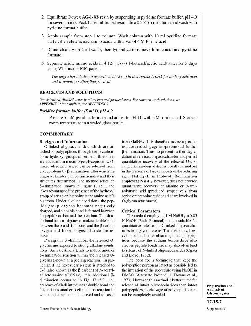
2. Equilibrate Dowex AG-1-X8 resin by suspending in pyridine formate buffer, pH 4.0for several hours. Pack 0.5 equilibrated resin into a 0.5 × 5–cm column and wash withpyridine format buffer.
3. Apply sample from step 1 to column. Wash column with 10 ml pyridine formatebuffer, then elute acidic amino acids with 5 vol of 4 M formic acid.
4. Dilute eluate with 2 ml water, then lyophilize to remove formic acid and pyridineformate.
5. Separate acidic amino acids in 4:1:5 (v/v/v) 1-butanol/acetic acid/water for 5 daysusing Whatman 3 MM paper.
The migration relative to aspartic acid (RAsp) in this system is 0.42 for both cysteic acidand α-amino-β-sulfonylbutyric acid.
REAGENTS AND SOLUTIONS
Use deionized, distilled water in all recipes and protocol steps. For common stock solutions, seeAPPENDIX 2; for suppliers, see APPENDIX 5.
Pyridine formate buffer (5 mM), pH 4.0
Prepare 5 mM pyridine formate and adjust to pH 4.0 with 6 M formic acid. Store atroom temperature in a sealed glass bottle.
COMMENTARY
Background InformationO-linked oligosaccharides, which are at-
tached to polypeptides through the β-carbon-borne hydroxyl groups of serine or threonine,are abundant in mucin-type glycoproteins. O-linked oligosaccharides can be released fromglycoproteins by β-elimination, after which theoligosaccharides can be fractionated and theirstructures determined. The method relies onβ-elimination, shown in Figure 17.15.1, andtakes advantage of the presence of the hydroxylgroup of serine or threonine at the amino acid’sβ carbon. Under alkaline conditions, the pep-tide-group oxygen becomes negativelycharged, and a double bond is formed betweenthe peptide carbon and the α carbon. This dou-ble bond in turn migrates to make a double bondbetween the α and β carbons, and the β-carbonoxygen and linked oligosaccharide are re-leased.
During this β-elimination, the released O-glycans are exposed to strong alkaline condi-tions. Such treatment tends to induce anotherβ-elimination reaction within the released O-glycans (known as a peeling reaction). In par-ticular, if the next sugar residue is attached toC-3 (also known as the β carbon) of N-acetyl-galactosamine (GalNAc), this additional β-elimination occurs as in Fig. 17.15.2—i.e.,presence of alkali introduces a double bond andthis induces another β-elimination reaction inwhich the sugar chain is cleaved and released
from GalNAc. It is therefore necessary to in-troduce a reducing agent to prevent such furtherβ-elimination. Thus, to prevent further degra-dation of released oligosaccharides and permitquantitative recovery of the released O-gly-cans, alkaline degradation is usually carried outin the presence of large amounts of the reducingagent NaBH4 (Basic Protocol). β-eliminationemploying NaBH4, however, does not providequantitative recovery of alanine or α-ami-nobutyric acid (produced, respectively, fromserine or threonine residues that are involved inO-glycan attachment).
Critical ParametersThe method employing 1 M NaBH4 in 0.05
N NaOH (Basic Protocol) is most suitable forquantitative release of O-linked oligosaccha-rides from glycoproteins. This method is, how-ever, not suitable for obtaining intact polypep-tides because the sodium borohydride alsocleaves peptide bonds and may also often leadto release of N-linked oligosaccharides (Ogataand Lloyd, 1982).
The need for a technique that kept thepolypeptide portion as intact as possible led tothe invention of the procedure using NaOH inDMSO (Alternate Protocol 1; Downs et al.,1973). However, this method is better suited forrelease of intact oligosaccharides than intactpolypeptides, as cleavage of polypeptides can-not be completely avoided.
Current Protocols in Molecular Biology Supplement 31
17.15.7
Preparation andAnalysis ofGlyconjugates
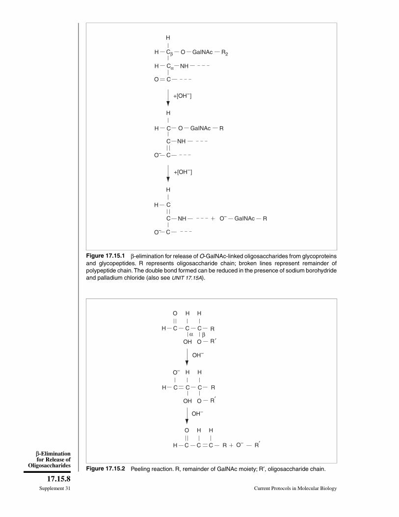
H Cβ O GaINAc R2
H Cα NH
O C
+[OH–]
H
C NH
C
H
CH
NHC
C
O– GaINAc
O–
H
H C O GaINAc R
O–
+[OH–]
R
Figure 17.15.1 β-elimination for release of O-GalNAc-linked oligosaccharides from glycoproteinsand glycopeptides. R represents oligosaccharide chain; broken lines represent remainder ofpolypeptide chain. The double bond formed can be reduced in the presence of sodium borohydrideand palladium chloride (also see UNIT 17.15A).
OH–
OH O
R
OH–
H C C C
R′
H H
O
OH O
RH C C C
R
H H
O–
O
H C C C R′
H H
R O–
α β′
Figure 17.15.2 Peeling reaction. R, remainder of GalNAc moiety; R′, oligosaccharide chain.
Supplement 31 Current Protocols in Molecular Biology
17.15.8
β-Eliminationfor Release of
Oligosaccharides
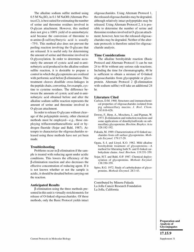
The alkaline sodium sulfite method using0.5 M Na2SO3 in 0.1 M NaOH (Alternate Pro-tocol 2), is best suited for estimating the numberof serine and threonine residues involved inO-glycan attachment. However, this methoddoes not give a 100% yield of α-aminobutyricacid because the conversion of threonine toα-amino-β-sulfonylbutyric acid is usually∼75%. This method also does not prevent thepeeling reaction involving the O-glycans thatare released. It is useful only for determiningthe amount of serine and threonine involved inO-glycosylation. In order to determine accu-rately the amount of cysteic acid and α-ami-nobutyric acid produced in the alkaline sodiumsulfite reaction, it is desirable to prepare acontrol in which the glycoproteins are oxidizedwith performic acid before β-elimination. Thistreatment cleaves disulfide cross-linkages inthe peptide chain, converting, for example, cys-tine to cysteine residues. The difference be-tween the amounts of cysteic acid and α-ami-nobutyric acid obtained before and after thealkaline sodium sulfite reaction represents theamount of serine and threonine involved inO-glycan attachment.
In order to release O-glycans without cleav-age of the polypeptide moiety, other chemicalmethods must be employed—e.g., those em-ploying trifluoromethanesulfonic acid or hy-drogen fluoride (Sojar and Bahl, 1987). At-tempts to characterize the oligosaccharides re-leased using these methods have not yet beenmade.
TroubleshootingProblems occur in β-elimination if the sam-
ple is treated with reducing agent under acidicconditions. This lowers the efficiency of theβ-elimination reaction and also decreases theeffective concentration of reducing agent. If itis not known whether or not the sample isacidic, it should be desalted before carrying outthe reaction.
Anticipated Resultsβ-elimination using the three methods pre-
sented in this unit is virtually stoichiometric forrelease of O-linked oligosaccharides. Of thesemethods, only the Basic Protocol yields intact
oligosaccharides. Using Alternate Protocol 1,the released oligosaccharides may be degraded,although relatively intact polypeptides may bereleased. Using Alternate Protocol 2, it is pos-sible to determine the number of serine andthreonine residues involved in O-glycan attach-ment; however, here too the released oligosac-charides may be degraded. Neither of the alter-nate protocols is therefore suited for oligosac-charide analysis.
Time ConsiderationsThe alkaline borohydride reaction (Basic
Protocol and Alternate Protocol 1) can be run24 to 48 hr without any serious side reactions.Including the time for chromatography, 48 hris sufficient to obtain a mixture of O-linkedoligosaccharides from glycopeptide or glyco-protein. Alternate Protocol 2 (β-eliminationwith sodium sulfite) will take an additional 24hr.
Literature CitedCarlson, D.M. 1968. Structures and immunochemi-
cal properties of oligosaccharides isolated frompig submaxillary mucins. J. Biol. Chem.243:616-626.
Downs, F., Herp, A., Moschera, J., and Pigman, W.1973. β-elimination and reduction reactions andsome applications of dimethylsulfoxide on sub-maxillary glycoproteins. Biochim. Biophys. Acta328:182-192.
Fukuda, M. 1989. Characterization of O-linked sac-charides from cell surface glycoproteins. Meth-ods Enzymol. 179:17-29.
Ogata, S.-I. and Lloyd, K.O. 1982. Mild alkalineborohydride treatment of glycoproteins—Amethod for liberating both N- and O-linked car-bohydrate chains. Anal. Biochem. 119:351-359.
Sojar, H.T. and Bahl, O.P. 1987. Chemical deglyo-sylation of glycoproteins. Methods Enzymol.138:350-359.
Spiro, R.G. 1972. Study of carbohydrates of glyco-proteins. Methods Enzymol. 28:3-43.
Contributed by Minoru FukudaLa Jolla Cancer Research FoundationLa Jolla, California
Current Protocols in Molecular Biology Supplement 31
17.15.9
Preparation andAnalysis ofGlyconjugates

UNIT 17.16Acid Hydrolysis for Release ofMonosaccharides
The first step in obtaining a compositional analysis of a glycoconjugate is the release ofthe individual monosaccharide constituents. How these are released and processeddepends on what methods are available for their analysis and identification. When theanalysis is to be performed by high-performance liquid chromatography (HPLC), themonosaccharides must be released by acid hydrolysis of the glycoconjugate. Free glycoseresidues can then be separated using a pellicular-resin anion-exchange column anddetected by pulsed amperometry (see UNIT 17.19A). When a gas-liquid chromatography(GLC) approach is to be taken in the analysis—and the glycoconjugate contains aminosugars and/or uronic acids—methanolysis, followed by trimethylsilylation of hydroxyland carboxyl groups, is the method of choice (UNIT 17.19A). Often a prediction can be madeof the type of monosaccharides present in a glycoconjugate by analogy with otherglycoconjugates from closely related sources. If the sample is not expected to containamino sugars or uronic acids—or if only neutral sugars must be measured—acid hydroly-sis, followed by reduction and peracetylation (UNIT 17.19A), is preferred.
When an HPLC approach is taken, neutral sugars, hexosamines, and uronic acids can bedirectly analyzed after strong acid hydrolysis (see Basic Protocol 3). If sialic acids areinvolved, they must be analyzed separately by submitting an aliquot of the sample to mildacid conditions (see Basic Protocol 2), after which the sialic acids are purified andanalyzed by HPLC without derivatization (see UNIT 17.19A) or after derivatization (see UNIT
17.18). In some cases, enzymatic release of sialic acids (see UNIT 17.12) may be chosen topreserve the O-acyl substitutions.
Mild acid hydrolysis to release fucosyl residues from glycoconjugates is described inBasic Protocol 1. Mild acid hydrolysis to release sialic acids from glycoconjugates, alongwith dialysis and column chromatography procedures to purify the sialic acids, aredescribed in Basic Protocol 2. Strong acid hydrolysis to release all monosaccharides fromglycoconjugates is described in Basic Protocol 3. Conditions required for successfulrelease of all the different monosaccharides in a glycoconjugate vary to a great extentwhen hydrochloric or sulfuric acid is used for hydrolysis. However, the conditionsrequired for hydrolysis using trifluoroacetic acid (TFA; see Basic Protocol 3) are consid-erably more uniform. Apart from sialic acids, which are known to be destroyed understrong acid conditions, all sugar residues—including fucose, hexosamines, and uronicacids—can be confidently determined after optimized treatment with TFA. TFA also hasthe advantage of being easily eliminated by evaporation without leaving a residue. Forthe release of sialic acids, however, mild acid hydrolysis using acetic acid (see BasicProtocol 2) and/or enzymatic release (UNIT 17.12) is recommended. The Alternate Protocoldescribes a harsher acid hydrolysis procedure for 4-O-acetylated sialic acids, which aremore difficult to release.
A precise quantitative ratio of the different types of monosaccharides in a glycoconjugateis very difficult to obtain. This arises from the fact that all glycosidic bonds must be splitwhile avoiding destruction of the liberated monosaccharides. Susceptibility to acidhydrolysis, as well as stability of the released unit, is different for each monosaccharide—e.g., pyranosides and furanosides hydrolyze at different rates; the presence of substituentsaffects the rate of hydrolysis; and α and β anomers have different rates of hydrolysis. Insome cases, therefore, more than one set of conditions may be required to achieve acomplete analysis (see Critical Parameters).
Supplement 32
Contributed by Adriana ManziCurrent Protocols in Molecular Biology (1995) 17.16.1-17.16.11Copyright © 2000 by John Wiley & Sons, Inc.
17.16.1
Preparation andAnalysis ofGlycoconjugates
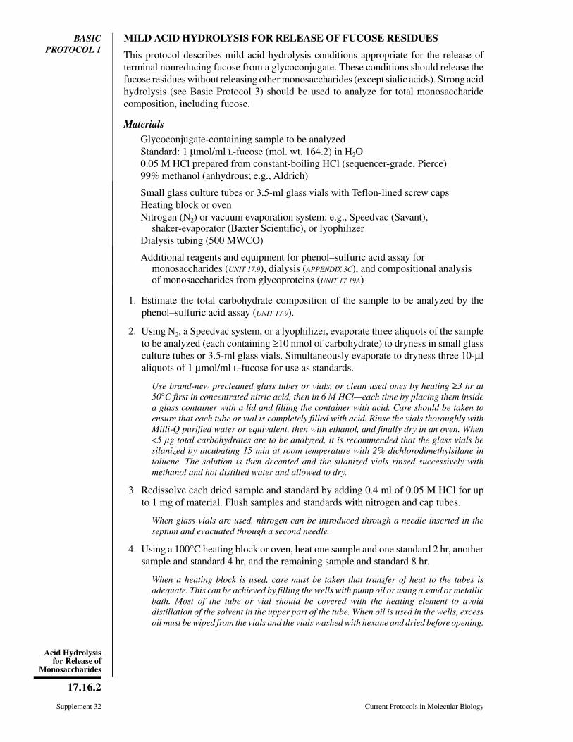
BASICPROTOCOL 1
MILD ACID HYDROLYSIS FOR RELEASE OF FUCOSE RESIDUES
This protocol describes mild acid hydrolysis conditions appropriate for the release ofterminal nonreducing fucose from a glycoconjugate. These conditions should release thefucose residues without releasing other monosaccharides (except sialic acids). Strong acidhydrolysis (see Basic Protocol 3) should be used to analyze for total monosaccharidecomposition, including fucose.
Materials
Glycoconjugate-containing sample to be analyzedStandard: 1 µmol/ml L-fucose (mol. wt. 164.2) in H2O0.05 M HCl prepared from constant-boiling HCl (sequencer-grade, Pierce)99% methanol (anhydrous; e.g., Aldrich)
Small glass culture tubes or 3.5-ml glass vials with Teflon-lined screw capsHeating block or ovenNitrogen (N2) or vacuum evaporation system: e.g., Speedvac (Savant),
shaker-evaporator (Baxter Scientific), or lyophilizerDialysis tubing (500 MWCO)
Additional reagents and equipment for phenol–sulfuric acid assay formonosaccharides (UNIT 17.9), dialysis (APPENDIX 3C), and compositional analysisof monosaccharides from glycoproteins (UNIT 17.19A)
1. Estimate the total carbohydrate composition of the sample to be analyzed by thephenol–sulfuric acid assay (UNIT 17.9).
2. Using N2, a Speedvac system, or a lyophilizer, evaporate three aliquots of the sampleto be analyzed (each containing ≥10 nmol of carbohydrate) to dryness in small glassculture tubes or 3.5-ml glass vials. Simultaneously evaporate to dryness three 10-µlaliquots of 1 µmol/ml L-fucose for use as standards.
Use brand-new precleaned glass tubes or vials, or clean used ones by heating ≥3 hr at50°C first in concentrated nitric acid, then in 6 M HCl—each time by placing them insidea glass container with a lid and filling the container with acid. Care should be taken toensure that each tube or vial is completely filled with acid. Rinse the vials thoroughly withMilli-Q purified water or equivalent, then with ethanol, and finally dry in an oven. When<5 g total carbohydrates are to be analyzed, it is recommended that the glass vials besilanized by incubating 15 min at room temperature with 2% dichlorodimethylsilane intoluene. The solution is then decanted and the silanized vials rinsed successively withmethanol and hot distilled water and allowed to dry.
3. Redissolve each dried sample and standard by adding 0.4 ml of 0.05 M HCl for upto 1 mg of material. Flush samples and standards with nitrogen and cap tubes.
When glass vials are used, nitrogen can be introduced through a needle inserted in theseptum and evacuated through a second needle.
4. Using a 100°C heating block or oven, heat one sample and one standard 2 hr, anothersample and standard 4 hr, and the remaining sample and standard 8 hr.
When a heating block is used, care must be taken that transfer of heat to the tubes isadequate. This can be achieved by filling the wells with pump oil or using a sand or metallicbath. Most of the tube or vial should be covered with the heating element to avoiddistillation of the solvent in the upper part of the tube. When oil is used in the wells, excessoil must be wiped from the vials and the vials washed with hexane and dried before opening.
Supplement 32 Current Protocols in Molecular Biology
17.16.2
Acid Hydrolysisfor Release of
Monosaccharides

5. After each sample and standard has been heated for the required time period,evaporate the solution using a nitrogen or vacuum evaporation system. Wash residuestwice with 99% methanol, then evaporate again.
CAUTION: This procedure should be carried out in a chemical fume hood.
If this is of interest, a defucosylated glycoconjugate can be prepared following this protocol.After acid hydrolysis under optimal conditions, neutralize reaction mixture with 0.05 MNaOH, dilute, and dialyze at 4°C against 20 vol water using 500-MWCO dialysis tubingto recover defucosylated glycoconjugate. Alternatively, the defucosylated product can berecovered by gel filtration chromatography (UNIT 10.9) on Bio-Gel P2 or Sephadex G-15,using water for elution.
6. Store the dried hydrolysates at −20°C until ready for compositional analysis. Resultswill indicate the best time for complete hydrolysis with minimal decomposition.
Procedures for compositional analysis are found in UNIT 17.19.
BASICPROTOCOL 2
RELEASE OF SIALIC ACIDS (EXCLUDING 4-O-ACETYLATEDSPECIES) BY MILD ACID HYDROLYSIS AND PURIFICATION OFTHE PRODUCT
This procedure allows the quantitative release of most types of known sialic acids, otherthan those acetylated at the 4 position, with minimal destruction. Subsequent handling ofthe sialic acid hydrolysate, including purification, is also designed to avoid migration orloss of O-acetyl groups. After purification, the mixtures of sialic acids can be analyzedby HPLC with (UNIT 17.18) or without derivatization (UNIT 17.19A). For release of sialic acidsacetylated at the 4 position, see Alternate Protocol. This protocol can be used to preparea mixture of sialic acids from commercially available glycoconjugate standards (e.g.,bovine submaxillary mucin) for use as a standard in sialic acid analysis.
Materials
Glycoconjugate-containing sample to be analyzedSodium formate buffer, pH 5.5 (see recipe)0.1 N H2SO4
10 M and 2 M acetic acid1% butylated hydroxytoluene (BHT; Sigma) in ethanolDowex AG 50W-X2 ion-exchange resin (200 to 400 mesh, hydrogen form;
Bio-Rad)Dowex AG 3-X4A ion-exchange resin (100 to 200 mesh, hydroxyl form; Bio-Rad)10 mM and 1 M formic acid (ACS certified), ice-cold
Dialysis tubing (1,000 and 12,000 MWCO; Spectrapor, Spectrum)Heating block5-ml glass culture tubes with Teflon-lined screw caps0.5 × 10–cm glass chromatography columns or Pasteur pipets plugged with glass
wool20-ml glass test tubesLyophilizer or shaker-evaporator (Baxter Scientific)
Additional reagents and equipment for dialysis (APPENDIX 3C), de-O-acetylation ofsialic acids and TBA assay (UNIT 17.18), and ion-exchange chromatography(UNIT 10.10)
Prepare sample and determine optimal conditions1. Thoroughly homogenize sample to be analyzed and dialyze (APPENDIX 3C) overnight
at 4°C against a 100-fold excess of sodium formate buffer, pH 5.5, using 12,000-
Current Protocols in Molecular Biology Supplement 32
17.16.3
Preparation andAnalysis ofGlycoconjugates

MWCO tubing. Lyophilize dialysate and store frozen at −20°C until ready foranalysis.
Dialysis is not necessary for relatively purified biological samples that do not containlow-molecular-weight contaminants. If not to be analyzed immediately, samples should belyophilized and stored frozen at −20°C.
Crude biological samples may contain O-acetylesterases, which could cause de-O-acety-lation of sialic acids during the isolation procedure. These enzymes can be inactivated byadding a 100 mM stock solution of diisopropyl fluorophosphate (DFP) in isopropanol toa final concentration of 1 mM, then incubating 15 min on ice. As DFP is extremely toxic,safety recommendations should be studied carefully; gloves should be worn and thecompound should be used in the fume hood.
2. Dissolve the sample in water. De-O-acetylate an aliquot of sample, release sialic acidswith 0.1 N H2SO4, and determine total sialic acid content by the TBA assay (UNIT
17.18).
Although the TBA assay uses harsher conditions for releasing the sialic acids and willcause some destruction, it is used here in a comparative fashion. Therefore, it is a validindication of the recoveries from step to step.
3. Transfer three aliquots of the sample from step 1 (each containing ∼10 nmol of sialicacids) to 5-ml glass culture tubes.
4. Add 10 M acetic acid to a final acetic acid concentration of 2 M, then add 1 vol of1% BHT per 100 vol sample.
The BHT retards lipid peroxidation.
5. Using an 80°C heating block or oven, heat one aliquot 2 hr, another aliquot 4 hr, andthe remaining aliquot 5 hr.
When a heating block is used, care must be taken that the transfer of heat to the tubes isadequate. See Basic Protocol 1, step 4 annotation, for proper technique.
6. After each sample has been heated for the required time period, lyophilize andde-O-acetylate, then determine total free sialic acid by the TBA assay (UNIT 17.18).Compare the numbers obtained to determine the time required for maximal releaseof sialic acids.
Release and purify sialic acids7. In a 5-ml glass culture tube add 2 M acetic acid to the lyophilized sample from step
1 (0.4 ml for up to 1 mg) and incubate at 80°C for the time determined in steps 5 and6. Cool to room temperature.
8. Transfer reaction mixture into 1000-MWCO dialysis tubing, then wash the test tubetwice with water, each time transferring the washings into the dialysis tubing. Dialyze(APPENDIX 3C) overnight at 4°C against 10 vol water and retain the dialysate (solutionoutside the tubing).
An MWCO of 1000 is sufficient to allow the passage of free sialic acids. Bigger pores mayallow more contamination by permitting bigger molecules to dialyze out.
When total volumes are small (100 l to 1 ml), the dialysis tubing can be placed inside atest tube. In this case, it must be closed with knots instead of plastic clamps.
Assuming that equilibrium is achieved, a 10% loss (sialic acids remaining in the tubing)should be expected when dialyzing against 10 vol water. The recovery can be improved byrepeating the dialysis.
Supplement 32 Current Protocols in Molecular Biology
17.16.4
Acid Hydrolysisfor Release of
Monosaccharides

9. Using a 0.5 × 10–cm chromatography column or Pasteur pipet plugged with glasswool, prepare a 1-ml column of Dowex AG 50W-X2 ion-exchange resin (see UNIT
10.10) in a cold room or cold box, using ice-cold water to suspend the resin and washthe column.
The resin must be extensively washed with ice-cold water before use. All chromatographysteps must be performed at 4°C and eluants must be ice cold.
10. Load the dialysate from step 8 directly to the column. Wash column with 6 ml coldwater and collect eluant in a 15-ml glass test tube on ice.
For large preparations, every step can be scaled up, but it may be necessary to lyophilizethe dialysate and dissolve it in a small volume of water before loading it onto the column.The content of the dialysis tubing (the retentate, containing the desialylated glycoconju-gates under study) can also be frozen, lyophilized, and reserved for other analyses.
11. Check pH of eluant with pH paper. If >3, go to step 12. If <3, add 10 mM sodiumformate buffer, pH 5.5, until pH is >3.
12. Using a 0.5 × 10–cm chromatography column or Pasteur pipet plugged with glasswool, prepare a 1-ml column of Dowex 3-X4A ion-exchange resin (hydroxyl form).Convert resin to formate form by equilibrating in 3 vol of 1 M formic acid. Let columnstand in contact with formic acid for 15 min, then wash with 10 mM sodium formate(pH 5.5) until effluent pH is stable at 5.5 (see UNIT 10.10).
Large batches of resin can be prepared and stored at 4°C. Always take into considerationthe capacity of the resin and calculate the volume of the column required to bind the totalsialic acids present in the sample as determined by the TBA assay in step 2.
13. Load sample from step 11 on column. Wash column immediately with 7 ml ice-cold10 mM formic acid, and discard washings.
14. Elute sialic acids with 10 ml ice-cold 1 M formic acid. Collect eluant on ice in a 20-mlglass test tube, then evaporate it to dryness by lyophilization or in a shaker-evaporatorwith the temperature of the water bath maintained at <37°C. Store at −20°C untilanalysis.
The purified mixture of sialic acids can be analyzed by HPLC (UNIT 17.19A) or derivatizedand analyzed by reversed-phase HPLC with fluorometric detection (UNIT 17.18).
ALTERNATEPROTOCOL
RELEASE AND PURIFICATION OF 4-O-ACETYLATED SIALIC ACIDS
A harsher acid hydrolysis procedure is used to liberate 4-O-acetylated sialic acids, whichare resistant to release by the method indicated in Basic Protocol 2.
Additional Materials (also see Basic Protocol 2)
23.4 M (concentrated) formic acidHeating block
1. Prepare the sample and determine total sialic acid content in an aliquot of the materialby TBA assay (see Basic Protocol 2, steps 1 and 2).
2. Adjust dialyzed sample to pH 2.0 by dropwise addition of concentrated formic acid.Heat at 70°C for 1 hr. Cool to room temperature, and centrifuge at room temperatureusing a tabletop centrifuge to separate supernatant and pellet. Separate supernatantby aspiration and retain both supernatant and pellet.
When a heating block is used, care must be taken that the transfer of heat to the tubes isadequate. See Basic Protocol 2 step 4 annotation for proper technique.
Current Protocols in Molecular Biology Supplement 32
17.16.5
Preparation andAnalysis ofGlycoconjugates

3. Dialyze supernatant overnight at 4°C against 10 vol water using 1000-MWCO tubing,then lyophilize the solution outside the tubing. Repeat the dialysis twice again andpool the recovered material.
When total volumes are small (100 l to 1 ml), the dialysis tubing can be placed inside atest tube. In this case, it must be closed with knots instead of plastic clamps.
4. Treat the pellet obtained in step 2, resuspended in a similar volume of water, twicemore at pH 2 with heating as described in step 2, centrifuging and dialyzing thesupernatants each time as described in step 3 to recover the hydrolysis products.
5. Pool the lyophilized dialysates and purify the mixture of sialic acids by ion-exchangechromatography (see Basic Protocol 2, steps 9 to 14). The annotation to BasicProtocol 2, step 14 describes options for analysis.
BASICPROTOCOL 3
STRONG ACID HYDROLYSIS FOR QUANTITATIVE RELEASE OFHEXOSES, PENTOSES, HEXOSAMINES, AND URONIC ACIDS FROMGLYCOCONJUGATES
This protocol describes several sets of conditions for the acid hydrolysis of monosaccha-rides from glycoconjugates. When no data is available from previous experimentsregarding optimal conditions, it is recommended that one try more than one set ofconditions to determine which ones are optimal. It is also important to prove thereproducibility of the method under the conditions chosen. The mixtures of free monosac-charides obtained by acid hydrolysis can be directly analyzed by high-pH anion-exchangechromatography with pulsed amperometric detection (HPAE-PAD) or by derivatizationfollowed by GLC (see UNIT 17.19A for both techniques).
Materials
Glycoconjugate-containing sample to be analyzedInternal standard (i.e., a monosaccharide that does not occur naturally in the
sample)Standard mixture (see recipe)2 M and 4 M trifluoracetic acid prepared from concentrated TFA (HPLC/spectra
grade sequanal quality, Pierce)99% methanol (anhydrous; e.g., Aldrich)
Heating block or ovenSmall glass culture tubes or 3.5-ml glass vials with Teflon-lined screw capsNitrogen (N2) or vacuum evaporation system: e.g., Speedvac (Savant) or
shaker-evaporator (Baxter Scientific)
Additional reagents and equipment for phenol–sulfuric acid assay formonosaccharides (UNIT 17.9) and compositional analysis of monosaccharidesreleased from glycoproteins (UNIT 17.19A)
1. If sufficient material is available, estimate the total carbohydrate content of the sampleto be analyzed by the phenol–sulfuric acid assay (UNIT 17.9).
Analysis can be done without having this information when sample is precious, as theanalytical tools used for the compositional analysis of acid hydrolysates (i.e., HPAEC-PAD,GLC) are far more sensitive than the colorimetric assay.
2. Using N2, a Speedvac system, or a lyophilizer, evaporate three aliquots of the sampleto be analyzed (each containing 5 to 50 µg carbohydrate) to dryness in small glasstubes or 3.5-ml glass vials. If the planned compositional analysis will be done byGLC, add 1 to 10 µg of internal standard to the sample before evaporating it.
Supplement 32 Current Protocols in Molecular Biology
17.16.6
Acid Hydrolysisfor Release of
Monosaccharides

Rhamnitol and arabinitol are good choices as internal standards for analysis of animalglycoconjugates, and inositol for plant polysaccharides.
3. Prepare (as in step 2) three aliquots of the standard mixture appropriate for themonosaccharides to be determined and the analytical method to be used, and threeblanks to detect possible contamination. For GLC analysis, also add the internalstandard.
The time involved in HPLC or GPC analysis of each sample is quite long (see UNIT 17.19A);therefore, preparation of a mixture of the appropriate standards in equimolar amountshelps reduce the analysis time. On the other hand, when identification of one particularcomponent is doubtful, coinjection of one aliquot of the sample with one aliquot of thesuspected standard into the GLC or HPLC column gives a definitive answer on the presenceof that particular monosaccharide. In such cases, processing each monosaccharide stand-ard individually in parallel can be helpful.
An appropriate blank is a material that has been processed exactly as the sample was (e.g.,a pool of fractions from a chromatography step that showed only background response tothe method used for monitoring). Blanks are especially useful when very small amounts ofmaterial are processed, as in such cases the limit of sensitivity will be determined by theaccumulated contaminants (e.g., from Sephadex columns or dirty glassware).
4. Dissolve the three evaporated aliquots of each sample, standard, and blank in 100 to500 µl of 2 M TFA. Flush with N2 and cap the tubes.
5. Using a 100°C heating block or oven, heat one sample, one standard, and one blank3 hr; another sample, standard, and blank 4 hr; and the remaining sample, standard,and blank 6 hr.
This time course will determine the best conditions for maximum recovery with minimumdestruction. When insufficient material is available for a time-course experiment, use 3 hrof hydrolysis for glycoproteins, glycopeptides, or free oligosaccharides of the N- and/orO-linked type that contain hexosamines and neutral monosaccharides. Use 6 hr ofhydrolysis when the sample is suspected to contain glycosyluronic acids (e.g., proteogly-cans or glycosaminoglycan chains).
When a heating block is used, care must be taken that the transfer of heat to the tubes isadequate. See Basic Protocol 1 step 4 annotation for proper technique.
6. After heating, evaporate solution in each tube using a nitrogen or vacuum evaporationsystem. Wash residues twice with 99% methanol, then evaporate again.
Store the dry hydrolysates at −20°C until ready for compositional analysis.
7. Analyze the hydrolysate by HPAE-PAD or derivatize and analyze by GLC (see UNIT
17.19A).
8. Compare the recovery of each monosaccharide obtained using the different hydroly-sis times and decide upon the most suitable hydrolysis time for the sample understudy. Repeat the analysis using such conditions and check for reproducibility.
If quantitation remains uncertain, hydrolysis can be repeated with the addition of a knownamount of an internal standard, even when HPLC is used for the analysis. Because theconstituents will be known at this point, any noninterfering monosaccharide or alditol canbe used as internal standard. The choice will depend on the method used for analysis. Whenthe analysis is done by HPAE-PAD, monosaccharide alditols cannot be used as internalstandards because they are not retained by the Carbo-Pak PA-1 column used for monosac-charide separation. In this case, 2-deoxyrhamnose or 2-deoxyglucose are appropriate asinternal standards. Prepare an aqueous solution of the standard and add a known amountto the sample, evaporating them together before dissolving in the acid at step 4. Thestandard must be present in an amount comparable to that of the major constituents of thesample.
Current Protocols in Molecular Biology Supplement 32
17.16.7
Preparation andAnalysis ofGlycoconjugates

REAGENTS AND SOLUTIONS
Use deionized, distilled water in all recipes and protocol steps. For common stock solutions, seeAPPENDIX 2; for suppliers, see APPENDIX 4.
Sodium formate buffer, pH 5.5Weigh 68 mg sodium formate, dissolve in water, then bring volume to 1 liter. Adjustto pH 5.5 by adding 1 M formic acid dropwise.
Standard mixtureFor GLC of alditol acetates: Prepare an equimolar mixture of the following:D-(+)-glucose (anhydrous, mixed anomers; Sigma)D-(+)-galactose (crystalline; Sigma)D-(+)-mannose (crystalline; Sigma)D-(+)-xylose (99%; Sigma)For HPAE-PAD: To the above mixture (without xylose), add equimolar amounts of:N-acetyl-D-galactosamine (Sigma)N-acetyl-D-glucosamine (Sigma)D-glucuronolactone (Sigma; when uronic acid is expected)
If xylose is expected, include it in the mixture, but be careful to use the right conditions forHPAEC-PAD (UNIT 17.19A).
COMMENTARY
Background Information
Fucose releaseFucosyl residues are typically in nonreduc-
ing terminal positions and are therefore suscep-tible to acid hydrolysis under relatively mildconditions. However, specific sites of attach-ment can increase the stability of the glycosidicbond (e.g., a positively charged amino groupproduced by de-N-acetylation of an N-acetyl-hexosamine residue will lead to a stable gly-cosidic linkage at the 3 position). Thus, it isnecessary to determine for each specific glyco-conjugate the conditions required for completeliberation with minimal destruction. The pa-rameters to be varied are the type of acid,concentration of acid, and time and temperatureused for the hydrolysis. The set of conditionssuggested in Basic Protocol 1 are the best start-ing point.
Most of the problems encountered whentrying to determine the best hydrolysis condi-tions for releasing fucose residues from a givenglycoconjugate have been reviewed by Gott-schalk (1972). Typical conditions are 0.5 Mhydrochloric acid at 100°C for 16 hr (Schragerand Oates, 1968), although 15 min at 100°C in1 M HCl are enough to release fucose fromorosomucoid (Gottschalk, 1972), and 0.6 Nsulfuric acid at 100°C for 1 hr is required torelease all the fucose from total serum proteins(Gyorky and Houck, 1965).
Sialic acid releaseMore than 25 different kinds of modified
sialic acids have now been reported to be foundin nature. Most arise from substitution of theparent molecule with a variety of differentgroups. These modifications have been shownto affect a wide spectrum of biological phenom-ena. Many studies of sialic acids have failed torecognize the complexity of this family ofsugars, because conventional methods for ana-lyzing glycoconjugates—e.g., methanolysis,hydrazinolysis, methylation analysis, and β-elimination—result in the destruction of O-substituted species, especially O-acetyl esters.
The classical method for releasing sialicacids from glycoconjugates (incubation with0.1 N H2SO4 for 1 hr at 80°C) results in exten-sive destruction of O-acetyl groups (Schauer,1978). In addition, the presence of O-acetylgroups makes the sialic acid molecule partiallyor completely resistant to release by all avail-able bacterial and viral neuraminidases(Schauer, 1978, 1982; Schauer and Corfield,1982). The use of milder conditions (incubationwith 0.5 M formic acid for 1 hr at 80°C) allowedthe release and identification of many pre-viously undetected O-acetylated sialic acids(Schauer 1978, 1982). However, even underthese conditions quantitative analysis is notpossible as a result of significant destructionand incomplete release of O-acetylated speciesusing formic acid (pH 2.1) for acid hydrolysis(Schauer, 1978; Varki and Kornfeld, 1980;
Supplement 32 Current Protocols in Molecular Biology
17.16.8
Acid Hydrolysisfor Release of
Monosaccharides

Varki and Diaz, 1983). Prolonged hydrolysis in2 M acetic acid (pH 2.4 to 2.5) at 80°C wasfound to achieve maximal release of O-acety-lated sialic acids with minimal loss of O-acetylgroups (Varki and Diaz, 1983). These latterconditions have been successfully used to re-lease and purify labile substituted sialic acidsfrom different biological sources (Manzi et al.,1990). Hydrolysis times vary for different gly-coconjugates. When sialic acids are acetylatedat the 4 position, complete hydrolysis is diffi-cult to achieve, but once released, the acetylgroup becomes very labile, producing a 4,7-an-hydro species.
Further complexity arises from the migra-tion of O-acetyl groups from the 7 or 8 positionsto the thermodynamically more stable 9 posi-tion of the sialic acid exocyclic side chain. Thismigration occurs rapidly, with t1⁄2 ranging fromminutes to hours, depending on the pH andtemperature (Kamerling and Vliegenthart,1989; Varki and Diaz, 1983). Migration is mini-mal between pH 3 and 5.
A recent review (Varki, 1992) covers thediversity in the sialic acids, including the dif-ferent problems encountered during theiranalysis and the methods available to overcomethese problems.
Strong acid hydrolysis with TFAHydrolysis of glycosidic linkages involving
hexoses requires more vigorous conditionsthan the ones used for fucose or sialic acids.Typically, hydrochloric (1 or 2 M, 100°C, 1 to6 hr), sulfuric (0.1 to 2 N, 100°C, 4 to 12 hr),or trifluoroacetic acid (2 M, 121°C, 1 to 2 hr)have been used for this purpose, under condi-tions that vary depending on the substrate. Pro-vided that the most labile species—e.g., sialicacids—are quantitated separately, the most se-vere problem encountered when determiningthe monosaccharide composition is the incom-plete release of 2-acetamido sugars and glyco-syluronic acids. The presence of these sugarscan often be predicted based on the origin ofthe glycoprotein under study. In the case ofacetamido sugars, if N-deacetylation occursfirst, the stability of the glycosidic linkage isincreased. Therefore, the conditions must besuch that hydrolysis of the glycosidic linkageis substantially faster than the de-N-acetylationreaction. The glycosidic linkage of glycosy-luronic acids is particularly resistant to acidhydrolysis, and further inhibition of their com-plete release occurs when the adjacent sugar inthe chain is an N-sulfated or N-acetylatedhexosamine. This problem is routinely encoun-
tered in glycosaminoglycan chains, wherequantitative release requires a previous reduc-tion of the carboxylic acid. On the other hand,liberated glycosyluronic acids are very suscep-tible to degradation, particularly by decarboxy-lation. Therefore, analysis of glycosaminogly-can chains is better achieved using enzymes(see UNIT 17.19B).
When enough material is available, recoveryand reproducibility can be assessed under sev-eral different sets of conditions. In many casesthis is not possible, and incubation with 2 MTFA for 3 hr at 100°C is recommended undersuch circumstances.
A review of the different conditions used forthe release of monosaccharides from glycopro-teins, including a detailed analysis of themechanisms of acid hydrolysis of glycosides,is provided by Gottschalk (1972). In mostcases, hydrolysis with TFA will produce asgood a recovery of fucose as hydrolysis with0.5 M HCl, although in the former case othersugars are also released. Where the sample isincubated with 2 M TFA at 121°C, 25% of thefucose decomposes after 6 hr. However, be-cause only 1 hr is required for complete release,this rate of decomposition is tolerable (Alber-sheim et al., 1967). Where the sample is heatedat 100°C, 98% recovery of fucose is achievedeven after 6 hr (Biermann, 1988). Usually, es-timation of the content of hexosamines in gly-coproteins has been based on HCl hydrolysisat different concentrations (ranging from 2 Mto 6 M) at temperatures close to 100°C forvarying periods of time (ranging from 1 to 24hr; Gottshalk, 1972). Also, incubation withtrifluoroacetic acid (0.5 M, 100°C, 19 hr; 4 M,125°C, 1 hr; or 4 M, 100°C, 4 hr) has beenshown to give good results (Keene et al., 1983;Neeser and Schweizer, 1984). The variety ofmethods clearly indicates that the effectivenessof the hydrolysis greatly depends on the glyco-conjugate—e.g., the type of linkage, the typeof aglycone (noncarbohydrate portion), and thepresence of substituents such as N-acetylgroups. Mannosamine is less acid-stable thanglucosamine and galactosamine (Ludowiegand Benmaman, 1967). Analysis of the rates ofdecomposition of monosaccharides in the pres-ence of 2 M (10 mg/ml) TFA—by continuinghydrolysis of polysaccharides after most of themonosaccharides have been liberated—haveindicated that after 6 hr at 121°C, >50% of thexylose and arabinose and >25% of the galac-tose, rhamnose, and fucose are decomposed(Albersheim et al., 1967). However, as most ofthe monosaccharides are liberated within 1 hr,
Current Protocols in Molecular Biology Supplement 32
17.16.9
Preparation andAnalysis ofGlycoconjugates

these rates of decomposition are tolerable. Hy-drolysis of urinary glycoconjugates with 2 MTFA for up to 15 hr at 100°C indicated that themaximum yield of aldoses and uronic acids isobtained after 6 hr. The recovery of the individ-ual monosaccharides added at the beginning ofthe treatment was >93% in all cases, includingthat of glucuronic acid (Honda et al., 1981). Acomparison between the action of different ac-ids on each type of carbohydrate-containingmolecule was done by Biermann (1988). Con-ditions used for acid hydrolysis of glycoconju-gates before GLC analysis have also been re-viewed by Kamerling and Vliegenthart (1989).
Critical ParametersFucose release. Use of high-quality acids
and highly purified deionized water is recom-mended. In the case of glycoproteins, destruc-tion arising from the interaction between liber-ated sugars and amino acids can be minimizedby using a low concentration of glycoprotein toperform the hydrolysis (<1 mg/ml) and exclud-ing oxygen (by displacing it with dry nitrogenor performing the hydrolysis in a tube sealedunder vacuum).
Sialic acid release. All the parameters in-volved in the protocol (e.g., temperature, pH,and concentration) must be carefully control-led. Because pH values outside the range of 3to 7 should be avoided, strongly basic anion-exchange resins (e.g., Dowex 1 or 2) should notbe used. Samples must be fresh, and must beprocessed and analyzed immediately. It is alsocritical that purified samples be kept dry at −70°or −80°C while stored. However, even storageat these temperatures for extended periods oftime can result in migration or loss of O-acetylgroups. It should be pointed out that optimalmethods for the release and purification of therare forms of sialic acids (e.g., O-methylated,O-sulfated, or multiply modified) have not beenadequately worked out.
Strong acid hydrolysis with TFA. High-quality acids must be used and highly purifieddeionized water must be used to dilute the acid,because trace iron can destroy hexosamines.Degradation is also avoided by excluding oxy-gen (usually by flushing with nitrogen, al-though it is also possible to work under vac-uum). Working at a low concentration of gly-coconjugate (<1 mg/ml) reduces thedestruction of sugars. A competing sugar notpresent in the glycoprotein can also be addedto reduce destruction (ribose has been used forthis purpose). It must be kept in mind that—
even when treating the standards in parallel—they may not reflect the decomposition sufferedby the glycosidically bound monosaccharidesin the sample, because in the latter case theyspend less time as free glycoses. Therefore, amethod that yields less decomposition is al-ways preferred to a correction factor worked upon the basis of monosaccharide standards.
Anticipated ResultsFucose release. Basic Protocol 1 will cleave
the glycosidic linkages of all terminal nonre-ducing fucosyl residues. Once free, the fucosylresidues will remain intact, giving a reasonableestimation of the amount of fucose in the gly-coconjugate (UNIT 17.19). Complete defucosyla-tion of a sample can be determined by analysisof the amount of fucose in the hydrolysate atdifferent time points. However, the use of TFA(strong acid hydrolysis; Basic Protocol 3) per-mits an accurate estimation of fucose togetherwith the rest of the sugars. Therefore, strongacid hydrolysis is recommended when ananalysis of the total glycosyl composition isrequired. The most valuable use of Basic Pro-tocol 1 is therefore the defucosylation of gly-coconjugates for further analysis.
Sialic acid release. The conditions for re-lease and purification of sialic acids used inthese protocols ensure maximum release ofmost modified sialic acids (with the exemptionof methylated or sulfated species), prevent mi-gration of O-acetyl groups from the 7 positionto the 9 position, and prevent de-O-acylation.Therefore, careful use of the suggested proto-cols will result in the most accurate possiblequantitation of the different sialic acid speciespresent in a glycoconjugate. Sialic acids ob-tained by these procedures can be analyzed byHPAEC-PAD (UNIT 17.19A) or other HPLC tech-niques with (UNIT 17.18) or without derivatization(UNIT 17.19A). Therefore, an accurate quantitationalso depends on the careful use of the finalmethod of analysis. Basic Protocol 2 can beused to obtain a mixture of Neu5Ac, Neu5Gc,and of some of the mono-, di-, and tri-O-acety-lated derivatives of these sialic acids from com-mercially available standards such as bovinesubmaxillary mucin. A mixture of standardswill be very useful when analyzing the presenceof sialic acid modifications in a glycoconjugate.
Strong acid hydrolysis with TFA. Carefulapplication of Basic Protocol 3 will result inhydrolysis of the glycosidic linkages ofhexoses, hexosamines, and uronic acids in gly-coconjugates with minimal destruction.
Supplement 32 Current Protocols in Molecular Biology
17.16.10
Acid Hydrolysisfor Release of
Monosaccharides
Time ConsiderationsFucose release. Because an 8-hr reaction is
involved, it is better to prepare the samples theprevious day. All hydrolysis and recovery pro-cedures can be completed during the secondday. Any additional time involved depends onthe method used for analysis.
Sialic acids. After the time course to deter-mine the time required for complete hydrolysis,release and purification require 2 days. Analy-sis must then be done immediately.
Strong acid hydrolysis with TFA. Prepara-tion, hydrolysis, and drying of the hydrolysateshould take ≤8 hr. The time required for analy-sis (by HPLC or GLC) is indicated in UNIT 17.19A.
Literature CitedAlbersheim, P., Nevins, D.J., English, P.D., and
Karr, A. 1967. A method for the analysis ofsugars in plant cell-wall polysaccharides by gas-liquid chromatography. Carbohydr. Res. 5:340-345.
Biermann, C.H. 1988. Hydrolysis and other cleav-ages of glycosidic linkages in polysaccharides.Adv. Carbohydr. Chem. Biochem. 46:251-271.
Gottschalk, A. 1972. Glycoproteins: Their Compo-sition, Structure, and Function (A. Gottschalk,ed.), 2nd ed., Part A, pp. 225-299. El-sevier/North-Holland, Amsterdam.
Gyorky, G. and Houck, J.C. 1965. The determina-tion of terminal protein-bound fucose. Can. J.Biochem. 43:1807-1811.
Honda, S., Suzuki, S., Kakahi, K., Honda, A., andTakai, T. 1981. Analysis of the monosaccharidecompositions of total nondialyzable urinary gly-coconjugates by the dithioacetal method. J.Chromatogr. 226:341-350.
Kamerling, J.P. and Vliegenthart, J.F.G. 1989. Car-bohydrates. In Clinical Biochemistry: Princi-ples, Methods, Applications. Vol. 1, Mass Spec-trometry (A.M. Lawson, ed.) pp. 175-263. Wal-ter de Gruyter & Co., Berlin.
Keene, L., Lindberg, B., Madden, J.K., Lindberg,A.A., and Gemski, P. 1983. Structural studies ofthe Escherichia coli O-antigen 25. Carbohydr.Res. 122:249-256.
Ludowieg, J. and Benmaman, J.D. 1967. Colorimet-ric differentiations of hexosamines. Anal. Bio-chem. 19:80-88.
Manzi, A.E., Dell, A., Azadi, P., and Varki, A. 1990.Studies of naturally occurring modifications ofsialic acids by fast-atom bombardment–massspectrometry. Analysis of positional isomers by
periodate cleavage. J. Biol. Chem. 265:8094-8107.
Neeser, J.R. and Schweizer, T.F. 1984. A quantita-tive determination by capillary gas-liquid chro-matography of neutral and amino sugars (as O-methyloxime acetates), and a study on hydrolyticconditions for glycoproteins and polysaccha-rides in order to increase sugar recoveries. Anal.Biochem. 142:58-67.
Schauer, R. 1978. Biosynthesis of sialic acids. Meth-ods Enzymol. 50:64-89.
Schauer, R. 1982. Chemistry, metabolism, and bio-logical functions of sialic acids. Adv. Carbohydr.Chem. Biochem. 40:131-234.
Schauer, R. and Corfield, A.P. 1982. In Sialic Acids:Chemistry, Metabolism and Function (R.Schauer, ed.) pp. 51-57. Springer-Verlag, NewYork.
Schrager, J. and Oates, M.D.G. 1968. The carbohy-drate components of the hydrolysates of gastricsecretion and extracts from mucuous glands ofthe gastric body mucosa and antrum. Biochem.J. 106:523-529.
Varki, A. 1992. Diversity in the sialic acids. Glyco-biology 2:25-40.
Varki, A. and Diaz, S. 1983. The release and purifi-cation of sialic acids from glycoconjugates:Methods to minimize the loss and migration ofO-acetyl groups. Anal. Biochem. 137:236-247.
Varki, A. and Kornfeld, S. 1980. An autosomaldominant gene regulates the extent of 9-O-acty-lation of murine erythrocyte sialic acids. J. Exp.Med. 152:532-544.
Key ReferencesBiermann, 1988. See above.
Gottschalk, 1972. See above.
These references discuss the mechanisms of acidhydrolysis of the different monosaccharides, and theinfluence of structure in determining alternativemechanisms. They also review many different hy-drolysis conditions, and illustrate application ofparticular conditions to different types of glycocon-jugates and the results obtained.
Varki and Diaz, 1983. See above.
Describes the parameters that must be varied toarrive at the optimal conditions used in the methoddescribed here.
Contributed by Adriana ManziUniversity of California San DiegoLa Jolla, California
Current Protocols in Molecular Biology Supplement 32
17.16.11
Preparation andAnalysis ofGlycoconjugates
UNIT 17.17AEnzymatic Release of Oligosaccharidesfrom Glycolipids
BASICPROTOCOL
USE OF ENDOGLYCOCERAMIDASE FOR SELECTIVE CLEAVAGEOF GLYCOLIPIDS
Selective cleavage of the oligosaccharide moiety of glycolipids for further structuralanalysis can be achieved by means of endoglycoceramidase (EGCase), an enzymespecific for the linkages between oligosaccharide and ceramide residues in glycolipids.The method can be used for analysis of glycolipids present in macroscopic amounts orfor glycolipids that have been radiolabeled in their carbohydrate moiety. A variety ofdifferent methods can be used for analysis of the released oligosaccharides.
Materials
Sample to be analyzed—neutral glycolipids (UNIT 17.2), gangliosides (UNIT 17.17B), ordesialylated gangliosides (see UNIT 17.12)—in organic solvent
1 mg/ml disialoganglioside-GD1a (Sigma) in 2:1 (v/v) chloroform/methanol0.5% (w/v) sodium taurocholate in 0.05 M sodium acetate buffer, pH 6.0Endoglycoceramidase (EGCase; from Rhodococcus sp.; for purification see Ito
and Yamagata, 1989a)TolueneMethanol60:40:9 (v/v/v) chloroform/methanol/0.2% (w/v; aqueous) calcium chlorideOrcinol/sulfuric acid reagent (see recipe)2:1 (v/v) chloroform/methanol
Bath sonicatorPrecoated high-performance thin-layer (HPTLC) silica gel 60 plates (10 × 10–cm;
Merck)Glass chromatography tank140°C ovenTabletop centrifuge
Digest sample with EGCase1. Evaporate an aliquot of the sample to be analyzed to dryness in a glass vial.
Simultaneously evaporate 10 µl of 1 mg/ml disialoganglioside-GD1a in the samemanner, as a positive control.
When analyzing complex mixtures of unknown composition, estimate the amount of sampleto be aliquotted by carrying out this protocol with two or three different amounts of sample.
Evaporation can be done by simply leaving the glass vial containing the chloroform/metha-nol solution open to the atmosphere or by using a Speedvac evaporator.
2. Dissolve the evaporated residues in 50 µl 0.5% sodium taurocholate in 0.05 M sodiumacetate buffer, pH 6.0 by brief sonication in a bath sonicator.
0.4% Triton X-100 can be used in place of sodium taurocholate. EGCase moleculesaggregate in the absence of detergents.
3. Add 12 mU of EGCase to each sample. Place the vials containing the samples, withcaps loosened, inside a closed container in which an open tube of toluene is alsoplaced to create a toluene atmosphere. Begin incubating at 37°C.
Supplement 32
Contributed by Adriana E. ManziCurrent Protocols in Molecular Biology (1995) 17.17.1-17.17.5Copyright © 2000 by John Wiley & Sons, Inc.
17.17.1
Preparation andAnalysis ofGlycoconjugates
4. At intervals of 0, 1, 3, 6, 15, and 20 hr of incubation, take an aliquot of the reactionmixture with a volume corresponding to ≥1 nmol of each glycolipid. Dilute eachaliquot with 9 vol methanol.
Determine optimal incubation time for cleavage5. Apply an aliquot of each diluted sample hydrolysate and GD1a hydrolysate (1 to 2
nmol) along a line drawn 1 cm from the bottom of an HPTLC silica gel 60 plate.
Depending on how many points are used, multiple plates may be required. Five to sevensamples can be spotted per plate.
The plates must be marked in advance with pencil lines 1 cm from the top and bottom ofthe plate. The bottom line should be further divided with pencil marks into 1-cm lanes with∼0.5-cm spacings between lanes. The lanes will correspond to the different incubation timesused in step 4.
6. Air dry plate and place in a glass chromatography tank that has been equilibratedwith 60:40:9 chloroform:methanol:0.02% aqueous calcium chloride.
The HPTLC is to separate the glycolipids. Released oligosaccharides can be analyzed byHPTLC on silica gel 60 plates using 2:1:1 (v/v/v) 1-butanol:glacial acetic acid:water asthe developing solvent.
7. Let the solvent ascend until it reaches the upper line, then take the plate out of thetank and allow to thoroughly air dry in a fume hood.
8. Spray with orcinol-sulfuric acid reagent and heat at 140°C for 5 min.
Alternatively, if the carbohydrate moiety has been radiolabeled, the plate can be submittedto autoradiography.
9. Note the lane in which complete release of oligosaccharides is indicated by thedisappearance of the glycolipid bands that are present in the starting material.
If no lane indicating complete reaction is seen, longer incubation periods should be tried.The time points indicated to step 4 are for analysis of purified glycolipids for which someinformation is available. A time course up to 72 hr is suggested for unknown glycolipidstructures. In these cases, add 36 mU of EGCase instead of 12 mU.
Analyze released oligosaccharides10. Set up reaction as described in steps 1 to 3, incubate for the time determined to be
optimal for complete release of oligosaccharides, then stop the reaction by adding 2vol of 2:1 chloroform:methanol.
11. Shake the mixture of aqueous cleaved glycolipid and organic solvent, then centrifuge10 min at 2000 rpm in a tabletop centrifuge. Remove the upper (aqueous) phasecontaining the oligosaccharides and lyophilize. Store at −20°C.
The remaining glycolipids and ceramides will partition into the lower (organic) phase.
The dried sample can be dissolved in water and submitted to a variety of different methodsof oligosaccharide analysis. The method to be chosen will depend on the availability ofinstrumentation, the complexity of the sample, the amount of material available, and thequestions to be answered. Possible methods for the analysis of the released oligosaccha-rides include: ion-exchange HPLC of anionic oligosaccharides (UNIT 17.21A) and sizingHPLC of neutral oligosaccharides (UNIT 17.21B). High-performance anion-exchange chro-matography on pellicular-resin columns can also be used for both anionic and neutraloligosaccharides. Radiolabeled samples are monitored by liquid scintillation counting ofcollected fractions, and nonradiolabeled ones by pulsed amperometric detection (PAD).
Supplement 32 Current Protocols in Molecular Biology
17.17.2
EnzymaticRelease of
Oligosaccharidesfrom Glycolipids

In most cases, dilution of the reaction mixture before injection into an HPLC system willsuffice. When desalting by gel-filtration chromatography (UNIT 10.9) is required prior tofurther analysis, the released oligosaccharides can be separated from remaining gly-colipids and ceramides and at the same time desalted by passing the reaction mixture fromstep 10 (without adding organic solvent) through a Dowex 50 (hydrogen form) gel-filtrationcolumn and washing with water. Long-chain nonpolar molecules will be retained byhydrophobic interactions and cations will be exchanged. The oligosaccharides that arerecovered should be lyophilized. Alternatively, gel-filtration chromatography may beperformed on the upper phase (step 11) using an 0.6 × 20–cm Sephadex G-10 column,eluting with water. The elution may be monitored by spotting aliquots of each fraction ona piece of silica gel–coated TLC plate and staining the carbohydrates with orcinol-sulfuricacid reagent (i.e., by spraying the dried plate and heating 15 min at 110°C). All fractionscontaining carbohydrates are then pooled and lyophilized.
REAGENTS AND SOLUTIONS
Use deionized, distilled water in all recipes and protocol steps. For common stock solutions, seeAPPENDIX 2; for suppliers, see APPENDIX 4.
Orcinol/sulfuric acid reagentWeigh 200 mg orcinol (5-methylresorcinol⋅H2O, formula weight 142.15; Aldrich).Dissolve in 100 ml ice-cold 2 M sulfuric acid. Store in a glass container at 4°C;discard when solution turns brownish.
COMMENTARY
Background InformationEndoglycoceramidase (EGCase) is an en-
zyme capable of hydrolyzing the linkage be-tween the oligosaccharides and ceramides ofacidic and neutral glycolipids to produce free,intact oligosaccharide and ceramide residues.The enzyme does not cleave the linkages be-tween monosaccharides and ceramides (incerebrosides) or between oligosaccharides anddiacylglycerol (in glycoglycerolipids). Com-mercially available EGCase is isolated from theculture supernatant of Rhodococcus sp. and hasan apparent molecular weight >16 kDa as de-termined by gel-filtration chromatography onSephadex G-100. This preparation containsthree isozymes with molecular weights of60,000 to 75 kDa as determined by SDS-PAGE.
One unit of enzyme is defined as the amountneeded to catalyze the release of 1 µmol/minof reducing power (as glucose) from a substrateof mixed bovine brain gangliosides at 37°C, pH6.0. The specific activity is not less than 15mU/mg of protein.
Chemical treatments—e.g., ozonolysis andperiodate oxidation—have been used to obtainfree oligosaccharides from glycosphingolipids.However, these reactions cause disruption ofthe ceramide residues.
Ito and Yamagata (1986) reported a novelglycosphingolipid-specific endoglycosidase—endoglycosylceramidase—or “endoglyco-
ceramidase” for short. The enzyme, isolatedfrom the culture supernatant of Rhodococcussp. strain G-74-2, was capable of cleaving thelinkage between oligosaccharides andceramide, releasing both parts intact. Later,similar enzymes called ceramide-glycanaseswere found in leeches (Li et al., 1986) andearthworms (Li et al., 1987). The crude en-zyme, however, could not be used to releaseoligosaccharides from living cells because con-tamination with hemolysin caused cell death.
A variety of methods for the analysis of thereleased oligosaccharides have been devel-oped. Some of them include a derivatizationstep to incorporate a fluorescent or hydropho-bic tag, followed by HPLC analysis of theproducts (Shimamura et al., 1988; Rasilo et al.,1989). Analysis of the released oligosaccha-rides by HPAE-PAD (see UNIT 17.19A) has alsobeen reported (Ito et al., 1991), as well as theuse of the enzyme in the structural charac-terization of metabolically radiolabeled gan-gliosides (Manzi et al., 1990).
In 1989, a mutant strain of Rhodococcus sp.,M-750, was isolated. This organism was capa-ble of producing three forms of endoglyco-ceramidase, and two of these forms were puri-fied (Ito and Yamagata, 1989a). The three isoen-zymes were separated from each other and theirsubstrate-specificity examined with variousglycosphingolipids. EGCase I (with a pI of 5.3)
Current Protocols in Molecular Biology Supplement 32
17.17.3
Preparation andAnalysis ofGlycoconjugates

and EGCase II (with a pI of 4.5) hydrolyze theglucosyl-ceramide linkage of globo-, lacto-,and ganglio-type glycosphingolipids. EGCaseI hydrolyzes globo-type glycosphingolipidsmuch faster than EGCase II. Neither of thesetwo isoenzymes hydrolyzes the galactosyl-ceramide linkage for which EGCase III is spe-cific (Ito and Yamagata, 1989b). A new mutant,M-777, produced EGCases at levels 5-foldhigher than M-750 (Ito et al., 1991).
In spite of extensive purification of the en-zymes, effective EGCase cleavage of glyco-sphingolipids in vivo while maintaining viabil-ity of the cells still presents two problems. First,the activity of the enzymes is very low in theabsence of detergents, and a detergent capableof stimulating activity without impairing cellviability has not been found. Second, the opti-mal pH is quite acidic, which is another factorthat prevents the study of the biological func-tions of glycosphingolipids. Recently, Ito et al.(1991) reported finding two activator proteinsin the culture supernatant of Rhodococcus sp.strain M-777. Activator protein II was purifiedand shown to stimulate the activity of EGCaseII much more than that of EGCase I, but wasnonspecific with respect to the glycolipid. Ac-tivator protein I seems to be specific for stimu-lating the activity of EGCase I. Following theaddition of activator protein II, EGCase II hy-drolyzed cell-surface glycosphingolipids quiteefficiently at a neutral pH where almost nohydrolysis occurred with EGCase II alone (Itoet al., 1991). In the near future, the use ofactivator proteins might permit the study of thebiological functions of glycosphingolipids inliving cells using EGCase.
Critical ParametersIf the activity of EGCase towards disialo-
ganglioside-GD1a is defined as 100% at pH 5 to6, only 50% of that activity is observed at pH7, and 20% at pH 4. The enzyme is completelyinactive at pH 3. Maximum stability of theenzyme is observed between pH 6 and 8.
The enzyme is inhibited by Cu2+, Ba2+, andHg2+. It is stable in buffered solution for 6months at 4°C. Excessive freeze-thawingshould be avoided.
The commercially available enzyme doesnot contain sphingomyelinase, β-galactosi-dase, β-mannosidase, β-galactosaminidase, β-glucosidase, α-fucosidase, or sialidase activity.Trace amounts of β-glucosaminidase, or α-mannosidase, as well as protease activity, can
sometimes be detected—this is indicated in thedata sheet provided by the manufacturer.
Sonication of the substrate prior to the ad-dition of enzyme is recommended.
It is always necessary to determine empiri-cally the time required for complete cleavage,as different glycolipids exhibit different ratesof hydrolysis.
Anticipated ResultsThe use of this protocol, in combination with
appropriate analysis of the released oligosac-charides, will allow the structural charac-terization of the types of carbohydrate chainsthat are linked to glycolipids in a given system.However, as mixtures of glycolipids may becomplicated, the usefulness of this protocol canbe driven one step forward by studying purifiedglycolipids. Where this is done, the releasedoligosaccharides can be submitted to acid hy-drolysis for composition analysis (UNITS 17.19A &
17.19B).
Time ConsiderationsUp to 3 days of incubation with the enzyme
may be required for complete release. The timerequired for analysis of the released oligosacha-rides depends on the method used.
Literature CitedIto, M. and Yamagata, T. 1986. A novel glycosphin-
golipid-degrading enzyme cleaves the linkagebetween the oligosaccharide and ceramide ofneutral and acidic glycosphingolipids. J. Biol.Chem. 261:14278-14282.
Ito, M. and Yamagata, T. 1989a. Endoglycocerami-dase from Rhodococcus species G-74-2. Meth-ods Enzymol. 179:488-495.
Ito, M. and Yamagata, T. 1989b. Purification andcharacterization of glycosphingolipid-specificendoglycosidases (endoglycoceramidases) froma mutant strain of Rhodococcus sp. Evidence forthree species of endoglycoceramidase with dif-ferent specificities. J. Biol. Chem. 264:9510-9519.
Ito, M., Ikegami, Y., and Yamagata, T. 1991. Activa-tor proteins for glycosphingolipid hydrolysis byendoglycoceramidases. Elucidation of biologi-cal functions of cell-surface glycosphingolipidsin situ by endoglycoceramidases made possibleusing these activator proteins. J. Biol. Chem.266:7919-7926.
Li, S.-C., Degasperi, R., Muldrey, J.E., and Li, Y.-T.1986. A unique glycosphingolipid-splitting en-zyme (ceramide-glycanase from leech) cleavesthe linkage between the oligosaccharide and theceramide. Biochem. Biophys. Res. Commun.141:346-352.
Supplement 32 Current Protocols in Molecular Biology
17.17.4
EnzymaticRelease of
Oligosaccharidesfrom Glycolipids
Li, Y.-T., Ishikawa, Y., and Li, S.-C. 1987. Occur-rence of ceramide-glycanase in the earthworm,Lumbricus terrestris. Biochem. Biophys. Res.Commun. 149:167-172.
Manzi, A.E., Sjoberg, E.R., Diaz, S., and Varki, A.1990. Biosynthesis and turnover of O-acetyl andN-acetyl groups in the gangliosides of humanmelanoma cells. J. Biol. Chem. 265:13091-13103.
Rasilo, M.-L., Ito, M., and Yamagata, T. 1989. Lib-eration of oligosaccharides from glycosphingo-lipids on PC12 cell surface with endoglyco-ceramidase. Biochem. Biophys. Res. Commun.162:1093-1099.
Shimamura, M., Hayase, T., Ito, M., Rasilo, M.-L.,and Yamagata, T. 1988. Characterization of amajor neutral glycolipid in PC12 cells asIII3Galα-globotriaosylceramide by the methodfor determining glycosphingolipid saccharidesequence with endoglycoceramidase. J. Biol.Chem. 263:12124-12128.
Key ReferencesIto and Yamagata, 1986. See above.
Describes the purification of the enzyme, its speci-ficity, and examples of its use.
Ito and Yamagata, 1989a. See above.
Describes the purification and properties of theenzyme and contains many references regarding itsapplication.
Contributed by Adriana E. ManziUniversity of California San DiegoLa Jolla, California
Current Protocols in Molecular Biology Supplement 32
17.17.5
Preparation andAnalysis ofGlycoconjugates
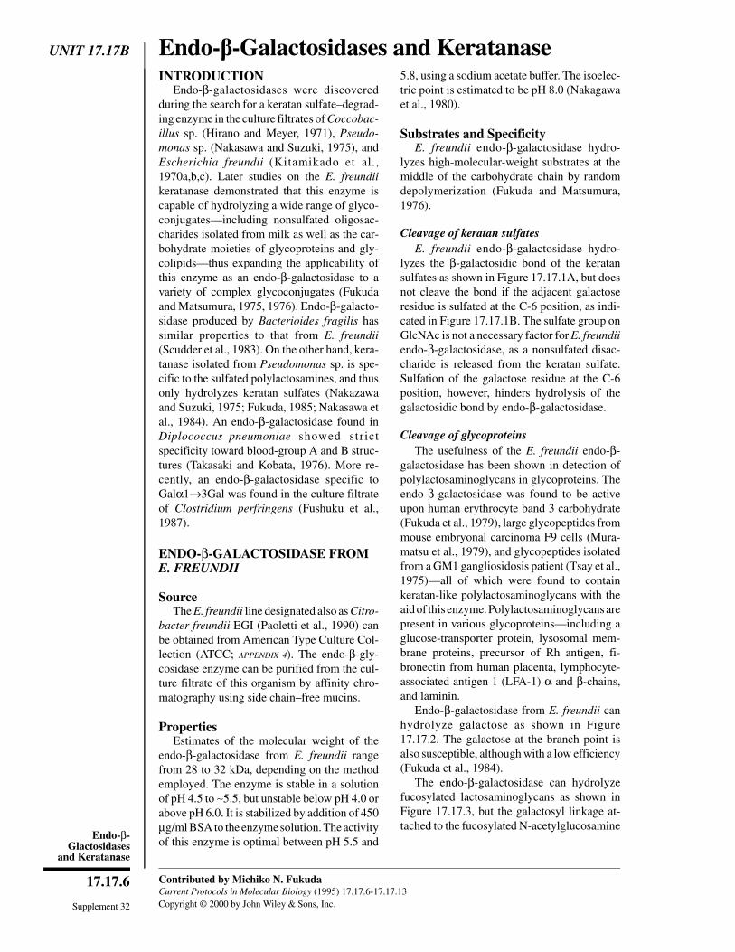
UNIT 17.17B Endo--Galactosidases and KeratanaseINTRODUCTION
Endo-β-galactosidases were discoveredduring the search for a keratan sulfate–degrad-ing enzyme in the culture filtrates of Coccobac-illus sp. (Hirano and Meyer, 1971), Pseudo-monas sp. (Nakasawa and Suzuki, 1975), andEscherichia freundii (Kitamikado et al.,1970a,b,c). Later studies on the E. freundiikeratanase demonstrated that this enzyme iscapable of hydrolyzing a wide range of glyco-conjugates—including nonsulfated oligosac-charides isolated from milk as well as the car-bohydrate moieties of glycoproteins and gly-colipids—thus expanding the applicability ofthis enzyme as an endo-β-galactosidase to avariety of complex glycoconjugates (Fukudaand Matsumura, 1975, 1976). Endo-β-galacto-sidase produced by Bacterioides fragilis hassimilar properties to that from E. freundii(Scudder et al., 1983). On the other hand, kera-tanase isolated from Pseudomonas sp. is spe-cific to the sulfated polylactosamines, and thusonly hydrolyzes keratan sulfates (Nakazawaand Suzuki, 1975; Fukuda, 1985; Nakasawa etal., 1984). An endo-β-galactosidase found inDiplococcus pneumoniae showed strictspecificity toward blood-group A and B struc-tures (Takasaki and Kobata, 1976). More re-cently, an endo-β-galactosidase specific toGalα1→3Gal was found in the culture filtrateof Clostridium perfringens (Fushuku et al.,1987).
ENDO-β-GALACTOSIDASE FROME. FREUNDII
SourceThe E. freundii line designated also as Citro-
bacter freundii EGI (Paoletti et al., 1990) canbe obtained from American Type Culture Col-lection (ATCC; APPENDIX 4). The endo-β-gly-cosidase enzyme can be purified from the cul-ture filtrate of this organism by affinity chro-matography using side chain–free mucins.
PropertiesEstimates of the molecular weight of the
endo-β-galactosidase from E. freundii rangefrom 28 to 32 kDa, depending on the methodemployed. The enzyme is stable in a solutionof pH 4.5 to ∼5.5, but unstable below pH 4.0 orabove pH 6.0. It is stabilized by addition of 450µg/ml BSA to the enzyme solution. The activityof this enzyme is optimal between pH 5.5 and
5.8, using a sodium acetate buffer. The isoelec-tric point is estimated to be pH 8.0 (Nakagawaet al., 1980).
Substrates and SpecificityE. freundii endo-β-galactosidase hydro-
lyzes high-molecular-weight substrates at themiddle of the carbohydrate chain by randomdepolymerization (Fukuda and Matsumura,1976).
Cleavage of keratan sulfatesE. freundii endo-β-galactosidase hydro-
lyzes the β-galactosidic bond of the keratansulfates as shown in Figure 17.17.1A, but doesnot cleave the bond if the adjacent galactoseresidue is sulfated at the C-6 position, as indi-cated in Figure 17.17.1B. The sulfate group onGlcNAc is not a necessary factor for E. freundiiendo-β-galactosidase, as a nonsulfated disac-charide is released from the keratan sulfate.Sulfation of the galactose residue at the C-6position, however, hinders hydrolysis of thegalactosidic bond by endo-β-galactosidase.
Cleavage of glycoproteinsThe usefulness of the E. freundii endo-β-
galactosidase has been shown in detection ofpolylactosaminoglycans in glycoproteins. Theendo-β-galactosidase was found to be activeupon human erythrocyte band 3 carbohydrate(Fukuda et al., 1979), large glycopeptides frommouse embryonal carcinoma F9 cells (Mura-matsu et al., 1979), and glycopeptides isolatedfrom a GM1 gangliosidosis patient (Tsay et al.,1975)—all of which were found to containkeratan-like polylactosaminoglycans with theaid of this enzyme. Polylactosaminoglycans arepresent in various glycoproteins—including aglucose-transporter protein, lysosomal mem-brane proteins, precursor of Rh antigen, fi-bronectin from human placenta, lymphocyte-associated antigen 1 (LFA-1) α and β-chains,and laminin.
Endo-β-galactosidase from E. freundii canhydrolyze galactose as shown in Figure17.17.2. The galactose at the branch point isalso susceptible, although with a low efficiency(Fukuda et al., 1984).
The endo-β-galactosidase can hydrolyzefucosylated lactosaminoglycans as shown inFigure 17.17.3, but the galactosyl linkage at-tached to the fucosylated N-acetylglucosamine
Contributed by Michiko N. FukudaCurrent Protocols in Molecular Biology (1995) 17.17.6-17.17.13Copyright © 2000 by John Wiley & Sons, Inc.Supplement 32
17.17.6
Endo-β-Glactosidases
and Keratanase

is poorly susceptible to the enzyme (Spoonceret al., 1984; Kannagi et al., 1982a).
Cleavage of glycolipidsThe so-called lacto-series glycolipids
(R3→GlcNAcβ1→3-Galβ1→4Glc→Cer) aresusceptible to endo-β-galactosidase (Fukuda etal., 1976). For example, endo-β-galactosidasehydrolyzes lacto-N-neotetraosylceramide,producing trisaccharide and glucosylceramideas shown in Figure 17.17.4A and lacto-N-neo-hexaosylceramide is hydrolyzed to producetwo oligosaccharides—lacto-N-triaosylcera-mide and glucosylceramide—as shown in Fig-ure 17.17.4B. Branched lacto-series gly-colipids—e.g., lacto-iso-N-neooctaosyl-ceramide—are hydrolyzed to a large
oligosaccharide and glucosylceramide asshown in Figure 17.17.4C.
Although the β-galactosidic linkage at thebranch point in the erythrocyte polylactosami-noglycan-peptides is cleaved by endo-β-galac-tosidase, under practical conditions the hy-drolysis of glycolipids at the branch point is toopoor to be detected.
Hydrolysis of x2-glycolipid (Kannagi et al.,1982b) indicates that not only theGlcNAcβ1→3Galβ1→ structure, but also theGalNAcβ1→3Galβ1→ structure is susceptibleto endo-β-galactosidase.
Cleavage of oligosaccharidesE. freundii endo-β-galactosidase can hydro-
lyze oligosaccharides isolated from human
SO4
64GlcNAcβ1 3Galβ1 4GlcNAcβ1
SO4
6
susceptible
A
SO4
64GlcNAcβ1 3Galβ1 4GlcNAcβ1
SO4
6
not susceptible
B SO4
6
Figure 17.17.1 Hydrolysis of keratan sulfates by E. freundii endo-β-galactosidase. (A) Endo-β-galactosidase hydrolyzes β-galactosidic bond of keratan sulfates. (B) Hydrolysis hindered bysulfation of galactose residue at C-6 position.
64GlcNAcβ1 3Galβ1 4GlcNAcβ13Galβ1Siaα2 Galβ1 4GlcNAcβ1 3Galβ1 4GlcNAcβ1
Galβ1 4GlcNAcβ1
4Galβ1
poorlysusceptible
Figure 17.17.2 Hydrolysis of galactose in glycoproteins by E. freundii endo-β-galactosidase.
β4GlcNAcβ1 3Galβ1 4GlcNAcβ1
susceptible
3
Fucα1
3
Fucα1
3Galβ1 4GlcNAcβ1
poorlysusceptible
Figure 17.17.3 Hydrolysis of fucosylated lactosaminoglycans by E. freundii endo-β-galactosi-dase. The galactosyl residue attached to the fucosylated N-acetylglucosamine is poorly susceptible.
Current Protocols in Molecular Biology Supplement 32
17.17.7
Preparation andAnalysis ofGlycoconjugates

milk (Fukuda, 1981; Nakagawa et al., 1980).Affinity of the enzyme for oligosaccharide sub-strates is weaker than that for polylactosamino-glycan, keratan sulfate, and glycolipid sub-strates. A high concentration of the enzyme isneeded for an efficient enzymic hydrolysis ofoligosaccharides.
Application of Endo-β-Galactosidaseto Various glycoconjugates
GlycopeptidesTo digest glycopeptides with endo-β-galac-
tosidase, mix 10 to 100 µg of the glycopep-tide—metabolically labeled (UNIT 17.4) or unla-beled—and 25 mU of endo-β-galactosidasewith 200 µl of 0.2 M sodium acetate buffer, pH5.0(APPENDIX 2), then incubate at 37°C over-night. The oligosaccharides released by endo-β-galactosidase are usually analyzed by gelfiltration (UNIT 10.9). In preparation for this, thedigest of unlabeled glycopeptides is reducedwith NaB[3H]4 (see UNIT 17.5). The resulting
[3H]-labeled oligosaccharide alcohols are thenapplied to a gel-filtration column packed withSephadex G-50 (Pharmacia Biotech) and/or acolumn packed with Bio-Gel P-4 (Bio-Rad).
Upon Bio-Gel P-4 gel filtration, the oli-gosaccharide components are eluted at the elu-tion volumes of disaccharides, trisaccharides,and tetrasaccharides; larger (or sialylated)molecules are eluted in the void volume. Theresults obtained for erythrocyte band 3 showedthat the oligosaccharide alcohols released are:GlcNAcβ1→3GalOH,Galβ1→4GlcNAcβ1→3GalOH, andFucα1→2Galβ1→4GlcNAcβ1→3GalOH.
The oligosaccharide components eluted inthe void volume from a Bio-Gel P-4 columnare sialylated oligosaccharides—because afterdesialylation these components are often elutedat the elution volumes of trisaccharides orlarger oligosaccharides. The structure of thesmallest sialylated oligosaccharide componentreleased from band 3 was:NeuNAcα2→3Galβ1→4GlcNAcβ1→3GalOH.
Galβ1 4GlcNAcβ1 3Galβ1 4Glc Cer
endo–β–galactosidase
Galβ1 4GlcNAcβ1 3Gal Glc Cer
A
Galβ1 4GlcNAcβ1 3Galβ1
endo–β–galactosidase
4GlcNAcβ1 3Gal
B 4GlcNAcβ1 3Galβ1 4Glc Cer
Galβ1 GlcNAcβ1 3Galβ1 4Glc Cer
GlcNAcβ1 3Gal Glc Cer)(and
Galβ1 4GlcNAcβ1Galβ1
endo–β–galactosidase
C4GlcNAcβ1 3Galβ1 4Glc Cer
6
Galβ1 4GlcNAcβ1 3
Galβ1 4GlcNAcβ1Galβ1 4GlcNAcβ1
6
Galβ1 4GlcNAcβ1 33Gal Glc Cer
Figure 17.17.4 Hydrolysis of glycolipids by E. freundii endo-β-galactosidase. (A) Enzyme hydro-lyzes lacto-N-neotetraosylceramide producing trisaccharide and glucosylceramide. (B) Enzymehydrolyzes lacto-N-neohexaosylceramide to produce lacto-N-triaosylceramide and glucosyl-ceramide. (C) Enzyme hydrolyzes branched lacto-series glycolipid to produce large oligosaccharideand glucosylceramide.
Supplement 32 Current Protocols in Molecular Biology
17.17.8
Endo-β-Glactosidases
and Keratanase
Because disaccharides should be producedfrom the internal moieties of polylactosaminein which tri- and tetrasaccharides representnonreducing terminals. Thus, in the absence offucosylation or sialylation the ratio of disaccha-rides to tri- and tetrasaccharides indicates thelength of the polylactosamine repeats. Theseanalyses, combined with methylation analysesof intact polylactosamines have yielded sub-stantial structural information on glycoprote-ins.
GlycolipidsTo hydrolyze glycolipids with endo-β-
galactosidase, take an aliquot corresponding to10 µg of glycolipid dissolved in 2:1 (v/v) chlo-roform/methanol and place in a small glass testtube or microcentrifuge tube. Dry the gly-colipid sample under N2 gas. To the dried sam-ple, add 10 µl of 0.2 M sodium acetate buffer,pH 5.8 (APPENDIX 2) containing 20 µg sodiumdeoxytaurocholate, as well as 10 µl of 0.25mU/µl endo-β-galactosidase. Incubate 2 hr toovernight to achieve enzymatic hydrolysis. Sia-lylated glycolipid—e.g., sialylparaglobosideand linear lacto-series glycolipids—can be hy-drolyzed easily under these conditions;stronger conditions—e.g., 10 × more en-zyme—are required for hydrolysis of branchedand/or highly fucosylated glycolipids.
Intact cells (Fukuda et al., 1979)Endo-β-galactosidase can directly modify
cell-surface glycoproteins and glycolipids.When human erythrocytes are treated withendo-β-galactosidase, Ii antigens and ABOblood-group antigens are abolished or de-creased. In addition to these antigenic changes,polylactosamines on band 3 and glycoproteinson band 4.5 are hydrolyzed, though O-linkedoligosaccharides in glycophorins remain unaf-fected. Glycolipids on the cell surface are alsohydrolyzed by endo-β-galactosidase, but onlylacto-series glycolipids with long carbohydratechains are hydrolyzed by this treatment. Theinnermost galactosyl linkage of glycolipids,R→Galβ1→4Glc→Cer, is not susceptible tothe enzyme treatment on the cell surface. Endo-β-galactosidase treatment of intact cells, com-bined with cell-surface labeling using galactoseoxidase or galactosyltransferase, has been use-ful for identifying polylactosamine-bearingglycoproteins.
To perform cell-surface labeling and endo-β-galactosidase digestion, the galactose and
N-acetylgalactosamine residues on the cell sur-face can be specifically labeled by the galactoseoxidase/NaB[3H]4 method (Gahmberg andHakomori, 1973). Wash the erythrocytes threetimes with cold PBS (APPENDIX 2), then suspend∼2 × 10⁷ cells (or 200 µl of packed cells) in 1ml PBS pH 7.4. Add 10 µl of 1 U/ml galactoseoxidase (Sigma) in PBS, pH 7.4 and incubate2 hr at room temperature. After incubation, add2.5 mCi NaB[3H]4 in 50 µl 0.01 N NaOH andincubate 30 min at room temperature. Finally,add ∼0.5 mg nonradioactive NaBH4 and wash3 × with cold PBS (pH 7.0), centrifuging be-tween washes. Resuspend 0.1 ml of these sur-face-labeled cells (as packed cells) in PBS pH7.0 to a final volume of 200 µl. To this cellsuspension, add 25 µl of 1 U/ml endo-β-galac-tosidase , then incubate 2 hr at room tempera-ture. Wash the cells with 3 ml cold PBS (pH7.0) and centrifuge. The pelleted cells can beused for glycoprotein and glycolipid analysis,and the supernatant can be analyzed for re-leased oligosaccharides. This procedure can beused not only for erythrocytes, but also fornucleated cells.
To analyze glycoproteins and glycolipids ofsurface-labeled cells, prepare erythrocytemembranes from the surface-labeled cells thathave been treated with or without endo-β-galactosidase as described above. Membranelysates from nucleated cells—e.g., lympho-cytes and fibroblasts—can be prepared byvortexing the cells in an equal volume of coldPBS containing 0.5% NP-40 and 1 mM PMSF.The nuclei are then separated out by centrifu-gation, after which the supernatant containingthe cell lysate may be dissolved in SDS samplebuffer and subjected to SDS-polyacrylamidegel electrophoresis (UNIT 10.2), followed byfluorography (APPENDIX 3A) to detect radioactiveprotein components.
To extract glycolipids from erythrocytemembranes or intact cells, these are dissolvedin 10 vol of 2:1 (v/v) chloroform/methanol andthe extract fractionated by Folch partition (UNIT
17.3) into lower and upper phases. If necessary,upper-phase glycolipids can be further sepa-rated into neutral and acidic glycolipids byDEAE-Sephadex. Each glycolipid fraction isfinally analyzed by thin-layer chromatographyfollowed by fluorography. Oligosaccharidesreleased from the cell surface by endo-β-galac-tosidase treatment can also be analyzed bygel-filtration chromatography on Sephadex G-50 and by paper chromatography.
Current Protocols in Molecular Biology Supplement 32
17.17.9
Preparation andAnalysis ofGlycoconjugates
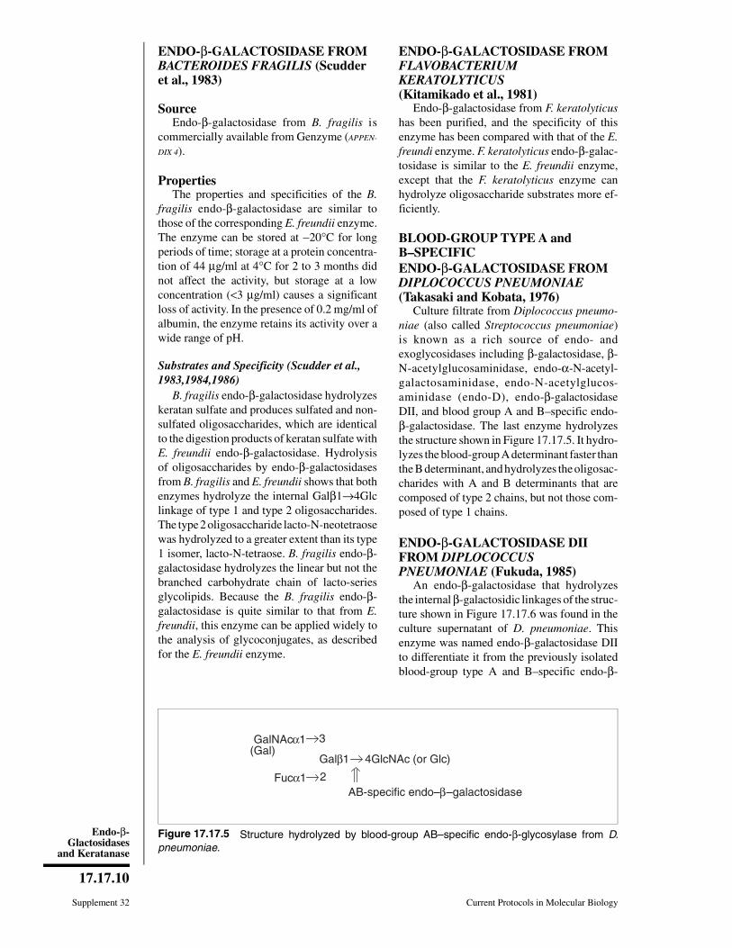
ENDO-β-GALACTOSIDASE FROMBACTEROIDES FRAGILIS (Scudderet al., 1983)
SourceEndo-β-galactosidase from B. fragilis is
commercially available from Genzyme (APPEN-
DIX 4).
PropertiesThe properties and specificities of the B.
fragilis endo-β-galactosidase are similar tothose of the corresponding E. freundii enzyme.The enzyme can be stored at −20°C for longperiods of time; storage at a protein concentra-tion of 44 µg/ml at 4°C for 2 to 3 months didnot affect the activity, but storage at a lowconcentration (<3 µg/ml) causes a significantloss of activity. In the presence of 0.2 mg/ml ofalbumin, the enzyme retains its activity over awide range of pH.
Substrates and Specificity (Scudder et al.,1983,1984,1986)
B. fragilis endo-β-galactosidase hydrolyzeskeratan sulfate and produces sulfated and non-sulfated oligosaccharides, which are identicalto the digestion products of keratan sulfate withE. freundii endo-β-galactosidase. Hydrolysisof oligosaccharides by endo-β-galactosidasesfrom B. fragilis and E. freundii shows that bothenzymes hydrolyze the internal Galβ1→4Glclinkage of type 1 and type 2 oligosaccharides.The type 2 oligosaccharide lacto-N-neotetraosewas hydrolyzed to a greater extent than its type1 isomer, lacto-N-tetraose. B. fragilis endo-β-galactosidase hydrolyzes the linear but not thebranched carbohydrate chain of lacto-seriesglycolipids. Because the B. fragilis endo-β-galactosidase is quite similar to that from E.freundii, this enzyme can be applied widely tothe analysis of glycoconjugates, as describedfor the E. freundii enzyme.
ENDO-β-GALACTOSIDASE FROMFLAVOBACTERIUMKERATOLYTICUS(Kitamikado et al., 1981)
Endo-β-galactosidase from F. keratolyticushas been purified, and the specificity of thisenzyme has been compared with that of the E.freundi enzyme. F. keratolyticus endo-β-galac-tosidase is similar to the E. freundii enzyme,except that the F. keratolyticus enzyme canhydrolyze oligosaccharide substrates more ef-ficiently.
BLOOD-GROUP TYPE A andB–SPECIFICENDO-β-GALACTOSIDASE FROMDIPLOCOCCUS PNEUMONIAE(Takasaki and Kobata, 1976)
Culture filtrate from Diplococcus pneumo-niae (also called Streptococcus pneumoniae)is known as a rich source of endo- andexoglycosidases including β-galactosidase, β-N-acetylglucosaminidase, endo-α-N-acetyl-galactosaminidase, endo-N-acetylglucos-aminidase (endo-D), endo-β-galactosidaseDII, and blood group A and B–specific endo-β-galactosidase. The last enzyme hydrolyzesthe structure shown in Figure 17.17.5. It hydro-lyzes the blood-group A determinant faster thanthe B determinant, and hydrolyzes the oligosac-charides with A and B determinants that arecomposed of type 2 chains, but not those com-posed of type 1 chains.
ENDO-β-GALACTOSIDASE DIIFROM DIPLOCOCCUSPNEUMONIAE (Fukuda, 1985)
An endo-β-galactosidase that hydrolyzesthe internal β-galactosidic linkages of the struc-ture shown in Figure 17.17.6 was found in theculture supernatant of D. pneumoniae. Thisenzyme was named endo-β-galactosidase DIIto differentiate it from the previously isolatedblood-group type A and B–specific endo-β-
Galβ1 4GlcNAc (or Glc)
AB-specific endo–β–galactosidase
3
2Fucα1
GalNAcα1 (Gal)
Figure 17.17.5 Structure hydrolyzed by blood-group AB–specific endo-β-glycosylase from D.pneumoniae.
Supplement 32 Current Protocols in Molecular Biology
17.17.10
Endo-β-Glactosidases
and Keratanase
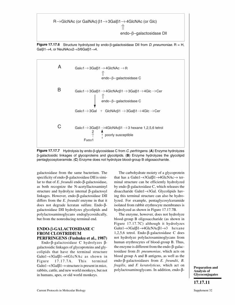
galactosidase from the same bacterium. Thespecificity of endo-β-galactosidase DII is simi-lar to that of E. freundii endo-β-galactosidase,as both recognize the N-acetyllactosaminylstructure and hydrolyze internal β-galactosyllinkages. However, endo-β-galactosidase DIIdiffers from the E. freundii enzyme in that itdoes not degrade keratan sulfate. Endo-β-galactosidase DII hydrolyzes glycolipids andpolylactosaminoglycans endoglycosidically,but from the nonreducing-terminal end.
ENDO-β-GALACTOSIDASE CFROM CLOSTRIDIUMPERFRINGENS (Fushuku et al., 1987)
Endo-β-galactosidase C hydrolyzes β-galactosidic linkages of glycoproteins and gly-colipids that have the terminal structureGalα1→3Galβ1→4GlcNAc as shown inFigure 17.17.7A. This terminalGalα1→3Galβ1→ structure is present in mice,rabbits, cattle, and new world monkeys, but notin humans, apes, or old world monkeys.
The carbohydrate moiety of a glycoproteinthat has a Galα1→3Galβ1→4GlcNAc→ ter-minal structure can be efficiently hydrolyzedby endo-β-galactosidase C, which releases thedisaccharide Galα1→3Gal. Glycolipids hav-ing this terminal structure can also be hydro-lyzed. For example, pentaglycosylceramideisolated from rabbit erythrocyte membranes ishydrolyzed as shown in Figure 17.17.7B.
The enzyme, however, does not hydrolyzeblood-group B oligosaccharide (as shown inFigure 17.17.7C) although it hydrolyzesGalα1→3Galβ1→4GlcNAcβ1→3 hexane1,2,5,6 tetrol. Endo-β-galactosidase C doesnot hydrolyze polylactosaminoglycans fromhuman erythrocytes of blood-group B. Thus,the enzyme is different from the endo-β-galac-tosidase from D. pneumoniae, which acts onblood group A and B antigens, as well as theendo-β-galactosidases from E. freundii, B.fragilis, and F. keratolyticus, which act onpolylactosaminoglycans. In addition, endo-β-
3Galβ1 4GlcNAc (or Glc)
endo–β–galactosidase Dll
β1GlcNAc (or GalNAc)R
Figure 17.17.6 Structure hydrolyzed by endo-β-galactosidase DII from D. pneumoniae. R = H,Galβ1→4, or NeuNAcα2→3/6Galβ1→4.
Galα1 3Galβ1
endo–β– galactosidase C
A
B
C
4GlcNAc R
Galα1 3Galβ1 4GlcNAcβ1 3Galβ1 4Glc Cer
Galα1 3Gal GlcNAcβ1 3Galβ1 4Glc Cer
Galα1 4GlcNAcβ1 3 hexane 1,2,5,6 tetrol3Galβ1
poorly susceptible2
Fucα1
endo–β– galactosidase C
Figure 17.17.7 Hydrolysis by endo-β-glycosidase C from C. perfringens. (A) Enzyme hydrolyzesβ-galactosidic linkages of glycoproteins and glycolipids. (B) Enzyme hydrolyzes the glycolipidpentaglycosylceramide. (C) Enzyme does not hydrolyze blood-group B oligosaccharide.
Current Protocols in Molecular Biology Supplement 32
17.17.11
Preparation andAnalysis ofGlycoconjugates

galactosidase C does not hydrolyze keratansulfate.
KERATANASE FROMPSEUDOMONAS SP. (Nakazawa andSuzuki, 1975)
SourceKeratanase (or keratan sulfate–specific
endo-β-galactosidase) has been purified fromPseudomonas sp. cell extract and is commer-cially available from Miles (APPENDIX 4).
PropertiesThe purified enzyme has been stored in ice.
The pH optimum is between 7.2 and 7.4 usingcorneal keratan sulfate as substrate. At a 1 mMconcentration, Ca2+, Mg2+, and Mn2+ have noeffect on the enzymatic activity. Acetate andmaleate elicit a slight increase in activity(∼15%) at concentrations of 0.05 M.
Substrates and SpecificityKeratanase from Pseudomonas sp. hydro-
lyzes keratan sulfate but not chondroitin sul-fates, hyaluronic acid, dermatan sulfate, orheparin. In contrast to E. freundii endo-β-galac-tosidase, this enzyme does not hydrolyze lacto-N-tetraose, lacto-series glycolipids, or erythro-cyte polylactosaminoglycans—thus Pseudo-monas endo-β-galactosidase is specific forkeratan sulfate. Two sulfated oligosaccha-rides—6-sulfo-GlcNAcβ1→3Gal and 6-sulfo-GlcNAcβ1→3-6-sulfo-GlcNAcβ1→4-6-sulfo-GlcNAcβ1→3Gal—have been identified in the digestion productsfrom keratan sulfates produced by this enzyme.However, the nonsulfated disaccharideGlcNAcβ1→3Gal is not produced.
ApplicationsThis enzyme has been quite useful in distin-
guishing keratan sulfates or sulfated polylac-tosaminoglycans from nonsulfated polylactos-aminoglycans (Nakazawa et al., 1984).
ENDO-β-GALACTOSIDASESPECIFIC TO THE REGIONLINKING CHONDROITINSULFATE AND THE COREPROTEIN (Takagaki et al., 1990a,b)
Endo-β-galactosidase that degrades thelinkage between a chondroitin sulfate and coreprotein has been reported in rabbit liver and inthe midgut gland of the mollusk Patnopectensp. Chondroitin sulfate–containing proteogly-
cans were digested with endo-β-xylosidasefrom the midgut gland of Patnopecten sp., andthe released chondroitin sulfate (with an R-Galβ1→3Galβ1→3Galβ1→4Xyl terminus)was conjugated with pyridylamine (PA) by re-ductive amination. This chondroitin sulfate–PAwas incubated with a crude enzyme solutionfrom the midgut gland of Patnopecten sp.HPLC analysis of the PA-oligosaccharide prod-uct identified Galβ1→4Xyl-PA, suggestingstrongly that an endo-β-galactosidase had spe-cifically hydrolyzed the Galβ1→3Gal linkagein the linkage region of chondroitin sulfate.
Literature CitedFukuda, M.N. 1981. Purification and charac-
terization of endo-β-galactosidase from Es-cherichia freundii induced by hog gastric mucin.J. Biol. Chem. 256:3900-3905.
Fukuda, M.N. 1985. Isolation and characterizationof a new endo-β-galactosidase from Diplococ-cus pneumoniae. Biochemistry 24:2154-2163.
Fukuda, M.N. and Matsumura, G. 1975. Endo-β-galactosidase of Escherichia freundii. Hydroly-sis of pig colonic mucin and milk oligosaccha-rides by endoglycosidic action. Biochem. Bio-phys. Res. Commun. 64:465-471.
Fukuda, M.N. and Matsumura, G. 1976. Endo-β-galactosidase of Escherichia freundii. Purifica-tion and endoglycosidic action on keratansul-fates, oligosaccharides and blood group activeglycoprotein. J. Biol. Chem. 251:6218-6225.
Fukuda, M.N., Fukuda, M., and Hakomori, S. 1979.Cell surface modification by endo-β-galactosi-dase, change of blood group activities and re-lease of oligosaccharides from glycoproteins andglycosphingolipids of human erythrocytes. J.Biol. Chem. 254:5458-5465.
Fukuda, M., Dell, A., Oates, J.E., and Fukuda, M.N.1984. Structure of branched lactosaminoglycan,the carbohydrate moiety of band 3 isolated fromadult human erythrocytes. J. Biol. Chem.259:8260-8273.
Fushuku, N., Muramatsu, H., Uezono, M.M., andMuramatsu, T. 1987. A new endo-β-galactosi-dase releasing Galα1→3Gal from carbohydratemoieties of glycoproteins and from a glycolipid.J. Biol. Chem. 262:10086-10092.
Gahmberg, C.G. and Hakomori, S. 1973. Externallabeling of cell surface galactose and galactos-amine in glycolipid and glycoprotein of humanerythrocytes. J. Biol. Chem. 248:4311-4317.
Hirano, S. and Meyer, K. 1971. Enzymatic degrada-tion of corneal and cartilaginous keratosulfates.Biochem. Biophys. Res. Commun. 44:1371-1375.
Kannagi, R., Nudelman, E., Levery, S.B., and Hak-omori, S. 1982a. A series of human erythrocyteglycosphingolipids reacting to the monoclonalantibody directed to a developmentally regulatedantigen, SSEA-1. J. Biol. Chem. 257:14865-14874.
Supplement 32 Current Protocols in Molecular Biology
17.17.12
Endo-β-Glactosidases
and Keratanase

Kannagi, R., Fukuda, M.N., and Hakomori, S.1982b. A new glycolipid antigen isolated fromhuman erythrocyte membranes reacting with an-tibodies directed to globo-N-tetraosylceramide(globoside). J. Biol. Chem. 257:4438-4442.
Kitamikado, M., Ueno, R., and Nakamura, T. 1970a.Enzymatic degradation of Whale cartilage kera-tosulfate-I, isolation of keratosulfate-degradingbacterium. Bull. Jap. Soc. Sci. Fish. 36:592-596.
Kitamikado, M., Ueno, R., and Nakamura, T. 1970b.Enzymatic degradation of Whale cartilage kera-tosulfate-II, identification of a keratosulfate-de-grading bacterium. Bull. Jap. Soc. Sci. Fish.36:1172-1174.
Kitamikado, M. and Ueno, R. 1970c. Enzymaticdegradation of Whale cartilage keratosulfate-III,purification of a bacterial keratosulfate-degrad-ing enzyme. Bull. Jap. Soc. Sci. Fish. 36:1175-1180.
Kitamikado, M. Ito, M., and Li, Y.-T. 1981. Isolationand characterization of a keratan sulfate–degrad-ing endo-β-galactosidase from FlavobacteriumKeratolyticus. J. Biol. Chem. 256:3906-3909.
Muramatsu, T., Gachelin, G., Damonneville, M.,Delarbre, C., and Jacob, F. 1979. Cell surfacecarbohydrates of embryonal carcinoma cells:Polysaccharidic side chains of F9 antigens andof receptors to two lectins, FBP and PNA. Cell18:183-191.
Nakagawa, H., Yamada, T., Chien, J.-L., Gardas, A.,Kitamikado, M., Li, S.-C., and Li, Y.-T. 1980.Isolation and characterization of an endo-β-galactosidase from a new strain of Escherichiafreundii. J. Biol. Chem. 255:5955-5959.
Nakazawa, K. and Suzuki, S. 1975. Purification ofkeratansulfate-endogalactosidase and its actionon keratan sulfates of different origin. J. Biol.Chem. 250:912-917.
Nakazawa, K., Hassell, J.R., Hascal, V.C., Lohman-der, S., Newsome, D.A., and Krachmer, J. 1984.Defective processing of keratan sulfate in macu-lar corneal dystrophy. J. Biol. Chem. 259:13751-13757.
Paoletti, L.C., Kasper, D.L., Michon, F., DiFabio, J.,Holme, K., Jennings, H.J., and Wessels, M.R.1990. An oligosaccharide-tetanus toxoid conju-gate vaccine against type III group B streptococ-cus. J. Biol. Chem. 265:18278-18283.
Scudder, P., Uemura, K., Kolby, J., Fukuda, M.N.,and Feizi, T. 1983. Isolation and characterizationof an endo-β-galactosidase from Bacterioidesfragilis. Biochem. J. 213:485-494.
Scudder, P., Hanfland, P., Uemura, K., and Feizi, T.1984. Endo-β-galactosidase of Bacterioidesfragilis and Escherichia freundii hydrolyze lin-ear but not branched oligosaccharide domains ofglycolipids of the neolactoseries. J. Biol. Chem.259:6586-6592.
Scudder, P., Tang, P.W., Hounsell, E.F., Lawson,A.M., Mehmet, H., and Feizi, T. 1986. Isolationand characterization of sulfated oligosaccharidesreleased from bovine corneal keratansulfate bythe action of endo-β-galactosidase. Eur. J. Bio-chem. 157:365-373.
Spooncer, E., Fukuda, M., Klock, J.C., Oates, J.E.,and Dell, A. 1984. Isolation and characterizationof polyfucosylated lactosaminoglycan from hu-man granulocytes. J. Biol. Chem. 259:4792-4801.
Takagaki, K., Kon, A., Kawasaki, H., Nakamura, T.,Tamura, S., and Endo, M. 1990a. Presence of anendo-β-galactosidase degrading the linkage re-gion between the chondroitin sulfate chain andcore peptide of proteoglycan. Biochem. Biophys.Res. Commun. 169:15-21.
Takagaki, K., Kon, A., Kawasaki, H., Nakamura, T.,Tamura, S., and Endo, M. 1990b. Isolation andcharacterization of Patnopecten mid-gut glandendo-β-xylosidase active on peptide chondroitinsulfate. J. Biol. Chem. 265:854-860.
Takasaki, S. and Kobata, A. 1976. Purification andcharacterization of an endo-β-galactosidase pro-duced by Diplococcus pneumoniae. J. Biol.Chem. 251:3603-3609.
Tsay, G.C., Dawson, G., and Li, Y.-T. 1975. Struc-ture of the glycopeptide storage material in Gm1gangliosides. Sequence determination with spe-cific endo- and exoglycosidases. Biochim. Bio-phys. Acta 385:305-311.
Contributed by Michiko N. FukudaLa Jolla Cancer ResearchLa Jolla, California
Current Protocols in Molecular Biology Supplement 32
17.17.13
Preparation andAnalysis ofGlycoconjugates
SECTION IVANALYSIS OF SACCHARIDES RELEASEDFROM GLYCOCONJUGATESMany of the more specific and sensitive methods for analyzing the structure of oligosac-charides require that the oligosaccharides be first released from the glycoconjugate towhich they are attached and, if necessary, purified away from the protein moiety. Theunits presented in this section describe various ways in which such released oligosaccha-rides can be analyzed. It must be recognized that no matter how good such a method is,it is highly dependent upon the quality of the preceding steps—i.e., the specific andquantitative release of the oligosaccharide from the glycoconjugate and the use of anappropriate purification scheme that does not result in selective losses. The methodspresented in this section include those designed for the analysis of monosaccharides (UNIT
17.18) and those designed for the study of oligosaccharides (or glycopeptides; UNITS
17.19-17.23). The first group primarily provide monosaccharide compositional informationand are a good starting point for the analysis of a previously unexplored glycoconjugate.Methods in the latter group range from simple techniques for determining basic propertiesof an oligosaccharide, to complex and sophisticated methodologies requiring the use ofexpensive instruments.
There are of course many other well-established techniques and approaches to thestructural analysis of oligosaccharides, some of which require specialized instrumentation(e.g., mass spectrometry and nuclear magnetic resonance). The choice of techniques forinclusion in this section was based partly on the criteria of ease of use and broad generalutility to the average molecular biologist.
UNIT 17.18Analysis of MonosaccharidesThis unit presents methods for assaying sialic acids, reducing sugars, and hexosamines.The BCA assay (Basic Protocol 1) detects free reducing terminii in sugars released fromglycoconjugates by appropriate treatments. Assays employing Ehrlich reagent (DMAB)detect hexosamines and N-acetylhexosamines (Basic Protocol 2 and Alternate Protocol);Basic Protocol 2 also includes a method for hydrolyzing the glycosidic linkages of thehexosamines and a method for re-N-acetylation is described in Support Protocol 1). TheTBA (Basic Protocol 3) and DMB assays (Basic Protocol 4) can be used to quantitate andfractionate free forms of many types of sialic acids; techniques for liberating the sialicacids from the parent glycoconjugates are provided in Support Protocols 2 and 3. (Anotheralternative, the ferric orcinol method presented in UNIT 17.9, can measure both free andglycosidically bound sialic acids irrespective of modifications, but with much reducedsensitivity.)
Note that the acid hydrolysis conditions used to release sialic acids are much milder thanthose used for other monosaccharides—indeed, sialic acids are destroyed under theharsher conditions. Therefore, a sialic acid–containing molecule needs to be subjected toat least two separate analyses—one for sialic acids and one for the other monosaccharides.
BASICPROTOCOL 1
BCA ASSAY FOR REDUCING SUGARS WITHSPECTROPHOTOMETRIC DETECTIONThis protocol presents the most sensitive method developed to date to measure freereducing termini in sugars. This type of assay is useful for determining the total amountof oligosaccharides released from N-linked or O-linked glycoconjugates by enzymatictreatment (UNIT 17.13A) or from glycolipids by endoglycoceramidase (UNIT 17.17A). It mayalso be used to determine the total sugars present after acid hydrolysis.
Supplement 32
Contributed by Adriana E. Manzi, Leland D. Powell, and Ajit VarkiCurrent Protocols in Molecular Biology (1995) 17.18.1-17.18.19Copyright © 2000 by John Wiley & Sons, Inc.
17.18.1
Preparation andAnalysis ofGlycoconjugates
Materials
Sample in water or aqueous bufferStandard: e.g., glucosamine hydrochloride or N-acetyl-D-glucosamine (both
available from Sigma)Bovine serum albumin (BSA)BCA reagent (reagent C; see recipe)
16 × 115–mm Pyrex test tubesClean glass marblesHeating block, 80°CSpectrophotometer and 1.0-ml glass cuvettes
1. In 16 × 115–mm Pyrex test tubes, prepare duplicates containing aliquots of sampleand of 2 to 25 nmol of an appropriate standard in 300 µl water. Also prepare a blankconsisting of 300 µl of water or the buffer in which sample is dissolved, and a controlcontaining BSA at a concentration equal to the maximum protein concentrationexpected in the samples.
Avoid borate ions in the buffer, but if these cannot be avoided, make sure to adjust the pHto 10.6 to avoid interference.
The expected reducing sugar for the type of sample under analysis should be used asstandard. N-acetylglucosamine should be used for oligosaccharides released from N-linkedglycoproteins; glucose for oligosaccharides released from gangliosides.
2. Add 700 µl BCA reagent to each tube, vortex, and cover with glass marbles to preventevaporation. Heat 30 min at 80°C.
3. Allow tubes to cool to room temperature. Transfer solutions to 1.0-ml glass cuvettesand measure the absorbance at 560 nm, adjusting against the values obtained for theblank.
4. Prepare a standard curve of absorbance vs. nmol reducing sugar.
5. Determine the amount of reducing sugar in each sample by reference to the standardcurve.
Reagents and SolutionsUse deionized, distilled water in all recipes and protocol steps. For common stock solutions, seeAPPENDIX 2; for suppliers, see APPENDIX 4.
BCA reagentReagent A: Dissolve 1.5 g disodium 2,2′-bicinchoninate (4,4′-dicarboxy-2,2′-biqui-noline; FW 388.3; Sigma) in 1 liter water. Add 71.6 g anhydrous sodium carbonate(ACS reagent grade, FW 105.99; Sigma) while stirring. Dilute to 1.15 liters withwater. Store in a dark bottle at room temperature (stable several months).Reagent B: Dissolve 3.5 g aspartic acid (FW 133.1; Sigma) and 5.0 g anhydroussodium carbonate in 100 ml water, shaking to dissolve (this produces foam). In aseparate container, dissolve 1.09 g copper sulfate (CuSO4⋅5 H2O, FW 249.6; ACSreagent grade) in 40 ml water. Mix the two solutions and dilute to 150 ml with water(a temporary fluffy blue precipitate may appear). Store at 4°C (stable severalmonths).Reagent C (BCA reagent): Mix 23 ml reagent A and 1 ml reagent B with 6 ml of100% ethanol. The solution will have an intense blue color and strong fluorescence.Store in a brown bottle up to 2 weeks at 4°C (discard earlier if purple backgroundcolor increases). Let stand 2 hr before use.
Supplement 32 Current Protocols in Molecular Biology
17.18.2
Analyis ofMonosaccharides

Commentary
Background InformationMost assays proposed for reduction of sug-
ars make use of inorganic oxidants that acceptelectrons from sugar aldehyde groups. Aldosesare converted to aldonic acids (and higher oxi-dation products) and either the metal ions di-rectly formed, metal oxides, or the complexa-tion products of the latter with chromogenicchelators are quantitated. The appropriate oxi-dation/reduction potential for this reaction de-pends on the pH and complexation constants.
The BCA reagent forms colored complexeswith copper(I); it is possible to couple thisreaction to the reduction from copper(II) tocopper(I) produced by reducing sugars in alka-line solution.
The most commonly used reduction assayfor sugars is the one developed by Park andJohnson (1949) as a modification of the col-orimetric method of Folin and Malmros (1929).This method, based on the production of Prus-sian blue following the reduction of ferricy-anide ions in alkaline solution, has a detectionlimit of ∼1 µg (5 nmol) reducing sugar.
Mopper and Gindler (1973) later proposeda new colorimetric reagent for automatic sugarchromatography that was sensitive to ∼10−10
mol of sugar in the ethanol/water mobile phase.The dye is based on the formation of a deep-blue complex with Cu(I) ions and 2,2′-bicinch-oninic acid in alkaline solution. The reaction isfirst order over a wide range. However, thesensitivity of the reaction is lower and thelinearity range is reduced in water solution.Moreover, when borate ions are present, theMopper-Gindler reagent produces only a faintcolor. Sinner and Puls (1978) achieved in-creased color formation by adjusting the re-agent/borate buffer mixture, when used, to theoptimal pH of the reaction (10.6), and increasedthe linear detection range by doubling the BCAconcentration. Using these conditions, 55 pmolcan be detected in the effluent from an HPLCcolumn with an on-line UV spectrophotometer.McFeeters (1980) adapted the method to man-ual sugar determinations using ethylene glycolinstead of ethanol to increase color formation.The conditions used for on-line detection canalso be applied to manual analysis providedspecial care is taken to compensate for thevolatility of ethanol.
Waffenschmidt and Jaenicke (1987) havesuggested that for the manual spectrophotomet-
ric quantitation of sugar samples, the sensitivityof the reaction could be improved by replacingthe aspartic acid in the BCA reagent with serine.
Critical ParametersUsing manual spectrophotometry, reliable
detection is achieved with <3 nmol of mono-,oligo-, or polysaccharides. Buffering salts, sul-furic acid, sodium hydroxide, hydrochloricacid, and acetic acid do not interfere. It is alsopossible to work in the presence of borate ionsif the pH is adjusted. The expected reducingsugar for the type of sample under analysis isused as a standard. However, linear responsesmay not be obtained with increasing size of theoligosaccharide (Doner and Irwin, 1992). Pro-teins cause a low level of interference of ~50:1(w/w) protein/sugar.
Anticipated ResultsWith this protocol it is possible to detect with
confidence 3 nmol of reducing sugar (mono-,oligo- or polysaccharide) in aqueous solution.
Time ConsiderationsThe assay can be completed in ∼1 hr.
Literature CitedDoner, L.W. and Irwin, P.L. 1992. Assay of reducing
end-groups in oligosaccharide homologues with2,2′-bicinchoninate. Anal. Biochem. 202:50-53.
Folin, O. and Malmros, H. 1929. An improved formof Folin’s micro method for blood sugar deter-minations. J. Biol. Chem. 83:115.
McFeeters, R.F. 1980. A manual method for reduc-ing sugar determinations with 2,2′-bicinchoni-nate reagent. Anal. Biochem. 103:302-306.
Mopper, K. and Gindler, E.M. 1973. A new noncor-rosive dye reagent for automatic sugar chroma-tography. Anal. Biochem. 56:440-442.
Park, J.T. and Johnson, M.J. 1949. A submicrode-termination of glucose. J. Biol. Chem. 181:149-151.
Sinner, M. and Puls, J. 1978. Noncorrosive dyereagent for detection of reducing sugars in boratecomplex ion-exchange chromatography. J.Chromatogr. 156:197-204.
Waffenschmidt, S. and Jaenicke, L. 1987. Assay ofreducing sugars in the nanomole range with 2,2′-bicinchoninate. Anal. Biochem. 165:337-340.
Key ReferenceWaffenschmidt and Jaenicke, 1987. See above.
Describes the modification of the original methodfor improved sensitivity that is used in this protocol.
Current Protocols in Molecular Biology Supplement 32
17.18.3
Preparation andAnalysis ofGlycoconjugates

BASICPROTOCOL 2
ASSAYS FOR FREE HEXOSAMINES WITH SPECTROPHOTOMETRICDETECTION
Free hexosamines can be quantitated spectrophotometrically by a series of proceduresderived from the Elson-Morgan and Morgan-Elson reactions that can be used for bothhexosamines and N-acetylated hexosamines. These assays require a hydrolysis step (seeBasic Protocol 2) to cleave the glycosidic linkages of the hexosamines. This hydrolysisalso produces de-N-acetylation. Thus, a re-N-acetylation step is required (see SupportProtocol 1) for the Morgan-Elson assay.
Acetylacetone/DMAB (Elson-Morgan) Assay for Free Hexosamines
Materials
Hexosamine-containing sample (≥6 µg hexosamine) in aqueous solution1 mM hexosamine standard stock solution(s): N-acetylglucosamine and/or
N-acetylgalactosamine (see recipe)4 M HCl (diluted from ACS reagent-grade concentrated HCl)Acetylacetone reagent (see recipe)95% ethanolEhrlich reagent (DMAB)/HCl (see recipe)
13 × 100–mm Pyrex test tubes with Teflon-lined screw capsNitrogen or vacuum evaporation system (Speedvac or shaker-evaporator)Heating block or ovenBoiling water bathSpectrophotometer and 1.0-ml glass cuvettes
Liberate hexosamines from glycosidic linkages1. Dry triplicate aliquots of the sample (containing ≥6 µg hexosamine) in 13 × 100-mm
Pyrex test tubes with Teflon-lined screw caps.
2. From hexosamine standard stock solution(s), prepare triplicate standards containing10, 20, 30, 40, 50, and 60 µg N-acetylglucosamine and/or N-acetylgalactosamine inwater. Prepare a blank containing the same total volume of water.
3. Dissolve samples, standards, and blank in 4 M HCl (0.4 ml/mg dry sample). Flushwith nitrogen and cap tubes. Heat 4 hr at 100°C.
Because the stability of this type of linkage depends on the structure of the glycoconjugate,optimal conditions for hydrolysis should be determined for each particular case (see UNIT
17.16). When using HCl, concentrations from 2 to 6 M and hydrolysis times from 2 to 10 hrshould be tried.
4. Evaporate the HCl in a vacuum evaporation system or under nitrogen. Place the tubesovernight in a desiccator containing sodium hydroxide pellets under vacuum.
Quantitate hexosamines by condensation with alkaline acetylacetone and DMAB5. Dissolve the hydrolysates in 250 µl water.
6. Add 500 µl acetylacetone reagent to each sample, standard, and blank. Vortex welland cap. Heat 10 min in a boiling water bath.
7. Cool to room temperature. Add 2.5 ml of 95% ethanol and vortex. Incubate 5 min at75 ± 2°C.
8. Add 500 µl DMAB/HCl and heat 30 min at 75 ± 2°C.
9. Cool tubes to room temperature and add 2.5 ml of 95% ethanol. Let stand 30 min.
Supplement 32 Current Protocols in Molecular Biology
17.18.4
Analyis ofMonosaccharides
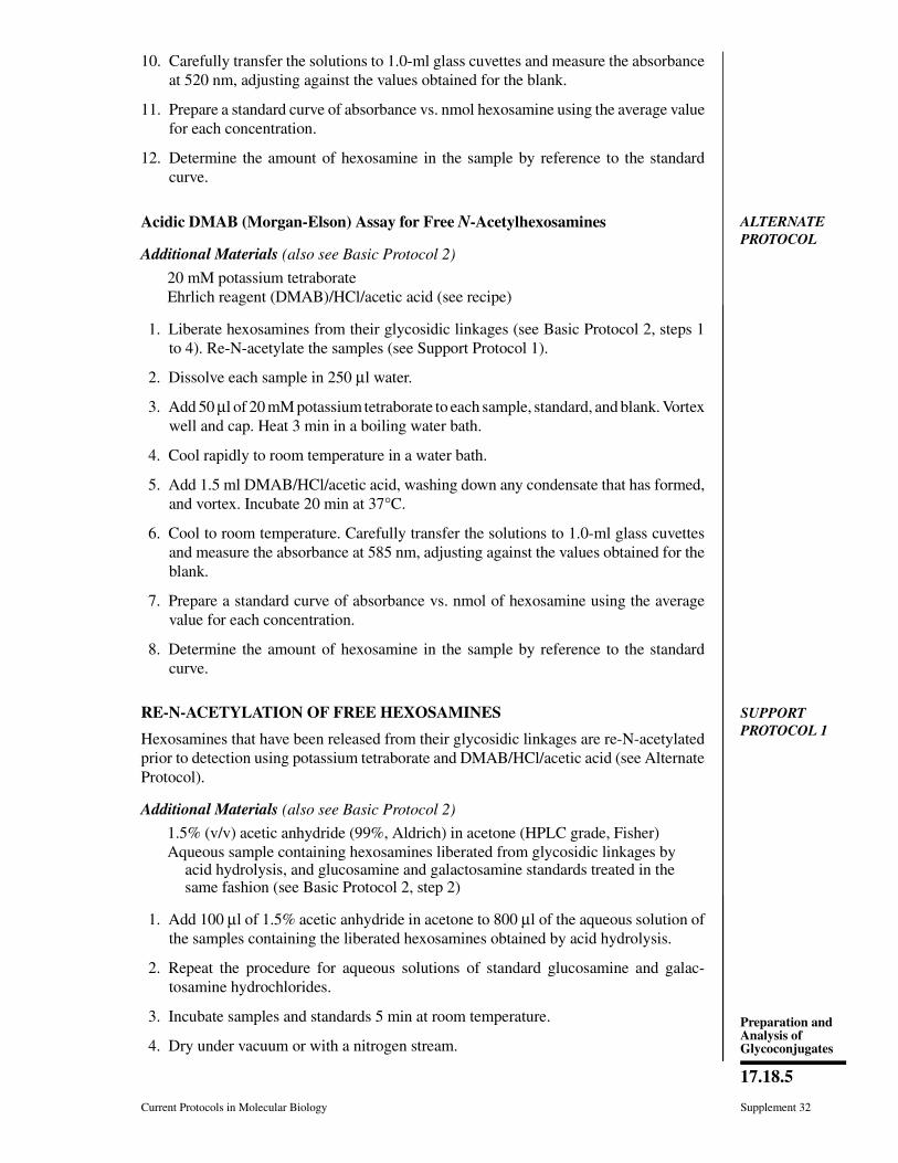
10. Carefully transfer the solutions to 1.0-ml glass cuvettes and measure the absorbanceat 520 nm, adjusting against the values obtained for the blank.
11. Prepare a standard curve of absorbance vs. nmol hexosamine using the average valuefor each concentration.
12. Determine the amount of hexosamine in the sample by reference to the standardcurve.
ALTERNATEPROTOCOL
Acidic DMAB (Morgan-Elson) Assay for Free N-Acetylhexosamines
Additional Materials (also see Basic Protocol 2)
20 mM potassium tetraborateEhrlich reagent (DMAB)/HCl/acetic acid (see recipe)
1. Liberate hexosamines from their glycosidic linkages (see Basic Protocol 2, steps 1to 4). Re-N-acetylate the samples (see Support Protocol 1).
2. Dissolve each sample in 250 µl water.
3. Add 50 µl of 20 mM potassium tetraborate to each sample, standard, and blank. Vortexwell and cap. Heat 3 min in a boiling water bath.
4. Cool rapidly to room temperature in a water bath.
5. Add 1.5 ml DMAB/HCl/acetic acid, washing down any condensate that has formed,and vortex. Incubate 20 min at 37°C.
6. Cool to room temperature. Carefully transfer the solutions to 1.0-ml glass cuvettesand measure the absorbance at 585 nm, adjusting against the values obtained for theblank.
7. Prepare a standard curve of absorbance vs. nmol of hexosamine using the averagevalue for each concentration.
8. Determine the amount of hexosamine in the sample by reference to the standardcurve.
SUPPORTPROTOCOL 1
RE-N-ACETYLATION OF FREE HEXOSAMINES
Hexosamines that have been released from their glycosidic linkages are re-N-acetylatedprior to detection using potassium tetraborate and DMAB/HCl/acetic acid (see AlternateProtocol).
Additional Materials (also see Basic Protocol 2)
1.5% (v/v) acetic anhydride (99%, Aldrich) in acetone (HPLC grade, Fisher)Aqueous sample containing hexosamines liberated from glycosidic linkages by
acid hydrolysis, and glucosamine and galactosamine standards treated in thesame fashion (see Basic Protocol 2, step 2)
1. Add 100 µl of 1.5% acetic anhydride in acetone to 800 µl of the aqueous solution ofthe samples containing the liberated hexosamines obtained by acid hydrolysis.
2. Repeat the procedure for aqueous solutions of standard glucosamine and galac-tosamine hydrochlorides.
3. Incubate samples and standards 5 min at room temperature.
4. Dry under vacuum or with a nitrogen stream.
Current Protocols in Molecular Biology Supplement 32
17.18.5
Preparation andAnalysis ofGlycoconjugates
Reagents and SolutionsUse deionized, distilled water in all recipes and protocol steps. For common stock solutions, seeAPPENDIX 2; for suppliers, see APPENDIX 4.
Acetylacetone reagentPrepare 48 ml of 0.625 M sodium carbonate (FW 106; anhydrous, ACS reagentgrade) in water. Add 2 ml acetylacetone (2,4-pentanedione, FW 100.1; Sigma).Prepare just prior to use.
Ehrlich reagent (DMAB)/HCl/acetic acidAdd 1.5 ml water to 11 ml concentrated HCl. Add 87.5 ml glacial acetic acid (FW60.05; 99.99%; Aldrich). Dissolve 10 g p-dimethylaminobenzaldehyde (DMAB)—4-(N,N-dimethylamino)benzaldehyde, DMAB; crystalline, ACS reagent grade, FW149.2—in the HCl/acetic acid mixture. Store in a dark bottle at room temperature(stable several weeks). Dilute 10 ml of mixture to 100 ml with glacial acetic acidimmediately before using.
Ehrlich reagent (DMAB)/HClWeigh 1.6 g p-dimethylaminobenzaldehyde (DMAB)—4-(N,N-dimethylamino)-benzaldehyde, DMAB; crystalline, ACS reagent grade, FW 149.2. Dissolve in 30ml concentrated HCl. Store in a dark bottle at room temperature (stable severalweeks).
Hexosamine standard stock solutions, 1 mMGlucosamine: Dissolve 9 mg D-glucosamine hydrochloride (2-deoxy-D-glucosehydrochloride, FW 179.2) or 11 mg of N-acetyl-D-glucosamine (2-acetamido-2-de-oxy-D-glucose, FW 221.2) in water. Dilute to 50 ml with more water. Store inaliquots at −20°C and thaw as needed.Galactosamine: Dissolve 9 g N-acetyl-D-galactosamine hydrochloride (2-deoxy-D-galactose hydrochloride, FW 179.2) or 11 g N-acetyl-D-galactosamine (2-acetamido-2-deoxy-D-galactose, FW 221.2) in water. Dilute to 50 ml with morewater. Store in aliquots at −20°C and thaw as needed.
Hexosamines may be obtained from Sigma.
Commentary
Background InformationThe hexosamines glucosamine and galact-
osamine are the only amino sugars detected todate as structural components of glycoproteinsand glycolipids. They generally occur in N-ace-tylated form. Provided they are quantitativelyreleased from their glycosidic linkages,hexosamines can be quantitated colorimetri-cally. The primary problem, however, is achiev-ing this quantitative liberation, because aminosugars are extremely stable under stronglyacidic conditions (see UNIT 17.16).
Most methods reported for colorimetricquantitation of glucosamine and galac-tosamine, N-acetylated or not, are based on theprocedures described by Elson and Morgan(1933) and Morgan and Elson (1934).
The basis of the Morgan-Elson reaction forquantitating N-acetylglucosamine (2-acetami-
do-2-deoxy-D-glucose) and N-acetylgalactos-amine (2-acetamido-2-deoxy-D-galactose) isthe red color produced by various N-acetyl-hexosamine derivatives on treatment withacidic Ehrlich reagent (p-dimethylaminoben-zaldehyde, or DMAB) following pretreatmentwith alkali. Detection of free amino sugarsusing this reaction can be achieved by includinga re-N-acetylation step in the procedure (Rose-man and Ludowieg, 1954; Levvy and McAllan,1959). A more sensitive method was developedbased on the observation that the presence ofborate buffer increases the intensity of color inthe reaction (Reissig et al., 1955). Differentstructures have been proposed for the chromo-gens formed during the alkaline treatment ofN-acetylhexosamines in the Morgan-Elson re-action (Foster and Horton, 1959).
Supplement 32 Current Protocols in Molecular Biology
17.18.6
Analyis ofMonosaccharides
The Elson-Morgan reaction for quantitatingglucosamine (2-amino-2-deoxy-D-glucose)and galactosamine (2-amino-2-deoxy-D-galac-tose) involves the production of a red colorupon successive treatment with alkaline acety-lacetone (2,4-pentanedione) and the Ehrlichreagent. Both hexosamines produce similarcolor intensities (Rondle and Morgan, 1955).Several red compounds with different ultravio-let absorption maxima are produced in the El-son-Morgan reaction. The proportion of theseproducts depends on the conditions used for thereactions with acetylacetone and the Ehrlichreagent.
Different methods for quantitating bothhexosamines individually (when both are pre-sent), based on their different reactivities andthe differential effects of borate ions in thereactions, have also been developed (Rosemanand Daffner, 1956; Tracey, 1955; Ludowieg andBenmaman, 1967).
The protocols described here are those sug-gested by Chaplin (1986)—i.e., direct reactionwith Ehrlich reagent (Alternate Protocol)—andby Montreuil (1986)—i.e., condensation withacetylacetone/Ehrlich reagent (Basic Protocol2).
Critical ParametersThe presence of neutral sugars and amino
acids interferes with the Elson-Morgan assay(Basic Protocol 2), because these react to pro-duce orange chromophores. Therefore, whenworking with crude hydrolysis mixtures, inter-fering compounds should be removed beforethe assay is performed. The reactivity of all2-amino-2-deoxy sugars in this assay is equiva-lent. Very careful control of the reaction condi-tions is required to obtain reproducible results,however, as different chromophores may playthe significant role and some chromophores arevolatile (e.g., 2-methylpyrrole). An O-methylsubstitution at position 4 or 6 has little effect inthe reaction, but 6-O-phosphate and 3-O-methyl both cause a shift in the absorptionmaximum, and the latter also increases thecolor intensity.
In the Morgan-Elson reaction (AlternateProtocol), furanosidic intermediates are pro-duced in the course of chromogen formation.Therefore, some alkali-stable substituents (e.g.,4-O-methyl groups) can prevent color produc-tion, while other groups that are easily elimi-nated (e.g., 3-O-acetyl groups) can cause in-creased chromogen formation. N-acetylgalac-tosamine yields only 23% of the color producedby N-acetylglucosamine. The presence of bo-
rate buffer increases the intensity of color in thereaction. With borate, the interference ofhexoses and amino acids is less pronounced.The use of different alkaline buffers and heatingtimes during prochromogen formation pro-duces differences in color yield between thedifferent 2-acetamido sugars.
When the Alternate Protocol is used, theabsorption curve of hexosamines shows amaximum at 520 nm but the extinction coeffi-cients for glucosamine and galactosamine aredifferent. Sialic acids react to produce a bluecoloration, and muramic acids and N-acetyl-hexosamines interfere. Sodium tetraborate andsodium chloride decrease the intensity of thecoloration.
Anticipated ResultsAny of the protocols described here will give
a fair estimation of the total content ofhexosamines in a given glycoconjugate, pro-vided hexosamine release is complete.
Time ConsiderationsThe most time-consuming step of this pro-
cedure is hydrolysis of the hexosamines, par-ticularly when conditions for complete releaseneed to be determined. The colorimetric reac-tions take 1 hr (Alternate Protocol) or 2 hr(Basic Protocol 2), respectively. If re-N-actyla-tion is required, an additional hour will benecessary.
Literature CitedChaplin, M.F. 1986. Monosaccharides. In Carbohy-
drate Analysis: A Practical Approach (M.F.Chaplin and J.F. Kennedy, eds.) pp. 1-36. IRLPress, Oxford.
Elson, L.A. and Morgan, W.T.J. 1933. A colorimet-ric method for the determination of glucosamineand chondrosamine. Biochem. J. 27:1824-1828.
Foster, A.B. and Horton, D.H. 1959. Aspects of thechemistry of the amino sugars. Adv. Carbohydr.Chem. 14:213-281.
Levvy, G.A. and McAllan, A. 1959. The N-acetyla-tion and estimation of hexosamines. Biochem. J.73:127-132.
Ludowieg, J. and Benmaman, J.D. 1967. Colorimet-ric differentiation of hexosamines. Anal. Bio-chem. 19:80-88.
Montreuil, J., Bouquelet, S., Debray, H., Fournet,B., Spik, G., and Strecker, G. 1986. Glycoprote-ins. In Carbohydrate Analysis: A Practical Ap-proach. (M.F. Chaplin and J.F. Kennedy, eds.)pp. 143-204. IRL Press, Oxford.
Morgan, W.T.J. and Elson, L.A. 1934. A colorimet-ric method for the determination of N-acetylglu-cosamine and N-acetylchondrosamine. Bio-chem. J. 27:988-995.
Current Protocols in Molecular Biology Supplement 32
17.18.7
Preparation andAnalysis ofGlycoconjugates
Reissig, J.L., Strominger, J.L., and Leloir, L. 1955.A modified colorimetric method for the estima-tion of N-acetylamino sugars. J. Biol. Chem.217:959-966.
Rondle, C.J.M. and Morgan, W.T.J. 1955. The de-termination of glucosamine and galactosamine.Biochem. J. 61:586-589.
Roseman, S. and Ludowieg, J. 1954. N-acetylationof the hexosamines. J. Am. Chem. Soc. 76:301-302.
Roseman, S. and Daffner, I. 1956. Colorimetricmethod for determination of glucosamine andgalactosamine. Anal. Chem. 28:1743-1747.
Tracey, M.V. 1955. A rapid colorimetric distinctionbetween glucosamine and galactosamine. Bio-chim. Biophys. Acta. 17:159-160.
Key ReferencesReissig et al., 1955. See above.
Rondle and Morgan, 1955. See above.
Roseman and Ludowieg, 1954. See above.
Descriptions of the different modifications to theoriginal Elson-Morgan and Morgan-Elson reac-tions.
BASICPROTOCOL 3
THIOBARBITURIC ACID ASSAY FOR SIALIC ACIDS WITHSPECTROPHOTOMETRIC OR HPLC DETECTION
Unconjugated sialic acids can be measured by oxidation with periodate followed byreaction with the chromogen 2′-thiobarbituric acid (TBA) to form a salmon-pink com-pound. This chromophore can be detected either by conventional spectrophotometry, witha sensitivity of 1 to 10 nmol, or by HPLC, with a sensitivity of 2 to 10 pmol. The latterapproach requires more equipment, but provides substantially improved sensitivity andeliminates interference by some compounds. The most common types of sialic acid,N-acetyl- and N-glycolylneuraminic acid (Neu5Ac and Neu5Gc, respectively), can beaccurately quantitated by this assay. Modified (e.g., O-acetylated) sialic acids givevariably reduced color response. Glycosidically bound molecules must be released bytreatment with base (see Support Protocol 2) followed by mild acid (see Support Protocol3) or sialidase treatment (UNIT 17.12) before detection.
Materials
Sialic acid–containing sample in solutionPeriodate reagent (see recipe)1 mM N-acetylneuraminic acid (Neu5Ac; store at −20°C; stable indefinitely)1 mM 2-deoxyribose (optional; store at −20°C; stable indefinitely)Arsenite reagent (see recipe)TBA reagent (see recipe)Cyclohexanone (for spectrophotometric detection)HPLC elution buffer (see recipe; for HPLC detetion)
Heating block, 100°CFor spectrophotometric detection: New 12 × 75–mm glass tubes Clean glass marbles Tabletop centrifuge Spectrophotometer and 1.0-ml glass cuvettesFor HPLC detection: 0.4 × 25–cm C18 reversed-phase HPLC column Equipment for HPLC (UNIT 10.2) HPLC UV detector with flow detector cell of volume <20 µl, and integrating
chart recorder if available
Prepare sample, standards, and controls1. Prepare two identical 50-µl samples of free or released sialic acid in sample buffer.
Add 25 µl periodate reagent to the sample without touching the sides of the tube.Vortex briefly at low speed to mix.
Supplement 32 Current Protocols in Molecular Biology
17.18.8
Analyis ofMonosaccharides

Free sialic acid samples may be derived from biochemical samples (e.g., purified proteins,total serum glycoproteins, or lipid extracts) either by sialidase digestion (UNIT 17.12) or byacid treatment (see Support Protocol 2 or UNIT 17.16).
Use new 12 × 75–mm glass tubes if later detection will by by spectrophotometric assay or1.5-ml polypropylene microcentrifuge tubes for later HPLC assay.
2. Prepare a series of dilutions of Neu5Ac in the same buffer as that in which the sampleis dissolved. Use a range of 1, 4, 10, and 15 nmol for the spectrophotometric assayand 2, 5, 20, 100, and 500 pmol for the HPLC assay. Also prepare two blank samplescontaining the same total quantity of buffer only. Treat standards and blanks withperiodate as described in step 1.
Optionally, samples with 5 to 10 nmol 2-deoxyribose may also be prepared to be used asstandards for interfering compounds in the sample.
Oxidize and react with TBA3. Incubate all tubes 20 min at room temperature.
4. Slowly add 250 µl arsenite reagent along the rim of each tube so it flows down thesides. Watch for a brown color to begin to develop in the sample and standard tubes(up to 2 min), then vortex vigorously.
The brown color results from the reduction of the periodate, which forms iodine. If anyperiodate remains unquenched, the TBA chromophore will not be produced. Occasionallythe brown color will not appear (Troubleshooting).
5. Incubate tubes 5 min at room temperature.
At this point the tubes can be stored overnight at 4°C if desired.
6. Add 1 ml TBA reagent to each tube, mix well, and heat 15 min at 100°C. Cover eachglass tube with a clean glass marble to minimize evaporation, or pierce microcentri-fuge tube caps with a needle after closing. A pink color will develop in samplescontaining >0.5 nmol sialic acid. Chill 2 to 3 min on ice and keep on ice untilchromophore detection is performed (beginning at step 7a or 7b).
Volumes of sample, periodate, and arsenite can be changed as long as the proportionsremain the same. However, the volume of TBA can be decreased independently if necessary,to accommodate a small tube size (e.g., microcentrifuge tube).
Detect chromophore by spectrophotometric measurement7a. Add 1 ml cyclohexanone to each tube. Vortex well to extract the pink chromophore
into the upper (cyclohexanone) layer, then centrifuge 5 min at 500 to 2000 × g (e.g.,1500 rpm in a tabletop centrifuge), room temperature, to separate the phases.
Depending on the size of the cuvettes, the volume of cyclohexanone can be reduced toimprove sensitivity.
8a. Transfer an appropriate volume of the cyclohexanone layer (0.3 to 0.8 ml, dependingon cuvette size) to a 1.0-ml glass cuvette. Read its absorbance at both 549 nm and532 nm, adjusting against the values obtained for the blank.
The chromophore generated from sialic acids has an absorption maximum of 549 nm.Interfering substances, including 2-deoxyribose and peroxidation products of fatty acids,produce a chromophore with an absorption maximum of 532 nm.
9a. Calculate the ratios of A549/A532 for the sialic acid standards and the sample. If theratios match, this indicates that there is no interference. If the ratio is lower for thesample than the standard, perform the calculation indicated in steps 10a to 12a tocompensate for interference.
Current Protocols in Molecular Biology Supplement 32
17.18.9
Preparation andAnalysis ofGlycoconjugates

10a. Calculate the value of (0.9 × A549 − 0.3 × A532) for samples and standards.
11a. Plot a standard curve with these values and determine amount of sialic acid in thesamples.
12a. Apply the calculations to the 2-deoxyribose sample to assure validity (this valueshould be close to zero after correction).
These correction coefficients are based on the original publications of Warren (1959) andAminoff (1959) and generally work for most spectrophotometers. However, variations inslit width between instruments may decrease the correction coefficient accuracy. If unac-ceptable variance is found, correction coefficients should be determined directly asdescribed in those original publications. The correction becomes less accurate if A532 is>0.7 × A549, and the result is practically meaningless if A532 is >1.5 × A549.
Detect chromophore by HPLC measurement 7b. Equilibrate an 0.4 × 25–cm C18 reversed-phase HPLC column in HPLC elution
buffer running at 0.7 to 1.0 ml/min. Connect column to a UV detector set at 549 nm.Connect the UV detector to an integrating data module, if available.
Faster flow rates, within the pressure limits of the column, will reduce the time requiredand will not adversely affect sensitivity. Columns 10 to 15 cm long may also be utilized.
All HPLC buffers must be filtered and degassed . A flow detector cell with a volume of <20l is essential for maximal sensitivity.
8b. Keep the tubes on ice. Immediately prior to performing HPLC, microcentrifuge alltubes 2 to 4 min at >5,000 × g, 4°C, to remove any precipitated reagents.
Keeping the samples chilled maximizes the precipitation of unused TBA, reducing the riskthat it will precipitate in the HPLC apparatus.
9b. Inject aliquots of the derivatized Neu5Ac standards (and 2-deoxyribose standard, ifincluded) and collect data using UV detector with flow recorder.
Chromophores resulting from these two sugars will elute at 5 to 10 min and 10 to 20 min,respectively, depending on the particular column and the methanol concentration in theelution buffer.
The concentration of methanol in the HPLC elution buffer should result in elution of theTBA chromophore at 6 to 10 min. If the chromophore elutes too early on the particularcolumn in use, decrease the methanol concentration; likewise, increase methanol concen-tration if the peak elutes too late.
10b. Quantitate the signal from each standard, and blank by integration and construct astandard curve.
If a data module capable of integration is not available, the signal can be quantitated bymeasuring the peak height from a chart recorder.
11b. Inject 10 to 400 µl of each sample and collect data as in step 10b. Calculate theamount of sialic acid present based on the standard curve.
The amount of sample injected should be based on the amount of sialic acid believed to bepresent. Quantitation is not affected by the volume injected. Total volumes of sample andreagents can be decreased proportionately to further improve sensitivity by increasing theamount of chromophere present in the injected volume.
When setting up the column for the first time, inject sample of buffer blank or (derivatized)Neu5Ac (100 to 500 pmol) to verify elution times.
At the end of each day wash the column thoroughly with 50% acetonitrile or 50% methanol.
Supplement 32 Current Protocols in Molecular Biology
17.18.10
Analyis ofMonosaccharides

SUPPORTPROTOCOL 2
DE-O-ACETYLATION OF SIALIC ACIDS
O-acetylated sialic acids are detected with reduced sensitivity by the TBA assay. Further-more, O-acetylation may reduce the release of sialic acids from glycoconjugates by eitheracid hydrolysis (see Support Protocol 3) or some sialidases (UNIT 17.12). Because sialicacids from some biological sourse may be as much as 50% to 100% O-acetylated,de-O-acetylation with base treatment is necessary for accurate quantitation. This fact canbe useful, as the amount of color yield in the TBA assay before and after base treatmentcan be used to recognize the presence of O-acetylated sialic acids. Alternatively, specificO-acetylated sialic acid species can be identified by the DMB assay (see Basic Protocol4).
Materials
Sialic acid–containing sample in solution0.2 M NaOH (store at room temperature)0.9 M H2SO4 (for acid release) or 0.2 M acetic acid (for enzymatic release)
1. Mix 20 µl sialic acid–containing sample with 40 µl of 0.1 M NaOH and incubate 30min at 37°C.
2a. For acid release: Add 4.5 µl of 0.9 M H2SO4) to bring to a final concentration of 0.1M H2SO4 (in excess of Na2SO4) and proceed directly to BHT treatment (see SupportProtocol 3, steps 2 to 4).
If the sample is free of potential lipid contaminants, the BHT step may be omitted and thesample utilized directly in the TBA or DMB assay.
2b. For enzymatic release: Neutralize with 20 µl of 0.2 M acetic acid (check pH on ablank sample to verify it is between 5 and 7). Proceed to sialidase treatment (UNIT
17.12).
SUPPORTPROTOCOL 3
ACID RELEASE OF DE-O-ACETYLATED SIALIC ACIDS
Acid treatment (0.1 M H2SO4) can be used as an alternative to sialidase digestion (UNIT
17.12) to release glycosidically bound sialic acid molecules for subsequent quantitation bythe TBA assay. O-acetylation is incompletely and variably destroyed by this type of acidtreatment. On the other hand, O-acetylation can itself hinder the release of sialic acids.Thus, it is best to eliminate all O-acetyl esters prior to acid hydrolysis by base release(see Support Protocol 2). Alternatively, to preserve O-acetylation (for study with the DMBassay; see Basic Protocol 4), sialic acid may be released by mild acid treatment asdescribed in UNIT 17.16.
Materials
Sialic acid–containing sample in solution, preferably de-O-acetylated (see SupportProtocol 2)
0.2 M H2SO4
1% (w/v) BHT in ethanol (see recipe)
Clean glass marblesHeating block, 80°C
1. Mix 25 µl sample with 25 µl of 0.2 M H2SO4 in the bottom of a clean glass tube.
If the sample has been de-O-acetylated in 40 l of 0.1 M NaOH and 4.5 l of 0.9 M H2SO4added (in Support Protocol 2), this will result in a final excess acid concentration of 0.1 Mand the same final volume.
Current Protocols in Molecular Biology Supplement 32
17.18.11
Preparation andAnalysis ofGlycoconjugates
2. Add 0.5 µl of 1% BHT in ethanol to sample.
BHT prevents interference caused by peroxidation of hydroxylated fatty acids.
3. Cover the tube with a clean glass marble to minimize evaporation and heat 1 hr at80°C.
4. Cool sample on ice. Proceed with TBA assay (see Basic Protocol 3, starting at step 1)without further adjustment.
Reagents and SolutionsUse deionized, distilled water in all recipes and protocol steps. For common stock solutions, seeAPPENDIX 2; for suppliers, see APPENDIX 4.
Arsenite reagentDissolve 10.0 g sodium arsenite and 7.1 g sodium sulfate in 70 ml of 0.1 M sulfuricacid. Add additional acid to bring volume to 100 ml. Store indefinitely at roomtemperature.
The final sodium arsenite concentration is 10% (w/v).
Butylated hydroxytoluene (BHT) in ethanolDissolve 100 mg BHT (2,6-di-tert-butyl-4-hydroxyanisole, FW 220.4; Sigma) in10 ml of 100% ethanol. Store tightly capped at room temperature indefinitely.
HPLC elution bufferPrepare a 2:3:5 (v/v/v) mixture of water/HPLC-grade methanol/0.2% (v/v) ortho-phosphoric acid (85%, ACS reagent grade). Filter mixture and degas.
Periodate reagent128 mg sodium meta-periodate (Sigma; 0.2 M final)1.3 ml H2O1.7 ml phosphoric acid (ortho-phosphoric acid, 85%, ACS reagent grade)Mix well to dissolveStore indefinitely at 4°C in a dark, foil-covered bottle
TBA reagentDissolve 14.2 g sodium sulfate in 170 ml water. Add 1.2 g 2-thiobarbituric acid(4,6-dihydroxypyrimidine-2-thiol, 98%; Sigma). Complete the dissolution of theTBA by adding 1 M NaOH dropwise to ∼pH 9, then dilute with water to 200 ml.Store at room temperature.
The solution is stable almost indefinitely—even when a yellow precipitate appears, thereagent can still be used if care is taken to avoid the particulate material.
If HPLC detection is used, sodium sulfate may be omitted from both the TBA and ansenitesolutions, as its function is to “salt out” the TBA chromophore into the cyclohexanone layer.
Commentary
Background InformationThe original assay for sialic acids described
independently by Warren (1959) and Aminoff(1959) is based upon the formation of an adductbetween the periodate oxidation product of freeunsubstituted sialic acids and 2′-thiobarbituricacid (TBA). The procedure has been modified
over the years (Schauer, 1978; Varki and Korn-feld 1980; Powell and Hart, 1986) to maximizeits sensitivity and specificity and to minimizeinterference. The most useful modificationshave been the following:
(1) Prior de-O-acetylation to eliminate theinterference of O-acetyl esters with the perio-
Supplement 32 Current Protocols in Molecular Biology
17.18.12
Analyis ofMonosaccharides
date oxidation of sialic acid. An increase incolor yield after de-O-acetylation indicates thepresence of O-acetylation.
(2) An increase in the acid concentration inthe arsenite reagent. This seems to avoid sub-sequent reaction failure by ensuring completedestruction of the periodate.
(3) Addition of BHT (butylated hydroxytol-uene) during acid or enzyme release of sialicacids. This minimizes lipid peroxidation andformation of interfering malonaldehyde (Criti-cal Parameters).
(4) Use of reversed-phase HPLC to removeinterfering chromophores and improve sensi-tivity.
Critical ParametersComplete oxidation of sialic acids in the
presence of high concentrations of phosphoricacid and periodate yields β-formyl pyruvicacid. One molecule of this compound con-denses with two molecules of TBA (under ele-vated temperature) to produce the brightly col-ored TBA adduct (Paerels & Schut, 1965).O-acetyl esters on the hydroxyl groups of sialicacids can cause varying degrees of interference,ranging from none (4-O-acetylation) or 50%(9-O-acetylation) to >90% (7-O-acetylation,multiple O-acetylations). Prior de-O-acetyla-tion prevents this interference in the color yield.Less common substitutions (e.g., O-methylgroups) can also cause varying degrees of lowerreactivity in comparison to Neu5Ac. For un-known reasons, the color yield also varies ac-cording to the substitution at the 5 position, inthe approximate ratio of 1:0.8:2.0 for Neu5Ac,Neu5Gc, and 2-keto-3-deoxyoctonate(KDO—as well as, presumably, 2-keto-3-de-oxynonulosonic acid, KDN) respectively. Forworking purposes, Neu5Ac is initially used asa standard. Once the approximate amount ofsialic acids in the sample is known, more de-tailed analysis of the types present can be per-formed (see Basic Protocol 4).
Compounds which, under the conditions ofstrong acid and periodate, yield β-formylpyru-vic acid will yield a salmon-pink TBA chromo-phore of λmax 549 nm. These compounds in-clude Neu5Ac, Neu5Gc, KDO, KDN, and the3-keto-2-deoxyaldulosonic acids. Under theseconditions other compounds yield malonalde-hyde which, following condensation with TBA,yields an orange-yellow chromophore of λmax
532 nm. These compounds include 2-deoxyri-bose and the oxidation products of unsaturatedlipids. Spontaneous peroxidation of lipids tomalonaldehyde frequently occurs during sam-
ple handling and is accelerated by heat, light,acid pH, and some cations (lipids are frequentlyencountered, as sialic acids are often associatedwith membranes). Spontaneous peroxidationcan be reduced or prevented by adding antioxi-dants such as BHT prior to storage or hydrolysisof samples. The presence of malonaldehyde-yielding substances can be detected and cor-rected for by measuring A549 and A532 (see BasicProtocol 1). Moreover, the two different chro-mophores arising from β-formylpyruvic acidand malonaldehyde are easily resolved byHPLC.
TroubleshootingFailure of brown color to develop after ad-
dition of arsenite reagent may be caused byoverly rapid addition of the arsenite or inade-quate acid in the sample. This latter indicatesthat the periodate has not been fully consumedand will interfere with formation of the TBAchromophore. Alternatively, even if the browncolor is seen, traces of periodate remaining onthe walls of the reaction vessel (and thereforenot exposed to the arsenite) may still also in-terfere. Certain buffers (particularly formate,which is frequently used during sialic acidpurification) interfere when present in largequantities, giving a bright yellow color insteadof the expected salmon pink. The predictedelution times of the chromophores from the C18
column are based on experience with severaldifferent columns from different suppliers. Iftrouble is encountered in detecting elution ofthe chromophores (from sialic acid or 2-de-oxyribose), inject 10 to 50 ml of a visibly pinkstandard solution (e.g., 5 nmol). Two peaksshould be seen eluting after the breakthroughpeak (V0), corresponding to the TBA adductsformed from β-formylpyruvic acid (elutingfirst) and malonaldehyde (eluting second). Aneven later-eluting reagent peak may also beseen, at an elution time 2 to 3 times that of theTBA chromophore, depending on the injectedvolume. If these peaks are not seen, or elutelater than the indicated times, increase the con-centration of methanol and allow the column toequilibrate at least 30 min between changes inmethanol concentration.
Anticipated ResultsFor both the spectrophotometric and HPLC
assays, the standard curve for A592 vs. nmolsialic acid should be linear over the suggestedconcentration ranges. Significant deviationfrom linearity indicates either improper dilu-tion of the sialic acid standard or improper
Current Protocols in Molecular Biology Supplement 32
17.18.13
Preparation andAnalysis ofGlycoconjugates
concentration of one of the reagents. If modifi-cations are made in the outlined protocol, theratios of sample/periodate/arsenite must bekeep constant, as these are optimized for con-centrations of periodate, phosphoric acid, andarsenite. An excessive amounts of any reagentcan be as detrimental as an insufficient amount.
Time ConsiderationsPreparation of the standard curve and sam-
ples should take <3 hr. Determining the absor-bance of each sample by HPLC will take ∼15to 20 min. Setup time for HPLC is indicated inUNIT 10.12.
Literature CitedAminoff, D. 1959. The determination of free sialic
acid in the presence of the bound compound.Virology 7:355-357.
Paerels, G.B. and Schut, J. 1965. The mechanism ofthe periodate-thiobarbituric acid reaction ofsialic acids. Biochem. J. 96:787-792.
Powell, L.D. and Hart, G.W. 1986. Quantitation ofpicomole levels of sialic acid by a HPLC-adap-tation of the thiobarbituric acid assay. Anal. Bio-chem. 157:179-185.
Schauer, R. 1978. Characterization of sialic acids.Methods Enzymol. 50:64-89.
Varki, A. and Kornfeld, S. 1980. An autosomaldominant gene regulates the extent of 9-O-ace-tylation of murine erythrocyte sialic acids: Aprobable explanation for the variation in capacityto activate the alternate complement pathway. J.Exp. Med. 152:532-544.
Warren, L. 1959. The thiobarbituric acid assay ofsialic acids. J. Biol. Chem. 234:1971-1975.
Key ReferencesAminoff, D. 1959. See above.
Warren, L. 1959. See above.
Original descriptions of the TBA assay; outline fac-tors affecting its validity and reproducibility, includ-ing reaction times, reagent concentrations, andinterfering compounds.
BASICPROTOCOL 4
DMB ASSAY FOR SIALIC ACIDS WITH HPLC DETECTION
The method of choice for characterizing the individual components of a sialic acid mixtureis the preparation of fluorescent derivatives. Mixtures of free sialic acids obtained usingthe protocols described in UNIT 17.16 or UNIT 17.12 can be reacted directly with 1,2-diamino-4,5-methylenedioxybenzene dihydrochloride (DMB) without the need for further purifi-cation. DMB derivatives of sialic acids are analyzed by HPLC on a reversed-phase columnand detected fluorometrically.
Materials
N-acetylneuraminic acid (Neu5Ac, MW 309.3; Boehringer Mannheim)Standard mixture: sialic acids prepared from bovine submaxillary mucin (BSM)
using the procedure in UNIT 17.16, or mixture of 100 pmol each Neu5Ac andN-glycolylneuraminic acid (Neu5Gc, MW 325.3; Sigma)
Sample containing mixture of free sialic acids in solution (5 to 1000 pmol; UNIT
17.16 or UNIT 17.12)DMB reagent (see recipe)Acetonitrile (HPLC grade)50% methanol (HPLC grade) in Milli-Q-purified waterMilli-Q-purified water: deionized water passed through a five-stage Milli-Q Plus
system (Millipore)
Helium (for HPLC apparatus)Heating block, 50°CHPLC apparatus (preferably ternary system) including pump and fluorescent
detector (UNIT 10.12)TSK gel ODS-120T column (250 mm × 4.6 mm i.d., 5 µm particle size; TosoHass)Guard cartridge TSK gel ODS-120T (1.5 × 3.2 mm)Filtering unit with 0.45-µm Nylon 66 membranes (Alltech)
Supplement 32 Current Protocols in Molecular Biology
17.18.14
Analyis ofMonosaccharides
Prepare fluorescent derivatives of sialic acids1. Transfer sample containing 5 to 1000 pmol free sialic acids to a 1.5-ml microcentri-
fuge tube and dry in a Speedvac. Prepare 1 nmol of each standard (Neu5Ac andstandard mixture) in the same fashion.
2. Dissolve the samples in 1 to 5 µl water.
3. Add 30 µl DMB reagent and wrap each tube in aluminum foil. Heat 2.5 hr in a heatingblock at 50°C.
4. Remove and place on ice.
Analysis should be done as soon as possible, but the DMB derivatives can be kept overnightat 4°C.
Analyze the fluorescent derivatives by HPLC with fluorometric detection5. Turn on the fluorescent detector. Set emission wavelength to 448 nm, excitation
wavelength to 373 nm, high voltage to 900, range to 500, and response time to 0.5sec.
6. Fill the three HPLC bottles with (respectively) acetonitrile, 50% methanol, andfiltered Milli-Q-purified water. Degas for 15 min and filter through 0.45-µm mem-branes.
7. Run each solvent through the attached lines of the HPLC apparatus to eliminate anybubbles.
8. Set the pump control unit for an isocratic elution with a ratio of 9:7:84 (v/v/v)acetonitrile/methanol/water.
Because 50% methanol is used (to avoid the production of bubbles that occurs when mixingwater and pure methanol), 14% of this solvent needs to be run to obtain 7% methanol (i.e.,run 9 parts acetonitrile, 14 parts methanol/water mix, and 77 parts water). When a ternaryHPLC system is not available, the eluant can be prepared ahead in the ratio stated above.
9. Begin running the solvent at 0.3 ml/min, then connect the TSK gel column to theGuard cartridge. Increase the flow 0.1 ml/min at a time, waiting for stable backpres-sure between steps, until 0.9 ml/min flow is attained. Wait until a stable baseline isobtained.
The following settings are adequate for most integrators: attenuation, 512; peak threshold,10000.
10. Inject 100 pmol DMB-derivatized Neu5Ac standard. Check the sensitivity and adjustthe integrator settings as required (the way this step is performed will depend on theparticular integrator used). Repeat the run if necessary.
11. Inject DMB-derivatized standard mixture (100 pmol each Neu5Ac and Neu5Gc, ormixture containing O-substituted standards prepared from BSM, if available).
12. Inject the derivatized samples. Adjust the integrator settings as necessary, and repeatto obtain all peaks in scale. Calculate the retention time of each peak relative toNeu5Ac and compare with those listed in Table 17.18.1.
Keep the volume of DMB reaction mixture injected as low as possible to maintain columnperformance. In other words, it is better to use lower attenuation in the integrator andinject a smaller volume of reagent into the HPLC column. When the total amount of sialicacids in the sample is not known, first inject 10% of the total reaction mixture with a lowattenuation setting, then if necessary inject more or change the settings.
Current Protocols in Molecular Biology Supplement 32
17.18.15
Preparation andAnalysis ofGlycoconjugates
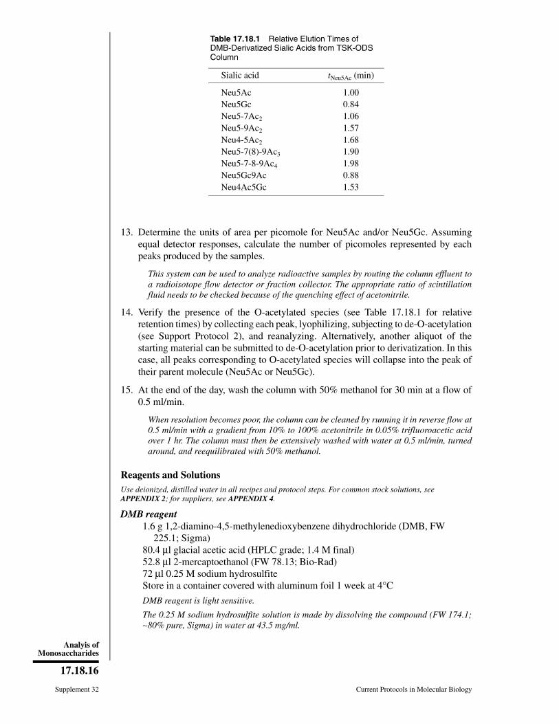
13. Determine the units of area per picomole for Neu5Ac and/or Neu5Gc. Assumingequal detector responses, calculate the number of picomoles represented by eachpeaks produced by the samples.
This system can be used to analyze radioactive samples by routing the column effluent toa radioisotope flow detector or fraction collector. The appropriate ratio of scintillationfluid needs to be checked because of the quenching effect of acetonitrile.
14. Verify the presence of the O-acetylated species (see Table 17.18.1 for relativeretention times) by collecting each peak, lyophilizing, subjecting to de-O-acetylation(see Support Protocol 2), and reanalyzing. Alternatively, another aliquot of thestarting material can be submitted to de-O-acetylation prior to derivatization. In thiscase, all peaks corresponding to O-acetylated species will collapse into the peak oftheir parent molecule (Neu5Ac or Neu5Gc).
15. At the end of the day, wash the column with 50% methanol for 30 min at a flow of0.5 ml/min.
When resolution becomes poor, the column can be cleaned by running it in reverse flow at0.5 ml/min with a gradient from 10% to 100% acetonitrile in 0.05% trifluoroacetic acidover 1 hr. The column must then be extensively washed with water at 0.5 ml/min, turnedaround, and reequilibrated with 50% methanol.
Reagents and SolutionsUse deionized, distilled water in all recipes and protocol steps. For common stock solutions, seeAPPENDIX 2; for suppliers, see APPENDIX 4.
DMB reagent1.6 g 1,2-diamino-4,5-methylenedioxybenzene dihydrochloride (DMB, FW
225.1; Sigma)80.4 µl glacial acetic acid (HPLC grade; 1.4 M final)52.8 µl 2-mercaptoethanol (FW 78.13; Bio-Rad)72 µl 0.25 M sodium hydrosulfiteStore in a container covered with aluminum foil 1 week at 4°C
DMB reagent is light sensitive.
The 0.25 M sodium hydrosulfite solution is made by dissolving the compound (FW 174.1;∼80% pure, Sigma) in water at 43.5 mg/ml.
Table 17.18.1 Relative Elution Times ofDMB-Derivatized Sialic Acids from TSK-ODSColumn
Sialic acid tNeu5Ac (min)
Neu5Ac 1.00Neu5Gc 0.84Neu5-7Ac2 1.06Neu5-9Ac2 1.57Neu4-5Ac2 1.68Neu5-7(8)-9Ac3 1.90Neu5-7-8-9Ac4 1.98Neu5Gc9Ac 0.88Neu4Ac5Gc 1.53
Supplement 32 Current Protocols in Molecular Biology
17.18.16
Analyis ofMonosaccharides

Commentary
Background InformationSialic acids released by hydrolysis in 2 M
acetic acid or by enzymatic treatment are con-verted with 1,2-diamino-4,5-methylenedioxy-benzene (DMB, a fluorogenic reagent for α-keto acids) to highly fluorescent derivatives(see Fig. 17.18.1) without the occurrence ofO-acetyl migration and de-O-acetylation.
The derivatives are separated using a re-versed-phase TSK ODS-120T column elutedisocratically with 9:7:84 (v/v/v) acetoni-trile/methanol/water at room temperature in∼25 min. Fluorescence of the eluate is moni-tored at an excitation wavelength of 373 nm andan emission wavelength of 448 nm. The aver-age limits of detection are 1 to 2 pmol at asignal-to-noise ratio of 3.
Fluorogenic reagents may be used for detec-tion of α-keto acids in very dilute solutionsbecause of their quantitative conversion into thecorresponding substituted quinoxalines
(Spiker and Towne, 1962; Hara et al., 1985a).Because sialic acids are α-keto acids, Hara etal. (1985b) tried several of these reagents andfound that 1,2-diamino-4,5-dimethoxybenz-ene reacts with Neu5Ac and Neu5Gc underconditions different than those for other α-ketoacids and forms fluorescent compounds. Theydeveloped a method for detecting smallamounts of Neu5Ac and Neu5Gc in serum andurine using HPLC (Hara et al., 1986) and fur-ther extended the method to the analysis ofglycoproteins and glycolipids (Hara et al.,1987a). Screening different diaminobenzenesas fluorogenic reagents revealed that 1,2-diam-ino-4,5-methylenedioxybenzene (DMB) is thebest reagent for sensitive and rapid detection ofα-keto acids (Hara et al., 1987b).
The DMB method could not be applied toO-acetylated sialic acids because of de-O-ace-tylation and O-acetyl migration in the sulfuricacid used in the reaction. However, O-acety-
O +NH3Cl–
+NH3Cl–
C
H
OH
7 8 9
65
10
H
1
4
2
3
+ 2H20
O
R
fluorescent derivative
For sialic acids, R =CH2
CHOH
CHNHAc
CHOH
CHOH
CHOH
CH2OH
O
C
H
OH
N
N
+
HO OC
CO R
H+
1,2-diamino-4,5-methylenedioxybenzene
dihydrochloride
α-keto acid
Figure 17.18.1 Reaction of sialic acids with 1,2-diamino-4,5-methylenedioxybenzene (DMB) toform fluorescent derivatives.
Current Protocols in Molecular Biology Supplement 32
17.18.17
Preparation andAnalysis ofGlycoconjugates
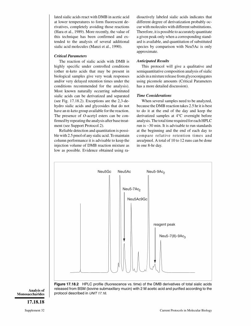
lated sialic acids react with DMB in acetic acidat lower temperatures to form fluorescent de-rivatives, completely avoiding those reactions(Hara et al., 1989). More recently, the value ofthis technique has been confirmed and ex-tended to the analysis of several additionalsialic acid molecules (Manzi et al., 1990).
Critical ParametersThe reaction of sialic acids with DMB is
highly specific under controlled conditions(other α-keto acids that may be present inbiological samples give very weak responsesand/or very delayed retention times under theconditions recommended for the analysis).Most known naturally occurring substitutedsialic acids can be derivatized and separated(see Fig. 17.18.2). Exceptions are the 2,3-de-hydro sialic acids and glycosides that do nothave an α-keto group available for the reaction.The presence of O-acetyl esters can be con-firmed by repeating the analysis after base treat-ment (see Support Protocol 2).
Reliable detection and quantitation is possi-ble with 2.5 pmol of any sialic acid. To maintaincolumn performance it is advisable to keep theinjection volume of DMB reaction mixture aslow as possible. Evidence obtained using ra-
dioactively labeled sialic acids indicates thatdifferent degree of derivatization probably oc-cur with molecules with different substitutions.Therefore, it is possible to accurately quantitatea given peak only when a corresponding stand-ard is available, and quantitation of substitutedspecies by comparison with Neu5Ac is onlyapproximate.
Anticipated ResultsThis protocol will give a qualitative and
semiquantitative composition analysis of sialicacids in a mixture release from glycoconjugatesusing picomole amounts (Critical Parametershas a more detailed discussion).
Time ConsiderationsWhen several samples need to be analyzed,
because the DMB reaction takes 2.5 hr it is bestto do it at the end of the day and keep thederivatized samples at 4°C overnight beforeanalysis. The total time required for each HPLCrun is ∼30 min. It is advisable to run standardsat the beginning and the end of each day tocompare relat ive retention t imes andarea/pmol. A total of 10 to 12 runs can be donein one 8-hr day.
Neu5Gc Neu5Ac Neu5-9Ac2
Neu5-7Ac2
Neu5Ac9Gc
reagent peak
Neu5-7(8)-9Ac3
Figure 17.18.2 HPLC profile (fluorescence vs. time) of the DMB derivatives of total sialic acidsreleased from BSM (bovine submaxillary mucin) with 2 M acetic acid and purified according to theprotocol described in UNIT 17.16.
Supplement 32 Current Protocols in Molecular Biology
17.18.18
Analyis ofMonosaccharides

Literature CitedHara, S., Yamaguchi, M., Nakamura, M., and Ok-
hura, Y. 1985a. Fluorescent products of reactionbetween α-keto acids and 1,2-diamino-4,5-di-methoxybenzene. Chem. Pharm. Bull. 33:3493-3498.
Hara, S., Takemori, Y., Yamaguchi, M., Nakamura,M., and Okhura, Y. 1985b. Determination ofα-keto acids in serum and urine by high-per-formance liquid chromatography with fluores-cence detection. J. Chromatogr. 344:33-39.
Hara, S., Yamaguchi, M., Takemori, Y., and Naka-mura, M. 1986. Highly sensitive determinationof N-acetyl- and N-glycolylneuraminic acids inhuman serum and urine and rat serum by re-versed-phase liquid chromatography with fluo-rescence detection. J. Chromatogr. 377:111-119.
Hara, S., Takemori, Y., Yamaguchi, M., Nakamura,M., and Okhura, Y. 1987a. Fluorometric high-performance liquid chromatography of N-ace-tyl- and N-glycolylneuraminic acids and its ap-plication to their microdetermination in humanand animal sera, glycoproteins, and glycolipids.Anal. Biochem. 164:138-145.
Hara, S., Yamaguchi, M., Takemori, Y., Nakamura,M., and Okhura, Y. 1987b. 1,2-diamino-4,5-methylendioxybenzene as a highly sensitivefluorogenic reagent for α-keto-acids. Chem.Pharm. Bull. 35:687-692.
Hara, S., Yamaguchi, M., Takemori, Y., Furuhata, K.,Ogura, H., and Nakamura, M. 1989. Determina-tion of mono-O-acetylated N-acetylneuraminicacids in human and rat sera by fluorometric
high-performance liquid chromatography. Anal.Biochem. 179:162-166.
Manzi, A.E., Diaz, S., and Varki, A. 1990. Highpressure liquid chromatography of sialic acidson a pellicular resin anion-exchange columnwith pulsed amperometric detection: A compari-son with six other systems. Anal. Biochem.188:20-32.
Spiker, J.E. and Towne, J.C. 1962. Fluorometricmicrodetermination of α-keto acids. Anal.Chem. 34:1468-1471.
Key ReferencesHara et al., 1989. See above.
Describes the use of the DMB method for the detec-tion and quantitation of Neu5Ac, Neu5Gc, andmono-O-acetylated derivatives of Neu5Ac.
Manzi et al., 1990. See above.
Describes the application of the DMB method toother members of the sialic acid family; discussesdifferences in reactivity with the fluorophore and inresponse factors between different DMB adducts.
Contributed by Adriana E. ManziUniversity of California San DiegoLa Jolla, California
Leland D. Powell and Ajit Varki (thiobarbituric acid assay; de-O-acetylation)University of California San DiegoLa Jolla, California
Current Protocols in Molecular Biology Supplement 32
17.18.19
Preparation andAnalysis ofGlycoconjugates

UNIT 17.19ATotal Compositional Analysis byHigh-Performance Liquid Chromatographyor Gas-Liquid Chromatography
Once the presence of carbohydrate in a glycoprotein has been confirmed, the next step isto determine the precise molar ratio of its monosaccharide constituents using protocolsin this unit and in the accompanying UNIT 17.19B (which presents the compositional analysisof labeled monosaccharides from glycosaminoglycans). This information helps to predictthe type of oligosaccharide present and to determine the approach to detailed structuralcharacterization. In some cases, it can also provide the first clues to the presence of a newtype of sugar chain. The analysis involves two major phases. First, the release of theindividual monosaccharides is achieved by methanolysis (Support Protocol 1), total acidhydrolysis (UNIT 17.16), or enzymatic release of sialic acids (UNIT 17.12). The second phaseinvolves analysis by fractionation, characterization, and quantitation of the mixtures ofmonosaccharides by high-performance liquid chromatography using anion-exchangechromatography with pulsed amperometric detection (HPAEC-PAD; Basic Protocols 1,2, and 3) and other HPLC systems (e.g., Alternate Protocol 1) or by gas-liquid chroma-tography with flame ionization detection (GLC-FID; Basic Protocol 4). The identity ofthe individual monosaccharides is determined by comparison with known standardsprocessed and analyzed in the same way. Use of these protocols requires availability ofinstrumentation and understanding of setup maintenance and use.
STRATEGIC PLANNING
The flow chart in Figure 17.19.1 illustrates the alternative pathways and sequence of stepsinvolved in compositional analysis. The protocols presented in this unit describe alterna-tive approaches for the analysis of monosaccharide mixtures obtained by total acidhydrolysis (UNIT 17.16) or methanolysis (Support Protocol 1). The following considerationsmay help the investigator to plan the approach most appropriate for the starting materialand intended results.
The choice of protocol to be used to release the individual monosaccharides from thestarting glycoconjugate sample depends upon the nature of the material to be analyzed aswell as the analysis method to be used. In general, glycoconjugates may be cleaved totheir constituent monosaccharides by acid hydrolysis (UNIT 17.16) or methanolysis (SupportProtocol 1). For the analysis of the constituents of neutral glycoconjugates, total acidhydrolysis of the sample is recommended. Glycoconjugates that may contain hexosami-nes or uronic acid can be broken down either by acid hydrolysis or methanolysis, andsialic acid residues can be released either enzymatically (as described in UNIT 17.12) or byhydrolysis in the presence of mild acid (UNIT 17.16). Note that the harsher acid conditionsused to release the other monosaccharides cause destruction of sialic acids. For mostglycoconjugates, the use of methanolysis to cleave glycosidic linkages is quite effectiveand generally results in less destruction of the monosaccharides than does acid hydrolysis(see Commentary for a more explicit discussion).
Frequently, the availability of instrumentation determines the choice between comparableoptions. For most HPLC approaches, such as those described in Basic Protocols 1, 2, and3, the mixtures of monosaccharides released by acid hydrolysis is directly analysedwithout further treatment. Mixtures of monosaccharides, released by acid hydrolysis orby methanolysis, being subjected to analysis by GLC-FID (Basic Protocol 4), however,require an initial derivatization step (or series of steps) to convert the mixture to volatile
Supplement 32
Contributed by Adriana E. ManziCurrent Protocols in Molecular Biology (1995) 17.19.1-17.19.25Copyright © 2000 by John Wiley & Sons, Inc.
17.19.1
Preparation andAnalysis ofGlycoconjugates

compounds. These volatile derivatives can be produced from monosaccharide mixturesusing one of the two alternative methods described in the support protocols. In the firstderivatization approach, monosaccharide mixtures obtained from acid hydrolysis undergoreduction and peracetylation to produce volatile alditol acetates (Support Protocol 2). Inthe second approach, the products of methanolysis are converted to volatile trimethylsilylderivatives (Support Protocol 3). In both cases, the resulting volatile compounds are thenanalyzed by GLC-FID.
Many different methods have been developed for each strategy, each with its ownadvantages and disadvantages (a more complete discussion and comparison of alterna-tives is presented in the Commentary). This unit presents reliable methods that have beenproven to be widely applicable.
glycoconjugate
acid hydrolysis(UNIT 17.16 )
free glycoses methyl glycosides
re-N-acetylationde-O-acetylation
pertrimethyl-silylation
(Support Protocol 3)
mixtures oftrimethylsilylatedmethyl glycosidesand methyl estersmethyl glycosidesincluding hexoses,
pentoses,deoxyhexoses,hexosamines,uronic acids,
and sialic acids
reduction andperacetylation
(Support Protocol 2)
mixtures of freeglycoses includinghexoses, pentoses,
de-N-acetylatedhexosamines,uronic acids,
and sialic acids
mixtures ofperacetylated
alditol acetatesincluding hexoses,
pentoses, anddeoxyhexoses
HPAEC-PAD(Basic Protocol
1, 2, or 3 orAlternate Protocol 1)
analysis
productionof volatile
derivatives
production of mono-
saccharideunits
GLC-FID(Basic Protocol 4)
methanolysis(Support Protocol 1)
enzymatic releaseof sialic acids
(UNIT 17.12)
Figure 17.19.1 Flow chart showing the different methods included in this section for compositionalanalysis of the carbohydrate moiety of glycoconjugates.
Supplement 32 Current Protocols in Molecular Biology
17.19.2
TotalCompositional
Analysis byHPLC or GLC
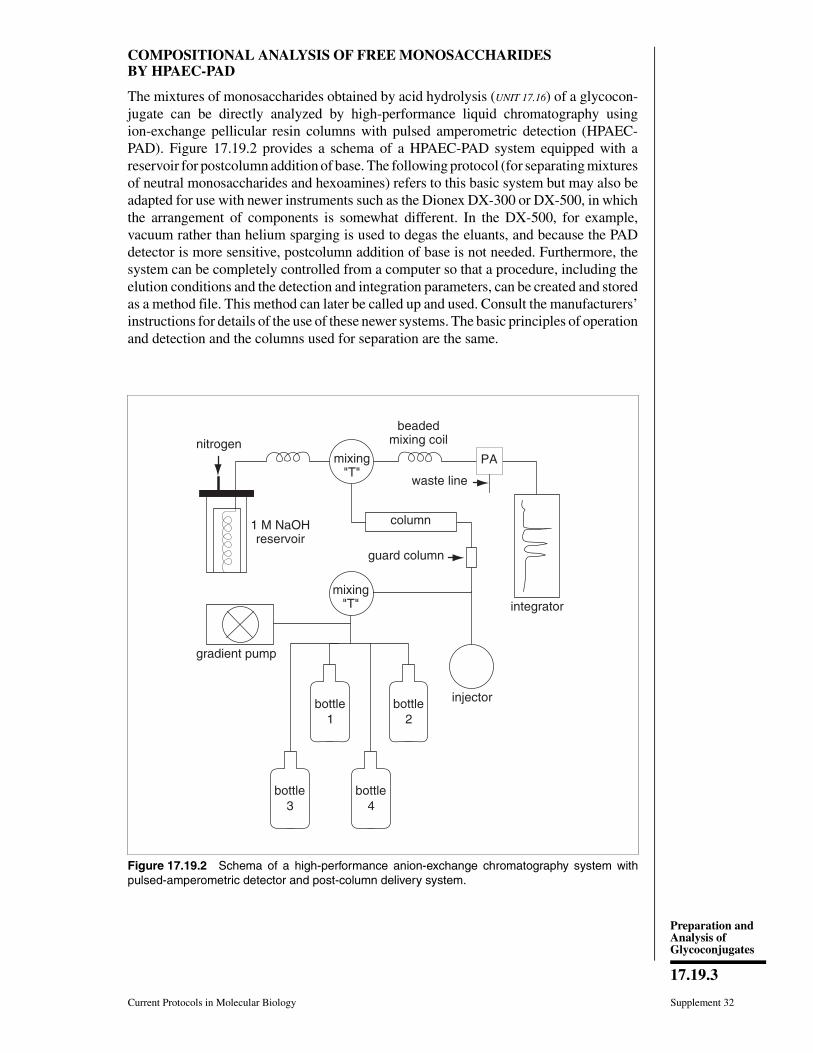
COMPOSITIONAL ANALYSIS OF FREE MONOSACCHARIDESBY HPAEC-PAD
The mixtures of monosaccharides obtained by acid hydrolysis (UNIT 17.16) of a glycocon-jugate can be directly analyzed by high-performance liquid chromatography usingion-exchange pellicular resin columns with pulsed amperometric detection (HPAEC-PAD). Figure 17.19.2 provides a schema of a HPAEC-PAD system equipped with areservoir for postcolumn addition of base. The following protocol (for separating mixturesof neutral monosaccharides and hexoamines) refers to this basic system but may also beadapted for use with newer instruments such as the Dionex DX-300 or DX-500, in whichthe arrangement of components is somewhat different. In the DX-500, for example,vacuum rather than helium sparging is used to degas the eluants, and because the PADdetector is more sensitive, postcolumn addition of base is not needed. Furthermore, thesystem can be completely controlled from a computer so that a procedure, including theelution conditions and the detection and integration parameters, can be created and storedas a method file. This method can later be called up and used. Consult the manufacturers’instructions for details of the use of these newer systems. The basic principles of operationand detection and the columns used for separation are the same.
bottle2
bottle4
bottle1
mixing"T"
column
guard column
mixing"T"
PA
waste line
injector
integrator
nitrogen
1 M NaOHreservoir
bottle3
gradient pump
beadedmixing coil
Figure 17.19.2 Schema of a high-performance anion-exchange chromatography system withpulsed-amperometric detector and post-column delivery system.
Current Protocols in Molecular Biology Supplement 32
17.19.3
Preparation andAnalysis ofGlycoconjugates

BASICPROTOCOL 1
Analysis of Mixtures of Neutral Monosaccharides and Hexosamines
Materials
Milli-Q-purified water: water deionized by passage through a five-stage Milli-QPlus system (Millipore)
Certified 50% (19.3 M) sodium hydroxide containing <0.1% sodium carbonate(Fisher)
NitrogenMonosaccharide standard mixture (see Support Protocol 4)Monosaccharide sample prepared by acid hydrolysis (UNIT 17.16: Basic Protocol 3,
Basic Protocol 1 when analyzing only fucose)Filtering unit with 0.45-µm Nylon 66 membranes (Alltech)High-pH anion-exchange chromatography system with pulsed amperometric
detector consisting of gradient pump, eluant degassing module, and PAD cellequipped with thin gasket (Dionex)
CarboPac PA-1 column (250 × 4 mm; Dionex)CarboPac PA Guard column (3 × 25 mm; Dionex)
Prepare the HPAEC-PAD system1. Filter 6 liters Milli-Q-purified water through a 0.45-µm membrane. Transfer 2 liters
to bottle 1, 1997 ml to bottle 2, and 1980 ml to bottle 3. Connect to helium spargeline and degas 15 min.
Always rinse the bottles before using them and make sure the lines are equipped withpolyethylene inlet frit filters. If 4-liter bottles are available, double the volumes of liquidin the bottles.
2. Prepare 25 mM and 200 mM sodium hydroxide solutions by adding (using disposableplastic pipets) 2.6 and 20.8 ml of 50% sodium hydroxide solution to bottles 2 and 3,respectively. Avoid bubbling air when adding. Degas for another 15 min.
IMPORTANT NOTE: It is critical that the sodium hydroxide solution be free of carbonate.Do not use sodium hydroxide pellets, as these are usually coated with a film of sodiumcarbonate produced by absorption of CO2 from the air; instead, use only 50% (19.3 M)NaOH solution that is free of carbonate. Take the solution from the middle of the bottle.Any CO2 absorbed will precipitate in 50% NaOH.
Keep all eluants under helium or vacuum (depending on the instrument used) at all times.Discard any remaining eluants after ∼2 weeks and prepare fresh ones. Do not store theMilli-Q-purified water to be used with this system in plastic containers. Rather, use freshlyprocessed water taken directly from the Milli-Q purification system. Do not use plastictubing extensions out of the Milli-Q system because these can become contaminated withmicroorganisms.
3. Run solvent from each bottle individually through the attached lines to remove anybubbles.
4. Connect the 4 × 250–mm CarboPac PA-1 column to the 3 × 25–mm CarboPac PAGuard colum.
5. Set the pump control unit (or computer, if using a computerized system) to deliver aflow rate of 1.0 ml/min with an appropriate elution regimen (see Table 17.19A.1 forexamples).
When both Xyl and Man are present in the sample, a very diluted eluent is used to achieveresolution. This considerably increases the time of analysis. Thus, when Xyl is not expected,an isocratic elution using 16 mM NaOH is preferred. These conditions are appropriate forthe analysis of N- and O-linked oligosaccharides, and considerably shorter. However, it isimportant to keep in mind that Xyl and Man will coelute if both are present.
Supplement 32 Current Protocols in Molecular Biology
17.19.4
TotalCompositional
Analysis byHPLC or GLC
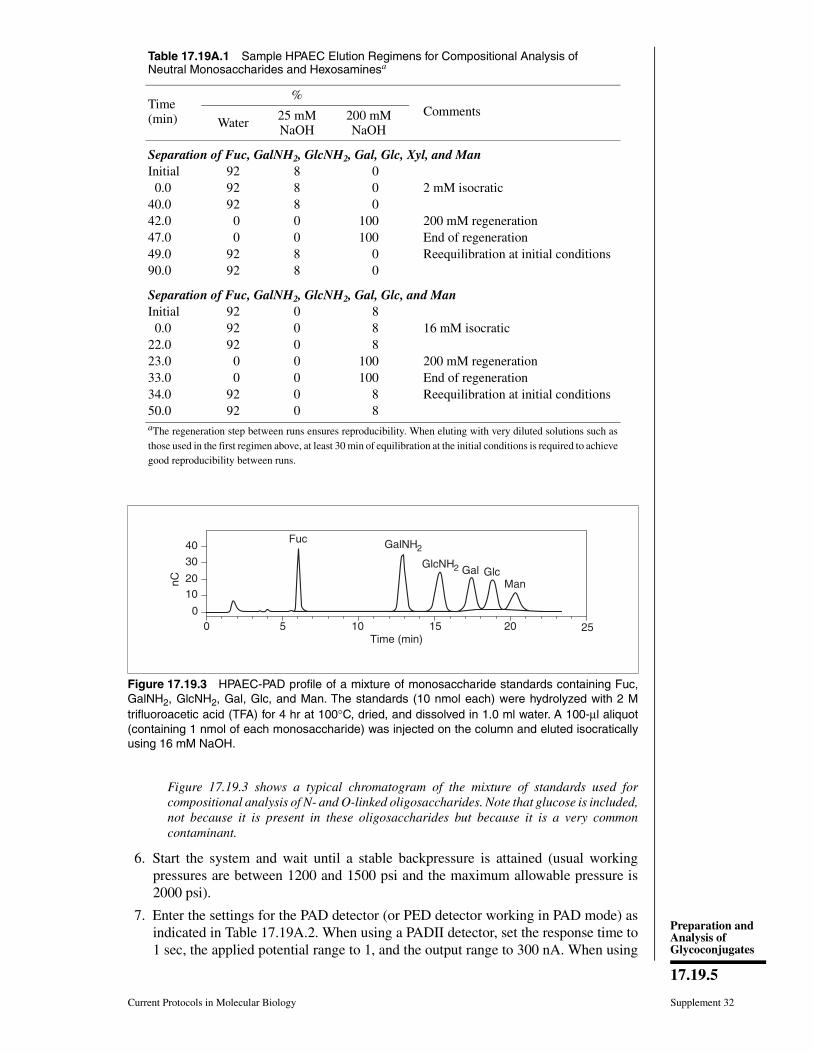
Figure 17.19.3 shows a typical chromatogram of the mixture of standards used forcompositional analysis of N- and O-linked oligosaccharides. Note that glucose is included,not because it is present in these oligosaccharides but because it is a very commoncontaminant.
6. Start the system and wait until a stable backpressure is attained (usual workingpressures are between 1200 and 1500 psi and the maximum allowable pressure is2000 psi).
7. Enter the settings for the PAD detector (or PED detector working in PAD mode) asindicated in Table 17.19A.2. When using a PADII detector, set the response time to1 sec, the applied potential range to 1, and the output range to 300 nA. When using
Table 17.19A.1 Sample HPAEC Elution Regimens for Compositional Analysis ofNeutral Monosaccharides and Hexosaminesa
Time(min)
%Comments
Water 25 mMNaOH
200 mMNaOH
Separation of Fuc, GalNH2, GlcNH2, Gal, Glc, Xyl, and ManInitial 92 8 0 0.0 92 8 0 2 mM isocratic40.0 92 8 042.0 0 0 100 200 mM regeneration47.0 0 0 100 End of regeneration49.0 92 8 0 Reequilibration at initial conditions90.0 92 8 0
Separation of Fuc, GalNH2, GlcNH2, Gal, Glc, and ManInitial 92 0 8 0.0 92 0 8 16 mM isocratic22.0 92 0 823.0 0 0 100 200 mM regeneration33.0 0 0 100 End of regeneration34.0 92 0 8 Reequilibration at initial conditions50.0 92 0 8aThe regeneration step between runs ensures reproducibility. When eluting with very diluted solutions such asthose used in the first regimen above, at least 30 min of equilibration at the initial conditions is required to achievegood reproducibility between runs.
40
30
2010
00 5 10 15 20
Time (min)
nC
Fuc GalNH2
GlcNH2 Gal GlcMan
25
Figure 17.19.3 HPAEC-PAD profile of a mixture of monosaccharide standards containing Fuc,GalNH2, GlcNH2, Gal, Glc, and Man. The standards (10 nmol each) were hydrolyzed with 2 Mtrifluoroacetic acid (TFA) for 4 hr at 100°C, dried, and dissolved in 1.0 ml water. A 100-µl aliquot(containing 1 nmol of each monosaccharide) was injected on the column and eluted isocraticallyusing 16 mM NaOH.
Current Protocols in Molecular Biology Supplement 32
17.19.5
Preparation andAnalysis ofGlycoconjugates
an ED40 detector in a DX300 or DX500 instrument, select the carbohydrate wave-form.
In the DX300 and DX500 systems, each method includes elution conditions, detectorsettings, and integration parameters. These can be manually selected from the controlpanels or entered using the software provided by the manufacturer. In both cases, when amethod is loaded, the pump starts and the detector is turned on.
Perform postcolumn addition of base (when using a PADI or early versions of thePADII detector only)8. Pressurize the postcolumn reservoir to deliver 0.3 to 0.4 ml/min of 1 M NaOH solution
(helium pressure should be ∼32 psi, resulting in a 360 mM sodium hydroxideconcentration at the electrode). Check the amount delivered by measuring the totalflow out of the PAD cell with a small graduated cylinder.
A regular piston pump cannot be used because this produces a wavy baseline due topulsation.
When using newer versions of the PADII cell, this step (designed to improve the sensitivityof detection with the older models) is unnecessary. In this case proceed directly to step 10.
9. Make sure that there are no bubbles inside the reference electrode. Wait 15 min forthe detector to stabilize, then press the Auto Offset control.
Check the baseline10. Check the detector baseline using an integrator or the software provided by the
manufacturer, and do not proceed until it is stable. Offset as necessary. When usingan integrator, the appropriate settings are attenuation, 1024 and peak threshold,10,000.
Analyze the standards11. Dissolve dried acid hydrolyzed (UNIT 17.16) monosaccharide standard mixture in 100
to 1000 µl water. Inject 10 to 100 µl of mixture onto column.
Use 100 l when the mixture contains 2.5 nmol of each monosacharide and 1000 ml whenthe mixture contains 10 nmol of each monosaccharide. Sufficient quantities of the standardmixture should be hydrolyzed to allow for several injections if necessary.
The standard mixture should contain 250 pmol to 1 nmol each of Fuc, GalNH2,GlcNH2, Gal, Glc, Xyl, and Man; 250 pmol to 1 nmol each of Fuc, GalNH2, GlcNH2,Gal,Glc, and Man; or 250 pmol to 1 nmol Fuc (when only analyzing fucose). The
Table 17.19A.2 PAD Detector Settings forDetection of Neutral Monosaccharides andHexosamines
Time (sec) Potential (V) Integration
0.00 0.05
0.20 0.05 Begin
0.40 0.05 End
0.41 0.75
0.60 0.75
0.61 −0.15
1.00 −0.15
Supplement 32 Current Protocols in Molecular Biology
17.19.6
TotalCompositional
Analysis byHPLC or GLC
amount of each monosaccharide standard injected depends on the sensitivity of the par-ticular detector and needs to be determined empirically in each case. The amount ofstandard mixture injected will depend on the amount of sample available. When the sampleis limited, it is necessary to work at high sensitivity, and in this case will be more difficultto obtain a good baseline; also, the amount of standards injected should be adjusted to bein the same range for improved quantitation.
Always prepare standards with extreme care (see Support Protocol 3), as their quality willdetermine the accuracy of sample quantitation. Always use standards that have beenhydrolyzed under the same conditions as the samples. This will correct for the differentrates of hydrolysis and different recoveries of the different monosaccharides.
12. Check the resolution between monosaccharides (resolution of Gal/GlcN andXyl/Man are usually critical, and inversion of the relative order of the componentsin these pairs may be observed). If resolution is poor, change the concentration ofbase (when retention times are too short, less base is required).
When using very dilute eluants (e.g., 2 mM NaOH), the concentration required for completeresolution may change when new 25 mM NaOH solution is prepared. This optimumconcentration is also different for different columns. Therefore, when a new solution isprepared or a new column is used, always check the resolution obtained for the standardmixture. When using higher concentrations of NaOH as eluant, this sort of variation is notobserved.
Analyze the samples13. When good resolution of the standards is achieved, inject the samples.
14. When using PADI or PADII detectors, it may be necessary (depending on the responseobtained for the samples) to change the working range to 100 nA or 1 µA. Wait untila good baseline is obtained, then inject another sample.
When using newer detectors, these adjustments are made automatically.
15. Quantitate the standard run and use these data (area per nmol or pmol of eachmonosaccharide) to calculate the amount in nmol or pmol of each component in thesample.
It is advisable to average the results from at least three runs of standards to obtain moreaccurate response factors for each monosaccharide and improve the accuracy of thedetermination. This can be easily accomplished when using the software provided by themanufacturer by doing an average calibration and using those parameters to analyze thesample runs.
Maintain the system running at maximum performance16. If resolution is lost and retention times become too short, wash the column for 30
min with 1 M sodium hydroxide. Wash extensively with water, then return to theworking eluant. Wait for the baseline to stabilize under running conditions and checkthe performance of the column by injecting 10 µl standard mixture. If performanceis not recovered, wash the column with 1 M HCl for 30 min at 1.0 ml/min. Washextensively with water, then with 1 M NaOH for 30 min at the same flow rate. Washextensively with water, reequilibrate with the working eluant, and check again.
Prepare high-concentration eluants used for regeneration in separate bottles, or wash thebottles extensively after they have been used to hold these solutions. Also, remember towash the lines that have carried concentrated solutions before connecting them to thecolumn for regular analysis.
Current Protocols in Molecular Biology Supplement 32
17.19.7
Preparation andAnalysis ofGlycoconjugates
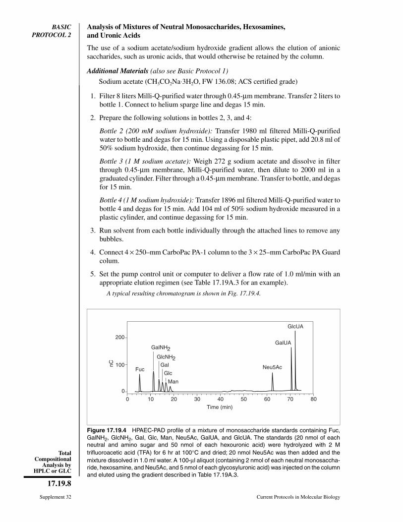
BASICPROTOCOL 2
Analysis of Mixtures of Neutral Monosaccharides, Hexosamines,and Uronic Acids
The use of a sodium acetate/sodium hydroxide gradient allows the elution of anionicsaccharides, such as uronic acids, that would otherwise be retained by the column.
Additional Materials (also see Basic Protocol 1)
Sodium acetate (CH3CO2Na⋅3H2O, FW 136.08; ACS certified grade)
1. Filter 8 liters Milli-Q-purified water through 0.45-µm membrane. Transfer 2 liters tobottle 1. Connect to helium sparge line and degas 15 min.
2. Prepare the following solutions in bottles 2, 3, and 4:
Bottle 2 (200 mM sodium hydroxide): Transfer 1980 ml filtered Milli-Q-purifiedwater to bottle and degas for 15 min. Using a disposable plastic pipet, add 20.8 ml of50% sodium hydroxide, then continue degassing for 15 min.
Bottle 3 (1 M sodium acetate): Weigh 272 g sodium acetate and dissolve in filterthrough 0.45-µm membrane, Milli-Q-purified water, then dilute to 2000 ml in agraduated cylinder. Filter through a 0.45-µm membrane. Transfer to bottle, and degasfor 15 min.
Bottle 4 (1 M sodium hydroxide): Transfer 1896 ml filtered Milli-Q-purified water tobottle 4 and degas for 15 min. Add 104 ml of 50% sodium hydroxide measured in aplastic cylinder, and continue degassing for 15 min.
3. Run solvent from each bottle individually through the attached lines to remove anybubbles.
4. Connect 4 × 250–mm CarboPac PA-1 column to the 3 × 25–mm CarboPac PA Guardcolum.
5. Set the pump control unit or computer to deliver a flow rate of 1.0 ml/min with anappropriate elution regimen (see Table 17.19A.3 for an example).
A typical resulting chromatogram is shown in Fig. 17.19.4.
0
100
200
nC
Time (min)
Fuc
GalNH2
GlcNH2Gal
Glc
Man
10 20 30 40 50 60 70 800
Neu5Ac
GalUA
GlcUA
Figure 17.19.4 HPAEC-PAD profile of a mixture of monosaccharide standards containing Fuc,GalNH2, GlcNH2, Gal, Glc, Man, Neu5Ac, GalUA, and GlcUA. The standards (20 nmol of eachneutral and amino sugar and 50 nmol of each hexouronic acid) were hydrolyzed with 2 Mtrifluoroacetic acid (TFA) for 6 hr at 100°C and dried; 20 nmol Neu5Ac was then added and themixture dissolved in 1.0 ml water. A 100-µl aliquot (containing 2 nmol of each neutral monosaccha-ride, hexosamine, and Neu5Ac, and 5 nmol of each glycosyluronic acid) was injected on the columnand eluted using the gradient described in Table 17.19A.3.
Supplement 32 Current Protocols in Molecular Biology
17.19.8
TotalCompositional
Analysis byHPLC or GLC
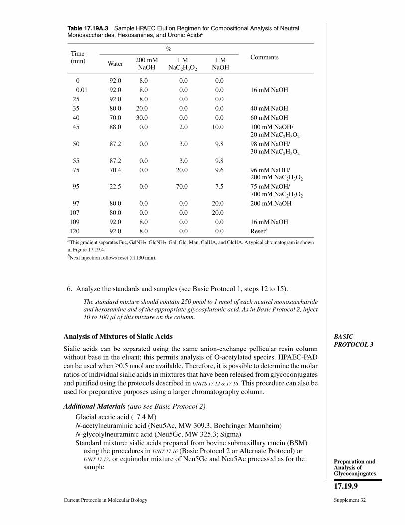
6. Analyze the standards and samples (see Basic Protocol 1, steps 12 to 15).
The standard mixture should contain 250 pmol to 1 nmol of each neutral monosaccharideand hexosamine and of the appropriate glycosyluronic acid. As in Basic Protocol 2, inject10 to 100 l of this mixture on the column.
BASICPROTOCOL 3
Analysis of Mixtures of Sialic Acids
Sialic acids can be separated using the same anion-exchange pellicular resin columnwithout base in the eluant; this permits analysis of O-acetylated species. HPAEC-PADcan be used when ≥0.5 nmol are available. Therefore, it is possible to determine the molarratios of individual sialic acids in mixtures that have been released from glycoconjugatesand purified using the protocols described in UNITS 17.12 & 17.16. This procedure can also beused for preparative purposes using a larger chromatography column.
Additional Materials (also see Basic Protocol 2)
Glacial acetic acid (17.4 M)N-acetylneuraminic acid (Neu5Ac, MW 309.3; Boehringer Mannheim)N-glycolylneuraminic acid (Neu5Gc, MW 325.3; Sigma)Standard mixture: sialic acids prepared from bovine submaxillary mucin (BSM)
using the procedures in UNIT 17.16 (Basic Protocol 2 or Alternate Protocol) orUNIT 17.12, or equimolar mixture of Neu5Gc and Neu5Ac processed as for thesample
Table 17.19A.3 Sample HPAEC Elution Regimen for Compositional Analysis of NeutralMonosaccharides, Hexosamines, and Uronic Acidsa
Time (min)
%Comments
Water 200 mMNaOH
1 MNaC2H3O2
1 MNaOH
0 92.0 8.0 0.0 0.0
0.01 92.0 8.0 0.0 0.0 16 mM NaOH
25 92.0 8.0 0.0 0.0
35 80.0 20.0 0.0 0.0 40 mM NaOH
40 70.0 30.0 0.0 0.0 60 mM NaOH
45 88.0 0.0 2.0 10.0 100 mM NaOH/20 mM NaC2H3O2
50 87.2 0.0 3.0 9.8 98 mM NaOH/30 mM NaC2H3O2
55 87.2 0.0 3.0 9.8
75 70.4 0.0 20.0 9.6 96 mM NaOH/200 mM NaC2H3O2
95 22.5 0.0 70.0 7.5 75 mM NaOH/700 mM NaC2H3O2
97 80.0 0.0 0.0 20.0 200 mM NaOH
107 80.0 0.0 0.0 20.0
109 92.0 8.0 0.0 0.0 16 mM NaOH
120 92.0 8.0 0.0 0.0 Resetb
aThis gradient separates Fuc, GalNH2, GlcNH2, Gal, Glc, Man, GalUA, and GlcUA. A typical chromatogram is shownin Figure 17.19.4.bNext injection follows reset (at 130 min).
Current Protocols in Molecular Biology Supplement 32
17.19.9
Preparation andAnalysis ofGlycoconjugates

Prepare the HPAEC-PAD system1. Filter 2 liters Milli-Q-purified water through 0.45-µm membrane. Transfer 1 liter to
bottle 1, connect to helium sparge line, and degas 15 min.
2. Prepare 1 M sodium hydroxide solution (see Basic Protocol 1, step 2).
3. Weigh 0.68 g sodium acetate and dissolve in Milli-Q-purified water, diluting thesolution to 1 liter final. Filter the resulting 5 mM sodium acetate buffer, transfer tobottle 2, connect to helium sparge line, and degas for 15 min.
4. Prepare 1 liter of 5 mM acetic acid using filtered Milli-Q-purified water and glacialacetic acid. Transfer to bottle 3 and degas for 15 min.
5. Run each solvent through the attached lines to remove any bubbles.
6. Connect the 4 × 250 mM CarboPac PA-1 column with the 3 × 25–mm CarboPac PAGuard column.
7. Set the pump control unit or computer to deliver a flow rate of 1.0 ml/min with anappropriate two-step elution regimen (see Table 17.19A.4 for an example).
For preparative purposes, a 9 × 250–mm column at a flow rate of 5 ml/min should be used.
8. Start the system and wait until a stable back pressure is attained (usual workingpressures are between 950 and 1400 psi and the maximum allowable pressure is 2000psi).
9. Perform postcolumn addition of base (if using PADI or earlier PADII system) anddetermine detector baseline (see Basic Protocol 1, steps 8 to 11).
Analyze the standards10. Inject 10 µl of a water solution containing 5 nmol Neu5Ac, working in the 300 nA
range.
If the samples to be analyzed have been submitted to acid hydrolysis for the release of sialicacids, use a Neu5Ac standard treated in the same manner for quantitation.
Table 17.19A.4 HPAEC Elution Regimen for Compositional Analysis at Sialic Acids
Time (min)
%Comments
Water 5 mM NaC2H3O2
5 mMC2H4O2
1 MNaOH
Initial 0 100 0 00.0 0 100 0 0 5 mM NaC2H3O2
0.01 0 100 0 05 0 100 0 0
35 0 50 50 0 2.5 mM NaC2H3O2/2.5 mM C2H4O2
36 0 0 100 0 5 mM AcOH46 0 0 100 048 80 0 0 20 200 mM NaOH68 80 0 0 2070 0 100 0 0 5 mM NaC2H3O2
100 0 100 0 0
Supplement 32 Current Protocols in Molecular Biology
17.19.10
TotalCompositional
Analysis byHPLC or GLC
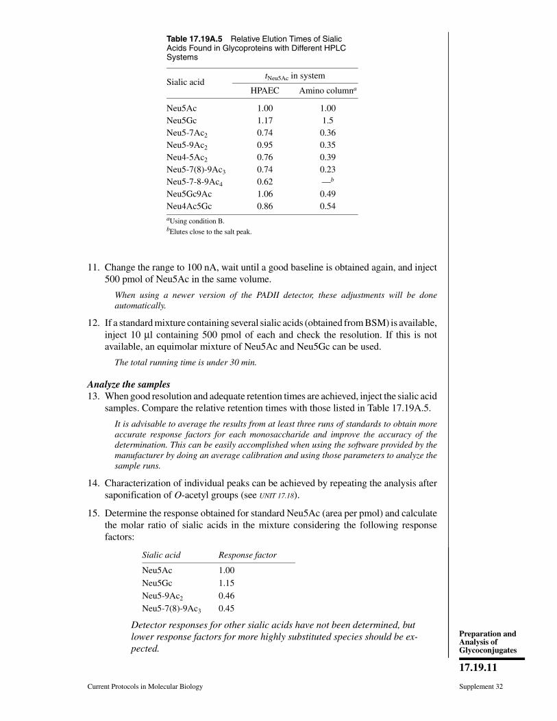
11. Change the range to 100 nA, wait until a good baseline is obtained again, and inject500 pmol of Neu5Ac in the same volume.
When using a newer version of the PADII detector, these adjustments will be doneautomatically.
12. If a standard mixture containing several sialic acids (obtained from BSM) is available,inject 10 µl containing 500 pmol of each and check the resolution. If this is notavailable, an equimolar mixture of Neu5Ac and Neu5Gc can be used.
The total running time is under 30 min.
Analyze the samples13. When good resolution and adequate retention times are achieved, inject the sialic acid
samples. Compare the relative retention times with those listed in Table 17.19A.5.
It is advisable to average the results from at least three runs of standards to obtain moreaccurate response factors for each monosaccharide and improve the accuracy of thedetermination. This can be easily accomplished when using the software provided by themanufacturer by doing an average calibration and using those parameters to analyze thesample runs.
14. Characterization of individual peaks can be achieved by repeating the analysis aftersaponification of O-acetyl groups (see UNIT 17.18).
15. Determine the response obtained for standard Neu5Ac (area per pmol) and calculatethe molar ratio of sialic acids in the mixture considering the following responsefactors:
Sialic acid Response factor
Neu5Ac 1.00
Neu5Gc 1.15
Neu5-9Ac2 0.46
Neu5-7(8)-9Ac3 0.45
Detector responses for other sialic acids have not been determined, butlower response factors for more highly substituted species should be ex-pected.
Table 17.19A.5 Relative Elution Times of SialicAcids Found in Glycoproteins with Different HPLCSystems
Sialic acidtNeu5Ac in system
HPAEC Amino columna
Neu5Ac 1.00 1.00Neu5Gc 1.17 1.5Neu5-7Ac2 0.74 0.36Neu5-9Ac2 0.95 0.35Neu4-5Ac2 0.76 0.39Neu5-7(8)-9Ac3 0.74 0.23Neu5-7-8-9Ac4 0.62 —b
Neu5Gc9Ac 1.06 0.49Neu4Ac5Gc 0.86 0.54aUsing condition B.bElutes close to the salt peak.
Current Protocols in Molecular Biology Supplement 32
17.19.11
Preparation andAnalysis ofGlycoconjugates

ALTERNATEPROTOCOL 1
COMPOSITIONAL ANALYSIS OF FREE SIALIC ACIDS BY AMINE-ADSORPTION ION-SUPPRESSION HPLC WITH UV DETECTION
As an alternative to Basic Protocol 3, mixtures of free sialic acids can be analyzed byamine-adsorption ion-suppression HPLC with UV detection. The choice depends on theinstrumentation available and the level of sensitivity required for a particular sample.
This system can be used to analyze radioactive samples if the column effluent is routedto a flow detector or fraction collector. When a flow detector is used, the appropriate ratioof scintillation fluid to be used has to be checked because of the quenching effect ofacetonitrile. When fractions are collected and 72% acetonitrile is used, it is necessary todry them, redissolve them in water, and then measure their radioactivity in a β-counterafter addition of scintillation cocktail. When 64% acetonitrile is used, the addition of 1vol water followed by 10 vol scintillation cocktail yields good sensitivity.
Materials
Monobasic sodium phosphate (anhydrous, MW 120.0; reagent grade, Sigma)Milli-Q-purified water: water deionized by passage through five-stage Milli-Q
Plus system (Millipore)Acetonitrile (HPLC grade, Fisher)N-acetylneuraminic acid (Neu5Ac, MW 309.3; Boehringer Mannheim)N-glycolylneuraminic acid (Neu5Gc, MW 325.3; Sigma)Standard mixture: sialic acids from bovine submaxillary mucin (BSM) prepared
using procedures in UNIT 17.16 or UNIT 17.12, or equimolar mixture of Neu5Ac andNeu5Gc processed as for the sample
Sialic acid sample prepared by acid hydrolysis (UNIT 17.16, Basic Protocol 2 orAlternate Protocol) or enzymatic release (UNIT 17.12)
HPLC apparatus with pump and UV detectorFiltering unit with 0.45-µm Nylon 66 membranes (Alltech)Micropak AX-5 (300 mm × 7.8 mm i.d., particle size 9 µm; Varian)Micropak AX-5 Guard column (4 cm × 4 mm)
Additional reagents and equipment for de-O-acetylation (UNIT 17.18)
Prepare the HPLC system1. Turn on the UV detector and set the wavelength to 200 nm.
2. Prepare 1 liter of 0.25 M monobasic sodium phosphate (30 g/liter) in Milli-Q-purifiedwater and filter through 0.45-µm membrane.
3. Fill the bottles in the HPLC sytem with (1) acetonitrile, (2) Milli-Q-purified waterfiltered through 0.45-µm membrane, and (3) filtered 0.25 M monobasic sodiumphosphate from step 2. Degas 15 min by helium sparging.
4. Run each solvent through the attached lines to eliminate any bubbles.
CAUTION: Do not allow direct mixing of stock acetonitrile and sodium phosphate, as thiswill cause precipitation of salts in the lines.
5. Set the pump control unit to deliver a flow rate of 1 ml/min with an appropriateisocratic elution regimen as indicated in Table 17.19A.6.
6. Start running the solvent, then connect the 300 × 7.8–mm Micropak AX-5 column tothe 4 cm × 4 mm Guard column.
7. Wait until the back pressure stabilizes and check the baseline. Wait until a stablebaseline is obtained.
Supplement 32 Current Protocols in Molecular Biology
17.19.12
TotalCompositional
Analysis byHPLC or GLC
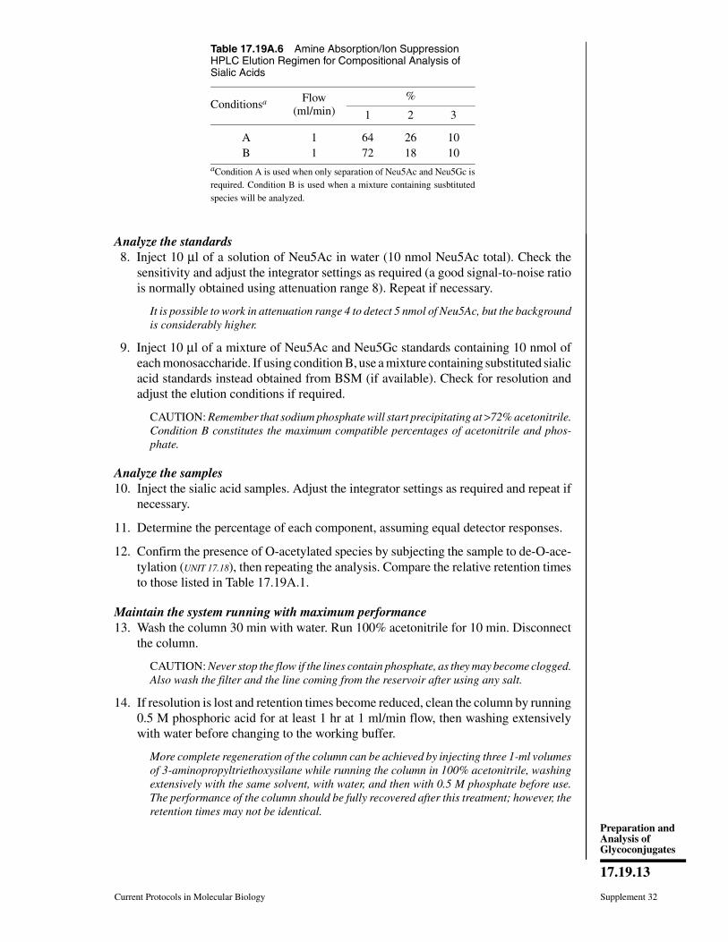
Analyze the standards8. Inject 10 µl of a solution of Neu5Ac in water (10 nmol Neu5Ac total). Check the
sensitivity and adjust the integrator settings as required (a good signal-to-noise ratiois normally obtained using attenuation range 8). Repeat if necessary.
It is possible to work in attenuation range 4 to detect 5 nmol of Neu5Ac, but the backgroundis considerably higher.
9. Inject 10 µl of a mixture of Neu5Ac and Neu5Gc standards containing 10 nmol ofeach monosaccharide. If using condition B, use a mixture containing substituted sialicacid standards instead obtained from BSM (if available). Check for resolution andadjust the elution conditions if required.
CAUTION: Remember that sodium phosphate will start precipitating at >72% acetonitrile.Condition B constitutes the maximum compatible percentages of acetonitrile and phos-phate.
Analyze the samples10. Inject the sialic acid samples. Adjust the integrator settings as required and repeat if
necessary.
11. Determine the percentage of each component, assuming equal detector responses.
12. Confirm the presence of O-acetylated species by subjecting the sample to de-O-ace-tylation (UNIT 17.18), then repeating the analysis. Compare the relative retention timesto those listed in Table 17.19A.1.
Maintain the system running with maximum performance13. Wash the column 30 min with water. Run 100% acetonitrile for 10 min. Disconnect
the column.
CAUTION: Never stop the flow if the lines contain phosphate, as they may become clogged.Also wash the filter and the line coming from the reservoir after using any salt.
14. If resolution is lost and retention times become reduced, clean the column by running0.5 M phosphoric acid for at least 1 hr at 1 ml/min flow, then washing extensivelywith water before changing to the working buffer.
More complete regeneration of the column can be achieved by injecting three 1-ml volumesof 3-aminopropyltriethoxysilane while running the column in 100% acetonitrile, washingextensively with the same solvent, with water, and then with 0.5 M phosphate before use.The performance of the column should be fully recovered after this treatment; however, theretention times may not be identical.
Table 17.19A.6 Amine Absorption/Ion SuppressionHPLC Elution Regimen for Compositional Analysis ofSialic Acids
Conditionsa Flow(ml/min)
%
1 2 3
A 1 64 26 10B 1 72 18 10
aCondition A is used when only separation of Neu5Ac and Neu5Gc isrequired. Condition B is used when a mixture containing susbtitutedspecies will be analyzed.
Current Protocols in Molecular Biology Supplement 32
17.19.13
Preparation andAnalysis ofGlycoconjugates

BASICPROTOCOL 4
COMPOSITIONAL ANALYSIS BY GLC-FID
This protocol describes the quantitative analysis of derivatized methyl glycosides andalditols through the use of a gas-liquid chromatograph with a flame-ionization detector(see Fig. 17.19.5). With this apparatus, it is quite straightforward to determine the molarratio of the monosaccharide components of a given glycoconjugate. This analysis can beperformed after complete acid hydrolysis (UNIT 17.16) and conversion of the free monosac-charides to volatile peracetylated alditols (see Support Protocol 2). It is also possible tocleave the glycosidic linkages with methanol/HCl to produce methyl glycosides, whichare converted into volatile trimethylsilylethers (see Support Protocol 3) for further GLCanalysis. The last method is preferred when hexosamines or uronic acid are expected tobe found in the sample.
Materials
Monosaccharide standard mixture (see Support Protocol 3) prepared in the samemanner as the sample
Monosaccharide sample prepared by acid hydrolysis (UNIT 17.16, Basic Protocol 3)followed by derivatization by peracetylation (see Support Protocol 2) and/or bymethanolysis (Support Protocol 1) followed by derivatization bypertrimethylsilylation (see Support Protocol 3)
Internal standard prepared in the same manner as the sample
0.25 mm × 30 m 5% DB-5 fused silica capillary column (J&W Scientific)Gas chromatograph with dual-flame ionization detectorHelium (research grade)Hydrogen and air lines (for FID detector only)
Gas-liquid chromatograph with flame-ionization detector (GLC-FID)1. Connect the capillary column to the gas chromatograph and begin running the carrier
gas at a flow rate of 1.5 ml/min. Set the makeup gas flow rate of 35 ml/min and thesplit ratio to 10:1.
When <25 g of total carbohydrates are processed, a splitless injection is required.
heliu
m capillarycolumn
oven
injector
pressureregulator
FID
hydr
ogen
air
integrator
Figure 17.19.5 Schema of a gas-liquid chromatograph with flame-ionization detector.
Supplement 32 Current Protocols in Molecular Biology
17.19.14
TotalCompositional
Analysis byHPLC or GLC
Several other types of stationary phases can be used for the analysis of acetylated alditols:e.g., CP-Sil 5 WCOT, SP-1000, Silar 10C, OV-1, SE-54, 5% DB-5, DB-1, Carbowax 20M,SE-30, OV-101, and OV-275. In the less polar stationary phases, amino sugar alditols canalso be analyzed. Other stationary phases used for the analysis of trimethylsilylatedglycosides and methyl glycoside methyl esters include CP-Sil 5 WCOT, 5% DB-5, DB-1,SE-30, and OV-101. Appropriate conditions vary from one to another. Typical chromato-grams and relative retention times can be found in Kammerling and Vliegenthart (1989)and references therein.
2. Set the GLC oven temperature to 50°C, the injector-port temperature to 150°C, andthe FID detector temperature to 250°C, and wait for stabilization.
3. Open the air and hydrogen lines and adjust the flows. Ignite the detector. Check thebackground.
4. Set up an appropriate temperature program:
a. Alditol acetates: Start at 50°C and keep at 50°C for 2 min; then go from 50°C to150°C at 20°C/min (5 min); then go from 150°C to 250°C at 4°C/min (25.0 min);then keep at 4°C for 5 min. (Total running time is 37 min.)
b. Trimethylsilylethers of methyl glycosides: Start at 50°C and keep at 50°C for 3min; then go from 50°C to 170°C at 20°C/min (6 min); then go from 170°C to250°C at 6°C/min (13.3 min); then keep at 250°C for 2.7 min. (Total running timeis 25 min.)
5. Inject 1 µl of the mixture of derivatized standards (see Support Protocol 2 and SupportProtocol 3). Adjust the settings of the integrator and repeat as necessary to obtain allthe peaks on scale.
6. Determine the retention times of each peak relative to the internal standard.
7. Inject the standards (containing internal standard) one at a time to assign the peaksand determine the response factors. In the case of TMS derivatives, also determinethe contribution of each peak to the total (%), and select the peak of each monosac-charide to be used for quantitation:
R = nmol internal standard × area monosaccharide
nmol monosaccharide × area internal standard
8. Inject 1 µl of sample (containing internal standard). Repeat as needed to obtain agood profile.
9. Tentatively assign the peaks by comparison of the relative retention times with thoseof the standards. If a discrepancy arises, repeat analysis, coinjecting the sample withthe individual standard. The questioned peak should appear separated from thestandard if it is not the same compound.
10. Determine the molar percentage of individual components by reference to the internalstandard:
nmol sugar ⁄ µg sample = area of monosaccharide peak × nmoles internal standard
area of internal standard peak × R × µg of sample
Current Protocols in Molecular Biology Supplement 32
17.19.15
Preparation andAnalysis ofGlycoconjugates
SUPPORTPROTOCOL 1
QUANTITATIVE RELEASE OF NEUTRAL MONOSACCHARIDES,HEXOSAMINES, AND URONIC ACIDS BY METHANOLYSIS
Glycoconjugate samples are hydrolyzed by heating in the presence of methanolic HCl,followed by treatment with pyridine and acetic anhydride.
Materials
Glycoconjugate sample containing 5 to 50 µg total sugarInternal monosaccharide standard: monosaccharide that does not occur naturally in
the sample (e.g., monosaccharide alditol that yields only one peak upon gaschromatography, see UNIT 17.16; see Support Protocol 4 for monosaccharidepreparation)
Monosaccharide standards: monosaccharides expected to be found in the sample(see Support Protocol 4 for preparation)
Phosphorus pentoxide (Fisher)Acetyl chloride (FW 78.50, 98% pure; Aldrich)Methanol (anhydrous, 99% pure, Aldrich; store in desiccator)Pyridine (anhydrous silylation grade, Pierce; store in desiccator)Acetic anhydride
1.5-ml glass Reacti-Vials (Pierce) with Teflon-lined screw caps, either new (rinsedwith water, then with ethanol, and dried) or acid-cleaned (see recipe)
Heating block or ovenNitrogen or vacuum evaporation system (Speedvac or shaker-evaporator)
1. Combine the glycoconjugate sample and 1 to 10 µg internal monosaccharide standardin a 1.5-ml precleaned or acid-cleaned Reacti-Vial. Lyophilize mixture.
2. Prepare a set of standards consisting of separate solutions containing 50 nmol of eachmonosaccharide that is expected to be found in the sample, each one also containing5 nmol of the same internal standard used for the sample. Also prepare a mixture ofall the expected monosaccharides plus internal standard. Treat all standards in parallelwith the sample.
3. Dry the lyophilized sample and standards overnight in a vacuum desiccator overphosphorus pentoxide.
4. Prepare methanolic HCl (either reagent A or reagent B) by adding acetyl chloride toanhydrous methanol in the following proportions:
Acetyl chloride Anhydrous methanol Final concentrationof HCl in methanol
Reagent A 3.50 vol 100 vol 0.50 MReagent B 7.00 100 vol 1.00 M
CAUTION: Add the acetyl chloride carefully, as the resulting reaction is highly exothermic.
Methanolic HCl reagent A is used except when glycosyluronic acids are present; in suchcases reagent B is preferred. Alternatively, an initial acid hydrolysis may be performedusing 200 l of 2 M trifluoroacetic acid (TFA) for 1 hr at 121°C. TFA should then beevaporated under a nitrogen stream or in Speedvac system and samples washed twice withmethanol, then evaporated again. This procedure ensures complete recovery of hexosami-nes but destroys sialic acids. Afterwards, proceed to step 4, and use reagent A.
Prepare a large enough volume of methanolic HCl to minimize pipetting errors, but onlyin the range required for the experiment, as it cannot be stored.
Supplement 32 Current Protocols in Molecular Biology
17.19.16
TotalCompositional
Analysis byHPLC or GLC
5. Dissolve dried sample and standards (from step 3) in 200 µl methanolic HCl (reagentA or B), then cap the vials.
6. Heat 16 hr at 65°C if using reagent A or 24 hr at 85°C if using reagent B. Check thescrew caps for tightness after the first 15 min of heating, then vortex and continueheating.
This procedure yields methyl glycosides of each monosaccharide residue in the glycocon-jugate. Hexosamines will be de-N-acetylated.
7. Evaporate sample and standards using a stream of nitrogen at room temperature or aSpeedvac system.
8. Add 100 µl anhydrous pyridine and vortex to resuspend the residue. Add 100 µl aceticanhydride, vortex 1 min, and evaporate to dryness under a stream of nitrogen.
This treatment produces re-N-acetylation of hexosamines. Some O-acetyl groups may beincorporated.
9. Dissolve the residue in 200 µl of 0.5 M methanolic HCl, then heat 1 hr at 65°C.
This treatment assures the clevage of any O-acetyl groups incorporated in step 8.
10. Evaporate under a stream of nitrogen, then dry further overnight in a vacuumdesiccator over phosphorus pentoxide.
SUPPORTPROTOCOL 2
PREPARATION OF VOLATILE DERIVATIVES OF FREE GLYCOSES
Volatile derivatives of free glycoses are prepared by first reducing the free sugars toalditols with sodium borohydride, then peracetylating with acetic anhydride. The result-ing alditol acetates can be analyzed immediately by GLC-FID (see Basic Protocol 2).
Materials
10 mg/ml sodium borohydride in 1 M ammonium hydroxide (ACS reagent grade)Monosaccharide sample prepared by acid hydrolysis (UNIT 17.16, Basic Protocol 3,
or Basic Protocol 1 when analyzing only fucose)Neutral sugar standard mixture (see Support Protocol 4) submitted to acid
hydrolysis (UNIT 17.16, Basic Protocol 3)Sodium borohydride (FW 37.8; store in desiccator)Glacial acetic acid (FW 60.05; 99.99% pure)1% acetic acid in methanol (anhydrous, 99% pure)Acetic anhydride (99% pure)TolueneChloroform (anhydrous, 99% pure)Acetone (99.9% pure, HPLC grade)Phosphorous pentoxide (as desiccant)
3.5-ml glass vials with Teflon-lined screw capsNitrogen evaporation unit (Reacti-Vap Evaporator, Reacti-Therm Heating Module,
and Reacti-Block B-1 or S-1, Pierce)Heating block or oven, 100°CTabletop centrifugeReacti-Vials (Pierce) or other glass test tubes
Reduce mixtures of free glycoses to alditols1. Add 250 µl of 1 M ammonium hydroxide/10 mg/ml sodium borohydride to the
monosaccharide sample and the neutral sugar standard mixture in 3.5-ml glass vialswith Teflon-lined screw caps. Let stand 10 min, then check for persistence of
Current Protocols in Molecular Biology Supplement 32
17.19.17
Preparation andAnalysis ofGlycoconjugates
microbubbles in the reaction mixture. If these cannot be seen, add a few microgramsof solid sodium borohydride.
It is important that no residue of acid from the hydrolysis be present or the hydride will berapidly destroyed. If excessive bubbling occurs when adding the borohydride, add methanoland evaporate two or three times, then repeat the reduction.
3. Incubate 2 hr at room temperature.
This reaction can also be run overnight.
4. Add glacial acetic acid dropwise until no further bubbling is observed.
Care must be taken to avoid losing sample with the bubbling, as the reaction is violent.This problem may be circumvented by using a 20% (v/v) solution of acetic acid in methanolinstead of glacial acetic acid.
5. Add 250 µl of 1% acetic acid in methanol to each tube. Evaporate to dryness usinga nitrogen evaporation unit or under vacuum using a Speedvac system with the heatingelement set at 40°C. Repeat four more times.
This treatment removes volatile methyl borate, producing sodium acetate. Completeremoval of borate is indicated by colorless crystals, as compared with white powderpreviously present. When a nitrogen evaporation unit is employed, care must be taken notto blow away the solid residue, particularly in the final steps.
6. Dry sample and standards in a vacuum dessicator overnight over phosphorus pentox-ide.
Peracetylate the alditols7. Add 200 µl acetic anhydride, vortex, and cap the tubes.
8. Heat 3 hr at 100°C, mixing after the first 15 min of heating to make sure that the solidresidue is suspended. Let cool to room temperature.
9. Add 200 µl toluene. Evaporate to dryness at 40°C using a shaker-evaporator orSpeedvac or under nitrogen. Add toluene as needed to dry completely.
Toluene helps to eliminate the reagents by forming an azeotrope.
Clean reaction mixtures by extraction10. Add 0.5 ml water and 0.5 ml chloroform to each tube, cap, and vortex well. Centrifuge
briefly at low speed to clearly separate the two phases.
Salts will be extracted into the water layer, leaving the peracetylated alditols in the organiclayer.
11. Remove the upper (water) layer with a Pasteur pipet. Wash the organic layer withwater two more times.
12. Transfer the chloroform layer to a clean tube or Reacti-Vial and dry under nitrogenat room temperature or in a Speedvac system.
13. Dissolve in 5 to 50 µl acetone, depending on the amount of starting material, and use1 µl for GLC-FID analysis (Basic Protocol 2).
Supplement 32 Current Protocols in Molecular Biology
17.19.18
TotalCompositional
Analysis byHPLC or GLC
SUPPORTPROTOCOL 3
PREPARATION OF VOLATILE DERIVATIVES OF METHYLGLYCOSIDES
Volatile derivatives of methyl glycosides (trimethylsilylethers of neutral sugars andhexosamines and methyl esters of uronic acids) are prepared from the dried methylglycosides (see Support Protocol 1) using a commercial silylation reagent. The silylatedsamples must then be analyzed immediately by GLC-FID (see Basic Protocol 2).
Materials
Dried methanolyzed samples and standards (see Support Protocol 1, step 10)Tri-Sil Reagent [2:1:10 (v/v/v) hexamethyldisilazane/trimethylchlorosilane/
pyridine; Pierce]96% hexane (HPLC grade)
Nitrogen streamTabletop centrifugeReacti-Vials (Pierce)
1. Suspend each dried sample and standard in 100 µl Tri-Sil and cap vial. Incubate 30min at room temperature.
Samples and all required standards must be processed in parallel.
2. Evaporate the silylating reagent under a stream of dry nitrogen.
Nitrogen can be dried by including a drying tube in the line.
3. Add 0.5 ml hexane, dissolve the derivatives, and centrifuge briefly at low speed toseparate insoluble salts.
4. Transfer supernatant to a clean Reacti-Vial and evaporate under dry nitrogen streamas in step 2.
5. Dissolve the residue 5 to 50 µl hexane, depending on the amount of starting material.Use 1 µl of solution for GLC analysis.
SUPPORTPROTOCOL 4
PREPARATION OF MONOSACCHARIDE STANDARD SOLUTIONS
Proper preparation and storage of monosaccharide standard solutions is critical to achievegood quantitation in a compositional analysis (Hardy and Townsend, 1994).
Materials
NaOH pelletsMonosaccharide standardsMilli-Q-purified water: water deionized by passage through a Milli-Q Plus system
(Millipore)
Vacuum desiccatorGlass vials100-ml volumetric flasks
1. Prepare a vacuum desiccator containing a plastic beaker with NaOH pellets.
2. Tare a group of glass vials (twice the number of standards to be used). Weight ∼500mg of each monosaccharide standard in separate vials and cover each vial withaluminum foil or a loosely placed clean cap. Cover the extra vials in the same mannerand place all the vials in the desiccator.
3. Put the desiccator under continuous vacuum for 1 day.
Current Protocols in Molecular Biology Supplement 32
17.19.19
Preparation andAnalysis ofGlycoconjugates
4. Weigh the vials and return them to the desiccator for another day under vacuum.Continue to do this every day until the change in weight from one day to the next is<1%.
This is normally achieved in 3 to 4 days.
5. Transfer a sample (e.g., 15 mg) of each standard to one of the empty, desiccated taredvials and determine the exact weight of the monosaccharides.
6. Add Milli-Q-purified water to each vial to give a 100 mM solution. Mix well todissolve.
7. Prepare a dilution of each of these primary standard solutions by transferring 100 µlof each solution to a 100-ml volumetric flask (100 pmol/µl). Dilute to 100 ml withMill-Q-purified water and mix.
8. Divide each standard into 1-ml aliquots and store these at −20°C until needed.
9. Prepare a mixture of all the required monosaccharides by transferring 100 µl of eachprimary standard solution to a 100-ml volumetric flask (100 pmol/µl). Bring up to100 ml with Milli-Q-purified water and mix.
10. Divide the standard mixture into 1-ml aliquots and store these at −20°C until needed.
11. When required, defrost one aliquot of each desired standard solution and use themwithin 1 or 2 days. Do not refreeze the standards.
Standards prepared in this manner can be used directly for HPAEC or HPLC analysis orlyophilized and used for GLC analysis.
REAGENTS AND SOLUTIONS
Use deionized, distilled water in all recipes and protocol steps. For common stock solutions, seeAPPENDIX 2; for suppliers, see APPENDIX 4.
Acid-cleaned glass vialsHeat 1.5-ml glass vials (e.g., Reacti-Vials, Pierce) with Teflon-lined screw caps at50°C for at least 3 hr each time in concentrated nitric acid and then in 6 M HCl.Rinse thoroughly with water, then with ethanol, and dry in an oven. When <5g oftotal carbohydrates is analyzed, it is recommended to silanize the glass vials byincubating them 15 min at room temperature with a 2% solution of dichlorodi-methylsilane in toluene, decanting the solution, rinsing successively with methanoland hot distilled water, and allowing the vials to dry.
New vials should be at least rinsed with water, then with ethanol, and dried.
COMMENTARY
Background InformationHigh-performance anion exchange chroma-
tography with pulsed amperometric detection(HPAEC-PAD) and gas-liquid chromatogra-phy with gas-liquid chromatography with massspectroscopic detection (GLC-MS) are themethods most widely used today for composi-tional analysis of the carbohydrate moieties ofglycoproteins. Both methods are able to detectall the possible known monosaccharides. Withthe HPAEC-PAD system it is possible to ana-lyze qualitatively and quantitatively a mixtureof free monosaccharides containing as little as
50 pmol of each individual component. WithGLC methods, the use of internal standardshelps improve quantitation because the injec-tion of reproducible proportions of the sampleinto the column is not easy.
HPAEC-PAD and other HPLC techniquesDifferent high-performance liquid chroma-
tography (HPLC) systems have been used forthe analysis of free glycoses obtained by acidhydrolysis, but sensitivity and resolution be-tween individual monosaccharides were tradi-tionally poor. The technique became popular
Supplement 32 Current Protocols in Molecular Biology
17.19.20
TotalCompositional
Analysis byHPLC or GLC
for analyzing glycoproteins and other glyco-conjugates available in limited amounts frombiological sources only after pellicular resinanion-exchange columns and the pulse-am-perometric detector were developed. Neutralsaccharides are analyzed at high pH (12 to 14),at which they are partially or completely ion-ized. Separation is achieved by adjusting theeluant pH. Anionic saccharides can be sepa-rated using sodium acetate/sodium hydroxidegradients. Therefore, with appropriate adjust-ment of the elution conditions, this system per-mits not only the compositional analysis of freemonosaccharide mixtures but also the analysisof oligo- and polysaccharides (Lee, 1990).
Pulsed amperometric detection (PAD) util-izes a repeating sequence of three potentialsapplied for specific durations. The resultingtotal current is the sum of (1) the carbohydrateoxidation current, (2) the current due to thecharging of the electrode surface, and (3) thecurrent caused by the oxidation of the goldelectrode. The current is only measured duringthe first applied potential and has been shownto be linear over >41⁄2 orders of magnitude.Recently, the parameters used for these threesteps were optimized (LaCourse and Johnson,1991). Molecules with similar size and struc-ture are reported to have similar response fac-tors. On the other hand, substituents on hy-droxyl groups are expected to affect the sensi-tivity of detection. The detector works only athigh pH. Thus, when base-labile species thatare separated without the use of base in theeluant are analyzed, it is necessary to add alkalito the effluent before it enters the PAD cell.
Isocratic elution with sodium hydroxide (2to 16 mM; the optimal concentration variesfrom column to column) allows for the separa-tion of neutral sugars and hexosamines. Differ-ent sodium acetate gradients have been em-ployed for the analysis of acidic monosaccha-rides and of mixtures of these and other sugars.The system is also useful for sialic acids, witha limit of detection of ∼200 pmol (Manzi et al.,1990). Oligo- and polysaccharides can also beanalyzed with this system using appropriateconditions (Hardy and Townsend, 1988; 1989).Although monosaccharide alditols are not re-tained by this column (they will come out inthe void volume), oligosaccharide alditols canbe analyzed (Reddy and Bush, 1991). Mainte-nance of a stable column temperature is criticalto obtaining reproducible results; failure to doso can cause lack of separation of critical pairsof monosaccharides (Van Riel and Olieman,1991).
Because only 1% of the sample is oxidizedin the electrode, the system can be used prepa-ratively without the need for a bypass. Whenanalyzing base-labile sialic acids, this methodcannot be used, because the postcolumn addi-tion of base required for detection will causesaponification of the O-acetyl esters. However,the pellicular resin column can be used forfractionation and the eluate routed to a fractioncollector. After the fractions are monitored bythe TBA assay (UNIT 17.18), they can be pooledand desalted. This method has proved to be thebest HPLC technique available for preparativefractionation of mixtures of sialic acids con-taining labile substituents (Manzi et al., 1990).
Another useful system for the analysis offree sialic acids is amine adsorption–ion sup-pression HPLC. This is based upon hydrogenbonding between the hydroxyl groups of sac-charides and the amine moieties of the station-ary phase (Mellis and Baenziger, 1981; Berghet al., 1981). To fractionate anionic molecules,phosphate is added to the mobile phase tosuppress the ionic effects while retaining hy-drogen bonding (Mellis and Baenziger, 1983).Good separation of sialic acids is obtained inisocratic mode with a mixture of acetoni-trile/water/0.25 M monobasic sodium phos-phate at 1 ml/min (Diaz and Varki, 1985). It isnecessary to maintain a minimum concentra-tion of 10% phosphate buffer with a maximumworking percentage of acetonitrile of 72%(above this the phosphate starts to precipitate).A 64:26:10 (v/v/v) rat io of acetoni-trile/water/0.25 M sodium phosphate can beused to separate Neu5Ac and Neu5Gc. ForO-acetylated sialic acids, however, it is neces-sary to increase the percentage of acetonitrileto a ratio of 72:18:10 (v/v/v) so as to increasethe retention times of the sialic acids (Manzi etal., 1990). Chromatography is monitored byabsorption at 200 nm, which requires extremelyhigh-purity samples and reagents. The sameanalytical column can be used for preparativepurposes (up to 400 nmol of sialic acids with agood resolution of the peaks). Recovery ofsialic acids is ∼65%, but some loss and migra-tion of O-acetyl groups occur.
GLC-FIDTo analyze mixtures of glycoses or methyl-
glycosides by gas-liquid chromatography, it isnecessary to prepare volatile derivatives. Aldi-tol acetates are the most widely used derivativesfor monosaccharides obtained by acid hydroly-sis, whereas trimethylsilylethers of methyl gly-cosides or methyl glycoside methyl esters are
Current Protocols in Molecular Biology Supplement 32
17.19.21
Preparation andAnalysis ofGlycoconjugates

the preferred derivatives for methanolysis prod-ucts. These are the derivatives of choice for anyglycoconjugate-derived sugar because of theamount of information that can be obtainedfrom them (e.g., retention times in differentcolumns). Other volatile compounds, such asaldononitrile acetates and trifluoroacetylatedalditols, have also been successfully employed(as reviewed by Kammerling and Vleigenthart,1989).
Acetylesters of alditolsWhen only neutral sugars need to be ana-
lyzed, the most common derivatization methodis the conversion of the free monosaccharidesinto their alditol acetates. Each monosaccha-ride produces a single derivative, giving a sim-ple chromatogram that can be easily interpretedand quantitated. The total analysis time is alsoshorter because all required standards can beprocessed as a mixture, requiring only one GLCrun.
The procedure involves the reduction of theglycoses to alditols with sodium borohydride;elimination of the excess of hydride with acid;elimination of the boric acid produced astrimethylborate by coevaporation with acidi-fied methanol; and peracetylation of all freehydroxyl groups. Different protocols to achievethis last step have been developed (Kammerlingand Vliegenthart, 1989; Albersheim et al.,1967; York et al., 1985). It is possible to simplyuse the sodium acetate produced in the reactionas the catalyst, and achieve complete acetyla-tion by heating with acetic anhydride. Becauseleftover borate could inhibit the acetylation,some protocols add pyridine to drive the reac-tion to completion.
Cleavage of glycosidic linkages bymethanolysis
Cleavage of all glycosidic linkages by meth-anolysis is very effective and causes less destruc-tion of the monosaccharides than acid hydrolysis.Monosaccharides are converted to their methylglycosides and glycosyluronic acids to theirmethyl esters methyl glycosides. The N-acetylgroup of amino sugars are completely cleavedby extensive methanolysis, and it is advisableto incorporate a re-N-acetylation step to avoidthe production of additional peaks in the chro-matogram. A drawback to the reacetylation stepis that some O-acetylation occurs (in someprimary hydroxyl groups), but a short addi-tional treatment with methanolic HCl elimi-nates these groups (Rickert and Sweeley,
1978). Because of anomerization, eachmonosaccharide produces several methylgly-cosides (α and β anomers of the pyranosidicand furanosidic forms), which yields a charac-teristic pattern of peaks in the GLC run. Whenthe sample contains several monosaccharides,however, the pattern is quite complex, with somepeaks superimposed, and this makes quantita-tion difficult. Nevertheless, by processing eachof the individual monosaccharides that are ex-pected in parallel, running each one separatelyunder the same conditions, and determining thecontribution of each peak to the total, an accuratequantitation can still be achieved.
Different conditions have been used formethanolysis of carbohydrates in glycoprote-ins using methanolic HCl, including 0.5 M for16 hr at 65°C (Reinhold, 1972) and 0.75 M for3 hr at 80°C (Wong et al., 1980). Conditions forthe simultaneous release of neutral monosac-charides, acetamido sugars, uronic acids, octu-losonic acids, and sialic acids (2 M TFA for 12hr at 100°C under vacuum) have been reported(Clarke et al., 1991). Nevertheless, the cleavageof the uronic acid glycosidic linkage may beincomplete in some cases, and several glycosy-luronic acids produce a certain percentage of3,6-lactones. The glycosidic linkages of sialicacids are cleaved with good yield, but a signifi-cant proportion of the released molecules aredestroyed. Although special conditions thatminimize this destruction have been developed(Schauer, 1978), N-acyl groups and most O-acetyl esters are eliminated. Thus, release ofintact sialic acids requires much milder acidhydrolysis of a separate aliquot of the sample.
Volatile derivatives of methyl glycosidesThe content of neutral sugars, amino sugars,
and glycosyluronic acids can be simultane-ously determined by this method. Differentreagents and procedures have been used tosilylate mixtures of methyl glycosides (Kam-merling and Vliegenthart, 1989). The mostwidely used procedure uses 5:1:5 (v/v/v) pyri-dine/hexamethyldisilazane/trimethylchlorosilane and an incubation that can range from 15min at room temperature to 20 min at 80°C.
Sialic acids have also been analyzed as tri-methylsilyl esters trimethylsilyl ethers ormethyl esters trimethylsilyl ethers. Althoughconditions have been worked out for preparingthese two types of derivatives while preservinglabile natural substituents such as O-acylgroups (Schauer and Corfield, 1982), thederivatization of sialic acids is frequently in-
Supplement 32 Current Protocols in Molecular Biology
17.19.22
TotalCompositional
Analysis byHPLC or GLC

complete. Further problems are encounteredduring the subsequent chromatographic sepa-ration because the high temperatures requiredto elute these molecules result in partial de-struction of the labile substituents. Therefore,this approach to sialic acid analysis has beenreplaced by HPLC methods with or withoutderivatization (see UNIT 17.18).
Qualitative and quantitative analysisAnalysis of volatile monosaccharide deriva-
tives is achieved by gas-liquid chromatography.A combined adsorption/partition mechanismtakes place between the volatile derivatives andthe particular stationary phase that is a liquid atworking temperatures. Through the large num-ber of theoretical plates available in the lengthof the fused-silica capillary columns, the indi-vidual components of the mixtures underanalysis are separated according to their par-ticular structural characteristics. The mobilephase (helium) carries the molecules through-out the column and to the detector, where theionization of the flame changes upon their pas-sage, producing an electrical output. This signalis amplified and registered.
Initial characterization of individual peaksis done by comparison with standards run in thesame conditions. The retention times of eachstandard relative to the component used asinternal standard in the sample are calculatedand compared with the relative retention timesof each peak in the chromatogram of the sam-ple. Typical elution profiles as well as relativeretention times in the different columns underdifferent conditions can be found in the litera-ture (Kammerling and Vliegenthart, 1989, andreferences therein).
A known amount of an internal standard (amonosaccharide or alditol that does not occurnaturally) is added to each glycoconjugate atthe beginning of its processing. Losses duringthe preparation of the sample for GLC will beproportionally the same for each component ofthe sample and for the internal standard. Thus,the ratio of analyte to internal standard willremain the same, even though the absoluteamounts of both may be lower at the end.Quantitative analysis is performed by deter-mining the detector responses for individualmonosaccharides relative to the internal stand-ard (known amounts are injected and the areasdetermined).
Critical Parameters
HPAEC-PAD and other HPLC techniquesWhen HPAEC-PAD is used for analysis of
sialic acids, the sialic acids (which have a pKa
of ∼2.0) bind to the resin at neutral pH, and itis necessary to use an anion to elute them.Samples are loaded in water to ensure bindingand submitted to a pH gradient elution withdilute sodium acetate and acetic acid. Thispermits the separation of different O-acetylatedsialic acid species, whose binding to the resinis relatively weaker with higher numbers ofsubstituents. The relative detector responsesvary significantly with the presence of differentnumbers of free hydroxyl groups. The responseobserved for Neu5Gc is 15% higher than thatfor Neu5Ac, which bears one fewer hydroxylgroup. In contrast, the responses of the detectorfor Neu5-9Ac2 and Neu5-7(8)-9Ac3 are only46% and 45% of the response for Neu5Ac,respectively. However, because of the possibil-ity of partial de-O-acetylation between thepoint of addition of postcolumn alkali and thePAD detector, the detector response factorsmay vary somewhat in different laboratories.For preparative purposes, a maximum of 2µmol of total sialic acids can be injected with-out overloading the 9 mm × 25 cm semiprepa-rative column. After removal by Dowex 50 (H+)chromatography of the small amount of acetaterequired for separation using the HPAEC col-umn, an excellent recovery (>90%) of acety-lated species is possible.
When gradients that end in high concentra-tions of acetate (>100 mM) are used, the slowreequilibration of the column with low concen-trations of hydroxide and acetate results inlower retention times and impaired separation.Resolution of some pairs of monosaccharides(e.g., Gal-GlcN and Xyl-Man) during the firstisocratic step is extremely sensitive to minorchanges in solvent composition. Therefore, ex-tended equilibration periods between runs arenecessary to obtain reproducible results. Ashorter delay between runs, resulting in a col-umn that is not fully equilibrated but that stillachieves adequate separation for a set ofmonosaccharides, can produce repeatable re-sults if an automatic sample injector with con-stant intervals between injections is employed.
When using amine adsorption–ion suppres-sion chromatography, the relative proportion ofsolvents can be varied according to the kind ofsialic acids to be separated. Because the eluantis monitored by UV absorbance at 200 nm,residual amounts of purification reagents pro-
Current Protocols in Molecular Biology Supplement 32
17.19.23
Preparation andAnalysis ofGlycoconjugates

duce an absorption peak close to the void vol-ume. The lower limit of reliable detection is 2nmol, and the detector responses are equal forall sialic acids. For preparative purposes, theeluant is monitored by UV and the peaks col-lected directly on ice, diluted 10-fold with coldwater, and purified by ion-exchange chroma-tography on Dowex 3-X4A (formate; see UNIT
17.16) at 4°C. The column is washed with 10 volof 10 mM formic acid and eluted with 7 vol of1 M formic acid, and sialic acids are recoveredby lyophillization.
GLC with FID detectionWhen the assignment of some peaks in gas-
liquid chromatography (a GLC) run remainsuncertain, coinjection with the suspectedmonosaccharide allows confirmation.
When analyzing trimethylsilylated methylglycosides, the sample is quantitated by choos-ing one nonsuperimposed peak for eachmonosaccharide and considering how much ofthe compound this area represents. Absolutemolar content of monosaccharide in the samplecan be calculated when a known amount of aninternal standard was added to the sample be-fore processing.
Because the linkage between N-acetylglu-cosamine and asparagine is barely cleavedby methanolysis, this fact has to be consideredwhen quantitation is required. Dehydration ofmonosaccharides can also occur to a small extent.
Anticipated ResultsWhen analyzing a neutral glycoconjugate
(one that does not contain hexosamines, uronicacids, or sialic acids), total acid hydrolysisfollowed by HPAEC-PAD or reduction/perace-tylation and GLC-FID will indicate the relativecomposition or absolute composition (when an
internal standard is added) of individualmonosaccharides. When fucose is present, mildacid hydrolysis conditions can be tried to assureminimal destruction.
When hexosamines or uronic acids are pre-sent, acid hydrolysis followed by HPAEC-PADor methanolysis followed by re-N-acetyla-tion/pertrimethylsilylation and GLC-FID canbe used. Optimal conditions for maximum re-lease (for both types of sugars) and minimaldestruction (for uronic acids) must be em-ployed for both approaches.
When sialic acids are present, HPAEC-PADor amine adsorption–ion suppression HPLCwith UV detection can be employed for com-positional analysis of the different species aftermild acid hydrolysis or enzymatic release. Al-ternatively, released sialic acids can be deriva-tized with DMB and submitted to HPLC (seeUNIT 17.18).
In some cases it is necessary to use morethan one protocol to obtain the complete com-positional analysis of sugars.
Analysis of monosaccharides using one ofthe HPLC-PAD methods described in BasicProtocols 1, 2, or 3 results in the successfulseparation of the mixture into constituentmonosaccharides. Figure 17.19.3 and Figure17.19.4. illustrate separation profiles obtainedfrom a mixture of monosaccharids subjected toHPAC-PAD.
As mentioned before, a characteristic pat-tern is obtained for each monosaccharide in thecase of trimethylsilylated methyl glycosides.By doing individual runs of each monosaccha-ride and including an internal standard, it ispossible to calculate the percentage of the totalmonosaccharide that each peak represents.Generally a minimum of 100 ng of each com-ponent is required for confident detection. Mo-
Table 17.19A.7 Molecular Weights of Monosaccharides Found in Glycoproteins and TheirVolatile Derivativesa
Monosaccharide
Molecular weight of:
Aldoses AlditolsAcetylatedalditols
Methyl glycosidesand methyl estermethyl glycosides
TMS ethers of methylglycosides and methylester methyl glycosides
Hexoses 180 182 434 194 482Pentoses 150 152 362 164 380Deoxyhexoses 164 166 376 178 394Hexosamines 179 181 431 193 481N-acetylhexosamines 221 223 473 235 523Hexosyluronic acids 194 NA NA 222 438aNA, not applicable.
Supplement 32 Current Protocols in Molecular Biology
17.19.24
TotalCompositional
Analysis byHPLC or GLC
lecular weights of the monosaccharides foundin glycoproteins, their alditols and methyl gly-cosides, and their volatile derivatives used inthe protocols included in this section are listedin Table 17.19A.7.
Time ConsiderationsThe total time of analysis for each particular
method depends on how many runs of HPLCor GLC are required per sample. This dependsin turn on the complexity of the sample, assometimes coinjection with known standardsmay be required.
Literature CitedAlbersheim, P., Nevins, D.J., English, P.D., and
Karr, A. 1967. A method for the analysis ofsugars in plant cell-wall polysaccharides by gas-liquid chromatography. Carbohydr. Res. 5:340-345.
Bergh, M.L., Koppen, P., and Van den Eijnden, D.H.1981. High-performance liquid chromatographyof sialic acid–containing oligosaccharides. Car-bohydr. Res. 94:225-229.
Clarke, A.J., Sarabia, V.S. Keenleyside, W.,MacLachlan, P.R., and Whitfield, C. 1991. Thecompositional analysis of bacterial extracellularpolysaccharides by high-performance–anion-exchange chromatography. Anal. Biochem.199:68-74.
Diaz, S. and Varki, A. 1985. Metabollic labeling ofsialic acids in tissue culture cell lines: Methodsto identify substituted and modified radioactiveneuraminic acids. Anal. Biochem. 150:32-46.
Hardy, M.R. and Townsend, R.R. 1988. Separationof positional isomers of oligosaccharides andglycopeptides by high-performance anion-ex-change chromatography with pulsed am-perometric detection. Proc. Natl. Acad. Sci.U.S.A. 85:3289-3293.
Hardy, M.R. and Townsend, R.R. 1989. Separationof fucosylated oligosaccharides using high pHanion-exchange chromatography with pulsedamperometric detection. Carbohydr. Res. 188:1-7.
Hardy, M.R. and Townsend, R.R. 1994. High-pHanion exchange chromatography of glycopro-tein-derived carbohydrates. Methods Enzymol.230:208-225.
Kamerling, J.P. and Vliegenthart, J.F.G. 1989. InClinical Biochemistry, Vol. 1: Principles, Meth-ods, Applications. Mass Spectrometry. (A.M.Lawson, ed.) pp. 175-263. Walter de Gruyter,Berlin.
LaCourse, W.R. and Johnson, D.C. 1991. Optimiza-tion of waveforms for pulsed amperometric de-tection (p.a.d.) of carbohydrates following sepa-ration by liquid chromatography. Carbohydr.Res. 215:159-178.
Lee, Y.C. 1990. High-performance ion-exchangechromatography for carbohydrate analysis.Anal. Biochem. 189:151-162.
Manzi, A.E., Diaz, S., and Varki, A. 1990. High-pressure liquid chromatography of sialic acidson a pellicular resin anion-exchange columnwith pulsed amperometric detection: A compari-son with six other systems. Anal. Biochem.188:20-32.
Mellis, S.J. and Baenziger, J.U. 1981. Separation ofneutral oligosaccharides by high-performanceliquid chromatography. Anal. Biochem.114:276-280.
Mellis, S.J. and Baenziger, J.U. 1983. Size frac-tionation of anionic oligosaccharides and gly-copeptides by high-performance liquid chroma-tography. Anal. Biochem. 134:442-449.
Reddy, G.P. and Bush, C.A. 1991. High-perform-ance anion-exchange chromatography of neutralmilk oligosaccharides and oligosaccharide aldi-tols derived from mucin-type glycoproteins.Anal. Biochem. 198:278-284.
Reinhold, V.N., 1972. Gas-liquid chromatographicanalysis of constituents carbohydrates in glyco-proteins. Methods Enzymol. (Part B) 25:244-260.
Rickert., S.J. and Sweeley, C.C. 1978. Quantitativeanalysis of carbohydrate residues of glycoprote-ins and glycolipids by gas-liquid chromatogra-phy. An appraisal of experimental details. J.Chromatogr. 147:317-326.
Schauer, R. 1978. Characterization of sialic acids.Methods Enzymol. 50:64-89.
Schauer, R. and Corfield, A.P. 1982. In Sialic Acids:Chemistry, Metabolism and Function (R.Schauer, ed.) pp. 51-57. Springer-Verlag, NewYork.
Van Riel, J. and Olieman, C. 1991. Selectivity con-trol in the anion-exchange chromatographicseparation of saccharides in dairy products usingpulsed amperometric detection. Carbohydr. Res.215:39-46.
Wong, C.G., Sung, S.-S., J., and Sweeley, C.C. 1980.Analysis and structural characterization ofamino sugars by gas-liquid chromatography andmass spectrometry. Methods Carbohyd. Chem.8:5565.
York, W.S., Darvill, A.G., McNeil, M., Stevenson,T.T., and Albersheim, P.1985. Isolation and char-acterization of plant cell wall and cell wall com-ponents. Methods Enzymol. 118:3-54.
Key ReferencesKamerling and Vliegenthart, 1989. See above.
Hardy and Townsend, 1994. See above.
Manzi et al., 1990. See above.
Descriptions of the principles of each method withexamples of their application.
Contributed by Adriana E. ManziUniversity of California San DiegoLa Jolla, California
Current Protocols in Molecular Biology Supplement 32
17.19.25
Preparation andAnalysis ofGlycoconjugates
UNIT 17.19B Composition of Labeled Monosaccharidesfrom Glycosaminoglycans
BASICPROTOCOL
Proteoglycans can be labeled metabolically with [3Hlglucosamine (UNIT 17.4) to incorpo-rate label into glucosamine and galactosamine. After isolation of proteoglycans (UNIT 17.2),glycosaminoglycans can be released from the proteoglycans by β-elimination (UNIT 17.15)and the distribution of labeled glucosamine and galactosamine in the isolated glycosami-noglycans can be determined. The glycosaminoglycans are hydrolyzed in acid until allthe N-acetyl groups and N- and O-sulfates are removed. This procedure yields oligosac-charides in which some but not all of the uronic acid and amino sugar glycosidic bondshave been hydrolyzed. This partially hydrolyzed product is then treated with nitrous acidat pH 4 (UNIT 17.22A) to obtain a deaminative cleavage of the remaining amino sugarglycosidic bonds in which the reducing terminal of each of the resulting products is ananhydro-D-mannose (from D-GlcN) or an anhydro-D-talose (from D-GalN). The finaldepolymerization mixture, which contains disaccharides (uronosyl-anhydro sugars), freeuronic acids, and free anhydro sugars, is reduced with NaBH4 to convert the anhydroman-nose and androtalose residues to anhydromannitol and anhydrotalitol residues, respec-tively. The mixtures are then separated by paper chromatography and the amount of eachlabeled component is quantified by scintillation counting.
This procedure can also be used to determine the composition of glycosaminoglycans thathave not been labeled metabolically. Samples of unlabeled glycosaminoglycans can bedepolymerized in the same manner and the hydrolysis/deamination mixture can bereduced with NaB3H4 to give stoichiometric 3H labeling of each reducing sugar compo-nent of the mixture.
The protocol has been described under the assumption that the user has a workingknowledge of the chromatographic procedures required and of the use of radioisotopesin quantitative procedures.
Materials
Glycosaminoglycan sample (optionally, metabolically labeled with[3H]glucosamine; UNIT 17.4)
20 µCi/ml [l4C]glucose solution (>40 mCi/mmol; optional internal standard)3 M and 0.5 M H2SO4
White mineral oil5.5 M NaNO2
1 M Na2CO3
0.25 M NaBH4 or 0.25 M ∼500 mCi/mmol NaB3H4, in 0.25 M NaOH0.1 M and 1 M NaOHPaper chromatography System 1 and System 2 (see recipes)Scintillation cocktail (see recipe)
6 × 150–mm test tubesSand baths: heating elements (Pierce) filled with sand, 99° and 50°CHamilton syringeWhatman no. 3 chromatography paper (grade 3 Chr)Whatman cellulose phosphate chromatography paper (grade P 81)Paper chromatography jars for descending chromatographyScintillation counter suitable for dual-label counting
Contributed by H. Edward ConradCurrent Protocols in Molecular Biology (1995) 17.19.26-17.19.32Copyright © 2000 by John Wiley & Sons, Inc.Supplement 32
17.19.26
Composition ofLabeled
Monosaccharidesfrom
Glycosamino-glycans
Hydrolyze glycosaminoglycans with acid1. Dissolve glycosaminoglycan sample in water and dialyze exhaustively against water
to remove salts.
If glycosaminoglycan sample is already radiolabeled (UNIT 17.4), its specific activity mustbe sufficient to permit detection following the procedure. If unlabeled glycosaminoglycansare to be analyzed after labeling by NaB3H4 reduction, a glycosaminoglycan concentrationof 1 to 10 mg/ml is desirable. More specifically, the glycosaminoglycan sample should yield0.1 to 1000 mol of each mono- or disaccharide to be assayed. This amount of materialwill incorporate >104 cpm into each product after reduction with NaB3H4 with a specificactivity of ∼500 mCi/mmol.
2. Transfer 40 µl of dissolved sample to a 6 × 150–mm test tube, add 20 µl of 20 µCi/ml[14C]glucose, and mix.
The [14C]glucose is an optional addition that provides an internal standard. It can bereplaced with 20 l water if desired. The [l4C]glucose introduced should give ∼50,000cpm/l glycosaminoglycan solution and should have a specific activity high enough (>40mCi/mmol) so that the amount of 3H incorporated upon reduction with NaB3H4 will notbe significant.
3. Add 12 µl of 3 M H2S04 to the sample, cover the meniscus with a layer of whitemineral oil to prevent evaporation of water (which would concentrate the acid duringhydrolysis), and heat the mixture 6 hr at 99°C in a sand bath.
The sample should not be heated at a temperature that would cause the water (acid) to boilduring the hydrolysis. Boiling will disrupt the mineral oil layer and allow water toevaporate, concentrating the acid.
Cleave with nitrous acid4. Cool the hydrolysate. Remove 8 µl with a Hamilton syringe and place in a 6 ×
150–mm test tube, then add 20 µl of 5.5 M NaNO2 (a large excess) to the sample.Allow deamination reaction to proceed 10 min at room temperature.
If a [14C]glucose internal standard is used, the volume of sample taken should be reflectedby the amount of l4C in the aliquot, but the same ratio of sample to NaNO2 solution mustbe used in order to bring the pH to 4 (the NaNO2 neutralizes the acid). The sample pH,which may be measured with pH paper, should be ∼4 during the deamination reaction.
Reduce with sodium borohydride5. Add 8 µl of 1 M Na2CO3 (pH ∼8.5) followed by 5 µl of 0.25 M NaBH4 or NaB3H4.
Unlabeled NaBH4 is used if the sample has previously been metabolically radiolabeled,and NaB3H4 is used if the sample has not been labeled.
CAUTION: All NaB3H4 manipulations (steps 5 to 9) must be carried out in a well-venti-lated fume hood.
6. Cork the reaction tube and heat 30 min at 50°C in a sand bath to reduce the aldehydegroups formed in the deamination reaction.
7. Cool the reduced sample to room temperature. Working in the fume hood, addsufficient 0.5 M H2SO4 (∼20 µl) to the sample to reduce the pH to <3 (this destroysexcess NaBH4 or NaB3H4).
8. After ∼30 min, dry the sample in a stream of air. Redissolve sample in water and dryagain to remove residual H2 or 3H2 gas.
9. Dissolve the sample in 50 µl water and add 1 M NaOH until the solution becomeslightly alkaline (pH 8 to 10), as observed with pH paper.
Current Protocols in Molecular Biology Supplement 32
17.19.27
Preparation andAnalysis ofGlycoconjugates

Spot samples and separate by chromatography10. Mark sheets of Whatman no. 3 and Whatman cellulose phosphate chromatography
paper with pencil lines 1⁄2 in. apart, then cut the sheets perpendicularly to the lines tocreate strips 1 × ∼22 in. (2.5 × 57 cm).
11. Spot 5- to 10-µl aliquots of each reduced sample from step 8 onto two strips ofWhatman no. 3 paper and one strip of cellulose phosphate paper. Place the aliquotsin the middle of the sixth 1⁄2-in. segment from one end of each strip; this end is thewick. Allow the spots to dry.
IMPORTANT NOTE: No more than 5 to 10 l of the original hydrolysate should beanalyzed on a chromatogram, because excess salt in the sample may interfere with theresolution of the components of the mixture. The borate remaining in the sample after thedestruction of the NaBH4 has no effect on the migration of the products.
12. Fold the wick ends so that they will fit properly into a descending paper chromatog-raphy trough. Place the two Whatman no. 3 strips in chromatography jar containingpaper chromatography System 1 reagent and allow them to develop for 15 and 40 hr,respectively. Place the cellulose phosphate strip in a jar containing paper chromatog-raphy System 2 reagent and allow it to develop for 10 to 12 hr.
Determine radioactivity by scintillation counting13. Remove developed strips from solvents after the appropriate times have elapsed.
Allow the strips to dry, then cut strips into 1⁄2-in. segments along the pencil markingsmade in step 10. Place the segments in separate scintillation vials.
l4. Add scintillation cocktail to each scintillation vial to cover the paper segment.Determine the radioactivity of each segment by counting in a scintillation counterunder optimal conditions for dual-label counting.
15. Plot the data for each strip as cpm vs. segment number.
Analyze data16. If a [14C]glucitol internal standard was used, compare the migration position of each
peak with that of the standard to identify the 3H-labeled peaks (see Table 17.19B.1).3H is counted with an efficiency of ∼5%. 3H peaks that overlap with the [14C]glucose peakmust be corrected for the 14C spillover into the 3H channel.
Table 17.19B.1 shows the Rglucitol values (the distance that each reduced component onthe chromatogram migrates relative to the distance that the [14C]glucitol internal standardmigrates) for the expected hydrolysis products in the two chromatography systems. The15-hr run in System 1 gives a value for the fast-moving anhydromannose and anhydrotaloseformed from glucosamine and galactosamine, respectively. The 40-hr run in System 1separates unhydrolyzed disaccharides, while the [14C]glucitol peak migrates to the end ofthe strip. Separation of glucuronic and iduronic acids, which migrate together in System1, is obtained on the System 2 chromatogram.
17. Sum the 3H cpm in each peak and normalize the result to the same number of[l4C]glucitol cpm (the value for the 14C cpm used for the normalization may be chosenarbitrarily, but it must be the same for each paper strip used for the glycosaminoglycansample being analyzed).
18. Calculate the percentage of each monosaccharide in the hydrolysate as [(total cpmfor the monosaccharide in question)/(total cpm in all components)] × 100. Calculatethe relative amounts of each monosaccharide component from Table 17.19B.2.
Supplement 32 Current Protocols in Molecular Biology
17.19.28
Composition ofLabeled
Monosaccharidesfrom
Glycosamino-glycans
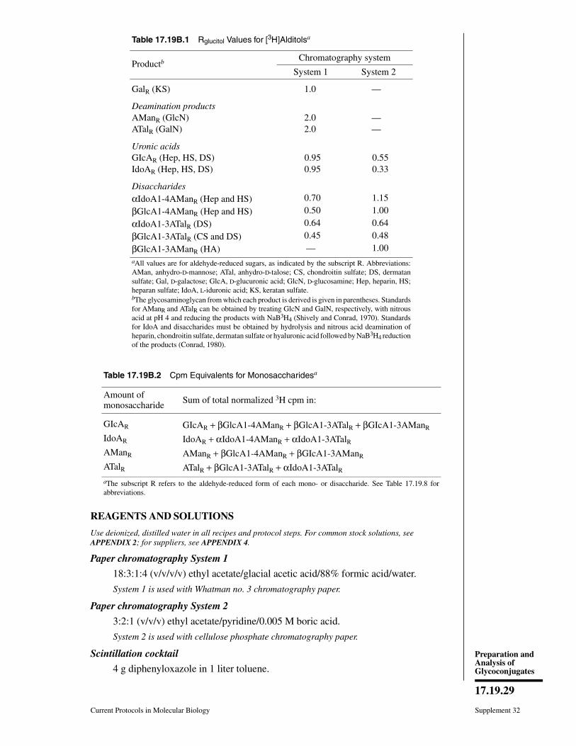
REAGENTS AND SOLUTIONS
Use deionized, distilled water in all recipes and protocol steps. For common stock solutions, seeAPPENDIX 2; for suppliers, see APPENDIX 4.
Paper chromatography System 1
18:3:1:4 (v/v/v/v) ethyl acetate/glacial acetic acid/88% formic acid/water.
System 1 is used with Whatman no. 3 chromatography paper.
Paper chromatography System 2
3:2:1 (v/v/v) ethyl acetate/pyridine/0.005 M boric acid.
System 2 is used with cellulose phosphate chromatography paper.
Scintillation cocktail
4 g diphenyloxazole in 1 liter toluene.
Table 17.19B.1 Rglucitol Values for [3H]Alditolsa
ProductbChromatography system
System 1 System 2
GalR (KS) 1.0 —
Deamination productsAManR (GlcN) 2.0 — ATalR (GalN) 2.0 —
Uronic acidsGIcAR (Hep, HS, DS) 0.95 0.55IdoAR (Hep, HS, DS) 0.95 0.33
DisaccharidesαIdoA1-4AManR (Hep and HS) 0.70 1.15
βGlcA1-4AManR (Hep and HS) 0.50 1.00
αIdoA1-3ATalR (DS) 0.64 0.64
βGlcA1-3ATalR (CS and DS) 0.45 0.48
βGlcA1-3AManR (HA) — 1.00aAll values are for aldehyde-reduced sugars, as indicated by the subscript R. Abbreviations:AMan, anhydro-D-mannose; ATal, anhydro-D-talose; CS, chondroitin sulfate; DS, dermatansulfate; Gal, D-galactose; GlcA, D-glucuronic acid; GlcN, D-glucosamine; Hep, heparin, HS;heparan sulfate; IdoA, L-iduronic acid; KS, keratan sulfate.bThe glycosaminoglycan from which each product is derived is given in parentheses. Standardsfor AManR and ATalR can be obtained by treating GlcN and GalN, respectively, with nitrousacid at pH 4 and reducing the products with NaB3H4 (Shively and Conrad, 1970). Standardsfor IdoA and disaccharides must be obtained by hydrolysis and nitrous acid deamination ofheparin, chondroitin sulfate, dermatan sulfate or hyaluronic acid followed by NaB3H4 reductionof the products (Conrad, 1980).
Table 17.19B.2 Cpm Equivalents for Monosaccharidesa
Amount ofmonosaccharide Sum of total normalized 3H cpm in:
GIcAR GIcAR + βGlcA1-4AManR + βGlcA1-3ATalR + βGIcA1-3AManR
IdoAR IdoAR + αIdoA1-4AManR + αIdoA1-3ATalRAManR AManR + βGlcA1-4AManR + βGIcA1-3AManR
ATalR ATalR + βGlcA1-3ATalR + αIdoA1-3ATalRaThe subscript R refers to the aldehyde-reduced form of each mono- or disaccharide. See Table 17.19.8 forabbreviations.
Current Protocols in Molecular Biology Supplement 32
17.19.29
Preparation andAnalysis ofGlycoconjugates
COMMENTARY
Background InforrnationWith the exception of keratan sulfate, which
contains alternating D-galactose and N-acetyl-D
glucosamine residues, all glycosaminoglycansare sequences of disaccharide units containinga uronic acid (D-glucuronic acid and/or L-iduronic acid) and an amino sugar (D-glu-cosamine or D-galactosamine). Disaccharidesof chondroitin sulfate contain D-GalNAc andD-GlcA, whereas those from dermatan sulfatecontain D-GalNAc and either D-GlcA or L-IdoA. Disaccharides from heparin and heparansulfate contain D-GlcN, which may be eitherN-acetylated or N-sulfated, and either D-GlcAor L-IdoA. Thus, if one has identified a glycos-aminoglycan that is free of other glycosamino-glycans, the primary purpose of monosaccha-ride analysis is to determine the relative pro-portions of the two uronic acid types. However,when glycosaminoglycans are isolated fromnatural sources, they are often recovered asmixtures of several types of glycosaminogly-cans that are difficult to resolve completely. Insuch cases, monosaccharide analysis can oftendemonstrate the nature of the mixtures byshowing the relative proportions of GlcNAcand GalNAc as well as the specific disaccharideunit in which they are found. Individual gly-cosaminoglycans in a mixture can be recog-nized by their susceptibility to cleavage byspecific enzymes (UNIT 17.13B) or nitrous acid(UNIT 17.22A). In the present method, the gly-cosaminoglycans are cleaved by acid hydro-lysis, which results in loss of the sulfates. Pro-cedures for determination of glycosaminogly-can disaccharides that have retained theirsulfate substituents are described in UNITS 17.13B
& 17.22A.Following acid hydrolysis and nitrous acid
cleavage of unlabeled glycosaminoglycans, itwould be equally feasible to analyze the prod-ucts of this cleavage reaction sequence by gaschromatography or HPLC without use ofNaB3H4. Although such an analytical schemehas not been developed, it would represent away to avoid use of radioisotopes. The paperchromatographic procedure used here does of-fer the possibility of analyzing many samplessimultaneously, which cannot be done with theGC or HPLC approaches where samples mustbe analyzed sequentially. Furthermore, whenunknown peaks occur in this analysis, it is clearthat the relative amounts of the unknowns arereflected by the amounts of 3H in the peaks,because (1) 3H labeling of carbohydrates is
identical for all reducing carbohydrates whenexpressed on a molar basis and (2) carbohy-drates are virtually the only naturally occurringcompounds that can be reduced by NaBH4 orNaB3H4. Moreover, it is easy to elute labeledproducts from the paper segments after analysisfor further characterization. This is readily ac-complished by rinsing the segments in toluene(to remove the diphenyloxazole remainingfrom the scintillation cocktail), then eluting thedried segment with water or buffer.
Critical ParametersThe challenge in analyzing the monosaccha-
ride composition of glycosaminoglycans is notin the quantitation of the monosaccharides butin the depolymerization step. The impossibilityof obtaining complete hydrolysis of glycosami-noglycans to monosaccharides without decom-position of some of the monosaccharide resi-dues cannot be emphasized too strongly. Thelabilities of the glycosidic bonds of N-acety-lated amino sugars (Shively and Conrad, 1970)and L-iduronic acid (Conrad, 1980) are similarto those for neutral hexoses, but the well-knownstability of glucuronic acid glycosides virtuallyprecludes the complete hydrolysis of thesebonds without destruction of these residues aswell as the other monosaccharide componentsof the glycosaminoglycan samples. Further-more, hydrolytic release of the N-acetyl groupsof the amino sugars occurs at a rate similar tothat for the GlcNAc or GalNAc glycosides, sothat many of the acetyl groups are released priorto complete hydrolysis of the amino sugar gly-cosidic bonds. This precludes complete hy-drolysis of the amino sugar glycosides, as theglycosidic bonds of the N-unsubstituted GlcNand GalN are extremely stable to acid (Shivelyand Conrad, 1970). For the N-sulfated GlcNresidues in heparin and heparan sulfate, thisproblem is particularly serious, as the N-sulfatesubstituents are very labile and are removedduring the first few minutes of hydrolysis.
The tack taken here is to circumvent thesehydrolysis problems by taking advantage of theelimination reaction that occurs when N-un-substituted amino sugars are treated with ni-trous acid (in spite of the misstatements oftenseen in the literature, this is not a hydrolysisreaction), thus cleaving these amino sugar gly-cosides under extremely mild conditions. Thisapproach requires only that hydrolysis be con-tinued long enough for complete removal ofN-sulfate and N-acetyl groups. This avoids the
Supplement 32 Current Protocols in Molecular Biology
17.19.30
Composition ofLabeled
Monosaccharidesfrom
Glycosamino-glycans

destruction of monosaccharide residues thatoccurs when forcing conditions of hydrolysisare used and allows almost stoichiometric re-covery of these glycosaminoglycans as mono-and disaccharides, which can be analyzed to-gether as described. Identical results for theanalytical procedure described here are ob-tained whether the acid hydrolysis is carried outfor 4, 6, or 8 hr before adding the nitrous acidreagent (Conrad, 1980). This is a reflection ofthe fact that all N- and O-sulfate groups and allN-acetyl groups will have been completely re-moved after 4 hr of hydrolysis. During thesubsequent period (from the fourth to the eighthhour) there is some further hydrolysis, whichreduces the yields of disaccharides and in-creases the yield of monosaccharides in theassay mixture, but this does not change the finalanalytical results.
For accurate results, it is advisable to firstseparate the glycosaminoglycan chains fromthe core protein and to isolate the free glycos-aminoglycan chains for the analysis (UNIT 17.15).This avoids the possibility of loss of monosac-charides due to the Browning reaction, whichoccurs when carbohydrates and amino acids areheated together in acid.
The nitrous acid reaction is virtuallystoichiometric for β-linked amino sugars(Shively and Conrad, 1970). Consequently, di-saccharides are obtained in stoichiometricyields from glycosaminoglycans that containβ-linked amino sugars (chondroitin sulfate,dermatan sulfate, hyaluronic acid, and keratansulfate). However, a side reaction, the ring con-traction reaction, occurs at a frequency of 10%to 15% for α-linked amino sugars such as thosefound in heparin and heparan sulfate (Shivelyand Conrad, 1970; 1976). The ring contractionreaction results in deamination without bondcleavage and may convert a heparin tetrasacch-aride into a new tetrasaccharide instead of twodisaccharides. Thus, heparin and heparan sul-fate yield significant amounts of ring contrac-tion tetrasaccharides (Bienkowski and Conrad,1985). However, there seems to be little struc-tural selectivity for the ring contraction: the ringcontraction tetrasaccharides contain disaccha-ride units in the same proportions as are foundin the original glycosaminoglycan. Thus, evenfor heparin and heparan sulfate, the disaccha-rides formed during nitrous acid cleavage areobtained in the same proportions that are foundin the original glycosaminoglycan. Conse-quently, when yields are expressed in percentof total uronic acids or percent of total amino
sugars, the ring contraction reaction does notinterfere with the quantitation, and the yieldsof di- and monosaccharides are truly repre-sentative of their proportions in the startingglycosaminoglycan. Some of these heparinring contraction tetrasaccharides have beenidentified (UNIT 17.22B).
TroubleshootingIn order to obtain reproducible migration of
peaks, it is necessary to have chromatographytanks well-equilibrated with freshly preparedsolvents. If many strips are placed in the tank,it may be necessary to add more chromatogra-phy solvent after the first day of the 40-hrdevelopment.
Anticipated ResultsThe results obtained here yield the distribu-
tion of labeled monosaccharides in metabo-lically labeled starting material or the monosac-charide compositions of the unlabeled glycos-aminoglycan starting material. This is possibleeven though complete hydrolysis is notachieved, because one can separate and quan-titate disaccharides of known composition.
Time ConsiderationsThe analytical procedure can be completed
in 3 days, with 1 day for the depolymerizationand NaB3H4 reduction, two overnight periodsfor paper chromatography, and finally scintil-lation counting and calculations. The procedurecan be interrupted at any stage. A major advan-tage of the paper chromatography procedureover HPLC or GC methods is that many sam-ples can be analyzed simultaneously. The onlysequential phase of the analysis is scintillationcounting, but the conditions described are de-signed to yield the mono- and disaccharides insufficient yields (i.e., total 3H cpm in each peak)so that 30-sec counting times for each papersegment result in good counting statistics.
Literature CitedBienkowski, M.J., and Conrad, H.E. 1985. Struc-
tural characterization of the oligosaccharidesformed by depolymerization of heparin with ni-trous acid. J. Biol. Chem. 260:356-365.
Conrad, H.E. 1980. The acid lability ofthe gly-cosidic bonds of L-iduronic acid residues in gly-cosaminoglycans. Biochem. J. 191:355-363.
Shively, J.E., and Conrad, H.E. 1970. Stoichiometryofthe nitrous acid deaminative cleavage of modelamino sugar glycosides and glycosaminoglycur-onans. Biochemistry 9:33-41.
Current Protocols in Molecular Biology Supplement 32
17.19.31
Preparation andAnalysis ofGlycoconjugates
Shively, J.E., and Conrad, H.E. 1976. Formation ofanhydrosugars in the chemical depolymerizationof heparin. Biochemistry 15:3932-3942.
Key ReferenceConrad, 1980. See above.
Describes the basic procedure used here.
Contributed by H. Edward ConradUniversity of IllinoisUrbana, Illinois
Supplement 32 Current Protocols in Molecular Biology
17.19.32
Composition ofLabeled
Monosaccharidesfrom
Glycosamino-glycans
UNIT 17.20Analysis of Oligosaccharide Negative Chargeby Anion-Exchange ChromatographyThis unit presents the analysis of negative charge on labeled N- or O-linked oligosaccha-rides. These protocols may be used in the initial screening of oligosaccharides to detectnegative charge, for analytical or preparative separation of oligosaccharides based on theirnegative charge, or to analyze the type of negative charge found on the oligosaccharides.The Basic Protocol describes the use of anion-exchange (QAE-Sephadex) chromatogra-phy with stepwise elution for estimating the number of negative charges on an oligosac-charide sample derived from glycosidase treatment of a glycoprotein. In the AlternateProtocol, gradient elution is used for the preparative separation of oligosaccharides basedon negative charge. The Support Protocol describes a method for measuring loss of orchange in negative charge after treatment of the oligosaccharide sample with mild acidand/or phosphatases.
BASICPROTOCOL
SEPARATION AND ANALYSIS OF ANIONIC OLIGOSACCHARIDES BYQAE-SEPHADEX CHROMATOGRAPHY WITH STEPWISE ELUTIONNegative charges on peptide-free radioactive N- or O-linked oligosaccharides can bedetected by binding the molecules to the strong anion exchanger QAE-Sephadex (see alsoUNIT 10.10). Stepwise batch elution (as described in this protocol) or gradient elution (seeAlternate Protocol) with salt separates the bound molecules according to the approximatenumber of negative charges. Loss or change of negative charge following treatment withsialidases (UNIT 17.12), solvolysis (UNIT 17.23), mild acid (UNIT 17.16), or phosphatase (seeSupport Protocol) can also be monitored (see Fig. 17.20.1). Because of charge-to-masseffects, this method gives only a general estimate of the number of negative charges onan oligosaccharide. If more precise analysis is desired, protocols using high-performanceliquid chromatography (HPLC; UNIT 17.21) are recommended.
Materials
Radiolabeled mixture of oligosaccharides released from glycoprotein(UNITS 17.12-17.17)
Equilibrated QAE-Sephadex chromatography resin (see recipe)2 mM Tris baseElution buffers (see recipe)
1- to 2-ml Pasteur pipets plugged with glass wool or 1- to 2-ml disposable plasticcolumns
Sintered-glass funnel
Additional reagents and equipment for metabolic radiolabeling (UNIT 17.4) andautoradiography (APPENDIX 3A)
1. Pour 0.75-ml column of equilibrated QAE-Sephadex in a 1- to 2-ml Pasteur pipetplugged with glass wool or in a 1- to 2-ml disposable plastic column. Wash with 5ml of 2 mM Tris base.
It is acceptable to let the top of the column bed go dry during washing.
2. Dilute or dissolve the sample in 0.75 ml of 2 mM Tris base and load onto the column.Wash with seven 0.75-ml aliquots of 2 mM Tris base and collect 1.5-ml fractions.
Try not to disturb the top of the column bed during application. Allow the top of the columnbed to go dry between aliquots.
The concentration of salts in the applied sample should be <5 mM and the pH must be>7. If the original sample contains acidic buffers or other salts, it may be necessary
Supplement 32
Contributed by Ajit VarkiCurrent Protocols in Molecular Biology (1995) 17.20.1-17.20.7Copyright © 2000 by John Wiley & Sons, Inc.
17.20.1
Preparation andAnalysis ofGlycoconjugates

020 70 100
–1 –2 –3140
0 –1
–1.5
–2
–2.5
–3
5 9 13 17
Rad
ioac
tivity
Fraction number
mM NaCl
no treatmentor alkalinephosphatase
sialidase
mild acid
mild acidand alkalinephosphatase
A
B
0.3 M
ammoniumacetate
Rad
ioac
tivity
Figure 17.20.1 Schematic examples of QAE-Sephadex analysis (based on Varki and Kornfeld,1983). Numbers in italics indicate the charges on the high mannose–type oligosaccharides elutingat that position. (A) Gradient fractionation of mixture of N-linked oligosaccharides with differentcombinations of sialic acid phosphomonoesters or phosphodiesters. (B) Peak marked “−1” consistsof a mixture of oligosaccharides with one negative charge due to either one sialic acid residue (acid-or sialidase-sensitive) or one phosphodiester (increased negative charge after mild acid, sensitiveto alkaline phosphatase only after mild acid treatment). The procedures used to evaluate oligosac-charide mixtures are listed on the right with examples of resulting shifts in the elution peaks for eachtype of treatment illustrated on the left.
Supplement 32 Current Protocols in Molecular Biology
17.20.2
Analysis ofOligosaccharide
NegativeCharge by Anion-
ExchangeChromatography

to neutralize with Tris base and/or dilute the sample to >0.75 ml to reduce the saltconcentration. In these cases, the first wash-through tube will contain >1.5 ml fluid.
3. Elute sample from the column with elution buffers containing increasing concentra-tions of salt (i.e., 20, 70, 100, 140, 200, 400, and 1000 mM NaCl) in 2 mM Tris base.For each salt concentration, use eight 0.75-ml aliquots and collect four 1.5-mlfractions.
The salt elution series suggested above was devised for analysis of phosphorylated highmannose–type oligosaccharides from lysosomal enzymes. Salt concentrations may need tobe adjusted to optimize the separation of other types of oligosaccharides (see CriticalParameters).
4. Monitor elution of the oligosaccharides by analyzing aliquots of the collectedfractions (or the entire fractions) for radioactivity. In the latter case, collect the eluatedirectly into large-sized scintillation vials. If the first wash fraction is >1.5 ml (seestep 2 annotation), monitor only an aliquot.
When comparing different treatments of the same oligosaccharide—e.g., sialidase (UNIT
17.12), mild acid, and alkaline phosphatase (see Support Protocol)—it is convenient to runall samples and controls in parallel. Approximately ten columns can be easily run at thesame time. A repeating pipettor makes the process more reproducible and less taxing.
5a. The first time the experiment is done, and anytime it is possible that some of theoligosaccharides may not elute with the highest concentration of salt, add 1.5 ml of2 mM Tris base to the column and transfer the contents of the column into ascintillation vial. Add scintillation fluid and monitor for any remaining radioactivity.
5b. Otherwise, discard the column contents (do not reuse).
ALTERNATEPROTOCOL
SEPARATION AND ANALYSIS OF ANIONIC OLIGOSACCHARIDES BYQAE-SEPHADEX CHROMATOGRAPHY WITH GRADIENT ELUTION
After the initial studies of a mixture of oligosaccharides on a QAE-Sephadex column, itmay be desirable to preparatively separate the oligosaccharides with better resolution.Although HPLC methods are preferable (see UNIT 17.21), the following protocol can beused if an HPLC unit or column is not available or if a large capacity is needed.
Additional Materials (also see Basic Protocol)
5- to 20-ml disposable plastic columnGradient mixer
Additional materials and equipment for salt gradient preparation (UNIT 10.10)
1. Prepare a 3- to 10-ml column of QAE-Sephadex (see Basic Protocol, step 1, but use5- to 20-ml disposable plastic column). Wash column with 10 column volumes of 2mM Tris base.
2. Prepare and load the oligosaccharide sample (see Basic Protocol, step 2).
3. Place 50 ml of 2 mM Tris base in the first chamber of the gradient mixer; place 50ml of an appropriate concentration of NaCl dissolved in 2 mM Tris base in the secondchamber (choose salt concentration based on the results of the Basic Protocol).
An ammonium acetate gradient at pH 5.3 can be used to fractionate phosphomonoestersand phosphodiesters (see Critical Parameters).
The concentration of NaCl or ammonium acetate in the second chamber is selected basedon prior knowledge of the concentration required to elute the most anionic species in themixture.
Current Protocols in Molecular Biology Supplement 32
17.20.3
Preparation andAnalysis ofGlycoconjugates

4. Elute the sample from the column with the selected linear gradient regimen.Collect1-ml fractions and monitor for radioactivity.
SUPPORTPROTOCOL
DETECTION AND REMOVAL OF PHOSPHODIESTERS ORPHOSPHOMONOESTERS
Oligosaccharide samples thought to contain phosphodiesters and phosphomonoesters(e.g., released from lysosomal enzymes) can be treated with mild acid and alkalinephosphatase to detect and remove them. Phosphomonoester-containing molecules willlose negative charge upon phosphatase treatment. Phosphodiester-containing moleculesare resistant to alkaline phosphatase alone, but will increase in negative charge followingmild acid treatment because of the generation of phosphomonoesters. A combination ofmild acid and phosphatase treatment will neutralize phosphodiesters; the mild acidconditions used here will also result in removal of sialic acids (see Fig. 17.20.1).
Additional Materials (also see Basic Protocol)
Radiolabeled mixture of oligosaccharides released from glycoprotein (UNITS
17.12-17.17), desalted and lyophilized10 U/ml E. coli alkaline phosphatase2 M acetic acid200 mM Tris⋅Cl, pH 8.0 (APPENDIX 2)
Strong 10-ml conical glass tubes or 1-ml Reacti-Vials (Pierce)Water bath or heating block 80°C
1. Dissolve the oligosaccharide sample in 0.5 ml of 2 M acetic acid in a strong 10-mlconical glass tube or 1-ml Reacti-Vial. Tightly cap or stopper the tube and heat 120min at 80°C.
This step is designed to detect phosphodiesters. A second, identical sample should beprepared and processed starting with step 3; if phosphodiesters are present, the acid-treated sample will show a higher negative charge than this non-acid-treated control uponanalysis.
2. Flash-freeze and lyophilize the sample.
3. Dissolve in 20 µl water, using a pipettor to wash the walls of the tube. Vortex welland centrifuge briefly at room temperature to get sample to bottom of tube.
4. Remove a 10-µl aliquot of sample and dilute to 1.5 ml with 2 mM Tris base. Applyto a 0.75-ml QAE-Sephadex column for analysis (see Basic Protocol).
5. To the remaining sample, add 10 µl of 200 mM Tris⋅Cl (pH 8.0) and mix. Add 1 µlof 10 U/ml E. coli alkaline phosphatase (10 mU). Incubate 1 hr at 37°C.
6. Dilute sample to 1.5 ml with water and apply to a 0.75-ml QAE-Sephadex column(see Basic Protocol).
REAGENTS AND SOLUTIONS
Use deionized, distilled water in all recipes and protocol steps. For common stock solutions, seeAPPENDIX 2; for suppliers, see APPENDIX 4.
Ammonium acetate (pH 5.3), 1 MDissolve 38.54 g ammonium acetate in H2O. Add H2O to 500 ml. Adjust pH to 5.3with 1 M acetic acid. Store at 4°C.
Supplement 32 Current Protocols in Molecular Biology
17.20.4
Analysis ofOligosaccharide
NegativeCharge by Anion-
ExchangeChromatography
Elution buffersPrepare stock solutions of 1 M Tris base (pH unadjusted; store at 4°C) and 2 M NaCl(store at room temperature). Use these stocks to make elution buffers containing 2mM Tris base and different concentrations of NaCl. For gradient elution, makeappropriate dilutions of the 1 M ammonium acetate (pH 5.3) stock solution (seerecipe). It is not necessary to check the final pH of of the diluted solutions.
QAE-Sephadex chromatography resin, equilibratedSwell QAE-Sephadex (Pharmacia Biotech) in 1 M Tris base (using 1 g dry resin/20ml Tris) overnight at 4°C, then decant. Repeat once. Pour slurry over a sintered-glassfunnel and wash with >20 vol of 2 mM Tris base. Check the pH of the final washwith pH paper to make sure it is similar to that of the Tris base; if not, wash againuntil appropriate pH is achieved. Store equilibrated resin in 2 mM Tris base at 4°C.
There is no need to remove “fines” (small particles) from the resin because the columns usedare small and flow rates are not limiting. With storage and exposure to air, the pH of the Trisbase will drift towards neutral; however, this is not of serious concern.
COMMENTARY
Background InformationNegatively charged monosaccharides and
highly anionic oligosaccharides bind well tomost anion-exchange resins. However, largeoligosaccharides with very few negativecharges have a poor charge-to-mass ratio andmay fail to bind to some anion-exchange resins.The strong anion exchanger QAE-Sephadexwas first used (Tabas and Kornfeld, 1980) topermit analysis of high mannose–type N-linkedoligosaccharides with single phosphodiesters.The column was equilibrated in 2 mM Tris baseto ensure that the pH was above neutral (maxi-mizing the deprotonation of all anions) and tolimit the amount of salt competing for bindingsites. The protocol described here is based uponmodifications of this method (Varki and Korn-feld, 1980, 1983; Goldberg and Kornfeld,1981).
In the time since the original description,modifications have been developed to separatea wide variety of anionic oligosaccharides, in-cluding O-linked oligosaccharides, N-linkedglycopeptides, and sulfated oligosaccharides(Roux et al., 1988; Fukuda, 1989; Cummingset al., 1989). In each case, column sizes andelution conditions were adjusted to allow ap-propriate separations and yield. In recent years,Mono-Q FPLC columns (Pharmacia Biotech)have permitted the use of the same QAE ex-changer group for more rigorous and completeseparations (van Pelt et al., 1987; UNIT 10.10).
Critical ParametersThe QAE-Sephadex resin must be thor-
oughly equilibrated in 2 mM Tris base. Theinitial loading must be carried out in a minimum
amount of salt (<2.5 mM) to ensure that weaklycharged molecules do not escape binding. Thiscan be achieved by desalting samples thor-oughly (UNIT 10.9) and/or diluting them suffi-ciently with water or 2 mM Tris base beforeloading. To make the dilution volume reason-able, the preceding steps (e.g., enzyme reac-tions) should be carried out in the minimumvolumes and lowest salt concentrations possi-ble. Acid treatments are carried out with vola-tile acids that can be lyophilized before analy-sis. The charge-to-mass ratio of an oligosaccha-ride affects the amount of NaCl required forelution. Thus, a high mannose–type N-linkedoligosaccharide with a single mannose-6-phos-phate residue will elute with 50 to 70 mM NaCl,while free mannose-6-phosphate (with thesame anionic group) requires 100 to 120 mMNaCl. The amount of salt needed to elute aparticular charge or size class of moleculesmust be determined empirically. Thus, thistechnique is most useful when a related seriesof oligosaccharides with a limited range of sizeand charge (e.g., phosphorylated N-linked oli-gosaccharides with one to four negativecharges) are being separated and analyzed.However, even when there is very extensiveheterogeneity in size and charge (e.g., sulfatedsialylated N-linked oligosaccharides), infor-mation can be obtained by following relativeshifts in the elution position of groups of mole-cules (Roux et al., 1988). Gradient elution givesmore defined separation between individualtypes of molecules, and is used primarily forpreparative separations. If the molecules con-tain mixtures of phosphomonoesters and phos-phodiesters, separation can be obtained with a
Current Protocols in Molecular Biology Supplement 32
17.20.5
Preparation andAnalysis ofGlycoconjugates
gradient of ammonium acetate adjusted to pH5.3 (close to the pKa of the second negativecharge of a phosphomonoester).
Analysis of glycopeptides by QAE-Sepha-dex chromatography is less satisfactory, be-cause the peptide portion of the molecules cancarry variable amounts of negative and/or posi-tive charge. However, if complete proteolyticcleavage is performed (see UNIT 17.14A), eachglycopeptide should have only one carboxylgroup and separations can be made on the basisof the additional negative charges on the oli-gosaccharide. Molecules with very high chargedensity (e.g., heparin chains) are not well re-solved on QAE columns because they are dif-ficult to elute.
TroubleshootingIf molecules known to be anionic do not bind
to the QAE-Sephadex column, the most likelyproblems are failure to properly equilibrate thecolumn in 2 mM Tris base and the presence ofexcessive salt in the sample. If peaks are broadand slurred, the most likely problem is exces-sive disturbance of the top of the column bedduring the application of elution buffers.
Anticipated ResultsThe method presented in this unit can be
used for the following analyses.1. Initial screening of oligosaccharides for
the presence of negative charge. The sample isapplied and the column washed and then di-rectly eluted with 1 M NaCl in 2 mM Tris base.As a control, a known neutral oligosaccharideand an anionic oligosaccharide should be rununder exactly the same conditions to obtainbackground numbers. A small quantity of theanionic oligosaccharide (<5%) can be expectedto escape binding to the column, and a smallamount of label from the neutral molecule(<2%) will be nonspecifically found in the salteluate.
2. Analytical separation of oligosaccha-rides by negative charge. A mixture of oligosac-charides is applied and sequentially elutedbatchwise with a defined series of salt elutionsteps (e.g., 20, 70, 100, 140, 200, 400, and 1000mM NaCl), and the profile is monitored.
3. Preparative separation of oligosaccha-rides by negative charge. This is based upon theprofile obtained by analytical separation. Gra-dient elution using NaCl or ammonium acetatemay be preferred. Peaks are pooled, dried, anddesalted on columns of Sephadex G-25 inwater.
4. Analysis of the type of negative chargeon oligosaccharides. Individual peaks obtainedby preparative separation are analyzed beforeand after treatment or combinations of treat-ments with mild acid (see Support Protocol),with sialidase (UNIT 17.12), by solvolysis (UNIT
17.23), and/or with alkaline phosphatase (seeSupport Protocol). Fig. 17.20.1 schematicallydepicts an example of such analyses.
Time ConsiderationsThe initial setup of small columns should
take 10 to 20 min. The setup for gradient elutiontakes somewhat longer. Stepwise elutions ofapproximately ten samples can usually be car-ried out in parallel in ∼1 hr. If sample prepara-tion for scintillation counting is performed dur-ing time gaps in the elution procedure, the entireprocess can be completed in 2 to 3 hr. The timefor scintillation counting will depend upon theamount of radioactivity available in each sam-ple.
Literature CitedCummings, R.D., Merkle, R.K., and Stults, N.L.
1989. Separation and analysis of glycoproteinoligosaccharides. Methods Cell. Biol. 32:141-183.
Fukuda, M. 1989. Characterization of O-linked sac-charides from cell surface glycoproteins. Meth-ods Enzymol. 179:17-29.
Goldberg, D.E. and Kornfeld, S. 1981. The phos-phorylation of β-glucuronidase oligosaccharidesin mouse P388D1 cells. J. Biol. Chem.256:13060-13067.
Roux, L., Holoyda, S., Sundblad, G., Freeze, H.H.,and Varki, A. 1988. Sulfated N-linked oligosac-charides in mammalian cells I: Complex-typechains with sialic acids and O-sulfate esters. J.Biol. Chem. 236:8879-8889.
Tabas, I. and Kornfeld, S. 1980. Biosynthetic inter-mediates of β-glucuronidase contain high man-nose oligosaccharides with blocked phosphateresidues. J. Biol. Chem. 255:6633-6639.
van Pelt, J., Damm, J.B., Kamerling, J.P., andVliegenthart, J.F.G. 1987. Separation of sialyl-oligosaccharides by medium pressure anion-ex-change chromatography on Mono Q. Carbohydr.Res. 169:43-51.
Varki, A. and Kornfeld, S. 1980. Structural studiesof phosphorylated high mannose–type oligosac-charides. J. Biol. Chem. 255:10847-10858.
Varki, A. and Kornfeld, S. 1983. The spectrum ofanionic oligosaccharides released by endo-β-N-acetylglucosaminidase H from glycoproteins.Structural studies and interactions with the phos-phomannosyl receptor. J. Biol. Chem. 258:2808-2818.
Supplement 32 Current Protocols in Molecular Biology
17.20.6
Analysis ofOligosaccharide
NegativeCharge by Anion-
ExchangeChromatography

Key ReferenceVarki, A. and Kornfeld, S. 1980. See above.
Figures in the miniprint section of this paper provideseveral examples of the types of analyses describedhere.
Contributed by Ajit VarkiUniversity of California San DiegoLa Jolla, California
Current Protocols in Molecular Biology Supplement 32
17.20.7
Preparation andAnalysis ofGlycoconjugates

UNIT 17.21AHPLC Methods for the Fractionation andAnalysis of Negatively ChargedOligosaccharides and Gangliosides
This unit describes the fractionation and analysis of anionic oligosaccharides (BasicProtocol 1) and gangliosides (Basic Protocol 2) using anion-exchange high-performanceliquid chromatography (HPLC). The sample is loaded onto the column in a low-ionic-strength solvent. The anionic saccharides or gangliosides are retained by the positivelycharged matrix by ionic interactions; the more negative charge on the compound, the moretightly it binds to the column. Neutral molecules pass through the column unretained.Elution is effected by a gradient of increasing ionic strength that progressively disruptsthe ionic interactions between the analyte and the column. Saccharides or gangliosidesare eluted in order of the number of negative charges they possess, although the charge-to-mass ratio can also contribute to elution position. Hence, a molecule with a highcharge-to-mass ratio may elute later than one with the same net charge but a smallercharge-to-mass ratio. Those unfamiliar with the basic principles of HPLC are referred toUNITS 10.12-10.14 or to Snyder et al. (1988).
BASICPROTOCOL 1
FRACTIONATION OF RELEASED N-LINKED OLIGOSACCHARIDES ONDEAE HPLC COLUMNS
The use of DEAE HPLC columns to separate sialylated N-linked oligosaccharides isdescribed. The DEAE column is equilibrated in water and eluted with a gradient ofincreasing NaCl concentration. Sialic acids are detected based on their radioactivity (ifpreviously radiolabeled) or by the TBA assay for sialic acids (if unlabeled) and compari-son with known standards.
Materials
HPLC-grade or high-quality deionized water (e.g., 18 MΩ Milli-Q type)0.5 M sodium chloride (NaCl) in HPLC-grade water (see recipe)Oligosaccharide sample (Peptide N:glycosidase F–released and desalted;
UNIT 17.13A)Sialylated oligosaccharide standards (e.g., Dionex or Oxford Glycosystems)
HPLC apparatus capable of two-component gradient formationDEAE HPLC column (TosoHaas TSK-gel DEAE-2SW, 25-cm × 4.6-mm i.d.,
5-mm particle size, or equivalent)
Additional reagents and equipment for desalting sample (UNITS 17.13A & 17.14A) and(if analyzing unlabeled sample) TBA assay (UNIT 17.18)
1. Before using any column, wash the column thoroughly. First, wash with at least 10column volumes of water. Next, wash the column with a linear gradient from 0 to 0.5M NaCl over 100 min, and then from 0.5 M NaCl to 100% water over 100 min. Usea flow rate of 0.4 to 0.6 ml/min for all washes.
The water and all buffers used should be prefiltered through a 0.45-m filter and degassed.Column prefilters and/or guard columns provide additional protection for the HPLCcolumn and increase its productive lifetime.
A flow rate of 0.4 to 0.6 ml/min is recommended for all column washings and elutions. Donot exceed the manufacturer’s recommendations for either the flow rate or maximumpressure.
Supplement 32
Contributed by Adriana E. Manzi and Bradley K. HayesCurrent Protocols in Molecular Biology (1995) 17.21.11-17.21.8Copyright © 2000 by John Wiley & Sons, Inc.
17.21.1
Preparation andAnalysis ofGlycoconjugates

2. Thoroughly clean the injection loop with water.
3. Equilibrate the column at room temperature with 100% water at a flow rate of 0.6ml/min. Wash with at least 10 column volumes of water.
4. Check the baseline obtained with the detection method to be used for the sample. Donot proceed until a stable baseline is obtained.
For analytical work, on-line radioactivity detectors or UV detectors may be used. For UVdetection, absorbance at 190 to 210 nm is measured. Because many compounds absorb atthese wavelengths, this method of detection demands high purity of the sample. Alterna-tively, individual fractions may be collected and analyzed by one of several methods (seestep 8). Collect a portion of the eluate and establish that the solvents do not interfere withthe detection method chosen and that the baseline is stable.
5. Set up a program from the pump control unit for washing unbound material, elutingthe bound species, and bringing the column back to the starting conditions, asindicated in Table 17.21A.1.
The NaCl gradient should be shallow for two reasons. First, the difference in NaClconcentration needed to elute oligosaccharides differing by a single negative charge is ∼20mM NaCl, depending on the column. Second, the DEAE resin will shrink and swell withincreasing and decreasing NaCl concentrations, respectively. Therefore, rapid changes inionic strength may damage the column and degrade its performance.
6. Make a “sham” run (injecting water in place of sample), monitoring the eluant, if anon-line detection system is used. If fractions are collected and individually analyzed,collect them every 1 min. Load the water into the injection loop using a syringe witha flat-tip needle compatible with the HPLC injector. Inject the water onto the columnand start the gradient program.
If contaminant peaks are observed (peaks >5% full scale when using the detector settingsrequired for the sample), the column must be washed until the peaks disappear. (Whenusing UV detection, a peak will be observed at the void volume, indicating the change inabsorbance produced when the water injected passes through the column.) To wash, repeatthe gradient and hold at the maximum concentration of NaCl for at least 10 columnvolumes. Wash extensively with water, then repeat the blank run.
7. Inject and analyze the sialylated oligosaccharide standards individually as indicatedin step 6. Determine the elution time for each of the standards. If the separation ofthe individual standards is adequate, proceed to the analysis of the samples. If not, itmay be necessary to adjust the rate of increase in the NaCl concentration to obtainbetter separation.
The column should be routinely standardized. At least one standard run, preferably with amixture of sialylated oligosaccharide standards, should be performed at the start of eachday.
Table 17.21A.1 DEAE HPLC Elution Regimenfor Analysis of N-Linked Oligosaccharides
Time (min) Eluant
0 100% H2O
5 100% H2O
95 50% 0.25 M NaCl
100 50% 0.25 M Nacl
140 100% H2O
155 100% H2O
Supplement 32 Current Protocols in Molecular Biology
17.21.2
HPLC Methodsfor the Analysis ofOligosaccharidesand Gangliosides
8. Analyze the desalted samples using the optimum conditions determined in step 7. Ifthe amount of sample is small or the column is being used to obtain individual speciesfor subsequent analysis, collect and analyze fractions every 0.5 to 1.0 min. Analyzethe individual fractions by using either liquid scintillation counting or the TBA assayfor sialic acids (UNIT 17.18).
The sample must be desalted (as described in UNITS 17.13A & 17.14A) prior to analysis,because even low concentrations of NaCl can prevent the initial binding of the anionicoligosaccharides to the cationic matrix. The desalted oligosaccharides can be concentratedby lyophilization and resuspended in a small volume of water. The main factor in deter-mining the appropriate sample volume is the size of the injection loop, although injectionloops of ≤500 l are preferred.
9. Estimate the number of sialic acid residues present by comparing the elution positionof each peak to that of the sialylated oligosaccharide standards run in the sameconditions.
The linkage of the sialic acid residues can also be determined using sialidase digestion(UNIT 17.12) in combination with repeated anion-exchange HPLC. If, for instance, a samplethat originally comigrated with a trisialylated triantennary oligosaccharide standard istreated with Newcastle disease virus sialidase, under conditions in which only α2-3-linkedsialic acids are released, and now comigrates with a monosialylated species, it can beinferred that the oligosaccharide in question has two α2-3-linked sialic acid residues.Additionally, if the oligosaccharide in question is not retained by the DEAE columnfollowing treatment with the broad-spectrum sialidase from Arthrobacter ureafaciens, itcan be inferred that the negative charge on the oligosaccharide in question was due solelyto sialic acid residues. Although this type of analysis can be performed on a mixture ofoligosaccharides, it is preferable to first isolate the individual HPLC peaks and then totreat with sialidase and reanalyze each separately.
CAUTION: Remember to desalt the samples submitted to sialidase digestion prior toion-exchange HPLC analysis.
BASICPROTOCOL 2
FRACTIONATION OF GANGLIOSIDES ON DEAE HPLC COLUMNS
The use of DEAE HPLC columns to separate gangliosides from neutral lipids andglycolipids and fractionate them according to the number of sialic acid residues isdescribed. Total lipid extracts, partially fractionated negatively charged glycolipids (totallipid extracts or the fraction enriched in polar lipid obtained by Folch partitioning of thetotal lipid extracts; see UNIT 17.3), or the total ganglioside fraction eluted from a DEAEanion-exchange column (see UNIT 17.3) can be further fractionated and analyzed by thismethod. The DEAE column is equilibrated in organic solvent and eluted with a gradientof increasing ammonium acetate concentration. Fraction components are detected basedon their radioactivity (if previously radiolabeled) or by HPTLC analysis with chemicalor immmunological detection (if unlabeled) and gangliosides identified by comparisonwith known standards.
Materials
HPLC-grade or high-quality deionized (e.g., 18 M Ω Milli-Q type) waterHPLC-grade chloroformHPLC-grade methanol1:8:1 (v/v/v) chloroform/methanol/1 M aqueous ammonium acetate (see recipe)Ganglioside sample (extracted and desalted; UNIT 17.3)Ganglioside standards (GM1, GD1a and GT1b; e.g., Sigma, Boehringer Mannheim, or
Calbiochem)
HPLC apparatus capable of two-component gradient formationDEAE HPLC column (TosoHaas TSK DEAE-2SW, or comparable)
Current Protocols in Molecular Biology Supplement 32
17.21.3
Preparation andAnalysis ofGlycoconjugates

Additional reagents and equipment for high-performance thin-layerchromatography (HPTLC; if analyzing unlabeled sample, UNIT 17.17A) withchemical detection (UNIT 17.10B) and for TBA assay (UNIT 17.18)
1. Equilibrate the column in organic solvent by running a linear gradient from 100%water to 1:8:1 (v/v/v) chloroform/methanol/water at 1 ml/min, over 1 hr. If the columnwas stored in buffer, first wash extensively with water.
The water and all aqueous buffers used should be prefiltered through a 0.45-m filter anddegassed. Do not degas mixtures containing organic solvents prior to chromatography;moreover, for systems that require continuous helium bubbling during chromatography, alow flow is sufficient. Column prefilters and/or guard columns provide additional protec-tion for the HPLC column and increase its productive lifetime. When using a guard column,be sure to connect it prior to performing the equilibration with organic solvent.
2. Convert the column to the acetate form by running a 1-hr linear gradient from 1:8:1(v/v/v) chloroform/methanol/water to 1:8:1 (v/v/v) chloroform/methanol/1 M aque-ous ammonium acetate at 1 ml/min, and holding at this concentration of salt foranother 1 hr. Wash the column extensively with 1:8:1 (v/v/v) chloroform/metha-nol/water to remove the salt. Use a flow rate of 1 ml/min throughout this step.
3. Flush out the injection loop with water, then methanol, then chloroform, and finallywith 1:8:1 (v/v/v) chloroform/methanol/water.
4. For columns previously used to analyze radioactive samples, check the backgroundwith an on-line radioactivity detector or by collecting fractions and counting.
5. Set up a program from the pump control unit for washing unbound material, elutingthe bound species, and bringing the column back to the starting conditions at 1 ml/minflow as indicated in Table 17.21A.2.
The DEAE resin will shrink and swell with increasing and decreasing salt concentrations,respectively. Therefore, transition from high salt concentration to no salt has to be doneslowly. Rapid changes in ionic strength may cause damage to the column and degradecolumn performance.
Table 17.21A.2 DEAE HPLC Elution Regimen for Analysis ofGangliosides
Step Time (min) Eluant
Wash 0 1:8:1 (v/v/v)chloroform/methanol/water
Elution 10 1:8:1 (v/v/v)chloroform/methanol/water
70 1:8:1 (v/v/v)chloroform/methanol/aqueous 1 M ammonium acetate
Reequilibration 100 1:8:1 (v/v/v)chloroform/methanol/water
130 1:8:1 (v/v/v)chloroform/methanol/water
Supplement 32 Current Protocols in Molecular Biology
17.21.4
HPLC Methodsfor the Analysis ofOligosaccharidesand Gangliosides
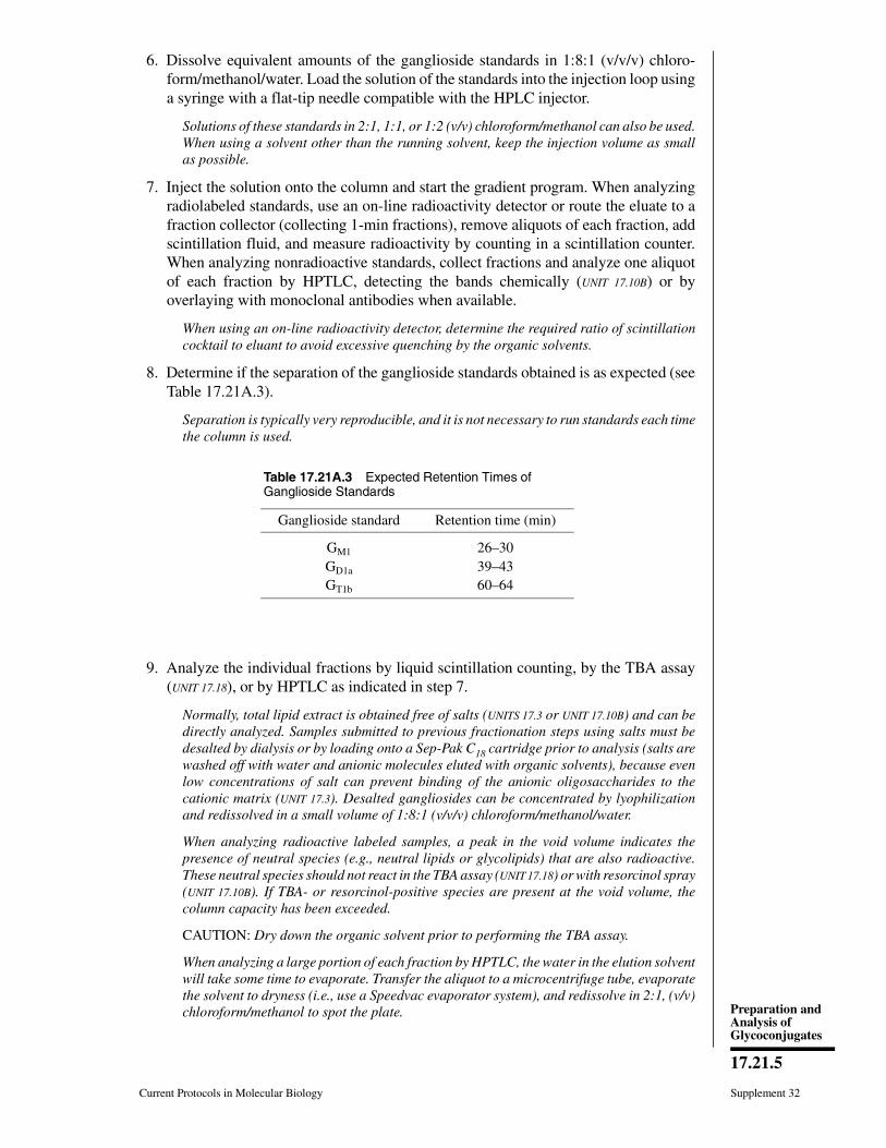
6. Dissolve equivalent amounts of the ganglioside standards in 1:8:1 (v/v/v) chloro-form/methanol/water. Load the solution of the standards into the injection loop usinga syringe with a flat-tip needle compatible with the HPLC injector.
Solutions of these standards in 2:1, 1:1, or 1:2 (v/v) chloroform/methanol can also be used.When using a solvent other than the running solvent, keep the injection volume as smallas possible.
7. Inject the solution onto the column and start the gradient program. When analyzingradiolabeled standards, use an on-line radioactivity detector or route the eluate to afraction collector (collecting 1-min fractions), remove aliquots of each fraction, addscintillation fluid, and measure radioactivity by counting in a scintillation counter.When analyzing nonradioactive standards, collect fractions and analyze one aliquotof each fraction by HPTLC, detecting the bands chemically (UNIT 17.10B) or byoverlaying with monoclonal antibodies when available.
When using an on-line radioactivity detector, determine the required ratio of scintillationcocktail to eluant to avoid excessive quenching by the organic solvents.
8. Determine if the separation of the ganglioside standards obtained is as expected (seeTable 17.21A.3).
Separation is typically very reproducible, and it is not necessary to run standards each timethe column is used.
9. Analyze the individual fractions by liquid scintillation counting, by the TBA assay(UNIT 17.18), or by HPTLC as indicated in step 7.
Normally, total lipid extract is obtained free of salts (UNITS 17.3 or UNIT 17.10B) and can bedirectly analyzed. Samples submitted to previous fractionation steps using salts must bedesalted by dialysis or by loading onto a Sep-Pak C18 cartridge prior to analysis (salts arewashed off with water and anionic molecules eluted with organic solvents), because evenlow concentrations of salt can prevent binding of the anionic oligosaccharides to thecationic matrix (UNIT 17.3). Desalted gangliosides can be concentrated by lyophilizationand redissolved in a small volume of 1:8:1 (v/v/v) chloroform/methanol/water.
When analyzing radioactive labeled samples, a peak in the void volume indicates thepresence of neutral species (e.g., neutral lipids or glycolipids) that are also radioactive.These neutral species should not react in the TBA assay (UNIT 17.18) or with resorcinol spray(UNIT 17.10B). If TBA- or resorcinol-positive species are present at the void volume, thecolumn capacity has been exceeded.
CAUTION: Dry down the organic solvent prior to performing the TBA assay.
When analyzing a large portion of each fraction by HPTLC, the water in the elution solventwill take some time to evaporate. Transfer the aliquot to a microcentrifuge tube, evaporatethe solvent to dryness (i.e., use a Speedvac evaporator system), and redissolve in 2:1, (v/v)chloroform/methanol to spot the plate.
Table 17.21A.3 Expected Retention Times ofGanglioside Standards
Ganglioside standard Retention time (min)
GM1 26–30GD1a 39–43GT1b 60–64
Current Protocols in Molecular Biology Supplement 32
17.21.5
Preparation andAnalysis ofGlycoconjugates
10. Determine the type of ganglioside (e.g., mono-, di-, or trisialylated) present bycomparing the elution position of each peak to that of known ganglioside standardsrun in the same conditions.
11. Determine the number of gangliosides in each fraction by analyzing the HPTLCpatterns obtained with resorcinol staining (UNIT 17.10B).
12. When doing semipreparative chromatography, pool the fractions according to theprofile obtained. Concentrate under vacuum, dissolve in a small volume of 1:8:1(v/v/v) chloroform/methanol/water, dilute with 10 vol water, and desalt by overnightdialysis against water using 500- or 1000-MWCO tubing. Recover the gangliosidesby lyophilization.
Alternatively, dry sample, resuspend in water, and desalt using a Sep-Pak C18 cartridgepreviously washed successively with 20 ml each chloroform, dimethyl sulfoxide, acetoni-trile, methanol, and water (see also UNIT 17.3).
IMPORTANT NOTE: Semipreparative chromatography is defined as the fractionation ofthe individual species obtained by HPLC with a standard-sized column.
REAGENTS AND SOLUTIONS
Use HPLC-grade or other high-quality deionized water (e.g., 18 MΩ Milli-Q type) in all recipes andprotocol steps, and filter through a 0.45-m filter and degas before use. For common stock solutions, seeAPPENDIX 2; for suppliers, see APPENDIX 4.
Chloroform/methanol/1 M aqueous ammonium acetate, 1:8:1 (v/v/v)Dissolve 77 g ammonium acetate in HPLC-grade water. Adjust the volume to 1 literand filter through a 0.45-µm filter. Combine 1 vol of this solution with 1 volchloroform and 8 vol methanol, mix, and use for HPLC. Store in a tightly cappedbottle.
Sodium chloride (NaCl), 0.5 MDissolve 29.25 g NaCl in 900 ml water. Adjust the volume to 1 liter and filter througha 0.45-µm filter before use.
COMMENTARY
Background InformationIon-exchange HPLC using weak anion ex-
change chromatography columns has beenused to analyze and/or fractionate a variety ofanionic oligosaccharides and glycoconjugates.Negatively charged saccharides bind to thepositively charged resin of the column and areeluted in order of increasing negative chargewith a gradient of increasing ionic strength.DEAE HPLC columns have been run in waterfor the fractionation of released N- and O-linked oligosaccharides and in organic solventsfor the fractionation of gangliosides. AlthoughHPLC analysis requires more equipment thanchromatography on QAE-Sephadex (UNIT 17.19),the profiles obtained are sharper and saccha-rides containing few negative charges can bebetter resolved.
DEAE and amine-bonded (Baenziger andNatowicz, 1981) HPLC columns have beenused to fractionate released N-linked oligosac-
charides that differ in their number of sialicacids. Sialidase digestion (UNIT 17.12) followedby repeated anion-exchange chromatographyyields information about the number and link-age of the sialic acid residues on the oligosac-charide. The decrease in negative charge fol-lowing sialidase treatment indicates the numberof sialic acid residues removed, and the linkageis inferred from knowledge of sialidase speci-ficity. Similarly, oligosaccharides can be ana-lyzed for the presence of phosphate by subject-ing them to ion-exchange HPLC analysis fol-lowing alkaline phosphatase digestion with andwithout prior mild acid treatment (Varki andKornfeld, 1983). If the phosphate is “covered”by N-acetyl-D-glucosamine (GlcNAc), it willbe resistant to alkaline phosphatase. Treatmentwith mild acid removes the GlcNAc residue andexposes the phosphate for removal by alkalinephosphatase.
Supplement 32 Current Protocols in Molecular Biology
17.21.6
HPLC Methodsfor the Analysis ofOligosaccharidesand Gangliosides
Sulfated and sialylated/sulfated oligosac-charides have also been separated using weakanion-exchange HPLC columns. Amine-bonded columns (Micropak AX-5, Varian) havebeen used to analyze sulfated/sialylated oli-gosaccharides (Green and Baenziger, 1986).When the column eluant is buffered to pH 4,both sialic acid and sulfate have one negativecharge. However, at pH 1.7 sialic acid is onlypartially ionized and, therefore, has a net nega-tive charge of <1. Thus, performing the HPLCanalysis at two different pHs, near the pK ofone of the anionic species, permits the frac-tionation of sulfated and sulfated/sialylated oli-gosaccharides. This approach has been adaptedto DEAE HPLC columns by buffering the col-umn with phosphate buffer at pH 2.5 for theseparation of dermatan sulfate fragments(Hayes and Varki, 1993a).
The separation of more negatively chargedsaccharides requires the use of strong anion-ex-change columns. Polysialic acids with 2 to 20sialic acids have been separated using a Mono-Q column (Hallenbeck et al., 1987).
Several methods to detect oligosaccharidesare available. The choice depends upon thenature of the sample. Radiolabeled oligosac-charides can be detected using an on-line radio-activity detector or by collecting fractions andanalyzing all or a portion of the individualfractions by liquid scintillation counting. Oli-gosaccharides can be radiolabeled, either me-tabolically (UNIT 17.4), enzymatically (UNIT 17.6),or chemically (UNIT 17.5). Oligosaccharides canalso be detected spectrophotometrically, asmany monosaccharides absorb between 190and 205 nm. This method, though simple, re-quires quantities of oligosaccharide that areoften prohibitive (>5 nmol). Moreover, thesample and HPLC solvents must be of highpurity and the elution medium cannot containsalts that absorb at these wavelengths (e.g.,acetate). An alternative approach is to assay thefractions for the presence of sialic acid usingthe TBA assay (UNIT 17.18), or, in the case ofgangliosides, by HPTLC with chemical detec-tion (UNIT 17.10B).
Interpretation of data depends on the use ofappropriate standards. Several sialylated N-and O-linked oligosaccharide standards arecommercially available. The same standardsused to calibrate the HPLC column can be usedas controls for sialidase digestions. It should benoted that several distinct oligosaccharidestructures may have equivalent net negativecharge, resulting in coelution; therefore, DEAE
HPLC is only one facet of oligosaccharidestructural analysis.
DEAE HPLC is also useful for separatinggangliosides according to the number of sialicacids they contain. All monosialylated gan-gliosides coelute, although some fractionationcan occur because of other structural differ-ences. The same is true for the higher-order(e.g., di- or trisialylated) gangliosides. Com-plete purification of individual species usuallyrequires a further chromatographic step basedon other principles (e.g., HPLC columns of aspecial silica gel called Iatrobeads, from IatronLaboratories). This method has been used forthe analysis and semipreparative fractionationof labeled and unlabeled gangliosides (Manziet al., 1990; Sjoberg et al., 1992). When gan-gliosides contain sulfate groups, interaction isstronger and, in some cases, it is necessary tokeep eluting at maximum salt concentration forlong periods (A. Manzi, unpub. observ.).
Critical ParametersThe separation of anionic oligosaccharides
is greatly affected by the steepness of the NaClgradient. If the concentration of NaCl is in-creased too rapidly, the resolution of the differ-ently charged species will be reduced. Addi-tionally, the column may be damaged becauseof shrinking and swelling of the matrix, whichoccur with changes in ionic strength. A finalconcentration of 125 mM NaCl should be suf-ficient to elute saccharides with up to five nega-tive charges. More negatively charged speciescan be eluted by increasing the final salt con-centration. The highest concentration of NaClcompatible with a given column is indicated bythe supplier (e.g., some DEAE columns shouldnot be exposed to NaCl concentrations >1 M).
Failure of acidic saccharides to bind to thecolumn can be caused by several factors. Saltin the sample may impair the ionic interactionsresponsible for binding to the column. Desalt-ing the sample prior to analysis should preventthis. Alternatively, the acidic moieties may beprotonated and hence uncharged. This is un-likely, because the pKa of the carboxylic acidsof acidic sugars is typically <3, with sulfateshaving even lower pKa values. On the otherhand, phosphodiesters have pKa values around5, and CO2 dissolved in column buffers maylower the pH of the column eluant sufficientlyto protonate them. This is easily addressed bybuffering the column to a pH >6 with 2 to 5 mMTris base, pH 9.5. In this case, UV cannot beused for detection.
Current Protocols in Molecular Biology Supplement 32
17.21.7
Preparation andAnalysis ofGlycoconjugates
It is not advisable to change the solvent fromaqueous to organic and back to analyze anionicoligosaccharides and gangliosides, because theperformance of the column in both applicationsis markedly affected. If both types of analysesare regularly done, two different columns mustbe used.
Anticipated ResultsOligosaccharides or gangliosides with sialic
acid as their only anionic moiety can be sepa-rated using the above protocols. When one tofour sialic acids/molecule are present, the spe-cies are readily resolved. Sialidase treatment ofreleased N-linked oligosaccharides with re-peated HPLC analysis provides informationabout the linkage of the sialic acid residues. Inthe case of gangliosides, the method is particu-larly useful for semipreparative purposes.
Time ConsiderationsA typical separation of released N-linked
oligosaccharides requires 1 to 2 hr, dependingon the length and steepness of the gradient andthe length of time the column is allowed towash. At least one standard run, preferable witha mixture of standards, should be performed atthe start of each day. Thus three to six samplescan be analyzed in an 8-hr day. When analyzinggangliosides, one run and equilibration of thecolumn back to the starting conditions usuallytakes 2 hr.
Literature CitedBaenziger, J.U. and Natowicz, M. 1981. Rapid sepa-
ration of anionic oligosaccharide species by highperformance liquid chromatography. Anal. Bio-chem. 112:357-361.
Green, E.D. and Baenziger, J.U. 1986. Separation ofanionic oligosaccharides by high-performanceliquid chromatography. Anal. Biochem. 158:42-49.
Hallenbeck, P.C., Yu, F., and Troy, F.A. 1987. Rapidseparation of polysialic acid by high-perform-ance liquid chromatography. Anal. Biochem.161:181-186.
Hayes, B.K. and Varki, A. 1993a. Biosynthesis ofoligosaccharides in intact Golgi preparationsfrom rat liver. Analysis of N-linked and O-linkedglycans labeled by UDP-[6-3H]N-acetylgalac-tosamine. J. Biol. Chem. 268:16170-16184.
Manzi, A.E., Sjoberg, E.R., Diaz, S., and Varki, A.1990. Biosynthesis and turnover of O-acetyl andN-acetyl groups in the gangliosides of humanmelanoma cells. J. Biol. Chem. 265:13091-13103.
Sjoberg, E.R., Manzi, A.E., Dell, A., Khoo, K.-H.,and Varki, A. 1992. Structural and immunologi-cal characterization of O-acetyl GD2: Evidencethat GD2 is an acceptor for ganglioside O-acetyl-transferase in human melanoma cells. J. Biol.Chem. 267:16200-16211.
Snyder, L.R., Glajch, J.L., and Kirkland, J.J. 1988.Practical HPLC Method Development. JohnWiley & Sons, New York.
Varki, A. and Kornfeld, S. 1983. The spectrum ofanionic oligosaccharides released by endo-β-N-acetylglucosaminidase H from glycoproteins. J.Biol. Chem. 258:2808-2818.
Key ReferencesHayes, B.K. and Varki, A. 1993b. Biosynthesis of
oligosaccharides in intact Golgi preparationsfrom rat liver. Analysis of N-linked glycans la-beled by UDP-[6-3H]galactose, CMP-[9-3H]N-acetylneuraminic acid and [3H-acetyl]AcCoA. J.Biol. Chem. 268:16155-16169.
Describes analysis of sialylated N-linked oligosac-charides with and without prior sialidase treatment.
Manzi et al., 1990. See above.
Describes qualitative and semipreparative analysisof mono-, di-, and trisialylated gangliosides.
Contributed by Adriana E. ManziUniversity of California San DiegoLa Jolla, California
Bradley K. HayesUniversity of Alabama at BirminghamBirmingham, Alabama
Supplement 32 Current Protocols in Molecular Biology
17.21.8
HPLC Methodsfor the Analysis ofOligosaccharidesand Gangliosides

UNIT 17.21BFractionation and Analysis of NeutralOligosaccharides by HPLC
This unit describes the fractionation and analysis of neutral oligosaccharides by high-per-formance liquid chromatography (HPLC) on bonded amine columns. Separation isachieved with gradients of acetonitrile and water and is based upon hydrogen bondingbetween the NH2 groups of the column and the hydroxyl groups of the oligosaccharides,as described in the Basic Protocol. The Support Protocol describes the reduction anddesalting of neutral oligosaccharides with sodium borohydride. The basic principles ofHPLC are discussed in UNITS 10.12-10.14 and in Snyder et al. (1988).
BASICPROTOCOL
SEPARATION OF NEUTRAL OLIGOSACCHARIDES ON A BONDEDAMINE HPLC COLUMN
Neutral oligosaccharides bind to amine-bonded HPLC columns because of hydrogenbonding via hydroxyl groups, which is promoted by acetonitrile and disrupted by water.The sample is loaded in a high concentration of acetonitrile and the column is developedwith a gradient of increasing water. Neutral oligosaccharides are initially retained by thecolumn and elute in order of increasing size (increasing numbers of hydroxyl groups).Oligosaccharides are detected based on their radioactivity (if previously radiolabeled) orby physical or chemical means (if unlabeled). Information about the number and size ofoligosaccharide species in a mixture is thus obtained.
Materials
Oligosaccharide sampleOligosaccharide standards (e.g., Dionex or Oxford Glycosystems)HPLC-grade waterHPLC-grade acetonitrile
HPLC apparatus capable of two-component gradient formationBonded amine HPLC column (e.g., Varian Micropak AX-5 or Rainin LC-NH2)
Additional reagents and equipment for exoglycosidase digestion (optional; UNITS
17.13 & 17.18), and reduction and desalting of neutral oligosaccharides (seeSupport Protocol)
1. Reduce and desalt the sample prior to analysis (see Support Protocol). If desired,prepare parallel samples treated by sequential or combined exoglycosidase digestion(UNIT 17.13 & 17.18) chosen to help elucidate the structure.
Reduction of samples gives sharper peaks and better baseline resolution.
The column is also a weak anion exchanger. Salts or buffers can compete with theoligosaccharide for binding sites, altering elution time and resolution. Anionic oligosac-charides will bind to the column and will not elute with water. However, the method canbe adapted to study anionic oligosaccharides.
2. Equilibrate the column in 70% acetonitrile/30% water at a flow rate of 1 ml/min.
Water and acetonitrile should be prefiltered through a 0.45-m filter and degassed. SeeUNIT 10.12 support protocol for preparation and degassing of solvents. Column prefiltersand/or guard columns provide additional protection and increase the productive lifetimeof the column.
All common neutral oligosaccharides should bind to the column in 70% acetonitrile. If anoligosaccharide fails to bind, the starting concentration of acetonitrile should be increased.
Supplement 32
Contributed by Bradley K. Hayes and Ajit VarkiCurrent Protocols in Molecular Biology (1995) 17.21.9-17.13Copyright © 2000 by John Wiley & Sons, Inc.
17.21.9
Preparation andAnalysis ofGlycoconjugates
3. Dissolve the sample in a small volume of water and add 3 vol acetonitrile.
The main factor determining the appropriate sample volume is the size of the injection loop.Injection loops of ≤500 l are preferred.
4. Load and inject the sample. Immediately start a 70% to 30% acetonitrile gradientover 80 min, maintaining the flow rate of 1 ml/min.
Typical neutral N-linked oligosaccharides elute at water concentrations of <70%. A secondgradient of 30% to 0% acetonitrile over 30 min may be included to elute unexpectedly largeneutral oligosaccharides and to help keep the column clean.
5. Detect the oligosaccharides by physical, chemical, or radiometric methods. Foranalytical work, in-line radioactivity detectors or UV detectors may be used. If theamount of radioactive sample is low, or if the column is being used to obtain individualspecies for subsequent analysis, collect and analyze fractions every 0.3 to 0.5 min.For UV detection, measure absorbance at 190 to 210 nm. Determine the number ofoligosaccharide species present and compare their retention times to those of knownstandards prepared similarly.
Because many compounds absorb at 190 to 210 nm, UV detection demands a sample ofhigh purity.
6. Infer features of the oligosaccharide structure from the retention time relative tostandards and from changes in retention caused by sequential or combined exogly-cosidase digestion.
Because retention time is also a reflection of column performance, it is important toproperly care for the column. Routine washing with water (step 4 annotation) helps removecontaminants that are hydrogen bonded to the column. Salts or other ionic contaminantscan be removed by periodic washing with 0.5 M potassium phosphate, pH 1.7. Columnperformance will deteriorate over time, even with washing, due to gradual dissolution ofthe bonded phase. Standards must therefore be run at the beginning and end of each set ofsamples.
SUPPORTPROTOCOL
SODIUM BOROHYDRIDE REDUCTION AND DESALTING OFNEUTRAL OLIGOSACCHARIDES
Oligosaccharides with a reducing sugar at their inner terminus elute as broader peaks thanthose that have been reduced to alditols. Whenever possible, therefore, oligosaccharidesshould be reduced prior to HPLC analysis. Samples and standards should be similarlytreated. Recoveries of oligosaccharides after reduction and desalting are typically ∼70%.Note that a radioactive label can be introduced at this stage if 3H-labeled sodiumborohydride ([3H]NaBH4) is used (UNIT 17.5) and that O-N-acetyl-D-galactosamine (O-GalNAc)-linked oligosaccharides are reduced during their release by β-elimination (UNIT
17.15).
Additional Materials (also see Basic Protocol)
1 M sodium borohydride (NaBH4) in 0.2 M sodium borate (Na2B4O7; see recipe)5% acetic acid/95% methanolMixed-bed anion and cation resin (Amberlite MB-3 or equivalent)
1. Dissolve NaBH4 to 1 M final concentration in 0.2 M Na2B4O7 (pH 9.5) immediatelybefore use. Dissolve the dried oligosaccharides in 100 µl of this reagent and incubate3 hr at room temperature. Because hydrogen gas may be evolved by decomposingNaBH4, cautiously vent the tube occasionally or use a tall, loosely capped tube.
Sodium borate buffer at pH 9.5 is preferred to more basic NaOH solutions, because thelatter promote epimerization of the core N-glucosamine to N-mannitosamine, which canresult in doublet peaks in the HPLC analysis.
Supplement 32 Current Protocols in Molecular Biology
17.21.10
Fractionation andAnalysis of
NeutralOligosaccharides
by HPLC
2. Stop the reduction by adding dropwise a 5% acetic acid/95% methanol mixture. Thesample will bubble due to evolution of hydrogen gas. Continue until no more gas isevolved.
3. Dry the sample under a stream of N2 or using a shaker/evaporator, Speedvacevaporator, or similar device. Take to complete dryness.
4. Repeat steps 2 and 3 three times using 0.5 ml of 5% acetic acid/95% methanol eachtime.
Under acidic conditions the methanol and Na2B4O7 form volatile methyl borates. The saltleft behind is sodium acetate.
5. Prewash a 2 to 5 ml column of mixed-bed resin with at least 10 column volumes ofwater. Dissolve the dried sample in water, load, and elute with water. Collect a single10 to 15 ml fraction.
Only neutral reduced oligosaccharides should pass through the column unretarded.
6. Lyophilize or dry the sample. Store the oligosaccharide at −20°C, either dry or inwater.
Presence of white powder following lyophilization indicates that not all the salt wasremoved by the resin. If so, repeat the desalting. A 26-G needle added to the column outletcan slow the flow and increase the time for the salts to interact with the resin. Alternatively,an anion-exchange column (e.g., Dowex 3×4a, hydroxide form, prewashed with ≥10 volwater) can be used followed by a cation-exchange column (e.g., Dowex 50, hydrogen form,prewashed with ≥10 vol water).
REAGENTS AND SOLUTIONS
Use HPLC-grade or other high-quality deionized water (e.g., 18 MΩ Milli-Q type) in all recipes andprotocol steps, and filter through a 0.45-m filter and degas before use. For common stock solutions, seeAPPENDIX 2; for suppliers, see APPENDIX 4.
1 M sodium borohydride in 0.2 M sodium boratePrepare 0.2 M sodium borate by dissolving 7.63 g sodium borate (Na2B4O7⋅10H2O)in 90 ml H2O. Adjust pH to 9.5 with NaOH and volume to 100 ml. Store at roomtemperature.Immediately before use, dissolve 37.8 mg sodium borohydride (NaBH4) per ml 0.2M sodium borate, pH 9.5. Do not mix excessively.
COMMENTARY
Background InformationThe fractionation and analysis of neutral
oligosaccharides on the basis of size using thisHPLC method yield several important piecesof information. A minimum estimate of thenumber of species present is obtained from thenumber of peaks observed. The size of an N-linked oligosaccharide may be indicative of theextent to which it has been processed. Singleor sequential exoglycosidase digestion(s) (UNIT
17.18), in conjunction with repeated HPLCanalysis, yield information about the identityand linkage of the monosaccharides present.Although HPLC analysis requires more equip-ment than conventional chromatography onBio-Gel P-4, the elution profiles obtained are
sharper and larger oligosaccharides can be bet-ter resolved.
Bonded amine HPLC columns have freeNH2 groups that hydrogen bond to the hydroxylgroups of oligosaccharides. Elution is accom-plished by increasing the water content, whichprogressively disrupts the hydrogen bonds.Other factors affecting elution time include themonosaccharide composition and the branch-ing pattern of the oligosaccharide. Severalmethods of detecting the oligosaccharides areavailable. Radiolabeled molecules can be de-tected with an in-line radioactivity detector orby liquid scintillation counting of fractions.Oligosaccharides can be radiolabeled, eithermetabolically (UNIT 17.4), enzymatically (UNIT
Current Protocols in Molecular Biology Supplement 32
17.21.11
Preparation andAnalysis ofGlycoconjugates

17.6), or chemically (UNIT 17.5). Because somemonosaccharides absorb UV light between 190and 205 nm, they can also be detected spectro-photometrically. However, this requires quan-tities of oligosaccharide that are sometimesprohibitive (>5 nmol) and a sample of highpurity. An alternative approach is to label thereducing terminus of the oligosaccharide witha fluorescent tag (see Tomiya et al., 1988, andreferences therein). However, some hydropho-bic tags can produce anomalous behavior onthese HPLC columns.
Interpretation of the data is dependent on theuse of appropriate standards. Complex, hybrid,and high-mannose N-linked oligosaccharidesand several O-linked oligosaccharide standardsare commercially available. The same stand-ards used to calibrate the HPLC column canalso be used as controls for exoglycosidasedigestions.
The use of bonded amine HPLC columns toseparate neutral oligosaccharides was intro-duced in the early 1980s. Since then severalarticles have been published describing the be-havior of model oligosaccharides on these col-umns and elucidating the factors that affectelution time (Mellis and Baenziger, 1981;Blanken et al., 1985). Such systems have beenused to study the substrate specificity of glyco-syltransferases (e.g., Schachter et al., 1989;Brockhausen et al., 1988; Koenderman et al.,1989) and to deduce the order of release ofmonosaccharides from oligosaccharides byexoglycosidase digestion (e.g., Tomiya et al.,1991). Sequential exoglycosidase digestioncoupled with HPLC analysis has been usedextensively to elucidate oligosaccharide struc-ture (e.g., Sampath et al., 1992).
A modification of this method has beendescribed for the fractionation of anionic oli-gosaccharides on the basis of size (Mellis andBaenziger, 1983). Triethylamine acetate is usedto suppress the charge of sialic acid, phosphate,and sulfate, thus allowing separation of oli-gosaccharides on the basis of monosaccharidecontent alone. The reader is referred to theoriginal description (and to UNIT 17.17B) for fur-ther details.
Critical ParametersThe most important parameter is column
performance. There is some variability in thequality of separation by columns from differentmanufacturers and between columns from agiven manufacturer. Decreases in column per-formance can be expected to occur over timedue to the gradual dissolution of the bonded
phase (Blanken et al., 1985). Improper care cangreatly accelerate the decay of column per-formance. Inadequate cleaning, the use of un-filtered solvents, and the presence of contami-nants in the sample can lead to higher backpressure, broader peaks, and decreased reten-tion time.
The nature of the gradient also affects theability of the column to resolve individual oli-gosaccharide species (Mellis and Baenziger,1981). Most oligosaccharides are retained bythe column if the starting acetonitrile/waterratio is 70:30. Typical gradients increase thewater content by 0.5%/min. However, for sepa-ration of larger oligosaccharides the water con-tent may need to be increased by only 0.3%/min(Blanken et al., 1985). Oligosaccharides thathave not been reduced and desalted givebroader peaks than reduced molecules. Reduc-tion should be performed in sodium boratebuffer and not in NaOH, as strongly alkalineconditions promote the epimerization of theGlcNAcitol to ManNAcitol, which can lead todoublet peaks (Mellis and Baenziger, 1981).Reduced oligosaccharides may have longer re-tention times than their unreduced counterparts(Koenderman et al., 1989). Thus, the standardsused to calibrate the column should be treatedthe same way as the samples.
Anticipated ResultsHomopolymers of increasing size of
GlcNAc (chitin) and glucose (dextrans) havebeen separated into a series of well-spacedpeaks, corresponding to oligomers that differin size by one monosaccharide unit (Mellis andBaenziger, 1981). The elution time of oligosac-charides depends on the number of monosac-charides present and their composition.GlcNAc- and fucose-containing oligosaccha-rides elute sooner than oligosaccharides havingan equal number of hexoses (Blanken et al.,1985), presumably because GlcNAc and fucosehave one fewer hydroxyl group. The branchingpattern of an oligosaccharide also affects itselution time. Oligosaccharides containing a β1-6 linkage elute later, presumably because theflexibility of this linkage allows a strongerinteraction with the column (Blanken et al.,1985). Thus, it is possible to separate isomersof the same size given good column perform-ance.
Time ConsiderationsA typical separation requires 1 to 2 hr, de-
pending upon the length and steepness of thegradient and the length of time the column is
Supplement 32 Current Protocols in Molecular Biology
17.21.12
Fractionation andAnalysis of
NeutralOligosaccharides
by HPLC
allowed to wash. At least one standard run,preferably with a mixture of standards, shouldbe performed at the start of each day. Thus threeto six samples can be analyzed in an 8-hr day.
Literature CitedBlanken, W.M., Bergh, M.L.E., Koppen, P.L., and
van den Eijnden, D.H. 1985. High pressure liq-uid chromatography of neutral oligosaccharides:Effects of structural parameters. Anal. Biochem.145:322-330.
Brockhausen, I., Narasimhan, S., and Schachter, H.1988. The biosynthesis of highly branched N-glycans: Studies on the sequential pathway andfunctional role of N-acetylglucosaminyltransfer-ases I, II, III, IV, V and VI. Biochimie 70:1521-1533.
Koenderman, A.H.L., Koppen, P.L., Koeleman,A.M., and van den Eijnden, D.H. 1989. N-ace-tylglucosaminyltransferases III, IV and V activi-ties in Novikoff ascites tumour cells, mouse lym-phoma cells and hen oviduct. Eur. J. Biochem.181:651-655.
Mellis, S.J. and Baenziger, J.U. 1981. Separation ofneutral oligosaccharides by high pressure liquidchromatography. Anal. Biochem. 114:276-280.
Mellis, S.J. and Baenziger, J.U. 1983. Size frac-tionation of anionic oligosaccharides and gly-copeptides by high-pressure liquid chromatogra-phy. Anal. Biochem. 134:442-449.
Sampath, D., Varki, A., and Freeze, H.H. 1992. Thespectrum of incomplete N-linked oligosaccha-rides synthesized by endothelial cells in the pres-ence of Brefeldin A. J. Biol. Chem. 279:4440-4455.
Schacter, H., Brockhausen, I., and Hull, E. 1989.High-performance liquid chromatography as-says of N-acetylglucosaminyltransferases in-volved in N- and O-linked synthesis. MethodsEnzymol. 179:351-396.
Snyder, L.R., Glajch, J.L., and Kirkland, J.J. 1988.Practical HPLC Method Development. JohnWiley & Sons, New York.
Tomiya, N., Awaya, J., Kurono, M., Endo, S., Arata,Y., and Takahashi, N. 1988. Analysis of N-linkedoligosaccharides using a two-dimensional map-ping technique. Anal. Biochem. 171:73-90.
Tomiya, N., Yuan, Y.C., Yoshida, T., Wada, Y., Away,J., Kurono, M., and Takahashi, N. 1991. Calcu-lated two-dimensional sugar map of pyridylami-nated oligosaccharides: Elucidation of the JackBean α-mannosidase digestion pathway ofMan9GlcNAc2. Anal. Biochem. 193:90-100.
Key ReferencesBlanken et al., 1985. See above.
Describes important parameters for optimal resolu-tion of neutral oligosaccharides.
Mellis and Baenziger, 1981. See above.
Describes important parameters for optimal resolu-tion of neutral oligosaccharides.
Contributed by Bradley K. HayesUniversity of Alabama at BirminghamBirmingham, Alabama
Ajit VarkiUniversity of California San DiegoLa Jolla, California
Current Protocols in Molecular Biology Supplement 32
17.21.13
Preparation andAnalysis ofGlycoconjugates
UNIT 17.22ANitrous Acid Degradation ofGlycosaminoglycans
BASICPROTOCOL
Glycosaminoglycans (GAGs) are made up of disaccharide units that are distinguishedfrom each other by the monosaccharide units of which they are composed and by thedegree and position of sulfation. These disaccharide units represent the monomeric unitsof the GAG; thus, measurement of the disaccharide composition of a GAG represents thefirst step in the characterization of the polymer—just as an amino acid analysis representsthe first step in characterization of a protein.
In this protocol, alternative sets of steps are presented—the first set for cleavage of theglycosidic bonds of the N-sulfated GlcN residues in heparin and heparan sulfate, and thesecond set for cleavage of the bonds between the N-acetylated amino sugar residues inheparin, heparan sulfate, chondroitin sulfate, dermatan sulfate, and keratan sulfate, as wellas hyaluronic acid. The glycosidic bonds of N-sulfated GlcN residues can be rapidlycleaved with nitrous acid at pH 1.5 at room temperature to yield oligosaccharides.N-acetylated amino sugar residues do not react with nitrous acid. However, these residuescan first be deacetylated by hydrazinolysis to generate GaIN or GlcN residues, and theglycosidic bonds of these N-unsubstituted amino sugar residues can then be cleaved atroom temperature with nitrous acid at pH 4. The N-sulfate groups in heparin and heparansulfate are stable under the hydrazinolysis conditions. Thus, following N-deacetylationby hydrazinolysis, all GAGs can be converted completely to their constituent disaccha-rides by treatment with nitrous acid at pH 4 alone (for chondroitin, dermatan, , and keratansulfates, as well as hyaluronic acid) or with nitrous acid at pH 4 and then at pH 1.5 (forheparin and heparan sulfates). The resulting cleavage products contain reducing-terminal2,5-anhydro-D-mannose residues (from GlcN) or 2,5-anhydro-D-talose residues (fromGaIN), which can be reduced stoichiometrically with tritiated sodium borohydride(NaB[3H]4) to [3H]anhydromannitol or [3H]anhydrotalitol residues—thus labeling thedisaccharides so that they can be assayed qualitatively or quantitatively. The proceduredescribed in the Basic Protocol may also be used to analyze metabolically labeled GAGs(with or without the use of NaB[3H]4).
Materials
Reference standard (see recipe)Unknown sample suspected of containing N-sulfated or N-acetylated GAGInternal standard (see recipe)Nitrous acid reagent, pH 1.5 (see recipe)1 M Na2CO3
10 µg/µl hydrazine sulfate in anhydrous hydrazine3 M H2SO4
Nitrous acid reagent, pH 4.0 (see recipe)0.5 M tritiated sodium borohydride (NaB[3H]4; ∼500 mCi/mmol) in 0.1 M NaOH
100-µl Reacti-Vials (Pierce)Sand bath: heating block (e.g., Pierce) with wells filled with sand
Cleave GAGs
To cleave N-sulfated GAGs1a. Mix 20 µl of reference standard solution for N-sulfated GAG assay with 5 µl
D-[14C]glucose internal standard. Prepare the same reaction mixture using 20 µl ofunknown sample suspected of containing N-sulfated GAG in place of the standard,and carry both mixtures through all steps in parallel.
Supplement 32
Contributed by H. Edward ConradCurrent Protocols in Molecular Biology (1995) 17.22.1-17.22.5Copyright © 2000 by John Wiley & Sons, Inc.
17.22.1
Preparation andAnalysis ofGlycoconjugates

2a. Cool a 5-µl aliquot of the reaction mixture to 0°C and add 20 µl nitrous acid reagent,pH 1.5.
3a. Let mixture warm to room temperature for 10 min to complete the deaminationreaction.
4a. Bring cleaved reaction mixture to pH 8.5 with 1 M Na2CO3.
The cleaved product is then subjected to borohydride reduction and analysis (steps 5 and6). After borohydride reduction but prior to analysis, it may be desirable to separate thereaction products (a mixture of di- and tetrasaccharides) according to size by gel filtration(see UNIT 10.9) on a Bio-Gel P-10 column, eluting with 1 M Na2CO3.
To cleave N-acetylated GAGs1b. Mix 200 µl of reference standard for N-acetylated GAG assay with 50 µl of
[14C]glucitol internal standard. Prepare the same reaction mixture using 20 µl ofunknown sample suspected of containing N-acetylated GAG in place of the standard,and carry both mixtures through all steps in parallel.
2b. Place a 15-µl aliquot of the reaction mixture in a 100-µl Reacti-Vial and evaporateto dryness in a stream of air. Redissolve dried sample in 20 µl of 10 µg/µl hydrazinesulfate in anhydrous hydrazine, then cap the vial and incubate 6 hr at 100°C in a sandbath.
CAUTION: Hydrazine is a toxic, corrosive, and flammable reagent and should be handledaccordingly.
3b. Cool reaction mixture, evaporate until as dry as possible in a stream of air, thenlyophilize partially dried sample to remove as much hydrazine as possible. Add 20µl nitrous acid reagent, pH 4 to the dried sample. Check pH with pH paper and adjustto pH 4 using 5 to 10 µl of 3 M H2SO4, if necessary. Let stand 15 min at roomtemperature.
Because of residual hydrazine sulfate and hydrazine, the solution is usually at ∼pH 6 beforeadjustment.
The hydrazinolysis procedure removes N-acetyl substituents from the N-acetylated aminosugar residues, but the N-sulfated substituents in heparin or heparan sulfate remain. Hence,steps 1b to 3b result in cleavage at the N-deacylated hexosamine residues but not at theN-sulfated hexamines. Deamination (step 4b) cleaves at the N-sulfated residues as well;thus, steps 1b to 4b result in the complete conversion of heparin and heparan sulfate (aswell as all of the other GAGs) to disaccharides.
4b. Adjust reaction mixture to pH 1.5 with 3 M H2SO4, then cool solution to 0°C, carryout the deamination reaction, and adjust the reaction mixture to pH 8.5 as in steps 2ato 4a.
The cleaved product is then subjected to borohydride reduction and analysis (steps 5 and6). All of the products obtained after hydrazinolysis and nitrous acid cleavage aredisaccharides, and therefore may be analyzed without further sample purification.
Reduce cleavage products with sodium borohydride5. Mix cleaved sample with 10 µl of 0.5 M Na[B3H]4 in 0.1 M NaOH and incubate 15
min at 50°C.
CAUTION: Carry out all NaB[3H]4 manipulations in a a well-ventilated fume hooddesignated for radioactive use.
Supplement 32 Current Protocols in Molecular Biology
17.22.2
Nitrous AcidDegradation of
Glycosamino-glycans
6. Add 5 µl of 3 M H2SO4 and evaporate samples to dryness in a stream of air, thenredissolve sample in water and again evaporate to dryness in a stream of air to removeas much [3H]2 as possible.
The sample is now ready for analysis. For analysis by paper chromatography or paperelectrophoresis, it should be redissolved in 60 l of water. For analysis by HPLC, it shouldbe dissolved in the starting buffer to be used the chromatographic run (see UNIT 17.22B forboth procedures).
REAGENTS AND SOLUTIONS
Use deionized, distilled water in all recipes and protocol steps. For common stock solutions, seeAPPENDIX 2; for suppliers, see APPENDIX 4.
Internal standardsD-[14C]glucose standard for N-sulfated GAG assay: Prepare a stock solution con-taining 20 µCi/ml D-[14C]glucose (specific activity >40 mCi/mmol; Amersham orDu Pont NEN). Dilute an aliquot of the stock solution in water to obtain a solutioncontaining ∼1 × 10⁵ cpm/µl of radioactivity.[14C] glucitol standard for N-acetylated GAG assay: Reduce D-[14C]glucose withsodium borohydride as in steps 5 and 6 of the Basic Protocol, except use cold NaBH4.Dilute the resulting [14C]glucitol in water to obtain a solution containing ∼1 × 10⁵
cpm/µl of radioactivity.
Because D-[14C]glucose reacts with hydrazine, it cannot be used directly as an internalstandard in the hydrazinolysis reaction. Consequently, it must be reduced for use in theN-acetylated GAG assay.
Nitrous acid reagent, pH 1.5Prepare 0.5 M H2SO4 and 0.5 M Ba(NO2)2 (114 mg/ml) and cool separately to 0°Cin an ice bath. Prepare a mixture containing 1 ml of each solution—i.e., 0.5 mmoleach of H2SO4 and Ba(NO2)2—at 0°C, then pellet the BaSO4 precipitate in a clinicalcentrifuge. Draw off the supernatant with a Pasteur pipet, and keep on ice until readyto use.
This reagent should be prepared when needed and used immediately.
Nitrous acid reagent, pH 4.0Add 5 ml of 5.5 M NaNO2 to 2 ml of 0.5 M H2SO4.
This reagent should be prepared when needed and used immediately.
Reference standardsFor N-sulfated GAG assay: 25 mg/ml aqueous solution of heparin or heparan sulfate.For N-acetylated GAG assay: 25 mg/ml aqueous solution of chondroitin-4-sulfate(chondroitin sulfate type A), chonodroitin-6-sulfate (chondroitin sulfate type C),dermatan sulfate (chondroitin sulfate type B), or keratan sulfate.
All of the above GAGs are available from Sigma. The standard solutions are stored frozenat −20°C.
COMMENTARY
Background InformationGlycosaminoglycans (GAGs) are families
of structurally related polymers composed ofsequences of different disaccharides that repre-sent the monomeric units of these polymers.Chondroitin sulfates, dermatan sulfates, andhyaluronic acid represent one family of GAGs.
Chondroitin sulfates contain only GlcA andGalNAc residues, and are sulfated at positions4 and/or 6 of the GalNAc (and on some of theuronic acid residues as well). In dermatan sul-fates, the amino sugar is GalNAc, but bothGlcA and IdoA are present in varying propor-tions. Hyaluronic acid contains GlcNAc and
Current Protocols in Molecular Biology Supplement 32
17.22.3
Preparation andAnalysis ofGlycoconjugates
GlcA. Keratan sulfate contains Gal andGlcNAc, both of which may be sulfated at the6 position. Thus, in contrast to heparin andheparan sulfate, which contain both N-acety-lated and N-sulfated amino sugars as describedbelow, chondroitin sulfate, dermatan sulfate,hyaluronic acid, and keratan sulfate containonly N-acetylated amino sugars. The gly-cosidic bonds of these N-acetylated aminosugar residues are not cleaved by nitrous acid.
Heparan sulfate and heparin represent an-other family of glycosaminoglycans, whichcontain GlcN as the amino sugar and both GlcAand IdoA in varying proportions. A uniquestructural feature of heparin and heparan sulfateis that a large proportion of their GlcN residuesare N-sulfated (85% to 90% in heparin; 40% to60% in a typical heparan sulfate); the rest areN-acetylated. The glycosidic bonds of theseN-sulfated GlcN residues can be cleaved rap-idly at room temperature with nitrous acid atpH 1.5 to yield oligosaccharides. This nitrousacid susceptibility distinguishes heparin andheparan sulfate from all other glycosaminogly-cans. Because of the low percentage of GlcNAcresidues in heparin, the GlcNAcs are found inisolated disaccharide units, surrounded by di-saccharides that have N-sulfated GlcNs. Thus,the pH 1.5 nitrous acid cleavage reaction prod-ucts of heparin are primarily disaccharides,with much smaller quantities of tetrasaccha-rides from sequences in which the GlcNAcs arelocated. However, heparan sulfate contains se-quences with multiple contiguous GlcNAc-containing disaccharide units, and these se-quences yield larger oligosaccharides amongthe nitrous acid cleavage products. Thus, toanalyze the disaccharides formed followingcleavage with the pH 1.5 nitrous acid reagent,the saccharides are first separated from thelarger oligosaccharides by gel filtration on aBioGel P-10 column using 1 M Na2CO3 forelution. The products are then analyzed bypaper chromatography, paper electrophoresis,or high-performance liquid chromatography(HPLC) as described in UNIT 17.22B.
The N-acetylated amino sugars can be de-acetylated by hydrazinolysis to generate GalNor GlcN residues, and the glycosidic bonds ofthese N-unsubstituted amino sugars can thenbe cleaved at room temperature with the pH 4nitrous acid reagent. Thus, following N-deace-tylation by hydrazinolysis, those glycosami-noglycans containing only N-acetylatedamino sugars are converted completely to di-saccharides by the pH 4 nitrous acid reagent(in contrast to heparin and heparan sulfate,
which yield higher oligosaccharides in additionto disaccharides). To convert heparin andheparan sulfate completely to disaccharides,these GAGs must first be N-deacetylated andthen treated with nitrous acid at both pH 4 andpH 1.5.
Critical ParametersThe nitrous acid reagents are prepared by
mixing nitrite salts with acid to yield the desiredpH. Once the nitrite and acid are mixed, a seriesof complex reactions occurs involving the re-sulting oxides of nitrogen. Within a short time,the active species of “nitrous acid” undergoeschanges that result in the loss the reagent’sability to cleave the glycosidic bonds of theamino sugar (Shively and Conrad, 1976). Con-sequently, the nitrous acid reagents must beused within a few minutes after preparation.This is of less concern for the pH 4 reagent thanfor the pH 1.5 reagent, because the pH 4 reagenthas a higher concentration of nitrite.
Also, pH is important for maintaining theselectivity of the cleavage. Although there isgood selectivity for N-unsubstituted GlcNs andN-sulfated GlcNs at pH 4 and pH 1.5, respec-tively, the glycosidic bonds of both types ofGlcN residues are cleaved at pHs between thesevalues. Thus, it is desirable to let the reactionsat the respective pHs proceed only for 10 to 15min. Also, when samples are derived from buff-ered solutions, it is necessary to check the pHwith pH paper before addition of the nitrousacid reagent. In fact, it is desirable to dialyzethe glycosaminoglycan solution to remove allsalts before beginning the cleavage step. An-other reason for the pre-dialysis is that theNaB[3H]4 reagent is catalytically destroyed byoxyanions, such as PO4
3− (Conrad et al., 1973).
Anticipated ResultsWhen samples containing mixtures of dif-
ferent glycosaminoglycans are treated with ni-trous acid at pH 1.5, only heparin and heparanSO4 are cleaved; thus, the total amount of gly-cosaminoglycan that is reduced in molecularsize by pH 1.5 nitrous acid represents the hepa-rin/heparan sulfate family. Just as the nitrousacid cleavage is diagnostic for the hepa-rin/heparan sulfate family, the chondroitin sul-fate/dermatan sulfate/hyaluronic acid family isspecifically cleaved by chondroitinases (orchondroitin lyases) ABC or AC (UNIT 17.13B).These selective cleavages can be used for char-acterization of glycosaminoglycans that aremetabolically labeled (e.g., with [35S]O4
2− or[3H]glucosamine). The use of nitrous acid and
Supplement 32 Current Protocols in Molecular Biology
17.22.4
Nitrous AcidDegradation of
Glycosamino-glycans
the chondroitinases for more definitive charac-terization of glycosaminoglycans is describedin UNIT 17.22B.
Time ConsiderationsThe nitrous acid cleavage is complete within
a few minutes after the reagent is added to thesample. When hydrazinolysis precedes the ni-trous acid cleavage, an additional 6 to 8 hr isrequired for the hydrazinolysis step and prepa-ration of the sample for nitrous acid cleavage.The overall procedure can be interrupted afterhydrazinolysis, nitrous acid cleavage, orNaB[3H]4 reduction without any loss of preci-sion in the analytical stage.
Literature CitedConrad, H.E., Varboncoeur, E., and James, M.E.
1973. Qualitative and quantitative analysis ofreducing carbohydrates by radiochromatogra-phy on ion exchange papers. Anal. Biochem.51:486-500.
Shively, J.E., and Conrad, H.E. 1976. Formation ofanhydrosugars in the chemical depolymerizationof heparin. Biochemistry 15:3932-3942.
Key ReferenceGuo, Y. and Conrad, H.E. 1989. The disaccharide
composition of heparins and heparan sulfates.Anal. Biochem. 176:96-104.
Shaklee, P.N. and Conrad, H.E. 1984 Hydrazi-nolysis of heparin and other glycosaminogly-cans. Biochemistry 217:187-197.
Shaklee, P.N. and Conrad, H.E. 1986. The disaccha-rides formed by deaminative cleavage of N-deacetylated glycosaminoglycans. Biochem. J.235:225-236.
These references describe the basic procedures inthis unit and the background for those procedures.
Contributed by H. Edward ConradUniversity of IllinoisUrbana, Illinois
Current Protocols in Molecular Biology Supplement 32
17.22.5
Preparation andAnalysis ofGlycoconjugates
UNIT 17.22B Analysis of Disaccharides andTetrasaccharides Released fromGlycosaminoglycansGlycosaminoglycans (GAGs) are converted to disaccharides by cleavage with lyases orby treatment with nitrous acid either before or after N-deacetylation. Enzymatic (lyase)cleavage of GAGs to disaccharides is described in UNIT 17.13B. Nitrous acid cleavage ofGAGs to release disaccharides (or tetrasaccharides, from heparin and heparan sulfate),and the NaB[3H]4 reduction of these products is described in UNIT 17.22A.
As described in this unit, individual disaccharides are separated by paper chromatographyor paper electrophoresis (Basic Protocol 1) or high-performance liquid chromatography(HPLC; Basic Protocol 2). Lyase-released disaccharides can also be monitored by UVabsorbance as described in Background Information. The disaccharides can be radiola-beled for subsequent detection either by metabolic labeling of the cells that produce them(UNIT 17.4) or by reduction of the disaccharides from unlabeled GAGs with NaB[3H]4 toincorporate one gram atom of 3H per mole of disaccharide (UNIT 17.22A). The labelincorporated into the separated disaccharides can then be used for qualitative or quanti-tative assay of each disaccharide.
Support Protocol 1 describes mild conditions for borohydride reduction of of alkali-labiledisaccharides obtained by cleavage of GAGs with lyases. Support Protocol 2 describesscintillation counting of samples obtained from HPLC separation of radiolabeled saccha-rides, and Support Protocol 3 describes calculations for the quantitation of disaccharides.
BASICPROTOCOL 1
ANALYSIS OF DISACCHARIDES AND OLIGOSACCHARIDES FROMGLYCOSAMINOGLYCANS BY PAPER CHROMATOGRAPHY AND PAPERELECTROPHORESIS
Materials
Sample of lyase-degraded (UNIT 17.13B; also see Support Protocol 1) or nitrousacid–degraded glycosaminoglycan (UNIT 17.22A)
Paper chromatography or paper electrophoresis system appropriate to saccharidemixture to be analyzed (see Table 17.22B.1 and Table 17.22B.2; also see recipein Reagents and Solutions)
Apparatus for paper electrophoresis
NOTE: [14C]glucose can be added as an internal standard. See UNIT 17.22A for details.
1. Run descending paper chromatograms and paper electrophoretograms on 1 × 22–in.strips of chromatography paper in the appropriate solvent system for the saccharidemixture to be analyzed. Spot the desired volume of each sample on a 0.5-in. segmentof the strip that has been marked off in advance. For paper electrophoresis, dampenthe paper strips with the solvent up to the point where the sample was spotted andelectrophorese the samples toward the anode at 25 V/cm for 2.5 hr.
2. Count radioactivity on 0.5-in. segments from the paper strips.
Contributed by H. Edward ConradCurrent Protocols in Molecular Biology (1995) 17.20.1-17.20.7Copyright © 2000 by John Wiley & Sons, Inc.Supplement 32
17.22.6
Analysis ofDisaccharides and
TetrasaccharidesReleased fromGlycosamino-
glycans
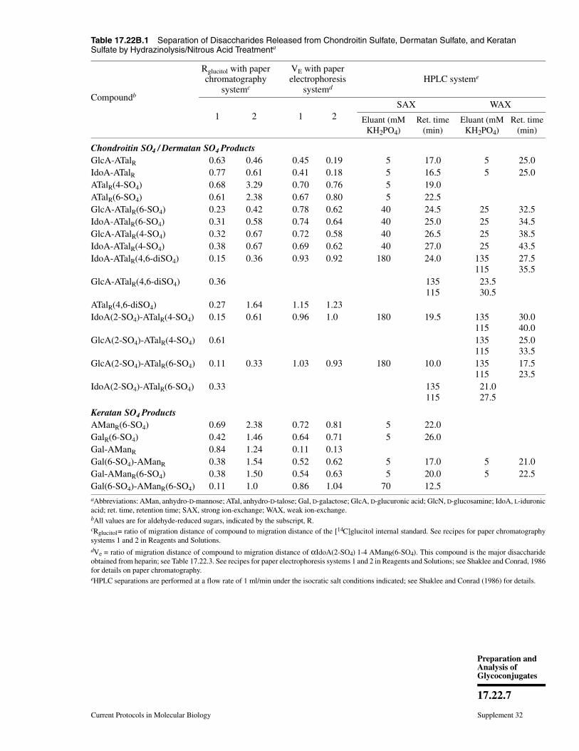
Table 17.22B.1 Separation of Disaccharides Released from Chondroitin Sulfate, Dermatan Sulfate, and KeratanSulfate by Hydrazinolysis/Nitrous Acid Treatmenta
Compoundb
Rglucitol with paperchromatography
systemc
VE with paperelectrophoresis
systemdHPLC systeme
1 2 1 2SAX WAX
Eluant (mMKH2PO4)
Ret. time(min)
Eluant (mMKH2PO4)
Ret. time(min)
Chondroitin SO4 / Dermatan SO4 ProductsGlcA-ATalR 0.63 0.46 0.45 0.19 5 17.0 5 25.0IdoA-ATalR 0.77 0.61 0.41 0.18 5 16.5 5 25.0ATalR(4-SO4) 0.68 3.29 0.70 0.76 5 19.0ATalR(6-SO4) 0.61 2.38 0.67 0.80 5 22.5GlcA-ATalR(6-SO4) 0.23 0.42 0.78 0.62 40 24.5 25 32.5IdoA-ATalR(6-SO4) 0.31 0.58 0.74 0.64 40 25.0 25 34.5GlcA-ATalR(4-SO4) 0.32 0.67 0.72 0.58 40 26.5 25 38.5IdoA-ATalR(4-SO4) 0.38 0.67 0.69 0.62 40 27.0 25 43.5IdoA-ATalR(4,6-diSO4) 0.15 0.36 0.93 0.92 180 24.0 135
11527.535.5
GlcA-ATalR(4,6-diSO4) 0.36 135115
23.5 30.5
ATalR(4,6-diSO4) 0.27 1.64 1.15 1.23IdoA(2-SO4)-ATalR(4-SO4) 0.15 0.61 0.96 1.0 180 19.5 135
11530.040.0
GlcA(2-SO4)-ATalR(4-SO4) 0.61 135115
25.033.5
GlcA(2-SO4)-ATalR(6-SO4) 0.11 0.33 1.03 0.93 180 10.0 135115
17.523.5
IdoA(2-SO4)-ATalR(6-SO4) 0.33 135115
21.0 27.5
Keratan SO4 ProductsAManR(6-SO4) 0.69 2.38 0.72 0.81 5 22.0GalR(6-SO4) 0.42 1.46 0.64 0.71 5 26.0Gal-AManR 0.84 1.24 0.11 0.13Gal(6-SO4)-AManR 0.38 1.54 0.52 0.62 5 17.0 5 21.0Gal-AManR(6-SO4) 0.38 1.50 0.54 0.63 5 20.0 5 22.5Gal(6-SO4)-AManR(6-SO4) 0.11 1.0 0.86 1.04 70 12.5aAbbreviations: AMan, anhydro-D-mannose; ATal, anhydro-D-talose; Gal, D-galactose; GlcA, D-glucuronic acid; GlcN, D-glucosamine; IdoA, L-iduronicacid; ret. time, retention time; SAX, strong ion-exchange; WAX, weak ion-exchange.bAll values are for aldehyde-reduced sugars, indicated by the subscript, R.cRglucitol = ratio of migration distance of compound to migration distance of the [14C]glucitol internal standard. See recipes for paper chromatographysystems 1 and 2 in Reagents and Solutions.dVe = ratio of migration distance of compound to migration distance of αIdoA(2-SO4) 1-4 AManR(6-SO4). This compound is the major disaccharideobtained from heparin; see Table 17.22.3. See recipes for paper electrophoresis systems 1 and 2 in Reagents and Solutions; see Shaklee and Conrad, 1986for details on paper chromatography.eHPLC separations are performed at a flow rate of 1 ml/min under the isocratic salt conditions indicated; see Shaklee and Conrad (1986) for details.
Current Protocols in Molecular Biology Supplement 32
17.22.7
Preparation andAnalysis ofGlycoconjugates

BASICPROTOCOL 2
ANALYSIS OF DISACCHARIDES AND OLIGOSACCHARIDES FROMGLYCOSAMINOGLYCANS BY HPLC
Samples to be analyzed by high-performance liquid chromatography (HPLC) are ob-tained by nitrous acid cleavage of N-deacetylated glycosaminoglycans at pH 4 (UNIT
17.22A), by direct cleavage of heparin or heparan sulfate with nitrous acid at pH 1.5 (UNIT
17.22A), or by enzymatic cleavage with lyases (UNIT 17.13B). For HPLC, all samples must befree of particulate matter and should be dissolved in the initial solvent used to elute theHPLC column.
Materials
Sample: mixture of saccharides obtained from glycosaminoglycan by nitrous acidcleavage (UNIT 17.22A) or lyase cleavage (UNIT 17.13B)
4.5-mm × 25-cm Partisil SAX strong anion-exchange column (Whatman) or 4-mm× 30-cm Varian MicroPak AX-5 weak anion-exchange column (VarianAnalytical)
HPLC solvents (see recipe and Tables 17.22B.1-17.22B.5)Gradient solutions for HPLC (Tables 17.22B.1-17.22B.5)Bio-Gel P-10 gel filtration column (Bio-Rad)4.6 × 250–mm Hi-Chrom S-5 ODS C-18 column (Regis Technology)Fraction collector or in-line radioactivity flow detector (e.g., Packard Instrument)
Additional reagents and equipment for gel-filtration chromatography (UNIT 10.9),ion-exchange HPLC (UNIT 10.13), and reversed-phase HPLC (UNIT 10.12)
Table 17.22B.2 Separation of Dissacharides Released from ChondroitinSulfate by Lyase Treatmenta
Compound
Rglucitol with paperchromatography systemb HPLC systemc
1 2SAX
retentiontime (min)
WAX
∆Di-OSR 1.0 0.95
∆Di-4SR 0.59 0.67 32.0 49.0∆Di-6SR 0.56 0.82 44.0 49.0∆Di-diSBR 0.26 0.46 61.0 67.0∆Di-diSDR 0.30 66.5 66.5
∆Di-diSER 0.26 0.41 68.0 70.0
aAbbreviations are: ∆Di-OSR, 2-acetamido-2-deoxy-3-O-(β-D-gluco-4-enepyranosyluronicacid)-D-galactitol; ∆Di-4SR, 2-acetamido-2-deoxy-3-O-(β-D-gluco-4-enepyranosyluronic acid)-4-O-sulfo-D-galactitol; ∆Di-6SR, 2-acetamido-2-deoxy-3-O-(β-D-gluco-4-enepyranosyluronicacid)-6-O-sulfo-D-galactitol;∆Di-diSBR, 2-acetamido-2-deoxy-3-O-(2-O-sulfo-β-D-gluco-4-enepyranosyluronic acid)-4-O-sulfo-D-galactitol; ∆Di-diSDR, 2-acetamido-2-deoxy-3-O-(2-O-sulfo-β-D-gluco-4-enepyranosyluronic acid)-6-O-sulfo-D-galact i tol ; ∆Di-diSER,2-acetamido-2-deoxy-3-O-(β-D-gluco-4-enepyranosyluronic acid)-4,6-di-O-sulfo-D-galactitol;SAX, strong ion-exchange; WAX, weak ion-exchange.bRglucitol = ratio of migration distance of compound to migration distance of [14C]glucitol internalstandards.cThe HPLC gradient for both SAX and WAX columns consisted of a 30-min isocratic segmentat 40 mM KH2PO4, followed by a 40-min linear gradient of 400 mM KH2PO4. HPLC separationsare performed at a flow rate of 1 ml/min.
Supplement 32 Current Protocols in Molecular Biology
17.22.8
Analysis ofDisaccharides and
TetrasaccharidesReleased fromGlycosamino-
glycans
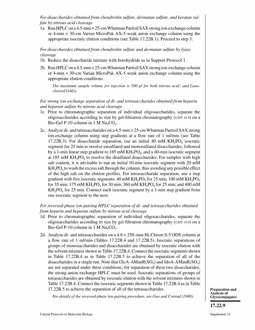
For disaccharides obtained from chondroitin sulfate, dermatan sulfate, and keratan sul-fate by nitrous acid cleavage1a. Run HPLC on a 4.5-mm × 25-cm Whatman Partisil SAX strong ion-exchange column
or 4-mm × 30-cm Varian MicroPak AX-5 weak anion exchange column using theappropriate isocratic elution conditions (see Table 17.22B.1). Proceed to step 3.
For disaccharides obtained from chondroitin sulfate and dermatan sulfate by lyasecleavage1b. Reduce the disaccharide mixture with borohydride as in Support Protocol 1.
2b. Run HPLC on a 4.5-mm × 25-cm Whatman Partisil SAX strong ion-exchange columnor 4-mm × 30-cm Varian MicroPak AX-5 weak anion exchange column using theappropriate elution conditions.
The maximum sample volume for injection is 500 l for both nitrous acid– and lyase-cleaved GAGs.
For strong ion-exchange separation of di- and tetrasaccharides obtained from heparinand heparan sulfate by nitrous acid cleavage1c. Prior to chromatographic separation of individual oligosaccharides, separate the
oligosaccharides according to size by gel-filtration chromatography (UNIT 10.9) on aBio-Gel P-10 column in 1 M Na2CO3.
2c. Analyze di- and tetrasaccharides on a 4.5-mm × 25-cm Whatman Partisil SAX strongion-exchange column using step gradients at a flow rate of 1 ml/min (see Table17.22B.3). For disaccharide separation, use an initial 40 mM KH2PO4 isocraticsegment for 25 min to resolve unsulfated and monosulfated disaccharides, followedby a 1-min linear step gradient to 185 mM KH2PO4, and a 40-min isocratic segmentat 185 mM KH2PO4 to resolve the disulfated disaccharides. For samples with highsalt content, it is advisable to run an initial 10-min isocratic segment with 20 mMKH2PO4 to wash the excess salt through the column, thus avoiding any possible effectof the high salt on the elution profiles. For tetrasaccharide separation, use a stepgradient with five isocratic segments: 40 mM KH2PO4 for 25 min; 100 mM KH2PO4
for 35 min; 175 mM KH2PO4 for 30 min; 360 mM KH2PO4 for 25 min; and 400 mMKH2PO4 for 25 min. Connect each isocratic segment by a 1-min step gradient fromone isocratic segment to the next.
For reversed-phase ion-pairing HPLC separation of di- and tetrasaccharides obtainedfrom heparin and heparan sulfate by nitrous acid cleavage1d. Prior to chromatographic separation of individual oligosaccharides, separate the
oligosaccharides according to size by gel-filtration chromatography (UNIT 10.9) on aBio-Gel P-10 column in 1 M Na2CO3.
2d. Analyze di- and tetrasaccharides on a 4.6 × 250–mm Hi-Chrom S-5 ODS column ata flow rate of 1 ml/min (Tables 17.22B.4 and 17.22B.5). Isocratic separations ofgroups of monosaccharides and disaccharides are obtained by isocratic elution withthe solvent mixtures shown in Table 17.22B.4. Connect the isocratic segments shownin Table 17.22B.4 as in Table 17.22B.5 to achieve the separation of all of thedisaccharides in a single run. Note that GlcA-AManR(SO4) and IdoA-AManR(SO4)are not separated under these conditions; for separation of these two disaccharides,the strong-anion exchange HPLC must be used. Isocratic separations of groups oftetrasaccharides are obtained by isocratic elution with the solvent mixtures shown inTable 17.22B.4. Connect the isocratic segments shown in Table 17.22B.4 as in Table17.22B.5 to achieve the separation of all of the tetrasaccharides.
For details of the reversed-phase ion-pairing procedure, see Guo and Conrad (1988).
Current Protocols in Molecular Biology Supplement 32
17.22.9
Preparation andAnalysis ofGlycoconjugates
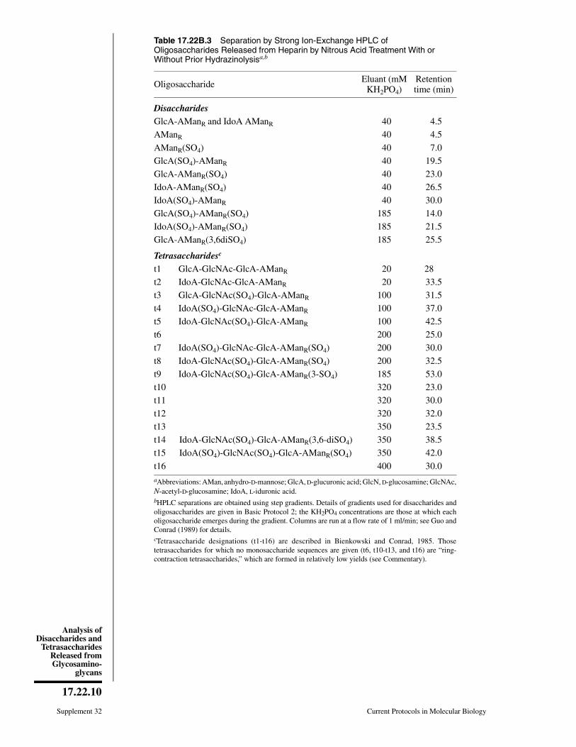
Table 17.22B.3 Separation by Strong Ion-Exchange HPLC ofOligosaccharides Released from Heparin by Nitrous Acid Treatment With orWithout Prior Hydrazinolysisa,b
Oligosaccharide Eluant (mMKH2PO4)
Retentiontime (min)
DisaccharidesGlcA-AManR and IdoA AManR 40 4.5
AManR 40 4.5
AManR(SO4) 40 7.0
GlcA(SO4)-AManR 40 19.5
GlcA-AManR(SO4) 40 23.0
IdoA-AManR(SO4) 40 26.5
IdoA(SO4)-AManR 40 30.0
GlcA(SO4)-AManR(SO4) 185 14.0
IdoA(SO4)-AManR(SO4) 185 21.5
GlcA-AManR(3,6diSO4) 185 25.5
Tetrasaccharidesc
t1 GlcA-GlcNAc-GlcA-AManR 20 28
t2 IdoA-GlcNAc-GlcA-AManR 20 33.5
t3 GlcA-GlcNAc(SO4)-GlcA-AManR 100 31.5
t4 IdoA(SO4)-GlcNAc-GlcA-AManR 100 37.0
t5 IdoA-GlcNAc(SO4)-GlcA-AManR 100 42.5
t6 200 25.0
t7 IdoA(SO4)-GlcNAc-GlcA-AManR(SO4) 200 30.0
t8 IdoA-GlcNAc(SO4)-GlcA-AManR(SO4) 200 32.5
t9 IdoA-GlcNAc(SO4)-GlcA-AManR(3-SO4) 185 53.0
t10 320 23.0
t11 320 30.0
t12 320 32.0
t13 350 23.5
t14 IdoA-GlcNAc(SO4)-GlcA-AManR(3,6-diSO4) 350 38.5
t15 IdoA(SO4)-GlcNAc(SO4)-GlcA-AManR(SO4) 350 42.0
t16 400 30.0aAbbreviations: AMan, anhydro-D-mannose; GlcA, D-glucuronic acid; GlcN, D-glucosamine; GlcNAc,N-acetyl-D-glucosamine; IdoA, L-iduronic acid.bHPLC separations are obtained using step gradients. Details of gradients used for disaccharides andoligosaccharides are given in Basic Protocol 2; the KH2PO4 concentrations are those at which eacholigosaccharide emerges during the gradient. Columns are run at a flow rate of 1 ml/min; see Guo andConrad (1989) for details.cTetrasaccharide designations (t1-t16) are described in Bienkowski and Conrad, 1985. Thosetetrasaccharides for which no monosaccharide sequences are given (t6, t10-t13, and t16) are “ring-contraction tetrasaccharides,” which are formed in relatively low yields (see Commentary).
Supplement 32 Current Protocols in Molecular Biology
17.22.10
Analysis ofDisaccharides and
TetrasaccharidesReleased fromGlycosamino-
glycans
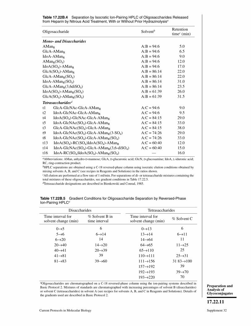
Table 17.22B.4 Separation by Isocratic Ion-Pairing HPLC of Oligosaccharides Releasedfrom Heparin by Nitrous Acid Treatment, With or Without Prior Hydrazinolysisa
Oligosaccharide Solventb Retention timec (min)
Mono- and DisaccharidesAManR A:B = 94:6 5.0GlcA-AManR A:B = 94:6 6.5IdoA-AManR A:B = 94:6 9.0AManR(SO4) A:B = 94:6 12.0IdoA(SO4)-AManR A:B = 94:6 17.0GlcA(SO4)-AManR A:B = 86:14 22.0GlcA-AManR(SO4) A:B = 86:14 22.0IdoA-AManR(SO4) A:B = 86:14 31.0GlcA-AManR(3,6diSO4) A:B = 86:14 23.5IdoA(SO4)-AManR(SO4) A:B = 61:39 26.0GlcA(SO4)-AManR(SO4) A:B = 61:39 31.5
Tetrasaccharidesd
t1 GlcA-GlcNAc-GlcA-AManR A:C = 94:6 9.0t2 IdoA-GlcNAc-GlcA-AManR A:C = 94:6 9.5t4 IdoA(SO4)-GlcNAc-GlcA-AManR A:C = 84:15 29.0t5 IdoA-GlcNAc(SO4)-GlcA-AManR A:C = 84:15 33.0t3 GlcA-GlcNAc(SO4)-GlcA-AManR A:C = 84:15 38.0t9 IdoA-GlcNAc(SO4)-GlcA-AManR(3-SO4) A:C = 74:26 29.0t8 IdoA-GlcNAc(SO4)-GlcA-AManR(SO4) A:C = 74:26 33.0t13 IdoA(SO4)-RC(SO4)IdoA(SO4)-AManR A:C = 60:40 12.0t14 IdoA-GlcNAc(SO4)-GlcA-AManR(3,6-diSO4) A:C = 60:40 15.0t16 IdoA-RC(SO4)IdoA(SO4)-AManR(SO4) — 16.0aAbbreviations: AMan, anhydro-D-mannose; GlcA, D-glucuronic acid; GlcN, D-glucosamine; IdoA, L-iduronic acid;RC, ring-contraction product.bHPLC separations are obtained using a C-18 reversed-phase column using isocratic elution conditions obtained bymixing solvents A, B, and C (see recipes in Reagents and Solutions) in the ratios shown.cAll elution are performed at a flow rate of 1 ml/min. For separations of di- or tetrasaccharide mixtures containing thetotal mixtures of these oligosaccharides, see gradient conditions in Table 17.22.5.dTetrasaccharide designations are described in Bienkowski and Conrad, 1985.
Table 17.22B.5 Gradient Conditions for Oligosaccharide Separation by Reversed-PhaseIon-Pairing HPLCa
Disaccharides Tetrasaccharides
Time interval for solvent change (min)
% Solvent B in time interval
Time interval for solvent change (min) % Solvent C
0→5 6 0→13 6
5→6 6→14 13→14 6→116→20 14 14→64 11
20→40 14→20 64→65 11→2540→41 20→39 65→110 25
41→81 39 110→111 25→3181→83 39→60 111→156 31 83→100
157→192 39
192→193 39→70193→220 70
aOligosaccharides are chromatographed on a C-18 reversed-phase column using the ion-pairing systems described inBasic Protocol 2. Mixtures of standards are chromatographed with increasing percentages of solvent B (disaccharides)or solvent C (tetrasaccharides) in solvent A (see recipes for solvents A, B, and C in Reagents and Solutions). Details ofthe gradients used are described in Basic Protocol 2.
Current Protocols in Molecular Biology Supplement 32
17.22.11
Preparation andAnalysis ofGlycoconjugates
3. Count radioactivity in 5-ml fractions collected in scintillation vials (see SupportProtocol 2) or use an in-line radioactivity flow detector. Identify disaccharides on thebasis of their chromatographic migration properties by referring to Tables 17.22B.1-17.22B.5. Quantify di- and tetrasaccharides (see Support Protocol 3).
SUPPORTPROTOCOL 1
BOROHYDRIDE REDUCTION OF ALKALI-LABILE DISACCHARIDESOBTAINED BY CLEAVAGE WITH LYASES
Disaccharides formed by cleavage of glycosaminoglycans with specific lyases (UNIT
17.13B) can be analyzed by reduction with NaB3H4 and separation of the labeled productseither by paper chromatography (Basic Protocol 1; Glaser and Conrad, 1979) or HPLC(Basic Protocol 2; Delaney et al., 1980; Shaklee and Conrad, 1986). However, thelyase-generated disaccharides are quite labile under the alkaline conditions required fortheir reduction with NaB3H4. Thus, a milder procedure than the 50°C reduction proceduredescribed in UNIT 17.22A must be used for the reduction. These labile disaccharides (orhigher oligosaccharides that may be obtained when the lyase does not convert theglycosaminoglycan completely to disaccharides) can be reduced at 0°C without cleavage.
Materials
Sample of lyase-degraded glycosaminoglycan1 M Na2CO3, pH 9.0 (ice-cold)Sodium borohydride reagent (see recipe)3 M H2SO4
6 × 150–mm test tubes
1. Place lyase-degraded GAG sample in a 6 × 150–mm test tube, dry sample in a streamof air, then chill tube in an ice bath.
2. Add 5 µl ice-cold 1 M Na2CO3 (pH 9.0) to sample, then add 10 µl of sodiumborohydride reagent. Cork or cap the tube and let it sit on ice 90 min.
IMPORTANT NOTE: If samples are already labeled by metabolically labeling of the GAG,0.5 M cold NaBH4 is used in preparing the sodium borohydride reagent instead of 0.5 MNaB[3H]4.
Carry out all NaB[3H]4 manipulations in a well-ventilated fume hood designated forradioactive use.
3. Add 2.5 µl of 3 M H2SO4 to destroy excess NaB3H4 and let sample stand ∼30 min.
4. Neutralize sample with 1 M Na2CO3 and dry it in a stream of air. Redissolve samplein water, then dry again to remove excess [3H]2 gas. Finally dissolve sample in 50 µlwater.
Aliquots of the sample may be analyzed by paper chromatography (Basic Protocol 1) orHPLC (Basic Protocol 2).
Supplement 32 Current Protocols in Molecular Biology
17.22.12
Analysis ofDisaccharides and
TetrasaccharidesReleased fromGlycosamino-
glycans
SUPPORTPROTOCOL 2
SCINTILLATION COUNTING OF FRACTIONS FROM HPLC ANALYSIS OFSACCHARIDES RELEASED FROM GLYCOSAMINOGLYCANS
Effluent from the HPLC column is collected in 0.5-ml fractions in scintillation vials usinga fraction collector. Alternatively the effluent may be run directly through an in-lineradioactivity flow detector (e.g., Radiomatic). If the total cpm in each peak are highenough to obtain good counting statistics in short counting intervals, a stream splitter maybe used to collect the bulk of each fraction (e.g., 0.4 ml) in a test tube while only a portionof each fraction (e.g., 0.1 ml) is taken in scintillation vials for counting. Add an appropriatevolume (4 to 5 ml) of any commercial scintillation fluid designed to accommodate thesalt and solvent concentrations in the eluants. Alternatively, add 4 ml of a scintillationcocktail containing 3 g diphenyloxazole in a mixture of 250 ml Triton X-114, 750 mlxylene, and 10 ml concentrated HCl. For KH2PO4 concentrations >10 mM, add 1 dropconcentrated HCl to the xylene-based scintillation fluid to to redissolve the salt precipitatethat begins to form in the scintillation vial.
SUPPORTPROTOCOL 3
CALCULATIONS FOR QUANTITATION OF DISACCHARIDES
Procedures described here allow calculation of the number of µmol of each disaccharidein the original sample taken for analysis. These calculations apply primarily to analysesin which disaccharides obtained from unlabeled glycosaminoglycans are mixed with a[14C]D-glucose internal standard and reduced with NaB3H4 (see UNIT 17.22A), but can alsobe applied to metabolically labeled materials if the specific radioactivity of the radiola-beled precursor that appears in the metabolically labeled glycosaminoglycan is deter-mined independently .
Calibrate NaB[3H]4 used in the analysis of unknownsBecause the counting efficiency for 3H varies in each type of counting experiment,cpm/µmol of a standard reducing sugar must be determined separately for each type ofseparation procedure. Mix a measured amount of a standard sugar (usually D-glucose)with the [14C]D-glucose internal standard (see UNIT 17.22A). Enough [14C]glucose should beadded so that ∼1 × 10⁵ 14C cpm will be recovered in the final separation step. Reduce analiquot of this mixture with the same NaB[3H]4 reagent to be used in the analysis (seeUNIT 17.22A). If NaB[3H]4 with a specific activity of ∼500 mCi/mmol (recommended) isused, the [3H]D-glucitol formed by reduction of the D-glucose should have a specificactivity of 125 mCi/mmol, and 1 µmol of the standard D-glucose should yield 778 × 106
dpm per µmol of [3H]glucitol. Counting efficiency for 3H in some of the separation stepsmay be as low as 5%. Chromatograph the resulting [3H]D-glucitol/[14C]glucitol mixturein the same type of analytical separation procedure that is being used for the unknowns(paper chromatography or HPLC). Determine the number of [14C]glucitol and [3H]gluci-tol cpm in the separated peak by scintillation counting under conditions optimized fordual-label counting. The 3H cpm must be corrected for the number of 14C cpm that spillover into the 3H counting channel. From the number of [14C]glucitol cpm, calculate thenumber of µmol of D-glucitol in the peak as described below.
When a known number of µmol of glucose are mixed with a known number of cpm ofthe [14C]glucose internal standard, the number of 14C cpm recovered in the analytical stage(e.g., in paper chromatography or HPLC) can be used to calculate the proportion oforiginal µmol of the standard glucose that are recovered in the analytical stage. Forexample, if the original D-glucose solution contained 1 × 10⁵ µmol of the D-glucosestandard and 5 × 10 5 cpm of [14C]glucose, and if 1 × 10⁵ cpm of [14C]glucitol wererecovered in the analytical run, then 2 × 10⁴ µmol of the D-glucitol standard would berecovered in the analytical run. Thus, if 1 × 10⁹ 3H cpm in the [3H]glucitol were recoveredin the analytical run, the number of 3H cpm/µmol for glucose would be 5 × 10⁴.
Current Protocols in Molecular Biology Supplement 32
17.22.13
Preparation andAnalysis ofGlycoconjugates

Using the [3H]glucitol cpm in the separated peak and the number of µmol of D-glucitolin the peak, calculate the number of [3H]glucitol cpm/µmol, which will be the same asthe number of 3H cpm/µmol for all mono-, di- or tetrasaccharides obtained in the analyticalrun, as all reducing sugars incorporate the same number of equivalents of 3H per µmolfrom the NaB[3H]4. Consequently, when the total number of 3H cpm is determined forany peak in the analytical run, division of this number by the 3H cpm/µmol of the D-glucosestandard yields the value for the number of µmol of the compound in the peak.
Calibrate internal standard [14C]glucoseBecause the counting efficiency for 14C varies in each type of counting experiment, thenumber of 14C cpm in a given aliquot of the [14C]glucose internal standard must bedetermined separately for each type of separation procedure. Reduce an aliquot of the[14C]glucose internal standard using the same reduction conditions used for the unknowns(see UNIT 17.22A for nitrous acid cleavage products; see Basic Protocol 2 for lyase cleavageproducts). To calibrate [14C]glucose for paper chromatography, transfer the resulting[14C]glucitol solution quantitatively to a paper chromatography strip and run the chroma-togram in the solvent for which the [14C]glucose calibration is being made. Develop thechromatogram, cut the strip into 0.5-in. segments for scintillation counting, then deter-mine the total number of 14C cpm in the original aliquot taken for analysis.
To calibrate [14C]glucose for HPLC, inject a measured aliquot of the [14C]glucitol solutionquantitatively into an HPLC column injector, elute the column as in a typical analyticalrun, then determine the number of 14C cpm in the [14C]glucitol peak. The [14C]glucosewill emerge in the void volume of the column in the HPLC procedures described here.
Chromatographic and electrophoretic properties of various glycosaminoglycan oligosac-charides are listed in Tables 17.22B.1-17.22B.5.
REAGENTS AND SOLUTIONS
Use deionized, distilled water in all recipes and protocol steps. For common stock solutions, seeAPPENDIX 2; for suppliers, see APPENDIX 4.
HPLC solvents
For anion-exchange chromatography: HPLC solvents for chromatography on weakanion-exchange or strong ion-exchange columns range from 0 mM to 400 mMKH2PO4. Degas and filter 1 liter distilled water by gently heating it to 80°C in a2-liter vacuum-filtration flask, sealing the flask with a stopper, then placing it in anice-water bath and evacuating with a water aspirator while gently stirring with amagnetic stirrer. Continue aspirating until no further bubbles are apparent (15 min),then transferto a volumetric flask containing the amount of KH2PO4 required to givethe desired concentration. Gently stir the solution, then filter through a 0.45-µmMillipore filter under vacuum into a clean 500-ml bottle, which will serve as areservoir for the solvent.
For HPLC systems that allow continuous degassing during the run, it is not necessary todegas solvents before making the solutions.
For reversed-phase ion-pairing chromatography: Prepare solvents A, B, and C asfollows. Mix or deliver in gradient as described in Table 17.22B.4 and Table17.22B.5.
Solvent A: Prepare 38 mM NH4H2PO4, 2 mM H3PO4, and 1 mM tetrabutylam-monium phosphate in water. Adjust pH to 3.6 with 0.1 M NaOH or HCl.
Solvent B: Prepare 38 mM NH4H2PO4, 2 mM H3PO4, and 1 mM tetrabutylam-monium phosphate in 30/70 (v/v) acetonitrile/water. Adjust pH to 4.2 with 0.1 MNaOH or HCl.
continued
Supplement 32 Current Protocols in Molecular Biology
17.22.14
Analysis ofDisaccharides and
TetrasaccharidesReleased fromGlycosamino-
glycans

Solvent C: Prepare 38 mM NH4H2PO4, 2 mM H3PO4, and 1 mM tetrabutylam-monium phosphate in 50:50 (v/v) acetonitrile /water. Adjust pH to 4.4 with 0.1 MNaOH or HCl.
Paper chromatography and paper electrophoresis systems
Use the following paper chromatography or paper electrophoresis systems inconjunction with the data in Table 17.22B.1 or Table 17.22B.2:
Paper chromatography system 1: 3:2:1 (v/v/v) 1-butanol/glacial acetic acid/1 MNH4OH. Use 1 × 22–in. Whatman no. 3 paper chromatography strips.
Paper chromatography system 2: 3:2:1 (v/v/v) ethyl acetate/pyridine/5 mM boricacid. Use 1 × 22–in. Whatman cellulose phosphate paper chromatography strips.
Paper electrophoresis system 1: 50 ml glacial acetic acid and 10 ml pyridine in 4liters water (pH 4.0). Use 1 × 22–in. cellulose phosphate paper chromatographystrips.
Paper electrophoresis system 2: 350 ml of glacial acetic acid and 100 ml of 88%formic acid in 4 liters water (pH 1.7). Use 1 × 22–in. cellulose phosphate paperchromatography strips.
Sodium borohydride reagent
Just prior to use, dissolve 1.89 mg NaB[3H]4 in 100 µl chilled 0.2 M Na2CO3, pH10.2, to give a 0.5 M NaB[3H]4 solution. Keep on ice until use.
COMMENTARY
Background InformationA number of high-performance liquid chro-
matography procedures have been developedto separate and quantitate the disaccharidesformed by deaminative cleavage of N-deacety-lated glycosaminoglcans with nitrous acid (UNIT
17.22A) or cleavage with lyases (UNIT 17.13B).These include chromatography on strong an-ion-exchange columns (SAX; Bienkowski andConrad, 1985); weak anion exchange columns(WAX; Shaklee and Conrad, 1986); and re-versed-phase ion-pairing columns (Guo andConrad, 1988). Alternatively, some of the di-saccharides can be separated by paper chroma-tography (Hopwood and Elliot, 1983; Hop-wood and Muller, 1983), paper electrophoresis(Hopwood and Elliot, 1983; Hopwood andMuller, 1983), thin-layer chromatography(Edge and Spiro, 1985), or capillary-zone elec-trophoresis (Al-Hakim and Linhardt, 1991; De-sai et al., 1993). When lyases are used to cleavethe glycosaminoglycan (UNIT 17.13B), the lyase-derived di- and oligosaccharides can be assayedby the absorbance of their unsaturated uronicacid residues at 233 nm. This UV assay hasbeen used for detection of of oligosaccharideseluted from SAX HPLC columns with a linearsodium chloride gradient running from 0 to 1M (Rice et al., 1985; Linhardt et al., 1988).Glycosaminoglycans that have been metabo-
lically labeled with [3H]D-glucosamine or[35S]O4
2− yield disaccharides with the corre-sponding labels. Disaccharides from both typesof cleavage can also be detected and quantifiedfollowing their reduction with NaB[3H]4 toincorporate one gram atom of 3H per mol ofdisaccharide. In this approach, the elution po-sitions of the 3H-labeled disaccharide peaksseparated by HPLC, paper chromatography, orpaper electrophoresis are used to identify thedisaccharides, and the 3H cpm recovered ineach peak are used to obtain ratios of disaccha-rides or quantitative measures of the amountsof each disaccharide present in the glycosami-noglycan.
Critical ParametersThe important parameters for the separa-
tions described in this unit are those that mustnormally be observed for chromatographicseparations. The critical parameters for the ana-lytical procedures are primarily those involvedin the treatment with nitrous acid andNaB[3H]4. These are described in UNIT 17.22A. Itmay be noted that the elution times given inTables 17.22B.1-17.22B.5 may vary some-what, but the relative positions of elution re-main the same. Positive identification may re-quire chromatographic runs of mixtures of pre-viously characterized disaccharides before or
Current Protocols in Molecular Biology Supplement 32
17.22.15
Preparation andAnalysis ofGlycoconjugates

after the sample run. As columns age, the elu-tion times become shorter and resolution be-comes poorer. When resolution becomes unsat-isfactory, the column must be replaced.
Anticipated ResultsThe nitrous acid reaction (see UNIT 17.22A) is
virtually stoichiometric for β-linked aminosugars (Shively and Conrad, 1970). Conse-quently, the yield of disaccharides isstoichiometric from those glycosaminoglycansthat contain β-linked amino sugars (i.e., chon-droitin sulfate, dermatan sulfate, hyaluronicacid, and keratan sulfate). However, a side re-action called the “ring-contraction reaction”occurs to the extent of 10% to 15% for α-linkedamino sugars—e.g., those found in heparin andheparan sulfate (Shively and Conrad, 1970,1976). The ring-contraction reaction results indeamination without bond cleavage, and mayconvert a segment in heparin that contains twodisaccharides having N-sulfated GlcN residuesinto a tetrasaccharide fragment instead of thetwo disaccharides that would be expected.Thus, heparin and heparan sulfate yield smallbut significant amounts of the ring-contractiontetrasaccharides (Bienkowski and Conrad,1985). However, there seems to be little struc-tural selectivity for the ring-contraction reac-tion, as the ring-contraction tetrasaccharidescontain disaccharide units in the same propor-tions as those found in the original glycosami-noglycan. Thus, even for heparin and heparansulfate, the disaccharides formed during nitrousacid cleavage are obtained in the same propor-tions that are found in the original glycosami-noglycan. Consequently, when yields are ex-pressed in percent of total uronic acids or per-cent of total amino sugars, the ring-contractionreaction does not interfere with the quantita-tion, and the yields of di- and monosaccharidesare truly representative of their proportions inthe starting glycosaminoglycan. Some of theseheparin ring-contraction tetrasaccharides havebeen identified (Bienkowski and Conrad,1985).
Time ConsiderationsThe times for chromatography are apparent
from the retention times reported in Tables17.22B.1 to 17.22B.3. Time for processing ofthe data will depend in great part on the type ofscintillation counter used and the data-process-ing equipment available on site where the meth-ods will be used.
Literature CitedAl-Hakim, A. and Linhardt, R.J. 1991. Capillary
Electrophoresis for the analysis of chondroitinsulfate and dermatan sulfate-derived disaccha-rides. Anal. Biochem. 195:68-73.
Bienkowski, M.J. and Conrad, H.E. 1985. Structuralcharacterization of the oligosaccharides formedby depolymerization of heparin with nitrousacid. J. Biol. Chem. 260:356-365.
Delaney, S.R., Conrad, H.E., and Glaser, J.H. 1980.A new approach for isolation and sequencing ofpure chondroitin SO4 oligosaccharides. Anal.Biochem. 108:25-34.
Desai, U.R., Wang, H.-M., Ampofo, S.A., Linhardt,R.J. 1993. Oligosaccharide composition of hepa-rin and low-molecular-weight heparins by capil-lary electrophoresis. Anal. Biochem. 213:120-127.
Edge, A.S.B., and Spiro, R.G. 1985. Structural Elu-cidation of glycosaminoglycans through charac-terization of disaccharides obtained after frag-mentation by hydrazine-nitrous acid treatment.Arch. Biochem. Biophys. 240:560-572.
Glaser, J.G., and Conrad, H.E. 1979. Chick embryoliver B-glucuronidase. Comparison of activityon natural and artificial substrates. J. Biol. Chem.254:6588-6597.
Guo, Y. and Conrad, H.E. 1988. Analysis of oli-gosaccharides from heparin by reversed phaseion-pairing high performance liquid chromatog-raphy. Anal. Biochem. 168:54-62.
Guo, Y. and Conrad, H.E. 1989. The disaccharidecomposition of heparins and heparan sulfates.Anal. Biochem. 176:96-104.
Hopwood, J.J. and Elliott, H. 1983. Selective depo-lymerisation of keratan sulfate: Production ofradiolabeled substrates for 6-O-sulfogalactosesulfatase and β-D-galactosidase. Carbohydr.Res. 117:263-274.
Hopwood, J.J. and Muller, V.J. 1983. Selective de-polymerisation of dermatan sulfate: Productionof radiolabelled substrates for α-L-iduronidase,sulfoiduronate sulfatase, and β-D-glucuronidase.Carbohydr. Res. 122:227-239.
Linhardt, R.J., Rice, K.G., Kim, Y.S., Lohse, D.L.,Wang, H.M., and Loganathan, D. 1988. Mappingand quantification of the major oligosaccharidecomponents of heparin. Biochem. J. 254:781-787.
Rice, K.G., Kim, Y.S., Grant, A.C., Merchant, Z.M.,and Linhardt, R.J. 1985. High-performance liq-uid chromatographic separation of heparin de-rived oligosaccharides. Anal. Biochem. 150:325-331.
Shaklee, P.N. and Conrad, H.E. 1986. The disaccha-rides formed by deaminative cleavage of N-deacetylated glycosaminoglycans. Biochem. J.235:225-236.
Supplement 32 Current Protocols in Molecular Biology
17.22.16
Analysis ofDisaccharides and
TetrasaccharidesReleased fromGlycosamino-
glycans
Shively, J.E. and Conrad, H.E. 1970. Stoichiometryof the nitrous acid deaminative cleavage ofmodel amino sugar glycosides and glycosamino-glycuronans. Biochemistry 9:33-41.
Shively, J.E. and Conrad, H.E. 1976. Formation ofanhydrosugars in the chemical depolymerizationof heparin. Biochemistry 15:3932-3942.
Key ReferencesBienkowski and Conrad, 1985. See above.
Describes the separation of heparin di- andtetrasaccharides on SAX columns.
Guo and Conrad, 1988. See above.
Describes the separation of heparin di- and tetra-saccharides by reversed-phase ion-pairing HPLC.
Shaklee and Conrad, 1986. See above.
Describes the separation of disaccharides fromchondroitin sulfate, dermatan sulfate, and keratansulfate formed by both nitrous acid and lyase cleav-age.
Contributed by H. Edward ConradUniversity of IllinoisUrbana, Illinois
Current Protocols in Molecular Biology Supplement 32
17.22.17
Preparation andAnalysis ofGlycoconjugates

UNIT 17.23Analysis of Sulfate Esters by Solvolysis orHydrolysisSulfate esters are found on N- and O-linked sugar chains or glycosaminoglycan (GAG)chains. Few sulfatases are available that can enzymatically remove them, so chemicalprocedures must be used. These procedures rely on the differential sensitivity of sulfateslocated in different linkages on the sugar. In comparison to the conditions used forenzymatic digestion, those used for chemical digestion are very harsh and cannot be usedon protein-bound carbohydrates (except for analytical purposes, as in Basic Protocol 2).With protein-bound carbohydrates, for preparative purposes, the chains must first bereleased by one of the procedures described in UNITS 17.13 & 17.15. Basic Protocol 1 describesrelease of sulfate esters by solvolysis. A Support Protocol describes a method formonitoring the efficiency of the solvolysis reaction, and Alternate Protocol 1 provides ascale-up method for using the solvolysis reaction with large amounts of material. BasicProtocol 2 presents a technique for acid hydrolysis to release sulfate esters, and AlternateProtocol 2 describes basic hydrolysis for the same purpose.
BASICPROTOCOL 1
SOLVOLYSIS FOR RELEASE OF SULFATE ESTERS FROM GLYCANS
Solvolysis is analogous to hydrolysis, except that a nonaqueous solvent is used. Bothsolvolysis procedures in this unit (also see Alternate Protocol 1) require conversion of thesulfated molecule to the pyridinium salt before heating in dimethyl sulfoxide (DMSO) toremove sulfate. This procedure is for 35SO4- or 3H-radiolabeled GAG samples, and appliesto microgram amounts of material. For scale-up, see Alternate Protocol 1.
Materials
Sulfated sample (UNIT 17.15), radiolabeled (UNIT 17.4 or UNIT 17.5)Glucose-6-sulfate (Sigma)0.5 × 50–cm Sephadex G-25 gel-filtration column (Pharmacia Biotech)2-cm column of Dowex-50 X8 (hydrogen form; see recipe for washing procedure)
packed in a Pasteur pipet plugged with glass woolPyridine1 M NaCl9:1 (v/v) dimethyl sulfoxide (DMSO; reagent grade)/methanolQAE-Sephadex (Pharmacia Biotech)
Thick-walled conical glass tubesHeating blockLyophilizer or shaker-evaporator
Additional reagents and equipment for desalting by gel-filtration chromatography(UNIT 10.9)
1. Desalt sample on a 0.5 × 50–cm Sephadex G-25 column, eluting with water.
It is essential to remove all nonvolatile salts from the sample. If free 35SO4 is present in thesample, sulfate will run through the water-eluted Sephadex G-25 column with an apparentmolecular weight much larger than its actual molecular weight, and may overlap withsmall 35SO4-labeled oligosaccharides. It may be therefore be necessary to remove free35SO4 by dialysis from the protein-bound 35SO4, prior to the release of the carbohydratechains from the protein.
2. Add 5 µg of glucose-6-sulfate to the sample, which will be in a volume of 1 to 5 mlof water. Pass the sample through a 2-cm column of water-washed Dowex-50 X8(hydrogen form) packed in a glass wool–plugged Pasteur pipet. Wash column with
Supplement 32
Contributed by Hudson H. FreezeCurrent Protocols in Molecular Biology (1995) 17.23.1-17.23.8Copyright © 2000 by John Wiley & Sons, Inc.
17.23.1
Preparation andAnalysis ofGlycoconjugates
5 ml water and combine washings with the original flowthrough of the column. CheckpH of the combined washings with pH paper; it should be within one unit of the pHof the combined sample and wash water that will be used in the subsequent steps.
The Dowex-50 column removes any residual cations and replaces them with protons. Theglucose-6-sulfate provides further insurance that any residual cations will not interferewith the solvolysis reaction, but the amount of this reagent must be kept low to preventunwanted side reactions.
3. Add 50 µl pyridine to the sample and split it into two aliquots. Add 2 µl of 1 M NaClto one sample and nothing to the other. Transfer samples to thick-walled glass conicaltubes and lyophilize or dry on a shaker-evaporator.
The sample with the NaCl serves as a negative control for desulfation and for the sideeffects of the DMSO-treatment step (step 4), because solvolysis does not proceed when thesalt is present. In the experimental (i.e., no-salt) sample, most of the pyridine evaporatesduring lyophilization, except for that which forms the pyridinium salt with the sample orglucose-6-sulfate.
4. Add 0.2 ml of 9:1 (v/v) DMSO/methanol to each sample. Using a heating block, heat2 hr at 85° to 90°C for O-sulfate esters or at 55°C for N-sulfate esters.
5. Remove DMSO under vacuum using a lyophilizer or shaker-evaporator.
Depending upon the next step—e.g., in the case of QAE-Sephadex chromatography (UNIT
17.20)—it may not be necessary to remove the solvent. If it is necessary to do this, it shouldbe noted that DMSO is not very volatile and will take several hours to evaporate undervacuum. In some cases the sample may need to be diluted.
SUPPORTPROTOCOL 1
MONITORING SOLVOLYSIS REACTION
If it is important to monitor the efficiency of the reaction in Basic Protocol 1, this can bedone by running a sample of [3H]glucitol-6-sulfate in parallel with the experimentalsamples.
Additional Materials (also see Basic Protocol 1)
[3H]glucitol-6-sulfate: glucose-6-sulfate (Sigma) reduced with NaB[3H]4 (UNIT 17.5)2-cm QAE-Sephadex column (Pharmacia Biotech)35 mM and 200 mM NaClScintillation fluid compatible with aqueous samples
Additional reagents and equipment for QAE-Sephadex analysis (UNIT 17.20)
1. Carry out steps 1 and 2 of Basic Protocol 1, running ∼10,000 cpm of [3H]glucitol-6-sulfate in parallel with the experimental sample. In step 3, split the pyridine treatedcolumn effluent from this glucitol-6-sulfate sample into three aliquots instead of two.Add 2 µl of 1 M NaCl to one aliquot. Dry all three aliquots as described in BasicProtocol step 3. Keep one of the non–salt-treated aliquots in reserve, then carry outsteps 4 and 5 (solvolysis) on the remaining salt-treated and non–salt-treated aliquots.
Alternatively, this 3H-labeled material may be added directly to the sample itself as aninternal control. This cannot, however, be done if it interferes with subsequent analysis ofthe sample products (e.g., if the sample itself is labeled with 3H).
2. Dilute the three [3H]glucitol samples to 1.5 ml with water and treat each of them asfollows. Load on a 2-cm QAE-Sephadex column. Wash column three times using 1.5ml of water each time and collect each effluent in a scintillation vial. Wash three moretimes using 1.5 ml of 35 mM NaCl each time, and collect the effluents in three more
Supplement 32 Current Protocols in Molecular Biology
17.23.2
Analysis ofSulfate Esters by
Solvolysis orHydrolysis
separate vials. Finally, wash once with 1.5 ml of 200 mM NaCl and collect the effluentin another vial.
Ten vials will thus be obtained for each of the three [3H] glucitol samples.
3. Add 15 ml of an aqueous-compatible scintillation fluid to each vial and count on ascintillation counter.
The [3H] glucitol in untreated sample and the solvolysis control incubation should elutein the 35mM NaCl wash. The [3H] glucitol in solvolyzed desulfated sample should elutewith water showing that it no longer carries a charge. Very little radioactivity will be seenin the high-salt wash. The efficiency of sulfation is then calculated as the percentage of[3H] glucitol eluted with water compared to the total [3H] glucitol recovered.
ALTERNATEPROTOCOL 1
SCALE-UP OF SOLVOLYSIS FOR LARGE AMOUNTS OF MATERIAL
Basic Protocol 1 is used to desulfate trace amounts of material, but it can be applied tolarger amounts of material such as glycosaminoglycans (GAGs). As with Basic Protocol1, the extent of the reaction can be monitored by the desulfation of [3H]glucitol-6-sulfateif the presence of this labeled compound will not interfere with the subsequent use of theproduct. The steps and the preparations of the procedure are essentially the same asdescribed for the Basic Protocol 1, except that no control incubation (i.e., with NaCl; seeBasic Protocol 1, step 3) is done on the material. This procedure describes the solvolysisof 10 mg of GAG (or other sulfated sugar chains) and can be further scaled up if necessary.
Additional Materials (also see Basic Protocol 1)
[3H]glucitol-6-sulfate: glucose-6-sulfate (Sigma) reduced with NaB[3H]4 (UNIT 17.5)Dialysis tubing (3000 MWCO)
Additional reagents and equipment for dialysis (APPENDIX 3C)
1. Desalt material containing 10 mg GAGs or other sulfated molecules on a SephadexG-25 column and pool the fractions that contain GAGs.
Free GAG chains can be monitored by the carbazole reaction (see UNIT 17.12) or by aminosugar tests. Follow the normal procedures for gel filtration and pool conservatively to avoidany salt.
2. Mix material with an appropriate amount of [3H]glucitol-6-sulfate and pass themixture over a 5 × 0.5–cm column of well-washed Dowex-50 X8 (hydrogen form).Wash with 2 to 3 column volumes water, then add 0.1 ml pyridine to the combinedeluates. Lyophilize.
The amount of [3H]glucitol-6-sulfate to be added will depend upon the fraction of the totalsample that can be sacrificed for the QAE-Sephadex analysis described in Support Protocol1. The pH of the eluate may be lower than that found in Basic Protocol 1 because the chains,which will be in protonated form, are found at a higher concentration.
3. Dissolve the lyophilized material in 1.0 ml of 90% dimethyl sulfoxide/10% methanoland heat 2 hr at 90°C.
The [3H]glucitol-6-sulfate can be used to confirm desulfation of the GAG chains and tofollow the course of the reaction. Take aliquots at different times and monitor its elutionfrom QAE-Sephadex as described in Support Protocol 1. Use <1% of the sample (100 g)so that the 2-cm QAE-Sephadex column is not overloaded (see Support Protocol 1). TheGAG chains may remain bound to the column, but this does not matter because only thedesulfation of the [3H]glucitol-6-sulfate is monitored.
Current Protocols in Molecular Biology Supplement 32
17.23.3
Preparation andAnalysis ofGlycoconjugates

4. Cool the reaction tube. Dialyze the sample against 3 liters of water using 3000-MWCO dialysis tubing.
Alternatively, remove the reagents on a Sephadex G-25 gel-filtration column. Either proce-dure will work, but the choice is determined by the future plans for the sample. The apparentsize of the molecules will decrease because the sulfate contributes disproportionately to theirapparent size, and some breakdown of chains may also occur.
BASICPROTOCOL 2
ACID HYDROLYSIS FOR RELEASE OF SULFATE ESTERS FROMGLYCOSAMINOGLYCANS
This protocol measures the amount of bound 35SO4 that remains soluble after thepreciptation of hydrolyzed sulfate as the barium salt. In this procedure, we assume thatthe released oligosaccharide is 35SO4-labled, but the sugars may also be labeled with 3H.If the sugars are not labeled with tritium, a tritiated molecule—e.g., 3H-labeled glucose-6-sulfate should be included as an internal standard for normalizing recovery of thematerial.
Materials
Sample: 35SO4-labeled oligosaccharide or glycopeptide0.50 N HCl (freshly diluted from 12 N reagent grade acid)0.50 N NaOH (accurately titrated)4 M NaCl0.01 N HCl in 1 M NaCl (diluted from 0.50 N HCl above)2 mM Na2SO4 in 2 M KCl20 mM barium acetateHeating block with wells filled with oil
1. Dispense ≥1000 to 2000 cpm of 35SO4-labeled oligosaccharide or glycopeptide in 50µl water into a series of 8 to 10 microcentrifuge tubes. Add 50 µl of 0.50 N HCl toeach tube.
2. Incubate the tubes in a heating block with wells filled with oil at 98° to 100°C for aseries of different time periods.
The amount of time will vary depending upon the sample. A preliminary experiment withtime points at 1 and 3 hr will help determine the best time points to try. All sulfate esterswill eventually be hydrolyzed and even the most resistant ones will be 75% degraded after3 hr. A reasonable set of time points to try is 0, 5, 15, 30, 60, 90, 120, 150, and 180 min.
3. At each time point, remove the tube from the oil, wipe it clean and place it on ice for1 min. Microcentrifuge 2 sec at maximum speed to collect the liquid in the bottomof the tube and add sufficient 0.5 N NaOH to neutralize the sample. Also add 50 µlof 4 M NaCl to maintain high salt concentration. Keep completed samples on icewhile remaining samples are incubating.
Determine the exact volume of 0.5 N NaOH (∼50 ) to add to the samples by firstneutralizing a dummy sample containing 50 l of 0.50 N HCl and 0.50 N water andchecking with pH paper. Acceptable pH after neutralization is 3 to 9.
4. After the time course has been completed for all samples and all samples have beenneutralized, add sufficient 0.01 N HCl in 1 M NaCl to each sample to adjust the pHto 2 to 3. Check with pH paper using 1 to 2 µl of sample.
Determine the exact amount of 0.01 N HCl (∼100 l) to add by adjusting the pH of thedummy sample neutralized above (see step 3, annotation). It is still necessary, however, tocheck the pH of the actual samples.
Supplement 32 Current Protocols in Molecular Biology
17.23.4
Analysis ofSulfate Esters by
Solvolysis orHydrolysis
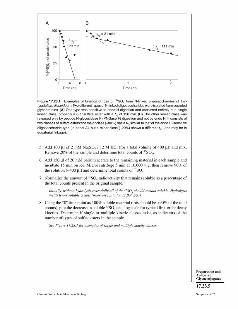
5. Add 100 µl of 2 mM Na2SO4 in 2 M KCl (for a total volume of 400 µl) and mix.Remove 20% of the sample and determine total counts of 35SO4.
6. Add 150 µl of 20 mM barium acetate to the remaining material in each sample andincubate 15 min on ice. Microcentrifuge 5 min at 10,000 × g, then remove 90% ofthe solution (∼400 µl) and determine total counts of 35SO4.
7. Normalize the amount of 35SO4 radioactivity that remains soluble as a percentage ofthe total counts present in the original sample.
Initially, without hydrolysis essentially all of the 35SO4 should remain soluble. Hydrolysisyields fewer soluble counts (more precipitaiton of Ba35SO4).
8. Using the “0" time point as 100% soluble material (this should be >90% of the totalcounts), plot the decrease in soluble 35SO4 on a log scale for typical first-order decaykinetics. Determine if single or multiple kinetic classes exist, as indicators of thenumber of types of sulfate esters in the sample.
See Figure 17.23.1 for examples of single and multiple kinetic classes.
t1/2 = 31 min
t1/2 = 111 min
0 1 2Time (hr)
100
50
25
00 4 8
t1/2 =
120 min
Time (hr)
%[3
5 S]0
4 n
ot p
reci
pita
ted
A B
Figure 17.23.1 Examples of kinetics of loss of 35SO4 from N-linked oligosaccharides of Dic-tyostelium discoideum. Two different types of N-linked oligosaccharides were isolated from secretedglycoproteins. (A) One type was sensitive to endo H digestion and consisted entirely of a singlekinetic class, probably a 6-O sulfate ester with a t1⁄2 of 120 min. (B) The other kinetic class wasreleased only by peptide:N-glycosidase F (PNGase F) digestion and not by endo H. It consists oftwo classes of sulfate esters: the major class (∼80%) has a t1⁄2 similar to that of the endo H–sensitiveoligosaccharide type (in panel A), but a minor class (∼20%) shows a different t1⁄2 (and may be inequatorial linkage).
Current Protocols in Molecular Biology Supplement 32
17.23.5
Preparation andAnalysis ofGlycoconjugates
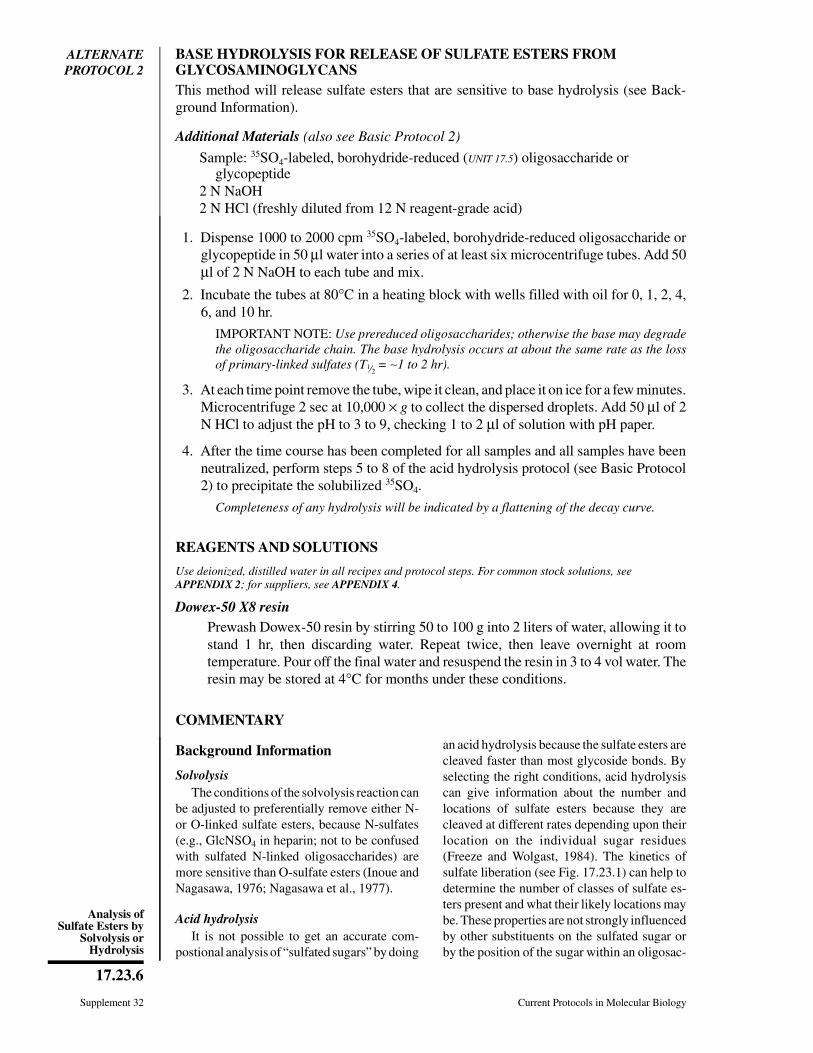
ALTERNATEPROTOCOL 2
BASE HYDROLYSIS FOR RELEASE OF SULFATE ESTERS FROMGLYCOSAMINOGLYCANSThis method will release sulfate esters that are sensitive to base hydrolysis (see Back-ground Information).
Additional Materials (also see Basic Protocol 2)
Sample: 35SO4-labeled, borohydride-reduced (UNIT 17.5) oligosaccharide orglycopeptide
2 N NaOH2 N HCl (freshly diluted from 12 N reagent-grade acid)
1. Dispense 1000 to 2000 cpm 35SO4-labeled, borohydride-reduced oligosaccharide orglycopeptide in 50 µl water into a series of at least six microcentrifuge tubes. Add 50µl of 2 N NaOH to each tube and mix.
2. Incubate the tubes at 80°C in a heating block with wells filled with oil for 0, 1, 2, 4,6, and 10 hr.
IMPORTANT NOTE: Use prereduced oligosaccharides; otherwise the base may degradethe oligosaccharide chain. The base hydrolysis occurs at about the same rate as the lossof primary-linked sulfates (T1⁄2 = ∼1 to 2 hr).
3. At each time point remove the tube, wipe it clean, and place it on ice for a few minutes.Microcentrifuge 2 sec at 10,000 × g to collect the dispersed droplets. Add 50 µl of 2N HCl to adjust the pH to 3 to 9, checking 1 to 2 µl of solution with pH paper.
4. After the time course has been completed for all samples and all samples have beenneutralized, perform steps 5 to 8 of the acid hydrolysis protocol (see Basic Protocol2) to precipitate the solubilized 35SO4.
Completeness of any hydrolysis will be indicated by a flattening of the decay curve.
REAGENTS AND SOLUTIONS
Use deionized, distilled water in all recipes and protocol steps. For common stock solutions, seeAPPENDIX 2; for suppliers, see APPENDIX 4.
Dowex-50 X8 resinPrewash Dowex-50 resin by stirring 50 to 100 g into 2 liters of water, allowing it tostand 1 hr, then discarding water. Repeat twice, then leave overnight at roomtemperature. Pour off the final water and resuspend the resin in 3 to 4 vol water. Theresin may be stored at 4°C for months under these conditions.
COMMENTARY
Background Information
SolvolysisThe conditions of the solvolysis reaction can
be adjusted to preferentially remove either N-or O-linked sulfate esters, because N-sulfates(e.g., GlcNSO4 in heparin; not to be confusedwith sulfated N-linked oligosaccharides) aremore sensitive than O-sulfate esters (Inoue andNagasawa, 1976; Nagasawa et al., 1977).
Acid hydrolysisIt is not possible to get an accurate com-
postional analysis of “sulfated sugars” by doing
an acid hydrolysis because the sulfate esters arecleaved faster than most glycoside bonds. Byselecting the right conditions, acid hydrolysiscan give information about the number andlocations of sulfate esters because they arecleaved at different rates depending upon theirlocation on the individual sugar residues(Freeze and Wolgast, 1984). The kinetics ofsulfate liberation (see Fig. 17.23.1) can help todetermine the number of classes of sulfate es-ters present and what their likely locations maybe. These properties are not strongly influencedby other substituents on the sulfated sugar orby the position of the sugar within an oligosac-
Supplement 32 Current Protocols in Molecular Biology
17.23.6
Analysis ofSulfate Esters by
Solvolysis orHydrolysis

charide chain. Since the hydrolysis can alsocleave glycosidic linkages and partially destroythe sugar chain, only the release of sulfate canbe measured.
The acid hydrolysis procedure differentiatesbetween sulfates linked to different hydroxylgroups on the sugar ring. Under the hydrolysisconditions in Basic Protocol 2, the T1⁄2 of sul-fates linked to equatorial hydroxyl groups (e.g.,glucose-3-sulfate, galactose-3-sulfate, N-ace-tylglucosamine-3-sulfate, and mannose-3-sul-fate) is 6 to 25 min; that of those linked to axialhydroxyl groups (e.g., galactose-4-sulfate, N-acetylglucosamine-4-sulfate, and mannose-2-sulfate) is 60 to 84 min, and that of those linkedto primary hydroxyl groups (e.g., glucose-6-sul-fate, galactose-6-sulfate, N-acetylglucosamine-6-sulfate, and mannose-6-sulfate) is 90 to 120min (Rees, 1963). All 6-OH positions are pri-mary, and all of the hydroxyl groups in glucoseare equatorial. Any hexose isomer of glucose(e.g., mannose or galactose) will change one ofthe equatorial positions to an axial position.Even though the time ranges for hydrolysislisted above have not been extensively docu-mented for sulfate-labeled oligosaccharides,they are useful because they will clearly differ-entiate between multiple classes of sulfate es-
ters. Using the acid hydrolysis technique, it isnot possible to determine which sugar is sul-fated or where it is found in the chain. If otherdata are available on the structure of the olig-soaccharide, this information may providelikely possibilities for the location of sulfategroups, especially if certain residues are resis-tant to exoglycosidase digestion.
Base hydrolysisOnly certain types of sulfate esters are sen-
sitive to base hydrolysis (Freeze and Wolgast,1984; Percival, 1978). Sensitivity is determinedby the location of the sulfate on the sugarresidue and—in contrast to acid hydrolysis de-scribed above—by the other substituents linkedto that sugar residue.
Base treatment will release sulfate estersfrom intact oligosaccharides under two differ-ent conditions. The first is when sulfate islocated in the 6 (or 3)-OH position and the 3(or 6)-OH group of the same sugar residue isnot substituted. The second is when the sulfateester is adjacent to an unsubstituted trans OHgroup. In the first case, the sulfate is eliminatedand an acid-labile 3,6-anhydrosugar is formed(see Fig. 17.23.2). In the second case, the sul-fate is released with the formation of an epoxide
OOH OH
OH
OCH2HSO3
OOH OH
OH
OCH2
O R
OOH
OH
CH2
O
OCH2O
OH OHOH O R
OH- OOH
OH
CH2
OH
mild H+
HOCH2O
OH OHOH O R
strong H+
HOCH2O
OH OHOHOH
+O
OHOH
CH2
OH
O
+
O
Figure 17.23.2 Formation of 3,6-anhydromannose by base treatment of an oligosaccharidecontaining mannose-6-sulfate. The formation of the anhydro sugar makes the glycosidic linkagevery sensitive to acid; mild hydrolysis causes cleavage. Stronger hydrolysis degrades the usualglycosidic linkages, but leaves the anhydrosugar. When reduced with sodium borohydride, themodified sugar can be quantified to show how much mannose-6-sulfate was originally present.
Current Protocols in Molecular Biology Supplement 32
17.23.7
Preparation andAnalysis ofGlycoconjugates

ring on the sugar. This is destroyed duringsubsequent acid hydrolysis of the oligosaccha-ride. The modified sugar can be identified if itis metabolically labeled—e.g., as in the discov-ery of mannose-6-sulfate (Freeze and Wolgast,1984)—but this is not routinely done. On theother hand, the loss of sulfate is easily meas-ured. When data on the acid and base hydrolysisof sulfate esters are combined with other struc-tural information on the molecules, several po-tential structures can be eliminated or sup-ported, but these procedures will not provideproof of the structure.
Critical ParametersSolvolysis is a harsh treatment that also
removes sialic acids and probably fucose resi-dues. If these sugars are normally present, theirloss may be an unacceptable side effect of thisprcedure. On the other hand, hexoses,hexuronic acids, and hexosamines in glycosidiclinkages are mostly stable to solvolysis. Theincubated control is very important because itwill monitor for any nonspecific destruction ofthe sugar chain.
For acid hydrolysis, the most critical pointsto observe are that the pH should be kept be-tween 2 and 3 and that the salt concentration bekept very high during the precipitation of sufatewith barium. Only then is the precipitationspecific for sulfate. Under less stringent condi-tions, the oligosaccharides or even monosac-charide sulfates can be precipitated.
Anticipated ResultsExpect ∼80% to 90% efficiency in desulfa-
tion, and <20% in the control incubation(Freeze and Wolgast, 1984).
Time ConsiderationsSolvolysis itself takes only a few hours to
complete. The rate-limiting step is the timeneeded to remove the water after the sample haspassed through the Dowex-50 column and thepyridine has been added. This will only take afew minutes using a shaker-evaporator, but willrequire an overnight lyophilization.
Beginning with the hydrolysis step, the pro-cedure can be completed within a day. If nec-essary, the procedure can be stopped after tak-ing all of the time points and neutralizing themwith base prior to precipitation. Where this isdone, the reaction mixtures can be stored at0°-4°C for 24 hr or more.
Literature CitedInoue, Y. and Nagasawa, K. 1976. Selective N-de-
sulfation of heparin with dimethyl sulfoxide con-taining water or methanol. Carbohydr. Res.46:87-95.
Nagasawa, K., Inoue, Y., and Kamata, T. 1977.Solvolytic desulfation of glycosaminoglycuro-nan sulfates with dimethyl sulfoxide containingwater or methanol. Carbohydr. Res. 58:47-55.
Freeze, H. and Wolgast, D. 1984. Structural analysisof N-linked oligosaccharides from glycoproteinssecreted by Dictyostelium discoideum. J. Biol.Chem. 261:127-133.
Rees, D.A. 1963. A note on the characterization ofcarbohydrate sulphates by acid hydrolysis. Bio-chem J. 88:343-346.
Percival, E. 1978. Sulfated polysaccharides of theRhodophycea. In Carbohydrate Sulfates. (R.G.Schweiger, ed.) pp. 213-224. American Chemi-cal Society, Washington, D.C.
Contributed by Hudson H. FreezeLa Jolla Cancer Research FoundationLa Jolla, California
Supplement 32 Current Protocols in Molecular Biology
17.23.8
Analysis ofSulfate Esters by
Solvolysis orHydrolysis
SECTION IIDETECTION OF SACCHARIDES ONGLYCOCONJUGATESThe first step toward analyzing the structure and function of oligosaccharides on aglycoconjugate is the detection of the glycan chains themselves. The units presented inthis section describe a variety of ways in which this can be achieved (e.g., UNIT 17.7).In some cases, such studies may provide the first evidence that the molecule beingexamined is a glycoconjugate. In other cases, these methods may be used to qualitativelyor quantitatively analyze the nature of the glycan chains on a molecule that is alreadyknown to be a glycoconjugate. For help in deciding which protocols to use, the user isreferred to the section entitled “Choice of Techniques” found in the introduction to thischapter (UNIT 17.0).
Introducing a label into the sugar chains of the glycoconjugate of interest is often in-formative, and can be very convenient for further analyses. This can be achieved bymetabolic radiolabeling (UNIT 17.4), chemical labeling (UNIT 17.5), or transferring labelfrom a radioactive sugar nucleotide using a glycosyltransferase (UNIT 17.6). Each of theseapproaches has its advantages and disadvantages in terms of specificity, sensitivity, andpractical utility. The user is advised to read the Commentary in of each of these unitsbefore choosing a particular approach.
Many membrane glycoproteins also acquire a glycophospholipid anchor during biosyn-thesis. A set of methods designed to detect and analyze such anchors is presented inUNIT 17.8. There are also many well established methods for direct chemical analysis ofthe oligosaccharides present in glycoconjugates, such as the classic phenol-sulfuric acidassay for hexoses and pentoses (UNIT 17.9).
Finally, oligosaccharides on many glycoconjugates can be altered with a variety ofcommercially available inhibitors and competitive substrates that can work in whole cellsor organisms. In this section, the prevention of N-linked glycosylation on glycoproteins(UNIT 17.10A) and the inhibition of glycolipid biosynthesis (UNIT 17.10B) is described, aswell as the use of synthetic glycosides as primers of oligosaccharide biosynthesis andinhibitors of glycoprotein and proteoglycan assembly (UNIT 17.11).
There are, of course, many other approaches to the direct analysis of oligosaccharides onglycoconjugates that could have been presented in this section. The current choices forinclusion (see Chapter 17 table of contents) were dictated by the need to “keep it simple”and to meet the criterion of providing broad general utility to the average molecularbiologist.
Current Protocols in Molecular Biology 1, July 2009Published online July 2009 in Wiley Interscience (www.interscience.wiley.com).DOI: 10.1002/0471142727.mb17200s87Copyright C© 2009 John Wiley & Sons, Inc.
Preparation andAnalysis ofGlycoconjugates
1
Supplement 87