Theme 1: Pulse Amplitude Modulation Fluorometry and the Stress ...
Quantitative fluorometry of abnormal mouse sperm nucleiG. C. Pogany and R. Balhorn
Biomedicai Division, Lawrence Livermore National Laboratory, CA 94550, USA
Summary. An extensive quantitative analysis of deformed mouse spermatozoa wasundertaken. Improvements over previous studies included the isolation and purifi-cation of sperm nuclei, a multifaceted analytical approach using several fluorochromesand the analysis of individual nuclei classified into shape categories.
Malformed sperm nuclei in BALB/c mice could not be distinguished from normalones in terms of total and basic proteins, sulfhydryl and disulfide group concentration,DNA concentration and chromatin organization. The shape of sperm nuclei is there-fore probably determined by the manner in which the internal biochemical componentsare assembled.
Keywords: quantitative fluorometry; sperm nuclei; mouse
Introduction
All species of mammal produce deformed spermatozoa during spermiogenesis. The proportionvaries depending upon the species or the strains (Beatty & Sharma, 1960; Beatty, 1972; Wyrobek,1979; Krzanowska, 1981), from 4% in SW mice (Wyrobek et ai, 1975; Pogany, 1987) to 99% in theazh/azh strain (Kot & Handel, 1987). The mechanisms responsible for nuclear deformation in thesecells are not understood, but deformed spermatozoa are known to penetrate the zona pellucida ofoocytes in vitro (Kot & Handel, 1987) and to fertilize eggs (Moore Smith, 1970; Krzanowska &Lorenc, 1983). Deformed spermatozoa may also contribute to reproductive dysfunctions and are
suspected to cause habitual abortions (Giarola & Ballerio, 1962).A large number of physical and chemical factors are known to temporarily increase the inci¬
dence of malformed spermatozoa (Wyrobek & Bruce, 1975; Wyrobek et al., 1983; Pogany, 1987).Although means of selecting against these deformed spermatozoa within the female reproductivetract have been demonstrated (Krzanowska, 1974), embryos conceived during the time that thefrequency of sperm shape abnormality is exacerbated may be at considerable genetic and/or terato-genic risks. Hence, it is important to determine the mechanism(s) behind the induction of headshape abnormalities.
The biochemical events responsible for the shape of spermatoza have not yet been resolvedalthough much is already known about the biochemistry of spermiogenesis. The process entails a
complex series of reactions during which somatic histones are replaced by gametic protamines(Bode et al., 1977; Willmitzer et al., 1977). This intricate sequence of biochemical steps is coupledwith the compaction of the haploid nucleus, which is probably the most important event in the finalshaping of the spermatozoa. Perturbations in these events could therefore significantly alter thestructural integrity of the sperm chromatin and influence the morphology of the cells.
A common approach to the analysis of sperm chromatin compaction involves the estimation ofdye binding during their morphological maturation. Results have revealed a progressive reduction
'Present address: Department of Biological Sciences, Box 5640, Northern Arizona University, Flagstaff, AZ 86011,USA.

in dye binding reflecting the increased condensation of the chromatin (Ringertz et al., 1970; Rod¬man et al., 1979; Wagner & Minhas, 1982; Gledhill, 1985). The progressive oxidation of sulf-hydryl groups in protamines (Bedford & Calvin, 1974; Saowaros & Panyim, 1979; Pelliciari et al.,1983; Shalgi et al., 1989; Balhorn et al., 1991) has also been suggested to assist in this compaction,although the precise manner in which this is accomplished in maturing spermatozoa is unknown.
Other investigations of chromatin in abnormally shaped spermatozoa have focused on DNAcontent, chromatin structure or on both (Pinkel et al., 1983; Evenson et al., 1985, 1986). In most
instances, these analyses involved large populations of spermatozoa without regard to their indi¬vidual shape. In the few cases where single cells were analysed for the structural integrity of mal¬formed chromatin (Gledhill et al., 1977; Gledhill, 1985), no attempts were made to distinguishbetween types of abnormality.
In an attempt to identify biochemical defects that contribute to abnormal shaping in spermato¬zoa, we used biochemical and histochemical tests to analyse normal and abnormally shaped spermnuclei individually. Three different fluorochromes were used to measure nuclear proteins, sulf-hydryl groups and DNA. 7-Aminoactinomycin D and Acridine Orange were used to assess theintegrity of sperm chromatin in deformed and normal nuclei.
Materials and Methods
AnimalsBALB/c mice were purchased from Bantin & Kingman, Inc. (Fremont, CA) and from Simonsen Laboratories,
Inc. (Gilroy, CA). They were kept individually in the animal facility of the Lawrence Livermore National Laboratoryunder a 12 h light : 12 h dark regimen and at a constant temperature of 22°C with free access to food (Certified RodentChow Checkers, Purina Mills, Inc., St Louis, MO) and water.
Isolation of sperm nucleiAnimals were killed with carbon dioxide and cervical dislocation. Cauda epididymides were removed, pooled in
cold Tris-saline (TS) buffer (001 mol Tris 1 in 0-9% saline, pH 80), and their contents gently expressed with theblunt edge of a scalpel. After filtration through a fine silk filter (no. 11; Standard Swiss Silk Doufour; AbbeEngineering Co., Brooklyn, NY), they were sonicated to remove somatic cell contaminants and washed three times in5 ml TS with intermittent centrifugation at 10 400 g for 4 min (Sorvall, Model RC2-B, HB-4 rotor: Norwalk, CT).Sperm nuclei were isolated (Balhorn et al., 1977; Pogany et al., 1981) in 4 ml TS to which 10 mg DTT (dithiothreitol;Boehringer-Mannheim, Indianapolis, IN) was added. After incubation on ice for 15 min, 1 ml of 5% MTAB stock(mixed alkyltrimethylammonium bromide; Sigma Chemical Co., St Louis, MO) in distilled water was added to a finalconcentration of 1 % and the nuclear suspension was further incubated on ice for 60 min with frequent but gentlemixing. After centrifugation, the pellet was resuspended in 5 ml 1 % MTAB and incubated for another 60 min at room
temperature. The suspension was then centrifuged and the pellet containing pure nuclei dispersed in 10 ml TS bysonication.
The nuclear suspension was further diluted by transferring 2-5 ml of the suspension to 10 ml of TS. Plain micro¬scope slides were cleaned in 70% ethanol and mounted into cytobuckets (International Equipment Company, no
1024: Needham Heights, MA). Aliquots of 0-25 ml were delivered to each of the three wells per slide and centrifugedat 350 g for 10 min (International Equipment Company, Model HN-SII). The slides were allowed to air dry overnight.They were subsequently rinsed with distilled water, air dried again and stored in a slide box for subsequent use. Ingeneral, fixation of the nuclei was omitted unless expressly required by the specific staining application.
Staining protocolsDAPI (4', 6-diamidino-2-phenylindole; Sigma Chemical Co., St Louis, MO) was used to determine the DNA
content of sperm nuclei (Lin et al., 1977). The dye was dissolved in distilled water to a stock concentration of 100 pgml"1. Just before use it was diluted to 0-5 pg ml"1 in antifadant (Johnson & Noguiera Araujo, 1981). Drops of thestain were placed on slides and the edges of the coverslip sealed with fingernail polish. They were left to incubate in thedark for 1 h before quantitation. Nuclei wre analysed at 365 nm (excitation) and 397 nm (emission).
Brilliant Sulfaflavine (Aldrich Chemical Co., Inc., Milwaukee, WI) was used to measure total and basic proteins(Leemann & Ruch, 1972). For total proteins, slides were first fixed in ethanofglacial acetic acid (3:1) and stained at
pH 2-8 in citric acid buffer (0· I mol citric acid 1" ', 0-3 mol disodium phosphate 1" ) with sulfaflavine at 01% concen¬tration for 30 min in the dark. Differentiation was obtained in buffer for 10 min. The slides were dehydrated, cleared inxylene, mounted in antifadant and analysed at 420 nm (excitation) and 520 nm (emission).
For the estimation of basic proteins with sulfaflavine, nuclei were fixed in buffered formalin (pH 70) for 10 min.The slides were then rinsed in water, and hydrolysed in 5% trichloroacetic acid at 60°C for 3 h. They were rinsed threetimes in 70% ethanol, twice in water (distilled), and once in TS. The slides were stained with Sulfaflavine at the same
concentration as above for 30 min and differentiated in buffer. After dehydration and clearing, they were mounted inantifadant and analysedat the same wavelengths used for neutral proteins.
Free thiol groups were determined using the thiol-specific dye DACM (N-(7-dimethylamino-4-methyl-coumarinyl)maleimide; Sigma Chemical Co., St Louis, MO) as reported by Pelliciari et al. (1983). A stock of 1 mg in1 ml acetone was kept in the freezer and diluted to a final concentration of 10"5 mol l"1 in 004mol PBS 1(phosphate-buffered saline, pH 70) just before use. Slides were washed in three changes in PBS and air dried. Slideswere stained in DACM for 20 min in the dark. They were rinsed three times in PBS, allowed to air dry, and sealed withantifadant. Their analysis was performed at 405 nm (excitation) and 515 nm (emission).
Sperm chromatin integrity (Gill et al., 1975) was assessed using the DNA intercalator 7-aminoactinomycin D (7-AMD; Calbiochem, San Diego, CA) as described by Balhorn et al. (1985). Slides of dried nuclei were stained for 16 hin phosphate buffer (pH 70) containing 5 µ mol 7-AMD l"1. Brief rinsing in phosphate buffer was followed by airdrying in the dark. Slides were immediately analysed at 546 nm (excitation) and 590 nm (emission).
Acridine orange (Polysciences, Inc., Warington, PA) staining was used because of its known metachromatic prop¬erty (Lerman, 1961; Darzynkiewicz et al., 1975) which permits the discrimination between native (green fluorescence)and denatured DNA (red fluorescence). The technique assesses the susceptibility of DNA to acid depurination anddenaturation when nuclei are exposed to acridine orange at a pH of 2-5-2-8. The one-step method of Tejada et al.(1984) was chosen because of its simplicity. Slides were fixed in fresh Carnoy's (3 parts methanol: 1 part glacial aceticacid) for 2 h and air dried. A working solution of acridine orange was used by adding 10 ml of a distilled water stock(1 mg ml"1) to a citric acid buffer (40 ml of 01 mol citric acid l"1 and 2-5ml of 0-3 mol disodium phosphate 1~\pH 2-55). Slides were stained for 5 min in the dark and briefly rinsed in distilled water. For the fluorometric analysis,slides were covered with water and the edges of the coverslip sealed with nail polish. The fluorescence of nuclei was
also scored according to whether they stained green (native DNA) or red (denatured DNA). Acridine orange bindingto nuclei was also quantitatively assessed at an emission of 490 nm and an excitation of 530 nm. All slides were
analysed immediately after staining.
Image analysisFields of dispersed nuclei were located at 100 (Pan-NEOFLUAR, 1-30 aperture) using a Zeiss Axiophot
microscope equipped with a COHU SIT Videcon camera (Model 5152; tube no 4804 HP2). A non-random approachwas chosen to obtain representative images of all shapes, some of which were too rare to select in a random fashion.The images were focused onto a black and white TV screen and pipelined through a Trapix 5500/64 processing unit(Recognition Concept, Inc., Carson City, NV) at 512 512 pixel resolution. Image capture and analysis wereexecuted by an ACUity program (version 6-2) specifically designed for object analysis (Young et al., 1982). Optimalimage segmentation parameters were selected for each stain to extract the best contour traces of the nuclei from thebackground. The digitized images were projected onto a colour monitor in pseudocolour video output and rastered tolocate each object. When an object was encountered which conformed to the user-selected segmentation parameters,a contour trace was delineated around it. The various morphological types were classified as 'normal', 'hammer','acrosome', 'truncated', 'collapsed' and Others' and given an identification number of 1 to 6 respectively. After theidentification of each nucleus, the digitized images were saved in individual files until the end of the session when theywere combined into a larger data file for subsequent statistical analyses. All results were expressed as total stain(brightness) per surface area of the nuclei.
Statistical analysisDigitization and the subsequent morphometric analysis of each object by ACUity were driven by a DEC,
MicroVAX II (Digital Equipment Corporation, Maynard, MA) computer and included a number of measurements.These have been described and defined by Young et al. (1982). Standard descriptive statistical analyses were per¬formed as well as one-way analysis of variance (anova) followed by the Scheffe multiple-range test of discriminationbetween the means.
Photographic records of the various nuclear shapes were obtained through a compound microscope. Intact spermsuspensions were first stained with 01 eosin Y for 60 min according to the method of Wyrobek & Bruce (1975),smeared onto clean slides, air dried, and covered with Permount. In the case of isolated nuclei, they were stained withToluidine blue for 60 min and rinsed in distilled water. All photographs were taken with a Zeiss Ultraphot microscopeat 100 magnification (Optovar = 1 -60) and recorded on Panatomic X film. Negatives were developed in MicrodolX for the recommended time and printed on multigrade RC paper.
Results
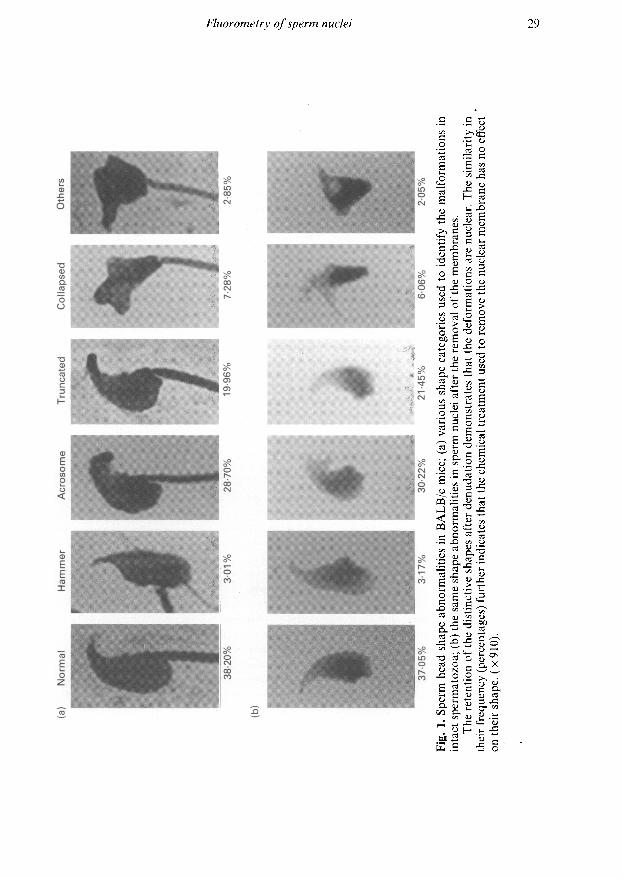
Approximately 60% of the sperm obtained from the cauda epididymides of BALB/c mice used inthis study have abnormally shaped heads. These were classified into five categories of shape

anomalies in increasing order of severity (Fig. 1). Even though the categories are based primarilyon the morphology of intact sperm, they apply equally well to isolated nuclei since the latter retainthe shape of the cells from which they are derived (Fig. 1). This clearly indicates that the removal ofthe membranes with DTT and MTAB does not preferentially affect any one particular shape. Thedistribution of nuclear deformities is virtually identical to that of untreated, intact spermatozoa.The analysis of these nuclei was, therefore, considered representative of intact epididymalspermatozoa.
The mildest deformation, defined as 'acrosome', pertains to various degrees of blunting andbending of the tip of the head. Although isolated nuclei no longer possess acrosomes, the desig¬nation is retained for purposes of consistency. 'Truncated' sperm heads and nuclei are defined as
having a tapered posterior aspect. Typically, these abnormalities include the deformation of theimplantation fossa. A third class of deformations involves the ventral shift of the implantationfossa to the extent that the insertion of the mid-piece is characteristically perpendicular to thelongitudinal axis of the head. These are designated 'hammer' types. The most severely deformedsperm and nuclei are classified as 'collapsed' and Others'. Both are amorphic and, upon closerexamination, reveal that they are thicker than the other types of deformity. The Others' categoryincludes nuclei that cannot easily be placed into any of the other morphological categories.Frequently, sperm heads are characterized by more than a single abnormality as seen in the'truncated' example where the blunting of the acrosome is also evident. In these instances, heads areclassified according to the most prominent aberration.
The most frequent shape anomaly in BALB/c mice is represented by 'acrosome' and 'truncated'types of deformity. In the former case, the proportion almost equals that of normal heads andnuclei. Most other shape aberrations are present less frequently, with the most severely distortedsperm heads and nuclei ('collapsed') being about twice as abundant as 'hammers' and 'others'.
A large number of slides were routinely prepared for analysis to minimize sample variationduring the fluorometric measurements. The likelihood that these samples could deteriorate uponstorage and, therefore, alter their quantitative fluorescence, was tested by performing the analysisat various time intervals up to 57 days. The results show that prolonged storage does not affectstainability and subsequent analyses (data not shown). Similarly, there is no evidence that theintegrity of the chromatin is altered upon storage as tested with acridine orange staining during thesame period (data not shown).
The image analysis capabilities of the program 'ACUity' were used to determine whether theapplication of the dyes affects the physical dimensions of the objects under consideration. Alterationsin the overall size of the nuclei, as a result of staining, would significantly influence the quantitativecomparisons between nuclei stained with different fluorochromes. When stained nuclei were com¬
pared, no change in size is observed except in the case of 7-AMD where a noticeable increase insurface area was noted (Table 1). Closer examination of the length and width parameters (routinelyprovided by ACUity) in 7-AMD-stained nuclei reveals that the increase is primarily due to nuclearelongation (42-11%). An increase in width is also observed but it is smaller (29-43%).
The distribution of the 5013 nuclei fluorometrically analysed in this investigation is shown(Table 2). As can be seen, the frequency of the various shapes analysed in this study deviatesslightly from the normal distribution of abnormally shaped spermatozoa in BALB/c (Fig. 1). Thisdeviation reflects the need to preferentially select certain abnormality types because of their lowinherent frequency.
The results of the quantitative fluorometric analysis of normal and malformed sperm nuclei areshown in Fig. 2. For most of the fluorochromes, the range of brightness is very similar and extendsfrom 30 (acridine orange) to 2000 (DAPI) fluorescence units. This latter stain stands out as a
particularly sensitive dye. Small quantitative differences in brightness values can be seen between'normal', 'acrosome', 'hammer' and 'truncated' shapes irrespective of the dyes used. Statisticalanalyses (anova and Scheffe multiple-range test) show that these differences are not significant.However, the fluorescence of 'collapsed' and 'other' shapes is significantly different from normal
._ „cd O
rg c.9 c« u
— cd
cd p
<« ca— —
•o -S
cd cj«> 3c cO CU'ß J3cd -"
gS o
O Ct)tí *-
3 u-SO- »Cd CJ
" < ._
3 ,2üC 3çd c
o
-S 3
cd - 5CO cdp CJ
«ao ü ^
cj co
.y c
ß ¿s< £ca £•
S X)co cd cj
'S o,— cdcd x¡E M
g
jj
«
e.-gcd
le-S £- £cl, -*-· ,2
3 cd <& a, x
: *-·
c c/3 a.
ta .S
b
cj cj cdU 3 -Cc a" w
u ii .3x: c; H >- -S
•5 o

Table 1. Effects of dye binding on the dimensions (pixels + sd) ofnormal mouse sperm nuclei
Dyes Surface area Length Width
Acridine orange7V-(7-dimethylamino-4-methyl-coumarinyl) maleimide(DACM)Brilliant sulfaflavine, pH 80Brilliant sulfaflavine, pH 2-84'6-Diamidino-2-diphenyl-indole (DAPI)Average7-Aminoactinomycin D nuclei% Increase:
743-5 +
881-2 ± 1306420 ±101812-7 ± 114
701-0 ± 79755-6
1055-0 ± 13739-6
44-9 ± 3-6 21-2 + 2-3
49-7 ± 5040-2 ± 3-848-7 ±4-7
43-6 ±4145-4
64-5 ± 4-742-1
221920
9 ± 2-89 ±2-19 + 2-3
20-8
27
±2-121-24 ±2-629-4
Table 2. Distribution of types of nuclear shapeDyes Normal Hammer Acrosome Truncated Collapsed Other Total
Acridine orange/V-(7-dimethylamino-4-methyl-coumarinyl) maleimide(DACM)Brilliant sulfaflavine pH 80Brilliant sulfalavine pH 2-84'6-Diamidino-2-diphenyl-indole (DAPI)7-Aminoactinomycin D(7-AMD)Totals:Frequency (%)
140
336432176
581
4292094
41-1
17
456315
51
65256
51
74
58136111
118
263760
15 2
147
177237163
255
4921471
29-3
29
705927
56
85326
6-5
11
19IX9
25
24106
2-
418
705945501
1086
13585013
100
nuclei when stained with DAPI, brilliant sulfaflavine 80, and 7-AMD (F > 2-381; = 0001). Theother stains (acridine orange, DACM, brilliant sulfaflavine 2-8) do not discriminate sufficientlybetween normal and these severely deformed nuclei to distinguish them.
Discussion
This study represents a comprehensive quantitative evaluation of sperm nuclear components. First,five different fluorochromes were used to quantify several critical components of sperm nuclei incontrast to previous investigations which were based on single parameter determinations. Second,six different types of sperm head morphology were recognized in an attempt to provide addeddelineation among them. The large number of individual sperm nuclei in each shape class providedsufficient statistical samples to generate meaningful comparisons between the various shapes.Lastly, the use of isolated and purified nuclei made it possible to dispense with concerns about thepotential influence of membranes on the quantitative uptake of dyes.
Most deformed BALB/c sperm nuclei could not be distinguished from normal sperm nuclei interms of total and basic proteins, sulfhydryl groups, DNA and chromatin organization as moni¬tored by 7-AMD binding and acridine orange metachromasia. The only significant differences were
those detected between the most deformed nuclei ('collapsed' and Others') and normal ones. How¬ever, these differences, although statistically significant, may be artifacts. In addition to beinghighly distorted, these nuclei were consistently thicker. This increased thickness may have affected

(b)3000
1 2 3 4 5 6
(e)40
30
20
10
1 2 3 4 5 6
(f)
Fig. 2. Quantitative fluorometry of mouse sperm nuclei using (a) jV-(7-dimethyl amino-4-methylcoumarinyl) maleimide (DACM), (b) 4'6-diamidino-2-diphenyl indole (DAPI), (c)brilliant sulfaflavine pH 80, (d) brilliant sulfaflavine pH 2-8, (e) acridine orange and (f) 7-aminoactinomycin D (7-AMD). All values are expressed as stain per area and are plotted as
arbitrary fluorescence units on the y axis. The numbers on the axes represent the variousnuclear shape categories (1: normal, 2: hammer, 3: acrosome, 4: truncated, 5: collapsed, 6:others). The total number of nuclei analysed in each shape category and for each dye isindicated within the graphs. The bars indicate the means and the lines represent the sem;*significant differences from average of other shapes ( < 005).
both the proper focusing and the segmentation of the objects. It is, therefore, unclear whether theobserved differences in staining for these two classes of nucleus are real or whether they arise fromtheir peculiar shape.
The absence of a relationship between the binding of the structural probes (acridine orange and7-AMD) and nuclear shapes contradicts a previous investigation in which binding of [3H]actino-mycin D was found to be higher in deformed bull spermatozoa than in their normal counterpart(Gledhill et al., 1971). This was subsequently related to structural defects in the chromatin (Gledhill,1972). However, the discrepancy may have resulted from determining dye binding on intactbull spermatozoa. It is therefore possible that the data from bulls may reflect only an altered
permeability of the membranes to the dye in deformed spermatozoa. Such a condition would nothave been detected in our investigation since only amembranous nuclei were analysed. Similarly, ithas been shown that spermatozoa from subfertile males are more susceptible to heat denaturation(Evenson et al., 1980). However, the relationship between their individual shape and theirpropensity to denature under heat was not established.
Our determinations of potential differences between normal and abnormal sperm nuclei interms of total and basic proteins are not shown. The harsh fixation and extraction treatments thatpreceded their quantitative assessments introduced uncertain experimental conditions that requirefurther monitoring. Accordingly, additional experiments, using milder conditions, are planned fora more quantitative re-assessment of these parameters.
Staining nuclei with 7-AMD induced an increase in the size of the nuclei that may have been dueto the prolonged exposure to either the dye or the buffer overnight (see Balhorn et al., 1985).However, the preferential increase in length may be related to the observation that chromatin,inside sperm nuclei, is in a cholesteric configuration (Sipski & Wagner, 1977). A greater increase inlength would, therefore, be anticipated, as a result of chromatin expansion, if the presumedcholesteric bundles were oriented along the longitudinal axis of the spermatozoa. The intercalationof 7-AMD between base pairs of the DNA in sperm chromatin could extend the length of thesebundles.
Our analyses of protein, DNA and total sulfhydryl groups in sperm nuclei support andcomplement previous reports in which the total protein contents of teratozoospermic cells wasfound to be identical to normal ones (Bustos-Obregon & Leiva, 1983). As demonstrated by de Boeret al. (1990), even aneuploid gametes could not be distinguished in terms of protamine content andcross-linking. Quantitative DNA analyses by Feulgen cytometry (Döring et al., 1972) failed todiscriminate abnormal sperm heads from normal ones. However, a UV absorption analysis of F,hybrids between Mus musculus and Mus poschiavinus showed a higher mean DNA content but thedifferences were not statistically significant (Stolla & Gropp, 1974). In a comprehensive analysis ofthe protein synthetic machinery of differentiating spermatozoa, Meistrich et al. (1988) showed thatazh/azh mice possessed normal components and expression levels compared with a strain where theincidence of sperm shape abnormality is considerably lower. These authors concluded that themolecular entities themselves were biochemically unaffected but could be subjected to faultyassembly processes. They further suggested that microtubular elements in the manchette could beinvolved in the formation and deformation of spermatozoa. However, the participation of themanchette in the shaping of sperm nuclei has been strongly disputed by Fawcett et al. (1971) on thebasis that there is insufficient diversity in its structure and distribution to account for the wide arrayof sperm shapes in animals. Accordingly, these latter authors propose that the final shaping of thesperm nucleus involves factors internal to the nucleus.
The similarity in the composition of nuclei from different shape classes suggests that the funda¬mental biochemical differentiation of the sperm nucleus antedates its formation during spermio¬genesis. The forces that impose a morphological character on each sperm nucleus must, therefore,use components that are quantitatively similar in all. Accordingly, the formative events instrumen¬tal in shaping sperm nuclei must exhibit considerable morphological plasticity. This extends fromsperm possessing minor acrosomal deformities to those having severely distorted nuclei ('others'and 'collapsed') and further suggests that it is the mode of packaging these similar components thatis radically different in abnormal spermatozoa. A genetic component for this disturbance inpackaging is strongly implied when one considers that the proportion of malformed spermatozoain any strain of mice is constant from one generation to the other (Wyrobek, 1979).
A number of internal formative elements have already been implicated in the morphologicaldevelopment of spermatozoa, including the characterized stable DNA-protein complexes in thesperm nucleus of rams (Avramova & Tsanev, 1987). DNA-anchoring proteins, located at theimplantation fossa of hamster spermatozoa, have also been characterized (Ward & Coffey, 1989).Residual histone components cannot be excluded either since they have been found in human
spermatozoa (Tanphaichitr et al., 1978) and are also suspected to occur in mice (R. Balhorn,unpublished) albeit in smaller concentrations. Finally, the presence of a structural nuclear elementsimilar to the nuclear matrix of somatic cells may need to be invoked to account for the shape ofspermatozoa. Such a matrix element has recently been suggested for human sperm nuclei (Wyrobeket al., 1990) and, if present, would have a significant impact on our understanding of normal andabnormal sperm maturation. The total absence of quantitative differences between malformedand normal sperm nuclei in BALB/c mice is consistent with the hypothesis that sperm nucleardeformation in this mouse strain may result from structural perturbations in a nuclear matrix.
Although the shape abnormalities analysed in BALB/c mice have an obvious genetic origin, itshould be instructive to compare our results with deformed nuclei experimentally generated withshape-altering agents such as X-rays (Pogany, 1987).
A. J. Wyrobek and P. Dean are thanked for their help with the Quips/ACUity, D. Sudar for hisgenerous assistance with computer programming. G. Pogany thanks R. Balhorn in whoselaboratory the research was conducted.
References
Avramova, . & Tsanev, R. (1987) Stable DNA-proteincomplexes in eukaryotic chromatin. Journal ofMolecular Biology 196, 437-440.
Balhorn, R., Gledhill, B.L. & Wyrobek, A.J. (1977)Mouse sperm chromatin proteins: quantitative iso¬lation and partial characterization. Biochemistry 16,4074-4080.
Balhorn, R., Kellaris, K., Corzett, M. & Clancy, C. (1985)7-Actinomycin D binding and the final stages ofsperm chromatin processing in the mouse. GameteResearch 12,411-422.
Balhorn, R., Corzett, M., Mazrimas, J. & Watkins, B.(1991) Identification of bull protamine disulfides.Biochemistry 30, 175-181.
Beatty, R.A. (1972) The genetics of size and shape ofspermatozoan organdíes. In Proceedings of theInternational Symposium: The Genetics of theSpermatozoan, pp. 97-115. Eds R. A. Beatty, &S. Gluecksohn-Waelsch. University of Edinburgh,Edinburgh.
Beatty, R. A. & Sharma, K.N. (1960) Genetics ofgametes: III. Strain differences in spermatozoa fromeight strains of mice. Proceedings of Royal Society ofEdinburgh 68, 25-53.
Bedford, J.M. & Calvin, H. (1974) The occurrence andpossible functional significance of -S-S-crosslinks insperm head with particular reference to eutherianmammals. Journal of Experimental Zoology 88,137-156.
Bode, J., Willmitzer, L. & Opatz, K. (1977) On thecompetition between protamines and histones: studiesdirected towards the understanding ofspermiogenesis.European Journal of Biochemistry 11, 393-403.
Bustos-Obregon, E. & Leiva, S. (1983) Chromatin pack¬ing in normal and teratospermic human ejaculatedspermatozoa. Andrologia 15, 468-478.
Darzynkiewicz, Z., Tráganos, F., Sharpless, T. &Melamed, M.R. (1975) Conformation of RNA in situas studied by acridine orange staining and automatedcytofluorometry. Experimental Cell Research 95,143-153.
de Boer, P., Redi, CA., Garagna, S. & Winking, H.(1990) Protamine amount and cross linking in mouse
teratospermatozoa and aneuploid spermatozoa.Molecular Reproduction and Development 15,297-301.
Döring, L., Gropp, A. & Tettenborn, U. (1972) DNA con¬tent and morphological properties of presumablyaneuploid spermatozoa of tobacco mouse hybrids.Journal of Reproduction and Fertility 30, 335-346.
Evenson, D.P., Darzynkiewicz, Z. & Melamed, M.R.(1980) Relation of mammalian sperm chromatinheterogeneity to fertility. Science 210, 1131-1133.
Evenson, D.P., Higgins, P.H., Grueneberg, D. & Ballachey,B.E. (1985) Flow cytometric analysis of mouse
spermatogenic function following exposure toethylnitrosourea. Cytometry 6, 238-253.
Evenson, D.P., Baer, R.K., Jost, L.K. & Gesh, R.W. ( 1986)Toxicity of thiopeta on mouse spermatogenesis as deter¬mined by dual parameter flow cytometry. Toxicologyand Applied Pharmacolog y 82,151 -163.
Fawcett, D.W., Anderson, W.A. & Phillips, D.M. (1971)Morphogenetic factors influencing the shape of thesperm head. Developmental Biology 26, 220-251.
Giarola, A. & Ballerio, C. (1962) Malformation ofspermatozoa as the probable cause of habitualabortion. Gynaecologia 153, 31-62.
Gill, J.E., Jotz, M.M., Young, S.G., Modest, E.J. &Sengupta, S.K. (1975) 7-Amino-actinomycin D as a
cytochemical probe. I Spectral properties. Journal ofHistochemistry and Cytochemistry 23, 793-799.
Gledhill, B.L. (1972) Further studies on the nuclearchromatin of morphologically abnormal bull sperma¬tozoa. Journal of Reproduction and Fertility 29,431-434.
Gledhill, B.L. (1985) Cytometry of mammalian sperm.Gamete Research 12, 423-438.
Gledhill, B.L., Darzynkeiwicz, Z. & Ringertz, N.R. ( 1971 )Changes in deoxyribonucleoprotein during spermio¬genesis in the bull: increased [3H]actinomycin Dbinding to nuclear chromatin of morphologicallyabnormal spermatozoa. Journal of Reproduction andFertility 26, 25-38.
Gledhill, B.L., Lake, S., Gray, J.W., Bennett, D.E. &Wyrobek, A.J. (1977) Two parameter flow cytometryto detect abnormally shaped sperm. Journal of CellBiology 75, 166a.

Johnson, G.D. & de C. Noguiera Araujo, G.M. (1981) Asimple method of reducing the fading of immunofluor¬escence during microscopy. Journal oflmmunologicalMethods 43, 349-350.
Kot, M.C. & Handel, M.A. (1987) Binding of morpholo¬gically abnormal sperm to mouse egg zonae pellucidaein vitro. Gamete Research 18, 57-66.
Krzanowska, H. (1974) The passage of abnormal sperma¬tozoa through the uterotubal junction of the mouse.Journal of Reproduction and Fertility 38, 81-90.
Krzanowska, H. (1981) Sperm head abnormalities inrelation to age and strain of mice. Journal ofReproduction and Fertility 62, 385-392.
Krzanowska, H. & Lorenc, E. (1983) Influence of egginvestments on in-vitro penetration of mouse eggs bymisshapen spermatozoa. Journal of Reproduction andFertility 68, 57-62.
Leemann, U. & Ruch, F. (1972) Cytofluorometric deter¬mination of basic and total proteins with sulfaflavine.Journal of Histochemislry and Cytochemistry 9,659-671.
'
Lerman, L.S. (1961) Structural considerations in theinteraction of DNA and acridines. Journal ofMolecular Biology 3, 18-30.
Lin, M.S., Comings, D.E. & Alti, O.S. ( 1977) Optical studiesof the interaction of 4'-6-diamidino-2-phenylindolewith DNA and metaphase chromosomes. Chromosoma60, 15-25.
Meistrich, M.L., Trostle-Weige, P.K., Yick, J., Ramagli,L.S., Rodriguez, L.V., Slaughter, G.R., Cabrai, F.,Hake, L., Hecht, N.B. & Balhorn, R. (1988) Bio¬chemical studies of azh mutant mice with abnormalsperm heads. In Development and Function of theReproductive Organs, pp. 231-235. Eds M. Parvinen,I. Huhtaniemi & L. J. Pelliniemi. Ares-SeronoSymposia, Rome.
Moore Smith, D. (1970) Fertilizing ability of structurallyabnormal spermatozoa. Nature 111, 79-80.
Pelliciari. C, Hosokawa, Y., Fukuda, M. & ManfrediRomanini, M.G. (1983) Cytofluorometric study ofnuclear sulphydryl and disulphide groups duringsperm maturation in the mouse. Journal ofReproduction and Fertility 68, 371-376.
Pinkel, D., Gledhill, B.L., Van Dilla, M.A., Lake, S. &Wyrobek, A.J. (1983) Radiation-induced DNA con¬tent variability in mouse sperm. Radiation Research95, 550-565.
Pogany, G.C. (1987) Effects of X-irradiation on thekinetics of abnormal sperm production and spermloss in the mouse. Journal of Reproduction andFertility 80, 1-12.
Pogany, G.C, Corzett, M., Weston, S. & Balhorn, R.(1981) DNA and protein content of mouse sperm.Implications regarding sperm chromatin structure.Experimental Cell Research 136, 127-136.
Ringertz, N.R., Gledhill, B.L. & Darzynkiewicz, Z.(1970) Changes in deoxyribonucleoprotein duringspermiogenesis in the bull. Sensitivity of DNA toheat denaturation. Experimental Cell Research 62,204-218.
Rodman, T.C, Litwin, S.D. & Romanini Vidali, G. (1979)Life history of mouse sperm protein. Journal of CellBiology 80, 605-620.
Saowaros, W. & Panyim, S. (1979) The formation ofdisulfide bonds in human protamines during spermmaturation. Experientia 35, 191-192.
Shalgi, R., Seligman, J. & Kosower, N.S. (1989)Dynamics of the thiol status of rat spermatozoa dur¬ing maturation: analysis with the fluorescent labelingagent monobromobimane. Biology of Reproduction40, 1037-1045.
Sipski, L.M. & Wagner, T.E. (1977) The total structureand organization of chromosomal fibers in Eutheriansperm nuclei. Biology of Reproduction 16, 428-440.
Stella, R. & Gropp, A. (1974) Variation of the DNAcontent of morphologically normal and abnormalspermatozoa in mice susceptible to irregular meioticsegregation. Journal of Reproduction and Fertility 38,335-346.
Tanphaichitr, N., Sobhon, P., Taluppeth, N. &Chalermisarachai, P. (1978) Basic nuclear proteins intesticular cells and ejaculated spermatozoa in man.
Experimental Cell Research 117, 347-356.Tejada, D.P., Cameron, J.C, Normal, ., Marie, J.J. &
Friedman, S. (1984) A test for the practical evaluationof male fertility by acridine orange (AO) fluorescence.Fertility and Sterility 42, 87-91.
Wagner, T.E. & Minnas, B.S. (1982) Condensation anddecondensation of spermatozoal DNA. In Prospectsfor Sexing Mammalian Sperm, pp. 49-68. Eds R. P.Amann & G. E. Seidel. Colorado AssociationUniversity Press, Colorado.
Ward, S.W. & Coffey, D.S. (1989) Identification ofa sperm nuclear annulus: a sperm DNA anchor.Biology of Reproduction 41, 361-370.
Willmitzer, L., Bode, J. & Wagner, K.G. (1977)Phosphorylated protamines. II Circular dichroism ofcomplexes with DNA, dependency on ionic strength.Nucleic Acids Research 4, 163-176.
Wyrobek, A.J. (1979) Changes in mammalian spermmorphology after X-ray and chemical exposure.Ge«c/ici92,S105-S119.
Wyrobek, A.J. & Bruce, W.R. (1975) Chemical inductionof sperm abnormalities in mice. Proceedings ofNational Academy of Sciences USA 11, 4425-4429.
Wyrobek, A.J., Heddle, J.A. & Bruce, W.R. (1975)Chromosomal abnormalities and the morphology ofmouse sperm heads. Canadian Journal ofGenetics andCytology 17,675-681.
Wyrobek, A.J., Gordon, L.A., Burkart, J.G., Francis,M.W., Kapp, R.W. Jr, Letz, G., Mailing, H.V.,Topham, J.C. & Whorton, M.D. (1983) An evaluationof the mouse sperm morphology test and othersperm tests in non-human mammals: a report of theUS Environmental Protection Agency Gene-ToxProgram. Mutation Research 115, 1-72.
Wyrobek, A.J., Alhborn, T., Balhorn, R., Stanker, L. &Pinkel, L. (1990) Fluorescence in situ hybridizationto Y chromosomes in decondensed human spermnuclei. Molecular Reproduction and Development 11,200-208.
Young, I.T., Gledhill, B.L., Lake, S. & Wyrobek, A.J.(1982) Quantitative analysis of radiation-inducedchanges in sperm morphology. Analytical andQuantitative Cytology 4, 207-216.
Received 11 June 1991