Quality Control of Full Field Digital Mammography...
67
Quality Control of Full Field Digital Mammography Units Melissa C. Martin, M.S., FACMP, FACR, FAAPM [email protected] 310-612-8127 ACMP Annual Meeting Virginia Beach, VA May 2, 2009
Transcript of Quality Control of Full Field Digital Mammography...
Quality Control of Full Field Digital Mammography Units
Melissa C. Martin, M.S., FACMP, FACR, FAAPM
ACMP Annual Meeting
Virginia Beach, VA
May 2, 2009
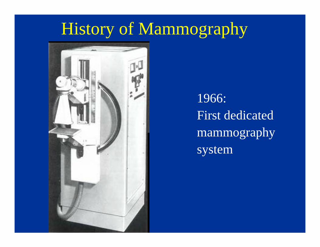
History of Mammography
1966:First dedicatedmammographysystem
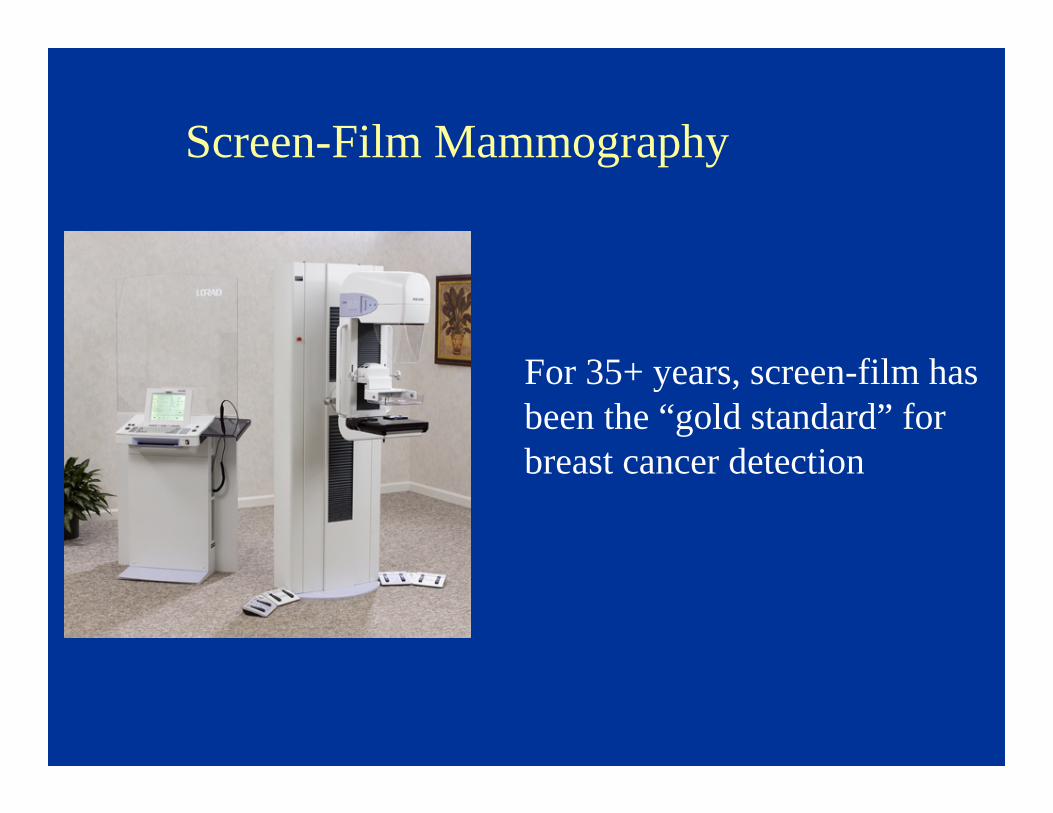
Screen-Film Mammography
For 35+ years, screen-film has been the “gold standard” for breast cancer detection
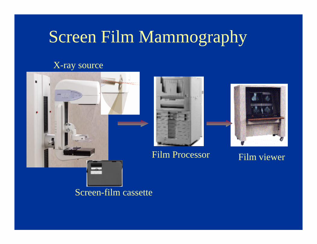
Screen Film Mammography
X-ray source
Screen-film cassette
Film Processor Film viewer
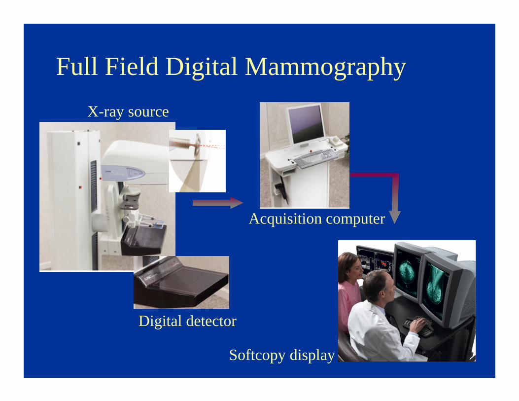
X-ray source
Digital detector
Full Field Digital Mammography
Acquisition computer
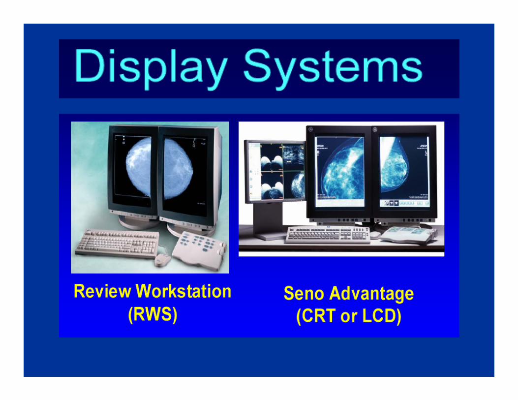
Softcopy display
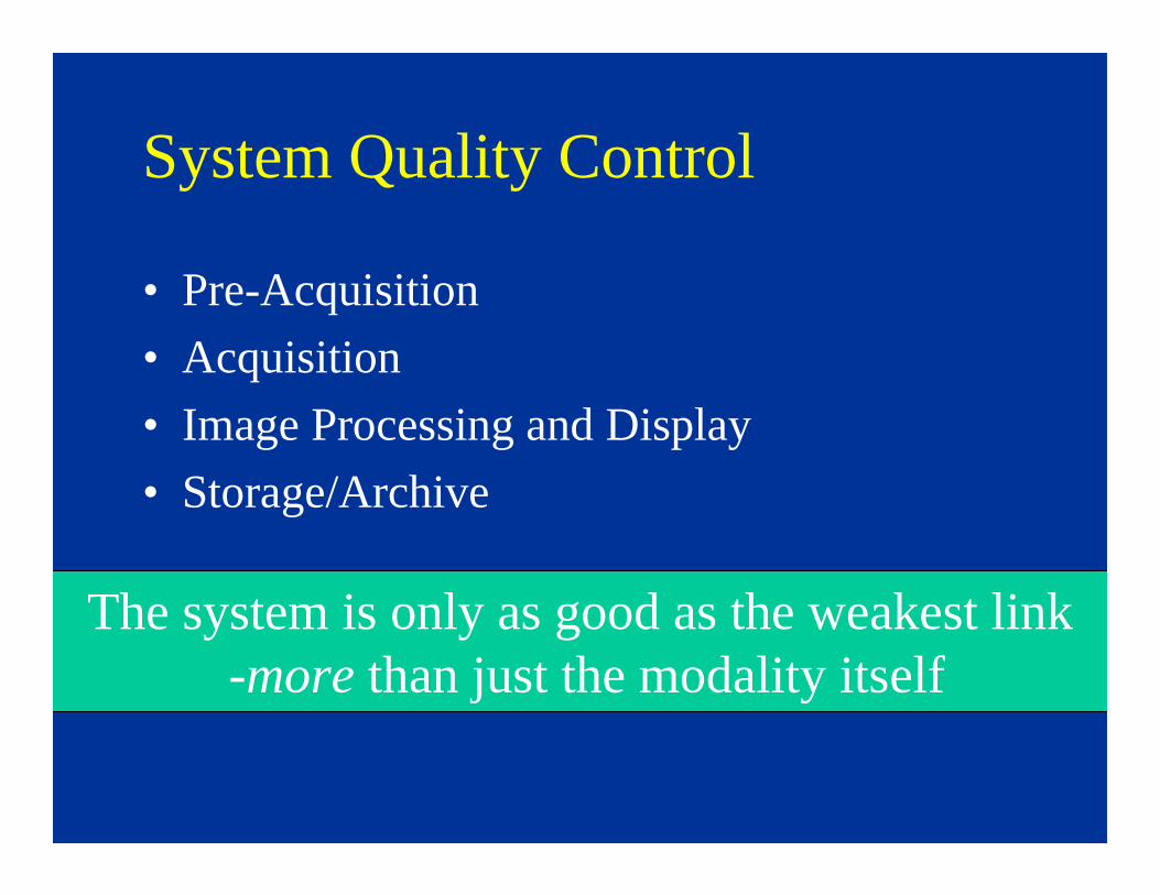
System Quality Control
• Pre-Acquisition
• Acquisition
• Image Processing and Display
• Storage/Archive
The system is only as good as the weakest link-more than just the modality itself
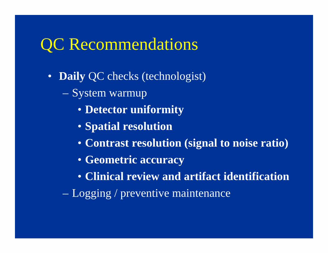
QC Recommendations
• Daily QC checks (technologist)
– System warmup
• Detector uniformity
• Spatial resolution
• Contrast resolution (signal to noise ratio)
• Geometric accuracy
• Clinical review and artifact identification
– Logging / preventive maintenance
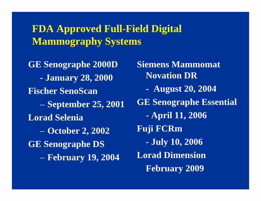
FDA Approved Full-Field Digital Mammography Systems
GE Senographe 2000D
- January 28, 2000
Fischer SenoScan
– September 25, 2001
Lorad Selenia
– October 2, 2002
GE Senographe DS
– February 19, 2004
Siemens MammomatNovation DR
- August 20, 2004
GE Senographe Essential
- April 11, 2006
Fuji FCRm
- July 10, 2006
Lorad Dimension
February 2009
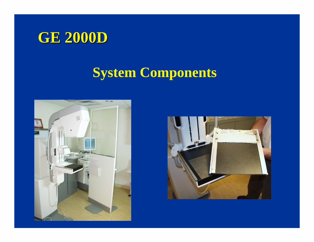
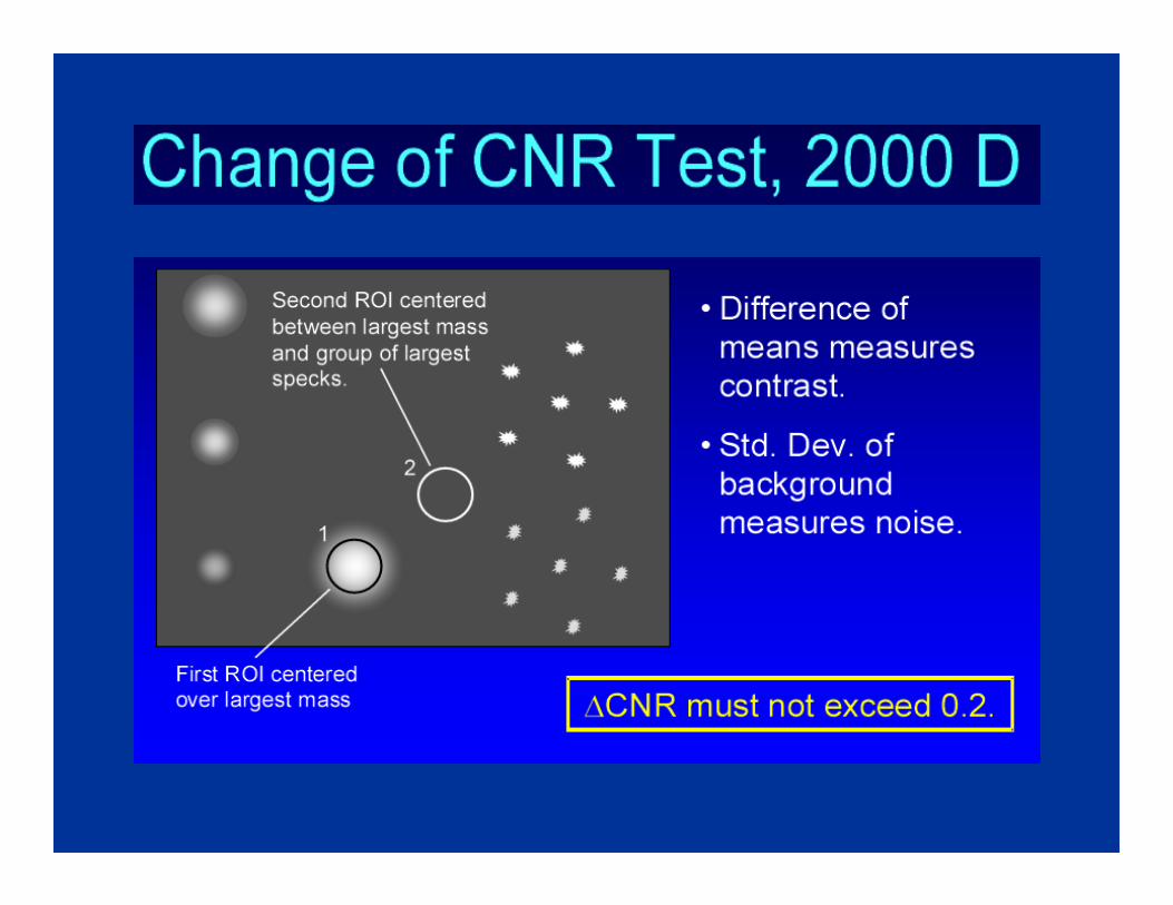
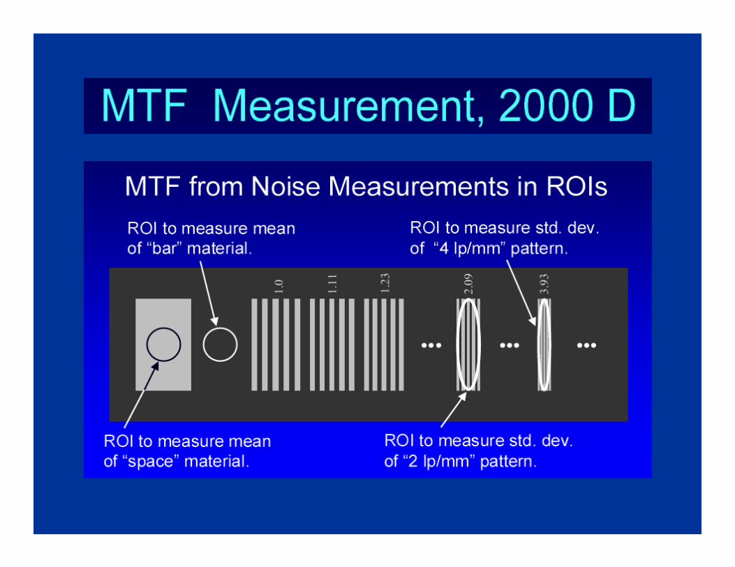
GE 2000DGE 2000D
System Components

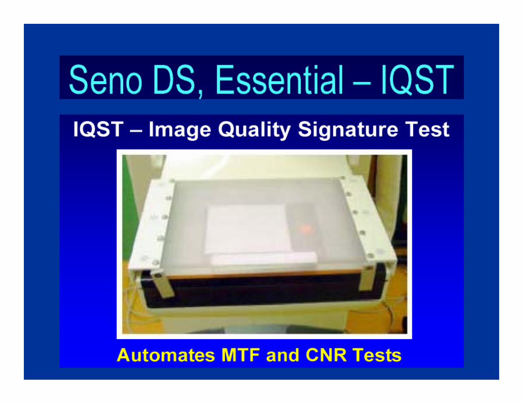
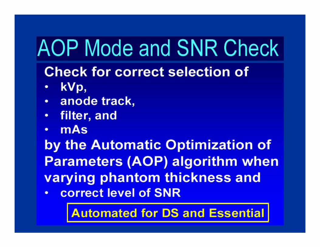
GE Seno DS & EssentialGE Seno DS & Essential
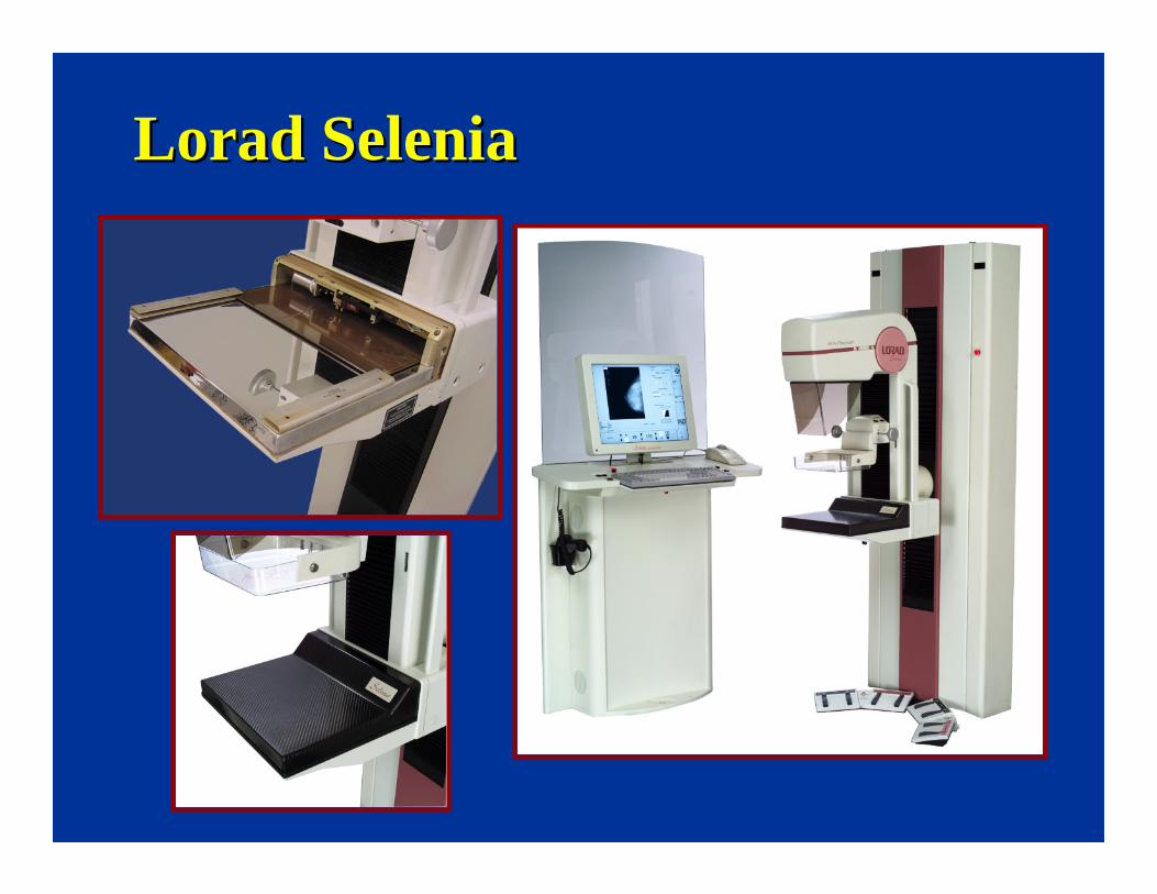
LoradLorad SeleniaSelenia
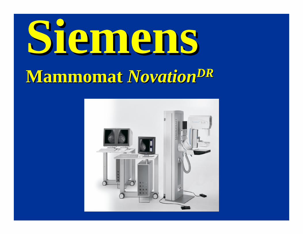
SiemensSiemensMammomatMammomat NovationNovationDRDR
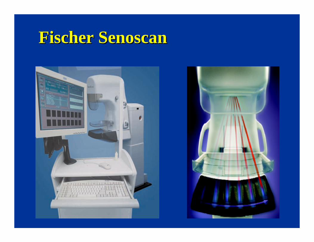
Fischer Fischer SenoscanSenoscan
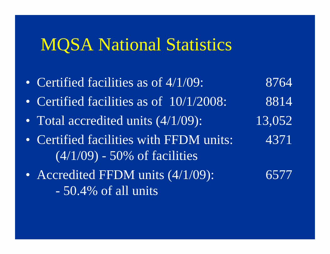
MQSA National Statistics
• Certified facilities as of 4/1/09: 8764
• Certified facilities as of 10/1/2008: 8814
• Total accredited units (4/1/09): 13,052
• Certified facilities with FFDM units: 4371(4/1/09) - 50% of facilities
• Accredited FFDM units (4/1/09): 6577- 50.4% of all units

Why Full Field Digital Mammography?Why Full Field Digital Mammography?
• Technical Reasons
• Clinical Reasons
• Practical Reasons
• Technical Reasons
• Clinical Reasons
• Practical Reasons
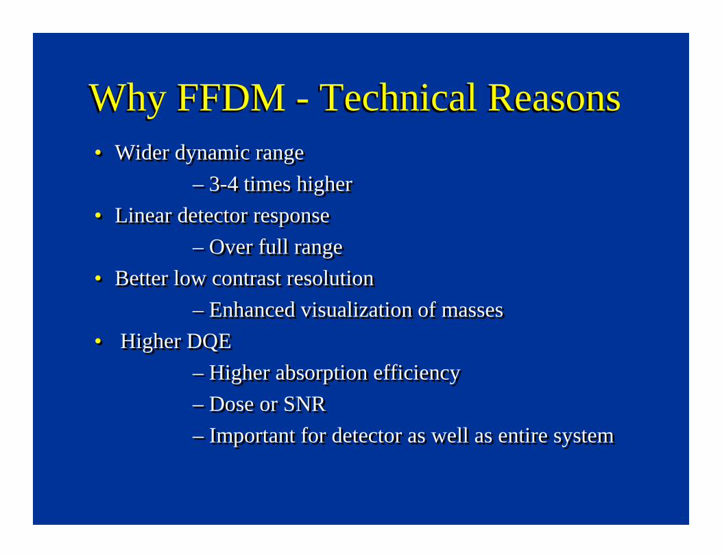
Why FFDM - Technical ReasonsWhy FFDM - Technical Reasons• Wider dynamic range
– 3-4 times higher
• Linear detector response
– Over full range
• Better low contrast resolution
– Enhanced visualization of masses
• Higher DQE
– Higher absorption efficiency
– Dose or SNR
– Important for detector as well as entire system
• Wider dynamic range
– 3-4 times higher
• Linear detector response
– Over full range
• Better low contrast resolution
– Enhanced visualization of masses
• Higher DQE
– Higher absorption efficiency
– Dose or SNR
– Important for detector as well as entire system
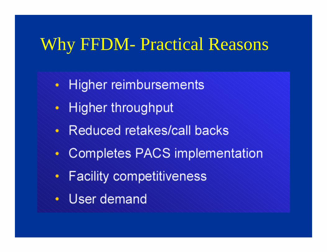
Why FFDM- Practical Reasons

Butler/wilcox - RSNA 2006
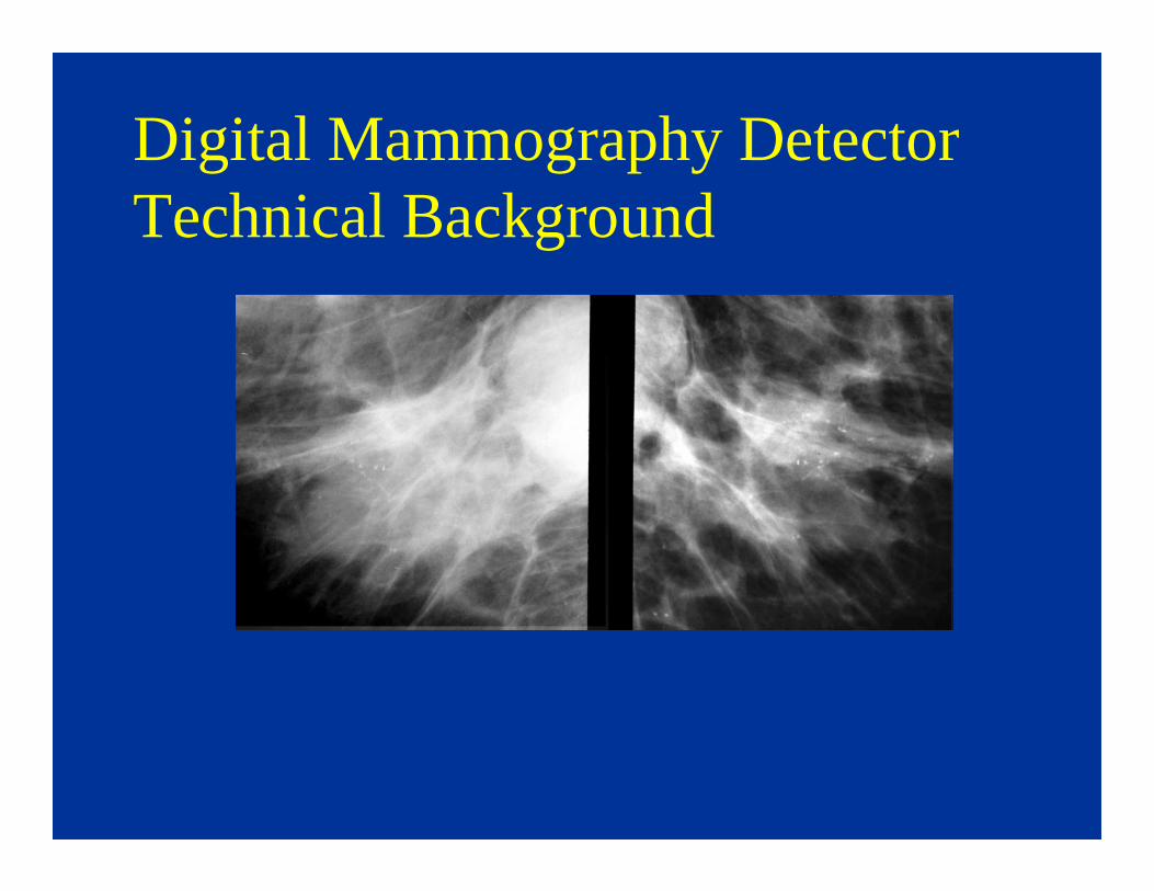
Digital Mammography Detector Technical Background
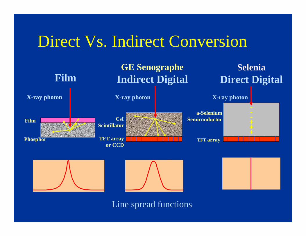
Direct Vs. Indirect Conversion
a-Selenium Semiconductor
TFT array
---+++
Selenia Direct Digital
CsI Scintillator
TFT array or CCD
GE SenographeIndirect Digital
X-ray photon X-ray photon
Line spread functions
Film
X-ray photon
Film
Phosphor

Currently Available FFDM Systems
direct conversion TFT
MammomatNovation (DR)
•Siemens Medical
direct conversion TFT
Selenia•Hologic/Lorad
indirect conversion, slot scanning
Senoscan•Fischer Imaging
(No longer available as new)
Indirect conversion TFT
Senographe 2000D, Senographe DS
and Essential
•GE Medical Systems
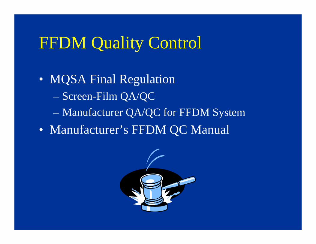
FFDM Quality Control
• MQSA Final Regulation– Screen-Film QA/QC
– Manufacturer QA/QC for FFDM System
• Manufacturer’s FFDM QC Manual

LORAD Selenia QC Tests
QC Manual tests:• Medical Physicist
– Annually
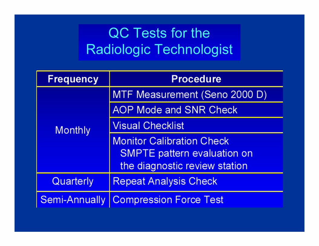
• Radiologic Technologist– Daily– Weekly– Biweekly– Monthly– Quarterly– Semiannually
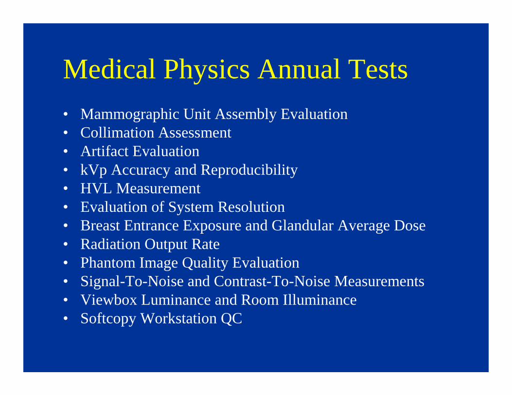
Medical Physics Annual Tests
• Mammographic Unit Assembly Evaluation• Collimation Assessment• Artifact Evaluation• kVp Accuracy and Reproducibility• HVL Measurement• Evaluation of System Resolution• Breast Entrance Exposure and Glandular Average Dose• Radiation Output Rate• Phantom Image Quality Evaluation• Signal-To-Noise and Contrast-To-Noise Measurements• Viewbox Luminance and Room Illuminance• Softcopy Workstation QC
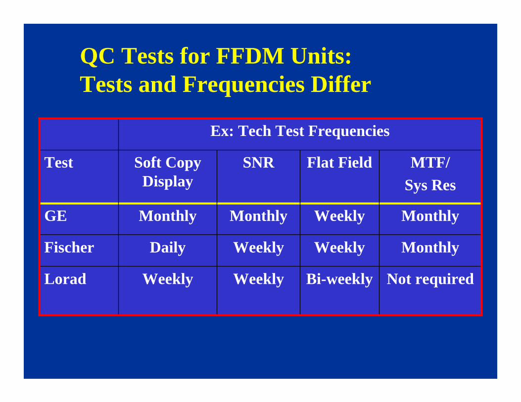
QC Tests for FFDM Units:Tests and Frequencies Differ
Not requiredBi-weeklyWeeklyWeeklyLorad
MonthlyWeeklyWeeklyDailyFischer
MonthlyWeeklyMonthlyMonthlyGE
MTF/ Sys Res
Flat FieldSNRSoft Copy Display
Test
Ex: Tech Test Frequencies

LORAD Selenia FFDM System
• RT QC Tests

Technologist QC Tests
• Laser Printer Quality Control (Weekly)
• SNR and CNR Measurements (Weekly)
• Softcopy Workstation QC (Weekly)
• Phantom Image (Weekly)
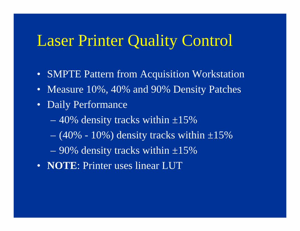
Laser Printer Quality Control
• SMPTE Pattern from Acquisition Workstation
• Measure 10%, 40% and 90% Density Patches
• Daily Performance
– 40% density tracks within ±15%
– (40% - 10%) density tracks within ±15%
– 90% density tracks within ±15%
• NOTE: Printer uses linear LUT
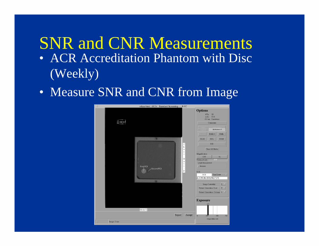
SNR and CNR Measurements• ACR Accreditation Phantom with Disc
(Weekly)
• Measure SNR and CNR from Image

SNR and CNR Measurements
• Passing Criteria
– SNR at least equal or greater than 40
– CNR should stay within ±15% of measurement obtained during acceptance testing of system
LORAD Selenia FFDM System
• Workstation QC
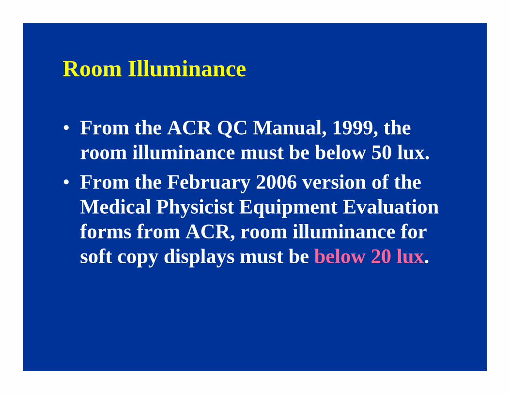
Room Illuminance
• From the ACR QC Manual, 1999, the room illuminance must be below 50 lux.
• From the February 2006 version of the Medical Physicist Equipment Evaluation forms from ACR, room illuminance for soft copy displays must be below 20 lux.
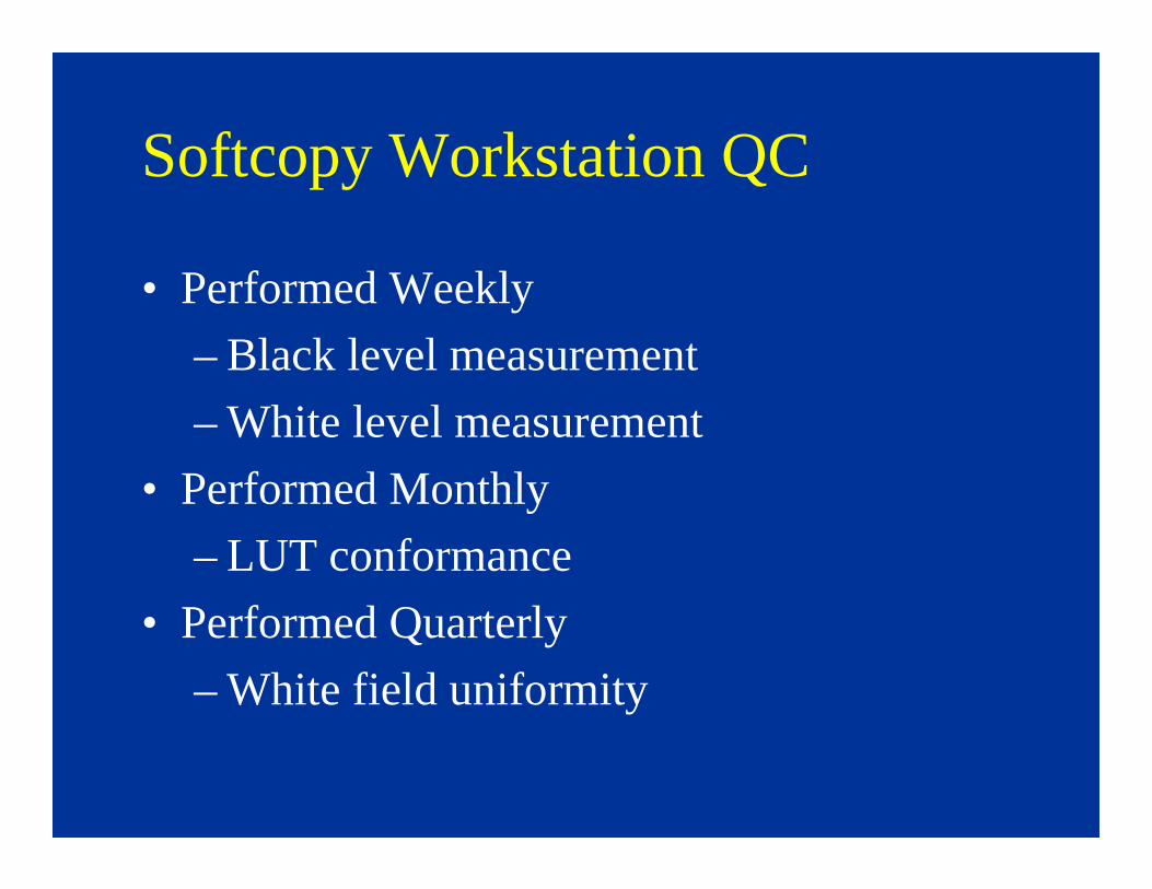
Softcopy Workstation QC
• Performed Weekly
– Black level measurement
– White level measurement
• Performed Monthly
– LUT conformance
• Performed Quarterly
– White field uniformity
Softcopy Workstation QC
• Data Logged by Software
• Warning Flags and Errors if Conformance Fails

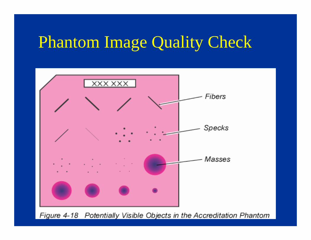
Phantom Image
• Review Weekly Phantom Image Results
– Measured film densities
– Phantom objects seen (printer, monitors)
• 5 fibers
• 4 speck groups
• 4 masses
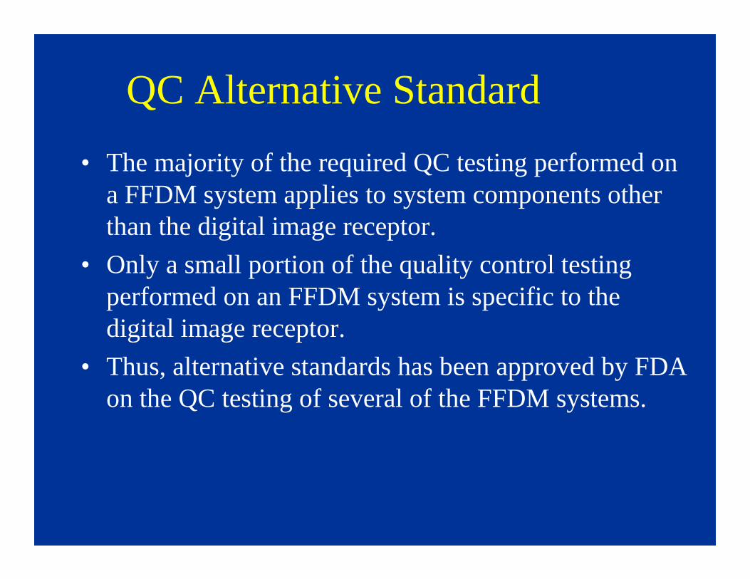
QC Alternative Standard
• The majority of the required QC testing performed on a FFDM system applies to system components other than the digital image receptor.
• Only a small portion of the quality control testing performed on an FFDM system is specific to the digital image receptor.
• Thus, alternative standards has been approved by FDA on the QC testing of several of the FFDM systems.
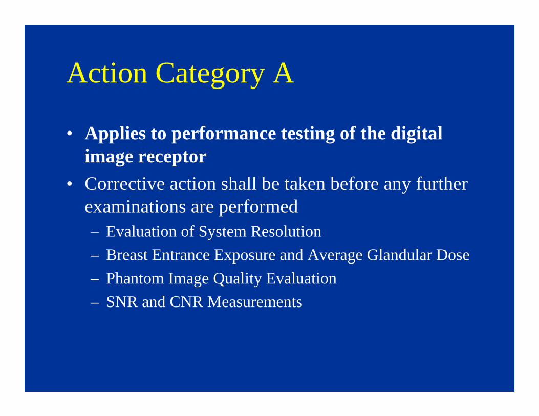
Action Category A
• Applies to performance testing of the digital image receptor
• Corrective action shall be taken before any further examinations are performed– Evaluation of System Resolution
– Breast Entrance Exposure and Average Glandular Dose
– Phantom Image Quality Evaluation
– SNR and CNR Measurements
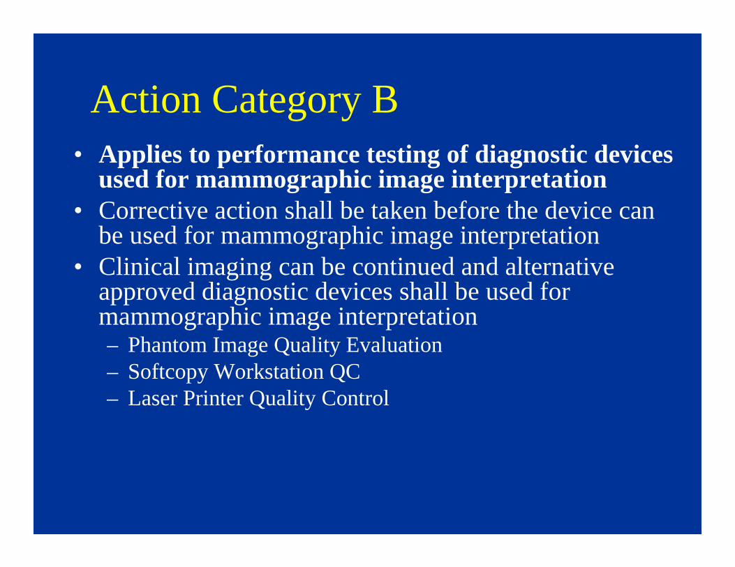
Action Category B• Applies to performance testing of diagnostic devices
used for mammographic image interpretation• Corrective action shall be taken before the device can
be used for mammographic image interpretation• Clinical imaging can be continued and alternative
approved diagnostic devices shall be used for mammographic image interpretation– Phantom Image Quality Evaluation– Softcopy Workstation QC– Laser Printer Quality Control

Action Category C
• Applies to performance testing of rest of the system
• Corrective action shall be taken within thirty days of the test date
• Clinical imaging and mammographic image interpretation can be continued during this period– All other tests


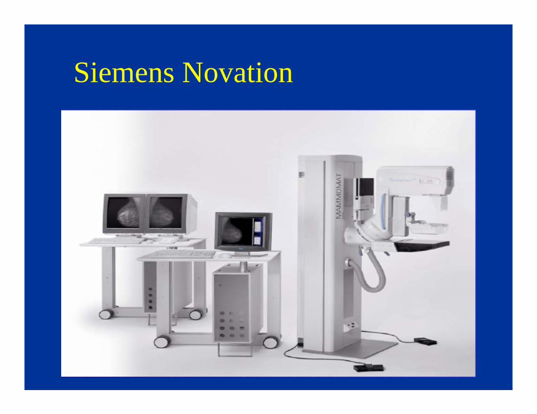
Siemens Novation

Phantom Image Quality Check
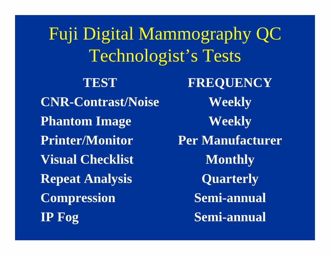
Fuji Digital Mammography QCTechnologist’s Tests
TESTCNR-Contrast/NoisePhantom ImagePrinter/MonitorVisual ChecklistRepeat AnalysisCompressionIP Fog
FREQUENCYWeeklyWeekly
Per ManufacturerMonthly
QuarterlySemi-annualSemi-annual
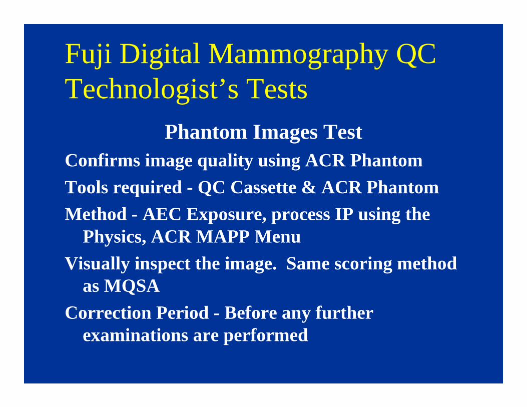
Fuji Digital Mammography QCTechnologist’s Tests
Phantom Images TestConfirms image quality using ACR Phantom
Tools required - QC Cassette & ACR Phantom
Method - AEC Exposure, process IP using the Physics, ACR MAPP Menu
Visually inspect the image. Same scoring method as MQSA
Correction Period - Before any further examinations are performed
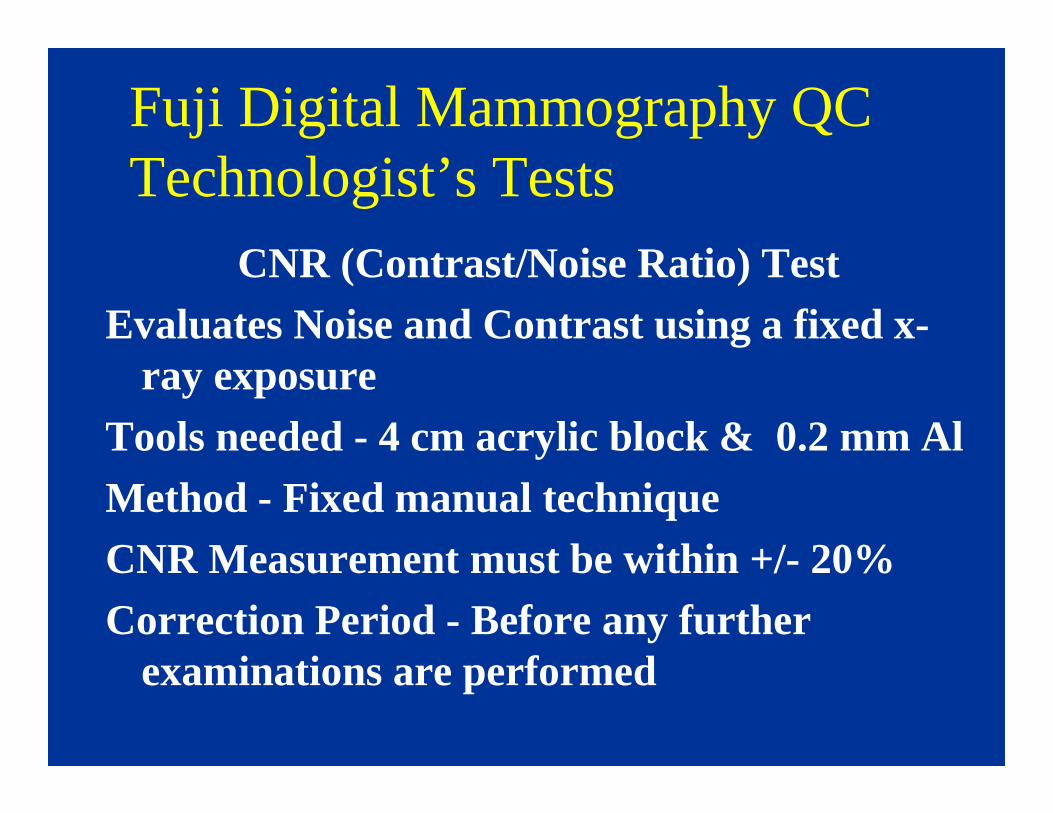
Fuji Digital Mammography QCTechnologist’s Tests
CNR (Contrast/Noise Ratio) TestEvaluates Noise and Contrast using a fixed x-
ray exposureTools needed - 4 cm acrylic block & 0.2 mm AlMethod - Fixed manual techniqueCNR Measurement must be within +/- 20%Correction Period - Before any further
examinations are performed

Fuji Digital Mammography QCTechnologist’s Tests
Tests in Accordance with ACR/MQSAVisual Checklist Monthly
Repeat Analysis Quarterly
Compression Test Semi-Annual
Same Action Limits and Corrective Action as MQSA
IP Fog replaces Darkroom Fog test - Semi Annual

There Are Currently Several FDAThere Are Currently Several FDA--Approved Laser Imagers for Digital Approved Laser Imagers for Digital MammographyMammography
•• Agfa DS4500MAgfa DS4500M
•• Kodak 8600 Laser ImagerKodak 8600 Laser Imager
•• Kodak 8610 Laser ImagerKodak 8610 Laser Imager
•• Kodak 8900MKodak 8900M
•• Fuji Fuji DrypixDrypix 7000 & 5000 7000 & 5000
•• Fuji Fuji DrypixDrypix FMFM--DP LDP L
•• Konica Konica DryProDryPro 793793
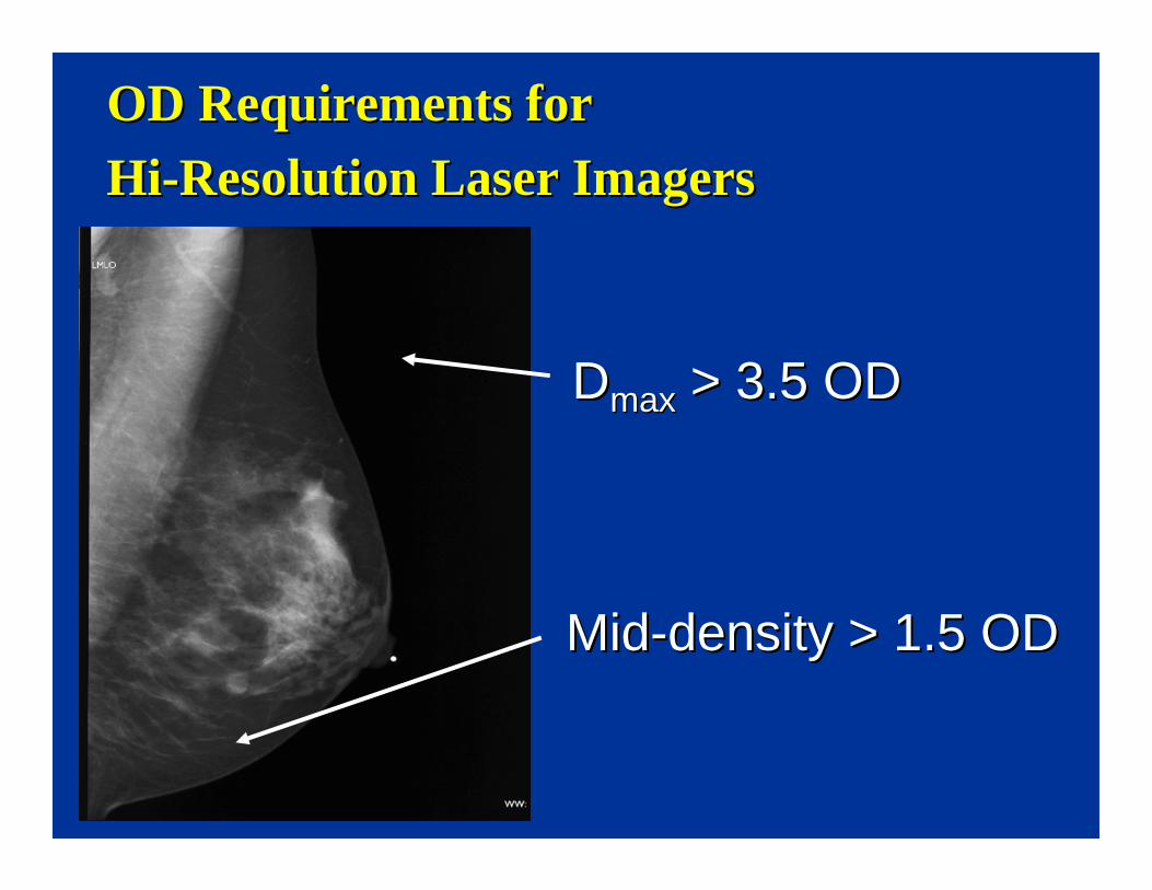
OD Requirements for OD Requirements for
HiHi--Resolution Laser ImagersResolution Laser Imagers
DDmaxmax > 3.5 OD> 3.5 OD
MidMid--density > 1.5 ODdensity > 1.5 OD
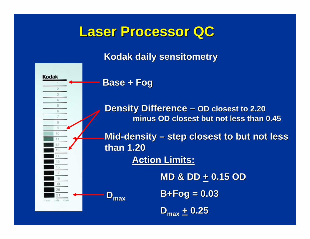
Kodak daily sensitometryKodak daily sensitometry
Laser Processor QCLaser Processor QC
Base + FogBase + Fog
Density Difference Density Difference –– OD closest to 2.20 OD closest to 2.20 minus OD closest but not less than 0.45minus OD closest but not less than 0.45
MidMid--density density –– step closest to but not less step closest to but not less than 1.20than 1.20
DDmaxmax
Action Limits:Action Limits:
MD & DD MD & DD ++ 0.15 OD0.15 OD
B+Fog = 0.03B+Fog = 0.03
DDmaxmax ++ 0.250.25

Butler/Wilcox - RSNA 2006

Thank You !!!
Melissa C. Martin, M.S., FACRTherapy Physics Inc.
879 West 190 St., Ste 419Gardena, CA 90248
Office Phone: 310-217-4114e-mail: [email protected]