Purpose of the study: To evaluate the diagnostic potential ...
4
Transcript of Purpose of the study: To evaluate the diagnostic potential ...

Image of the upcoming MARS arm scanner is indicative only
IntIntroduction: The scaphoid bone is the most frequently fractured carpal bone, most commonly after a fall onto an outstretched hand. The diagnostic modalities used to detect a scaphoid fracture include plain radiograph, computed tomography (CT), magnetic resonance imaging (MRI), and bone scintigraphy. In this study we used a a new high-resolution imaging modality, MARS, to analyse fracture non-union characteristics.
MARS uses a low power x-ray tube, photon counting detectors (Medipix3RX) and narrow energy ranges to differentiate and measure multiple materials (lipid, water and calcium) in a single scan. The smaller pixel size of the photon counting detectors (110 µm) enables visualisation and quantification of both cortical and trabecular density,density, as well as characterising the morphology of the fracture site. The scans are lower dose and can be retrospectively reconstructed in isotropic voxel sizes ranging from 60 µm to 500 µm.
We present three cases of scaphoid fracture non-union. These patients either had an x-ray or a CT scan to diagnose the fracture.
Purpose of the study: To evaluate the diagnostic potential of MARS spectral photon-counting CT in the quantitative and qualitative assessment of scaphoid fractures.
This study was done in association with:
Mr. Ram Chandru Orthopaedic Surgeon Christchurch, New Zealand
Dr. Jonathan Crighton RadiologistCanterbury District Health BoardCanterbury District Health Board
Dr. Luke Holmes Radiology RegistrarCanterbury District Health Board
Conclusion: The MARS scanner is an ultra- high-resolution, low-dose (<0.05 mSv), 3D advanced imaging modality which produces high contrast images containing tissue composition information and reduced metal artifact, facilitating improved diagnostic potential.
ClinicalClinical relevance: The MARS scanner produces high contrast bone detail, as well as assessment of bone density and morphology at the fracture site. This increased level of detail and information can aid in treatment decisions, as well as surgical planning. Postoperative imaging evaluation of fracture fixation with metalware is often impaired byby the metal artifact, which makes it difficult to evaluate the surrounding tissue. Due to reduced metal artifact and greatly improved visualisation of the bone-metal interface, MARS can be used to assess fracture healing and identify any metalware complications, such as implant loosening, migration of screw, or infection.
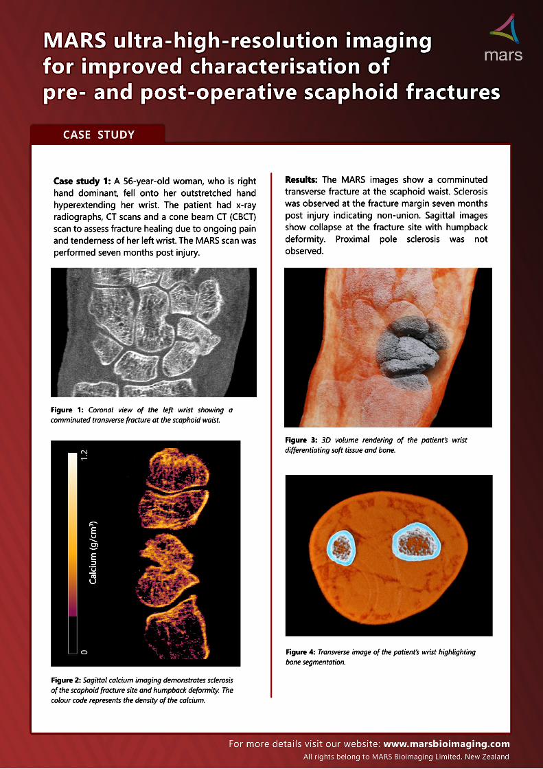
Figure 3: 3D volume rendering of the patient’s wrist differentiating soft tissue and bone.
Figure 2: Sagittal calcium imaging demonstrates sclerosis of the scaphoid fracture site and humpback deformity. The colour code represents the density of the calcium.
Figure 1: Coronal view of the left wrist showing a comminuted transverse fracture at the scaphoid waist.
Calcium (g/cm3 )
CaseCase study 1: A 56-year-old woman, who is right hand dominant, fell onto her outstretched hand hyperextending her wrist. The patient had x-ray radiographs, CT scans and a cone beam CT (CBCT) scan to assess fracture healing due to ongoing pain and tenderness of her left wrist. The MARS scan was performed seven months post injury.
RResults: The MARS images show a comminuted transverse fracture at the scaphoid waist. Sclerosis was observed at the fracture margin seven months post injury indicating non-union. Sagittal images show collapse at the fracture site with humpback deformity. Proximal pole sclerosis was not observed.
Figure 4: Transverse image of the patient’s wrist highlighting bone segmentation.

Figure 7: 3D rendering of the calcium imaging of patient’s wrist with scaphoid fracture.
Case study 2: A 55-year-old right hand dominant male was troubled with chronic right wrist pain following a bike crash. The patient had radiographs and CBCT scan to monitor the progress of fracture healing. Twelve weeks post injury, the patient had a MARS scan.
Figure 6: Sclerosis of the fracture margins was not observed on calcium imaging.
Figure 5: Coronal view of the right wrist demonstrates the separation between the fracture fragments.
Results: The MARS images show non-union with displacement. Small bony fragments were observed at the fracture margin. The separation between the fracture fragments was more than 3 mm. Sclerosis was not observed.
Calcium (g/cm3 )
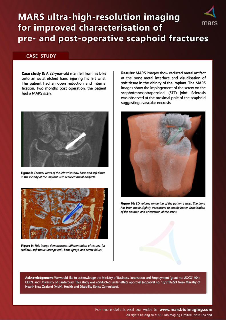
Case study 3: A 22-year-old man fell from his bike onto an outstretched hand injuring his left wrist. The patient had an open reduction and internal fixation. Two months post operation, the patient had a MARS scan.
Results: MARS images show reduced metal artifact at the bone-metal interface and visualisation of soft tissue in the vicinity of the implant. The MARS images show the impingement of the screw on the scaphotrapeziotrapezoidal (STT) joint. Sclerosis was observed at the proximal pole of the scaphoid suggesting avascular necrosis.
Figure 9: This image demonstrates differentiation of tissues, fat (yellow), soft tissue (orange red), bone (grey), and screw (blue).
Figure 10: 3D volume rendering of the patient’s wrist. The bone has been made slightly translucent to enable better visualisation of the position and orientation of the screw.
Figure 8: Coronal views of the left wrist show bone and soft tissue in the vicinity of the implant with reduced metal artifacts. (a)
Acknowledgement: We would like to acknowledge the Ministry of Business, Innovation and Employment (grant no: UOCX1404), CERN, and University of Canterbury. This study was conducted under ethics approval (approval no: 18/STH/221 from Ministry of Health New Zealand (MoH), Health and Disability Ethics Committee).