Pulse analysis: stroke volume, vascular stiffening and cardiovascular disease Tom Archer, MD, MBA...
91
Pulse analysis: stroke volume, vascular stiffening and cardiovascular disease Tom Archer, MD, MBA UCSD Anesthesia
-
Upload
garey-sims -
Category
Documents
-
view
218 -
download
0
Transcript of Pulse analysis: stroke volume, vascular stiffening and cardiovascular disease Tom Archer, MD, MBA...

Pulse analysis: stroke volume, vascular stiffening
and cardiovascular disease
Tom Archer, MD, MBA
UCSD Anesthesia

http://www.itmonline.org/image/pulse2.jpg
Pulse analysis is an ancient practice, now making a comeback.

Pablo Picasso, “Science and Charity”, 1897

Traditional pulse “analysis”: subjective and hard to quantify
• “Waterhammer pulse”-- AI
• “Slow upstroke”-- AS
• “Pulsus paradoxus”– cardiac tamponade
• Is this silly and obsolete stuff?

Pulse analysis was serious business in the 19th century
• Sphygmographs in common use.
• Insurance companies relied on their results.

http://www.mamweb.org/modules.php?name=Content&pa=showpage&pid=32000
Etienne-Jules Marey (1830-1904) invented the sphygmograph to record the arterial pulse on smoked paper. It was used by Engelmann, Mackenzie and Wenckebach.
Sphygmograph 1876

Life insurance examination manual from 1891 discussed pulse analysis by sphygmography.


Tom Archer, 58 y.o., good general health. Takes Crestor for high cholesterol.
Radial and predicted ascending aortic pressure waveform when subject is cold.

Scipione Riva-Rocci introduced the mercury sphygmo-manometer in 1896.
Measured systolic BP only.

history.library.ucsf.edu/.../chapter2_03.html
Harvey Cushing used it.
Korotkoff introduced auscultation for diastolic pressure in 1905.

In the 20th century, Riva-Rocci and Korotkoff’s sphygmomanometer
eclipsed pulse analysis
• Two simple numbers: systolic / diastolic.
• Easy to use.
• Pulse analysis fell into disuse.
• 20th century saw tremendous gains from simple sphygmomanometry: dangers of high BP.

K Hirata (Circ J 2006; 70: 1231–1239)
Increased diastolic BP is associated with CAD– multiple large studies.
High diastolic blood pressure is associated with increased risk of coronary disease.
K Hirata (Circ J 2006; 70: 1231–1239)
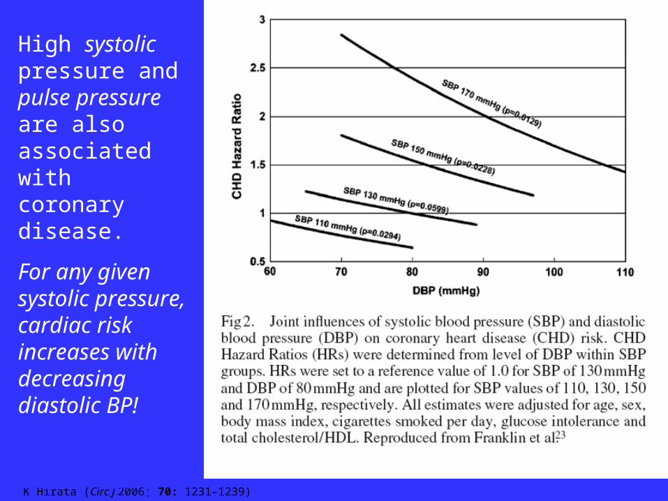
Both increased brachial SBP and increased brachial pulse pressure are associated with increased coronary disease.
High systolic pressure and pulse pressure are also associated with coronary disease.
For any given systolic pressure, cardiac risk increases with decreasing diastolic BP!

But simple sphygmomanometry ignores valuable information within
the pulse trace.
• Extra information can be extracted from the pulse using transducers and computers.
• Pulse analysis is becoming reproducible and objective.
• Pulse analysis is JUST SOFTWARE analyzing the BP signal.

Pulse analysis gives two types of information
• “Central blood pressure” – Ascending aortic blood pressure from
radial waveform.– Specifically, we get “Augmentation
Index” (AIx)– a measure of extra heart work.
• Stroke volume (CO, SVR)

Central blood pressure (CBP)
• Systolic pressure in the ascending aorta is NOT the same as brachial or radial systolic BP.
• Diastolic and mean pressures are very similar at radial / brachial and central sites.

health.yahoo.com/topic/heart/overview/article...
LV “sees” the SBP in the ascending aorta.
With normal aortic valve, LV wall tension depends on pressure in ascending aorta
(and diameter of LV chamber).
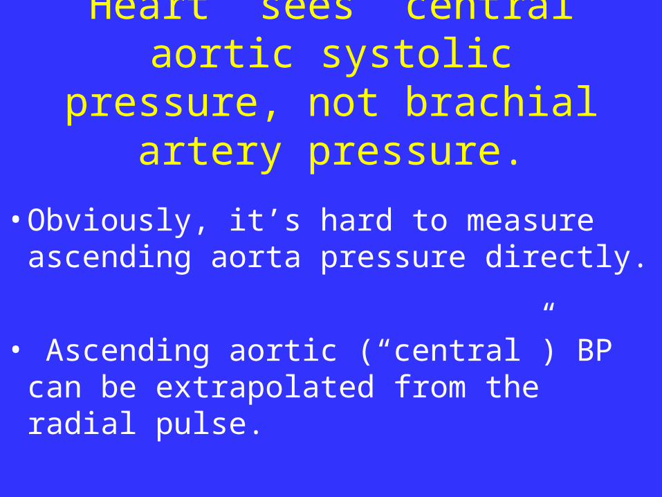
Heart “sees” central aortic systolic pressure, not brachial
artery pressure.
• Obviously, it’s hard to measure ascending aorta pressure directly.
• Ascending aortic (“central”) BP can be extrapolated from the radial pulse.

SphygmoCor system for measuring central blood pressures

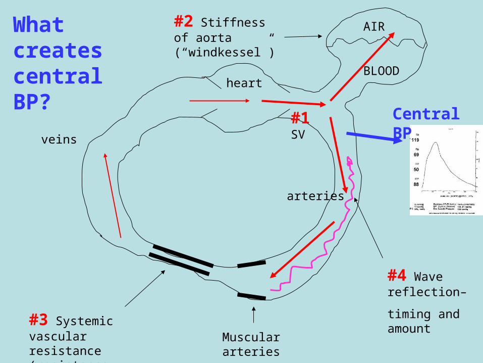
What creates central BP?
• Stroke volume
• Aortic stiffness (compliance / Windkessel)
• Systemic vascular resistance (“runoff”)
• Reflected pressure wave
AIR
BLOOD
#3 Systemic vascular resistance (resistance arterioles)
#2 Stiffness of aorta (“windkessel”)
heart
veins
arteries
#1 SV
#4 Wave reflection–
timing and amount
Muscular arteries
Central BP
What creates central BP?

Kozo Hirata, MD; Masanobu Kawakami, MD; Michael F O’Rourke, MD, DSc*Circ J 2006; 70: 1231–1239
AIx =
Augmentation Pressure /
Pulse Pressure
Augmentation index– extra cardiac work due to wave reflection

Augmentation index is a deadly backdraft of pressure which exhausts the heart over time.

Run animation
• Wave reflection animation can be found at:
• http://atcormedical.com/wave_reflection.html

Augmentation Index (AIx)
• AIx = unnecessary heart work.
• High AIx leads to LVH and cardiomyopathy.
• Lower AIx is better.
• Treatments that lower AIx help the patient.

Hypertensive patients treated to identical brachial BP
endpoints with
amlodipine
and
atenolol
show lower
central BPs and AIx
with amlodipine
CAFE / ASCOT study, M. O’Rourke (Circulation. 2006;113:1213-1225.)

A given brachial BP measurement does not say what central BP the heart
is actually generating.
Antihypertensive drugs may exert
their beneficial effects via effects on
central blood pressure.
These effects may not be appreciated
by just measuring brachial BP

Central BPs– ASCOT / CAFE study
• Lower central BPs are associated with better CV outcomes.
• Amlodipine achieved lower central BPs and had better CV outcomes than atenolol, despite achieving the same brachial artery BPs.
CAFE / ASCOT study, M. O’Rourke (Circulation. 2006;113:1213-1225.)

When is AIx high-- chronically?
• Normal aging
• Obesity
• Atherosclerosis
• Diabetes
• Pre-eclampsia
• Inflammatory arthritis
• Renal failure

WW Nichols Curr Opin Cardiol 2002, 17:543–551
As healthy individuals age, reflected wave arrives at ascending aorta earlier and increases augmentation index and central pulse pressure.

Tom Archer, 58 yo, after work, seated comfortably. Aix = 11%.

67 y.o. obese female-- AIx 24%

77 y.o. man. Never smoked.
Aix = 29%.

42 y.o. obese female, smoker.
Aix = 36%

77 yo male, ESRD, AV fistula, CAD, HBP
Takes atenolol, lisinopril, terazosin, finasteride. Aix = 39%

When is Aix high– acutely?
• Arterial compression in legs (squatting)
• Cold body temperature.
• Nicotine ingestion

Tom Archer, 58 y.o., while squatting.
Aix = 21%
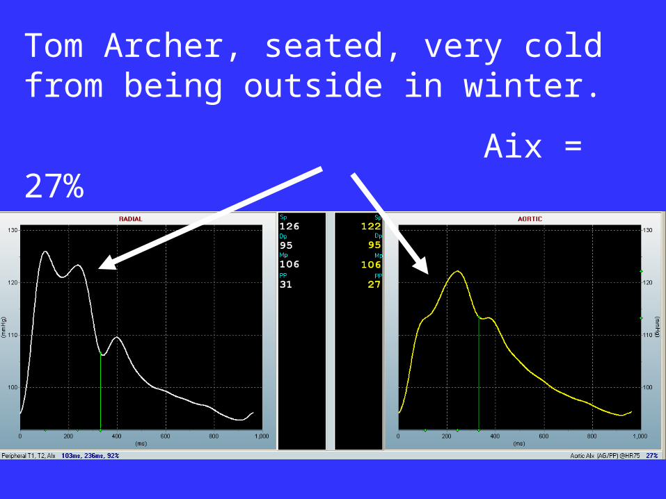
Tom Archer, seated, very cold from being outside in winter.
Aix = 27%

David G. Edwards,1 Amie L. Gauthier,2 Melissa A. Hayman,2 Jesse T. Lang,2 and Robert W. Kenefick2J Appl Physiol 100: 1210–1214, 2006.
Exposure of healthy young adults to cold air for 30 min increases augmentation index.

Does cold weather increase MI rate due to increased AIx?

Perioperative hypothermia increases cardiac event rate.
Is this due to increased AIx with hypothermia?

What makes AIx go down-- chronically?
• Exercise
• Weight loss
• Red wine
• Statins
• Control of blood pressure (ACEI and CCB)
• NTG


Ted A, 30 yo, at rest, seated. Subject runs marathons. Aix = -14%.

64 yo obese male. HBP on lisinopril. Moderate ETOH consumption.
Aix = 14%.

What makes Aix go down-- acutely?
• Exercise
• Red wine
• Lowering blood pressure
• NTG

Tom Archer, 58 yo, after exercise and wine.
AIx = 1%

S. C. MILLASSEAU, R. P. KELLY, J. M. RITTER and P. J. CHOWIENCZYK
Clinical Science (2002) 103, 371–377
NTG reduces wave reflection and AIx by dilating muscular arteries.
Is this its primary mechanism of action?

What determines augmentation index?
• Timing of wave reflection– pulse wave velocity. Faster wave return is bad.
• Amount of wave reflection– muscular artery tone.–NTG reduces muscular artery tone and
wave reflection–Does NTG work by decreasing AIx?
Yes, at least in part.

PWV (aortic stiffness)
increases with age.
AR Khoshdel Journal of Hypertension 2006, 24:1231–1237

AIx increases in “inflammatory” states:
Obesity
• OSA
• Hyperglycemia
• Sepsis
• Pre-eclampsia
• Lupus
• Cocaine
• Hypercholesterolemia

MT, 22 yo, healthy, in labor, epidural in place and she is comfortable.
Aix = -1%.

JM, 21 yo, in labor, recent onset lupus, on prednisone and plaquenil
Aix = 6%

Statins and ACE inhibitors can lower central BPs
and AIx

6 months Rx with atorvastatin decreased central aortic pulse pressure and augmentation index.
WW Nichols Curr Opin Cardiol 2002, 17:543–551

Four months Rx with lisinopril decreased central aortic pulse pressure and augmentation index.
WW Nichols Curr Opin Cardiol 2002, 17:543–551

DO, 56 yo female, hypertensive, diabetic May 31, 2007. Aix = 41%

DO, 56 yo female, hypertensive, diabetic January 3, 2008. After weight loss and 3 weeks Lipitor. AIx = 26%
ACE inhibitors and aldosterone antagonists
can reverse LV hypertrophy—
is this due to decreased AIx and strain on the heart?

Adams KF, Am J Health-Syst Pharm—Vol 61 May 1, 2004 Suppl 2
ACE inhibitors and aldosterone antagonists reverse LV hypertrophy– via central BP effects?.

Radial Pulse Analysis II:
Pulse analysis to determine stroke volume and cardiac
output from the arterial pressure trace.

Pulse contour analysis
• We’ll skip the math and validation studies.
• Intuitively– the bigger the waveform and the longer it lasts, the bigger the stroke volume.
• Calibration (e.g. with lithium dilution) vs. age / weight / height algorithm to predict probable aortic compliance and then follow trends.

Big stroke volume
Small stroke volume
P
R
E
S
S
U
R
E

Godje O Crit Care Med 2002 Vol. 30, No. 1
PiCCO system of pulse contour analysis
Aortic compliance
(low compliance and stiff aorta means high pulse pressure)
Systemic vascular resistance
(low resistance means rapid fall in diastolic pressure)

Stroke volume from arterial pressure trace--- PulseCO from LiDCO Ltd. (UK)

Stroke volume from arterial pressure trace--- Edwards Life Sciences, Irvine California

Stroke Volume Estimation by Pulse Contour Analysis
• Complicated
• Lots of physics and math
• Computerized
• Real time
• Needs a good waveform
• Has limitations, but it usually works

Stroke volume estimation by pulse contour analysis
• Just software for analyzing pulse contour.
• No additional patient risk or invasion over that of arterial line alone.

PulseCO:
Hemodynamic examples

Dramatic drop in SV, CO and BP with small dose of propofol (40 mg) after loading with fentanyl, lidocaine and esmolol.
CO
SVR
BP
SV HR

Continuous spinal in morbidly obese parturient at C/S causes fall in SVR, rise in
CO. Phenylephrine increases SVR and decreases CO.

GA induction with etomidate and sux associated with extreme hypertension in patient with pre-eclampsia and lupus. Hypertension due to both inc CO and inc SVR.

Repeat CS. Epidural anesthesia. Delivery with inc in HR and CO, oxytocin bolus with decrease SVR and BP, increase in CO and SV.

20
10
0
1000
500
0
Nominal cardiac output L/min
Nominal systemic vascular resistance dyn.sec.cm-5
Blood pressure mm Hg
Heart rate beats/min and nominal stroke volume mL
200
100
0
150
100
50
0 0 A 5 10 B 15 C/D 20
minutes
SV
Septic woman for CS. SVR does not rise with intubation (A) and incision (B).

Tourniquet inflation on leg increases SVR and MAP and decreases CO. Deflation reverses these changes.

Phenylephrine and vasopressin both increase SVR and decrease CO (in this patient).

Repeat CS, CSE. Severe pre-eclampsia. Multiple nicardipine boluses cause decreases in SVR and MAP, increases in SV and CO.

CS patient with severe pre-eclampsia superimposed on CRF: Antihypertensive therapy with labetalol 25 mg and hydralazine 5 mg (A) and nicardipine 250 µ total (B). Behind almost “stable” BP is drop in SVR, rise in CO.
8
4
0
3000
2000
1000
0
200
100
0
150
100
50
0
0 10 20 30 40
A minutes B V
Nominal cardiac output L/min
Nominal systemic vascular resistance dyn.sec.cm-5
Blood pressure mm Hg
Heart rate beats/min and nominal stroke volume mL

Methylene blue x 2 and indigo carmine x 1 scavenge NO and increase SVR. Cystoscopy after C-hysterectomy, GA, placenta percreta.

In conclusion
• Radial pulse wave analysis can give us two types of information:
–Central BPs and augmentation index, a measure of unnecessary heart work
–Stroke volume (and CO and SVR)

In conclusion
• Pulse wave analysis has two distinct techniques:
– Radial pulse tonometry (CBP and AIx)• AtCor Medical (SphygmoCor)
– Pulse contour analysis (SV, CO and SVR)• LiDCO• Vigileo (Edwards)• PiCCO• Others
In conclusion
• Radial pulse tonometry gives us central blood pressures and a measure of unnecessary heart work during systole– augmentation index (AIx).
• Central BPs and AIx may be the “smoking gun” for CV damage.

In conclusion
• Helpful therapeutic interventions such as ACE inhibitors, statins, calcium channel blockers and NTG may exert some of their therapeutic effects via their effects on central BP and AIx.
• Hence, measurement of these parameters may become part of the routine Dx and Rx of CV disorders.

In conclusion
• Measurement of CBP and AIx may become useful in acute CV care. This is speculative at this point.
• It behooves us to put this technology on our “radar screen”.

In conclusion
• Pulse contour analysis: several companies are working hard in this field.
• I am most familiar with LiDCO, but Edwards is pushing hard with Vigileo.

In conclusion
• Pulse contour analysis to estimate stroke volume, CO and SVR works well enough to be clinically useful.
• I find it fascinating and a great teaching and learning tool.
• Even if the numerical estimates are incorrect, the trends are correct.
• Both LiDCO and Edwards have “calibration-free” products.

Questions for the future
• Will radial pulse tonometry (and AIx) become a routine office evaluation of patients at risk for CV disease?
• Will it provide an “early warning system” for the development of CV disease?
• Will it guide drug therapy of CV disease?

Questions for the future
• Will measurement and pharmacological manipulation of AIx in critical care improve patient outcomes?

Questions for the future
• Will pulse contour analysis for CO and SVR be incorporated into standard arterial line monitoring as a routine?

The End
Questions?