Pulmonary Atresia With Intact Ventricular Septum ...
8
Rev Esp Cardiol. 2007;60(8):833-40 833 Introduction and objectives. In patients with pulmonary atresia with intact ventricular septum (PAIVS), radiofrequency-assisted perforation of the valve is the most widely used initial therapy when the anatomy is favorable. We report our experience with a modified mechanical technique that gave good results. Methods. Between November 2001 and October 2006, valve opening was carried out successfully in 11 consecutive neonates with a favorable anatomy (ie, Alwi groups A and B, and tricuspid valve Z-score –1.1 [1.3]). The technique involved snare-assisted anterograde or retrograde perforation with the soft tip of a special guidewire used for chronic total coronary artery occlusions, use of an arteriovenous loop, and progressive balloon dilatation from a diameter of 2 mm to a maximum diameter of 9.6 [1.2] mm. Results. Valve opening was achieved in all patients, and right ventricular (RV) systolic pressure fell from 97 [17] mm Hg to 48 [13] mm Hg (P<.001). No pericardial effusion or cardiac tamponade was observed, though 1 neonate died 24 hours after the procedure due to pulmonary embolism. Six patients (54%) were discharged without any further intervention, while 4 (36%) required an additional increase in pulmonary blood flow. During the follow-up period of 25 [21] months, 2 patients died. Eight (72%) survived and were in New York Heart Association functional class 1. Two required additional surgery on the outflow tract, one of whom also needed a one-and-a-half ventricular repair. Data indicate that the valves remain open as RV structures grow, though without any change in the tricuspid valve Z-score. Conclusions. Pulmonary valvuloplasty using a mechanical technique proved effective in patients with PAIVS. Modification of the standard mechanical Pulmonary Atresia With Intact Ventricular Septum. Perforation and Pulmonary Vavuloplasty Using a Modified Mechanical Technique. Medium-Term Follow-Up Juan Alcíbar-Villa, a Ainhoa Rubio, b Natividad Peña, a José M. Galdeano, b Maite Luis, b Josune Arriola, a Ramón Inguanzo, a Javier Pérez-Asenjo, a José I. Aramendi, c and José I. Barrenechea a a Sección de Hemodinámica, Servicio de Cardiología, Hospital de Cruces, Baracaldo, Vizcaya, Spain b Sección de Cardiología Pediátrica, Hospital de Cruces, Baracaldo, Vizcaya, Spain c Servicio de Cirugía Cardiaca, Hospital de Cruces, Baracaldo, Vizcaya, Spain ORIGINAL ARTICLES Correspondence: Dr. J. Alcíbar-Villa. Sección de Hemodinámica. Hospital de Cruces. Plaza Cruces, s/n. 48903 Baracaldo. Vizcaya. España. E-mail: [email protected] Received January 15, 2007. Accepted for publication May 10, 2007. technique by using the soft tip of a special guidewire used for chronic total coronary artery occlusions was less aggressive and improved results. In patients with a favorable anatomy, results were comparable to those obtained using the radiofrequency technique. Key words: Pulmonary atresia with intact ventricular septum. Mechanical perforation. Pulmonary valvuloplasty. Atresia pulmonar con septo íntegro. Perforación y valvuloplastia pulmonar mediante técnica mecánica modificada. Seguimiento a medio plazo Introducción y objetivos. En la atresia pulmonar con septo íntegro, si la anatomía es favorable, el tratamiento inicial más extendido es la apertura valvular mediante ra- diofrecuencia. Presentamos nuestra experiencia median- te una técnica mecánica modificada con buenos resulta- dos. Métodos. Entre noviembre de 2001 y octubre de 2006 realizamos apertura valvular en 11 casos consecutivos con anatomía favorable (grupo A y B de Alwi e índice z tricúspide de –1,1 ± 1,3), mediante perforación anterógra- da guiada por lazo o retrógrada mediante guías especia- les para la obstrucción crónica total coronaria por su par- te blanda y circuito arteriovenoso y dilatación progresiva con balón monorraíl coronario desde 2 mm de diámetro hasta un máximo de 9,6 ± 1,2 mm. Resultados. Se realizó la apertura valvular en todos los casos con reducción de la presión sistólica del ventrí- culo derecho de 97 ± 17 a 48 ± 13 mmHg (p < 0,001). No se observaron derrame ni taponamiento en ningún caso, y un neonato falleció a las 24 h por un tromboembolismo pulmonar. Seis casos (54%) fueron dados de alta sin otro procedimiento, y 4 (36%) precisaron un flujo pulmonar adicional. El seguimiento fue de 25 ± 21 meses. Dos pa- cientes fallecieron, mientras que 8 (72%) sobrevivieron y se encontraban en situación funcional I. Dos precisaron cirugía adicional del tracto de salida y en uno de ellos, además, del ventrículo y medio. Persisten datos de des- Document downloaded from https://www.revespcardiol.org/, day 01/02/2022. This copy is for personal use. Any transmission of this document by any media or format is strictly prohibited. Document downloaded from https://www.revespcardiol.org/, day 01/02/2022. This copy is for personal use. Any transmission of this document by any media or format is strictly prohibited.
Transcript of Pulmonary Atresia With Intact Ventricular Septum ...
Rev Esp Cardiol. 2007;60(8):833-40 833
Introduction and objectives. In patients withpulmonary atresia with intact ventricular septum (PAIVS),radiofrequency-assisted perforation of the valve is themost widely used initial therapy when the anatomy isfavorable. We report our experience with a modifiedmechanical technique that gave good results.
Methods. Between November 2001 and October 2006,valve opening was carried out successfully in 11consecutive neonates with a favorable anatomy (ie, Alwigroups A and B, and tricuspid valve Z-score –1.1 [1.3]).The technique involved snare-assisted anterograde orretrograde perforation with the soft tip of a specialguidewire used for chronic total coronary arteryocclusions, use of an arteriovenous loop, and progressiveballoon dilatation from a diameter of 2 mm to a maximumdiameter of 9.6 [1.2] mm.
Results. Valve opening was achieved in all patients,and right ventricular (RV) systolic pressure fell from 97[17] mm Hg to 48 [13] mm Hg (P<.001). No pericardialeffusion or cardiac tamponade was observed, though 1 neonate died 24 hours after the procedure due topulmonary embolism. Six patients (54%) were dischargedwithout any further intervention, while 4 (36%) required anadditional increase in pulmonary blood flow. During thefollow-up period of 25 [21] months, 2 patients died. Eight(72%) survived and were in New York Heart Associationfunctional class 1. Two required additional surgery on theoutflow tract, one of whom also needed a one-and-a-halfventricular repair. Data indicate that the valves remainopen as RV structures grow, though without any changein the tricuspid valve Z-score.
Conclusions. Pulmonary valvuloplasty using amechanical technique proved effective in patients withPAIVS. Modification of the standard mechanical
Pulmonary Atresia With Intact Ventricular Septum.Perforation and Pulmonary Vavuloplasty Using a ModifiedMechanical Technique. Medium-Term Follow-UpJuan Alcíbar-Villa,a Ainhoa Rubio,b Natividad Peña,a José M. Galdeano,b Maite Luis,b Josune Arriola,a
Ramón Inguanzo,a Javier Pérez-Asenjo,a José I. Aramendi,c and José I. Barrenecheaa
aSección de Hemodinámica, Servicio de Cardiología, Hospital de Cruces, Baracaldo, Vizcaya, SpainbSección de Cardiología Pediátrica, Hospital de Cruces, Baracaldo, Vizcaya, SpaincServicio de Cirugía Cardiaca, Hospital de Cruces, Baracaldo, Vizcaya, Spain
ORIGINAL ARTICLES
Correspondence: Dr. J. Alcíbar-Villa.Sección de Hemodinámica. Hospital de Cruces.Plaza Cruces, s/n. 48903 Baracaldo. Vizcaya. España.E-mail: [email protected]
Received January 15, 2007.Accepted for publication May 10, 2007.
technique by using the soft tip of a special guidewire usedfor chronic total coronary artery occlusions was lessaggressive and improved results. In patients with afavorable anatomy, results were comparable to thoseobtained using the radiofrequency technique.
Key words: Pulmonary atresia with intact ventricularseptum. Mechanical perforation. Pulmonary valvuloplasty.
Atresia pulmonar con septo íntegro. Perforación y valvuloplastia pulmonar mediante técnicamecánica modificada. Seguimiento a medioplazo
Introducción y objetivos. En la atresia pulmonar consepto íntegro, si la anatomía es favorable, el tratamientoinicial más extendido es la apertura valvular mediante ra-diofrecuencia. Presentamos nuestra experiencia median-te una técnica mecánica modificada con buenos resulta-dos.
Métodos. Entre noviembre de 2001 y octubre de 2006realizamos apertura valvular en 11 casos consecutivoscon anatomía favorable (grupo A y B de Alwi e índice ztricúspide de –1,1 ± 1,3), mediante perforación anterógra-da guiada por lazo o retrógrada mediante guías especia-les para la obstrucción crónica total coronaria por su par-te blanda y circuito arteriovenoso y dilatación progresivacon balón monorraíl coronario desde 2 mm de diámetrohasta un máximo de 9,6 ± 1,2 mm.
Resultados. Se realizó la apertura valvular en todoslos casos con reducción de la presión sistólica del ventrí-culo derecho de 97 ± 17 a 48 ± 13 mmHg (p < 0,001). Nose observaron derrame ni taponamiento en ningún caso,y un neonato falleció a las 24 h por un tromboembolismopulmonar. Seis casos (54%) fueron dados de alta sin otroprocedimiento, y 4 (36%) precisaron un flujo pulmonaradicional. El seguimiento fue de 25 ± 21 meses. Dos pa-cientes fallecieron, mientras que 8 (72%) sobrevivieron yse encontraban en situación funcional I. Dos precisaroncirugía adicional del tracto de salida y en uno de ellos,además, del ventrículo y medio. Persisten datos de des-
Document downloaded from https://www.revespcardiol.org/, day 01/02/2022. This copy is for personal use. Any transmission of this document by any media or format is strictly prohibited.Document downloaded from https://www.revespcardiol.org/, day 01/02/2022. This copy is for personal use. Any transmission of this document by any media or format is strictly prohibited.

obstrucción valvular con crecimiento de estructuras en elventrículo derecho, aunque sin cambios en el índice zvalvular.
Conclusiones. La valvuloplastia pulmonar con técnicamecánica sigue siendo válida en la atresia pulmonar consepto íntegro. La modificación de la técnica mecánicaclásica mediante guías especiales para la obstruccióncrónica total coronaria por su parte blanda es menosagresiva y mejora sus resultados. En esta serie con ana-tomía favorable los resultados son superponibles a losobtenidos mediante radiofrecuencia.
Palabras clave: Atresia pulmonar con septo íntegro. Per-foración mecánica. Valvuloplastia pulmonar.
INTRODUCTION
Pulmonary atresia with an intact septum (PAIS) is aserious cardiopathy that has an incidence of 0.7% to3.1% of the congenital cardiopathies.1 It is characterizedby great anatomical variability, where development ofthe right ventricle (RV) goes from a severely hypoplasicform and whose definitive treatment is a univentriculartype correction, to the presence of a normal RV or withslight hypodevelopment with good possibility for abiventricular type correction. In addition to the degreeof development of RV, tricuspid insufficiency (TI) isvery variable with or without Ebsteins malformationand the coronary circulation can be totally or partiallydependent on the RV.2-5 For these favorable cases withRV with acceptable development, and if the coronarydistribution allows it, the treatment in the first days oflife is valvular opening to favor the pulmonary flowand development of the right cavities. In 1991, Latson6
communicated the first case of valvular opening withthe rigid tip of a guidewire, and in that same year,Qureshi et al7 and Parsons et al,8 by means of laser; inthe following years, radio frequency with good resultswas also used.9-11 Our experience in valvular openingin PAIS began in 2001.12 Based on the mechanicaltechniques of Latson and with special guides for totalchronic coronary obstruction on the other hand, wedescribed a modified less aggressive form directed witha catheter loop in its anterograde form. since then wehave carried out 11 consecutive cases of valvular opening
in PAIS, with good initial results and with medium termfollow-up, which is the subject of this study.
METHODS
Patients
Eleven newborn patient were seen (7 girls and 4 boys)with an average age of 9 (18) days (2-64 days), withweight varying from 2.5 to 3.4 kg and a body surfacearea (BSA) of 0.20 (0.02) m2.
All of them were diagnosed in our center and thosewith PAIS were referenced. They were admited to thepediatric intensive care unit and stabilized by meansof the perfusion of prostaglandin E1 (PGE1); in spiteof this, in 4 cases the procedure was performed as anemergency due to clinical instability. Anechocardiographic study was carried out following thecriteria of Kleinman13: evaluation of the pulmonaryflow through a left pulmonary duct in all cases;presence or absence of ventriculocoronary sinus; sizeof RV and its formation in 1, 2, or 3 parts, and thecompetence of the pulmonary (PV) and tricuspid (TV)valves. The pulmonary ring and the pulmonary z, aswell as the tricuspid ring in apical 4 chamber viewand the z of the tricuspid were measured as anexpression of the development of the RV,14,15
determining these values according to the normogrammproposed by Hanley et al3:
z value=measured diameter-average normaldiameter/standard deviation of the normal average
diameter
Valvuloplasty Technique
The procedure was previously performed according tothe technique described6,12,16 by a femoral artery and veinpuncture, with 4 and 5 Fr introducers. An aortographywith the purpose of excluding a sinus dependent coronarycirculation, as well as a right anteroposterior and lateralprojections ventriculography, and positioning of the Goose-neck 5 mm loop (Microvena) through the duct was carriedout on the PV. A coronary Judkins 3.5 catheter wasemployed in 7 cases and Goodale Lubin catheter in 4cases, introduced straight to the RV and right under thePV “crowned” by the bow. The connection of the catheterwas done with a coronary angioplasty “Y” key and, throughthis, we introduced special 0.014 inch guidewires for thetotal chronic coronary obstruction: using Crossit 200 and300 (Guidant) in 7 cases; Choice P-T Graphix (Boston)in 3 cases and Asahi Confianza (Abbot) in 1 case. Withthe catheter supported on the PV, rotation maneuvers witha torque were made directing the guide to the loop and,once through it, it was extracted by the femoral artery,stablishing an arteriovenous circuit. With the stability that
834 Rev Esp Cardiol. 2007;60(8):833-40
Alcíbar-Villa J et al. Valvuloplasty With Modified Mechanical Technique in Pulmonary Atresia With Intact Ventricular Septum
ABREVIATIONS
PAIS: pulmonary atresia with an intact septumTI: tricuspid insufficiencyPGE1: prostaglandin E1BSA: body surface areaLV: left ventriclePV: pulmonary valveTV: tricuspid valve
Document downloaded from https://www.revespcardiol.org/, day 01/02/2022. This copy is for personal use. Any transmission of this document by any media or format is strictly prohibited.Document downloaded from https://www.revespcardiol.org/, day 01/02/2022. This copy is for personal use. Any transmission of this document by any media or format is strictly prohibited.

this circuit provides, we progressively achieved PV dilationwith a low profile coronary monorail 2 and 4 mm and,once the valve was opened, we slid a Goodale Lubincatheter to the descending aorta, though which weadvanced a 0.035 inches guide. With this guide weintroduced 6 mm balloons, reaching a maximum balloondiameter between 8 and 12 mm. In 3 cases it was notpossible to carry out this anterograde technique, leaving
a sentry catheter in the infundibulum itself, retrieving theloop and its sheath, positioning it over the valve, perforatingit in a retrograde form, capturing the guide with the loopin the vena cava and proceeding in similar form as in theprevious technique, establishing the arteriovenous circuit.Finally, the angiographic control and the pressure readingare done in the usual way (Figures 1-5). After theprocedure, the patients were again hospitalized in the
Alcíbar-Villa J et al. Valvuloplasty With Modified Mechanical Technique in Pulmonary Atresia With Intact Ventricular Septum
Rev Esp Cardiol. 2007;60(8):833-40 835
Figure 1. Right ventriculography.Tripartite ventricle with a good anteriorand posterior valvular aperturedevelopment (A and B).
Figure 2. Bipartite ventricle before andafter the aperture (A and B). Perforationa sequential anterograde valvuloplastysequence (1-4).
Document downloaded from https://www.revespcardiol.org/, day 01/02/2022. This copy is for personal use. Any transmission of this document by any media or format is strictly prohibited.Document downloaded from https://www.revespcardiol.org/, day 01/02/2022. This copy is for personal use. Any transmission of this document by any media or format is strictly prohibited.

neonatal intensive care unit, maintaining the PGE1perfusion during 5 (4) days, with a progressive dosereduction during the following days. Follow-up was done
through the pediatrics outpatient clinic of pediatriccardiology. Continuous variables were expressed as means(standard deviation) and the categorical ones as
836 Rev Esp Cardiol. 2007;60(8):833-40
Alcíbar-Villa J et al. Valvuloplasty With Modified Mechanical Technique in Pulmonary Atresia With Intact Ventricular Septum
Figure 3. Bipartite ventricle before andafter the valve aperture (A and B).Retrograde perforation sequence withguidewire capture by a loop in theinferior vena cava and sequentialanterograde valvuloplasty (1-4).
Figure 4. Tripartite ventricle with severetricuspid insufficiency before and afterthe valvular aperture (A and B).Retrograde perforation sequence withcapture of the guidewire through a loopin the superior vena cava and sequentialanterograde valvuloplasty (1-4).
Document downloaded from https://www.revespcardiol.org/, day 01/02/2022. This copy is for personal use. Any transmission of this document by any media or format is strictly prohibited.Document downloaded from https://www.revespcardiol.org/, day 01/02/2022. This copy is for personal use. Any transmission of this document by any media or format is strictly prohibited.
percentages. Student t test was applied for matched dataand a P value less than .05 was considered significant.All calculations were performed using SPSS 11.01.
RESULTS
In the 11 cases, the presence of an infundibulum andventriculopulmonary continuity data were confirmedthrough an echocardiogram. Eight had a tripartite RVand 3 a bipartite one due to obliteration of the trabecularsegment; 2 had, additionally, a muscular subpulmonaryobstruction and 1 a ventriculocoronary sinus, withoutanatomic obstructions and a preserved coronary flow onaortography.
With this technique, a maximum diameter of PVdilation with a balloon of 9.6 (1.2) mm (8-12) wasachieved with a balloon/ring ratio of 143 (28)%, obtainingthe following results:
1. The presence of a waist in the balloon catheters thatprogressively opened the PV.
2. Reduction in the RV pressure from 97 (17) to 48 (13) mm Hg (P<.001), with a final valvular gradientof 18 (16) mm Hg; in 2 cases a gradient greater than 40 mm Hg persisted and both presented a muscle-hypoplasia obstruction in the RV out flow tract.
3. Angiographic signs of PV aperture with a mobilevalve and good contrast of the trunk and pulmonarybranches.
4. Reduction in the degree of TI from 3.3 (0.9) to 2 (1.1) (P<.001) in the final ventriculography, measuredin 4 degrees according to the density of the contrast ofthe right atrium.
5. Absence of immediate complications, tamponade,pericardial effusion or cardiac, or pulmonary arteryperforation.
The anatomy of the RV and the hemodynamic databefore and after the valvuloplasty are showed in the Table.
After the procedure, patients were taken to the neonatalintensive care unit and were reported stable. Nonetheless,one of them died after 24 h due to a pulmonary embolism,demonstrated through pulmonary scintigraphy andthrombosis in the lower right extremity as demonstratedthrough Doppler.
Over the course of the following days, the infusion ofPGE1 was progressively reduced and in 6 cases (54%)no other procedure was done; in 4 cases (36%) additionalpulmonary irrigation was needed in the first month:2 central, a modified Blalock-Taussig and the implantationof a 4×18 coronary stent in the ductus was needed inanother case. Patients were discharged after a hospitalstay that lasted 30 (15) days.
During the 25 (21) months of follow-up (interval,2-60 months), 2 patients with a previous fistula died:1 of them at 2 months due to heart failure, undergoing asurgical closure of the fistula that evolved into anirreversible episode of shock; the other patient at 5 months,
Alcíbar-Villa J et al. Valvuloplasty With Modified Mechanical Technique in Pulmonary Atresia With Intact Ventricular Septum
Rev Esp Cardiol. 2007;60(8):833-40 837
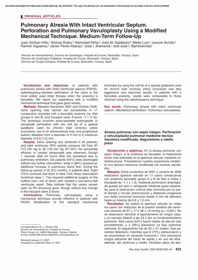
Figure 5. Tripartite ventricle and aGoose-Neck loop over the atresic valve(A). Anterograde perforation andprogressive dilation (1-4). Valve aperture(B).
Document downloaded from https://www.revespcardiol.org/, day 01/02/2022. This copy is for personal use. Any transmission of this document by any media or format is strictly prohibited.Document downloaded from https://www.revespcardiol.org/, day 01/02/2022. This copy is for personal use. Any transmission of this document by any media or format is strictly prohibited.
after being hospitalized for fever and pneumonia with adeteriorating evolution and, later, signs of low tissueperfusion; an urgent catheterisation was undertaken,showing a permeable fistula and the decision was takento perform a Glenn intervention, but the patient presenteda irreversible cardiac arrest during surgery.
In 2 symptomatic cases and in the presence of severegradients of 50 and 60 mm Hg and a very hypoplasicbipartite RV at the age of 2 and a half years, a RV wideningpatch of the out flow tract surgery was and, in 1 of them,a bidirectional Glenn intervention (one and a half ventricle)was also done with a good outcome in both cases.
In this follow-up, the most relevant data was:
1. The survival of 72% of the children in very goodfunctional conditions, achieving good development.
2. A reduction in RV pressure from 48 (13) mm Hg(hemodynamic) postvalvuloplasty to 33 (13) mm Hg(Doppler) (P<.05), including cases in which theobstruction was undone surgically.
3. An increase in the TV diameter from 10 (2) to 16.5 (2) mm (P<.01) and a reduction, though withoutsignificance, of the z score of the TV from –1.1 (1.3) to–1.3 (1) in spite of a good body development in thechildren.
4. Non-significant reduction of the TI degree from 2 (1) to 1.3 (0.7) (P=NS).
5. A growth in the PV ring from 6.7 (1.4) to 13 (1.8) mm(P<.001) and the pulmonary z from –2.2 (1.9) to 0.1 (1.7)though not significant (P=NS).
6. In all cases there was a residual pulmonaryinsufficiency observed that went from mild to moderate,quantified at 1.8 (0.5).
DISCUSSION
PAIS is a congenital cardiopathy with a greatmorphological diversity.1-5 This is demonstrated in theUnited Kingdom and Ireland collaboration study led byDaubeney et al.17 Of 183 cases, atresia was membranousin 74.7% and muscular in 25.3%; the RV was bipartitein 33.6% of cases and unipartite in 7%; coronary
anomalies were identified in 45.8% of cases, withstenosis/interruption/ectasia in 7.6% and an Ebsteinsmalformation in 18 patients.
Anatomical and morphological characteristics of theRV are the main determinant in the therapeutic approachthat is undertaken and the prognosis for the evolution ofpatients with this cardiopathy.17-19 Most of the authorsconsider that a biventricular repair cannot be viable whenthe z of the TV is less than –4. Wang et al20 estimatesthat treatment with a catheter can be definitive with a TV z of ≥–0,1, pulmonary z ≥–4.1, and a relation betweenventricular areas of ≥0.65, and for Cheatham,14 when theTV ring is >11, the PV ≥7 mm and the RV volume is≥30 mL/m2.
In an attempt to facilitate therapeutic approaches,Alwi21
recently published an algorithm on the therapeutic strategyin these patients: group A, patients with a tripartite RV,well developed and membranous atresia with a tricuspidz of >–2.5 whose initial, and maybe definitive, treatmentis valve opening through radiofrequency. Group C, patientswith severe hypoplasia of a unipartite RV, with a tricuspidz of <–5 where the initial treatment is an atrial septostomy,stent on ductus, or a modified Blalock-Taussig, and assomething definitive, the bidirectional Glenn techniqueat 6-12 months, followed by a Fontan intervention. GroupB or intermediate with a RV on the limit, a goodinfundibulum and a reduced trabecular segment with atricuspid z of –2.5 to –4.5 whose initial treatment isvalvulotomy, stent on ductus and a possible atrialseptostomy; in this group it is also common to repair theout flow tract of the RV or the PV ring and the “one anda half ventricle” when the RV is not adequately developed.
In some studies, with the radiofrequency techniquethere is a greater effectiveness than with surgicalvalvulotomy and a demonstrated mortality,22 although inothers this is not confirmed, but a smaller incidence oflow output after this procedure19 has been demonstrated.After the first data on valvular opening in PAIS,6-10 thelaser technique and, mainly, the radio frequency technique,experienced a great expansion, with several differentseries published.11,18,20,22-28 The results with this lasttechnique in the revision of Benson et al29 show a success
838 Rev Esp Cardiol. 2007;60(8):833-40
Alcíbar-Villa J et al. Valvuloplasty With Modified Mechanical Technique in Pulmonary Atresia With Intact Ventricular Septum
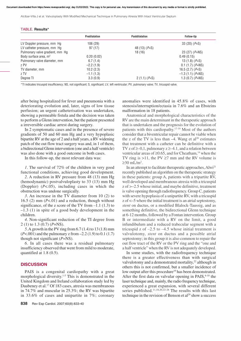
TABLE. Results*
Predilatation Postdilatation Follow-Up
LV Doppler pressure, mm Hg 106 (29) 33 (20) (P=S)
LV catheter pressure, mm Hg 97 (17) 48 (13) (P=S)
Pulmonary valve gradient, mm Hg 18 (16) 23 (27) (P=NS)
Body surface area, m2 0.20 (0.02) 0.49 (0.15)
Pulmonary valve diameter, mm 6.7 (1.4) 13 (1.8) (P=S)
z PV –2.2 (1.9) 0.1 (1.7) (P=NS)
TV diameter, mm 10.2 (2.3) 16.5 (2.7) (P=S)
z TV –1.1 (1.3) –1.3 (1.1) (P=NS)
Degree TI 3.3 (0.9) 2 (1.1) (P=S) 1.3 (0.7) (P=NS)
*TI indicates tricuspid insufficiency; NS, not significant; S, significant; LV, left ventricular; PV, pulmonary valve; TV, tricuspid valve.
Document downloaded from https://www.revespcardiol.org/, day 01/02/2022. This copy is for personal use. Any transmission of this document by any media or format is strictly prohibited.Document downloaded from https://www.revespcardiol.org/, day 01/02/2022. This copy is for personal use. Any transmission of this document by any media or format is strictly prohibited.

in 87% of the cases, with an incidence of complicationsof 15%, mortality of 8%, and the necessity of additionalpulmonary flow in 33%. The main difficulties that havebeen described with the mechanical technique of Latson6
are the aggressiveness of the rigid tip of the guide, whichcan perforate the out flow tract and/or the pulmonaryartery, as well as the unstable position of the subpulmonarycatheter when the firm part of guide14 progresses. Withthis technique, the results have been inferior to thoseobtained with radiofrequency, with an initial success rateof 68%, mortality of 4%, and the necessity of additionalpulmonary irrigation in 48%.14 In our experience withcases that in principle are favorable for valvular openingand that correspond to groups A and B of Alwi,21 we wereable to open the valves in all of them and in a consecutiveform during a period of 5 years. We used a modifiedLatson technique. We never used the rigid end of theguide, but special guides for total coronary chronicobstruction, always using the softer tip and directing tothe loop in a supravalvular fashion. It allows for a greaterstability of the catheter, that does not move from itsposition, and which is less aggressive, without any caseof tamponade or pericardial effusions. The most effectiveguide in our experience has been the “Cross it” which,in addition to having a certain distal firmness, displayssome sharpness in its tip from 0.014 to 0.010 inches,with capacity of lock and puncture when submitting toa rotational movement with the support of a catheter nextto the membrane which is to be perforated, directing itto the loop, ductus, and the descending aorta. With respectto the catheter, we have observed that, for ampleinfundibula, the most stable is the right Judkins, and fornarrow infundibula, the Goodale Lubin, with which onemust be cautious when passing the guide through thedistal orifice and avoid the 2 lateral ones. The exteriorizedarteriovenous circuit that facilitates support, along withthe low profile of the coronary monorail catheters, allowsthe initial valvular opening, proceeding in a progressiveform, which has been mentioned above.12,16 When theanterograde technique was not possible, we would makeit in an effective retrograde form, expanding progressivelywith balloon catheters in an anterograde manner. Weconsidered that with the use of special guides for the totalchronic coronary obstruction we have improved the resultsof the mechanical technique, being able to open the valvesin 100% of the cases and with a procedural global successof 90%, a mortality of 9% and the need for additionalirrigation in 36%. In the different medium and long termstudies, there is a great variety of results that depend toa large extent on the anatomical variability and on thetherapeutic approach that was followed.18,19,30 Mortalityhas been related to the aperture procedure, with thenecessity for late or definitive surgery, and also inherentto the cardiopathy in itself, in its evolution. In the largeststudies, survival is 60%-70% at 5 years19,30 and can becompared with ours of 72%, although with a shorterfollow-up. Valvular opening allows a greater degree of
flow by the TV, RV, and the PV, favoring the developmentof these structures; like in other studies, we observedthat, although the PV and the TV increase significantlytheir diameter, this increase is not proportional to thebody surface area and significative neither changes inthe z score of the TV nor in the PV are appreciated; insome way, this indicates that the presence of a certaindegree of hypodevelopment of the TV and the rightcavities is not essential for maintaining a sufficientpulmonary flow.18 The limitations that this study mayhave are due to the small number of patients and theabsence of a control group; nevertheless, its value whendealing with consecutive cases is reinforced. There mightbe some interobserver variability in the quantitative andsemiquantitative measurements during the follow-up. Inaddition, it must considered that this hemodynamics studygroup makes pediatric and adult interventionismprocedures simultaneously, with ample experience incoronary angioplasty, and their results should not begeneralized.
CONCLUSIONS
1. Pulmonary valvuloplasty with a mechanicaltechnique is still valid in PAIS.
2. The modification of the classic technique throughthe use of special guidewires for the total chronic coronaryobstruction by its directed soft part is less aggressive andimproves results.
3. In this series with favorable anatomy, the results aresimilar to those obtained through radiofrequency.
ADDENDUM
In the time this article was in print, 2 cases were treated
successfully.
THANK YOU
To Drs Alberto Cabrera, José I, Arana, and Carlos Romerofor their collaboration.
REFERENCES
1. Freedom RM. Etiology and incidence. En: Freedom RM, editor.
Pulmonary atresia with intact ventricular septum. Mount Kisco:
Futura; 1989. p. 1-8.
2. Freedom RM, Mawson JB, Yoo S-J, Benson LM. Pulmonary atresia
and intact ventricular septum. En: Freedom RM, editor. Congenital
Heart disease. Texbook of angiocardiography. Vol 1. Mount Kisco:
Futura; 1987. p. 617-62.
3. Hanley FL, Sade RM, Blackstone EH, Kirklin JW, Freedom RM,
Nanda NC. Outcomes in neonatal pulmonary atresia with intact
ventricular septum: a multiinstitutional study. J Thorac Cardiovasc
Surg. 1993;105:406-27.
4. Anderson RH, Anderson C, Zuberbuhler JR. Further morphologic
studies on hearts with pulmonary atresia and intact ventricular
septum. Cardiol Young. 1991;1:105-13.
Alcíbar-Villa J et al. Valvuloplasty With Modified Mechanical Technique in Pulmonary Atresia With Intact Ventricular Septum
Rev Esp Cardiol. 2007;60(8):833-40 839
Document downloaded from https://www.revespcardiol.org/, day 01/02/2022. This copy is for personal use. Any transmission of this document by any media or format is strictly prohibited.Document downloaded from https://www.revespcardiol.org/, day 01/02/2022. This copy is for personal use. Any transmission of this document by any media or format is strictly prohibited.

5. Freedom RM, Anderson RH, Perrin D. The significance of ventri-
culo-coronary arterial connections in the setting of pulmonary atresia
with an intact ventricular septum. Cardiol Young. 2005;15:447-68.
6. Latson LA. Non surgical treatment of a neonate with pulmonary
atresia and intact ventricular septum by transcatheter puncture and
balloon dilation of the atretic valve. Am J Cardiol. 1991;68:277-9.
7. Qureshi SA, Rosenthal W, Tynan M. Transcatheter Laser-assisted
balloon pulmonary valve dilation in pulmonary valve atresia. Am
J Cardiol. 1991;67:428-31.
8. Parsons JM, Rees MR, Gibbs JL. Pecutaneous Laser valvotomy
with balloon dilatation of the pulmonary valve as primary treatment
for pulmonary atresia. Br Heart J. 1991;66:36-8.
9. Rosenthal E, Qureshi SA, Chan KC, Martin RP, Skehan DJ, Jordan
SC, et al. Radiofrecuency-assisted balloon dilatation in patients with
pulmonary valve atresia and an intact ventricular septum. Br Heart
J. 1993;69:347-51.
10. Redington AN, Cullen S, Rigby MC. Laser or radiofrecuency
pulmonary valvotomy in neonates with pulmonary atresia and intact
ventricular septum: description of a new method avoiding arterial
catheterization. Cardiol Young. 1992;2:387-90.
11. Fontes VF, Esteves CA, Braga SL, Acuna V, Santana MV, de Oliveira
LM, et al. Pulmonary atresia with intact ventricular septum: valvar
perforation by radiofrecuency. Arq Bras Cardiol. 1995;64:231-3.
12. Alcíbar J, Cabrera A, Peña N, Baraldi C, Arriola J, Aramendi J.
Valvulotomía mecánica percutánea dirigida en la atresia pulmonar
con septo íntegro. Rev Esp Cardiol. 2003;56:822-5.
13. Kleinman CS. The Echocardiographic assessment of pulmonary
atresia with intact ventricular septum. Cathet Cardiovasc Int
2006;68:131-5.
14. Cheatham JP. The transcatheter management of the neonate and
infant with pulmonary atresia and intact ventricular septum. J Int
Cardiol. 1998;11:363-87.
15. Rowlatt JF, Rimoldi MJA, Lev M. The quantitative anatomy of the
normal child’s heart. Pediatr Clin Morth Am. 1963;10:499-588.
16. Latson L, Cheatham J, Froemming S, Kugler J. Transductal guide-
wire “rail” for balloon valvuloplasty in neonates with isolated critical
pulmonary valve stenosis or atresia. Am J Cardiol. 1994;73:713-4.
17. Daubeney PE, Delany DJ, Anderson RH, Sandor GGS, Slavik Z,
Keeton BR, et al. Pulmonary atresia with intact ventricular septum:
range of morphology in a population-based study. J Am Coll Cardiol.
2002;39:1670-9.
18. Humpl T, Söderberg B, McCrindle BW, Nykannen DG, Freedom
RM, Villiams WG, et al. Percutaneous balloon valvotomy in
pulmonary atresia with intact ventricular septum: impact on patient
care. Circulation. 2003;108:826-32.
19. Mi YP, Chau AKT, Chiu CSW, Yung TC, Lun KS, Cheung YF.
Evolution of the management approach for pulmonary atresia with
intact ventricular septum. Heart. 2005;91:657-63.
20. Wang JK, Wu MH, Chang CI, Chen YS, Lue HC. Outcomes of
transcatheter valvotomy in patients with pulmonary atresia and intact
ventricular septum. Am J Cardiol. 1999;84:1055-60.
21. Alwi M. Management algorithm in pulmonary atresia with intact
ventricular septum. Cathet Cardiovasc Interven. 2006;67:679-86.
22. Alwi M, Geetha K, Choo KK, Bilkis AA, Hasri S, Haifa AL. Risk
factors for augmentation of the flow of blood to the lungs in pulmonary
atresia with intact ventricular septum after radiofrecuency valvotomy.
Cardiol Young. 2005;15:141-7.
23. Camino M, Brugada J, Mortera C, Thio M, Rovirosa M, Bartrons
J. Valvulotomía pulmonar percutánea mediante radiofrecuenciaen
la atresia pulmonar con septo interventricular íntegro. Rev Esp
Cardiol. 2001;54:243-6.
24. Pedra CA, Sousa LN, Pedra SR, Ferreira WP, Braga SL, Esteves
CA, et al. New percutaneous techniques for perforating the pulmonary
valve in pulmonary atresia with intact ventricular septum. Arq Bras
Cardiol. 2001;77:479-86.
25. Cheung YF, Leung MP, Chan AKT. Usefulness of lasser-assisted
valvotomy with baloon valvoplasty for pulmonary valve atresia with
intact ventricular septum. Am J Cardiol. 2002;90:438-42.
26. Moskowitz WB, Titus JL, Topaz O. Excimer laser ablation for
valvular angioplasty in pulmonary atresia and intact ventricular
septum. Laser Surg Med. 2004;35:327-35.
27. Agnoletti G, Piechaud JF, Bonhoeffer P, Aggoun Y, Abdel-Masih
T, Boudjemline Y, et al. Perforation of the atretic pulmonary
valve: long term follow up. J Am Coll Cardiol. 2003;41:1399-
403.
28. Weber H. Initial and late results after chatheter intervention for
neonatal critical pulmonary valve stenosis and atresia with intact
ventricular septum: a technique in continual evolution. Cathet
Cardiovasc Int. 2002;56:394-9.
29. Benson LN, Nykanen D, Collison A. Radiofrequency perforation
in the treatment of congenital heart disease. Cathet Cardiovasc Int
2002;56:72-82.
30. Ashburn DA, Blackstone EH, Wells WJ, Jonas RA, Pigula FA,
Manning PB, et al. Determinants of mortality and type of repair in
neonates with pulmonary atresia and intact ventricular septum.
J Thorac Cardiovasc Surg. 2004;127:1000-8.
840 Rev Esp Cardiol. 2007;60(8):833-40
Alcíbar-Villa J et al. Valvuloplasty With Modified Mechanical Technique in Pulmonary Atresia With Intact Ventricular Septum
Document downloaded from https://www.revespcardiol.org/, day 01/02/2022. This copy is for personal use. Any transmission of this document by any media or format is strictly prohibited.Document downloaded from https://www.revespcardiol.org/, day 01/02/2022. This copy is for personal use. Any transmission of this document by any media or format is strictly prohibited.
![Special Report Interventional Cardiology · defect (ASD; Lutembacher syndrome) had fewer symp-toms than patients with an intact septum [16]. Also, the closure of congenital ASD has](https://static.fdocuments.us/doc/165x107/5e5b7e0b9f19eb1d7031026e/special-report-interventional-cardiology-defect-asd-lutembacher-syndrome-had.jpg)