Protocol for multiplexed ... - vaccine.uab.edu · Page 2 of 43 Filename: UAB-MOPA-E.02 C. FBS Lot...
43
www.vaccine.uab.edu Page 1 of 43 Filename: UAB-MOPA-E.02 Protocol for multiplexed opsonophagocytic killing assay (UAB-MOPA) for antibodies against Streptococcus pneumoniae (Version E.02, December 2014) By Moon H. Nahm and Robert L. Burton The Bacterial Respiratory Pathogen Reference Laboratory of the US NIH WHO Reference Laboratory for Pneumococcal Serology Departments of Pathology and Microbiology University of Alabama at Birmingham Birmingham AL 35294-2170 USA www.vaccine.uab.edu Procedures 1. Bacteria Stocks A. Master Stock Maintenance B. Production of Assay Stocks C. Characterization of Assay Stocks D. Acceptance Criteria for Assay Stocks 2. HL60 Cells A. Production of Master Cell Banks B. Initiation of Working Cell Cultures C. Propagation of Working Cell Cultures D. Differentiation of HL60 Cells E. Determination of HL60 Cell Phenotype F. Acceptance Criteria for HL60 Cells 3. Test Samples A. Sample Collection B. Sample Handling C. Heat Inactivation D. Testing Samples for Bactericidal Agents (Antibiotics) E. Production of QC Serum Pools 4. Complement A. Preparation of Working Aliquots of Complement B. Complement Screening 1 (CH50 Assay) C. Complement Screening 2 (MOPA) D. Complement Lot Acceptance Criteria 5. Fetal Bovine Serum (FBS) A. Heat Inactivation and Preparation of Assay Aliquots B. FBS Screening
Transcript of Protocol for multiplexed ... - vaccine.uab.edu · Page 2 of 43 Filename: UAB-MOPA-E.02 C. FBS Lot...

www.vaccine.uab.edu Page 1 of 43 Filename: UAB-MOPA-E.02
Protocol for multiplexed opsonophagocytic killing assay (UAB-MOPA) for antibodies against
Streptococcus pneumoniae
(Version E.02, December 2014)
By Moon H. Nahm and Robert L. Burton
The Bacterial Respiratory Pathogen Reference Laboratory of the US NIH WHO Reference Laboratory for Pneumococcal Serology
Departments of Pathology and Microbiology University of Alabama at Birmingham
Birmingham AL 35294-2170 USA
www.vaccine.uab.edu
Procedures 1. Bacteria Stocks
A. Master Stock Maintenance B. Production of Assay Stocks C. Characterization of Assay Stocks D. Acceptance Criteria for Assay Stocks
2. HL60 Cells A. Production of Master Cell Banks B. Initiation of Working Cell Cultures C. Propagation of Working Cell Cultures D. Differentiation of HL60 Cells E. Determination of HL60 Cell Phenotype F. Acceptance Criteria for HL60 Cells
3. Test Samples A. Sample Collection B. Sample Handling C. Heat Inactivation D. Testing Samples for Bactericidal Agents (Antibiotics) E. Production of QC Serum Pools
4. Complement A. Preparation of Working Aliquots of Complement B. Complement Screening 1 (CH50 Assay) C. Complement Screening 2 (MOPA) D. Complement Lot Acceptance Criteria
5. Fetal Bovine Serum (FBS) A. Heat Inactivation and Preparation of Assay Aliquots B. FBS Screening

www.vaccine.uab.edu Page 2 of 43 Filename: UAB-MOPA-E.02
C. FBS Lot Acceptance Criteria 6. UAB-MOPA Procedure 7. Data Handling
A. Data Conversion B. Assay Acceptance Criteria C. Acceptance Criteria for Individual Sample Data
8. Assay Notes 9. References 10. Materials and Reagents
A. Plasticware B. Solutions and Reagents C. Bacterial Culture Media D. Chemicals E. Equipment and Software F. Cell Line G. Bacteria H. Reagents for Flow Cytometry I. Recipes for Prepared Solutions
11. Cell Counting
Introduction Opsonization assay has become an important tool in assessing pneumococcal vaccine immunogenicity (reference 1). To facilitate the widespread use of the opsonization assay, we have prepared a detailed protocol for 4-fold multiplexed opsonophagocytic killing assay (UAB-MOPA). Although this protocol describes the multiplexed assay, this protocol can be used as a single serotype assay with the minor changes described in Note 9 in Assay Notes section. Also, this protocol can be used with minor alterations (e.g., a different serial dilution factor or different number of wells per sample). Additional information on this assay can be found in publications (references 2 and 3) and at a web site (www.vaccine.uab.edu). To facilitate data analysis, an Excel®-based data processing program was developed that converts colony counts to “opsonization index”. This program, “opsotiter3”, can be obtained from this reference laboratory by sending a written request to Rob Burton at [email protected]. We have collaborated with the US National Institute of Standards and Technology (NIST) to develop software that can efficiently enumerate colonies in a digital image (i.e., a digital camera picture or scanned image). This software is called “NICE” (NIST’s Integrated Colony Enumerator) and is available free of charge. For more information about NICE, contact: [email protected]. To download the software and/or the software manual, go to: ftp://ftp.nist.gov/pub/physics/mlclarke/NICE/. If academic investigators need MOPA target strains for non commercial uses, they can obtain the strains from BEI Resources (www.beiresources.org). For commercial uses, please contact Dr. Debbie Bidanset at the University of Alabama at Birmingham ([email protected]). Please note that registration with BEI resources is required to obtain the strains. BEI contact information:
BEI Resources 10801 University Boulevard Manassas, VA 20110-2209 Email: [email protected] Web site: www.beiresources.org

www.vaccine.uab.edu Page 3 of 43 Filename: UAB-MOPA-E.02
Telephone: 1-800-359-7370 The procedures outlined above are necessary to establish the assay. Afterwards, the UAB-MOPA procedure will be the primary protocol used, with the other procedures (bacteria working stock preparation/characterization, complement lot screening, etc) being used only occasionally. This protocol is based on more than 10 years of continuous development work by many people in Nahm laboratory, to whom we are indebted. We are also indebted to many other individuals who have provided assistance and critical comments as well as to the NIH and WHO for financial support. Pneumococci are human pathogens. As such, biosafety concerns apply. Check local guidelines for the proper handling of pneumococci. If there are additional questions, please contact Dr. Moon H. Nahm ([email protected]) or Mr. Robert L. Burton ([email protected]). Abbreviations: OBB, Opsonization Buffer B; HI, Heat-Inactivated; THYA, Todd-Hewitt Yeast Agar plates; THYB, Todd-Hewitt Yeast Broth; NSK, Non-Specific Killing; TTC, 2,3,5,-triphenyltetrazolium chloride; OD, Optical Density; HBSS, Hanks Balanced Salt Solution; CFU, Colony Forming Units; QC, Quality Control
Procedures
1. Bacteria Stocks
Pneumococci are human pathogens. As such, biosafety considerations apply. Check local guidelines for the proper handling of pneumococci.
A. Master Stock Maintenance To maintain the integrity of the pneumococcal bacterial stocks, the master stock vials from BEI Resources should never be thawed. All master stock vials should be stored at -80C. To produce assay stocks, retrieve the master stock vial from the freezer, quickly remove a fleck of ice from the vial, and streak it onto a blood agar plate (see below for more details). Immediately, return the master stock vial to the freezer. B. Production of Assay Stocks From the master stock tube, many aliquots of assay stocks can be produced and frozen. An aliquot of assay stock is thawed and used for only one experiment. The procedure below is for preparing 48 tubes of assay stock. If more aliquots are desired, scale up accordingly by increasing the number of 50 ml tubes used, i.e., do not remove more than 10 ml of culture from each tube.
1. Retrieve the master stock vial from the freezer, quickly remove a fleck of ice from the
vial, and streak it onto a blood agar plate. Immediately, return the master stock vial to the freezer.
2. Incubate the plate upside down overnight at 37C in a candle jar (Note 13). Pneumococci yield alpha-hemolytic colonies that can be identified by a green halo
Title: Bacteria Stocks Revision History: 2/1/08; 6/23/08; 9/20/12; 11/14/14

www.vaccine.uab.edu Page 4 of 43 Filename: UAB-MOPA-E.02
surrounding the colony. 3. Transfer ~20 isolated colonies to a 50 ml tube containing 50 ml of THY broth (see
Note 14). Incubate for 3-8 hours in a 37C water bath until the top 150 microliters of the culture broth has an OD600 of ~0.6-0.9.
4. Harvest the top 10 ml of the broth and transfer to a fresh 50 ml tube. Add 5 ml of 80% sterile glycerol and 10 ml of fresh THY broth to the 10 ml of bacteria.
5. Mix well, and dispense 0.5 ml aliquots into sterile 1.5 ml microcentrifuge tubes (~ 48 tubes). Indicate the last aliquot prepared with an identifiable mark (this vial will be used to determine purity later).
6. Randomly select an aliquot (not the last aliquot prepared) that will not be frozen and will be used to compare the density pre- and post-freezing (see below). Transfer the remaining tubes to a labeled cardboard freezer box and place the box (without a lid) into a -70ºC freezer. After stocks have frozen completely (overnight is best), attach an appropriately labeled lid. Store the tubes frozen at -70C until needed, up to ~18 months.
C. Characterization of Assay Stocks Before an assay stock can be accepted, it must be characterized. If the assay stock meets the criteria defined below, the lot of stock will be accepted.
1. Confirm the serotype of the strain using standard serotyping protocols (Quellung,
agglutination, latex bead-based, ELISA, etc). 2. Look for the presence of other microbial contamination by thawing the last aliquot
prepared and streaking ~10 microliters onto a blood agar plate. After overnight growth at 37°C, only alpha-hemolytic pneumococcal colonies should be visible.
3. Determine the antibiotic sensitivity and resistance of the frozen assay stocks.
a. Prepare OBB. b. Melt the overlay agar (you will need ~180 ml for each strain being tested) in a
microwave using 50% power. Gently and carefully swirl overlay agar to ensure that all agar clumps have melted. Alternatively, overlay can be prepared and autoclaved the day of the assay to obviate the need for melting in a microwave. After the agar is melted, aliquot 13 ml to each of 13 sterile tubes. Keep tubes in 50ºC water bath until needed (do not use until agar temperature has equilibrated to 50°C).
c. Dry small (10 cm x 10 cm) THYA plates (you will need 13 THYA plates for each strain being tested) by removing the lids and placing the plates in a laminar flow hood for 30-60 minutes (see Note 4). After plates are dry, replace lid to prevent over-drying, and keep at RT until needed. Plates should be labeled with strain name and antibiotic concentration (see below).
d. Rapidly thaw a frozen assay stock tube prepared above in a 37C water bath using gentle, constant agitation.
e. Serial dilutions of bacteria (in triplicate) can be conveniently prepared in a microtiter plate as follows:
i. Dilute the bacteria 10-fold by mixing 30 microliters of bacteria with 270 microliters of OBB in row A, columns 1 through 3 of a microtiter plate. Add 240 microliters OBB to rows B though H, columns 1 through 3.
ii. Prepare 5-fold dilutions by mixing 60 microliters of dilute bacteria from previous dilution (row A) with 240 microliters of OBB in row B. Continue 5-fold serial dilutions for a total of 8 dilutions (10-fold to 7.8 x 10⁵-fold).
f. Spot 10 microliters from each dilution from each of the three columns onto THYA plates (13 total plates are needed).

www.vaccine.uab.edu Page 5 of 43 Filename: UAB-MOPA-E.02
g. Incubate the THYA plates at RT to allow the fluid to absorb into the agar (10-20 minutes).
h. Remove one of the overlay tubes from the water bath, add 13 microliters of TTC, and add 12 ml of this overlay to one of the 13 THYA plates. This is the “no antibiotic” control plate.
i. Remove three of the overlay tubes from the water bath, and add 13 microliters of TTC to each tube. To one tube, add 6.5 microliters of optochin, and add 12 ml of this overlay to one of the 13 THYA plates. This is the “0.5X optochin” plate. To the second tube, add 13 microliters of optochin, and add 12 ml of this overlay to one of the 13 THYA plates. This is the “1X optochin” plate. To the third overlay tube, add 26 microliters of optochin, and add 12 ml of this overlay to one of the 13 THYA plates. This is the “2X optochin” plate.
j. Remove three of the overlay tubes from the water bath, and repeat the above for trimethoprim.
k. Remove three of the overlay tubes from the water bath, and repeat the above for streptomycin.
l. Remove three of the overlay tubes from the water bath, and repeat the above for spectinomycin.
m. After all overlay has been added, there should be 13 total plates:
n. After the overlay has set, incubate the plates overnight at 37C in a candle jar (Note 13).
o. Count the colonies. p. Calculate the average of each of the triplicates for a bacteria dilution that
produces 50-150 CFU/spot in the no antibiotic plate. Determine the sensitivity of the strain to each concentration of antibiotic by comparing the CFU/spot in the presence of antibiotic to the CFU/spot without antibiotic. A strain should be resistant to the desired antibiotic at 2X assay concentration (with <20% reduction in CFU/spot compared to the no antibiotic plate), and sensitive to the other antibiotics at 0.5X assay concentration (with >95% reduction in CFU/spot compared to the no antibiotic plate).
4. Determine the optimal dilution factor for the assay stock. Prior to being used as
targets in the UAB-MOPA assay, each assay stock must be titrated in assay conditions to determine the dilution necessary to yield about 120 CFU/spot on THYA plates. a. Prepare OBB. b. Melt the overlay agar (~150 ml is sufficient for up to 12 strains) in a microwave
using 50% power. Gently and carefully swirl overlay agar to ensure that all agar clumps have melted, and place bottle in 50ºC water bath until needed (do not use until agar temperature has equilibrated to 50°C). Alternatively, overlay can be prepared and autoclaved the day of the assay to obviate the need for melting in a microwave.
c. Dry small (10 cm x 10 cm) THYA plate (you will need 1 THYA plate for each
Plate # Antibiotic Plate # Antibiotic1 No antibiotic 8 streptomycin, 0.5X2 optochin, 0.5X 9 streptomycin, 1X3 optochin, 1X 10 streptomycin, 2X4 optochin, 2X 11 trimethoprim, 0.5X5 spectinomyin, 0.5X 12 trimethoprim, 1X6 spectinomyin, 1X 13 trimethoprim, 2X7 spectinomyin, 2X

www.vaccine.uab.edu Page 6 of 43 Filename: UAB-MOPA-E.02
strain being tested) by removing the lid and placing the plate in a laminar flow hood for 30-60 minutes (see Note 4). After plate is dry, replace lid to prevent over-drying, and keep at RT until needed.
d. Add 20 microliters OBB to 16 wells (2 entire columns) of a fresh microtiter plate (in place of the 20 microliters of test serum that is used in the assay). This is the “assay plate”.
e. Prepare the differentiated HL60 cells: i. Transfer the DMF-differentiated HL60 cells (differentiated for 5 or 6 days)
from the culture flasks (1 flask will be sufficient to test up to 12 strains) to 50 ml centrifuge tubes.
ii. Centrifuge the HL60 cells at ~350g (1200 rpm using a Sorvall RT7 with RTH-250 rotor) for 5 minutes at RT.
iii. Remove the supernatant and combine all cell pellets in 50 ml of 1X HBSS (without Ca++/Mg++). Centrifuge at ~350g for 5 minutes at RT.
iv. Remove the supernatant and add 50 ml of 1X HBSS (with Ca++/Mg++). Centrifuge at ~350g for 5 minutes at RT.
v. Remove the supernatant, and suspend the cells at 1 x 10⁷ cells/ml in Opsonization Buffer B (store the cells at RT until needed). See “Cell Counting” procedure for performing viable cell counts.
vi. When counting the cells, determine the viability using trypan blue exclusion. Record the number of live cells and the number of dead cells. The cell viability must be ≥90% for the cells to be considered acceptable for use in the assay.
f. Rapidly thaw a frozen assay stock vial and wash bacteria: i. Gently swirl the tube in a 37C water bath until thawed. ii. Centrifuge tube at 12,000g for 2 minutes in microcentrifuge. iii. Carefully remove supernatant and discard. iv. Add 1 ml Opsonization Buffer B to the tube, and mix. v. Centrifuge tube at 12,000g for 2 minutes. vi. Carefully remove supernatant and discard. vii. Suspend bacteria pellet in original volume of Opsonization Buffer B (i.e.,
0.5 ml). g. Dilutions of several bacterial cultures can be conveniently prepared in a fresh
microtiter plate as follows (this is the “dilution plate”): i. Dilute the thawed bacteria 10-fold by mixing 15 microliters of bacteria with
135 microliters of Opsonization Buffer B in columns 1 and 2 of row A. Add 120 microliters Opsonization Buffer B to columns 1 and 2, rows B though H.
ii. Prepare 5-fold dilutions by mixing 30 microliters of dilute bacteria from previous dilution (row A) with 120 microliters of Opsonization Buffer B in row B. Continue 5-fold serial dilutions for a total of 8 dilutions (10-fold to 7.8 x 10⁵-fold).
h. Transfer 10 microliters of the diluted bacteria from column 1 of the dilution plate to column 1 of the assay plate. Repeat for column 2.
i. Incubate plate at RT and room air on a mini-orbital shaker (700 rpm) for 30 minutes.
j. During this time, remove a vial containing an adequate volume of complement (~0.3 ml for each strain tested) from the freezer and leave at RT to thaw (immediately after complement has thawed completely, incubate on ice until needed).
k. After the 30-minute incubation, prepare HL60/complement mixture by mixing 1 ml of HL60 cell suspension and 0.25 ml of complement (for each strain tested).
l. Add 50 microliters of the HL60/complement mixture to each well of the plate (16
www.vaccine.uab.edu Page 7 of 43 Filename: UAB-MOPA-E.02
wells/strain). m. Incubate plate on a mini-orbital shaker (700 rpm) for 45 minutes at 37ºC with 5%
CO2 in a single layer, i.e., do not stack plates. In order to maintain a constant CO2 percent during this incubation step, do not open the incubator door.
n. After incubation, place plates on ice to stop phagocytic process for ~20 minutes. o. Using a multi-channel pipettor, mix the contents of each well, remove 10
microliters from each well in an 8 well column, and apply as eight-10 microliter spots to THYA plates on the left side. Immediately, tilt the plates to shape the spots into a small strip of fluid (~2-3 cm long). Apply the second 8 well column to the center of the THYA plate. You must tilt plate immediately to prevent spots from running together.
p. Incubate the THYA plates at RT for 10-20 minutes to allow the fluid to absorb into the agar.
q. Remove the overlay from the water bath, and add 150 microliters of TTC. Add 12 ml of this overlay to each plate.
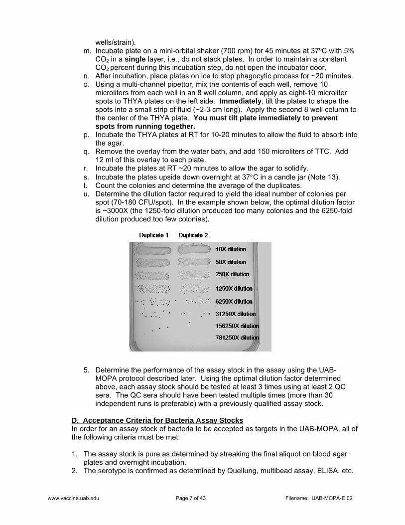
r. Incubate the plates at RT ~20 minutes to allow the agar to solidify. s. Incubate the plates upside down overnight at 37C in a candle jar (Note 13). t. Count the colonies and determine the average of the duplicates. u. Determine the dilution factor required to yield the ideal number of colonies per
spot (70-180 CFU/spot). In the example shown below, the optimal dilution factor is ~3000X (the 1250-fold dilution produced too many colonies and the 6250-fold dilution produced too few colonies).
5. Determine the performance of the assay stock in the assay using the UAB-MOPA protocol described later. Using the optimal dilution factor determined above, each assay stock should be tested at least 3 times using at least 2 QC sera. The QC sera should have been tested multiple times (more than 30 independent runs is preferable) with a previously qualified assay stock.
D. Acceptance Criteria for Bacteria Assay Stocks In order for an assay stock of bacteria to be accepted as targets in the UAB-MOPA, all of the following criteria must be met:
1. The assay stock is pure as determined by streaking the final aliquot on blood agar
plates and overnight incubation. 2. The serotype is confirmed as determined by Quellung, multibead assay, ELISA, etc.

www.vaccine.uab.edu Page 8 of 43 Filename: UAB-MOPA-E.02
3. The stock is resistant to the appropriate antibiotic and sensitive to the other three antibiotics. The stock should be resistant to the appropriate antibiotic at 2X assay concentration and sensitive to the appropriate antibiotics at 0.5X assay concentrations.
4. The assay dilution factor is ≥200. 5. The mean opsonic indices of at least two QC sera (tested at least 3 times in the
UAB-MOPA) using the new assay stock are within the range (mean ± 2SD) determined using the previous assay stock.
2. HL60 Cells HL60 is a human cell line. As such, biosafety considerations apply. Check local guidelines for the proper handling of human cell lines. Before you begin culturing HL60 cells, and periodically thereafter, you should check your incubator to ensure proper CO2 levels and humidity (Note 6). CO2 levels should be confirmed using an outside reference (such as a FYRITE® Gas Analyzer produced by Bacharach, Pittsburgh, PA). Proper humidity is maintained by ensuring the humidity pan is always full. Proper humidity is vital for two reasons. First, it helps minimize evaporation in cell cultures and assay plates. Second, the CO2 sensors of some incubators require proper humidity for proper function.
A. Production of Master Cell Banks When you receive the stock vial from ATCC, you should thaw, expand, and freeze the cells to produce a master cell bank (~60 tubes) directly from this stock tube. During the expansion and production of the master cell bank, it is critical that the culture is properly maintained so as to ensure the quality of the cells. Proper maintenance includes monitoring of CO2 levels (Note 6), keeping the cell density below 5 x 10⁵ cells/ml, and proper humidity (Note 6). For freezing the cells, the use of a controlled-rate freezer system (Cryomed systems from Thermo-Forma, for example) is highly recommended. Whatever system is used, the sample temperature should decrease ~1°C/minute during the entire freezing process. 1. Add 10 ml CM3 to 15-ml centrifuge tube. 2. Thaw frozen HL60 cells (original tube obtained from ATCC) quickly in a 37ºC water
bath, and add the cells to the centrifuge tube containing 10 ml CM3. 3. Centrifuge the cells at ~350g (1200 rpm using a Sorvall RT7 with RTH-250 rotor) for
5-10 minutes at RT. Remove the supernatant, as much as possible (Note 5). 4. Suspend the cell pellet in CM3 (the volume of medium is indicated in the lot-specific
information received from ATCC), and transfer cells to the necessary number of 150 cm2 flasks (no more than 70 ml/flask). Place the flask(s) (lying flat) in a tissue culture incubator (37ºC, 5% CO2, See Note 6).
5. After 3 or 4 days, add ~50 ml fresh CM3 to the flask. Do not add more than 120 ml medium to each 150 cm2 flask. When more than 120 ml of medium is needed, use multiple flasks.
6. Monitor the cell density using a hemacytometer. When cells reach a density of ~5 x 10⁵ cells/ml, add fresh CM3 to adjust the cell density to ~2 x 10⁵ cells/ml. Do not add more than 120 ml medium to each 150 cm2 flask. When more than 120 ml of medium is needed, use multiple flasks.
7. When you have 10 flasks containing ~120 ml of medium per flask and a cell density of ~5 x 10⁵ cells/ml (usually ~3 weeks), freeze the cells.
a. Prepare fresh freezing medium:
Title: HL60 Cells Revision History: 2/1/08; 5/28/08
www.vaccine.uab.edu Page 9 of 43 Filename: UAB-MOPA-E.02
35 ml FBS (heat inactivated at 56°C for 30 minutes) 7 ml DMSO 28 ml RPMI 1640
b. Transfer the contents of all flasks to an appropriate number of 50 ml centrifuge tubes (~24). Keep a small volume (~1-2 ml) of culture to test for sterility.
c. Centrifuge the tubes at ~350g (1200 rpm using a Sorvall RT7 with RTH-250 rotor) for 5 minutes.
d. Remove supernatant (as much as possible), and discard supernatant. e. Add 2.5 ml of freezing medium to each 50 ml centrifuge tube and gently re-
suspend the cell pellets. Combine the cells from all 24 tubes together in a 150 cm2 flask (total volume should be ~60 ml).
f. Aliquot 1 ml of cells in freezing medium to each vial (each vial should contain ~107 cells), and place the vial on ice until all are ready.
g. Put all cryovials except one (that will be used to check for microbial contamination below) into controlled-rate freezer and begin freezing program. Sample temperature should decrease ~1°C per minute.
h. When freezing is completed, transfer cryovials to cryobiological storage system (liquid nitrogen storage system).
8. The small volume of culture collected above should be checked for mycoplasma contamination using standard tissue culture mycoplasma screening techniques. Also, it should be checked for other microbial contamination by streaking onto blood agar plates.
B. Initiation of Working Cell Cultures To maintain effector cell integrity, a new vial of cells from the master cell bank should be thawed every 3-4 months. 1. Add 10 ml CM3 to 15-ml centrifuge tube. 2. Remove one master cell bank Cryovial from liquid nitrogen storage. Gently swirl the
vial in a 37ºC water bath to rapidly thaw the frozen cells. Add the thawed cells to the centrifuge tube containing 10 ml CM3.
3. Centrifuge the cells at ~350g (1200 rpm using a Sorvall RT7 with RTH-250 rotor) for 5-10 minutes at RT. Remove the supernatant, as much as possible (Note 5).
4. Suspend the cell pellet in ~70 ml CM3. Transfer the cells to a 150 cm2 flask. Place the flask (lying flat) in a tissue culture incubator (37ºC, 5% CO2, See Note 6).
5. After 3-4 days of culture, add ~50 ml CM3. 6. Every 3-4 days, remove ~50 ml of cells and discard. Add 50 ml of fresh CM3. 7. After ~2 weeks of culture, begin feeding cells with CM1 (every 3-4 days, remove 50
ml of cells and discard, add 50 ml of fresh CM1). 8. After cells have been in culture for at least 3-4 weeks, begin regular propagation
schedule below. We have found that this is easily begun on a Wednesday or a Friday. If this is done on Wednesday, count cells, adjust cell concentration to ~8 x 10⁵ cells/ml in old culture medium, aliquot 40 ml of cells per T150 flask, and add 80 ml fresh CM1. If this is done on Friday, count cells, adjust cell concentration to ~8 x 10⁵ cells/ml in old culture medium, aliquot 20 ml of cell per T150 flask, and add 60 ml fresh CM1.
9. Follow the propagation schedule below.
C. Propagation of Working Cell Cultures This procedure is designed for differentiating cells up to two times per week. This helps to conserve the passage number of the culture. Cells differentiated on Wednesdays will be used in the assay the following Monday and Tuesday (days 5 and 6 of differentiation,
www.vaccine.uab.edu Page 10 of 43 Filename: UAB-MOPA-E.02
respectively). Cells differentiated on Fridays will be used the following Wednesday and Thursday (days 5 and 6 of differentiation, respectively). Cells not used after 6 days will be discarded.
Monday: Add 40 ml CM1 to each 150 cm2 flask of cells (there should be ~80 ml in each flask prior to feeding) to produce a final volume of ~120 ml/flask. Cell concentration should now be ~3 x 10⁵ cells/ml.
Wednesday: Mix contents of flask(s), and remove 80 ml from each flask (leaving ~40 ml). The removed cells can be pooled and used for differentiation (see below), or discarded if not needed. Add 80 ml CM1 to the remaining cells in each flask to produce a final volume of ~120 ml/flask, and return flask to incubator for continued propagation. Cell concentration should now be ~3 x 10⁵ cells/ml.
Friday: Mix contents of flask(s), and remove all volume except 20 ml from each flask (i.e., remove ~100 ml/flask). The removed cells can be pooled and used for differentiation (see below), or discarded if not needed. Add 60 ml CM1 to the remaining cells in each flask to produce a final volume of ~80 ml/flask, and return flask to incubator for continued propagation. Cell concentration should now be ~2 x 10⁵ cells/ml.
D. Differentiation of HL60 Cells 1. Centrifuge the HL60 cells at ~350g (1200 rpm using a Sorvall RT7 with RTH-250
rotor) for 5 minutes at RT. Remove ALL the supernatant (to completely remove any antibiotics).
2. Gently re-suspend the cell pellet in CM2 (containing 0.8% DMF), count the cells, and adjust the concentration to ~4 x 10⁵ cells/ml.
3. Add 100 ml of the cell suspension to each 150 cm2 flask needed. 4. Incubate the flask in an incubator (37ºC, 5% CO2, See Note 6) for 5-6 days, lying flat.
Do not feed the culture during this period. Cells not used within 6 days will be discarded.
5. Four flasks will usually yield enough differentiated cells to prepare 7 opsonization assay microtiter plates at ~200:1 (HL60:bacteria).
E. Determination of HL60 Cell Phenotype
We recommend determining the HL60 cell phenotype once every 2 to 4 weeks, more often if changes are noted in QC sera performance (low titers, incomplete killing, etc). We also recommend that both differentiated cells and undifferentiated cells are used for FACS analysis. The undifferentiated will be used as controls.
1. Label four 15 ml centrifuge tubes as “A”, “B”, “C”, or “D”. Label 22 FACS tubes “1”
through “22”. Also, label 14 wells of a 96-well V-bottom plate as indicated below (the numbers will correspond to the FACS tube numbers when the cells are transferred from the plate to the FACS tubes):

www.vaccine.uab.edu Page 11 of 43 Filename: UAB-MOPA-E.02
2. Mix contents of flasks containing HL60 cells, and count cells. We recommend testing both differentiated cells and undifferentiated cells.
3. Transfer indicated number of cells to indicated tube (typically ~3 ml of differentiated and ~4 ml of undifferentiated cells contain 2 x 10⁶ cells):
4. Keep Tubes B and D at room temperature until needed. 5. Centrifuge Tubes A and C at 350g for 5 minutes at 4°C. 6. While cells are centrifuging, prepare dilutions of antibodies, see Note 15 (store
diluted antibodies at 4°C protected from light until needed):
7. Remove supernatant from Tubes A and C. 8. Add 10 ml FACS Buffer to Tubes A and C, and centrifuge at 350g for 5 minutes at
4°C. 9. Remove supernatant from Tubes A and C, and suspend the cell pellets in 1 ml FACS
Buffer. 10. Aliquot 50 microliters of cell suspension from Tube A to wells 5, 6, 7, 8, 9, 10, and 11
of the 96-well, V-bottom plate labeled above. Aliquot 50 microliters of cell suspension from Tube C to wells 16, 17, 18, 19, 20, 21, and 22 of the 96-well, V-bottom plate labeled above.
11. Add 10 microliters of diluted CD11b PE to wells 6 and 17 (changing tips between wells) and gently mix.
12. Add 10 microliters of diluted CD35 PE to wells 7 and 18 (changing tips between wells) and gently mix.
Antibody FACS bufferCD11b PE 6 24CD35 PE 3 27CD71 PE 3 27IgG1 Isotype PE (1/5) 6 24IgG1 Isotype PE (1/80) 1 79IgG2a Isotype PE 2 32
Volume (microliters)
5
22
8
211917
1110
2018
16
976
5
22
8
211917
1110
2018
16
976
Tube Sample Intended Use # Cells Needed
A Undifferentiated Cells Surface Markers 2 X 10⁶
B Undifferentiated Cells Viability by FACS 1 X 10⁶
C Differentiated Cells Surface Markers 2 X 10⁶
D Differentiated Cells Viability by FACS 1 X 10⁶
www.vaccine.uab.edu Page 12 of 43 Filename: UAB-MOPA-E.02
13. Add 10 microliters of diluted CD71 PE to wells 8 and 19 (changing tips between wells) and gently mix.
14. Add 10 microliters of diluted IgG1 PE Isotype (1/5 dilution) to wells 9 and 20 (changing tips between wells) and gently mix.
15. Add 10 microliters of diluted IgG1 PE Isotype (1/80 dilution) to wells 10 and 21 (changing tips between wells) and gently mix.
16. Add 10 microliters of diluted IgG2a PE Isotype to wells 11 and 22 (changing tips between wells) and gently mix.
17. Add nothing to wells 5 and 16. These wells are unstained control wells. 18. Incubate at 4°C protected from light for 30 minutes. 19. Add 150 microliters FACS buffer to each well, and centrifuge plate at 350g for 5
minutes at 4°C. Prepare FACS/PI buffer by adding 30 microliters PI stock solution to 6 ml FACS buffer (final PI concentration is ~5 ug/ml).
20. Remove supernatant from each well using separate pipet tips—do not cross-contaminate wells.
21. Suspend cell pellets in 250 microliters FACS/PI buffer. 22. Using separate pipet tips, transfer the contents of well 5 to FACS tube 5, well 6 to
FACS tube 6, etc until all the contents of all wells have been transferred to FACS tubes. Store FACS tubes at 4°C protected from light until samples can be acquired (as soon as possible within 2 hours).
23. Begin processing Tubes B and D by adding cold 1X PBS up to 10 ml. 24. Centrifuge Tubes B and D at 350g for 5 minutes at 4°C. Prepare 50 micrograms/ml
PI solution by adding 3 microliters of PI stock solution to 57 microliters 1X Annexin V Binding Buffer. Store at room temperature protected from light until needed.
25. Remove supernatant from Tubes B and D. 26. Add 10 ml cold 1X PBS to Tubes B and D, and centrifuge at 350g for 5 minutes at
4°C. 27. Remove supernatant from Tubes B and D, and suspend the cell pellet in 1 ml 1X
Annexin V Binding Buffer. 28. Aliquot 100 microliters of cell suspension from Tube B to FACS tubes 1, 2, 3, and 4.
Aliquot 100 microliters of cell suspension from Tube D to FACS tubes 12, 13, 14, and 15.
29. To FACS tubes 2 and 13, add 5 microliters Annexin V FITC. 30. To FACS tubes 3 and 14, add 10 microliters diluted PI. 31. To FACS tubes 4 and 15, add 5 microliters Annexin V FITC and 10 microliters diluted
PI. 32. Add nothing to FACS tubes 1 and 12. They are unstained controls. 33. Incubate tubes at room temperature for 15 minutes, protected from light. 34. Add 300 microliters 1X Annexin V Binding Buffer to FACS tubes 1-4 and 12-15.
Store FACS tubes protected from light at room temperature until samples can be analyzed with a flow cytometer (as soon as possible within 1 hour).

www.vaccine.uab.edu Page 13 of 43 Filename: UAB-MOPA-E.02
Assay tube summary:
Cell Type Description Buffer FACS Tube #
Undifferentiated
Unstained AV Binding Buffer 1 Annexin V FITC AV Binding Buffer 2 PI AV Binding Buffer 3 Annexin V FITC, PI AV Binding Buffer 4 Unstained FACS/PI Buffer 5 CD11b PE FACS/PI Buffer 6 CD35 PE FACS/PI Buffer 7 CD71 PE FACS/PI Buffer 8 IgG1 PE Isotype, 1/5 (isotype control for CD11b) FACS/PI Buffer 9 IgG1 PE Isotype, 1/80 (isotype control for CD35) FACS/PI Buffer 10 IgG2a PE Isotype, 1/17 (isotype control for CD71) FACS/PI Buffer 11
Differentiated
Unstained AV Binding Buffer 12 Annexin V FITC AV Binding Buffer 13 PI AV Binding Buffer 14 Annexin V FITC, PI AV Binding Buffer 15 Unstained FACS/PI Buffer 16 CD11b PE FACS/PI Buffer 17 CD35 PE FACS/PI Buffer 18 CD71 PE FACS/PI Buffer 19 IgG1 PE Isotype, 1/5 (isotype control for CD11b) FACS/PI Buffer 20 IgG1 PE Isotype, 1/80 (isotype control for CD35) FACS/PI Buffer 21 IgG2a PE Isotype, 1/17 (isotype control for CD71) FACS/PI Buffer 22
Data analysis
1. On an FSC vs FL3 (PI) dot plot, create three gates similar to the ones shown below.
Gate 1 will be used for surface marker analysis of undifferentiated cells, Gate 3 will be used for surface marker analysis of differentiated cells, and Gate 2 will be used for viability analysis of both differentiated and undifferentiated cells.
FSC
PI
0 256 512 768 102410
0
101
102
103
104
Gate 2
Gate 1
Gate 3

www.vaccine.uab.edu Page 14 of 43 Filename: UAB-MOPA-E.02
2. For surface marker analysis: a. Create seven histograms of FL2 (one each for unstained, CD11b, CD35, CD71,
IgG1 Isotype 1/5, IgG1 Isotype 1/80, and IgG2a Isotype) for undifferentiated cells and seven histograms for differentiated cells.
b. Insert the appropriate data file into each histogram, and gate the histograms on Gate 1 for undifferentiated cells or Gate 3 for differentiated cells.
c. Create a marker (M1) that results in ~1% of the cells being positive for the appropriate isotype.
d. Record the percent positive cells (differentiated and undifferentiated) for each surface marker using M1.
3. For viability analysis:
a. Create two FSC versus FL2 dot plots (one for unstained and one for AnnexinV/PI) for undifferentiated cells and two dot plots for differentiated cells. Note, the FACS tubes with cells stained only for Annexin V and only for PI are used for adjusting compensation during data acquisition, and are not used during analysis.
b. Insert the appropriate data file into each dot plot, and gate the dot plots on Gate 2.
c. Create quadrants as shown below.
d. Record the percent positive cells (the unstained should be ~0%) for the upper right quadrant (AnV+/PI+ necrotic cells) and lower right quadrant (AnV+/PI- apoptotic cells). The percent in the upper left quadrant should be ~0%.
F. Acceptance Criteria for HL60 Cells HL60 cell master cell bank must meet all of the following criteria: 1. There is no detectable microbial contamination including mycoplasma. 2. The cultures have the microscopic features described in reference 4 (R. Fleck, et al). 3. Proper documentation from the source is maintained. Differentiated HL60 cells will be considered acceptable as effector cells in the UAB-MOPA if all of the following criteria are met: 1. The cultures have the microscopic features described in reference 4 (R. Fleck, et al). 2. Viability is ≥90% (trypan blue) or ≥65% (propidium iodide). 3. CD35 is expressed on ≥55% of the cells. 4. CD71 is expressed on ≤20% of the cells. 5. Apoptotic cells, defined as Annexin V+/PI-, represent ≤25% of the cells.
Annexin V FITC
010
110
210
310
410
PI
010
110
210
310
410

www.vaccine.uab.edu Page 15 of 43 Filename: UAB-MOPA-E.02
3. Test Samples Before being tested in the UAB-MOPA assay, samples need to be screened for bactericidal agents (most likely antibiotics) and also heat-inactivated (to destroy endogenous complement activity).
A. Sample Collection Serum (not plasma) should be used in the UAB-MOPA. Check local regulations for handling human serum.
B. Sample Storage For short-term storage (<1 month), samples may be kept at 4°C. Otherwise, samples should be stored at -80°C. Whenever samples are moved, make sure to enter this information into the sample logs.
C. Heat Inactivation
This can be done well in advance of the UAB-MOPA.
1. Remove samples from -80°C freezer and keep at room temperature. 2. Adjust temperature of water bath to 56°C. 3. After samples have thawed completely, mix samples thoroughly, and incubate
samples in 56°C water bath for 30 minutes. 4. Remove samples from water bath and cool to room temperature. 5. Store samples at 4°C for short term storage (<30 days) or -80°C for long term
storage.
D. Testing Samples for Bactericidal Agents (Antibiotics) Since some individuals may be receiving antibiotic therapy at the time of serum collection, each test serum should be tested for the presence of antibiotics. Alternatively, if the MOPA testing involves multiple serotypes, only those samples that do not have an undetectable titer for at least one serotype need to be screened for antibiotics. 1. Prepare frozen stocks of R36A bacteria using the protocol described above for
preparation of UAB-MOPA assay stocks. This can be done well in advance. 2. Melt the overlay agar (~13 ml is sufficient for up to 12 sera) in a microwave using
50% power. Gently and carefully swirl overlay agar to ensure that all agar clumps have melted, and place bottle in 50ºC water bath until needed. Alternatively, overlay can be prepared and autoclaved the day of the assay to obviate the need for melting in a microwave.
3. Dry THYA plate (you will need 1 THYA plate for up to 12 sera) by removing the lid and placing the plate in a laminar flow hood for 30-60 minutes (see Note 4). After plate is dry, replace lid to prevent over-drying, and keep at RT until needed.
4. Thaw vial of R36A stock in 37°C water bath with constant agitation. 5. Soak the tip of a cotton-tipped applicator in bacteria suspension and streak evenly
onto a THYA plate (see Note 8): streak the entire surface of the plate, rotate the plate 45°, streak the entire surface of the plate, rotate the plate 45°, streak the entire surface of the plate.
6. Allow absorption of the fluid for 5-10 minutes. 7. Spot 5 microliters of undiluted test serum onto the THYA plate, leaving ~2-3 cm
Title: Test Samples Revision History: 2/1/08; 6/23/08; 9/20/12

www.vaccine.uab.edu Page 16 of 43 Filename: UAB-MOPA-E.02
between spots. If serum volume is limited, it may be pre-diluted 2- or 4-fold, and 5 microliters of the diluted serum is spotted. Up to 12 sera may be spotted onto each THYA plate, leaving at least 2 cm between spots.
8. Allow the serum to be completely absorbed. 9. Add 12 ml of overlay (~50°C) containing 25 micrograms/ml TTC. 10. Incubate the plate overnight at 37°C and 5% CO2. 11. Inhibition of growth around where the serum was spotted is indicative of the
presence of antibiotics (generally inhibition zone is ≥1 cm) if undiluted serum is tested. Samples that may contain antibiotics should be analyzed further after the inactivation or removal of antibiotics.
E. Production of QC Serum Pools Ideally, multiple QC sera are included in each assay, and these sera have opsonization indices ranging from high to low for each serotype. When a pool is prepared, no preservatives (including sodium azide) can be added. Serum pools must be aliquoted and stored frozen. Once an aliquot is thawed, it may be kept at 4oC up to one month, but should not be re-frozen. In order to be considered for a pool, prospective single donor serum should:
Not contain antibiotics Not display “irregular” killing curves for serotypes of interest If possible, be age-matched (toddlers, old adults, elderly adults, etc.) to the
unknown test samples If possible, be vaccine-matched (polysaccharide, conjugate, etc.) to the unknown
samples Have a sufficient volume that when a pool of ~5 donors is prepared, the total
volume will be sufficient to last for 2-3 years Not contain sodium azide or other preservatives Be negative for infectious disease markers (HIV-1 and -2, HTLV-1 and -2,
Hepatitis B and C, West Nile Virus, Chagas, and Syphilis).
4. Complement Complement components are extremely heat-sensitive. Therefore, great care should be taken when handling complement and preparing working aliquots. When stock bottles (100 ml) arrive from the manufacturer, ensure that they are still frozen and dry ice remains in the shipping container (if not, immediately contact the manufacturer). Quickly transfer the bottles to the designated area in the -80°C freezer for storage. General tips:
Thaw complement on ice or in a cold water bath—never thaw using warm/hot water
Store complement in a -80°C freezer as far away from the door as possible When a working aliquot is thawed, do not re-freeze remaining complement—
discard it
Complement lots show considerable lot-to-lot variation in their potency and non-specific
Title: Complement Revision History: 2/1/08; 5/28/08; 6/23/08; 9/1/09; 11/1/11
www.vaccine.uab.edu Page 17 of 43 Filename: UAB-MOPA-E.02
killing. Therefore, prospective lots need to be carefully screened. Prospective lots of complement may be screened using the CH50 assay, but ultimately must be tested in the UAB-MOPA (at least three times) against all serotypes of interest. The opsonization indices of QC sera as well as the non-specific killing (NSK) are calculated and compared to those obtained with previously qualified lots of complement. A. Preparation of Working Aliquots of Complement 1. Remove 100 ml stock bottle of complement from -80°C. 2. Thaw stock bottle of complement under stream of cold water (~20°C), which
provides constant agitation. 3. Label 12 tubes (sterile 15-ml centrifuge tubes) with “Complement for UAB-MOPA”,
lot#, batch#, and date. Place tubes in ice bucket. 4. Immediately after the complement has completely thawed, place bottle on ice. 5. Quickly aliquot 8 ml complement to each of the pre-cooled tubes, replacing them
onto ice after aliquoting. 6. After the last aliquot has been prepared, remove ~10 microliters from this last aliquot
and streak onto a blood agar plate to ensure sterility. Incubate plate overnight at 37°C and look for microbial growth. There should be no growth.
7. For control A in the UAB-MOPA, you will need heat-inactivated (HI) complement: a. Transfer ~10 ml of complement to a sterile 15-ml centrifuge tube. b. Incubate in a 56°C water bath for 30 minutes. c. Aliquot ~0.2 ml of HI complement to 1.5 ml sterile microcentrifuge tubes. d. Store at -80°C until needed.
B. Complement Screening 1 (CH50) The CH50 assay is designed to measure total complement activity in a serum sample. Each test serum is co-incubated with either unmodified sheep red blood cells or with antibody-opsonized sheep red blood cells (opsonized with Hemolysin, rabbit anti-sera to sheep RBC). The Hemolysin antibodies should activate the complement cascade, ultimately resulting in the formation of membrane attack complexes that will puncture the membranes of the RBC and cause release of hemoglobin. There should be no significant lysis of the control RBC. After incubation, the reaction mixture is centrifuged, and the optical density (405 nm) of the supernatant is determined to measure the release of hemoglobin (protocol adapted from Manual of Clinical Immunology, 5th edition, Noel R. Rose, et al, ASM publication, 1997).
1. Prepare stock of working hemolysin:
a. Add 2 ml of water to the vial containing the lyophilized hemolysin. b. Rotate the vial gently until all hemolysin has dissolved. c. Heat inactivate the hemolysin at 56°C for 30 minutes. d. Transfer the 2 ml of hemolysin to a 200 ml bottle and add 198 ml of 0.9% NaCl. e. Aliquot 25 ml of diluted hemolysin to each of eight labeled 50-ml centrifuge tubes. f. Store at -80°C until needed.
2. Prepare control sheep red blood cells (ctRBC) and sensitized (opsonized) sheep red blood cells (sRBC): a. Prepare 1X GVB and place in ice bath. Throughout the assay, return the unused
buffer to the ice bath because cold buffer is needed near the end of the assay. b. Mix sheep blood well, and transfer 10 ml to each of two 50 ml centrifuge tubes
(labeled “A” and “B”). Add 40 ml 1X GVB to each tube. c. Centrifuge tubes at 1300g (2500 rpm using Sorvall H1000B rotor), 2°C for 5
minutes. d. Aspirate supernatant and discard.

www.vaccine.uab.edu Page 18 of 43 Filename: UAB-MOPA-E.02
e. Continue washing RBC with centrifugation (1300g, 2°C, 5 minutes) and 50 ml 1X GVB until supernatant is clear (usually one or two more washes after first centrifugation is adequate).
f. Suspend cells in each tube in 40 ml 1X GVB and adjust cell concentration to ~1 x 10⁹ cells/ml: i. Remove 150 microliters of cell suspension and add it to 2.1 ml of water. ii. Measure OD at 541 nm using spectrophotometer. A cell suspension
containing 1 x 10⁹ cells/ml should have an OD541 of ~0.7. iii. Adjust the volume of the cell suspension:
final volume (in ml)=57.1*OD541 The final volume should be ~50 ml and the cell concentration should be ~1 x 10⁹ cells/ml. If the final volume is less than 40 ml, discard the cells and prepare new batch of cells.
g. Thaw one tube of working Hemolysin (25 ml), and add 25 ml of 1X GVB. h. Place tubes containing RBC, tube with 50 ml 1X GVB, and tube containing
diluted Hemolysin in 37°C water bath for at least 10 minutes to pre-warm. i. Add the pre-warmed 1X GVB (volume added is equal to cell volume) to the cells
in tube “A” (these are the control RBC, “ctRBC”). Add the pre-warmed Hemolysin (volume added is equal to cell volume) to the cells in tube “B” (these are the sensitized RBC, “sRBC”) all at once, swirl, and place in 37°C water bath for 20 minutes, mixing tubes by gentle inversion every 5-10 minutes.
j. Centrifuge cells at 1,300g, 2°C for 5 minutes. k. Aspirate and discard supernatant. l. Suspend cells in each tube in 100 ml of ice cold 1X GVB (cell concentration
should be ~5 x 10⁸ cells/ml). Store cells at 4°C for up to two weeks. 3. CH50 assay:
This protocol is designed to test up to 6 samples. a. Prepare 1X GVB and place in ice bath. Throughout the assay, return the unused
buffer to the ice bath because cold buffer is needed near the end of the assay. b. Label assay plates (96 well, round bottom tissue culture treated plates):

www.vaccine.uab.edu Page 19 of 43 Filename: UAB-MOPA-E.02
c. Thaw test samples at room temperature. Immediately after samples have
thawed, store them on ice. d. Add 50 microliters of 1X GVB to plate 1, columns 1, 3, and 5, rows A-H. e. Add 50 microliters of water to plate 1, columns 2 and 4, rows A-H. f. Add 50 microliters of 1X GVB to plates 2, 3 and 4, columns 1 through 12, rows B-
H. g. Test samples can be tested neat or pre-diluted (2-fold or 4-fold) in 1X GVB if
there is insufficient volume to test undiluted. h. Add 100 microliters of sample 1 to plates 2, 3, and 4, columns 1 and 2, row A
only. Add 100 microliters of sample 2 to plates 2, 3, and 4, columns 3 and 4, row A only. Continue through sample 6, if necessary.
i. Perform 2-fold serial dilutions of samples in plates 2, 3, and 4 by mixing contents of wells in row A, transferring 50 microliters to row B, mixing contents of wells in
Plate 1: Assay Control Plate
1 2 3 4 5 6 7 8 9 10 11 12A Replicate 1 Replicate 1 Replicate 1 Replicate 1 Replicate 1B Replicate 2 Replicate 2 Replicate 2 Replicate 2 Replicate 2C Replicate 3 Replicate 3 Replicate 3 Replicate 3 Replicate 3D Replicate 4 Replicate 4 Replicate 4 Replicate 4 Replicate 4E Replicate 5 Replicate 5 Replicate 5 Replicate 5 Replicate 5F Replicate 6 Replicate 6 Replicate 6 Replicate 6 Replicate 6G Replicate 7 Replicate 7 Replicate 7 Replicate 7 Replicate 7H Replicate 8 Replicate 8 Replicate 8 Replicate 8 Replicate 8
0% Lysis 100% Lysis 0% Lysis 100% Lysis Buffer Alone
Plate 2: Serum Control Plate (No RBC)
1 2 3 4 5 6 7 8 9 10 11 12A Dilution 1 Dilution 1 Dilution 1 Dilution 1 Dilution 1 Dilution 1 Dilution 1 Dilution 1 Dilution 1 Dilution 1 Dilution 1 Dilution 1B Dilution 2 Dilution 2 Dilution 2 Dilution 2 Dilution 2 Dilution 2 Dilution 2 Dilution 2 Dilution 2 Dilution 2 Dilution 2 Dilution 2C Dilution 3 Dilution 3 Dilution 3 Dilution 3 Dilution 3 Dilution 3 Dilution 3 Dilution 3 Dilution 3 Dilution 3 Dilution 3 Dilution 3D Dilution 4 Dilution 4 Dilution 4 Dilution 4 Dilution 4 Dilution 4 Dilution 4 Dilution 4 Dilution 4 Dilution 4 Dilution 4 Dilution 4E Dilution 5 Dilution 5 Dilution 5 Dilution 5 Dilution 5 Dilution 5 Dilution 5 Dilution 5 Dilution 5 Dilution 5 Dilution 5 Dilution 5F Dilution 6 Dilution 6 Dilution 6 Dilution 6 Dilution 6 Dilution 6 Dilution 6 Dilution 6 Dilution 6 Dilution 6 Dilution 6 Dilution 6G Dilution 7 Dilution 7 Dilution 7 Dilution 7 Dilution 7 Dilution 7 Dilution 7 Dilution 7 Dilution 7 Dilution 7 Dilution 7 Dilution 7H Dilution 8 Dilution 8 Dilution 8 Dilution 8 Dilution 8 Dilution 8 Dilution 8 Dilution 8 Dilution 8 Dilution 8 Dilution 8 Dilution 8
Plate 3: ctRBC Plate
1 2 3 4 5 6 7 8 9 10 11 12A Dilution 1 Dilution 1 Dilution 1 Dilution 1 Dilution 1 Dilution 1 Dilution 1 Dilution 1 Dilution 1 Dilution 1 Dilution 1 Dilution 1B Dilution 2 Dilution 2 Dilution 2 Dilution 2 Dilution 2 Dilution 2 Dilution 2 Dilution 2 Dilution 2 Dilution 2 Dilution 2 Dilution 2C Dilution 3 Dilution 3 Dilution 3 Dilution 3 Dilution 3 Dilution 3 Dilution 3 Dilution 3 Dilution 3 Dilution 3 Dilution 3 Dilution 3D Dilution 4 Dilution 4 Dilution 4 Dilution 4 Dilution 4 Dilution 4 Dilution 4 Dilution 4 Dilution 4 Dilution 4 Dilution 4 Dilution 4E Dilution 5 Dilution 5 Dilution 5 Dilution 5 Dilution 5 Dilution 5 Dilution 5 Dilution 5 Dilution 5 Dilution 5 Dilution 5 Dilution 5F Dilution 6 Dilution 6 Dilution 6 Dilution 6 Dilution 6 Dilution 6 Dilution 6 Dilution 6 Dilution 6 Dilution 6 Dilution 6 Dilution 6G Dilution 7 Dilution 7 Dilution 7 Dilution 7 Dilution 7 Dilution 7 Dilution 7 Dilution 7 Dilution 7 Dilution 7 Dilution 7 Dilution 7H Dilution 8 Dilution 8 Dilution 8 Dilution 8 Dilution 8 Dilution 8 Dilution 8 Dilution 8 Dilution 8 Dilution 8 Dilution 8 Dilution 8
Plate 4: sRBC Plate
1 2 3 4 5 6 7 8 9 10 11 12A Dilution 1 Dilution 1 Dilution 1 Dilution 1 Dilution 1 Dilution 1 Dilution 1 Dilution 1 Dilution 1 Dilution 1 Dilution 1 Dilution 1B Dilution 2 Dilution 2 Dilution 2 Dilution 2 Dilution 2 Dilution 2 Dilution 2 Dilution 2 Dilution 2 Dilution 2 Dilution 2 Dilution 2C Dilution 3 Dilution 3 Dilution 3 Dilution 3 Dilution 3 Dilution 3 Dilution 3 Dilution 3 Dilution 3 Dilution 3 Dilution 3 Dilution 3D Dilution 4 Dilution 4 Dilution 4 Dilution 4 Dilution 4 Dilution 4 Dilution 4 Dilution 4 Dilution 4 Dilution 4 Dilution 4 Dilution 4E Dilution 5 Dilution 5 Dilution 5 Dilution 5 Dilution 5 Dilution 5 Dilution 5 Dilution 5 Dilution 5 Dilution 5 Dilution 5 Dilution 5F Dilution 6 Dilution 6 Dilution 6 Dilution 6 Dilution 6 Dilution 6 Dilution 6 Dilution 6 Dilution 6 Dilution 6 Dilution 6 Dilution 6G Dilution 7 Dilution 7 Dilution 7 Dilution 7 Dilution 7 Dilution 7 Dilution 7 Dilution 7 Dilution 7 Dilution 7 Dilution 7 Dilution 7H Dilution 8 Dilution 8 Dilution 8 Dilution 8 Dilution 8 Dilution 8 Dilution 8 Dilution 8 Dilution 8 Dilution 8 Dilution 8 Dilution 8
Sample 5 Sample 6Sample 1 Sample 2 Sample 3 Sample 4
Sample 1 Sample 2 Sample 3 Sample 4 Sample 5 Sample 6
ctRBC sRBC
Blank
Sample 5 Sample 6Sample 1 Sample 2 Sample 3 Sample 4
www.vaccine.uab.edu Page 20 of 43 Filename: UAB-MOPA-E.02
row B, transferring 50 microliters to row C, etc. After transferring 50 microliters from row G to row H, mix contents of wells in row H, remove 50 microliters from row H and discard. Store plate on ice until needed.
j. Transfer 5 ml of ctRBC from storage bottle to a labeled 15-ml centrifuge tube. Transfer 5 ml of sRBC from storage bottle to a second labeled 15-ml centrifuge tube. Add 10 ml of cold 1X GVB to each tube, and centrifuge tubes at 1300g (2500 rpm using Sorvall H1000B rotor), 2°C for 5 minutes.
k. Remove and discard supernatant from each tube. Add 15 ml of cold 1X GVB to each tube, and centrifuge tubes at 1300g, 2°C for 5 minutes.
l. Suspend each cell pellet in 5 ml of cold 1X GVB and adjust cell concentration to ~2 x 10⁸ cells/ml:
i. Remove 150 microliters cell suspension and add it to 2.1 ml water. ii. Measure OD at 541 nm using spectrophotometer. iii. Adjust the volume of the cell suspension:
final volume (ml)=35.7*OD541 The final volume should be ~12 ml.
m. Add 50 microliters of ctRBC to plate 1, columns 1 and 2, rows A through H and to
all wells of plate 3. n. Add 50 microliters of sRBC to plate 1, columns 3 and 4, rows A through H and to
all wells of plate 4. o. Add 50 microliters of 1X GVB to plate 1, column 5, rows A through H and to all
wells plate 2. p. Incubate plates in warm room (37°C) on microtiter plate shaker at ~500 rpm
(speed 5 on Lab-Line Instruments, model 4625) for 60 minutes. q. Remove plates and add 150 microliters of cold 1X GVB to all wells except plate
1, columns 2 and 4, rows A-H which receive 150 microliters of water. r. Centrifuge at 1300g, 2°C for 5 minutes. s. Immediately remove 150 microliters of supernatant only (avoid disturbing cell
pellet) to ELISA plate (flat-bottomed 96-well plate). t. Read OD 405. u. Data analysis:
i. Open “CH50 Analysis Template V.02.xls”. ii. Copy and paste raw OD data into the indicated wells. iii. Enter required assay information (name, assay name, sample names, etc)
into the indicated wells. iv. Save the file, and print the summary page and raw data page.
C. Complement Screening 2 (UAB-MOPA) The protocol below can be used to screen one lot of complement against 4 serotypes using up to 5 QC sera.
1. Prepare OBB. 2. Dry large (~12 cm x ~12 cm) THYA plates by removing the lids and placing the
plates in a laminar flow hood for 30-60 minutes (see Note 4). You will need 8 plates. After plates are dry, replace lid to prevent over-drying, and keep at RT until needed. Label the plates with the following information: assay plate identifier, row numbers, and strain name (or antibiotic added to overlay).
3. If not already done, prepare heat-inactivated (HI) complement for assay “Control A” (alternatively, many aliquots of heat-inactivated complement can be prepared ahead of time and kept at -80°C until needed):

www.vaccine.uab.edu Page 21 of 43 Filename: UAB-MOPA-E.02
a. Remove a vial of complement and allow to thaw completely at room temperature. b. Incubate complement in 56°C water bath for 30 minutes. c. Allow HI complement to cool to room temperature before using.
4. Melt the overlay agar (you will need ~210 ml) in a microwave using 50% power. Gently and carefully swirl overlay agar to ensure that all agar clumps have melted. Alternatively, overlay can be prepared and autoclaved the day of the assay to obviate the need for melting in a microwave. Aliquot 55 ml to each of four sterile bottles and place bottles in 50ºC water bath until needed (do not use until agar temperature has equilibrated to 50°C).
5. Prepare the microtiter plate following the plate layout diagram below. a. Add 20 microliters Opsonization Buffer B to columns 1 and 2, rows A through H.
Add 20 microliters of Opsonization Buffer B to columns 3 through 12, rows A through G only.
b. Add 30 microliters of heat-inactivated QC serum 1 to columns 3 and 4, row H only. Add 30 microliters of heat-inactivated QC serum 2 to columns 5 and 6, row H only. Continue for up to 5 total QC sera.
a. Perform 3-fold serial dilutions in columns 3 through 12 of plate A and all columns of plates B through E. Make sure to change pipet tips between rows.
i. Using a plate shaker, mix plate (800-1000 rpm) for ~5 seconds. ii. Transfer 10 microliters of serum from row H to row G. Discard tips. iii. Using a plate shaker, mix plate (800-1000 rpm) for ~5 seconds. iv. Using new pipet tips, transfer 10 microliters of serum from row G to row F.
Discard tips. v. Using a plate shaker, mix plate (800-1000 rpm) for ~5 seconds. vi. Continue through row A. After mixing plate, remove and discard 10
microliters from row A.
Plate layout diagram:
Control Conditions Use(s)
A bacteria, complement (HI), HL60, t=75 minutes Used to calculate non-specific killing
B bacteria, complement, HL60, t=75 minutes Used to calculate 0% killing in assay conditions
Used to calculate non-specific killing
6. Prepare the differentiated HL60 cells: a. Transfer the DMF-differentiated HL60 cells from the culture flasks (1 flask should
be sufficient for 2 assay plates) to 50 ml centrifuge tubes. b. Centrifuge the HL60 cells at ~350g (1200 rpm using a Sorvall RT7 with RTH-250
rotor) for 5 minutes at RT. c. Remove the supernatant and combine all cell pellets in 50 ml of 1X HBSS
(without Ca++/Mg++). Centrifuge at ~350g for 5 minutes at RT. d. Remove the supernatant and add 50 ml of 1X HBSS (with Ca++/Mg++).
Centrifuge at ~350g for 5 minutes at RT. e. Remove the supernatant, and suspend the cells at 1 x 10⁷ cells/ml in
Opsonization Buffer B (store the cells at RT until needed). See “Cell Counting”
1 2 3 4 5 6 7 8 9 10 11 12A Control A Control B Dilution 8 Dilution 8 Dilution 8 Dilution 8 Dilution 8 Dilution 8 Dilution 8 Dilution 8 Dilution 8 Dilution 8B Control A Control B Dilution 7 Dilution 7 Dilution 7 Dilution 7 Dilution 7 Dilution 7 Dilution 7 Dilution 7 Dilution 7 Dilution 7C Control A Control B Dilution 6 Dilution 6 Dilution 6 Dilution 6 Dilution 6 Dilution 6 Dilution 6 Dilution 6 Dilution 6 Dilution 6D Control A Control B Dilution 5 Dilution 5 Dilution 5 Dilution 5 Dilution 5 Dilution 5 Dilution 5 Dilution 5 Dilution 5 Dilution 5E Control A Control B Dilution 4 Dilution 4 Dilution 4 Dilution 4 Dilution 4 Dilution 4 Dilution 4 Dilution 4 Dilution 4 Dilution 4F Control A Control B Dilution 3 Dilution 3 Dilution 3 Dilution 3 Dilution 3 Dilution 3 Dilution 3 Dilution 3 Dilution 3 Dilution 3G Control A Control B Dilution 2 Dilution 2 Dilution 2 Dilution 2 Dilution 2 Dilution 2 Dilution 2 Dilution 2 Dilution 2 Dilution 2H Control A Control B Dilution 1 Dilution 1 Dilution 1 Dilution 1 Dilution 1 Dilution 1 Dilution 1 Dilution 1 Dilution 1 Dilution 1
QC Serum 5QC Serum 1 QC Serum 2 QC Serum 3 QC Serum 4
www.vaccine.uab.edu Page 22 of 43 Filename: UAB-MOPA-E.02
procedure for performing viable cell counts. f. When counting the cells, determine the viability using trypan blue exclusion.
Record the number of live cells and the number of dead cells. The cell viability must be ≥90% for the cells to be considered acceptable for use in the assay.
7. Rapidly thaw a frozen working stock tube of each of the four bacteria strains and wash: a. Gently swirl the tubes in a 37°C water bath. b. Centrifuge the tubes at 12,000g for 2 minutes in microcentrifuge. c. Carefully remove supernatant and discard. d. Add 1 ml Opsonization Buffer B to each tube, and mix. e. Centrifuge the tubes at 12,000g for 2 minutes. f. Carefully remove supernatant and discard. g. Suspend bacteria pellet in original volume of Opsonization Buffer B (ie, 0.5 ml).
8. Prepare a bacterial mixture by adding the appropriate volume of each of the four strains of bacteria to one tube containing 10 ml of Opsonization Buffer B (should be ~50,000 CFU/ml for each bacteria strain). Add 10 microliters of the bacterial mixture to each well including all control wells.
9. Incubate the microtiter plate for 30 minutes at RT and room air on a mini-orbital shaker (700 rpm).
10. During this time, remove a prospective test lot of complement from the freezer (~1.3 ml of active complement and ~0.1 ml of heat-inactivated complement are needed). Leave the complement at RT to thaw (immediately after complement has thawed completely, keep on ice until needed).
11. During this incubation, transfer 4.8 ml of the HL60 cell suspension prepared above to a clean, sterile 15-ml tube. Also, transfer 400 microliters of the HL60 cell suspension to a clean, sterile 1.5 ml centrifuge tube. Keep both tubes at room temperature until needed.
12. Following the 30 minute incubation, add 100 microliters of inactivated complement to the tube containing the 400 microliters of HL60 cell suspension. Mix well, and add 50 microliters of the mixture to each well in column 1, rows A through H.
13. Add 1.2 ml of the active complement to the tube containing 4.8 ml of HL60 cell suspension. Mix well, and add 50 microliters of the mixture to each well of columns 2 through 12, rows A through H.
14. Incubate the microtiter plates on a mini-orbital shaker (700 rpm) for 45 minutes at 37ºC with 5% CO2 in a single layer, i.e., do not stack plates. In order to maintain a constant CO2 percent during this incubation step, do not open the incubator door.
15. After the incubation period, place the microtiter plates on ice for ~20 minutes (to stop the phagocytic process).
16. Spot 10 microliters of reaction mixture from each well onto four THYA plates. Starting with row H, mix the contents of each well and remove 10 microliters from each well in a 12-well row, and apply as twelve-10 microliter spots to THYA plates on the first (bottom) row of the THYA plate. Immediately, tilt the plates to shape the spots into a small strip of fluid (~2-3 cm long). Repeat this procedure for rows G, F, and E (for a total of 48 individual wells per plate). You must tilt plate immediately to prevent spots from running together. Rows D, C, B, and A are spotted onto a second set of four plates.
17. Leave the plates at RT for ~20 minutes to let the excess fluid seep into the agar (refer to Note 7).
18. Remove one bottle containing 52 ml overlay prepared above from the water bath, and add 52 microliters TTC stock and 52 microliters optochin stock. Mix well, and add 25 ml of this overlay per plate to each of the first of the four replicate plates spotted above. There should be 2 THYA plates that receive this overlay.
19. Remove the second bottle containing 52 ml overlay prepared above from the water

www.vaccine.uab.edu Page 23 of 43 Filename: UAB-MOPA-E.02
bath, and add 52 microliters TTC stock and 52 microliters streptomycin stock. Mix well, and add 25 ml of this overlay per plate to each of the second of the four replicate plates spotted above. There should be 2 THYA plates that received this overlay.
20. Remove the third bottle containing 52 ml overlay prepared above from the water bath, and add 52 microliters TTC stock and 52 microliters spectinomycin stock. Mix well, and add 25 ml of this overlay per plate to each of the third of the four replicate plates spotted above. There should be 2 THYA plates that received this overlay.
21. Remove the fourth bottle containing 52 ml overlay prepared above from the water bath, and add 52 microliters TTC stock and 52 microliters trimethoprim stock. Mix well, and add 25 ml of this overlay per plate to each of the fourth of the four replicate plates spotted above. There should be 2 THYA plates that received this overlay.
22. Incubate the plates at RT for ~20 minutes. After the overlay has solidified, place the plates, upside down, in a candle jar and incubate them for 16-18 hours at 37ºC (Note 13). Colonies that grow on the THYA plates with the optochin overlay are bacteria of the “optochin resistant” serotype, those that grow on the THYA plates containing streptomycin are bacteria of the “streptomycin resistant” serotype, etc.
23. Count colonies manually or using an automated counter (see Note 11 for colony counting options).
24. Calculate the opsonic indices and non-specific killing (Note 18). Opsonization indices can be calculated using linear interpolation of the serum dilution killing the indicated percentage (usually 50%) of the bacteria. Since each bacteria strain likely has different values for control B, each strain must be analyzed separately. Therefore, when the analysis is completed, you should have 4 files (1 for each serotype). a. Open analysis template “opsotiter3”. b. Select the “RawData” worksheet of the template file. c. Enter raw data/assay information (cells requiring input are highlighted yellow):
i. Enter the raw data (CFU/spot) into the corresponding cells, noting the positions for control A and control B. This can be done manually, or by copying and pasting data from another file. If data is entered electronically from another file, make sure the format is the same as the format indicated.
ii. Enter the assay information into the indicated cells. iii. Make necessary changes to the adjustable assay parameters. Typically,
these do not change from assay to assay. iv. Enter the sample information (name and pre-dilution factor) into the
indicated cells. d. Save the file, and print the “RawData” and “Report” worksheets. e. To analyze another set of data, close the file, and re-open the template, i.e., do
not use one analyzed file as a template to analyze another set of data. 25. After each QC serum has been tested at least twice with the prospective
complement lot, calculate the average opsonization index for each serum for each serotype. Also, calculate the non-specific killing (NSK) for each run.
D. Complement Lot Acceptance Criteria A prospective lot of complement will be considered acceptable if:
1. The CH50 value of the new complement is in the middle 70th percentile, i.e. do
not use lots with CH50 values in the lower 15th or upper 15th percentile. 2. The CH50 value of the new complement for the control (unsensitized) RBC is <4.
From our experience, most lots of baby rabbit complement (from 3-4 week old rabbits) do not have a detectable CH50 value for the control RBC.

www.vaccine.uab.edu Page 24 of 43 Filename: UAB-MOPA-E.02
3. For most serotypes, the NSK is ≤30%. For some serotypes (e.g. 6A, 6B, and 7F), the NSK may be higher. NSK up to 70% is acceptable for these serotypes.
4. For each QC sera, the mean opsonic index (tested at least twice) for the new complement is within the accepted range determined using the previously qualified complement lot (mean ± 2 SD).
5. Fetal Bovine Serum (FBS) We have recently discovered that the high NSK against serotype 14 that we saw with most lots of complement was due at least in part to the bovine serum that we used in the assay. This high NSK of serotype 14 was seen with all lots of FetalClone I that we tested. However, when other grades of FBS from HyClone and Atlanta Biologicals were used, the NSK against type 14 was negligible for most complement lots. Thus, we have discontinued use of FetalClone I in the assay (although we still use it for HL60 cultivation) and began using FBS in the OBB. Due to NSK of other serotypes (6B and 7F for examples), lots of FBS should be screened.
A. Heat Inactivation and Preparation of Assay Aliquots 1. Remove bottle of FBS from -20°C storage and place in 37°C water bath. Once the
serum has completely thawed, remove from water bath and cool to room temperature.
2. In an equal-sized and equal–shaped bottle, add 500 ml of water and equilibrate to room temperature. This bottle will be used to monitor the temperature of the liquid in the bottle (“temperature monitor bottle”). Old empty FBS bottles work well for this purpose.
3. Place all bottles into 56°C water bath and position a thermometer into the temperature monitor bottle. The water level in the water bath should be ~1 cm above the liquid level in the bottles, but below the tops of the bottles.
4. When the temperature inside the temperature monitor bottle reaches 56°C, begin timing.
5. After 30 minutes at 56°C, remove FBS bottles from water bath. Allow to cool to room temperature.
6. Aliquot ~45 ml of heat-inactivated FBS to sterile 50-ml centrifuge tubes. For short-term storage (<1 month), aliquots can be stored at 4°C. For longer storage, aliquots should be stored at ≤ -20°C.
B. FBS Screening The protocol below can be used to screen one lot of FBS against 4 serotypes using up to 5 QC sera.
1. Prepare OBB using the prospective lot of FBS. 2. Dry large (~12 cm x ~12 cm) THYA plates by removing the lids and placing the
plates in a laminar flow hood for 30-60 minutes (see Note 4). You will need 8 plates. After plates are dry, replace lid to prevent over-drying, and keep at RT until needed. Label the plates with the following information: assay plate identifier, column numbers, and strain name (or antibiotic added to overlay).
3. If not already done, prepare heat-inactivated (HI) complement for assay “Control A” (alternatively, many aliquots of heat-inactivated complement can be prepared ahead of time and kept at -80°C until needed):
Title: Fetal Bovine Serum
Revision History: 2/1/08; 5/28/08; 6/23/08; 11/1/11; 9/20/12

www.vaccine.uab.edu Page 25 of 43 Filename: UAB-MOPA-E.02
a. Remove a vial of complement and allow to thaw completely at room temperature. b. Incubate complement in 56°C water bath for 30 minutes. c. Allow HI complement to cool to room temperature before using.
4. Melt the overlay agar (you will need ~210 ml) in a microwave using 50% power. Gently and carefully swirl overlay agar to ensure that all agar clumps have melted. Alternatively, overlay can be prepared and autoclaved the day of the assay to obviate the need for melting in a microwave. Aliquot 55 ml to each of four sterile bottles and place bottles in 50ºC water bath until needed (do not use until agar temperature has equilibrated to 50°C).
5. Prepare the microtiter plate following the plate layout diagram below. a. Add 20 microliters Opsonization Buffer B to columns 1 and 2, rows A through H.
Add 20 microliters of Opsonization Buffer B to columns 3 through 12, rows A through G only.
b. Add 30 microliters of heat-inactivated QC serum 1 to columns 3 and 4, row H only. Add 30 microliters of heat-inactivated QC serum 2 to columns 5 and 6, row H only. Continue for up to 5 total QC sera.
c. Perform 3-fold serial dilutions in columns 3 through 12 of plate A and all columns of plates B through E. Make sure to change pipet tips between rows.
i. Using a plate shaker, mix plate (800-1000 rpm) for ~5 seconds. ii. Transfer 10 microliters of serum from row H to row G. Discard tips. iii. Using a plate shaker, mix plate (800-1000 rpm) for ~5 seconds. iv. Using new pipet tips, transfer 10 microliters of serum from row G to row F.
Discard tips. v. Using a plate shaker, mix plate (800-1000 rpm) for ~5 seconds. vi. Continue through row A. After mixing plate, remove and discard 10
microliters from row A.
Plate layout diagram:
Control
A Contains bacteria + complement (HI) + HL60, t=75
minutes Used to calculate non-specific killing.
Control B
Contains bacteria + complement + HL60, t=75 minutes
Used to calculate maximum CFU/spot in assay conditions. Used to calculate non-specific killing.
6. Prepare the differentiated HL60 cells:
a. Transfer the DMF-differentiated HL60 cells from the culture flasks (1 flask should be sufficient for 2 plates) to 50 ml centrifuge tubes.
b. Centrifuge the HL60 cells at ~350g (1200 rpm using a Sorvall RT7 with RTH-250 rotor) for 5 minutes at RT.
c. Remove the supernatant and combine all cell pellets in 50 ml of 1X HBSS (without Ca++/Mg++). Centrifuge at ~350g for 5 minutes at RT.
d. Remove the supernatant and add 50 ml of 1X HBSS (with Ca++/Mg++). Centrifuge at ~350g for 5 minutes at RT.
e. Remove the supernatant, and suspend the cells at 1 x 10⁷ cells/ml in Opsonization Buffer B (store the cells at RT until needed). See “Cell Counting” procedure for performing viable cell counts.
1 2 3 4 5 6 7 8 9 10 11 12A Control A Control B Dilution 8 Dilution 8 Dilution 8 Dilution 8 Dilution 8 Dilution 8 Dilution 8 Dilution 8 Dilution 8 Dilution 8B Control A Control B Dilution 7 Dilution 7 Dilution 7 Dilution 7 Dilution 7 Dilution 7 Dilution 7 Dilution 7 Dilution 7 Dilution 7C Control A Control B Dilution 6 Dilution 6 Dilution 6 Dilution 6 Dilution 6 Dilution 6 Dilution 6 Dilution 6 Dilution 6 Dilution 6D Control A Control B Dilution 5 Dilution 5 Dilution 5 Dilution 5 Dilution 5 Dilution 5 Dilution 5 Dilution 5 Dilution 5 Dilution 5E Control A Control B Dilution 4 Dilution 4 Dilution 4 Dilution 4 Dilution 4 Dilution 4 Dilution 4 Dilution 4 Dilution 4 Dilution 4F Control A Control B Dilution 3 Dilution 3 Dilution 3 Dilution 3 Dilution 3 Dilution 3 Dilution 3 Dilution 3 Dilution 3 Dilution 3G Control A Control B Dilution 2 Dilution 2 Dilution 2 Dilution 2 Dilution 2 Dilution 2 Dilution 2 Dilution 2 Dilution 2 Dilution 2H Control A Control B Dilution 1 Dilution 1 Dilution 1 Dilution 1 Dilution 1 Dilution 1 Dilution 1 Dilution 1 Dilution 1 Dilution 1
QC Serum 5QC Serum 1 QC Serum 2 QC Serum 3 QC Serum 4

www.vaccine.uab.edu Page 26 of 43 Filename: UAB-MOPA-E.02
f. When counting the cells, determine the viability using trypan blue exclusion. Record the number of live cells and the number of dead cells. The cell viability must be ≥90% for the cells to be considered acceptable for use in the assay.
7. Rapidly thaw a frozen working stock tube of each of the four bacteria strains and wash: a. Gently swirl the tubes in a 37°C water bath. b. Centrifuge the tubes at 12,000g for 2 minutes in microcentrifuge. c. Carefully remove supernatant and discard. d. Add 1 ml Opsonization Buffer B to each tube, and mix. e. Centrifuge the tubes at 12,000g for 2 minutes. f. Carefully remove supernatant and discard. g. Suspend bacteria pellet in original volume of Opsonization Buffer B (ie, 0.5 ml).
8. Prepare a bacterial mixture by adding the appropriate volume of each of the four strains of bacteria to one tube containing 10 ml of Opsonization Buffer B (should be ~50,000 CFU/ml for each bacteria strain). Add 10 microliters of the bacterial mixture to each well including all control wells.
9. Incubate the microtiter plate for 30 minutes at RT and room air on a mini-orbital shaker (700 rpm).
10. During this time, remove complement from the freezer (~1.3 ml of active complement and ~0.1 ml of heat-inactivate complement are needed). Leave the complement at RT to thaw (immediately after complement has thawed completely, keep on ice until needed).
11. During this incubation, transfer 4.8 ml of the HL60 cell suspension prepared above to a clean, sterile 15-ml tube. Also, transfer 400 microliters of the HL60 cell suspension to a clean, sterile 1.5 ml centrifuge tube. Keep both tubes at room temperature until needed.
12. Following the 30 minute incubation, add 100 microliters of inactivated complement to the tube containing the 400 microliters of HL60 cell suspension. Mix well, and add 50 microliters of the mixture to each well in column 1, rows A through H.
13. Add 1.2 ml of the active complement to the tube containing 4.8 ml of HL60 cell suspension. Mix well, and add 50 microliters of the mixture to each well of columns 2 through 12, rows A through H.
14. Incubate the microtiter plates on a mini-orbital shaker (700 rpm) for 45 minutes at 37ºC with 5% CO2 in a single layer, i.e., do not stack plates. In order to maintain a constant CO2 percent during this incubation step, do not open the incubator door.
15. After the incubation period, place the microtiter plates on ice for ~20 minutes (to stop the phagocytic process).
16. Spot 10 microliters of reaction mixture from each well onto four THYA plates. Starting with row H, mix the contents of each well and remove 10 microliters from each well in a 12-well row, and apply as twelve-10 microliter spots to THYA plates on the first (bottom) row of the THYA plate. Immediately, tilt the plates to shape the spots into a small strip of fluid (~2-3 cm long). Repeat this procedure for rows G, F, and E (for a total of 48 individual wells per plate). You must tilt plate immediately to prevent spots from running together. Rows D, C, B, and A are spotted onto a second set of four plates.
17. Leave the plates at RT for ~20 minutes to let the excess fluid seep into the agar (refer to Note 7).
18. Remove one bottle containing 52 ml overlay prepared above from the water bath, and add 52 microliters TTC stock and 52 microliters optochin stock. Mix well, and add 25 ml of this overlay per plate to each of the first of the four replicate plates spotted above. There should be 2 THYA plates that received this overlay.
19. Remove the second bottle containing 52 ml overlay prepared above from the water bath, and add 52 microliters TTC stock and 52 microliters streptomycin stock. Mix

www.vaccine.uab.edu Page 27 of 43 Filename: UAB-MOPA-E.02
well, and add 25 ml of this overlay per plate to each of the second of the four replicate plates spotted above. There should be 2 THYA plates that received this overlay.
20. Remove the third bottle containing 52 ml overlay prepared above from the water bath, and add 52 microliters TTC stock and 52 microliters spectinomycin stock. Mix well, and add 25 ml of this overlay per plate to each of the third of the four replicate plates spotted above. There should be 2 THYA plates that received this overlay.
21. Remove the fourth bottle containing 52 ml overlay prepared above from the water bath, and add 52 microliters TTC stock and 52 microliters trimethoprim stock. Mix well, and add 25 ml of this overlay per plate to each of the fourth of the four replicate plates spotted above. There should be 2 THYA plates that received this overlay.
22. Incubate the plates at RT for ~20 minutes. After the overlay has solidified, place the plates, upside down, in a candle jar and incubate them for 16-18 hours at 37ºC (Note 13). Colonies that grow on the THYA plates with the optochin overlay are bacteria of the “optochin resistant” serotype, those that grow on the THYA plates containing streptomycin are bacteria of the “streptomycin resistant” serotype, etc.
23. Count colonies manually or using an automated counter (see Note 11 for colony counting options).
24. Calculate the opsonic indices and non-specific killing (Note 18). Opsonization indices can be calculated using linear interpolation of the serum dilution killing the indicated percentage (usually 50%) of the bacteria. Since each bacteria strain likely has different values for control B, each strain must be analyzed separately. Therefore, when the analysis is completed, you should have 4 files (1 for each serotype). a. Open analysis template “opsotiter3.XX.xls” (”XX” refers to version number that
will change over time). b. Select the “RawData” worksheet of the template file. c. Enter raw data/assay information for a single plate (cells requiring input are
highlighted yellow): i. Enter the raw data (CFU/spot) into the corresponding cells, noting the
positions for control A and control B. This can be done manually, or by copying and pasting data from another file. If data is entered electronically from another file, make sure the format is the same as the format indicated.
ii. Enter the assay information into the indicated cells. iii. Make necessary changes to the adjustable assay parameters. Typically,
these do not change from assay to assay. iv. Enter the sample information (name and pre-dilution factor) into the indicated
cells. d. Save the file, and print the “RawData” and “Report” worksheets. e. To analyze another set of data, close the file, and re-open the template, i.e., do
not use one analyzed file as a template to analyze another set of data. 25. After each QC serum has been tested at least twice with the prospective FBS lot,
calculate the average opsonization index for each serum for each serotype. Also, record the non-specific killing for each run.
C. FBS Lot Acceptance Criteria A prospective lot of FBS will be considered acceptable if:
1. For each QC sera, the mean opsonic index (n≥2) for the FBS is within the
accepted range determined using the previously qualified FBS lot (mean ± 2 SD). 2. For most serotypes, the NSK is ≤30%. For some serotypes (e.g. 6A, 6B, and
7F), the NSK may be higher. NSK up to 70% is acceptable for these serotypes.

www.vaccine.uab.edu Page 28 of 43 Filename: UAB-MOPA-E.02
6. UAB-MOPA Procedure The protocol below is used for a single “run”, with “run” defined as 7 microtiter plates. For experiments involving more than 41 samples, multiple “runs” should be performed (i.e., the first 7 microtiter plates are completely finished before beginning the next 7 microtiter plates). Test serum samples must be heat-inactivated as described previously prior to being tested in the UAB-MOPA. Target strains are grouped according to the table below:
Strain 1 Strain 2 Strain 3 Strain 4
Assay Cassette A OREP4 SPEC6B STREP14 EMC23F or TREP23F
Assay Cassette B OREP18C SPEC19F EMC9V or STREP9V
TREP6A
Assay Cassette C OREP3 SPEC1 STREP5 TREP19A Assay Cassette D OREP7F SPEC6C STREP33F TREP22F Assay Cassette E OREP17F SPEC9N STREP8 TREP11A Assay Cassette F OREP10A SPEC6D STREP2 TREP12F Assay Cassette G empty* SPEC20B empty* TREP15B *To maintain the E:T ratio, two irrelevant strains of the proper antibiotic resistance should be included. For example, OREP3 and STREP8 could be included.
1. Prepare OBB (100 ml should be sufficient). 2. Dry large (~12 cm x ~12 cm) THYA plates by removing the lids and placing the
plates in a laminar flow hood for 30-60 minutes (see Note 4). You will need 56 plates. After plates are dry, replace lid to prevent over-drying, and keep at RT until needed. Label the plates with the following information: assay plate identifier, row numbers, and strain name (or antibiotic added to overlay). For example, a plate labeled “C, H-E, OP” represents rows H, G, F, and E of microtiter plate C containing the optochin resistant strain.
3. Melt the overlay agar (you will need ~1500 ml for a seven microtiter plate assay) in a microwave using 50% power. Gently and carefully swirl overlay agar to ensure that all agar clumps have melted. Alternatively, overlay can be prepared and autoclaved the day of the assay to obviate the need for melting in a microwave. Aliquot 370 ml to each of four sterile bottles and place bottles in 50ºC water bath until needed .
4. If not already done, prepare heat-inactivated (HI) complement for assay “Control A” (alternatively, many aliquots of heat-inactivated complement can be prepared ahead of time and kept at -80°C until needed):
a. Remove a vial of complement and allow to thaw completely at room temperature.
b. Incubate complement in 56°C water bath for 30 minutes. c. Allow HI complement to cool to room temperature before using.
5. Prepare the microtiter plates following the plate layout diagram below. The procedure described below utilizes 8 dilutions of serum serially diluted 3-fold, starting at a 4-fold dilution. For alternative assay formats see Note 12.
a. Add 20 microliters Opsonization Buffer B to rows A through H, columns 1
Title: UAB-MOPA Procedure Revision History: 2/1/08; 8/1/13; 11/14/14

www.vaccine.uab.edu Page 29 of 43 Filename: UAB-MOPA-E.02
and 2 of plate A. b. Add 20 microliters Opsonization Buffer B to rows A through G, columns 3
through 12 of plate A. Also, add 20 microliters Opsonization Buffer B to rows A through G, columns 1 through 12 of plates B, C, D, E, F and G.
c. Add 30 microliters of serum sample #1 to plate A, row H, columns 3 and 4. Add 30 microliters of serum sample #2 to plate A, row H, columns 5 and 6. Continue with remaining samples according to template below.
d. Perform 3-fold serial dilutions in columns 3 through 12 of plate A and all columns of plates B through E. Make sure to change pipet tips between rows.
i. Using a plate shaker, mix plate (800-1000 rpm) for ~5 seconds. ii. Transfer 10 microliters of serum from row H to row G. Discard tips. iii. Using a plate shaker, mix plate (800-1000 rpm) for ~5 seconds. iv. Using new pipet tips, transfer 10 microliters of serum from row G to
row F. Discard tips. v. Using a plate shaker, mix plate (800-1000 rpm) for ~5 seconds. vi. Continue through row A. After mixing plate, remove and discard 10
microliters from row A. Plate layout: Plate A
Plates B, C, D, E, F, and G
6. Prepare the differentiated HL60 cells: a. Transfer the DMF-differentiated HL60 cells from the culture flasks (5 flasks
should be sufficient for 7 assay plates) to 50 ml centrifuge tubes.
1 2 3 4 5 6 7 8 9 10 11 12
A Control A Control B Dilution 8 Dilution 8 Dilution 8 Dilution 8 Dilution 8 Dilution 8 Dilution 8 Dilution 8 Dilution 8 Dilution 8
B Control A Control B Dilution 7 Dilution 7 Dilution 7 Dilution 7 Dilution 7 Dilution 7 Dilution 7 Dilution 7 Dilution 7 Dilution 7
C Control A Control B Dilution 6 Dilution 6 Dilution 6 Dilution 6 Dilution 6 Dilution 6 Dilution 6 Dilution 6 Dilution 6 Dilution 6
D Control A Control B Dilution 5 Dilution 5 Dilution 5 Dilution 5 Dilution 5 Dilution 5 Dilution 5 Dilution 5 Dilution 5 Dilution 5
E Control A Control B Dilution 4 Dilution 4 Dilution 4 Dilution 4 Dilution 4 Dilution 4 Dilution 4 Dilution 4 Dilution 4 Dilution 4
F Control A Control B Dilution 3 Dilution 3 Dilution 3 Dilution 3 Dilution 3 Dilution 3 Dilution 3 Dilution 3 Dilution 3 Dilution 3
G Control A Control B Dilution 2 Dilution 2 Dilution 2 Dilution 2 Dilution 2 Dilution 2 Dilution 2 Dilution 2 Dilution 2 Dilution 2
H Control A Control B Dilution 1 Dilution 1 Dilution 1 Dilution 1 Dilution 1 Dilution 1 Dilution 1 Dilution 1 Dilution 1 Dilution 1
Sample 1 Sample 2 Sample 3 Sample 4 Sample 5
1 2 3 4 5 6 7 8 9 10 11 12
A Dilution 8 Dilution 8 Dilution 8 Dilution 8 Dilution 8 Dilution 8 Dilution 8 Dilution 8 Dilution 8 Dilution 8 Dilution 8 Dilution 8
B Dilution 7 Dilution 7 Dilution 7 Dilution 7 Dilution 7 Dilution 7 Dilution 7 Dilution 7 Dilution 7 Dilution 7 Dilution 7 Dilution 7
C Dilution 6 Dilution 6 Dilution 6 Dilution 6 Dilution 6 Dilution 6 Dilution 6 Dilution 6 Dilution 6 Dilution 6 Dilution 6 Dilution 6
D Dilution 5 Dilution 5 Dilution 5 Dilution 5 Dilution 5 Dilution 5 Dilution 5 Dilution 5 Dilution 5 Dilution 5 Dilution 5 Dilution 5
E Dilution 4 Dilution 4 Dilution 4 Dilution 4 Dilution 4 Dilution 4 Dilution 4 Dilution 4 Dilution 4 Dilution 4 Dilution 4 Dilution 4
F Dilution 3 Dilution 3 Dilution 3 Dilution 3 Dilution 3 Dilution 3 Dilution 3 Dilution 3 Dilution 3 Dilution 3 Dilution 3 Dilution 3
G Dilution 2 Dilution 2 Dilution 2 Dilution 2 Dilution 2 Dilution 2 Dilution 2 Dilution 2 Dilution 2 Dilution 2 Dilution 2 Dilution 2
H Dilution 1 Dilution 1 Dilution 1 Dilution 1 Dilution 1 Dilution 1 Dilution 1 Dilution 1 Dilution 1 Dilution 1 Dilution 1 Dilution 1
Sample 11, 17, 23, etcSample 6, 12, 18, etc Sample 7, 13, 19, etc Sample 8, 14, 20, etc Sample 9, 15, 21, etc Sample 10, 16, 22, etc
Control A Contains bacteria + complement (HI) + HL60, t=75 minutes
Used to calculate non-specific killing.
Control B Contains bacteria + complement + HL60, t=75 minutes
Used to calculate maximum CFU/spot in assay conditions. Used to calculate non-specific killing.

www.vaccine.uab.edu Page 30 of 43 Filename: UAB-MOPA-E.02
b. Centrifuge the HL60 cells at ~350g (1200 rpm using a Sorvall RT7 with RTH-250 rotor) for 5 minutes at RT.
c. Remove the supernatant and combine all cell pellets in 50 ml of 1X HBSS (without Ca++/Mg++). Centrifuge at ~350g for 5 minutes at RT.
d. Remove the supernatant and add 50 ml of 1X HBSS (with Ca++/Mg++). Centrifuge at ~350g for 5 minutes at RT.
e. Remove the supernatant, and suspend the cells at 1 x 10⁷ cells/ml in Opsonization Buffer B (store the cells at RT until needed). See “Cell Counting” procedure for performing viable cell counts. You will need ~32 ml of cells.
f. When counting the cells, determine the viability using trypan blue exclusion. Record the number of live cells and the number of dead cells. The cell viability must be ≥90% for the cells to be considered acceptable for use in the assay.
7. Rapidly thaw a frozen working stock tube of each of the four bacteria strains and wash: a. Gently swirl the tubes in a 37°C water bath. b. Centrifuge the tubes at 12,000g for 2 minutes in microcentrifuge. c. Carefully remove supernatant and discard. d. Add 1 ml Opsonization Buffer B to each tube, and mix. e. Centrifuge the tubes at 12,000g for 2 minutes. f. Carefully remove supernatant and discard. g. Suspend bacteria pellet in original volume of Opsonization Buffer B (ie, 0.5
ml). 8. Prepare a bacterial mixture by adding the appropriate volume of each of the four
strains of bacteria (using the dilution factors calculated in the Bacteria Stocks section above) to one tube containing 10 ml of Opsonization Buffer B (should be ~50,000 CFU/ml for each bacteria strain). Add 10 microliters of the bacterial mixture to each well including all control wells.
9. Incubate the microtiter plates for 30 minutes at RT and room air on a mini-orbital shaker (700 rpm).
10. During this time, remove required number of complement vials (~0.2 ml of heat inactivated complement and ~8 ml of active complement are needed) from the freezer. Leave the complement vials at RT to thaw (immediately after complement has thawed completely, incubate on ice until needed).
11. Following the 30 minute incubation, prepare HL60/complement (HI) mixture by mixing 0.4 ml of HL60 cell suspension with 0.1 ml of heat-inactivated complement. Add 50 microliters/well of mixture containing heat inactivated complement to each well of plate A, column 1.
12. Prepare HL60/complement (active) mixture by mixing 30.4 ml of HL60 cell suspension with 7.6 ml of complement. Add 50 microliters/well of mixture containing active complement to each well of all plates except column 1 of plate A.
13. Incubate the microtiter plates on a mini-orbital shaker (700 rpm) for 45 minutes at 37ºC with 5% CO2 in a single layer, i.e., do not stack plates. In order to maintain a constant CO2 percent during this incubation step, do not open the incubator door.
14. After the incubation period, place the microtiter plates on ice for ~20 minutes (to stop the phagocytic process).
15. Spot 10 microliters of reaction mixture from each well onto four THYA plates. Starting with row H, mix the contents of each well and remove 10 microliters from each well in a 12-well row, and apply as twelve-10 microliter spots to THYA plates on the first (bottom) row of the THYA plate. Immediately, tilt the plates to

www.vaccine.uab.edu Page 31 of 43 Filename: UAB-MOPA-E.02
shape the spots into a small strip of fluid (~2-3 cm long). Repeat this procedure for rows G, F, and E (for a total of 48 individual wells per plate). You must tilt plate immediately to prevent spots from running together. Rows D, C, B, and A are spotted onto a second set of four plates.
16. Leave the plates at RT for ~20 minutes to let the excess fluid seep into the agar (refer to Note 7).
17. Remove one bottle containing 350 ml overlay prepared above from the water bath, and add 350 microliters TTC stock and 350 microliters optochin stock. Mix well, and add 25 ml of this overlay per plate to each of the first of the four replicate plates spotted above. There should be 14 THYA plates that receive this overlay.
18. Remove the second bottle containing 350 ml overlay prepared above from the water bath, and add 350 microliters TTC stock and 350 microliters streptomycin stock. Mix well, and add 25 ml of this overlay per plate to each of the second of the four replicate plates spotted above. There should be 14 THYA plates that receive this overlay.
19. Remove the third bottle containing 350 ml overlay prepared above from the water bath, and add 350 microliters TTC stock and 350 microliters spectinomycin stock. Mix well, and add 25 ml of this overlay per plate to each of the third of the four replicate plates spotted above. There should be 14 THYA plates that receive this overlay.
20. Remove the fourth bottle containing 350 ml overlay prepared above from the water bath, and add 350 microliters TTC stock and 350 microliters trimethoprim stock. Mix well, and add 25 ml of this overlay per plate to each of the fourth of the four replicate plates spotted above. There should be 14 THYA plates that receive this overlay.
21. Incubate the plates at RT for ~20 minutes. After the overlay has solidified, place the plates, upside down, in a candle jar and incubate them for 16-18 hours at 37ºC (Note 13). Colonies that grow on the THYA plates with the optochin overlay are bacteria of the “optochin resistant” serotype, those that grow on the THYA plates containing streptomycin are bacteria of the “streptomycin resistant” serotype, etc.
22. After overnight incubation, plates should look like this:

www.vaccine.uab.edu Page 32 of 43 Filename: UAB-MOPA-E.02
23. Count colonies manually or using an automated counter (see Note 11 for colony
counting options). 24. After data is analyzed, THYA plates should be discarded after sterilization (Note
10).
7. Data Handling
A. Data Conversion Opsonization indices can be calculated using linear interpolation of the serum dilution killing the desired percentage (usually 50%) of the bacteria using analysis template “opsotiter3” (Note 18). Since each bacteria strain likely has different values for control B, each strain must be analyzed separately. Therefore, when the analysis is completed, you should have 4 files (1 for each serotype). 1. Open analysis template “opsotiter3”.
Title: Data Revision History: 2/1/08; 11/1/11
www.vaccine.uab.edu Page 33 of 43 Filename: UAB-MOPA-E.02
2. Select the “RawData” worksheet of the template file. 3. Enter raw data/assay information (cells requiring input are highlighted yellow): 4. Enter the raw data (CFU/spot) into the corresponding cells, noting the positions for
control A and control B. This can be done manually, or by copying and pasting data from another file. If data is entered electronically from another file, make sure the format is the same as the format indicated.
5. Enter the assay information into the indicated cells. 6. Make necessary changes to the adjustable assay parameters. Typically, these do not
change from assay to assay. 7. Enter the sample information (name and pre-dilution factor) into the indicated cells. 8. Save the file, and print the “RawData” and “Report” worksheets. All analyzed data files
must be stored in a secure, backed-up location. 9. To analyze another set of data, close the file, and re-open the template, ie do not use
one analyzed file as a template to analyze another set of data. 10. Give the data to the laboratory supervisor for review to determine which, if any,
samples should be repeated (see Note 16).
B. Assay Acceptance Criteria Data from an entire run may be accepted if all of the following criteria are met (note that the data from individual samples may still be rejected, see below): 1. No micro-colonies are present. Micro-colonies are bacteria colonies that are ~5- to 10-
fold smaller than regular colonies. Micro-colonies are usually an indication of incomplete antibiotic sensitivity.
2. The maximum CFU/spot is ≥ 70 and ≤180. 3. The NSK for all serotypes is ≤ 70%. 4. The titers for QC sera are within the acceptable ranges (mean ± 2SD). If three QC
sera are included, the assay data will be accepted if two of the three are in range. C. Acceptance Criteria for Individual Sample Data For individual serum samples, the OI will be accepted if all of the above criteria are met and: 1. No bactericidal agents (antibiotics) were detected. 2. The replicate precision is acceptable as determined by the laboratory supervisor. 3. The sample’s killing curve is not irregular. We are currently developing criteria to
define regular (and irregular) curves. See Note 16 for tentative acceptance scheme.
8. Assay Notes
Note 1: Baby rabbit complement and/or HL60 cells can influence the number of viable bacterial cells. Therefore, complement and HL60 cells should be present when determining the dilution factor for each strain of bacteria used in the OPKA. Note 2: Brief description of the UAB-MOPA method: Serum 20 microliters Neat or diluted
Bacteria 10 microliters 2000 total CFU/well (500 CFU/well each serotype
Title: Assay Notes Revision History: 2/1/08; 6/23/08; 11/1/11; 9/20/12
www.vaccine.uab.edu Page 34 of 43 Filename: UAB-MOPA-E.02
X 4 serotypes) HL60 cells/Complement 50 microliters 400,000 HL60/well;
(4:1 mixture) final complement conc. is 12.5%
Total volume 80 microliters Note 3: TTC turns red upon heating and should not be added before autoclaving, or reheating in a microwave oven. Add TTC to the agar after it cools to ~50ºC. Note 4: Drying the THYA plates for the correct amount of time is very important. Usually 30-60 minutes is sufficient, although drying times vary depending on the temperature and humidity in the air (our laboratory is usually ~21°C with a relative humidity of ~70%). Under-drying the plates results in an excess number of colonies around the perimeter of the spot. This can affect the counting of the colonies. Over-drying the plates can cause the spots to run together when the plates are tilted. Note 5: It is important to remove as much of the DMSO-containing freezing medium as possible because DMSO causes the HL60 cells to differentiate. Note 6: It is important to maintain the CO2 concentration at 5% as HL60 cells are sensitive to subtle %CO2 changes. It is recommended the %CO2 be checked regularly with an outside reference (such as a FYRITE® Gas Analyzer produced by Bacharach, Pittsburgh, PA). Also, proper humidity is important. Make sure that water level within the incubator is maintained at levels specified by the manufacturer. Note 7: Usually, for absorption of excess liquid, plates can be incubated on the bench top in room air. However, if excessive contamination occurs (for example, due to poor air quality), plates may need to be incubated in a laminar flow hood. In this case, absorption times may need to be shortened. Note 8: Blood agar plates may also be used. If blood agar plates are used, do not add an overlay. Note 9: With the following modifications, this protocol can be used for a single serotype assay:
a. For the assay, bacteria should be diluted to ~100,000 CFU/ml (for UAB-MOPA, bacteria is diluted to ~50,000 CFU/ml). Adding 10 microliters of diluted bacteria to each well yields an effector:target ratio of 400:1 (UAB-MOPA uses a 200:1).
b. After the assay is complete, spot 5 microliters of the final reaction product onto a single THYA plate (for UAB-MOPA, 10 microliters is spotted on four THYA plates).
c. Since only one serotype is added to each well, antibiotics are not needed and should not be added to the overlay (TTC is still added to the overlay).
Note 10: Streptococcus pneumoniae is a human pathogen that must be handled in biosafety level BSL2 conditions. All items that come in contact with bacteria are considered biohazardous, and should be disposed of in accordance with local regulations. Note 11: You can take a picture of your THYA plates with a digital camera or scanner and email the images to someone with colony counting software (see reference 6). We have also collaborated with the US National Institute of Standards and Technology to develop software that can enumerate colonies in a digital image. This software is called
www.vaccine.uab.edu Page 35 of 43 Filename: UAB-MOPA-E.02
“NICE”. For more information about NICE contact: [email protected]. To download the software, go to: ftp://ftp.nist.gov/pub/physics/mlclarke/NICE/. Note 12: Alternate assay formats include the use of 11 serum dilutions, different serial dilution factor (i.e., 2 instead of 3), and/or 8-fold sensitivity. Note 13: We use 10 gallon glass fish tanks as candle jars. After positioning plates in tank, light candle, place aluminum foil over the top of the tank, and seal foil tightly with tape. To ensure consistent, adequate growth, tanks should not be filled to more than 50% capacity. Note 14: We have obtained inconsistent results with autoclaved THY broth. Therefore, we recommend filter sterilization. Note 15: Because different antibody clones and different lots of the same clone may provide different results, the optimal antibody concentrations should be determined by the individual users. The concentrations that are provided are clone- and lot-specific. Note 16: Tentative acceptance criteria for individual samples:
Good curves Currently, the best definition of a good curve is one that has the general sigmoid shape killing curve and does not fit into one of the categories below. U curves Samples that display a U-shaped killing curve with maximum killing between 40% and 70% (inclusive) will be repeated. If the second run data matches the first run data, the first run data will be accepted. If the second run data differs by more than 3-fold from the first run data, the sample will be tested a third time. If the data from the third run matches (within 3-fold) the first or the second run data, the first or second run data will be used, respectively. If the third run does not match either of the first two runs, the OI will be assigned as “TND”. N curves Samples that display an N-shaped killing curve with maximum killing of the right-most titer point between 40% and 70% will be repeated. If the second run data matches the first run data, the first run data will be accepted. If the second run data differs by more than 3-fold from the first run data, the sample will be tested a third time. If the data from the third run matches (within 3-fold) the first or the second run data, the first or second run data will be used, respectively. If the third run does not match either of the first two runs, the OI will be assigned as “TND”. Extraneous data In the UAB-MOPA, quite often a sample needs to be repeated for only one serotype in that bacteria group. Sometimes, we can mix/match the group partners to re-test only the serotypes that need to be re-tested. However, most of the time, we get extraneous repeat data for serotypes that did not need to be repeated. In these situations, follow the rules above for repeat data (first run, second run, third run, etc).
Note 17: Barbital is a schedule IV controlled substance, and thus requires a prescription. The barbital should be stored at room temperature in a secure location.

www.vaccine.uab.edu Page 36 of 43 Filename: UAB-MOPA-E.02
Note 18: Opsotiter3 is an Excel®-based program that was developed to analyze data from opsonophagocytic killing assays, specifically assays following the UAB MOPA format. After raw colony count data is pasted into the program, opsotiter3 estimates the opsonic index for each sample by determining the dilution of serum that kills 50% of the bacteria using a linear interpolation algorithm. Opsotiter3 helps to manage all of the assay information by tabulating the opsonic indices for all of the test samples, as well as other user-entered information and assay-specific information into a concise report sheet. Opsotiter3 also generates the point-to-point dose-response curves for each sample. Note 19: We have found lot-to-lot variations in the activity of optochin. Therefore, each new batch should be tested for adequate potency, and the concentration should be adjusted accordingly. The concentration used for the assay should kill the sensitive strains at ½ the assay concentration but have no effect on the resistant strains at twice the assay concentration. From our experience, the concentration used for the assay varies from 2 mg/L to 8 mg/L. Optochin is soluble in water up to at least 8 mg/ml, so a 1000X solution can be prepared. Click here for more information.
9. References
1) Romero-Steiner S, Frasch CE, Carlone G, Fleck RA, Goldblatt D, and Nahm MH. Use of opsonophagocytosis for serological evaluation of pneumococcal vaccines. Clin. Vacc. Immuno. 2006 Feb; 13(2):165-169. 2) Kim KH, Yu J, Nahm MH. Efficiency of a pneumococcal opsonophagocytic killing assay improved by multiplexing and by coloring colonies. Clin. Diag. Lab. Immunol. 2003 Jul; 10(4):616-21. 3) Burton RL and Nahm MH. Development and validation of a fourfold multiplexed opsonization assay (UAB-MOPA) for pneumococcal antibodies. Clin. Vacc. Immuno. 2006 Sep; 13(9):1001-1009. 4) Fleck RA, Romero-Steiner S, and Nahm MH. Use of HL-60 cell line to measure opsonic capacity of pneumococcal antibodies. Clin. Diag. Lab. Immunol. 2005 Jan; 12(1):19-27. 5) Romero-Steiner S, et al. Multilaboratory evaluation of a viability assay for measurement of opsonophagocytic antibodies specific to the capsular polysaccharides of streptococcus pneumoniae. Clin. Diag. Lab. Immunol. 2003 Nov ; 10(6) :1019-1024. 6) Putman M, Burton RL, and Nahm MH. Simplified method to automatically count bacterial colony forming unit. J. Immunol. Meth. 2005 July ; 302(1-2):99-102. 7) Burton RL, and Nahm MH. Development of a fourfold multiplexed opsonophagocytosis assay for pneumococcal antibodies against additional serotypes and discovery of serological subtypes in Streptococcus pneumoniae serotype 20. Clin Vaccine Immunol 2012;19:835-41.
Title: References Revision History: 2/1/08; 8/1/13

www.vaccine.uab.edu Page 37 of 43 Filename: UAB-MOPA-E.02
10. Materials, Reagents, Solutions, and Equipment Note that the items listed below that have specific manufacturers and catalog numbers represent those used at UAB. Comparable items from other manufacturers may work as well, but have not been tested by UAB.
B. Solutions and Reagents
Penicillin/Streptomycin stock (100X)
Invitrogen 15140-148
GlutaMax-1 (100X) Invitrogen 35050-061 RPMI 1640 CellGro MT 10-040-CM Bovine Serum (Fetalclone I, for HyClone SH30080.03
Title: Materials Revision History: 2/1/08; 9/20/12; 8/1/13
A. Plasticware and Glassware
Description Manufacturer Catalog Number Tissue culture flask, vent cap
(T150, 150 cm2) Falcon 355000
Microtiter plate (round bottom, tissue culture treated, for UAB-MOPA protocol)
CoStar 3799
Microtiter plate (ELISA plate, flat bottom, for CH50 assay)
CoStar 9017
Small square Petri dish (10 cm x 10 cm x 1.5 cm)
Nunc 4021
Large square Petri dish (12 cm x 12 cm x 1.5 cm)
VWR (Greiner Bio-One #688102)
82051-066
Sterile reagent reservoir* CosStar 4870 Cryovial (2 ml, self standing) VWR 66008-284 Microcentrifuge tubes (1.5 ml) Fisher 05-406-16 Centrifuge tubes (50 ml) Fisher 05-539-6 Centrifuge tubes (15 ml) Corning 430790 Pipets (50 ml) Falcon 357550 Pipets (25 ml) Falcon 357535 Pipets (10 ml) Falcon 357551 Pipets (5 ml) Falcon 357543 Filter, 0.22 micrometer, syringe top Millipore SLGP033RS Assorted pipet tips Any Any Cotton-tipped applicators General Medical Corp 24-806-2S Inoculation loops, disposable Nunc 254437 Filter, 0.2 micrometer, bottle top Millipore SCGPT05RE Glass bottles (1L, 500 ml, 250 ml,
100 ml) Any Any
Fiberboard boxes for freezing (2 inch)
Fisher 11-678-24A

www.vaccine.uab.edu Page 38 of 43 Filename: UAB-MOPA-E.02
HL60 cells) Fetal Bovine Serum (defined FBS
or premium FBS, for OBB) HyClone or Atlanta Biologicals SH30070.03 or 11150
10X HBSS (without Ca, Mg, phenol red)
Invitrogen 14185-052
10X HBSS (with Ca and Mg, without phenol red)
Invitrogen 14065-056
Baby Rabbit Complement (3-4 week, see complement section for lot acceptance criteria)
Pel-freez Biologicals (Rogers, AR, USA)
31061
Sheep blood (in alsevers solution) Colorado Serum Company CS1113
C. Bacterial Culture Reagents
Description Manufacturer Catalog Number Todd Hewitt Broth Becton-Dickinson 249240 Yeast Extract Becton-Dickinson 212750 Bacto Agar Becton-Dickinson 214010 Blood Agar Plates Remel 1202
D. Chemicals
Description Manufacturer Catalog Number Gelatin Sigma G-9391 Glycerol Sigma G-7893 N,N-dimethylformamide (DMF) Fisher D131-1 2,3,5,-triphenyltetrazolium chloride
(TTC) Sigma T-8877
Streptomycin sulfate Sigma S-6501 Optochin (Ethylhydrocupreine HCl) Sigma E-9876 Spectinomycin Sigma S-9007 Trimethoprim Sigma T-7883 Dimethylsulfoxide (DMSO) Sigma D-2650 Sodium azide Sigma S-2002 Calcium chloride Sigma C-4901 Magnesium chloride, 1M solution Fluka 63020 Sodium chloride Fisher S271 Barbital sodium C-IV (see Note 17) Sigma B-0500 Hemolysin Sigma S-1389 Hydrochloric acid Fisher A144 Trypan blue solution (0.4%) Sigma T-8154
E. Equipment and Software
Description Manufacturer Catalog Number Assorted pipettors Any Any Mini-orbital shaker (with rpm
display). You will need two: one at room temperature and one at 37ºC with 5% CO2)
Bellco Biotechnology 7644-20115

www.vaccine.uab.edu Page 39 of 43 Filename: UAB-MOPA-E.02
CO2 incubator, 37ºC, 5% CO2, humidified
Thermo-Fisher 13-255-25
Water bath (56ºC) Any Any Water bath (50ºC) Any Any Water bath (37ºC) Any Any Microwave oven (1.65 kW) General Electric Model JES1358WJ01 Controlled-rate freezer Thermo Forma Cryomed Systems Cryobiological storage system Thermo Forma (or equivalent) 8031 (or equivalent) Microcentrifuge Kendro (or equivalent) Biofuge fresco (or
equivalent) Water purification system Millipore Synergy 185 Laminar flow hood (biological
safety cabinet) Any Any
Colony counting software Synbiosis or NIST (See Note 11)
ProtoCOL or NICE (See Note 11)
Calculation programs www.vaccine.uab.edu www.vaccine.uab.edu Autoclave Any Any Computer (PC) with MS Excel® Any Any Microplate reader (with 405 nm
filter) Bio-Tek Instruments ELx808
Spectrophotometer (with 541 nm filter)
Bio-Rad Smart Spec 3000
Centrifuge (with 15-ml and 50-ml tube adaptors and microplate carriers)
Kendro RT7 Plus
Flow cytometer (with Cell Quest) Becton Dickinson FACS Caliber Flow cytometry analysis software DeNovo Software FCS Express
F. Cell Lines
Description Manufacturer Catalog Number HL60 cell line ATCC CCL-240
G. Bacteria
BEI Catalog #
Strain Name
Description Reference
NR-13388 SPEC1
Spectinomycin resistant variant of L82006 (serotype 1)
2
NR-31700 STREP2 Streptomycin resistant variant of DBL2 (serotype 2) 7 NR-13389 OREP3 Optochin resistant variant of Wu2 (serotype 3) 2
NR-13390 OREP4 Optochin resistant variant of DS2382-94 (serotype
4) 2
NR-13391 STREP5 Streptomycin resistant variant of DBL5 (serotype 5) 2
NR-13392 TREP6A Trimethoprim resistant variant of EF6796 (serotype
6A) 2
NR-13393 SPEC6B
Spectinomycin resistant variant of BG25-9 (serotype 6B)
2
NR-20805 SPEC6C
Spectinomycin resistant variant of BGO-2197 (serotype 6C)
7
NR-20806 SPEC6D Spectinomycin resistant variant of MNZ920 7

www.vaccine.uab.edu Page 40 of 43 Filename: UAB-MOPA-E.02
(serotype 6D)
NR-13394 OREP7F Optochin resistant variant of DS2617-97 (serotype
7F) 2
NR-31701 STREP8
Streptomycin resistant variant of DS5675-06 (serotype 8)
7
NR-31702 SPEC9N
Spectinomycin resistant variant of DS1398-00 (serotype 9N)
7
NR-13395 EMC9V
Streptomycin resistant variant of 1081748 (serotype 9V)
2
NR-31703 OREP10A
Optochin resistant variant of DS3032-06 (serotype 10A)
7
NR-31705 TREP11A
Trimethoprim resistant variant of DS3160-06 (serotype 11A)
7
NR-31704 TREP12F
Trimethoprim resistant variant of DS4031-06 (serotype 12F)
7
NR-13396 STREP14
Streptomycin resistant variant of DS2214-94 (serotype 14)
2
NR-33666 TREP15B
Trimethoprim resistant variant of DS0556-97 (serotype 15B)
7
NR-31706 OREP17F
Optochin resistant variant of DS3022-06 (serotype 17F)
7
NR-13397 OREP18C Optochin resistant variant of GP116 (serotype 18C) 2
NR-13398 TREP19A Trimethoprim resistant variant of DS3519-97
(serotype 19A) 2
NR-13399 SPEC19F
Spectinomycin resistant variant of 2217-94 (serotype 19F)
2
NR-33664 SPEC20B
Spectinomycin resistant variant of DS3014-06 (serotype 20B)
7
NR-31707 TREP22F
Trimethoprim resistant variant of DS3433-06 (serotype 22F)
7
NR-13400 EMC23F
Clinical isolate (1212458), naturally resistant to trimethoprim (serotype 23F)
2
NR-33665 STREP33F
Streptomycin resistant variant of DS3052-06 (serotype 33F)
7
H. Flow Cytometry Reagents
Description Manufacturer Catalog Number Anti-human CD11b PE BDIS 555388 Anti-human CD35 PE BDIS 559872 Anti-human CD71 PE BDIS 555537 IgG1 PE Isotype BDIS 556650 IgG2a PE Isotype BDIS 555574 Propidium Iodide Solution Sigma P4864 Annexin V FITC BD Pharmingen 51-65874X 10X Annexin V Binding Buffer BD Pharmingen 51-66121E Microtiter plate (V-bottom) Nunc 249570 FACS tubes Falcon 352008

www.vaccine.uab.edu Page 41 of 43 Filename: UAB-MOPA-E.02
I. Recipes for Prepared Solutions Water Only ultrapure water should be used for preparation of solutions. CM1 (tissue culture medium for HL60 propagation) To 1 liter of RPMI 1640, add 114 ml FetalClone I (heat inactivated at 56°C for 30 minutes), 11.4 ml GlutaMax-1, and 11.4 ml penicillin/streptomycin stock. If desired, penicillin/streptomycin may be omitted. CM2 (tissue culture medium for HL60 differentiation) To 1 liter of RPMI 1640, add 114 ml FetalClone I (heat inactivated at 56°C for 30 minutes), 11.4 ml GlutaMax-1, and 9.1 ml DMF (Dimethylformamide). Do not add penicillin/streptomycin. CM3 (tissue culture medium for recovering HL-60 after cryogenic storage) To 1 liter of RPMI 1640, add 256 ml FetalClone I (heat inactivated at 56°C for 30 minutes), 12.8 ml GlutaMax-1, and 12.8 ml penicillin/streptomycin stock. If desired, penicillin/streptomycin may be omitted. Todd-Hewitt-Yeast broth (THYB) In a 1L glass, sterile bottle, add 30 g of Todd-Hewitt broth and 5 g of yeast extract to 1000 ml water. Mix solution until all components have completely dissolved. Sterile filter using a 0.22 micrometer bottle top filter into a sterile 1L glass bottle. Store at 4ºC. See Note 14. Todd-Hewitt-yeast extract agar plate (THYA plate) In a 500 ml glass, autoclavable bottle, add 12 g of Todd-Hewitt broth, 2 g of yeast extract and 6 g of Bacto agar to 400 ml of water. Autoclave and let agar cool to 56ºC in a water bath. On a level surface, use a 25 ml pipet to add 25 ml to a square Petri dish (~12 cm x ~12 cm) and incubate at RT for ~20 minutes. Store in sealed plastic bags at 4ºC for up to one month. Overlay agar In a 1L glass, autoclavable bottle, add 24 g of Todd-Hewitt broth, 4 g of yeast extract and 6 g of Bacto agar to 800 ml of water. Autoclave and store at RT. Alternatively, overlay agar can be made the day of the assay, autoclaved, and stored in a 50°C water bath until needed for the assay (agar should be kept in water bath at least 1-2 hours to ensure cooling to 50°C). TTC stock Prepare a 25 mg/ml (1000X) stock solution in water by adding 1.25 g of TTC (2,3,5,-triphenyltetrazolium chloride) to 40 ml water. After dissolution, adjust volume to 50 ml with additional water and sterile filter using 0.2 micrometer filter. Store at 4ºC. Solution should have slight yellow color. If solution develops red color, discard and prepare new. TTC colorizes the bacterial colonies making them much easier to count. Streptomycin stock Prepare a 300 mg/ml (1000X) stock solution in water by adding 3 g of antibiotic to 5 ml of water. After dissolution, adjust volume to 10 ml with additional water, and sterile filter using 0.2 micrometer filter. Make 1 ml aliquots and store at -20ºC for up to 3 months.

www.vaccine.uab.edu Page 42 of 43 Filename: UAB-MOPA-E.02
Optochin stock See Note 19 (the current lot of optochin is used at 8 mg/L). Prepare an 8 mg/ml (1000X) stock solution in water by adding 80 mg of antibiotic to 5 ml of water. After dissolution, adjust volume to 10 ml with additional water, and sterile filter using 0.2 micrometer filter. Make 1 ml aliquots and store at -20ºC for up to 3 months. Trimethoprim stock Prepare a 25 mg/ml (1000X) stock solution in DMSO by adding 250 mg of antibiotic to 5 ml of DMSO. After dissolution, adjust volume to 10 ml with additional DMSO. Make 1 ml aliquots and store at -20ºC for up to 3 months. Spectinomycin stock Prepare a 300 mg/ml (1000X) stock solution in DMSO by adding 3 g of antibiotic to 5 ml of DMSO. After dissolution, adjust volume to 10 ml with additional DMSO. Make 1 ml aliquots and store at -20ºC for up to 3 months. 1% sterile gelatin solution Add 1 g of gelatin to 100 ml of water. Autoclave and store at RT. 80% (approximate) glycerol Mix 20 ml of water and 100 g glycerol. Autoclave and store at RT. Opsonization Buffer B (OBB) Mix 80 ml of sterile water, 10 ml of 10X HBSS (with Ca++/Mg++), 10 ml of 1% gelatin, and 5.3 ml of FBS (inactivated for 30 minutes at 56°C). This buffer is used only for 1 day. 1X HBSS (Hanks’ balanced salt solution) Add 50 ml 10X HBSS stock to 450 ml sterile water, and mix. 10% (w/v) Sodium Azide Add 5 g NaN3 to 40 ml of water. After complete dissolution, add water to 50 ml. 10X PBS (with azide) To 800 ml of water, add the ingredients in the table below. After complete dissolution, add water to 1L. This PBS is used only for FACS analysis—do not use for bacteria culture because it contains azide.
Dry chemical Weight (grams) NaCl 80.00 KH2PO4 3.14 Na2HPO4·7H2O 20.61 KCl 1.60 NaN3 10.00

www.vaccine.uab.edu Page 43 of 43 Filename: UAB-MOPA-E.02
1X PBS (with azide) Add 100 ml of 10X PBS (with azide) to 900 ml of water. FACS Buffer (with azide) Combine 100 ml of 10X PBS (with azide), 30 ml of FBS (heat-inactivated), and 870 ml of water. Calcium Chloride (0.3M) Add 1.66 g of CaCl2 to 40 ml of water. After complete dissolution, add water to 50 ml. Sodium Chloride (0.9%) Add 2.25 g NaCl to 200 ml water. After complete dissolution, add water to 250 ml. 5X Gelatin veronal buffer (GVB) In 1L glass bottle, add 750 ml of water, 41.5 g of NaCl, 5.1 g Barbital sodium C-IV (see Note 1), and 5 g gelatin. Insert stir bar, and place bottle on heated magnetic stirrer set at ~90°C. After all reagents dissolve (~30-60 minutes), remove from heat and add 2 ml of 10% NaN3, 1 ml of 0.3M CaCl2, and 2 ml of 1M MgCl2. After solution has cooled to room temperature, mix solution and check pH, which should be ~9. Adjust pH to 7.35 ± 0.05 with 6M HCl (should take about 2.5 ml). Add water up to 1L. Store at RT for up to 1 month. 1X Gelatin veronal buffer (GVB) In glass bottle, add 100 ml 5X GVB and 400 ml water.
11. Cell Counting 1. Prepare 5-fold dilution of sample by mixing 10 microliters of sample with 40
microliters of trypan blue solution. 2. Load one counting chamber of the hemacytometer with the diluted sample allowing
capillary action to fill the chamber. Do not over-fill the chamber. 3. Count the number of viable cells (cells that exclude the trypan blue) and the number
of nonviable cells (cells that take up the trypan blue dye) in the four corner counting fields and the middle counting field (there are 9 defined counting fields on the hemacytometer). Enter the cell counts onto the assay cover sheet.
4. Calculate the cell concentration: Number of cells/ml = (# of cells total in 5 counting fields) x 104